Skip to content

High-quality research proteins.

  • 1-800-805-6531

  • Mon - Sat 8.00-18.00, Sun - Closed

  • info@prospecbio.com

  • PO Box 6591 East Brunswick NJ 08816

  • Products
    • Cytokines
    • Growth factors
    • Chemokines
    • Cd antigens
    • Neurotrophins
    • Hormones
    • Enzymes
    • Viral antigens
    • Recombinant proteins
    • Natural proteins
    • Monoclonal antibodies
    • Polyclonal antibodies
    • Thermal Packaging
  • About
    • Company
    • Founder
    • Contribution
  • Customers
    • Eastern Asia
    • Eastern Europe
    • North America
    • South America
    • South East Asia
    • Southern Asia
    • Western Europe
  • Ordering
    • Ordering Method
    • Ordering - Credit Card
    • Ordering - PO
    • Shipping Fees
    • FAQs
  • Services
    • DNA Cloning
    • Protein Expression
    • Monoclonal Antibody
    • Fermentation
    • Technology License
  • Contact us
Log in

Country/region

  • USD $ | Albania
  • USD $ | Andorra
  • USD $ | Argentina
  • USD $ | Armenia
  • USD $ | Australia
  • USD $ | Austria
  • USD $ | Azerbaijan
  • USD $ | Belarus
  • USD $ | Belgium
  • USD $ | Bolivia
  • USD $ | Brazil
  • USD $ | Bulgaria
  • USD $ | Cambodia
  • USD $ | Canada
  • USD $ | Chile
  • USD $ | China
  • USD $ | Colombia
  • USD $ | Costa Rica
  • USD $ | Croatia
  • USD $ | Cyprus
  • USD $ | Czechia
  • USD $ | Denmark
  • USD $ | Estonia
  • USD $ | Finland
  • USD $ | France
  • USD $ | Georgia
  • USD $ | Germany
  • USD $ | Greece
  • USD $ | Hong Kong SAR
  • USD $ | Hungary
  • USD $ | Iceland
  • USD $ | India
  • USD $ | Ireland
  • USD $ | Israel
  • USD $ | Italy
  • USD $ | Japan
  • USD $ | Kazakhstan
  • USD $ | Latvia
  • USD $ | Liechtenstein
  • USD $ | Lithuania
  • USD $ | Madagascar
  • USD $ | Malta
  • USD $ | Mexico
  • USD $ | Moldova
  • USD $ | Monaco
  • USD $ | Netherlands
  • USD $ | New Zealand
  • USD $ | North Macedonia
  • USD $ | Norway
  • USD $ | Panama
  • USD $ | Paraguay
  • USD $ | Peru
  • USD $ | Philippines
  • USD $ | Poland
  • USD $ | Portugal
  • USD $ | Romania
  • USD $ | Singapore
  • USD $ | Slovakia
  • USD $ | Slovenia
  • USD $ | South Africa
  • USD $ | South Korea
  • USD $ | Spain
  • USD $ | Sri Lanka
  • USD $ | Sweden
  • USD $ | Switzerland
  • USD $ | Taiwan
  • USD $ | Thailand
  • USD $ | Türkiye
  • USD $ | Ukraine
  • USD $ | United Kingdom
  • USD $ | United States
  • USD $ | Uruguay
  • USD $ | Vatican City
  • USD $ | Venezuela
  • USD $ | Vietnam
  • Facebook
  • Instagram
logoProSpec - Recombinant Protein Manufacturer for academic and R&D industry
Become a distributor
Account Log in
Wishlist
Cart Cart(0)
  • Products
    +
    • Cytokines
    • Growth factors
    • Chemokines
    • Cd antigens
    • Neurotrophins
    • Hormones
    • Enzymes
    • Viral antigens
    • Recombinant proteins
    • Natural proteins
    • Monoclonal antibodies
    • Polyclonal antibodies
    • Thermal Packaging
  • About
    +
    • Company
    • Founder
    • Contribution
  • Customers
    +
    • Eastern Asia
    • Eastern Europe
    • North America
    • South America
    • South East Asia
    • Southern Asia
    • Western Europe
  • Ordering
    +
    • Ordering Method
    • Ordering - Credit Card
    • Ordering - PO
    • Shipping Fees
    • FAQs
  • Services
    +
    • DNA Cloning
    • Protein Expression
    • Monoclonal Antibody
    • Fermentation
    • Technology License
  • Contact us

Item added to your cart

View cart
View All
  • Cytokines
  • Growth factors
  • Chemokines
  • Cd antigens
  • Neurotrophins
  • Hormones
  • Enzymes
  • Viral antigens
  • Recombinant proteins
  • Natural proteins
  • Monoclonal antibodies
  • Polyclonal antibodies
  • Thermal Packaging
  • Tumor Necrosis Factor

    Tumor Necrosis Factor

    View All
  • 4-1BB

    4-1BB

    View All
  • Adiponectin

    Adiponectin

    View All
  • AIF1

    AIF1

    View All
  • Other Cytokines

    Other Cytokines

    View All
  • AITRL

    AITRL

    View All
  • Angiopoietin

    Angiopoietin

    View All
  • Apolipoprotein

    Apolipoprotein

    View All
  • B-Cell Activating Factor

    B-Cell Activating Factor

    View All
  • Beta Defensin

    Beta Defensin

    View All
  • Bone Morphogenetic Protein

    Bone Morphogenetic Protein

    View All
  • B type Natriuretic Peptide

    B type Natriuretic Peptide

    View All
  • BST

    BST

    View All
  • Betacellulin

    Betacellulin

    View All
  • Cardiotrophin

    Cardiotrophin

    View All
  • Activin

    Activin

    View All
  • Fibroblast Growth Factor

    Fibroblast Growth Factor

    View All
  • Other Growth Factors

    Other Growth Factors

    View All
  • CSF

    CSF

    View All
  • CTGF

    CTGF

    View All
  • IGFBP

    IGFBP

    View All
  • VEGF Protein

    VEGF Protein

    View All
  • EGF

    EGF

    View All
  • Epigen

    Epigen

    View All
  • Erythropoietin

    Erythropoietin

    View All
  • Insulin-Like Growth Factor

    Insulin-Like Growth Factor

    View All
  • Growth Hormone

    Growth Hormone

    View All
  • HDGF

    HDGF

    View All
  • Hepatocyte Growth Factor

    Hepatocyte Growth Factor

    View All
  • Insulin

    Insulin

    View All
  • BCA-1/ BLC (CXCL13)

    BCA-1/ BLC (CXCL13)

    View All
  • BRAK (CXCL14)

    BRAK (CXCL14)

    View All
  • C-10 (CCL6)

    C-10 (CCL6)

    View All
  • Other Chemokines

    Other Chemokines

    View All
  • MEC (CCL28)

    MEC (CCL28)

    View All
  • LD78-beta (CCL3L1)

    LD78-beta (CCL3L1)

    View All
  • CTACK (CCL27)

    CTACK (CCL27)

    View All
  • CXCL16

    CXCL16

    View All
  • CXCL17

    CXCL17

    View All
  • Platelet Factor-4 (CXCL4)

    Platelet Factor-4 (CXCL4)

    View All
  • ENA-78 (CXCL5)

    ENA-78 (CXCL5)

    View All
  • Eotaxin (CCL11,24,26)

    Eotaxin (CCL11,24,26)

    View All
  • Exodus-2 (CCL21)

    Exodus-2 (CCL21)

    View All
  • Fractalkine (CX3CL1)

    Fractalkine (CX3CL1)

    View All
  • CXCL6

    CXCL6

    View All
  • Other CD Antigens

    Other CD Antigens

    View All
  • CD14

    CD14

    View All
  • CD164

    CD164

    View All
  • CD1

    CD1

    View All
  • CD2

    CD2

    View All
  • CD200

    CD200

    View All
  • CD207

    CD207

    View All
  • CD226

    CD226

    View All
  • CD23

    CD23

    View All
  • CD244

    CD244

    View All
  • CD247

    CD247

    View All
  • CD27

    CD27

    View All
  • CD274

    CD274

    View All
  • CD300

    CD300

    View All
  • CD33

    CD33

    View All
  • Other Neurotrophins

    Other Neurotrophins

    View All
  • Beta-NGF

    Beta-NGF

    View All
  • BDNF

    BDNF

    View All
  • CDNF

    CDNF

    View All
  • CNTF

    CNTF

    View All
  • GDNF

    GDNF

    View All
  • Glia Maturation Factor

    Glia Maturation Factor

    View All
  • MANF

    MANF

    View All
  • Midkine

    Midkine

    View All
  • Neuroglobin

    Neuroglobin

    View All
  • Neurotrophic factors

    Neurotrophic factors

    View All
  • Neuregulin

    Neuregulin

    View All
  • Neuropilin

    Neuropilin

    View All
  • Pigment Epithelium-Derived Factor

    Pigment Epithelium-Derived Factor

    View All
  • Pleiotrophin

    Pleiotrophin

    View All
  • Peptide Hormones

    Peptide Hormones

    View All
  • Other Hormones

    Other Hormones

    View All
  • HCG

    HCG

    View All
  • Endothelin

    Endothelin

    View All
  • Exendin

    Exendin

    View All
  • FSH

    FSH

    View All
  • Glucagon

    Glucagon

    View All
  • GHRP

    GHRP

    View All
  • GLP

    GLP

    View All
  • Thyrostimulin

    Thyrostimulin

    View All
  • Inhibin A

    Inhibin A

    View All
  • LHRH

    LHRH

    View All
  • Melanotan

    Melanotan

    View All
  • Procalcitonin

    Procalcitonin

    View All
  • PTH

    PTH

    View All
  • Peroxiredoxin

    Peroxiredoxin

    View All
  • Peptidase

    Peptidase

    View All
  • Synthetase

    Synthetase

    View All
  • Transferase

    Transferase

    View All
  • Hydrolase

    Hydrolase

    View All
  • Dehydrogenase

    Dehydrogenase

    View All
  • ACE2

    ACE2

    View All
  • Esterase

    Esterase

    View All
  • Protein Kinases

    Protein Kinases

    View All
  • Other Enzymes

    Other Enzymes

    View All
  • Phosphatase

    Phosphatase

    View All
  • Deaminase

    Deaminase

    View All
  • Oxygenase

    Oxygenase

    View All
  • Adenylate Kinase

    Adenylate Kinase

    View All
  • Lyase

    Lyase

    View All
  • Chagas

    Chagas

    View All
  • SARS

    SARS

    View All
  • Influenza

    Influenza

    View All
  • ASFV

    ASFV

    View All
  • Astrovirus

    Astrovirus

    View All
  • Borrelia

    Borrelia

    View All
  • Helicobacter Pylori

    Helicobacter Pylori

    View All
  • Chikungunya

    Chikungunya

    View All
  • Chlamydia

    Chlamydia

    View All
  • Cytomegalo

    Cytomegalo

    View All
  • Dengue

    Dengue

    View All
  • EBV

    EBV

    View All
  • Ebola

    Ebola

    View All
  • Feline Leukemia Virus

    Feline Leukemia Virus

    View All
  • Hepatitis A

    Hepatitis A

    View All
  • Other

    Other

    View All
  • Actin

    Actin

    View All
  • ADAM

    ADAM

    View All
  • Complement Component

    Complement Component

    View All
  • Ag85

    Ag85

    View All
  • Eukaryotic Translation Initiation Factor

    Eukaryotic Translation Initiation Factor

    View All
  • Anterior Gradient Protein

    Anterior Gradient Protein

    View All
  • Heat Shock Protein

    Heat Shock Protein

    View All
  • Alpha-2-HS-Glycoprotein

    Alpha-2-HS-Glycoprotein

    View All
  • Hemoglobin

    Hemoglobin

    View All
  • Allergy

    Allergy

    View All
  • Anaplasma

    Anaplasma

    View All
  • Angiogenin

    Angiogenin

    View All
  • Ankyrin Repeat Domain

    Ankyrin Repeat Domain

    View All
  • Annexin

    Annexin

    View All
  • Other Natural Proteins

    Other Natural Proteins

    View All
  • Aprotinin

    Aprotinin

    View All
  • Transferrin

    Transferrin

    View All
  • Avidin

    Avidin

    View All
  • Anti Coagulation Factors

    Anti Coagulation Factors

    View All
  • Natural Albumin

    Natural Albumin

    View All
  • Natural Coagulation Factors

    Natural Coagulation Factors

    View All
  • Fibronectin

    Fibronectin

    View All
  • Thrombin

    Thrombin

    View All
  • Anti Human Enzyme

    Anti Human Enzyme

    View All
  • Other Monoclonal Antibodies

    Other Monoclonal Antibodies

    View All
  • Anti Human Cytokine

    Anti Human Cytokine

    View All
  • Anti Human Heat Shock Protein

    Anti Human Heat Shock Protein

    View All
  • Anti Mouse Lymphocyte

    Anti Mouse Lymphocyte

    View All
  • Anti Human Chemokine

    Anti Human Chemokine

    View All
  • Anti Human Lymphocyte

    Anti Human Lymphocyte

    View All
  • Anti Viral Monoclonal

    Anti Viral Monoclonal

    View All
  • Anti Mouse Cytokine

    Anti Mouse Cytokine

    View All
  • Anti-GST Monoclonal

    Anti-GST Monoclonal

    View All
  • Other Polyclonal Antibodies

    Other Polyclonal Antibodies

    View All
  • Anti Viral

    Anti Viral

    View All
  • Anti-GST Monoclonal

    Anti-GST Monoclonal

    View All
Back

Search results

967 results found for “Guanine Nucleotide Binding Protein”

Name

Description

Product #

Price

Quantity

Shipping Method

  • View Data Sheet

    Name :

    BMP6 Human

    Description:

    Bone Morphogenetic protein-6 Human Recombinant

    Bone morphogenetic protein 6, BMP-6, VG-1-related protein, VG-1-R, VGR-1, BMP6, VGR, VGR1.

    Product # :

    CYT-754

    Price :

    Quantity :

    Shipping Method :

    Ice Icon

    Shipped with Ice Packs

    Add To Cart

    More Info

    • description
    • source
    • formulation
    • purity
    • More Info
    • sds-page

    Description

    BMP6 Human Recombinant produced in E.coli is a single, non-glycosylated polypeptide chain containing 164 amino acids (375-513) and having a molecular mass of 18kDa.BMP6 is fused to a 25 amino acid His-tag at N-terminus & purified by proprietary chromatographic techniques.

    Source

    Escherichia Coli.

    Formulation

    The BMP6 solution (0.25mg/ml) contains 10mM Sodium citrate buffer (pH 3.5) and 10% glycerol.

    Purity

    Greater than 90.0% as determined by SDS-PAGE.

    sds-page

    BMP6-sds-page - Product image 1

    More Info

    • Introduction

      The bone morphogenetic proteins (BMPs) are a family of secreted signaling molecules, which can induce ectopic bone growth. Various BMPs are part of the transforming growth factor-beta (TGFB) superfamily. BMPs were initially identified by an ability of demineralized bone extract to induce endochondral osteogenesis in vivo in an extraskeletal site. Based upon its expression early in embryogenesis, BMP6 has a suggested role in early development. Moreover, the fact that the BMP6 is closely related to BMP5 and BMP7 leads to an assumption of possible bone inductive activity.

    • Synonyms

      Bone morphogenetic protein 6, BMP-6, VG-1-related protein, VG-1-R, VGR-1, BMP6, VGR, VGR1.

    • Physical Appearance

      Sterile filtered colorless solution.

    • Stability

      Store at 4°C if entire vial will be used within 2-4 weeks. Store, frozen at -20°C for longer periods of time. For long term storage it is recommended to add a carrier protein (0.1% HSA or BSA). Avoid multiple freeze-thaw cycles.

    • Amino Acid Sequence

      MGSSHHHHHH SSGLVPRGSH MGSHMSASSR RRQQSRNRST QSQDVARVSS ASDYNSSELK TACRKHELYV SFQDLGWQDW IIAPKGYAAN YCDGECSFPL NAHMNATNHA IVQTLVHLMN PEYVPKPCCA PTKLNAISVL YFDDNSNVIL KKYRNMVVRA CGCH.

    • Background

      Bone Morphogenetic Protein-6 Human Recombinant: Unraveling its Therapeutic Potential in Tissue Engineering and Regenerative Medicine

      Abstract:

      Bone Morphogenetic Protein-6 (BMP-6) human recombinant is a critical member of the bone morphogenetic protein family, known for its pivotal role in tissue development, repair, and regeneration. This research paper aims to provide a comprehensive analysis of BMP-6, including its characteristics, signaling pathways, and potential therapeutic applications. Moreover, innovative methodologies for the production and optimization of BMP-6 human recombinant are proposed, shedding light on its future implications in the field of tissue engineering and regenerative medicine.

      Introduction:

      Tissue engineering and regenerative medicine have emerged as promising approaches to address the challenges associated with tissue repair and regeneration. BMP-6, a prominent member of the BMP family, plays a key role in regulating cellular responses during tissue development and healing. This paper explores the distinctive features of BMP-6 and presents novel approaches for the production and optimization of BMP-6 human recombinant, aiming to unlock its therapeutic potential in diverse regenerative contexts.

      Characteristics and Signaling Pathways:

      BMP-6 is a secreted growth factor belonging to the transforming growth factor-beta (TGF-β) superfamily. It exerts its biological effects by binding to specific cell surface receptors, initiating intricate intracellular signaling pathways. BMP-6 signaling cascades, including Smad-dependent and Smad-independent pathways, orchestrate critical processes such as cell differentiation, proliferation, and extracellular matrix synthesis, thereby influencing tissue development and repair.

      Production of BMP-6 Human Recombinant:

      Efficient production methodologies are vital for harnessing the therapeutic potential of BMP-6 human recombinant. Recombinant protein expression systems, including mammalian cells or baculovirus-insect cell systems, have been employed for the production of functional BMP-6. Optimization strategies, such as codon optimization, signal peptide engineering, and protein folding enhancement, have been implemented to enhance the yield and bioactivity of BMP-6 recombinant protein.

      Potential Therapeutic Applications:

      BMP-6 human recombinant holds immense promise in the field of tissue engineering and regenerative medicine. Its regulatory role in bone formation, cartilage regeneration, and wound healing positions it as a potential therapeutic candidate for the treatment of skeletal disorders, osteoarthritis, and tissue injuries. Furthermore, the ability of BMP-6 to modulate cell behavior and tissue remodeling indicates its wider therapeutic applications in various regenerative processes.

      Conclusion:

      BMP-6 human recombinant emerges as a crucial regulator in tissue engineering and regenerative medicine, offering significant potential for tissue repair and regeneration. Optimizing production methodologies and further unraveling its signaling mechanisms will undoubtedly enhance its therapeutic applications. Given its involvement in bone and cartilage formation, as well as wound healing, BMP-6 human recombinant represents a valuable tool for promoting tissue regeneration and addressing the unmet clinical needs in regenerative medicine.

      What is the molecular weight/Mw of BMP6 Protein?
      BMP6 Protein has a total Mw of 18kDa.

      What is the source or expression system of BMP6 Protein?
      Escherichia Coli.

      What is the Purity of BMP6 Protein?
      BMP6 Protein is >90% pure as determined by SDS-PAGE.

      What is the Biological Activity of BMP6 Protein?
      The biological functionality of BMP6 Protein will be determined in the future.

      What is the amino acid sequence of BMP6 Protein?
      MGSSHHHHHH SSGLVPRGSH MGSHMSASSR RRQQSRNRST QSQDVARVSS ASDYNSSELK TACRKHELYV SFQDLGWQDW IIAPKGYAAN YCDGECSFPL NAHMNATNHA IVQTLVHLMN PEYVPKPCCA PTKLNAISVL YFDDNSNVIL KKYRNMVVRA CGCH.

      What applications can BMP6 Protein be used in?
      BMP6 Protein can probably be used in western blot, ELISA and Lateral Flow.

      What is the endotoxin level for BMP6 Protein?
      The endotoxin level is minimal, BMP6 Protein was purified using conventional chromatography techniques.

    ProSpec's products are furnished for LABORATORY RESEARCH USE ONLY. The product may not be used as drugs, agricultural or pesticidal products, food additives or household chemicals.

    Bmp6 Human
  • View Data Sheet

    Name :

    FGF 2 Human, Plant

    Description:

    Fibroblast Growth Factor-Basic Human Recombinant, Plant

    Prostatropin, HBGH-2, HBGF-2, FGF-2, FGF-b, Fibroblast growth factor 2, Basic fibroblast growth factor, Heparin-binding growth factor 2.

    Product # :

    CYT-123

    Price :

    Quantity :

    Shipping Method :

    Room Temp Icon

    Shipped at Room temp

    Add To Cart

    More Info

    • description
    • source
    • formulation
    • purity
    • biological activity
    • More Info

    Description

    FGF-2 Human Recombinant produced in rice is a single, non-glycosylated polypeptide chain containing 146 amino acids and having a molecular mass of ~17kDa.The FGF-b protein is purified by proprietary chromatographic techniques.

    Source

    Rice Grain (Oryza Sativa).

    Formulation

    FGF-b was lyophilized from a concentrated solution without any additives.

    Purity

    Greater than 95.0% as determined by SDS-PAGE.

    Biological Activity

    The ED50, as calculated by the dose-dependent proliferation of Balb/c 3T3 cells expressing FGF receptors is <1 ng/ml, corresponding to a specific activity of >1 x10⁶ Units/mg.

    More Info

    • Introduction

      Basic fibroblast growth factor is a member of the fibroblast growth factor (FGF) family. FGF family members possess broad mitogenic and cell survival activities, and are involved in a variety of biological processes, including embryonic development, cell growth, morphogenesis, tissue repair, tumor growth and invasion. This protein functions as a modifier of endothelial cell migration and proliferation, as well as an angiogenic factor. It acts as a mitogen for a variety of mesoderm- and neuroectoderm-derived cells in vitro, thus is thought to be involved in organogenesis. Three alternatively spliced variants encoding different isoforms have been described. The heparin-binding growth factors are angiogenic agents in vivo and are potent mitogens for a variety of cell types in vitro. There are differences in the tissue distribution and concentration of these 2 growth factors.

    • Synonyms

      Prostatropin, HBGH-2, HBGF-2, FGF-2, FGF-b, Fibroblast growth factor 2, Basic fibroblast growth factor, Heparin-binding growth factor 2.

    • Physical Appearance

      Sterile Filtered White lyophilized (freeze-dried) powder.

    • Stability

      Lyophilized Fibroblast Growth Factor-2 although stable at room temperature for 3 weeks, should be stored desiccated below -18°C. Upon reconstitution FGF-b should be stored at 4°C between 2-7 days and for future use below -18°C.For long term storage it is recommended to add a carrier protein (0.1% HSA or BSA).Please prevent freeze-thaw cycles.

    • Solubility

      It is recommended to reconstitute the lyophilized Fibroblast Growth Factor Basic in sterile 18MΩ-cm H2O at 100µg/ml, which can then be further diluted to other aqueous solutions.

    • Background

      What is the molecular weight/Mw of FGF 2 HUMAN, PLANT Protein?
      FGF 2 HUMAN, PLANT Protein has a total Mw of 17kDa.

      What is the source or expression system of FGF 2 HUMAN, PLANT Protein?
      Rice Grain (Oryza Sativa).

      What is the Purity of FGF 2 HUMAN, PLANT Protein?
      FGF 2 HUMAN, PLANT Protein is >95% pure as determined by SDS-PAGE.

      What is the Biological Activity of FGF 2 HUMAN, PLANT Protein?
      The ED50, as calculated by the dose-dependent proliferation of Balb/c 3T3 cells expressing FGF receptors is <1 ng/ml, corresponding to a specific activity of >1 x10⁶ Units/mg.

      What is the amino acid sequence of FGF 2 HUMAN, PLANT Protein?
      FGF 2 HUMAN, PLANT Protein is composed from 146 amino acids.

      What applications can FGF 2 HUMAN, PLANT Protein be used in?
      FGF 2 HUMAN, PLANT Protein can probably be used in western blot, ELISA and Lateral Flow.

      What is the endotoxin level for FGF 2 HUMAN, PLANT Protein?
      The endotoxin level is minimal, FGF 2 HUMAN, PLANT Protein was purified using conventional chromatography techniques.

    ProSpec's products are furnished for LABORATORY RESEARCH USE ONLY. The product may not be used as drugs, agricultural or pesticidal products, food additives or household chemicals.

    Bfgf Human Plant
  • View Data Sheet

    Name :

    IFNG Rat

    Description:

    IFN-Gamma Rat Recombinant

    Immune IFN, type II IFN, T cell IFN, MAF, IFNG, IFG, IFI, IFN-gamma.

    Product # :

    CYT-359

    Price :

    Quantity :

    Shipping Method :

    Room Temp Icon

    Shipped at Room temp

    Add To Cart

    More Info

    • description
    • source
    • formulation
    • purity
    • biological activity
    • More Info

    Description

    IFN-gamma Rat Recombinant produced in E.Coli is a single, non-glycosylated, polypeptide chain containing 135 amino acids and having a molecular mass of 15609 Dalton.The IFN-gamma is purified by proprietary chromatographic techniques.

    Source

    Escherichia Coli.

    Formulation

    Lyophilized from a 0.2μm filtered concentrated (1mg/ml) solution in PBS, pH 7.4.

    Purity

    Greater than 95.0% as determined by:
    (a) Analysis by RP-HPLC.
    (b) Analysis by SDS-PAGE.

    Biological Activity

    The specific activity as determined by the cytopathic affect inhibition assay with murine L929 cells chalenged with EMC virus was < 0.1 ng/ml, corresponding to a specific activity of 10,000,000units/mg.

    More Info

    • Introduction

      IFN-gamma produced by lymphocytes activated by specific antigens or mitogens.
      IFN-gamma, in addition to having antiviral activity, has important immunoregulatory functions, it is a potent activator of macrophages, and has antiproliferative effects on transformed cells and it can potentiate the antiviral and antitumor effects of the type I IFNs.

    • Synonyms

      Immune IFN, type II IFN, T cell IFN, MAF, IFNG, IFG, IFI, IFN-gamma.

    • Physical Appearance

      Sterile Filtered White lyophilized (freeze-dried) powder.

    • Stability

      Lyophilized IFN-gamma although stable at room temperature for 3 weeks, should be stored desiccated below -18°C. Upon reconstitution IFN-gamma should be stored at 4°C between 2-7 days and for future use below -18°C.For long term storage it is recommended to add a carrier protein (0.1% HSA or BSA).Please prevent freeze-thaw cycles.

    • Solubility

      It is recommended to reconstitute the lyophilized IFN-gamma in sterile 18MΩ-cm H2O not less than 100µg/ml, which can then be further diluted to other aqueous solutions.

    • Amino Acid Sequence

      The sequence of the first five N-terminal amino acids was determined and was found to be Met-Gln-Gly-Tyr-Leu.

    • Background

      What is the molecular weight/Mw of IFNG RAT Protein?
      IFNG RAT Protein has a total Mw of 15.6kDa.

      What is the source or expression system of IFNG RAT Protein?
      Escherichia Coli.

      What is the Purity of IFNG RAT Protein?
      IFNG RAT Protein is >95% pure as determined by SDS-PAGE.

      What is the Biological Activity of IFNG RAT Protein?
      The specific activity as determined by the cytopathic affect inhibition assay with murine L929 cells chalenged with EMC virus was < 0.1 ng/ml, corresponding to a specific activity of 10,000,000units/mg.

      What is the amino acid sequence of IFNG RAT Protein?
      he sequence of the first five N-terminal amino acids was determined and was found to be Met-Gln-Gly-Tyr-Leu.

      What applications can IFNG RAT Protein be used in?
      IFNG RAT Protein can probably be used in western blot, ELISA and Lateral Flow.

      What is the endotoxin level for IFNG RAT Protein?
      The endotoxin level is minimal, IFNG RAT Protein was purified using conventional chromatography techniques.


    ProSpec's products are furnished for LABORATORY RESEARCH USE ONLY. The product may not be used as drugs, agricultural or pesticidal products, food additives or household chemicals.

    Interferon Gamma Rat
  • View Data Sheet

    Name :

    HLA-DRB1 Human, Sf9

    Description:

    Major Histocompatibility Complex Class II DR Beta 1 Human Recombinant, Sf9

    DRB1, HLA DRB1, HLA-DR1B, HLA-DRB1, MHC class II antigen DRB1 16, DR-16, DR16, Human Leucocyte Antigen DRB1, MHC Class II HLA-DR-Beta Cell Surface Glycoprotein, MHC Class II HLA-DRw10-Beta.

    Product # :

    PRO-2553

    Price :

    Quantity :

    Shipping Method :

    Ice Icon

    Shipped with Ice Packs

    Add To Cart

    More Info

    • description
    • source
    • formulation
    • purity
    • More Info

    Description

    HLA-DRB1 produced in Sf9 Baculovirus cells is a single, glycosylated polypeptide chain containing 207 amino acids (30-227a.a.) and having a molecular mass of 24.0kDa. (Molecular size on SDS-PAGE will appear at approximately 28-40kDa). HLA-DRB1 is expressed with a 9 amino acid His tag at C-Terminus and purified by proprietary chromatographic techniques.

    Source

    Sf9, Baculovirus cells.

    Formulation

    HLA-DRB1 protein solution (0.25mg/ml) contains Phosphate Buffered Saline (pH 7.4) and 30% glycerol.

    Purity

    Greater than 85.0% as determined by SDS-PAGE.

    More Info

    • Introduction

      Major Histocompatibility Complex Class II DR Beta 1 also known as HLA-DRB1 ia a member of the HLA class II beta chain paralogs. Molecule class II is a heterodimer consisting of an alpha (DRA) and a beta chain (DRB), both anchored in the membrane. HLA-DRB1 takes an essential part in the immune system by presenting peptides derived from extracellular proteins. Class II molecules are expressed in antigen presenting cells (APC: B lymphocytes, dendritic cells, macrophages). Furthermore, the beta chain is approximately 26- 28 kDa. It is encoded by 6 exons. While exon one encodes the leader peptide; exons 2 and 3 encode the two extracellular domains; exon 4 encodes the transmembrane domain; and exon 5 encodes the cytoplasmic tail.

    • Synonyms

      DRB1, HLA DRB1, HLA-DR1B, HLA-DRB1, MHC class II antigen DRB1 16, DR-16, DR16, Human Leucocyte Antigen DRB1, MHC Class II HLA-DR-Beta Cell Surface Glycoprotein, MHC Class II HLA-DRw10-Beta.

    • Physical Appearance

      Sterile Filtered colorless solution.

    • Stability

      Store at 4°C if entire vial will be used within 2-4 weeks. Store, frozen at -20°C for longer periods of time. For long term storage it is recommended to add a carrier protein (0.1% HSA or BSA). Avoid multiple freeze-thaw cycles.

    • Amino Acid Sequence

      ADPGDTRPRF LWQPKRECHF FNGTERVRFL DRYFYNQEES VRFDSDVGEF RAVTELGRPD AEYWNSQKDI LEQARAAVDT YCRHNYGVVE SFTVQRRVQP KVTVYPSKTQ PLQHHNLLVC SVSGFYPGSI EVRWFLNGQE EKAGMVSTGL IQNGDWTFQT LVMLETVPRS GEVYTCQVEH PSVTSPLTVE WRARSESAQS KHHHHHH

    ProSpec's products are furnished for LABORATORY RESEARCH USE ONLY. The product may not be used as drugs, agricultural or pesticidal products, food additives or household chemicals.

    Hla Drb1
  • View Data Sheet

    Name :

    GPD1L Antibody

    Description:

    Glycerol-3-Phosphate Dehydrogenase 1 Like, Mouse Anti Human

    Glycerol-3-phosphate dehydrogenase 1-like protein, GPD1-L, GPD1L, KIAA0089.

    Product # :

    ANT-638

    Price :

    Quantity :

    Shipping Method :

    Ice Icon

    Shipped with Ice Packs

    Add To Cart

    More Info

    • formulation
    • More Info

    Formulation

    1mg/ml containing PBS, pH-7.4, 10% Glycerol and 0.02% Sodium Azide.

    More Info

    • Introduction

      Glycerol-3-phosphate dehydrogenase 1-like protein (GPD1L) converts sn-glycerol 3-phosphate to glycerone phosphate. GPD1L is found in the cytoplasm, associated with the plasma membrane, where it binds the sodium channel, voltage-gated, type V, alpha subunit (SCN5A). Mutations in the GPD1L gene are the cause of SIDS (sudden infant death syndrome) and Brugada syndrome type 2 (an autosomal dominant tachyarrhythmia).

    • Synonyms

      Glycerol-3-phosphate dehydrogenase 1-like protein, GPD1-L, GPD1L, KIAA0089.

    • Physical Appearance

      Sterile filtered colorless solution.

    • Immunogen

      Anti-human GPD1L mAb, is derived from hybridization of mouse F0 myeloma cells with spleen cells from BALB/c mice immunized with recombinant human GPD1L amino acids 1-351 purified from E. coli.

    • Ig Subclass

      Mouse IgG1 heavy chain and k light chain.

    • Clone

      PAT14E2AT.

    • Applications

      GPD1L antibody has been tested by ELISA, Western blot analysis, Flow cytometry and ICC/IF to assure specificity and reactivity. Since application varies, however, each investigation should be titrated by the reagent to obtain optimal results.

    • Type

      Mouse Anti Human Monoclonal.

    • Storage Procedures

      For periods up to 1 month store at 4°C, for longer periods of time, store at -20°C. Prevent freeze thaw cycles.

    • Purification Method

      GPD1L antibody was purified from mouse ascitic fluids by protein-A affinity chromatography.

    ProSpec's products are furnished for LABORATORY RESEARCH USE ONLY. The product may not be used as drugs, agricultural or pesticidal products, food additives or household chemicals.

    Gpd1L Antibody
  • View Data Sheet

    Name :

    IL 22 Mouse, PEG

    Description:

    Interleukin-22 Mouse Recombinant, Pegylated

    IL-TIF, TIFa, IL-10-related T-cell-derived-inducible factor, IL-22, ILTIF, IL-D110, zcyto18, MGC79382, MGC79384, TIFIL-23.

    Product # :

    CYT-701

    Price :

    Quantity :

    Shipping Method :

    Room Temp Icon

    Shipped at Room temp

    Add To Cart

    More Info

    • description
    • source
    • formulation
    • purity
    • biological activity
    • More Info

    Description

    Pegylated Interleukin-22 Mouse Recombinant produced in E.Coli is a single, non-glycosylated homodimeric polypeptide chain containing 147 amino acids and an aditional Ala amino acid at N-terminus having a molecular mass of 36 kDa as determioned by mass spectometry. However due to enlarged hydrodymanic volume it runs on the SDS-PAGE as a 50 kDa protein and in gel-filtration on Superdex 200 as over 200 kDa protein. The Murine IL-22 is Mono-pegylated (with 20 kDa PEG) purified by proprietary chromatographic techniques.

    Source

    Escherichia Coli.

    Formulation

    Lyophilized from a concentrated solution at 0.65mg/ml containing 0.003mM NaHCO3.

    Purity

    Greater than 98.0% as determined by:
    (a) Analysis by Gel-Filtration.
    (b) Analysis by SDS-PAGE.

    Biological Activity

    The ED50 as determined by STAT3 phosphorylation assay in HepG cells. The activity in vitro was found to be ~ 10% compared to the non-pegylated mouse IL22.

    More Info

    • Introduction

      Interleukin-22 (IL-22), also known as IL-10-related T cell-derived inducible factor (IL-TIF) was initially identified as a gene induced by IL-9 in mouse T cells and mast cells. Mouse IL-22 cDNA encodes a 179 amino acid residue protein with a putative 33 amino acids signal peptide that is cleaved to generate a 147 aa mature protein that shares approximately 79% and 22% aa sequence identity with human IL-22 and IL-10, respectively. IL-22 has been shown to activate STAT-1 and STAT-3 in several hepatoma cell lines and upregulate the production of acute phase proteins. IL-22 is produced by normal mouse T cells upon Con A activation. Mouse IL-22 expression is also induced in various organs upon lipopolysaccharide injection, suggesting that IL-22 may be involved in inflammatory responses. The functional IL-22 receptor complex consists of two receptor subunits, IL-22R (previously an orphan receptor named CRF2-9) and IL-10Rβ (previously known as CRF2-4), belonging to the class II cytokine recep

    • Synonyms

      IL-TIF, TIFa, IL-10-related T-cell-derived-inducible factor, IL-22, ILTIF, IL-D110, zcyto18, MGC79382, MGC79384, TIFIL-23.

    • Physical Appearance

      Sterile Filtered White lyophilized (freeze-dried) powder.

    • Stability

      Lyophilized pegylated murine IL22 although stable at room temperature for several days, should be stored desiccated below -20°C. Upon reconstitution at 0.1mg/ml pegylated mouse IL22 and up to 2mg/ml, filter and sterilized, the protein can be stored at 4 degrees Celsius for several weeks making it suitable for long term infusion studies using osmotic pumps. At lower concentration addition of a carrier protein (0.1% HSA or BSA) is suggested. Please prevent freeze-thaw cycles.

    • Solubility

      It is recommended to reconstitute the lyophilized pegylated mouse Interleukin -22 in sterile 0.4% NaHCO3 adjusted to pH 8-9 not less than 100µg/ml, which can then be further diluted to other aqueous solutions.

    ProSpec's products are furnished for LABORATORY RESEARCH USE ONLY. The product may not be used as drugs, agricultural or pesticidal products, food additives or household chemicals.

    Il 22 Mouse Pegylated
  • View Data Sheet

    Name :

    TNFRSF14 Mouse

    Description:

    HVEM Mouse Recombinant

    Tumor Necrosis Factor Receptor Superfamily Member 14, HVEM, TR2, Herpes Virus Entry Mediator A, Tumor Necrosis Factor Receptor-Like 2, Herpesvirus Entry Mediator, HVEA, ATAR, CD270, LIGHTR, CD40-Like Protein, Tumor Necrosis Factor Receptor-Like Gene2.

    Product # :

    CYT-1145

    Price :

    Quantity :

    Shipping Method :

    Ice Icon

    Shipped with Ice Packs

    Add To Cart

    More Info

    • description
    • source
    • formulation
    • purity
    • More Info

    Description

    TNFRSF14 Mouse produced in Sf9 Baculovirus cells is a single, glycosylated polypeptide chain containing 407 amino acids (39-206 aa) and having a molecular mass of 45.3kDa.TNFRSF14 is fused to a 239 amino acid hIgG-His-Tag at C-terminus and purified by proprietary chromatographic techniques.

    Source

    Sf9, Baculovirus cells.

    Formulation

    The TNFRSF14 solution (0.25mg/ml) contains 10% glycerol and Phosphate-Buffered Saline (pH 7.4).

    Purity

    Greater than 90.0% as determined by SDS-PAGE.

    More Info

    • Introduction

      Herpesvirus entry mediator or HVEM or tumour necrosis factor receptor superfamily member 14 or TNFRSF14, is part of the TNF receptors family that is a receptor located on the cell surface. The cytoplasmic area of this receptor can bind all sorts of TNF receptor associated factor (TRAF) protein. Those proteins can mediate pathways activating an immune response. The MITF is regulating TNFRSF14 gene expression.

    • Synonyms

      Tumor Necrosis Factor Receptor Superfamily Member 14, HVEM, TR2, Herpes Virus Entry Mediator A, Tumor Necrosis Factor Receptor-Like 2, Herpesvirus Entry Mediator, HVEA, ATAR, CD270, LIGHTR, CD40-Like Protein, Tumor Necrosis Factor Receptor-Like Gene2.

    • Physical Appearance

      Sterile Filtered colorless solution.

    • Stability

      Store at 4°C if entire vial will be used within 2-4 weeks. Store, frozen at -20°C for longer periods of time. For long term storage it is recommended to add a carrier protein (0.1% HSA or BSA).Avoid multiple freeze-thaw cycles.

    • Amino Acid Sequence

      QPSCRQEEFL VGDECCPMCN PGYHVKQVCS EHTGTVCAPC PPQTYTAHAN GLSKCLPCGV
      CDPDMGLLTW QECSSWKDTV CRCIPGYFCE NQDGSHCSTC LQHTTCPPGQ RVEKRGTHDQ
      DTVCADCLTG TFSLGGTQEE CLPWTNCSAF QQEVRRGTNS TDTTCSSQLE PKSCDKTHTC
      PPCPAPELLG GPSVFLFPPK PKDTLMISRT PEVTCVVVDV SHEDPEVKFN WYVDGVEVHN
      AKTKPREEQY NSTYRVVSVL TVLHQDWLNG KEYKCKVSNK ALPAPIEKTI SKAKGQPREP
      QVYTLPPSRD ELTKNQVSLT CLVKGFYPSD IAVEWESNGQ PENNYKTTPP VLDSDGSFFL
      YSKLTVDKSR WQQGNVFSCS VMHEALHNHY TQKSLSLSPG KHHHHHH

    ProSpec's products are furnished for LABORATORY RESEARCH USE ONLY. The product may not be used as drugs, agricultural or pesticidal products, food additives or household chemicals.

    Tnfrsf14 Mouse
  • View Data Sheet

    Name :

    BMP8B Antibody

    Description:

    Bone Morphogenetic protein-8b, Mouse Anti Human

    Bone morphogenetic protein 8B, BMP-8, BMP-8B, Osteogenic protein 2, OP-2, BMP8B, BMP8, Bone Morphogenetic protein-8b, OP2.

    Product # :

    ANT-558

    Price :

    Quantity :

    Shipping Method :

    Ice Icon

    Shipped with Ice Packs

    Add To Cart

    More Info

    • formulation
    • More Info

    Formulation

    1mg/ml containing PBS, pH-7.4, 10% Glycerol and 0.02% Sodium Azide.

    More Info

    • Introduction

      Bone Morphogenetic protein-8b (BMP8B) belongs to a family of secreted signaling molecules which can induce ectopic bone growth. BMP8B is known for having a possible bone inductive activity as it is related to BMP5 and BMP7. BMP8B is the osteoinductive factor accountable for epithelial osteogenesis.

    • Synonyms

      Bone morphogenetic protein 8B, BMP-8, BMP-8B, Osteogenic protein 2, OP-2, BMP8B, BMP8, Bone Morphogenetic protein-8b, OP2.

    • Physical Appearance

      Sterile filtered colorless solution.

    • Immunogen

      Anti-human BMP8B mAb, is derived from hybridization of mouse F0 myeloma cells with spleen cells from BALB/c mice immunized with a recombinant human BMP8B protein 264-402 amino acids purified from E. coli.

    • Ig Subclass

      Mouse IgG2a heavy chain and k light chain.

    • Clone

      PAT13E6AT.

    • Applications

      BMP8B antibody has been tested by ELISA, Western blot analysis to assure specificity and reactivity. Since application varies, however, each investigation should be titrated by the reagent to obtain optimal results. Recommended starting dilution is 1:1000.

    • Type

      Mouse Anti Human Monoclonal.

    • Storage Procedures

      For periods up to 1 month store at 4°C, for longer periods of time, store at -20°C. Prevent freeze thaw cycles.

    • Purification Method

      BMP8B antibody was purified from mouse ascitic fluids by protein-A affinity chromatography.

    ProSpec's products are furnished for LABORATORY RESEARCH USE ONLY. The product may not be used as drugs, agricultural or pesticidal products, food additives or household chemicals.

    Bmp8B Antibody
  • View Data Sheet

    Name :

    TSLPR Human, Sf9

    Description:

    Thymic Stromal Lymphopoietin Receptor Human Recombinant, Sf9

    Cytokine receptor-like factor 2, CRLF2, Cytokine receptor-like factor 2, Cytokine receptor-like 2, IL-XR, Thymic stromal lymphopoietin protein receptor, TSLP receptor, CRL2, ILXR, TSLPR, Cytokine receptor-like factor 2 isoform 1, CRLF2Y.

    Product # :

    CYT-995

    Price :

    Quantity :

    Shipping Method :

    Ice Icon

    Shipped with Ice Packs

    Add To Cart

    More Info

    • description
    • source
    • formulation
    • purity
    • More Info

    Description

    TSLPR Human Recombinant produced in Sf9 Baculovirus cells is a single, glycosylated polypeptide chain containing 218 amino acids (23-231) and having a molecular mass of 25.2kDa (Molecular size on SDS-PAGE will appear at approximately 28-40kDa).TSLPR is fused to a 6 amino acid His-Tag at C-terminus and purified by proprietary chromatographic techniques.

    Source

    Sf9, Baculovirus cells.

    Formulation

    TSLPR protein solution (1mg/ml) containing Phosphate Buffered Saline (pH 7.4) and 10% glycerol.

    Purity

    Greater than 95.0% as determined by SDS-PAGE.

    More Info

    • Introduction

      Thymic Stromal Lymphopoietin Receptor (TSLPR) is a member of the type I cytokine receptor family. TSLPR alongside the interleukin 7 receptor (IL7R) activate STAT3, STAT5, and JAK2 pathways, which is regulated processes such as cell proliferation and development of the hematopoietic system. Two transcript variants encoding dissimilar isoforms have been found for this gene. TSLPR is expressed in heart, skeletal muscle, kidney and adult and fetal liver.

    • Synonyms

      Cytokine receptor-like factor 2, CRLF2, Cytokine receptor-like factor 2, Cytokine receptor-like 2, IL-XR, Thymic stromal lymphopoietin protein receptor, TSLP receptor, CRL2, ILXR, TSLPR, Cytokine receptor-like factor 2 isoform 1, CRLF2Y.

    • Physical Appearance

      Sterile Filtered clear solution.

    • Stability

      Store at 4°C if entire vial will be used within 2-4 weeks. Store, frozen at -20°C for longer periods of time. For long term storage it is recommended to add a carrier protein (0.1% HSA or BSA).Avoid multiple freeze-thaw cycles.

    • Amino Acid Sequence

      ADPQGGAAEG VQIQIIYFNL ETVQVTWNAS KYSRTNLTFH YRFNGDEAYD QCTNYLLQEG HTSGCLLDAE QRDDILYFSI RNGTHPVFTA SRWMVYYLKP SSPKHVRFSW HQDAVTVTCS DLSYGDLLYE VQYRSPFDTE WQSKQENTCN VTIEGLDAEK CYSFWVRVKA MEDVYGPDTY PSDWSEVTCW QRGEIRDACA ETPTPPKPKL SKHHHHHH.

    ProSpec's products are furnished for LABORATORY RESEARCH USE ONLY. The product may not be used as drugs, agricultural or pesticidal products, food additives or household chemicals.

    Tslpr Human Sf9
  • View Data Sheet

    Name :

    Acrp30 Antibody

    Description:

    Adiponectin, Mouse Anti Human

    Acrp30, AdipoQ, GBP-28, APM-1, ACDC.

    Product # :

    ANT-232

    Price :

    Quantity :

    Shipping Method :

    Ice Icon

    Shipped with Ice Packs

    Add To Cart

    More Info

    • formulation
    • More Info

    Formulation

    containing PBS, pH-7.4, & 0.1% Sodium Azide

    More Info

    • Introduction

      Human Adiponectin is a secreted protein expressed exclusively in differentiated adipocyte (adipokine). Adiponectin contains a modular structure comprising an N-terminal collagenous domain followed by a C-terminal globular domain. APM-1 plays a role in various physiological processes such as energy homeostasis and obesity. Plasma levels of adiponectin are reduced in obese humans, and decreased levels are associated with insulin resistance and hyperinsulinemia.

    • Synonyms

      Acrp30, AdipoQ, GBP-28, APM-1, ACDC.

    • Physical Appearance

      Sterile Filtered solution.

    • Immunogen

      Anti-human adiponectin mAb, is derived from hybridization of mouse SP2/O myeloma cells with spleen cells from BALB/c mice immunized with Recombinant human adiponectin amino acids 15-244 purified from E. coli.

    • Ig Subclass

      Mouse IgG1 heavy chain and k light chain.

    • Clone

      P1G12AT.

    • Applications

      Adiponectin antibody has been tested by ELISA and Western blot analysis to assure specificity and reactivity. Recommended dilution range for Western blot analysis is 1:250 ~ 1:1,000. The antibody has the specificity against globular domain of adiponectin. Recommended starting dilution is 1:500.

    • Type

      Mouse Anti Human Monoclonal.

    • Storage Procedures

      For periods up to 1 month store at 4°C, for longer periods of time, store at -20°C. Prevent freeze thaw cycles.

    • Purification Method

      Adiponectin antibody was purified from mouse ascitic fluids by protein-G affinity chromatography.

    ProSpec's products are furnished for LABORATORY RESEARCH USE ONLY. The product may not be used as drugs, agricultural or pesticidal products, food additives or household chemicals.

    Acrp30 Antibody
  • View Data Sheet

    Name :

    CTGF Human, His

    Description:

    Connective Tissue Growth Factor Human Recombinant, His Tag

    CCN2, NOV2, HCS24, IGFBP8, MGC102839, CTGF, Connective Tissue Growth Factor.

    Product # :

    CYT-438

    Price :

    Quantity :

    Shipping Method :

    Ice Icon

    Shipped with Ice Packs

    Add To Cart

    More Info

    • description
    • source
    • formulation
    • purity
    • More Info
    • sds-page

    Description

    CTGF Human Recombinant produced in E.coli is a single, non-glycosylated polypeptide chain containing 344 amino acids (27-349) and having a molecular mass of 37.7kDa.The CTGF is fused to a 21 amino acid His-Tag at N-terminus and purified by proprietary chromatographic techniques.

    Source

    Escherichia Coli.

    Formulation

    CTGF protein (1mg/ml) is supplied in 20mM Tris-HCl, pH-8 and 10% Glycerol.

    Purity

    Greater than 85.0% as determined by Analysis by SDS-PAGE.

    sds-page

    CTGF-sds-page - Product image 1

    More Info

    • Introduction

      Connective Tissue Growth Factor belongs to the CCN family of proteins. The CCN family presently consists of six members in human also known as: Cyr61 (Cystein rich 61), CTGF (Connective Tissue Growth Factor), Nov (Nephroblastoma Overexpressed gene), WISP-1, 2 and 3 (Wnt-1 Induced Secreted Proteins). The CCN genes encode secreted proteins associated with the Extracellular Matrix (ECM) and cell membrane. CCN proteins are matricellular proteins which are involved in the regulation of various cellular functions including: proliferation, differentiation, survival, adhesion and migration. They are expressed in derivatives of the three embryonic sheets and are implicated in the development of kidney, nervous system, muscle, bone marrow, cartilage and bone. During adulthood, they are implicated in wound healing, bone fracture repair, and pathologies such as: fibrosis, vascular ailments and tumorigenesis.
      Full length secreted CCN proteins can show an antiproliferative activity, whereas truncated isoforms are likely to stimulate proliferation and behave as oncogenes. The full length protein consists of four modulesModule I shares partial identity with the N-terminal part of the Insulin-like Growth Factor Binding Proteins (IGFBPs).
      Module II includes a stretch of 70amino acid residues – which shares sequence identity with the Von Willebrand Factor Type C repeat (VWC).
      Module III contains sequences sharing identity with the Thrombospondin type 1 repeat (TSP1) (WSXCSXXCG), which is thought to be implicated in the binding of sulfated glycoconjugates and to be important for cell adhesion. Module IV, also designated CT, is encoded by exon5. It is the leasts conserved one of the four domains at the level of nucleotide sequence, but it appears to be critical for several of the biological functions attributed to the CCN proteins. Module IV resembles the CT domain of several extracellular protein including, Von Willebrand's factor and mucins. Sequence similarities to heparin-binding motifs are also found within this domain.
      Proteolysis of the secreted full-length CCN proteins that has been reported in the case of CCN2 and CCN3 might result in the production of CCN-derived peptides with high affinity for ligands that full-length CNN proteins bind only poorly. Amino-truncated CCN2 isoforms were biologically active whereas no specific biological activity has been attributed to the truncated CCN3. Although the molecular processes underlying the production of these secreted isoforms is presently unknown, it is important to note that proteolysis occur at the same amino acid residues in both CCN2 and CCN3. An elevated expression of CCN2 has also been detected by Northern blotting in human invasive mammary ductal carcinomas, dermatofibromas, pyogenic granuloma, endothelial cells of angiolipomas and angioleiomyomas, and in pancreatic tumors. A study performed with chondrosarcomas representative of various histological grades established that CCN2 expression was closely correlated with increasing levels of malignancy. In agreement with CCN2 playing a role in brain tumor angiogenesis, immunocytochemistry studies indicated that both glioblastoma tumor cells and proliferating endothelial cells stained positive for CCN2. In astrocytomas, CCN2 expression was particularly elevated in high grade tumors, with a marked effect of CCN2 on cell proliferation. Downregulation of CCN2 expression in these cells was associated with a growth arrest at the G1/S transition while over-expression of CCN2 induced a two-fold increase of the number of cells in the G1 phase. Gene profiling analysis allowed to identify a set of about 50 genes whose expression might account for the proliferative activity of CCN2 in these cells. CCN2 was seen in a higher proportion of mononuclear cells of patients with acute lymphoblastic leukemia.

    • Synonyms

      CCN2, NOV2, HCS24, IGFBP8, MGC102839, CTGF, Connective Tissue Growth Factor.

    • Physical Appearance

      Sterile Filtered clear solution.

    • Stability

      Store at 4°C if entire vial will be used within 2-4 weeks. Store, frozen at -20°C for longer periods of time. For long term storage it is recommended to add a carrier protein (0.1% HSA or BSA).Avoid multiple freeze-thaw cycles.

    • Amino Acid Sequence

      MGSSHHHHHH SSGLVPRGSH MQNCSGPCRC PDEPAPRCPA GVSLVLDGCG CCRVCAKQLG ELCTERDPCD PHKGLFCDFG SPANRKIGVC TAKDGAPCIF GGTVYRSGES FQSSCKYQCT CLDGAVGCMP LCSMDVRLPS PDCPFPRRVK LPGKCCEEWV CDEPKDQTVV GPALAAYRLE DTFGPDPTMI RANCLVQTTE WSACSKTCGM GISTRVTNDN ASCRLEKQSR LCMVRPCEAD LEENIKKGKK CIRTPKISKP IKFELSGCTS MKTYRAKFCG VCTDGRCCTP HRTTTLPVEF KCPDGEVMKK NMMFIKTCAC HYNCPGDNDI FESLYYRKMY GDMA.

    • Background

      What is the molecular weight/Mw of CTGF Protein?
      CTGF Protein has a total Mw of 37.7kDa.

      What is the source or expression system of CTGF Protein?
      Escherichia Coli.

      What is the Purity of CTGF Protein?
      CTGF Protein is >85% pure as determined by SDS-PAGE.

      What is the Biological Activity of CTGF Protein?
      The biological functionality of CTGF Protein will be determined in the future.

      What is the amino acid sequence of CTGF Protein?
      MGSSHHHHHH SSGLVPRGSH MQNCSGPCRC PDEPAPRCPA GVSLVLDGCG CCRVCAKQLG ELCTERDPCD PHKGLFCDFG SPANRKIGVC TAKDGAPCIF GGTVYRSGES FQSSCKYQCT CLDGAVGCMP LCSMDVRLPS PDCPFPRRVK LPGKCCEEWV CDEPKDQTVV GPALAAYRLE DTFGPDPTMI RANCLVQTTE WSACSKTCGM GISTRVTNDN ASCRLEKQSR LCMVRPCEAD LEENIKKGKK CIRTPKISKP IKFELSGCTS MKTYRAKFCG VCTDGRCCTP HRTTTLPVEF KCPDGEVMKK NMMFIKTCAC HYNCPGDNDI FESLYYRKMY GDMA.

      What applications can CTGF Protein be used in?
      CTGF Protein can probably be used in western blot, ELISA and Lateral Flow.

      What is the endotoxin level for CTGF Protein?
      The endotoxin level is minimal, CTGF Protein was purified using conventional chromatography techniques.

    ProSpec's products are furnished for LABORATORY RESEARCH USE ONLY. The product may not be used as drugs, agricultural or pesticidal products, food additives or household chemicals.

    Ctgf Human His
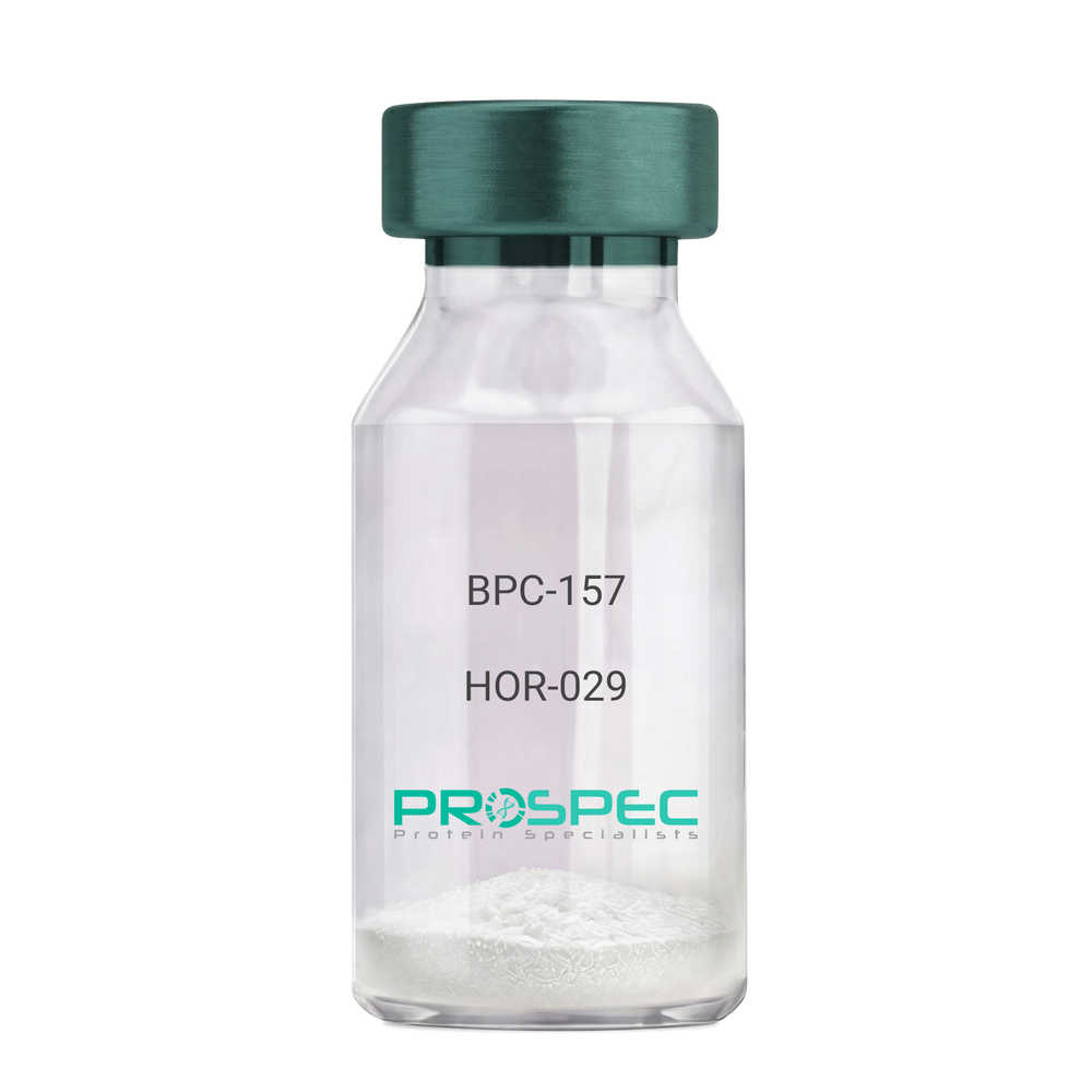
  • View Data Sheet

    Name :

    BPC-157

    Description:

    BPC-157 Pentadecapeptide

    Product # :

    HOR-029

    Price :

    Quantity :

    Shipping Method :

    Room Temp Icon

    Shipped at Room temp

    Add To Cart

    More Info

    • description
    • formulation
    • purity
    • More Info

    Description

    BPC-157 Synthetic is a single, non-glycosylated polypeptide chain containing 15 amino acids, having a molecular mass of 1419.55 Dalton and a Molecular formula of C62H98N16O22.

    Formulation

    The protein was lyophilized with no additives.

    Purity

    Greater than 97.0% as determined by analysis by RP-HPLC.

    More Info

    • Physical Appearance

      Sterile Filtered White lyophilized (freeze-dried) powder.

    • Stability

      Lyophilized BPC-157 although stable at room temperature for 3 weeks, should be stored desiccated below -18°C. Upon reconstitution bpc-157 should be stored at 4°C between 2-7 days and for future use below -18°C. For long term storage it is recommended to add a carrier protein (0.1% HSA or BSA). Please prevent freeze-thaw cycles.

    • Solubility

      It is recommended to reconstitute the lyophilized BPC-157 in sterile 18MΩ-cm H2O not less than 100 µg/ml, which can then be further diluted to other aqueous solutions.

    • Amino Acid Sequence

      H-Gly-Glu-Pro-Pro-Pro-Gly-Lys-Pro-Ala-Asp-Asp-Ala-Gly-Leu-Val-OH

    • Background

      BPC-157, short for Body Protection Compound-157, is a synthetic peptide that has garnered significant attention in the field of regenerative medicine and sports science. This peptide, derived from a portion of the human gastric juice protein known as BPC, exhibits remarkable healing and tissue regeneration properties. BPC-157 has shown promise in various preclinical and clinical studies, demonstrating its potential for the treatment of a wide range of injuries and disorders.

      The research on BPC-157 encompasses investigations into its mechanisms of action, efficacy, safety, and potential therapeutic applications. Studies have elucidated the peptide's ability to enhance angiogenesis, promote collagen synthesis, modulate inflammatory responses, and protect against oxidative stress. These properties make BPC-157 an intriguing candidate for accelerating tissue healing, reducing inflammation, and improving overall recovery outcomes.

      Preclinical studies have revealed the beneficial effects of BPC-157 in several injury models. For instance, BPC-157 has demonstrated its potential in accelerating tendon and ligament healing, mitigating muscle damage, and promoting bone regeneration. These findings suggest that BPC-157 could be a valuable therapeutic tool in orthopedic medicine and sports-related injuries.

      Furthermore, BPC-157 has exhibited promising effects on gastrointestinal health. Studies have highlighted its ability to protect and heal the gut lining, reduce ulcer formation, and alleviate symptoms associated with inflammatory bowel disease. These observations open up avenues for BPC-157 as a potential treatment for gastrointestinal disorders.

      In addition to its regenerative properties, BPC-157 has shown potential in neurological and psychiatric conditions. Research has indicated its neuroprotective effects, with implications for the treatment of traumatic brain injury, stroke, and neurodegenerative disorders. Preliminary studies also suggest BPC-157's potential as an antidepressant and anxiolytic agent.

      Despite the promising findings, further research is needed to fully understand the mechanisms underlying BPC-157's actions and to assess its long-term safety and efficacy. Clinical trials are underway to explore its potential therapeutic applications in humans, including its use in tendon and ligament repair, inflammatory bowel disease, and neurodegenerative disorders.

      This comprehensive review aims to summarize the current state of research on BPC-157, providing an overview of its mechanisms of action and therapeutic potential. By examining relevant studies and findings, we aim to shed light on the diverse applications of BPC-157 and its implications for regenerative medicine, sports science, and various disease

    ProSpec's products are furnished for LABORATORY RESEARCH USE ONLY. The product may not be used as drugs, agricultural or pesticidal products, food additives or household chemicals.

    Bpc 157
  • View Data Sheet

    Name :

    FDX1 Human

    Description:

    Ferredoxin-1 Human Recombinant

    Adrenodoxin mitochondrial, Adrenal ferredoxin, Ferredoxin-1, Hepatoredoxin, FDX1, ADX.

    Product # :

    PRO-713

    Price :

    Quantity :

    Shipping Method :

    Ice Icon

    Shipped with Ice Packs

    Add To Cart

    More Info

    • description
    • source
    • formulation
    • purity
    • More Info

    Description

    FDX1 Human Recombinant fused with a 15 amino acid T7 tag at N-terminus produced in E.Coli is a single, non-glycosylated, polypeptide chain containing 139 amino acids (61-184 a.a.) and having a molecular mass of 15.0kDa.The FDX1 is purified by proprietary chromatographic techniques.

    Source

    Escherichia Coli.

    Formulation

    The FDX1 solution contains 20mM Tris-HCl buffer (pH8.0) and 10% Glycerol.

    Purity

    Greater than 90.0% as determined by SDS-PAGE.

    More Info

    • Introduction

      Ferredoxin-1 is a small iron-sulfur protein who transfers electrons from NADPH through ferredoxin reductase to a terminal cytochrome P450. This specific oxidation/reduction system is located in steroidogenic tissues, and is involved with the synthesis of bile acid and vitamin D. FDX1 takes part in the synthesis of thyroid hormones and transfers electrons from adrenodoxin reductase to the cholesterol side chain cleavage cytochrome P450. FDX1 supports reactions catalyzed by human microsomal P450s- full length CYP17, truncated CYP17, and truncated CYP21. In addition to the FDX1 at the 11q22chromosomal locus, there are pseudogenes located on chromosomes 20 and 21.

    • Synonyms

      Adrenodoxin mitochondrial, Adrenal ferredoxin, Ferredoxin-1, Hepatoredoxin, FDX1, ADX.

    • Physical Appearance

      Sterile Filtered colorless solution.

    • Stability

      Store at 4°C if entire vial will be used within 2-4 weeks. Store, frozen at -20°C for longer periods of time. For long term storage it is recommended to add a carrier protein (0.1% HSA or BSA).Avoid multiple freeze-thaw cycles.

    • Amino Acid Sequence

      MASMTGGQQM GRGSMSSSED KITVHFINRD GETLTTKGKV GDSLLDVVVE NNLDIDGFGA CEGTLACSTC HLIFEDHIYE KLDAITDEEN DMLDLAYGLT DRSRLGCQIC LTKSMDNMTV RVPETVADAR QSIDVGKTS.

    ProSpec's products are furnished for LABORATORY RESEARCH USE ONLY. The product may not be used as drugs, agricultural or pesticidal products, food additives or household chemicals.

    Fdx1 Human
  • View Data Sheet

    Name :

    s100A4 Antibody

    Description:

    S100 Calcium Binding Protein A4, Mouse Anti Human

    Protein S100-A4, S100 calcium-binding protein A4, Metastasin, Protein Mts1, Placental calcium-binding protein, Calvasculin, S100A4, CAPL, MTS1, 42A, 18A2, FSP1, P9KA, PEL98.

    Product # :

    ANT-607

    Price :

    Quantity :

    Shipping Method :

    Ice Icon

    Shipped with Ice Packs

    Add To Cart

    More Info

    • formulation
    • More Info

    Formulation

    1mg/ml containing PBS, pH-7.4, 10% Glycerol and 0.02% Sodium Azide.

    More Info

    • Introduction

      S100A4 is a member of the S100 family of proteins containing 2 EF-hand calcium-binding motifs. S100 belongs to the family of calcium binding proteins such as calmodulin and troponin C. S100A is composed of an alpha and beta chain whereas S100B is composed of two beta chains. S100 protein is also expressed in the antigen presenting cells such as the Langerhans cells in skin and interdigitating reticulum cells in the paracortex of lymph nodes. S100 proteins are localized either in the cytoplasm or the nucleus of a wide range of cells. S100 proteins are involved in the regulation of a number of cellular processes such as cell cycle progression and differentiation. S100A4 may function in motility, invasion, and tubulin polymerization. There are at least 13 members in the S100 gene family, which are located as a cluster on chromosome 1q21. Chromosomal rearrangements and altered expression of the S100A4 gene have been implicated in tumor metastasis.

    • Synonyms

      Protein S100-A4, S100 calcium-binding protein A4, Metastasin, Protein Mts1, Placental calcium-binding protein, Calvasculin, S100A4, CAPL, MTS1, 42A, 18A2, FSP1, P9KA, PEL98.

    • Physical Appearance

      Sterile filtered colorless solution.

    • Immunogen

      Anti-human s100A4 mAb, is derived from hybridization of mouse F0 myeloma cells with spleen cells from BALB/c mice immunized with a recombinant human s100A4 protein 1-101 amino acids purified from E. coli.

    • Ig Subclass

      Mouse IgG2b heavy chain and k light chain.

    • Clone

      PAT1C3AT.

    • Applications

      s100A4 antibody has been tested by ELISA, Western blot analysis and ICC/IF to assure specificity and reactivity. Since application varies, however, each investigation should be titrated by the reagent to obtain optimal results.

    • Type

      Mouse Anti Human Monoclonal.

    • Storage Procedures

      For periods up to 1 month store at 4°C, for longer periods of time, store at -20°C. Prevent freeze thaw cycles.

    • Purification Method

      s100A4 antibody was purified from mouse ascetic fluids by protein-A affinity chromatography.

    ProSpec's products are furnished for LABORATORY RESEARCH USE ONLY. The product may not be used as drugs, agricultural or pesticidal products, food additives or household chemicals.

    S100A4 Antibody
  • View Data Sheet

    Name :

    UMOD Canine

    Description:

    Uromodulin Canine

    Tamm-Horsfall urinary glycoprotein, THP, FJHN, HNFJ, THGP, MCKD2, ADMCKD2, UMOD, Uromodulin.

    Product # :

    ENZ-334

    Price :

    Quantity :

    Shipping Method :

    Room Temp Icon

    Shipped at Room temp

    Add To Cart

    More Info

    • description
    • source
    • formulation
    • More Info

    Description

    UMOD is an 85-kDa glycoprotein which is produced in the thick ascending limb of Henle´s loop and early distal convoluted tubules of the nephron.

    Source

    Canine Urine.

    Formulation

    The UMOD protein was lyophilized from 0.4µm filtered solution at a concentration of 0.1mg/ml containing deionized water.

    More Info

    • Introduction

      Uromodulin is the most abundant protein in normal urine. Its secretion in urine follows proteolytic cleavage of the ectodomain of its glycosyl phosphatidylinosital-anchored counterpart that is situated on the luminal cell surface of the loop of Henle. Uromodulin plays a role as a constitutive inhibitor of calcium crystallization in renal fluids. Secretion of uromodulin in urine provides protection against urinary tract infections caused by uropathogenic bacteria. Defects in Uromodulin expression are associated with the autosomal dominant renal disorders medullary cystic kidney disease-2 (MCKD2) and familial juvenile hyperuricemic nephropathy (FJHN). These disorders are characterized by juvenile onset of hyperuricemia, gout, and progressive renal failure. While several transcript variants may exist for this gene, the full-length natures of only two have been described to date. UMOD is involved in regulating the circulating activity of cytokines as it binds to il-1, il-2 and tnf with high affinity.

    • Synonyms

      Tamm-Horsfall urinary glycoprotein, THP, FJHN, HNFJ, THGP, MCKD2, ADMCKD2, UMOD, Uromodulin.

    • Physical Appearance

      Filtered White lyophilized (freeze-dried) powder.

    • Stability

      Lyophilized UMOD although stable at room temperature for 3 weeks, should be stored desiccated below -18°C. Upon reconstitution UMOD should be stored at 4°C between 2-7 days and for future use below -18°C.For long term storage it is recommended to add a carrier protein (0.1% HSA or BSA).Please prevent freeze-thaw cycles.

    • Solubility

      Add deionized water to prepare a working stock solution of approximately 0.5mg/mL and let the lyophilized pellet dissolve completely. Product is not sterile! Please filter the product by an appropriate sterile filter before using it in the cell culture.

    • Amino Acid Sequence

      RSCSECHSNA TCMEDGMVTT CSCLVGFTGS GFECVDLDEC AIPGAHNCSE GSSCMNTLGS YLCTCPDGFR LTPGLGCIDV DECSEPGLSR CHALATCINN KGNYSCVCPA GYRGDGQHCE CSPGSCGPGL DCVPVGDALV CADPCQEHRI LDEYWRSTEY GAGYTCDVGL NGWYRFTGPG GVRLAETCVP VLHCNTAAPM WLNGTHPTRD QGIVNRTACA HWRGHCCLWD ASIQVKACAG GYYVYNLTET PECYLAYCTD PTSVLGTCEE CSVEEDCKSH DGMWSCQCKQ DFNVTDLFLL DRLECRPNDI KVSLSKCQLK SLGFEKVFMY LRDSQCSGFN ERGDRDWVSV VTPARDGPCG TVMVRNETHA TYSNTLYLAD EIVIRDRNIK INFECSYPLD MKVSLETSLQ PIVSSLNISV GGTGMFTVRM ALFQTPDYTQ PYQGSSVTLT TEAFLYVGTM LDGGDLSRFA LLMTNCYATP SSNATDPLKY FIIQDRCPRT TDSTIQVVEN GESPQGRFSV QMFRFAGNYD LVYLHCEVYL CDIINEKCKP TCSGTRFRSG GIIDQSRVLN LGPITRKNVQ AVVSRAASS.

    ProSpec's products are furnished for LABORATORY RESEARCH USE ONLY. The product may not be used as drugs, agricultural or pesticidal products, food additives or household chemicals.

    Umod Canine
  • View Data Sheet

    Name :

    IFNG 139 a.a. Human

    Description:

    Interferon-gamma 139 a.a Human Recombinant

    Interferon, Gamma, Immune Interferon, IFN-Gamma, IFG, IFI, Interferon Gamma 3, Interferon gamma.

    Product # :

    CYT-843

    Price :

    Quantity :

    Shipping Method :

    Ice Icon

    Shipped with Ice Packs

    Add To Cart

    More Info

    • description
    • source
    • formulation
    • purity
    • biological activity
    • More Info
    • SDS-PAGE

    Description

    IFNG 139 a.a. Human Recombinant produced in E.Coli is a single, non-glycosylated, polypeptide chain containing 139 amino acids (24-161 a.a.) and having a molecular mass of 16.3kDa. IFNG 139 a.a. is purified by proprietary chromatographic techniques.

    Source

    Escherichia Coli.

    Formulation

    IFNG 139 a.a. protein solution (1mg/ml) contains PBS buffer (pH 7.4) and 10% glycerol.

    Purity

    Greater than 90.0% as determined by analysis by SDS-PAGE.

    Biological Activity

    IFNG activity was wmasured in a cytotoxicity assay using WiDr cells. The ED50 was found to be less or equal to 0.5ng/ml.

    SDS-PAGE

    IFNG 139 a.a. Human - Product image 1

    More Info

    • Introduction

      IFN-gamma produced by lymphocytes activated by specific antigens or mitogens. IFN-gamma, in addition to having antiviral activity, has important immunoregulatory functions, it is a potent activator of macrophages, and has antiproliferative effects on transformed cells and it can potentiate the antiviral and antitumor effects of the type interferons.

    • Synonyms

      Interferon, Gamma, Immune Interferon, IFN-Gamma, IFG, IFI, Interferon Gamma 3, Interferon gamma.

    • Physical Appearance

      Sterile Filtered clear solution.

    • Stability

      Store at 4°C if entire vial will be used within 2-4 weeks. Store, frozen at -20°C for longer periods of time. Please avoid freeze thaw cycles.

    • Amino Acid Sequence

      MQDPYVKEAE NLKKYFNAGH SDVADNGTLF LGILKNWKEE SDRKIMQSQI VSFYFKLFKN FKDDQSIQKS VETIKEDMNV KFFNSNKKKR DDFEKLTNYS VTDLNVQRKA IHELIQVMAE LSPAAKTGKR KRSQMLFRG.

    • Background

      What is the molecular weight/Mw of IFNG 139 A.A. HUMAN Protein?
      IFNG 139 A.A. HUMAN Protein has a total Mw of 16.3kDa.

      What is the source or expression system of IFNG 139 A.A. HUMAN Protein?
      Escherichia Coli.

      What is the Purity of IFNG 139 A.A. HUMAN Protein?
      IFNG 139 A.A. HUMAN Protein is >90% pure as determined by SDS-PAGE.

      What is the Biological Activity of IFNG 139 A.A. HUMAN Protein?
      IFNG activity was wmasured in a cytotoxicity assay using WiDr cells. The ED50 was found to be less or equal to 0.5ng/ml.

      What is the amino acid sequence of IFNG 139 A.A. HUMAN Protein?
      MQDPYVKEAE NLKKYFNAGH SDVADNGTLF LGILKNWKEE SDRKIMQSQI VSFYFKLFKN FKDDQSIQKS VETIKEDMNV KFFNSNKKKR DDFEKLTNYS VTDLNVQRKA IHELIQVMAE LSPAAKTGKR KRSQMLFRG.

      What applications can IFNG 139 A.A. HUMAN Protein be used in?
      IFNG 139 A.A. HUMAN Protein can probably be used in western blot, ELISA and Lateral Flow.

      What is the endotoxin level for IFNG 139 A.A. HUMAN Protein?
      The endotoxin level is minimal, IFNG 139 A.A. HUMAN Protein was purified using conventional chromatography techniques.


    ProSpec's products are furnished for LABORATORY RESEARCH USE ONLY. The product may not be used as drugs, agricultural or pesticidal products, food additives or household chemicals.

    Ifng 139 Aa Human
  • View Data Sheet

    Name :

    BD 1 Mouse

    Description:

    BD 1 Mouse

    Beta-defensin 1, BD-1, Defensin beta 1, hBD-1, HBD1, HBP1, DEFB1, HBD-1, HBP-1, DEFB101, DEFB-1, MGC51822.

    Product # :

    CYT-044

    Price :

    Quantity :

    Shipping Method :

    Room Temp Icon

    Shipped at Room temp

    Add To Cart

    More Info

    • description
    • source
    • formulation
    • purity
    • biological activity
    • More Info

    Description

    BD 1 Mouse Recombinant produced in E.Coli is a single, non-glycosylated, polypeptide chain containing 37 amino acids and having a molecular mass of 4.1 KDa.The BD 1 Mouse is purified by proprietary chromatographic techniques.

    Source

    Escherichia Coli.

    Formulation

    Lyophilized from a 0.2µm filtered concentrated solution in PBS, pH7.4.

    Purity

    Greater than 97.0% as determined by:
    (a) Analysis by RP-HPLC.
    (b) Analysis by SDS-PAGE.

    Biological Activity

    Measured by its ability to chemoattract CD34+ dendritic cells using a concentration range of 0.1-1.0 ug/ml, corresponding to a specific activity of 1,000-10,000units/mg.

    More Info

    • Introduction

      The Defensin family are highly similar in their protein sequence and are microbicidal & cytotoxic peptides made by neutrophils. Beta Defensin-1 is an antimicrobial peptide having the resistance of epithelial surfaces to microbial colonization. Beta Defensin-1 has close proximity to Defensin Alpha-1 and has been implicated in the pathogenesis of cystic fibrosis.
      Skin of patients having atopic dermatitis patients and mycosis fungoides (non-lesional and lesional) show lower human Beta Defensin-1 mRNA expression and higher human Beta Defensin-2 and human Beta Defensin-3 mRNA expression.
      Beta Defensin is highly expressed by epithelial cells.
      Beta-defensin 1 may play a role in the pathogenesis of severe sepsis.

    • Synonyms

      Beta-defensin 1, BD-1, Defensin beta 1, hBD-1, HBD1, HBP1, DEFB1, HBD-1, HBP-1, DEFB101, DEFB-1, MGC51822.

    • Physical Appearance

      Sterile Filtered White lyophilized (freeze-dried) powder.

    • Stability

      Lyophilized Mouse BD 1 although stable at room temperature for 3 weeks, should be stored desiccated below -18°C. Upon reconstitution BD 1 should be stored at 4°C between 2-7 days and for future use below -18°C.For long term storage it is recommended to add a carrier protein (0.1% HSA or BSA).Please prevent freeze-thaw cycles.

    • Solubility

      It is recommended to reconstitute the lyophilized Mouse BD-1 in sterile 18M-cm H2O not less than 100µg/ml, which can then be further diluted to other aqueous solutions.

    • Amino Acid Sequence

      DQYKCLQHGG FCLRSSCPSN TKLQGTCKPD KPNCCKS.

    • Background

      What is the molecular weight/Mw of BD1 Protein?
      BD1 Protein has a total Mw of 4.1kDa.

      What is the source or expression system of BD1 Protein?
      Escherichia Coli.

      What is the Purity of BD1 Protein?
      BD1 Protein is >97% pure as determined by SDS-PAGE.

      What is the Biological Activity of BD1 Protein?
      Measured by its ability to chemoattract CD34+ dendritic cells using a concentration range of 0.1-1.0 ug/ml, corresponding to a specific activity of 1,000-10,000units/mg.

      What is the amino acid sequence of BD1 Protein?
      DQYKCLQHGG FCLRSSCPSN TKLQGTCKPD KPNCCKS.

      What applications can BD1 Protein be used in?
      BD1 Protein can probably be used in western blot, ELISA and Lateral Flow.

      What is the endotoxin level for BD1 Protein?
      The endotoxin level is minimal, BD1 Protein was purified using conventional chromatography techniques.

    ProSpec's products are furnished for LABORATORY RESEARCH USE ONLY. The product may not be used as drugs, agricultural or pesticidal products, food additives or household chemicals.

    Bd 1 Mouse
  • View Data Sheet

    Name :

    Nucleosome Bovine

    Description:

    Bovine Nucleosome

    Product # :

    PRO-2557

    Price :

    Quantity :

    Shipping Method :

    Ice Icon

    Shipped with Ice Packs

    Add To Cart

    More Info

    • description
    • source
    • formulation
    • purity
    • More Info

    Description

    Nucleosome Bovine shows multiple bands between 14-18kDa and is purified from bovine tissues by proprietary protein-chemical techniques.

    Source

    Bovine tissues.

    Formulation

    Nucleosome Bovine is supplied in 20mM HEPES buffer pH-7.5, 500mM NaCl, 1mM NaCl and 20% glycerol.

    Purity

    Greater than 95.0% as determined by SDS-PAGE.

    More Info

    • Introduction

      Nucleosome core proteins are a group of proteins which are necessary for DNA packaging in eukaryotes. Every core contains an octamer with 2 copies of each histone 2A, 2B, 3 and 4 around which an approx. 140 bp section of DNA is wound twice.

    • Physical Appearance

      Sterile Filtered clear solution.

    • Stability

      Store at 4°C if entire vial will be used within 2-4 weeks. Store, frozen at -20°C for longer periods of time. Avoid multiple freeze-thaw cycles.

    • Immunological Functions

      1. Binds IgG type human auto antibodies.2. Checkerboard analysis of positive/negative samples.

    • coating concentration

      0.2-0.5 μg/ml (depending on the type of ELISA plate and coating buffer). Suitable for labeling of functional groups.

    ProSpec's products are furnished for LABORATORY RESEARCH USE ONLY. The product may not be used as drugs, agricultural or pesticidal products, food additives or household chemicals.

    Nucleosome Bovine
  • View Data Sheet

    Name :

    CRP Canine

    Description:

    C-Reactive Protein Canine Recombinant

    C-reactive protein, CRP, PTX1, MGC88244, MGC149895.

    Product # :

    PRO-2663

    Price :

    Quantity :

    Shipping Method :

    Room Temp Icon

    Shipped at Room temp

    Add To Cart

    More Info

    • description
    • source
    • formulation
    • purity
    • More Info

    Description

    CRP Canine Recombinant is a single, glycosylated polypeptide chain containing 204 amino acids and having a molecular mass of 23.2kDa (calculated).

    Source

    HEK293 cells.

    Formulation

    CRP filtered (0.4 µm) and lyophilized from 0.5mg/ml in 20mM Tris, 50mM NaCl and 1mM CaCl2, pH 7.5.

    Purity

    Greater than 95.0% as determined by SDS-PAGE.

    More Info

    • Introduction

      CRP is an acute phase protein that correlates with inflammatory disease and is synthesized by hepatocytes during the acute phase response by certain cytokines (IL-1 and TNF Alpha and Beta). CRP levels increase dramatically and serve as a useful marker of inflammation in such conditions as bacterial infection, rheumatoid arthritis, viral infections, transplantation rejection, meningitis, myocardial infarction, septicemia, osteomyelitis and others. CRP is also highly correlated to Serum Amyloid A levels.

    • Synonyms

      C-reactive protein, CRP, PTX1, MGC88244, MGC149895.

    • Physical Appearance

      Filtered White lyophilized (freeze-dried) powder.

    • Stability

      Store lyophilized protein at -20°C. Aliquot the product after reconstitution to avoid repeated freezing/thawing cycles. Reconstituted protein can be stored at 4°C for a limited period of time; it does not show any change after two weeks at 4°C.

    • Solubility

      It is recommended to add deionized water to prepare a working stock solution of approximately 0.5mg/ml and let the lyophilized pellet dissolve completely.

    • Amino Acid Sequence

      QIDLDEKAFV FPRESENSYV ILFPQLQKPM KAFTVCLQVY TDLTRPHSLF SYATKSQSNE ILLFKERPGL FSVSVGGSDA FINFPQKFYA PQHFCVTWES VTGLTELWVD GKPMVRASLR RGYTVGSGAS IVLGQEQDSF GGGFDKNQSL VGDIEDVNMW DFVLSPSQIL TLYTTRALSP NVLNWRNLRY ETRGEVFLKK ELWS

    ProSpec's products are furnished for LABORATORY RESEARCH USE ONLY. The product may not be used as drugs, agricultural or pesticidal products, food additives or household chemicals.

    CRP Canine
  • View Data Sheet

    Name :

    LUM Human, sf9

    Description:

    Lumican Human Recombinant, Sf9

    Lumican, LDC, Lumican Proteoglycan, Keratan Sulfate Proteoglycan Lumican, SLRR2D, KSPG Lumican.

    Product # :

    PRO-2355

    Price :

    Quantity :

    Shipping Method :

    Ice Icon

    Shipped with Ice Packs

    Add To Cart

    More Info

    • description
    • source
    • formulation
    • purity
    • More Info

    Description

    LUM Human Recombinant produced in Sf9 Baculovirus cells is a single, glycosylated polypeptide chain containing 329 amino acids (19-338 aa) and having a molecular mass of 37.7kDa (Migrates at 40-57kDa on SDS-PAGE under reducing conditions).LUM is expressed with a 6 amino acid His tag at C-Terminus and purified by proprietary chromatographic techniques.

    Source

    Sf9, Baculovirus cells.

    Formulation

    LUM protein solution (0.5mg/ml) contains Phosphate buffered saline (pH7.4) and 10% glycerol.

    Purity

    Greater than 95.0% as determined by SDS-PAGE.

    More Info

    • Introduction

      Lumican also known as LUM belongs to the small leucine-rich proteoglycan (SLRP) family which comprises decorin, biglycan, fibromodulin, keratocan, epiphycan, and osteoglycin. Furthermore we can see that in these bifunctional molecules, the protein moiety binds collagen fibrils and the highly charged hydrophilic glycosaminoglycans regulate interfibrillar spacings. Lumican is the main keratan sulfate proteoglycan of the cornea however LUM is also distributed in interstitial collagenous matrices throughout the body. Lumican regulates collagen fibril organization and circumferential growth, corneal transparency, epithelial cell migration and tissue repair. Among the diseases associated with LUM is posterior amorphous corneal dystrophy.

    • Synonyms

      Lumican, LDC, Lumican Proteoglycan, Keratan Sulfate Proteoglycan Lumican, SLRR2D, KSPG Lumican.

    • Physical Appearance

      Sterile filtered colorless solution.

    • Stability

      Store at 4°C if entire vial will be used within 2-4 weeks. Store, frozen at -20°C for longer periods of time. For long term storage it is recommended to add a carrier protein (0.1% HSA or BSA).Avoid multiple freeze-thaw cycles.

    • Amino Acid Sequence

      ADLQYYDYDF PLSIYGQSSP NCAPECNCPE SYPSAMYCDE LKLKSVPMVP PGIKYLYLRN NQIDHIDEKA FENVTDLQWL ILDHNLLENS KIKGRVFSKL KQLKKLHINH NNLTESVGPL PKSLEDLQLT HNKITKLGSF EGLVNLTFIH LQHNRLKEDA VSAAFKGLKS LEYLDLSFNQ IARLPSGLPV SLLTLYLDNN KISNIPDEYF KRFNALQYLR LSHNELADSG IPGNSFNVSS LVELDLSYNK LKNIPTVNEN LENYYLEVNQ LEKFDIKSFC KILGPLSYSK IKHLRLDGNR ISETSLPPDM YECLRVANEV TLNHHHHHH

    ProSpec's products are furnished for LABORATORY RESEARCH USE ONLY. The product may not be used as drugs, agricultural or pesticidal products, food additives or household chemicals.

    Lum Human Sf9
  • View Data Sheet

    Name :

    Dengue NS1 Antibody

    Description:

    Monoclonal Mouse Anti Dengue NS1

    Product # :

    ANT-495

    Price :

    Quantity :

    Shipping Method :

    Ice Icon

    Shipped with Ice Packs

    Add To Cart

    More Info

    • description
    • formulation
    • purity
    • More Info

    Description

    Paired monoclonal antibodies developed for dengue NS1 antigen test by lateral flow immunoassay device. First antibody is for conjugation and the second antibody is for membrane coating. Please note that when ordering for example: 100µg antibody we ship 50µg from each of the antibody solutions (100µg in total).

    Formulation

    *Dengue NS1 Antibody solution for gold conjugation (3.6mg/ml) in PBS and Sodium nitrate.

    *Dengue NS1 Antibody solution for membrane coating (2.2mg/ml) in PBS and Sodium nitrate.

    Purity

    95% pure (12% SDS-PAGE and Coomassie staining).

    More Info

    • Physical Appearance

      2 vials of sterile Filtered clear colorless solution.

    • Stability

      Store at 4°C if entire vial will be used within 2-4 weeks. Store, frozen at -20°C for longer periods of time. For long term storage it is recommended to add a carrier protein (0.1% HSA or BSA).Avoid multiple freeze-thaw cycles.

    • Applications

      Lateral flow immunoassay.

    • Assay Conditions

      1. 1-2 days fresh prepared gold particle with maximal homogeneity in gold particle size, λ 525-529, OD 1.2-1.4. 2. Conjugation at pH 8.5, conjugation time should be 6-8 hours. 3. Blocking condition: remove supernatant and re-suspend gold pellet bound with anti-dengue NS1 to OD 8.0- 8.2, add casein to the final 0.3-0.4%. 4. Treat sample pad with 50mM sodium borate, 0.5% mouse serum and 0.5% Triton X-100, pH8.0.

    • Purification Method

      Purified monoclonal IgG by protein A chromatography.

    ProSpec's products are furnished for LABORATORY RESEARCH USE ONLY. The product may not be used as drugs, agricultural or pesticidal products, food additives or household chemicals.

    Dengue Ns1 Antibody
  • View Data Sheet

    Name :

    AICDA Antibody

    Description:

    Activation-induced cytidine deaminase, Mouse Anti Human

    Activation-induced cytidine deaminase, Cytidine aminohydrolase, AICDA, AID, ARP2, CDA2, HIGM2.

    Product # :

    ANT-411

    Price :

    Quantity :

    Shipping Method :

    Ice Icon

    Shipped with Ice Packs

    Add To Cart

    More Info

    • formulation
    • More Info

    Formulation

    1mg/ml containing PBS, pH-7.4, & 0.1% Sodium Azide.

    More Info

    • Introduction

      AICDA is an RNA-editing deaminase which belongs to the cytidine deaminase family. AICDA is involved in somatic hypermutation, gene conversion, and class-switch recombination of immunoglobulin genes. Defects in the AICDA gene are the cause of autosomal recessive hyper-IgM immunodeficiency syndrome type 2 (HIGM2).

    • Synonyms

      Activation-induced cytidine deaminase, Cytidine aminohydrolase, AICDA, AID, ARP2, CDA2, HIGM2.

    • Immunogen

      Anti-human AICDA mAb, is derived from hybridization of mouse SP2/0 myeloma cells with spleen cells from BALB/c mice immunized with recombinant human AICDA amino acids 1-54 purified from E. coli.

    • Ig Subclass

      Mouse IgG1 heavy chain and κ light chain.

    • Clone

      P2D3AT.

    • Applications

      AICDA antibody has been tested by ELISA and Western blot analysis to assure specificity and reactivity. Since application varies, however, each investigation should be titrated by the reagent to obtain optimal results. Recommended dilution range for Western blot analysis is 1:500 ~ 1,000. Recommended starting dilution is 1:500.

    • Type

      Mouse Anti Human Monoclonal.

    • Storage Procedures

      For periods up to 1 month store at 4°C, for longer periods of time, store at -20°C. Prevent freeze thaw cycles.

    • Purification Method

      AICDA antibody was purified from mouse ascitic fluids by protein-G affinity chromatography.

    ProSpec's products are furnished for LABORATORY RESEARCH USE ONLY. The product may not be used as drugs, agricultural or pesticidal products, food additives or household chemicals.

    Aicda Antibody
  • View Data Sheet

    Name :

    FABP9 Antibody

    Description:

    Fatty Acid Binding Protein-9, Mouse Anti Human

    Fatty acid-binding protein 9, Testis lipid-binding protein, TLBP, Testis-type fatty acid-binding protein, T-FABP, FABP9, PERF, PERF15.

    Product # :

    ANT-523

    Price :

    Quantity :

    Shipping Method :

    Ice Icon

    Shipped with Ice Packs

    Add To Cart

    More Info

    • formulation
    • More Info

    Formulation

    1mg/ml containing PBS, pH-7.4, 10% Glycerol and 0.02% Sodium Azide.

    More Info

    • Introduction

      Fatty acid binding protein 9 (FABP9) belongs to the fatty acid-binding proteins (FABPs) which are a family of small, extremely conserved, cytoplasmic proteins to bind long-chain fatty acids and other hydrophobic ligands. FABP9 is found in midpachytene spermatocytes and round spermatids, and comprises part of the perinuclear theca. FABP9 probably links intracellular membranes and signals abnormal sperm formation during spermatogenesis.

    • Synonyms

      Fatty acid-binding protein 9, Testis lipid-binding protein, TLBP, Testis-type fatty acid-binding protein, T-FABP, FABP9, PERF, PERF15.

    • Physical Appearance

      Sterile filtered colorless solution.

    • Immunogen

      Anti-human FABP9 mAb, clone PAT13F9AT, is derived from hybridization of mouse F0 myeloma cells with spleen cells from BALB/c mice immunized with a recombinant human FABP9 protein 1-132 amino acids purified from E. coli.

    • Ig Subclass

      Mouse IgG1 heavy chain and k light chain.

    • Clone

      PAT13F9AT.

    • Applications

      The antibody has been tested by ELISA, Western blot analysis to assure specificity and reactivity. Since application varies, however, each investigation should be titrated by the reagent to obtain optimal results. Recommended starting dilution is 1:1000.

    • Type

      Mouse Anti Human Monoclonal.

    • Storage Procedures

      For periods up to 1 month store at 4°C, for longer periods of time, store at -20°C. Prevent freeze thaw cycles.

    • Purification Method

      FABP9 antibody was purified from mouse ascitic fluids by protein-A affinity chromatography.

    ProSpec's products are furnished for LABORATORY RESEARCH USE ONLY. The product may not be used as drugs, agricultural or pesticidal products, food additives or household chemicals.

    Fabp9 Antibody
  • View Data Sheet

    Name :

    PPM1A Antibody

    Description:

    Protein Phosphatase 1A Alpha Isoform, Mouse Anti Human

    Protein phosphatase 1A, EC 3.1.3.16, Protein phosphatase 2C isoform alpha, PP2C-alpha, IA, PPM1A, PP2CA, MGC9201.

    Product # :

    ANT-309

    Price :

    Quantity :

    Shipping Method :

    Ice Icon

    Shipped with Ice Packs

    Add To Cart

    More Info

    • formulation
    • More Info

    Formulation

    1mg/ml containing PBS, pH-7.4, & 0.1% Sodium Azide.

    More Info

    • Introduction

      Protein Phosphatase 2C alpha is a member of the PP2C family of Ser/Thr protein phosphatases. PP2C family members are known to be negative regulators of cell stress response pathways. This phosphatase dephosphorylates, and negatively regulates the activities of, MAP kinases and MAP kinase kinases. It has been shown to inhibit the activation of p38 and JNK kinase cascades induced by environmental stresses. This phosphatase can also dephosphorylate cyclin-dependent kinases, and thus may be involved in cell cycle control. Overexpression of this phosphatase is reported to activate the expression of the tumor suppressor gene TP53/p53, which leads to G2/M cell cycle arrest and apoptosis. Three alternatively spliced transcript variants encoding two distinct isoforms have been described. Protein phosphatase 2C(PP2Cα) is a Mn2+- or Mg2+-dependent protein serine/threonine phosphatase that is essential for regulating cellular stress response in eukaryotes.

    • Synonyms

      Protein phosphatase 1A, EC 3.1.3.16, Protein phosphatase 2C isoform alpha, PP2C-alpha, IA, PPM1A, PP2CA, MGC9201.

    • Immunogen

      Anti-human PPM1A mAb, is derived from hybridization of mouse SP2/O myeloma cells with spleen cells from BALB/c mice immunized with recombinant human PPM1A amino acids 1-382 purified from E. coli.

    • Ig Subclass

      Mouse IgG2b heavy chain and κ light chain.

    • Clone

      Pp6c7AT.

    • Applications

      PPM1A antibody has been tested by ELISA and Western blot analysis to assure specificity and reactivity. Since application varies, however, each investigation should be titrated by the reagent to obtain optimal results. Recommended dilution range for Western blot analysis is 1:250 ~ 500. Recommended starting dilution is 1:250.

    • Type

      Mouse Anti Human Monoclonal.

    • Storage Procedures

      For periods up to 1 month store at 4°C, for longer periods of time, store at -20°C. Prevent freeze thaw cycles.

    • Purification Method

      PPM1A antibody was purified from mouse ascitic fluids by protein-G affinity chromatography.

    ProSpec's products are furnished for LABORATORY RESEARCH USE ONLY. The product may not be used as drugs, agricultural or pesticidal products, food additives or household chemicals.

    Ppm1A Antibody
  • 1
  • …
  • 37
  • 38
  • 39
  • 40
  • 41
Your browser does not support the video tag.

Products

  • Cytokines
  • Growth Factors
  • Chemokines
  • CD Antigens
  • Neurotrophins
  • Hormones
  • Enzymes
  • Viral Antigens
  • Recombinant Proteins
  • Natural Proteins
  • Monoclonal Antibodies
  • Polyclonal Antibodies
  • Thermal Packaging

About

  • Company
  • Founder
  • Contribution
  • Contact Us
  • My Account

Ordering

  • Ordering Method
  • Ordering - Credit Card
  • Ordering PO
  • Shipping Fees
  • FAQ's

Legal

  • Terms & Conditions
  • Privacy
  • Site Map
  • info@prospecbio.com
  • Facebook
  • Instagram
  • Linkedin Profile

© 2026 ProSpec-Tany TechnoGene Ltd. All Rights Reserved.

  • Choosing a selection results in a full page refresh.
  • Opens in a new window.