Skip to content

High-quality research proteins.

  • 1-800-805-6531

  • Mon - Sat 8.00-18.00, Sun - Closed

  • info@prospecbio.com

  • PO Box 6591 East Brunswick NJ 08816

  • Products
    • Cytokines
    • Growth factors
    • Chemokines
    • Cd antigens
    • Neurotrophins
    • Hormones
    • Enzymes
    • Viral antigens
    • Recombinant proteins
    • Natural proteins
    • Monoclonal antibodies
    • Polyclonal antibodies
    • Thermal Packaging
  • About
    • Company
    • Founder
    • Contribution
  • Customers
    • Eastern Asia
    • Eastern Europe
    • North America
    • South America
    • South East Asia
    • Southern Asia
    • Western Europe
  • Ordering
    • Ordering Method
    • Ordering - Credit Card
    • Ordering - PO
    • Shipping Fees
    • FAQs
  • Services
    • DNA Cloning
    • Protein Expression
    • Monoclonal Antibody
    • Fermentation
    • Technology License
  • Contact us
Log in

Country/region

  • USD $ | Albania
  • USD $ | Andorra
  • USD $ | Argentina
  • USD $ | Armenia
  • USD $ | Australia
  • USD $ | Austria
  • USD $ | Azerbaijan
  • USD $ | Belarus
  • USD $ | Belgium
  • USD $ | Bolivia
  • USD $ | Brazil
  • USD $ | Bulgaria
  • USD $ | Cambodia
  • USD $ | Canada
  • USD $ | Chile
  • USD $ | China
  • USD $ | Colombia
  • USD $ | Costa Rica
  • USD $ | Croatia
  • USD $ | Cyprus
  • USD $ | Czechia
  • USD $ | Denmark
  • USD $ | Estonia
  • USD $ | Finland
  • USD $ | France
  • USD $ | Georgia
  • USD $ | Germany
  • USD $ | Greece
  • USD $ | Hong Kong SAR
  • USD $ | Hungary
  • USD $ | Iceland
  • USD $ | India
  • USD $ | Ireland
  • USD $ | Israel
  • USD $ | Italy
  • USD $ | Japan
  • USD $ | Kazakhstan
  • USD $ | Latvia
  • USD $ | Liechtenstein
  • USD $ | Lithuania
  • USD $ | Madagascar
  • USD $ | Malta
  • USD $ | Mexico
  • USD $ | Moldova
  • USD $ | Monaco
  • USD $ | Netherlands
  • USD $ | New Zealand
  • USD $ | North Macedonia
  • USD $ | Norway
  • USD $ | Panama
  • USD $ | Paraguay
  • USD $ | Peru
  • USD $ | Philippines
  • USD $ | Poland
  • USD $ | Portugal
  • USD $ | Romania
  • USD $ | Singapore
  • USD $ | Slovakia
  • USD $ | Slovenia
  • USD $ | South Africa
  • USD $ | South Korea
  • USD $ | Spain
  • USD $ | Sri Lanka
  • USD $ | Sweden
  • USD $ | Switzerland
  • USD $ | Taiwan
  • USD $ | Thailand
  • USD $ | Türkiye
  • USD $ | Ukraine
  • USD $ | United Kingdom
  • USD $ | United States
  • USD $ | Uruguay
  • USD $ | Vatican City
  • USD $ | Venezuela
  • USD $ | Vietnam
  • Facebook
  • Instagram
logoProSpec - Recombinant Protein Manufacturer for academic and R&D industry
Become a distributor
Account Log in
Wishlist
Cart Cart(0)
  • Products
    +
    • Cytokines
    • Growth factors
    • Chemokines
    • Cd antigens
    • Neurotrophins
    • Hormones
    • Enzymes
    • Viral antigens
    • Recombinant proteins
    • Natural proteins
    • Monoclonal antibodies
    • Polyclonal antibodies
    • Thermal Packaging
  • About
    +
    • Company
    • Founder
    • Contribution
  • Customers
    +
    • Eastern Asia
    • Eastern Europe
    • North America
    • South America
    • South East Asia
    • Southern Asia
    • Western Europe
  • Ordering
    +
    • Ordering Method
    • Ordering - Credit Card
    • Ordering - PO
    • Shipping Fees
    • FAQs
  • Services
    +
    • DNA Cloning
    • Protein Expression
    • Monoclonal Antibody
    • Fermentation
    • Technology License
  • Contact us

Item added to your cart

View cart
View All
  • Cytokines
  • Growth factors
  • Chemokines
  • Cd antigens
  • Neurotrophins
  • Hormones
  • Enzymes
  • Viral antigens
  • Recombinant proteins
  • Natural proteins
  • Monoclonal antibodies
  • Polyclonal antibodies
  • Thermal Packaging
  • Tumor Necrosis Factor

    Tumor Necrosis Factor

    View All
  • 4-1BB

    4-1BB

    View All
  • Adiponectin

    Adiponectin

    View All
  • AIF1

    AIF1

    View All
  • Other Cytokines

    Other Cytokines

    View All
  • AITRL

    AITRL

    View All
  • Angiopoietin

    Angiopoietin

    View All
  • Apolipoprotein

    Apolipoprotein

    View All
  • B-Cell Activating Factor

    B-Cell Activating Factor

    View All
  • Beta Defensin

    Beta Defensin

    View All
  • Bone Morphogenetic Protein

    Bone Morphogenetic Protein

    View All
  • B type Natriuretic Peptide

    B type Natriuretic Peptide

    View All
  • BST

    BST

    View All
  • Betacellulin

    Betacellulin

    View All
  • Cardiotrophin

    Cardiotrophin

    View All
  • Activin

    Activin

    View All
  • Fibroblast Growth Factor

    Fibroblast Growth Factor

    View All
  • Other Growth Factors

    Other Growth Factors

    View All
  • CSF

    CSF

    View All
  • CTGF

    CTGF

    View All
  • IGFBP

    IGFBP

    View All
  • VEGF Protein

    VEGF Protein

    View All
  • EGF

    EGF

    View All
  • Epigen

    Epigen

    View All
  • Erythropoietin

    Erythropoietin

    View All
  • Insulin-Like Growth Factor

    Insulin-Like Growth Factor

    View All
  • Growth Hormone

    Growth Hormone

    View All
  • HDGF

    HDGF

    View All
  • Hepatocyte Growth Factor

    Hepatocyte Growth Factor

    View All
  • Insulin

    Insulin

    View All
  • BCA-1/ BLC (CXCL13)

    BCA-1/ BLC (CXCL13)

    View All
  • BRAK (CXCL14)

    BRAK (CXCL14)

    View All
  • C-10 (CCL6)

    C-10 (CCL6)

    View All
  • Other Chemokines

    Other Chemokines

    View All
  • MEC (CCL28)

    MEC (CCL28)

    View All
  • LD78-beta (CCL3L1)

    LD78-beta (CCL3L1)

    View All
  • CTACK (CCL27)

    CTACK (CCL27)

    View All
  • CXCL16

    CXCL16

    View All
  • CXCL17

    CXCL17

    View All
  • Platelet Factor-4 (CXCL4)

    Platelet Factor-4 (CXCL4)

    View All
  • ENA-78 (CXCL5)

    ENA-78 (CXCL5)

    View All
  • Eotaxin (CCL11,24,26)

    Eotaxin (CCL11,24,26)

    View All
  • Exodus-2 (CCL21)

    Exodus-2 (CCL21)

    View All
  • Fractalkine (CX3CL1)

    Fractalkine (CX3CL1)

    View All
  • CXCL6

    CXCL6

    View All
  • Other CD Antigens

    Other CD Antigens

    View All
  • CD14

    CD14

    View All
  • CD164

    CD164

    View All
  • CD1

    CD1

    View All
  • CD2

    CD2

    View All
  • CD200

    CD200

    View All
  • CD207

    CD207

    View All
  • CD226

    CD226

    View All
  • CD23

    CD23

    View All
  • CD244

    CD244

    View All
  • CD247

    CD247

    View All
  • CD27

    CD27

    View All
  • CD274

    CD274

    View All
  • CD300

    CD300

    View All
  • CD33

    CD33

    View All
  • Other Neurotrophins

    Other Neurotrophins

    View All
  • Beta-NGF

    Beta-NGF

    View All
  • BDNF

    BDNF

    View All
  • CDNF

    CDNF

    View All
  • CNTF

    CNTF

    View All
  • GDNF

    GDNF

    View All
  • Glia Maturation Factor

    Glia Maturation Factor

    View All
  • MANF

    MANF

    View All
  • Midkine

    Midkine

    View All
  • Neuroglobin

    Neuroglobin

    View All
  • Neurotrophic factors

    Neurotrophic factors

    View All
  • Neuregulin

    Neuregulin

    View All
  • Neuropilin

    Neuropilin

    View All
  • Pigment Epithelium-Derived Factor

    Pigment Epithelium-Derived Factor

    View All
  • Pleiotrophin

    Pleiotrophin

    View All
  • Peptide Hormones

    Peptide Hormones

    View All
  • Other Hormones

    Other Hormones

    View All
  • HCG

    HCG

    View All
  • Endothelin

    Endothelin

    View All
  • Exendin

    Exendin

    View All
  • FSH

    FSH

    View All
  • Glucagon

    Glucagon

    View All
  • GHRP

    GHRP

    View All
  • GLP

    GLP

    View All
  • Thyrostimulin

    Thyrostimulin

    View All
  • Inhibin A

    Inhibin A

    View All
  • LHRH

    LHRH

    View All
  • Melanotan

    Melanotan

    View All
  • Procalcitonin

    Procalcitonin

    View All
  • PTH

    PTH

    View All
  • Peroxiredoxin

    Peroxiredoxin

    View All
  • Peptidase

    Peptidase

    View All
  • Synthetase

    Synthetase

    View All
  • Transferase

    Transferase

    View All
  • Hydrolase

    Hydrolase

    View All
  • Dehydrogenase

    Dehydrogenase

    View All
  • ACE2

    ACE2

    View All
  • Esterase

    Esterase

    View All
  • Protein Kinases

    Protein Kinases

    View All
  • Other Enzymes

    Other Enzymes

    View All
  • Phosphatase

    Phosphatase

    View All
  • Deaminase

    Deaminase

    View All
  • Oxygenase

    Oxygenase

    View All
  • Adenylate Kinase

    Adenylate Kinase

    View All
  • Lyase

    Lyase

    View All
  • Chagas

    Chagas

    View All
  • SARS

    SARS

    View All
  • Influenza

    Influenza

    View All
  • ASFV

    ASFV

    View All
  • Astrovirus

    Astrovirus

    View All
  • Borrelia

    Borrelia

    View All
  • Helicobacter Pylori

    Helicobacter Pylori

    View All
  • Chikungunya

    Chikungunya

    View All
  • Chlamydia

    Chlamydia

    View All
  • Cytomegalo

    Cytomegalo

    View All
  • Dengue

    Dengue

    View All
  • EBV

    EBV

    View All
  • Ebola

    Ebola

    View All
  • Feline Leukemia Virus

    Feline Leukemia Virus

    View All
  • Hepatitis A

    Hepatitis A

    View All
  • Other

    Other

    View All
  • Actin

    Actin

    View All
  • ADAM

    ADAM

    View All
  • Complement Component

    Complement Component

    View All
  • Ag85

    Ag85

    View All
  • Eukaryotic Translation Initiation Factor

    Eukaryotic Translation Initiation Factor

    View All
  • Anterior Gradient Protein

    Anterior Gradient Protein

    View All
  • Heat Shock Protein

    Heat Shock Protein

    View All
  • Alpha-2-HS-Glycoprotein

    Alpha-2-HS-Glycoprotein

    View All
  • Hemoglobin

    Hemoglobin

    View All
  • Allergy

    Allergy

    View All
  • Anaplasma

    Anaplasma

    View All
  • Angiogenin

    Angiogenin

    View All
  • Ankyrin Repeat Domain

    Ankyrin Repeat Domain

    View All
  • Annexin

    Annexin

    View All
  • Other Natural Proteins

    Other Natural Proteins

    View All
  • Aprotinin

    Aprotinin

    View All
  • Transferrin

    Transferrin

    View All
  • Avidin

    Avidin

    View All
  • Anti Coagulation Factors

    Anti Coagulation Factors

    View All
  • Natural Albumin

    Natural Albumin

    View All
  • Natural Coagulation Factors

    Natural Coagulation Factors

    View All
  • Fibronectin

    Fibronectin

    View All
  • Thrombin

    Thrombin

    View All
  • Anti Human Enzyme

    Anti Human Enzyme

    View All
  • Other Monoclonal Antibodies

    Other Monoclonal Antibodies

    View All
  • Anti Human Cytokine

    Anti Human Cytokine

    View All
  • Anti Human Heat Shock Protein

    Anti Human Heat Shock Protein

    View All
  • Anti Mouse Lymphocyte

    Anti Mouse Lymphocyte

    View All
  • Anti Human Chemokine

    Anti Human Chemokine

    View All
  • Anti Human Lymphocyte

    Anti Human Lymphocyte

    View All
  • Anti Viral Monoclonal

    Anti Viral Monoclonal

    View All
  • Anti Mouse Cytokine

    Anti Mouse Cytokine

    View All
  • Anti-GST Monoclonal

    Anti-GST Monoclonal

    View All
  • Other Polyclonal Antibodies

    Other Polyclonal Antibodies

    View All
  • Anti Viral

    Anti Viral

    View All
  • Anti-GST Monoclonal

    Anti-GST Monoclonal

    View All
Back

Search results

1000 results found for “CNTF”

Name

Description

Product #

Price

Quantity

Shipping Method

  • View Data Sheet

    Name :

    CXCL3 Rat beta

    Description:

    GRO-gamma, CINC-2 beta Rat Recombinant (CXCL3)

    Macrophage inflammatory protein 2-beta, MIP2-beta, CXCL3, Growth-regulated protein gamma, GRO-gamma, GRO-gamma (1-73), GRO3, GROg, MIP2B, SCYB3, MIP-2b, CINC-2b, MGSA gamma.

    Product # :

    CHM-273

    Price :

    Quantity :

    Shipping Method :

    Room Temp Icon

    Shipped at Room temp

    Add To Cart

    More Info

    • description
    • source
    • formulation
    • purity
    • biological activity
    • More Info

    Description

    GRO-g Rat Recombinant produced in E.Coli is a single, non-glycosylated, polypeptide chain containing 68 amino acids and having a total molecular mass of 7.8kDa. GRO-g is purified by proprietary chromatographic techniques.

    Source

    Escherichia Coli.

    Formulation

    Lyophilized from a 0.2µm filtered concentrated (1.0mg/ml) solution in 1×PBS, pH 7.4.

    Purity

    Greater than 97.0% as determined by
    (a) Analysis by RP-HPLC.
    (b) Analysis by SDS-PAGE.

    Biological Activity

    Determined by its ability to chemoattract human CXCR2 transfected 293 cells using a concentration range of 10-100ng/ml.

    More Info

    • Introduction

      Chemokine (C-X-C motif) ligand 3 (CXCL3) is a small cytokine belonging to the CXC chemokine family that is also known as GRO3 oncogene (GRO3), GRO protein gamma (GROg) and macrophage inflammatory protein-2-beta (MIP2b). CXCL3 controls migration and adhesion of monocytes and mediates it effects on its target cell by interacting with a cell surface chemokine receptor called CXCR2. The gene for CXCL3 is located on chromosome 4 in a cluster of other CXC chemokines.

    • Synonyms

      Macrophage inflammatory protein 2-beta, MIP2-beta, CXCL3, Growth-regulated protein gamma, GRO-gamma, GRO-gamma (1-73), GRO3, GROg, MIP2B, SCYB3, MIP-2b, CINC-2b, MGSA gamma.

    • Physical Appearance

      Sterile Filtered White lyophilized (freeze-dried) powder.

    • Stability

      Lyophilized GRO-g although stable at room temperature for 3 weeks, should be stored desiccated below -18°C. Upon reconstitution GRO-g should be stored at 4°C between 2-7 days and for future use below -18°C.For long term storage it is recommended to add a carrier protein (0.1% HSA or BSA).Please prevent freeze-thaw cycles.

    • Solubility

      It is recommended to reconstitute the lyophilized GRO-g in sterile 18M-cm H2O not less than 100µg/ml, which can then be further diluted to other aqueous solutions.

    • Amino Acid Sequence

      RELRCQCLKT LPRVDFENIQ SLTVTPPGPH CTQTEVIATL KDGQEVCLNP QAPRLQKIIQ KLLKSPSL.

    • Background

      What is the molecular weight/Mw of CXCL3 RAT BETA Protein?
      CXCL3 RAT BETA Protein has a total Mw of 7.8kDa.

      What is the source or expression system of CXCL3 RAT BETA Protein?
      Escherichia Coli.

      What is the Purity of CXCL3 RAT BETA Protein?
      CXCL3 RAT BETA Protein is >97% pure as determined by SDS-PAGE.

      What is the Biological Activity of CXCL3 RAT BETA Protein?
      Determined by its ability to chemoattract human CXCR2 transfected 293 cells using a concentration range of 10-100ng/ml.

      What is the amino acid sequence of CXCL3 RAT BETA Protein?
      RELRCQCLKT LPRVDFENIQ SLTVTPPGPH CTQTEVIATL KDGQEVCLNP QAPRLQKIIQ KLLKSPSL.

      What applications can CXCL3 RAT BETA Protein be used in?
      CXCL3 RAT BETA Protein can probably be used in western blot, ELISA and Lateral Flow.

      What is the endotoxin level for CXCL3 RAT BETA Protein?
      The endotoxin level is minimal, CXCL3 RAT BETA Protein was purified using conventional chromatography techniques.


    ProSpec's products are furnished for LABORATORY RESEARCH USE ONLY. The product may not be used as drugs, agricultural or pesticidal products, food additives or household chemicals.

    Gro G Cinc 2B Rat
  • View Data Sheet

    Name :

    Protein-A/G Cys

    Description:

    Protein A/G Cys Recombinant

    Product # :

    PRO-1928

    Price :

    Quantity :

    Shipping Method :

    Room Temp Icon

    Shipped at Room temp

    Add To Cart

    More Info

    • description
    • source
    • formulation
    • purity
    • More Info
    • sds-page, HPLC

    Description

    Protein-A/G Recombinant produced in E.Coli is a single non-glycosylated polypeptide chain fused with a Cys at C-terminus. Protein-A/G is comprised of 5 IgG-binding regions of protein A (E-D-A-B-C) and 2 of protein G (C1-C3) containing 430 amino acids in total and having a molecular mass of 47.8kDa. Cell wall binding region, cell membrane binding region and albumin binding region have been eliminated from the recombinant Protein A/G to guarantee the maximum specific IgG binding.

    Source

    Escherichia Coli.

    Formulation

    Protein-A/G was lyophilized without any additives.

    Purity

    Greater than 96.0% as determined by:
    (a) Analysis by HPLC.
    (b) Analysis by SDS-PAGE.

    sds-page, HPLC

    protein a/g cys hplc - Product image 1
    protein a/g cys sds-page - Product image 2

    More Info

    • Introduction

      The recombinant Protein A/G is a genetically engineered protein comprised of 7 IgG-binding domains EDABC-C1C3, corresponding to the Protein A and G domains which are included in the recombinant sequence. The Protein A part is from Staphylococcus aureus segments E, D, A, B and C. The Protein G part is from Streptococcus segments C1 and C3. The recombinant Protein A/G has a broader binding capacity than either Protein A or Protein G alone. The recombinant Protein A/G is ideal for purification of monoclonal or polyclonal IgG antibodies. Protein A/G binds to various human, mouse and rat IgG subclasses such as the human IgG1, IgG2, IgG3, IgG4; mouse IgG2a, IgG2b, IgG3 and rat IgG2a, IgG2c. In addition, Protein A/G binds to total IgG from cow, goat, sheep, horse, rabbit, guinea pig, pig, dog and cat.

    • Physical Appearance

      Sterile Filtered White lyophilized (freeze-dried) powder.

    • Stability

      Lyophilized Protein-A/G although stable at room temperature for 3 weeks, should be stored desiccated below -18°C. Upon reconstitution Protein-A/G should be stored at 4°C between 2-7 days and for future use below -18°C. For long term storage it is recommended to add a carrier protein (0.1% HSA or BSA). Please prevent freeze-thaw cycles.

    • Solubility

      It is recommended to reconstitute the lyophilized Protein-A/G in sterile 18M-cm H2O not less than 0.1mg/ml, which can then be further diluted to other aqueous solutions.

    • Amino Acid Sequence

      NAAQHDEAQQ NAFYQVLNMP NLNADQRNGF IQSLKDDPSQ SANVLGEAQK LNDSQAPKAD AQQNNFNKDQ QSAFYEILNM PNLNEAQRNG FIQSLKDDPS QSTNVLGEAK KLNESQAPKA DNNFNKEQQN AFYEILNMPN LNEEQRNGFI QSLKDDPSQS ANLLSEAKKL NESQAPKADN KFNKEQQNAF YEILHLPNLN EEQRNGFIQS LKDDPSQSAN LLAEAKKLND AQAPKADNKF NKEQQNAFYE ILHLPNLTEE QRNGFIQSLK DDPSVSKEIL AEAKKLNDAQ APKEEDSLEG SGSGTYKLIL NGKTLKGETT TEAVDAATAE KVFKQYANDN GVDGEWTYDD ATKTFTVTEK PEVIDASELT PAVTTYKLVI NGKTLKGETT TKAVDAETAE KAFKQYANDN GVDGVWTYDD ATKTFTVTEC.

    ProSpec's products are furnished for LABORATORY RESEARCH USE ONLY. The product may not be used as drugs, agricultural or pesticidal products, food additives or household chemicals.

    Protein A G Cys
  • View Data Sheet

    Name :

    LGALS3 Mouse, Active

    Description:

    Galectin-3 Mouse Recombinant, BioActive

    Lectin galactose binding soluble 3, Lectin, galactose binding, soluble 3, CBP35, GAL3, GALBP, GALIG, LGALS2, MAC2.

    Product # :

    CYT-1151

    Price :

    Quantity :

    Shipping Method :

    Ice Icon

    Shipped with Ice Packs

    Add To Cart

    More Info

    • description
    • source
    • formulation
    • purity
    • biological activity
    • More Info
    • sds-page

    Description

    LGALS3 Mouse Recombinant produced in E.Coli is a single, non-glycosylated polypeptide chain containing 287 amino acids ( 1-264 a.a) and having a molecular mass of 29.8kDa.LGALS3 is fused to a 23 amino acid His-tag at N-terminus & purified by proprietary chromatographic techniques.

    Source

    E.coli.

    Formulation

    LGALS3 protein (0.5mg/ml) contains 20mM Tris-HCl buffer (pH 8.0), 0.15M NaCl, 50% glycerol,1mM DTT and 2mM EDTA.

    Purity

    Greater than 95% as determined by SDS-PAGE.

    Biological Activity

    Measured by its ability to agglutinate human red blood cells. The ED50 for this effect is ≥ 25ug/ml. 

    sds-page

    LGALS3-sds-page - Product image 1

    More Info

    • Introduction

      Galectin 3, or LGALS3, is a protein which belongs to the animal lectins family, that binds betagalactoside residues selectively. LGALS3 is originated and leaves cells through ectocytosis. The protein is capable of inhibition apoptosis and the development of cancer. Galectin 3 is found in epithelial tissues in organisms, it can be located in dendritic cells, Kupffer cells, macrophages etc. LGALS3 levels elevated when inflammation is generating, cell proliferation, trans-activation by viral proteins and cell differentiation.

    • Synonyms

      Lectin galactose binding soluble 3, Lectin, galactose binding, soluble 3, CBP35, GAL3, GALBP, GALIG, LGALS2, MAC2.

    • Physical Appearance

      Sterile Filtered colorless solution.

    • Stability

      Store at 4°C if entire vial will be used within 2-4 weeks. Store, frozen at -20°C for longer periods of time. For long term storage it is recommended to add a carrier protein (0.1% HSA or BSA).Avoid multiple freeze-thaw cycles.

    • Amino Acid Sequence

      MGSSHHHHHH SSGLVPRGSH MGSMADSFSL NDALAGSGNP NPQGYPGAWG NQPGAGGYPG AAYPGAYPGQ APPGAYPGQA PPGAYPGQAP PSAYPGPTAP GAYPGPTAPG AYPGSTAPGA FPGQPGAPGA YPSAPGGYPA AGPYGVPAGP LTVPYDLPLP GGVMPRMLIT IMGTVKPNAN RIVLDFRRGN DVAFHFNPRF NENNRRVIVC NTKQDNNWGK EERQSAFPFE SGKPFKIQVL VEADHFKVAV NDAHLLQYNH RMKNLREISQ LGISGDITLT SANHAMI.

    • Background

      What is the molecular weight/Mw of LGALS3 MOUSE Protein?
      LGALS3 MOUSE Protein has a total Mw of 29.8kDa.

      What is the source or expression system of LGALS3 MOUSE Protein?
      Escherichia Coli.

      What is the Purity of LGALS3 MOUSE Protein?
      LGALS3 MOUSE Protein is >95% pure as determined by SDS-PAGE.

      What is the Biological Activity of LGALS3 MOUSE Protein?
      Measured by its ability to agglutinate human red blood cells. The ED50 for this effect is ≥ 25ug/ml.

      What is the amino acid sequence of LGALS3 MOUSE Protein?
      MGSSHHHHHH SSGLVPRGSH MGSMADSFSL NDALAGSGNP NPQGYPGAWG NQPGAGGYPG AAYPGAYPGQ APPGAYPGQA PPGAYPGQAP PSAYPGPTAP GAYPGPTAPG AYPGSTAPGA FPGQPGAPGA YPSAPGGYPA AGPYGVPAGP LTVPYDLPLP GGVMPRMLIT IMGTVKPNAN RIVLDFRRGN DVAFHFNPRF NENNRRVIVC NTKQDNNWGK EERQSAFPFE SGKPFKIQVL VEADHFKVAV NDAHLLQYNH RMKNLREISQ LGISGDITLT SANHAMI.

      What applications can LGALS3 MOUSE Protein be used in?
      LGALS3 MOUSE Protein can probably be used in western blot, ELISA and Lateral Flow.

    ProSpec's products are furnished for LABORATORY RESEARCH USE ONLY. The product may not be used as drugs, agricultural or pesticidal products, food additives or household chemicals.

    Galectin 3 Mouse
  • View Data Sheet

    Name :

    SLAMF1 Human

    Description:

    SLAMF1 Human Recombinant

    Signaling lymphocytic activation molecule, CDw150, IPO-3, CD150, SLAMF1, SLAM.

    Product # :

    PRO-1342

    Price :

    Quantity :

    Shipping Method :

    Ice Icon

    Shipped with Ice Packs

    Add To Cart

    More Info

    • description
    • source
    • formulation
    • purity
    • More Info

    Description

    SLAMF1 Human Recombinant produced in E.Coli is a single, non-glycosylated polypeptide chain containing 240 amino acids (21-237 a.a) and having a molecular mass of 26.7kDa.SLAMF1 is fused to a 23 amino acid His-tag at N-terminus & purified by proprietary chromatographic techniques.

    Source

    Escherichia Coli.

    Formulation

    SLAMF1 protein solution (0.5mg/ml) containing 20mM Tris-HCl buffer (pH 8.0), 0.4M UREA and 10% glycerol.

    Purity

    Greater than 85.0% as determined by SDS-PAGE.

    More Info

    • Introduction

      SLAMF1 is a member of the immunoglobulin gene superfamily and is involved in T-cell stimulation. The SLAMF1 protein is constitutively expressed on peripheral blood memory T cells, T-cell clones, immature thymocytes, and a fraction of B cells, and is swiftly induced on naive T cells after activation. There are probably 2 modes of SLAM signaling: one in which the inhibitor SH2D1A serves as a negative regulator and another in which protein-tyrosine phosphatase 2C (PTPN11)-dependent signal transduction functions.

    • Synonyms

      Signaling lymphocytic activation molecule, CDw150, IPO-3, CD150, SLAMF1, SLAM.

    • Physical Appearance

      Sterile Filtered colorless solution.

    • Stability

      Store at 4°C if entire vial will be used within 2-4 weeks. Store, frozen at -20°C for longer periods of time. For long term storage it is recommended to add a carrier protein (0.1% HSA or BSA).Avoid multiple freeze-thaw cycles.

    • Amino Acid Sequence

      MGSSHHHHHH SSGLVPRGSH MGSASYGTGG RMMNCPKILR QLGSKVLLPL TYERINKSMN KSIHIVVTMA KSLENSVENK IVSLDPSEAG PPRYLGDRYK FYLENLTLGI RESRKEDEGW YLMTLEKNVS VQRFCLQLRL YEQVSTPEIK VLNKTQENGT CTLILGCTVE KGDHVAYSWS EKAGTHPLNP ANSSHLLSLT LGPQHADNIY ICTVSNPISN NSQTFSPWPG CRTDPSETKP.

    ProSpec's products are furnished for LABORATORY RESEARCH USE ONLY. The product may not be used as drugs, agricultural or pesticidal products, food additives or household chemicals.

    Slamf1 Human
  • View Data Sheet

    Name :

    EGF (1-51), Human

    Description:

    Epidermal Growth Factor (1-51 a.a.)Human Recombinant

    Urogastrone, URG, EGF.

    Product # :

    CYT-1115

    Price :

    Quantity :

    Shipping Method :

    Room Temp Icon

    Shipped at Room temp

    Add To Cart

    More Info

    • description
    • source
    • formulation
    • purity
    • biological activity
    • More Info

    Description

    Epidermal Growth Factor (1-51 a.a.) Human Recombinant produced in yeast is a single, glycosylated polypeptide chain containing 51 amino acids and having a molecular mass of 6.0kDa. The EGF is purified by proprietary chromatographic techniques.

    Source

    Saccharomyces cerevisiae

    Formulation

    Lyophilized from a 0.2μm filtered concentrated solution in PBS, pH 7.4.

    Purity

    Greater than 98.0% as determined by:
    (a) Analysis by RP-HPLC.
    (b) Analysis by SDS-PAGE.

    Biological Activity

    The ED50 is determined by a cell proliferation assay using murine Balb/c 3T3 cells and is < than 0.1 ng/ml, corresponding to a specific activity of > 1.0 × 10⁷ IU/mg.

    More Info

    • Introduction

      Epidermal growth factor has a profound effect on the differentiation of specific cells in vivo and is a potent mitogenic factor for a variety of cultured cells of both ectodermal and mesodermal origin. The EGF precursor is believed to exist as a membrane-bound molecule which is proteolytically cleaved to generate the 53-amino acid peptide hormone that stimulates cells to divide. EGF stimulates the growth of several epidermal and epithelial tissues in vivo and in vitro and of some fibroblasts in cell culture.

    • Synonyms

      Urogastrone, URG, EGF.

    • Physical Appearance

      Sterile Filtered White lyophilized (freeze-dried) powder.

    • Stability

      Lyophilized EGF although stable at room temperature for 3 weeks, should be stored desiccated below -18°C. Upon reconstitution Epidermal Growth Factor should be stored at 4°C between 2-7 days and for future use below -18°C.Please prevent freeze-thaw cycles.

    • Solubility

      It is recommended to reconstitute the lyophilized Epidermal Growth Factor in sterile 18MΩ-cm H2O not less than 100µg/ml, which can then be further diluted to other aqueous solutions.

    • Amino Acid Sequence

      NSDSECPLSH DGYCLHDGVC MYIEALDKYA CNCVVGYIGE RCQYRDLKWW E.

    • Background

      Exploring the Potential of Epidermal Growth Factor (1-51 a.a.) Human Recombinant: Novel Insights and Therapeutic Prospects

      Abstract:

      Epidermal Growth Factor (EGF) stands as a pivotal cytokine orchestrating essential cellular processes. This concise research paper delves into the unique realm of Epidermal Growth Factor (1-51 a.a.) Human Recombinant, unveiling its intricate molecular dynamics, signaling cascades, and therapeutic promise. Employing cutting-edge methodologies encompassing in vitro assays and animal models, this study elucidates the multifaceted cellular responses sparked by this truncated EGF variant, paving the way for potential clinical applications.

      Introduction:

      The truncated form of EGF, spanning amino acids 1 to 51 (a.a.), carries distinct attributes that set it apart from the full-length counterpart. This paper centers on exploring the intriguing dimensions of Epidermal Growth Factor (1-51 a.a.) Human Recombinant, offering new insights into its interactions and potential utility.

      Molecular Insights and Signaling Dynamics:

      At the heart of its function lies the interplay between EGF (1-51 a.a.) and the epidermal growth factor receptor (EGFR). High-resolution structural analyses unveil the nuances of their binding interface, initiating a cascade of phosphorylation events that trigger canonical and non-canonical signaling pathways. The MAPK pathway and the PI3K/Akt pathway, intricately modulated by EGF (1-51 a.a.), propel cellular processes like proliferation, migration, and evasion of apoptosis.

      In Vitro Profiling and Cellular Responses:

      In dissecting the cellular responses, diverse in vitro assays have been employed. These encompass cell viability assays, wound healing assays, and intricate fluorescence resonance energy transfer (FRET) studies. These assays converge to illuminate the dynamic orchestration of EGF-induced cellular behaviors, showcasing its role in promoting cellular migration, division, and wound closure.

      In Vivo Implications and Therapeutic Horizons:

      Translating these insights into tangible therapeutic possibilities, in vivo studies present a compelling narrative. In animal models, EGF (1-51 a.a.) emerges as a potent player in cutaneous wound healing, fostering accelerated tissue regeneration. Moreover, its potential extends to oncology, as it not only influences tumor microenvironments but also demonstrates anti-apoptotic effects, hinting at its role in tailored cancer interventions.

      Future Prospects and Challenges:

      While these discoveries hold immense promise, challenges persist. The intricate network of signaling events demands further scrutiny, considering potential cross-talk and off-target effects. Refining delivery mechanisms and dosing regimens is essential for realizing the clinical potential of EGF (1-51 a.a.).

      Conclusion:

      In a synthesis of complex molecular insights and tangible therapeutic potential, Epidermal Growth Factor (1-51 a.a.) Human Recombinant emerges as a captivating subject. Its truncated structure and distinctive signaling cascades paint a canvas of cellular orchestration. As research advances, harnessing its therapeutic benefits could usher in novel interventions for wound healing and cancer therapy.

      What is the molecular weight/Mw of EGF Protein?
      EGF Protein has a total Mw of 6kDa.

      What is the source or expression system of EGF Protein?
      Saccharomyces cerevisiae

      What is the Purity of EGF Protein?
      EGF Protein is >98% pure as determined by SDS-PAGE.

      What is the Biological Activity of EGF Protein?
      The ED50 is determined by a cell proliferation assay using murine Balb/c 3T3 cells and is < than 0.1 ng/ml, corresponding to a specific activity of > 1.0 × 10⁷ IU/mg.

      What is the amino acid sequence of EGF Protein?
      NSDSECPLSH DGYCLHDGVC MYIEALDKYA CNCVVGYIGE RCQYRDLKWW E.

      What applications can EGF Protein be used in?
      EGF Protein can probably be used in western blot, ELISA and Lateral Flow.

      What is the endotoxin level for EGF Protein?
      The endotoxin level is minimal, EGF Protein was purified using conventional chromatography techniques.

    ProSpec's products are furnished for LABORATORY RESEARCH USE ONLY. The product may not be used as drugs, agricultural or pesticidal products, food additives or household chemicals.

    Egf Protein
  • View Data Sheet

    Name :

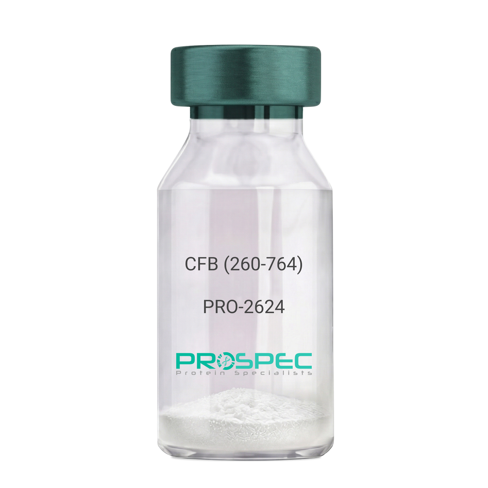
    CFB (260-764) Human, Sf9

    Description:

    Complement Factor B (260-764 a.a.) Human Recombinant, Sf9

    CFB, C3/C5 convertase, Glycine-rich beta glycoprotein, GBG, PBF2, Properdin factor B, BF, BFD, AHUS4, ARMD14, CFAB, CFBD, FB, FBI12, GBG, H2-Bf.

    Product # :

    PRO-2624

    Price :

    Quantity :

    Shipping Method :

    Ice Icon

    Shipped with Ice Packs

    Add To Cart

    More Info

    • description
    • source
    • formulation
    • purity
    • More Info

    Description

    CFB Human Recombinant produced in Sf9 Baculovirus cells is a single, glycosylated polypeptide chain containing 514 amino acids (260-764 a.a) and having a molecular mass of 58.1kDa.CFB is fused to an 9 amino acid His-tag at C-terminus & purified by proprietary chromatographic techniques.

    Source

    Sf9, Baculovirus cells.

    Formulation

    CFB protein solution (0.5mg/ml) containing Phosphate Buffered Saline (pH 7.4) and 20% glycerol.

    Purity

    Greater than 90.0% as determined by SDS-PAGE.

    More Info

    • Introduction

      Factor B is a protein, build from a single chain that circulates in the blood. Complement factor D cleaves and activates factor B, thus creating a catalytic subunit Bb and noncatalytic chain Ba. Subunit Bb is a serine protease that can bind to C3b and forms alternative pathway C3 convertase. Subunit Ba acts as an inhibitor for proliferation of lymphocytes, when Bb is part of the lymphocytes’ proliferation. The gene that codes for this protein is located on chromosome six.

    • Synonyms

      CFB, C3/C5 convertase, Glycine-rich beta glycoprotein, GBG, PBF2, Properdin factor B, BF, BFD, AHUS4, ARMD14, CFAB, CFBD, FB, FBI12, GBG, H2-Bf.

    • Physical Appearance

      Sterile Filtered colorless solution.

    • Stability

      Store at 4°C if entire vial will be used within 2-4 weeks. Store, frozen at -20°C for longer periods of time. For long term storage it is recommended to add a carrier protein (0.1% HSA or BSA).Avoid multiple freeze-thaw cycles.

    • Amino Acid Sequence

      ADPKIVLDPS GSMNIYLVLD GSDSIGASNF TGAKKCLVNL IEKVASYGVK PRYGLVTYAT
      YPKIWVKVSE ADSSNADWVT KQLNEINYED HKLKSGTNTK KALQAVYSMM SWPDDVPPEG
      WNRTRHVIIL MTDGLHNMGG DPITVIDEIR DLLYIGKDRK NPREDYLDVY VFGVGPLVNQ
      VNINALASKK DNEQHVFKVK DMENLEDVFY QMIDESQSLS LCGMVWEHRK GTDYHKQPWQ
      AKISVIRPSK GHESCMGAVV SEYFVLTAAH CFTVDDKEHS IKVSVGGEKR DLEIEVVLFH
      PNYNINGKKE AGIPEFYDYD VALIKLKNKL KYGQTIRPIC LPCTEGTTRA LRLPPTTTCQ
      QQKEELLPAQ DIKALFVSEE EKKLTRKEVY IKNGDKKGSC ERDAQYAPGY DKVKDISEVV
      TPRFLCTGGV SPYADPNTCR GDSGGPLIVH KRSRFIQVGV ISWGVVDVCK NQKRQKQVPA HARDFHINLF QVLPWLKEKL QDEDLGFLHH HHHH

    ProSpec's products are furnished for LABORATORY RESEARCH USE ONLY. The product may not be used as drugs, agricultural or pesticidal products, food additives or household chemicals.

    Complement Factor B
  • View Data Sheet

    Name :

    BAFF Antibody

    Description:

    B-cell Activating Factor, Mouse Anti Human

    BAFF, BLYS, CD257, TALL1, THANK, ZTNF4, TALL-1, TNFSF20, TNFSF13B.

    Product # :

    ANT-367

    Price :

    Quantity :

    Shipping Method :

    Ice Icon

    Shipped with Ice Packs

    Add To Cart

    More Info

    • formulation
    • More Info

    Formulation

    1mg/ml containing PBS, pH-7.4, & 0.1% Sodium Azide.

    More Info

    • Introduction

      Binds to tnfrsf13b/taci and tnfrsf17/bcma. tnfsf13/april binds to the same 2 receptors, together, they form a 2 ligands -2 receptors pathway involved in the stimulation of b- and t-cell function and the regulation of humoral immunity. A third b-cell specific baff-receptor (baffr/br3) promotes the survival of mature b-cells and the b-cell response. B Lymphocyte Stimulator functions as a potent B-cell growth factor in costimulation assays. Administration of BAFF Human recombinant to mice disrupts splenic B-cell and T-cell zones and results in elevated levels of serum immunoglobulin.

    • Synonyms

      BAFF, BLYS, CD257, TALL1, THANK, ZTNF4, TALL-1, TNFSF20, TNFSF13B.

    • Physical Appearance

      Sterile Filtered colorless solution.

    • Immunogen

      Anti-human BAFF mAb, is derived from hybridization of mouse F0 myeloma cells with spleen cells from BALB/c mice immunized with Recombinant human BAFF amino acids 134-285 purified from E. coli.

    • Ig Subclass

      Mouse IgG3 heavy chain and κ light chain.

    • Clone

      PH4C7AT.

    • Applications

      BAFF antibody has been tested by ELISA and Western blot analysis to assure specificity and reactivity. Since application varies, however, each investigation should be titrated by the reagent to obtain optimal results. Recommended dilution range for Western blot analysis is 1:500 ~ 1,000. Recommended starting dilution is 1:500.

    • Type

      Mouse Anti Human Monoclonal.

    • Storage Procedures

      For periods up to 1 month store at 4°C, for longer periods of time, store at -20°C. Prevent freeze thaw cycles.

    • Purification Method

      BAFF antibody was purified from mouse ascitic fluids by protein-G affinity chromatography.

    ProSpec's products are furnished for LABORATORY RESEARCH USE ONLY. The product may not be used as drugs, agricultural or pesticidal products, food additives or household chemicals.

    Baff Antibody
  • View Data Sheet

    Name :

    KRT19 Antibody

    Description:

    Cytokeratin 19, Mouse Anti Human

    Keratin type I cytoskeletal 19, Cytokeratin-19, CK-19, Keratin-19, K19, KRT19, CK19, K1CS, MGC15366.

    Product # :

    ANT-472

    Price :

    Quantity :

    Shipping Method :

    Ice Icon

    Shipped with Ice Packs

    Add To Cart

    More Info

    • formulation
    • More Info

    Formulation

    1mg/ml containing PBS, pH-7.4, 10% Glycerol and 0.02% Sodium Azide.

    More Info

    • Introduction

      CTK-19 is a member of the keratin family. The keratins are intermediate filament proteins responsible for the structural integrity of epithelial cells and are subdivided into cytokeratins and hair keratins. The type I cytokeratins consist of acidic proteins which are arranged in pairs of heterotypic keratin chains. Unlike its related family members, this smallest known acidic cytokeratin is not paired with a basic cytokeratin in epithelial cells. It is specifically expressed in the periderm, the transiently superficial layer that envelopes the developing epidermis. The type I cytokeratins are clustered in a region of chromosome 17q12-q21.

    • Synonyms

      Keratin type I cytoskeletal 19, Cytokeratin-19, CK-19, Keratin-19, K19, KRT19, CK19, K1CS, MGC15366.

    • Physical Appearance

      Sterile filtered colorless solution.

    • Immunogen

      Anti-human KRT19 mAb, clone PAT13D10A, is derived from hybridization of mouse F0 myeloma cells with spleen cells from BALB/c mice immunized with a recombinant human KRT19 protein purified from E. coli.

    • Ig Subclass

      Mouse IgG2b heavy chain and Kappa light chain.

    • Clone

      PAT13D10A.

    • Applications

      The antibody has been tested by ELISA, Western blot analysis to assure specificity and reactivity. Since application varies, however, each investigation should be titrated by the reagent to obtain optimal results. Recommended starting dilution is 1:1000.

    • Type

      Mouse Anti Human Monoclonal.

    • Storage Procedures

      For periods up to 1 month store at 4°C, for longer periods of time, store at -20°C. Prevent freeze thaw cycles.

    ProSpec's products are furnished for LABORATORY RESEARCH USE ONLY. The product may not be used as drugs, agricultural or pesticidal products, food additives or household chemicals.

    Krt19 Antibody
  • View Data Sheet

    Name :

    TTR Antibody

    Description:

    Transthyretin, Mouse Anti Human

    Transthyretin, ATTR, Prealbumin, TBPA, TTR, PALB, HsT2651.

    Product # :

    ANT-003

    Price :

    Quantity :

    Shipping Method :

    Ice Icon

    Shipped with Ice Packs

    Add To Cart

    More Info

    • formulation
    • More Info

    Formulation

    1mg/ml containing PBS, pH-7.4, & 0.1% Sodium Azide.

    More Info

    • Introduction

      Transthyretin (TTR) is a circulating and abundant tetramer, which functions as a carrier protein of thyroid hormone and retinol binding protein. It is primarily produced in the liver, but also in the brain and eye. Mutations in the TTR gene cause the protein to dissociate into monomers that after misfolding assemble into amyloid fibrils deposits in a variety of organs. TTR is also the only gene known to be associated with familial TTR amyloidosis. The diseases that originate from mutations in the TTR gene include amyloidotic polyneuropathy, euthyroid hyperthyroxinaemia, amyloidotic vitreous opacities, cardiomyopathy, oculoleptomeningeal amyloidosis, meningocerebrovascular amyloidosis, carpal tunnel syndrome, etc.

    • Synonyms

      Transthyretin, ATTR, Prealbumin, TBPA, TTR, PALB, HsT2651.

    • Physical Appearance

      Sterile filtered colorless solution.

    • Immunogen

      Anti-human TTR mAb, is derived from hybridization of mouse F0 myeloma cells with spleen cells from BALB/c mice immunized with recombinant human TTR amino acids 21-147 purified from E. coli.

    • Ig Subclass

      Mouse IgG1 heavy chain and κ light chain.

    • Clone

      PAT7B2AT.

    • Applications

      TTR antibody has been tested by ELISA and Western blot analysis to assure specificity and reactivity. Since application varies, however, each investigation should be titrated by the reagent to obtain optimal results. Recommended dilution range for Western blot analysis is 1:500 ~ 3000.
      Recommended starting dilution is 1:500.

    • Type

      Mouse Anti Human Monoclonal.

    • Storage Procedures

      For periods up to 1 month store at 4°C, for longer periods of time, store at -20°C. Prevent freeze thaw cycles.

    • Purification Method

      TTR antibody was purified from mouse ascitic fluids by protein-G affinity chromatography.

    ProSpec's products are furnished for LABORATORY RESEARCH USE ONLY. The product may not be used as drugs, agricultural or pesticidal products, food additives or household chemicals.

    Ttr Antibody
  • View Data Sheet

    Name :

    IGF1 N15 Human

    Description:

    Insulin Like Growth Factor-1 N15 Labeled Human Recombinant

    Somatomedin C, IGF-I, IGFI, IGF1, IGF-IA, Mechano growth factor, MGF.

    Product # :

    CYT-128

    Price :

    Quantity :

    Shipping Method :

    Room Temp Icon

    Shipped at Room temp

    Add To Cart

    More Info

    • description
    • source
    • formulation
    • purity
    • biological activity
    • More Info
    • sds-page

    Description

    IGF1 N15 Human Recombinant produced in E.coli is a single, non-glycosylated polypeptide chain containing 70 amino acids and having a molecular mass of 7.74kDa. The N15 is stable isotope labeled. The IGF1 N15 is purified by proprietary chromatographic techniques.

    Source

    Escherichia Coli.

    Formulation

    IGF1 N15 protein was lyophilized from a 0.2µm filtered concentrated solution in PBS, pH 7.2.

    Purity

    Greater than 97.0% as determined by:
    (a) Analysis by RP-HPLC.
    (b) Analysis by SDS-PAGE.

    Biological Activity

    The ED50 as determined by a cell proliferation assay using serum free human MCF-7 cells is less than 2ng/ml, corresponding to a specific activity of > 5.0 × 10⁵ IU/mg.

    sds-page

    IGF-1 15N Human sds-page - Product image 1

    More Info

    • Introduction

      The somatomedins, or insulin-like growth factors (IGFs), comprise a family of peptides that play important roles in mammalian growth and development. IGF1 mediates many of the growth-promoting effects of growth hormone (GH; MIM 139250). Early studies showed that growth hormone did not directly stimulate the incorporation of sulfate into cartilage, but rather acted through a serum factor, termed 'sulfation factor,' which later became known as 'somatomedin' (Daughaday et al., 1972). Three main somatomedins have been characterized: somatomedin C (IGF1), somatomedin A (IGF2; MIM 147470), and somatomedin B (MIM 193190) (Rotwein, 1986; Rosenfeld, 2003).

    • Synonyms

      Somatomedin C, IGF-I, IGFI, IGF1, IGF-IA, Mechano growth factor, MGF.

    • Physical Appearance

      Sterile Filtered White lyophilized (freeze-dried) powder.

    • Stability

      Lyophilized IGF1 N15 although stable at room temperature for 3 weeks, should be stored desiccated below -18°C. Upon reconstitution IGF1 N15 should be stored at 4°C between 2-7 days and for future use below -18°C. For long term storage it is recommended to add a carrier protein (0.1% HSA or BSA). Please prevent freeze-thaw cycles.

    • Solubility

      It is recommended to reconstitute the lyophilized IGF1 N15 in sterile 18M-cm H2O not less than 100µg/ml, which can then be further diluted to other aqueous solutions.

    • Amino Acid Sequence

      GPETLCGAEL VDALQFVCGD RGFYFNKPTG YGSSSRRAPQ TGIVDECCFR SCDLRRLEMY CAPLKPAKSA.

    ProSpec's products are furnished for LABORATORY RESEARCH USE ONLY. The product may not be used as drugs, agricultural or pesticidal products, food additives or household chemicals.

    Igf1 N15 Human
  • View Data Sheet

    Name :

    KRT8 Human

    Description:

    Cytokeratin 8 Human Recombinant

    Keratin type II cytoskeletal 8, Cytokeratin-8, CK-8, Keratin-8, K8, KRT8, CYK8, KO, CK8, K2C8, CARD2.

    Product # :

    PRO-347

    Price :

    Quantity :

    Shipping Method :

    Room Temp Icon

    Shipped at Room temp

    Add To Cart

    More Info

    • description
    • source
    • formulation
    • purity
    • More Info

    Description

    Cytokeratin 8 Human Recombinant produced in E.Coli is a single, non-glycosylated polypeptide chain having a molecular mass of 53,532 Dalton. The KRT8 is purified by proprietary chromatographic techniques.

    Source

    Escherichia Coli.

    Formulation

    The protein (1mg/ml) was lyophilized after from a sterile solution containing 30mM Tris-HCl pH-8, 9.5M urea, 2mM DTT, 2mM EDTA and 10mM methylammonium chloride.

    Purity

    Greater than 95.0% as determined by:
    (a) Analysis by RP-HPLC.
    (b) Analysis by SDS-PAGE.

    More Info

    • Introduction

      Keratin 8 and 18 (K8/18) are the major components of intermediate filament (IF) proteins of simple or single-layered epithelia.

    • Synonyms

      Keratin type II cytoskeletal 8, Cytokeratin-8, CK-8, Keratin-8, K8, KRT8, CYK8, KO, CK8, K2C8, CARD2.

    • Physical Appearance

      Sterile Filtered White lyophilized (freeze-dried) powder.

    • Stability

      Lyophilized KRT8 although stable at room temperature for 3 weeks, should be stored at 2-8°C. Upon reconstitution KRT8 should be stored at 4°C between 2-7 days and for future use below -18°C. For long term storage it is recommended to add a carrier protein (0.1% HSA or BSA). Please prevent freeze-thaw cycles.

    • Solubility

      It is recommended to reconstitute the lyophilized KRT8 in sterile 18MΩ-cm H2O not less than 100µg/ml, which can then be further diluted to other aqueous solutions.

    • Reconstitution to filaments

      Performed by mixing equimolar amounts of cytokeratins of type I and type II at concentrations of approx. 0.5 mg/ml, both dissolved in 9.5 M urea buffer (see above). Protofilaments and filament complexes are obtained by dialyzing the resulting polypeptide solution stepwise to a concentration of 4 M urea and then to low salt condition (50 mM NaCl, 2 mM dithiothreitol, 10 mM Tris-HCl, pH 7.4). For immunization purposes, the solution can be further dialyzed against PBS (phosphate buffered saline, e.g. Dulbecco's PBS).

    ProSpec's products are furnished for LABORATORY RESEARCH USE ONLY. The product may not be used as drugs, agricultural or pesticidal products, food additives or household chemicals.

    Krt8 Human
  • View Data Sheet

    Name :

    CTSS Antibody

    Description:

    Cathepsin-S, Mouse Anti Human

    Cathepsin S, MGC3886, CTSS, Cathepsin-S.

    Product # :

    ANT-485

    Price :

    Quantity :

    Shipping Method :

    Ice Icon

    Shipped with Ice Packs

    Add To Cart

    More Info

    • formulation
    • More Info

    Formulation

    1mg/ml containing PBS, pH-7.4, 10% Glycerol and 0.02% Sodium Azide.

    More Info

    • Introduction

      Cathepsin S (CTSS) belongs to the peptidase C1 family. CTSS is a lysosomal cysteine proteinase that participates in the degradation of antigenic proteins to peptides for presentation on MHC class II molecules. CTSS functions as an elastase over a broad pH range in alveolar macrophages.

    • Synonyms

      Cathepsin S, MGC3886, CTSS, Cathepsin-S.

    • Physical Appearance

      Sterile filtered colorless solution.

    • Immunogen

      Anti-human CTSS mAb, clone PAT1F9A, is derived from hybridization of mouse F0 myeloma cells with spleen cells from BALB/c mice immunized with a recombinant human CTSS protein 17-331 amino acids purified from E. coli.

    • Ig Subclass

      Mouse IgG2b heavy chain and Kappa light chain.

    • Clone

      PAT1F9AT.

    • Applications

      The antibody has been tested by ELISA, Western blot analysis to assure specificity and reactivity. Since application varies, however, each investigation should be titrated by the reagent to obtain optimal results. Recommended starting dilution is 1:500.

    • Type

      Mouse Anti Human Monoclonal.

    • Storage Procedures

      For periods up to 1 month store at 4°C, for longer periods of time, store at -20°C. Prevent freeze thaw cycles.

    • Purification Method

      CTSS antibody was purified from mouse ascitic fluids by protein-A affinity chromatography.

    ProSpec's products are furnished for LABORATORY RESEARCH USE ONLY. The product may not be used as drugs, agricultural or pesticidal products, food additives or household chemicals.

    Ctss Antibody
  • View Data Sheet

    Name :

    CFL1 Antibody

    Description:

    Cofilin-1, Mouse Anti Human

    CFL-1, CFL1, Cofilin1, Cofilin-1, Cofilin non-muscle isoform, 18 kDa phosphoprotein, p18.

    Product # :

    ANT-354

    Price :

    Quantity :

    Shipping Method :

    Ice Icon

    Shipped with Ice Packs

    Add To Cart

    More Info

    • formulation
    • More Info

    Formulation

    1mg/ml containing PBS, pH-7.4, & 0.1% Sodium Azide.

    More Info

    • Introduction

      Cofilin is a family of actin-binding proteins which disassembles actin filaments. It is a widely distributed intracellular actin-modulating protein that binds and depolymerizes filamentous F-actin and inhibits the polymerization of monomeric G-actin in a pH-dependent manner. It is involved in the translocation of actin-cofilin complex from cytoplasm to nucleus.

    • Synonyms

      CFL-1, CFL1, Cofilin1, Cofilin-1, Cofilin non-muscle isoform, 18 kDa phosphoprotein, p18.

    • Immunogen

      Anti-human CFL1 mAb is derived from hybridization of mouse F0 myeloma cells with spleen cells from BALB/c mice immunized with recombinant human CFL1 amino acids 1-166 purified from E. coli.

    • Ig Subclass

      Mouse IgG1 heavy chain and κ light chain.

    • Clone

      PAT1C1AT.

    • Applications

      CFL1 antibody has been tested by ELISA and Western blot analysis to assure specificity and reactivity. Since application varies, however, each investigation should be titrated by the reagent to obtain optimal results. Recommended dilution range for Western blot analysis is 1:1,000 ~ 2,000.Recommended starting dilution is 1:1,000.

    • Type

      Mouse Anti Human Monoclonal.

    • Storage Procedures

      For periods up to 1 month store at 4°C, for longer periods of time, store at -20°C. Prevent freeze thaw cycles.

    • Purification Method

      CFL1 antibody was purified from mouse ascitic fluids by protein-G affinity chromatography.

    ProSpec's products are furnished for LABORATORY RESEARCH USE ONLY. The product may not be used as drugs, agricultural or pesticidal products, food additives or household chemicals.

    Cfl1 Antibody
  • View Data Sheet

    Name :

    GMFB Antibody

    Description:

    Glia Maturation Factor Beta, Mouse Anti Human

    Glia maturation factor beta, GMFB, GMF-B, GMF-beta, GMF.

    Product # :

    ANT-680

    Price :

    Quantity :

    Shipping Method :

    Ice Icon

    Shipped with Ice Packs

    Add To Cart

    More Info

    • formulation
    • More Info

    Formulation

    1mg/ml containing PBS, pH-7.4, 10% Glycerol and 0.02% Sodium Azide.

    More Info

    • Introduction

      Glia Maturation Factor-Beta (GMF-Beta) is a 17 kDa protein nerve gorwth factor identified as a growth and differentiation factor in the vertebrate brain.

      Glia Maturation Factor-Beta stimulates differentiation of normal neurons as well as glial cells. GMFB inhibits the proliferation of the N-18 neuroblastoma line and the C6 glioma line while promoting their phenotypic expression.
      GMF-beta inhances the phenotypic expression of glia & neurons thus inhibits the proliferation of their respective tumors when added to cell culture. Although astrocytes produce GMF-b and stores it inside the cells, they don’t secrete the GMF-B into the cultured medium. Cell- surface GMFb acts on the target cells at close range when cells are in direct contact. GMF-Beta is produced by thymic epithelial cells and plays an important role in T cell development in favor of CD4+ T cells.
      GMF-Beta is a brain-specific protein which belongs to the actin-binding proteins (ADF) family. GMF-beta appears to play a role in the differentiation, maintenance, and regeneration of the nervous system. It also supports the progression of certain auto-immune diseases, possibly through its ability to induce the production and secretion of various pro-inflammatory cytokines.

    • Synonyms

      Glia maturation factor beta, GMFB, GMF-B, GMF-beta, GMF.

    • Physical Appearance

      Sterile filtered colorless solution.

    • Immunogen

      Anti-human GMFB mAb, is derived from hybridization of mouse F0 myeloma cells with spleen cells from BALB/c mice immunized with recombinant human GMFB amino acids 1-142 purified from E. coli.

    • Ig Subclass

      Mouse IgG2a heavy chain and Kappa light chain.

    • Clone

      PAT44D8AT.

    • Applications

      GMFB antibody has been tested by ELISA, Western blot and ICC/IF to assure specificity and reactivity. Since application varies, however, each investigation should be titrated by the reagent to obtain optimal results.

    • Type

      Mouse Anti Human Monoclonal.

    • Storage Procedures

      For periods up to 1 month store at 4°C, for longer periods of time, store at -20°C. Prevent freeze thaw cycles.

    • Purification Method

      GMFB antibody was purified from mouse ascitic fluids by protein-A affinity chromatography.

    ProSpec's products are furnished for LABORATORY RESEARCH USE ONLY. The product may not be used as drugs, agricultural or pesticidal products, food additives or household chemicals.

    Gmfb Antibody
  • View Data Sheet

    Name :

    GSTP1 Antibody

    Description:

    Glutathione S-Transferase pi 1, Mouse Anti Human

    Glutathione S-transferase P 1, Gst P1, GST YF-YF, GST class-pi, GST-piB, Preadipocyte growth factor.

    Product # :

    ANT-729

    Price :

    Quantity :

    Shipping Method :

    Ice Icon

    Shipped with Ice Packs

    Add To Cart

    More Info

    • formulation
    • More Info

    Formulation

    1mg/ml containing PBS, pH-7.4, 10% Glycerol and 0.02% Sodium Azide.

    More Info

    • Introduction

      GSTP1 is a polymorphic gene encoding active, functionally different GSTP1 variant proteins that are believed to function in xenobiotic metabolism and have a part in susceptibility to cancer, and other diseases. GSTP1 is a glutathione S-transferase belonging to the pi class. GST family enzymes play a significant role in detoxification by catalyzing the conjugation of many hydrophobic and electrophilic compounds with reduced glutathione. Based upon the biochemical, immunologic and structural properties of the soluble GSTs they are grouped into 4 main classes: alpha, mu, pi, and theta. The GSTP1 enzyme acts by catalyzing the reaction of glutathione with an acceptor molecule to form a Sulfur-substituted glutathione. The reactions employing glutathione contribute the transformation of a broad range of electrophiles, including reactive products of lipid, protein, carcinogens, therapeutic drugs, environmental toxins, and products of oxidative stress.
      The GSTP1 inactivation through CpG hypermethylation is frequent in pituitary adenomas and may be a factor in aggressive pituitary tumor behavior. GSTP1 is may be a transcriptional target of the p53 tumor suppressor gene. Single-nucleotide polymorphism in GSTP1 is linked to modified protein binding, which influence GSTP1's contribution to carcinogen and drug metabolism, and possibly disease pathogenesis and/or drug response. GST-pi might have central roles in proliferation of androgen-independent human prostate cancer cells.

    • Synonyms

      Glutathione S-transferase P 1, Gst P1, GST YF-YF, GST class-pi, GST-piB, Preadipocyte growth factor.

    • Physical Appearance

      Sterile filtered colorless solution.

    • Immunogen

      Anti-human GSTP1 mAb, is derived from hybridization of mouse F0 myeloma cells with spleen cells from BALB/c mice immunized with recombinant human GSTP1 amino acids 1-210 purified from E. coli.

    • Ig Subclass

      Mouse IgG1 heavy chain and κ light chain.

    • Clone

      PAT12C10AT

    • Applications

      GSTP1 antibody has been tested by ELISA, Western blot analysis, ICC/IF and Flow cytometry to assure specificity and reactivity. Since application varies, however, each investigation should be titrated by the reagent to obtain optimal results.

    • Type

      Mouse Anti Human Monoclonal.

    • Storage Procedures

      For periods up to 1 month store at 4°C, for longer periods of time, store at -20°C. Prevent freeze thaw cycles.

    • Purification Method

      GSTP1 antibody was purified from mouse ascitic fluids by protein-A affinity chromatography.

    ProSpec's products are furnished for LABORATORY RESEARCH USE ONLY. The product may not be used as drugs, agricultural or pesticidal products, food additives or household chemicals.

    Gstp1 Antibody
  • View Data Sheet

    Name :

    HCV NS3 Genotype-1b C33C

    Description:

    Hepatitis C Virus NS3 Genotype-1b C33C Recombinant

    Product # :

    HCV-271

    Price :

    Quantity :

    Shipping Method :

    Ice Icon

    Shipped with Ice Packs

    Add To Cart

    More Info

    • description
    • source
    • formulation
    • purity
    • More Info

    Description

    Recombinant Hepatitis C Virus NS3 Genotype-1b C33C (40-315 aa) produced in E. coli having 226 aa.Recombinant Hepatitis C Virus NS3 Genotype-1b C33C is fused to a 6xHis tag at C-Terminus and purified by proprietary chromatographic technique.

    Source

    Escherichia Coli.

    Formulation

    HCV NS3 Genotype-1b C33C solution contains 50mM potassium phosphate buffer.

    Purity

    Protein is >90% pure as determined by 10% PAGE (coomassie staining).

    More Info

    • Introduction

      HCV is a small 50nm, enveloped, single-stranded, positive sense RNAvirus in the family Flaviviridae. HCV has a high rate of replication with approximately one trillion particles produced each day in an infected individual. Due to lack of proofreading by the HCV RNA polymerase, the HCV has an exceptionally high mutation rate, a factor that may help it elude the host's immune response. Hepatitis C virus is classified into six genotypes(1-6) with several subtypes within each genotype. The preponderance and distribution of HCV genotypes varies globally. Genotype is clinically important in determining potential response to interferon-based therapy and the required duration of such therapy. Genotypes 1 and 4 are less responsive to interferon-based treatment than are the other genotypes (2, 3, 5 and 6). HCV NS3 (genotype 1b) contains 2 domains which are serine protinase and helicase. C33c is a fragment of NS3 containing the most important epitopes recognized by HCV antibody.

    • Physical Appearance

      Sterile Filtered clear solution.

    • Stability

      Store at 4°C if entire vial will be used within 2-4 weeks.Store, frozen at -20°C for longer periods of time.For long term storage it is recommended to add a carrier protein (0.1% HSA or BSA).Avoid multiple freeze-thaw cycles.

    • Applications

      ELISA and lateral flow assay.

    ProSpec's products are furnished for LABORATORY RESEARCH USE ONLY. The product may not be used as drugs, agricultural or pesticidal products, food additives or household chemicals.

    Hsv Ns3 C33C
  • View Data Sheet

    Name :

    HCV NS5 Genotype-1b

    Description:

    Hepatitis C Virus NS5 Genotype-1b Recombinant

    Product # :

    HCV-228

    Price :

    Quantity :

    Shipping Method :

    Ice Icon

    Shipped with Ice Packs

    Add To Cart

    More Info

    • description
    • source
    • formulation
    • purity
    • More Info

    Description

    The E.coli derived recombinant protein contains the HCV NS5 Genotype1b immunodominant regions, amino acids 2212-2313. The protein is fused to a GST tag at N-terminus.

    Source

    Escherichia Coli.

    Formulation

    1.5M urea, 25mM Tris-HCl pH 8.0, 0.2% Triton-X and 50% Glycerol.

    Purity

    HCV NS5 Genotype-1b protein is >95% pure as determined by SDS- PAGE (coomassie staining).

    More Info

    • Introduction

      HCV is a small 50nm, enveloped, single-stranded, positive sense RNAvirus in the family Flaviviridae.
      HCV has a high rate of replication with approximately one trillion particles produced each day in an infected individual. Due to lack of proofreading by the HCV RNA polymerase, the HCV has an exceptionally high mutation rate, a factor that may help it elude the host's immune response. Hepatitis C virus is classified into six genotypes(1-6) with several subtypes within each genotype. The preponderance and distribution of HCV genotypes varies globally. Genotype is clinically important in determining potential response to interferon-based therapy and the required duration of such therapy. Genotypes 1 and 4 are less responsive to interferon-based treatment than are the other genotypes (2, 3, 5 and 6).

    • Physical Appearance

      Sterile Filtered clear solution.

    • Stability

      HCV NS5 Genotype-1b although stable at 4°C for 1 week, should be stored below -18°C. Please prevent freeze thaw cycles.

    • Applications

      HCV NS5 Genotype-1b antigen is suitable for ELISA and Western blots, excellent antigen for detection of HCV with minimal specificity problems.

    • Specificity

      Immunoreactive with sera of HCV-infected individuals.

    • Purification Method

      HCV NS5 Genotype-1b protein was purified by proprietary chromatographic technique.

    ProSpec's products are furnished for LABORATORY RESEARCH USE ONLY. The product may not be used as drugs, agricultural or pesticidal products, food additives or household chemicals.

    Hcv Ns5 Genotype 1B
  • View Data Sheet

    Name :

    HCV NS5 Genotype-3a

    Description:

    Hepatitis C Virus NS5 Genotype-3a Recombinant

    Product # :

    HCV-232

    Price :

    Quantity :

    Shipping Method :

    Ice Icon

    Shipped with Ice Packs

    Add To Cart

    More Info

    • description
    • formulation
    • purity
    • More Info

    Description

    The E.coli derived recombinant protein contains the HCV NS5 Genotype 3a immunodominant regions, amino acids 2212-2313. The protein is fused to a GST tag at N-terminus.

    Formulation

    1.5M urea, 25 mM Tris-HCl, pH-8, 0.2% Triton-X & 50% Glycerol.

    Purity

    HCV NS5 Genotype-3a protein is >95% pure as determined by 10% PAGE (coomassie staining).

    More Info

    • Introduction

      HCV is a small 50nm, enveloped, single-stranded, positive sense RNAvirus in the family Flaviviridae.
      HCV has a high rate of replication with approximately one trillion particles produced each day in an infected individual. Due to lack of proofreading by the HCV RNA polymerase, the HCV has an exceptionally high mutation rate, a factor that may help it elude the host's immune response. Hepatitis C virus is classified into six genotypes(1-6) with several subtypes within each genotype. The preponderance and distribution of HCV genotypes varies globally. Genotype is clinically important in determining potential response to interferon-based therapy and the required duration of such therapy. Genotypes 1 and 4 are less responsive to interferon-based treatment than are the other genotypes (2, 3, 5 and 6).

    • Stability

      HCV NS5 Genotype-3a although stable at 4°C for 1 week, should be stored below -18°C. Please prevent freeze thaw cycles.

    • Applications

      HCV NS5 Genotype-3 antigen is suitable for ELISA and Western blots, excellent antigen for detection of HCV with minimal specificity problems.

    • Specificity

      Immunoreactive with sera of HCV-infected individuals.

    • Purification Method

      HCV NS5 Genotype-3a protein was purified by proprietary chromatographic technique.

    ProSpec's products are furnished for LABORATORY RESEARCH USE ONLY. The product may not be used as drugs, agricultural or pesticidal products, food additives or household chemicals.

    Hcv Ns5 Genotype 3A
  • View Data Sheet

    Name :

    HCV NS5 Genotype-4

    Description:

    Hepatitis C Virus NS5 Genotype-4 Recombinant

    Product # :

    HCV-222

    Price :

    Quantity :

    Shipping Method :

    Ice Icon

    Shipped with Ice Packs

    Add To Cart

    More Info

    • description
    • formulation
    • purity
    • More Info

    Description

    The E.coli derived recombinant protein contains the HCV NS5 Genotype 4 immunodominant regions, amino acids 2212-2313. The protein is fused with GST tag at N-terminus.

    Formulation

    1.5M urea, 25mM Tris-HCl, pH-8, 0.2% Triton-X & 50% Glycerol.

    Purity

    HCV NS5 Genotype-4 protein is >95% pure as determined by 10% PAGE (coomassie staining).

    More Info

    • Introduction

      HCV is a small 50nm, enveloped, single-stranded, positive sense RNAvirus in the family Flaviviridae.
      HCV has a high rate of replication with approximately one trillion particles produced each day in an infected individual. Due to lack of proofreading by the HCV RNA polymerase, the HCV has an exceptionally high mutation rate, a factor that may help it elude the host's immune response. Hepatitis C virus is classified into six genotypes(1-6) with several subtypes within each genotype. The preponderance and distribution of HCV genotypes varies globally. Genotype is clinically important in determining potential response to interferon-based therapy and the required duration of such therapy. Genotypes 1 and 4 are less responsive to interferon-based treatment than are the other genotypes (2, 3, 5 and 6).

    • Stability

      HCV NS5 Genotype-4 although stable at 4°C for 1 week, should be stored below -18°C. Please prevent freeze thaw cycles.

    • Applications

      HCV NS5 Genotype-4 antigen is suitable for ELISA and Western blots, excellent antigen for detection of HCV with minimal specificity problems.

    • Specificity

      Immunoreactive with sera of HCV-infected individuals.

    • Purification Method

      HCV NS5 Genotype-4 protein was purified by proprietary chromatographic technique.

    ProSpec's products are furnished for LABORATORY RESEARCH USE ONLY. The product may not be used as drugs, agricultural or pesticidal products, food additives or household chemicals.

    Hcv Ns5 Genotype 4
  • View Data Sheet

    Name :

    HCV NS4 (1916-1947 a.a.)

    Description:

    Hepatitis C Virus NS4 (1916-1947 a.a.) Recombinant

    Product # :

    HCV-202

    Price :

    Quantity :

    Shipping Method :

    Ice Icon

    Shipped with Ice Packs

    Add To Cart

    More Info

    • description
    • formulation
    • purity
    • More Info

    Description

    E.coli derived 30 kDa recombinant protein. Artificial mosaic polypeptide composite constructed from diagnostically relevant antigenic regions derived from the NS4 region.

    Formulation

    1.5M urea, 25mM Tris-HCl pH-8, 0.2% Triton-X & 50% Glycerol.

    Purity

    HCV NS4 protein is >95% pure as determined by 10% PAGE (coomassie staining).

    More Info

    • Introduction

      HCV is a small 50nm, enveloped, single-stranded, positive sense RNAvirus in the family Flaviviridae.
      HCV has a high rate of replication with approximately one trillion particles produced each day in an infected individual. Due to lack of proofreading by the HCV RNA polymerase, the HCV has an exceptionally high mutation rate, a factor that may help it elude the host's immune response. Hepatitis C virus is classified into six genotypes(1-6) with several subtypes within each genotype. The preponderance and distribution of HCV genotypes varies globally. Genotype is clinically important in determining potential response to IFN-based therapy and the required duration of such therapy. Genotypes 1 and 4 are less responsive to IFN-based treatment than are the other genotypes (2, 3, 5 and 6).

    • Stability

      HCV NS4 although stable at 4°C for 1 week, should be stored below -18°C. Please prevent freeze thaw cycles.

    • Applications

      HCV NS4 antigen is suitable for ELISA and Western blots, excellent antigen for detection of HCV with minimal specificity problems.

    • Specificity

      Immunoreactive with sera of HCV-infected individuals.

    • Purification Method

      HCV NS4 protein was purified by proprietary chromatographic technique.

    ProSpec's products are furnished for LABORATORY RESEARCH USE ONLY. The product may not be used as drugs, agricultural or pesticidal products, food additives or household chemicals.

    Hcv Ns4
  • View Data Sheet

    Name :

    DTL Human

    Description:

    Denticleless E3 Ubiquitin Protein Ligase Human Recombinant

    L2DTL, DCAF2, RAMP, CDT2.

    Product # :

    PRO-2826

    Price :

    Quantity :

    Shipping Method :

    Room Temp Icon

    Shipped at Room temp

    Add To Cart

    More Info

    • description
    • source
    • formulation
    • purity
    • More Info

    Description

    The DTL Human is created as a recombinant protein with a 4kda N-terminal fusion of His Tag. The DTL His-Tagged Fusion Protein, produced in E. coli, is a 31kDa protein containing 126 amino acid residues of the DTL Human, 1-231 amino acids.

    Source

    Escherichia Coli.

    Formulation

    Each mg was lyophilized with 1xPBS, 0.4% SDS and 4mM DTT.

    Purity

    Greater than 90% as determined by SDS-PAGE.

    More Info

    • Synonyms

      L2DTL, DCAF2, RAMP, CDT2.

    • Physical Appearance

      Filtered White lyophilized (freeze-dried) powder.

    • Stability

      Store lyophilized DTL at -20°C. Aliquot the product after reconstitution to avoid repeated freezing/thawing cycles. Reconstituted protein can be stored at 4°C for a limited period of time; it does not show any change after two weeks at 4°C.

    • Solubility

      It is recommended to add deionized water to prepare a working stock solution of approximately 0.5mg/ml and let the lyophilized pellet dissolve completely. Product is not sterile! Please filter the product by an appropriate sterile filter before using it on cell culture.

    • Background

      Denticleless E3 ubiquitin protein ligase (DTL) is part of the E3 ubiquitin ligase family, fundamental to the ubiquitin-proteasome system, which adjusts protein degradation and modification in cells. DTL also participates in numerous critical biological processes, which comprises cell cycle progression, DNA repair, as well as embryonic development. More than a few diseases have been linked with the dysregulation of DTL in particular Cancer, this highlights the importance of DTL as a potential therapeutic goal. DTL takes an important part in regulating the cell cycle, mainly the transition from the G1 phase to the S phase. The degradation of cell cycle regulators, such as cyclins, is also mediated by DTL in that way assuring appropriate cell division in addition to preventing uncontrolled proliferation. DTL is also part of the DNA damage response mechanisms. It ubiquitinates vital proteins which are involved in DNA repair pathways, thus influencing the cellular response to genotoxic stress and preserving genomic integrity.

    ProSpec's products are furnished for LABORATORY RESEARCH USE ONLY. The product may not be used as drugs, agricultural or pesticidal products, food additives or household chemicals.

    Dtl Protein
  • View Data Sheet

    Name :

    ESAT6-CFP10

    Description:

    ESAT6-CFP10 Chimeric Recombinant

    Early Secretory Target Mycobacterium Tuberculosis, ESAT-6

    Product # :

    PRO-2737

    Price :

    Quantity :

    Shipping Method :

    Ice Icon

    Shipped with Ice Packs

    Add To Cart

    More Info

    • description
    • source
    • formulation
    • purity
    • More Info

    Description

    Recombinant Chimeric ESAT6-CFP10 produced in E. coli is fused to a 6 amino acid his tag at its C-terminus and purified by proprietary chromatographic technique.

    Source

    Escherichia Coli.

    Formulation

    ESAT6-CFP10 solution contains 25mM K2CO3, 0.02% NaN3 and PBS.

    Purity

    Protein is >95% pure as determined by 12% PAGE (coomassie staining).

    More Info

    • Introduction

      Mycobacterium antigen ESAT6 has been isolated from low molecular weight fractions of the shot-term-culture filtrate (ST-CF) and it can easily be detected in tuberculosis patients. The export of ESAT-6 which is a potent T-cell antigen, and related proteins requires a dedicated secretory apparatus that is encoded by a group of genes, several of which also code for proteins that are recognized strongly by T cells. The ESAT-6 systems can consequently be considered as immunogenicity islands and there is mounting evidence that the equivalent genes are subject to selective pressure imposed by the immune system of the host. This antigen includes many epitopes detectable in the serum of most patients with tuberculosis (more than 90%). By the attempts to obtain the vaccine on the basis of ESAT-6 it was demonstrated that the optimization of adjuvant is very important when using the combination of dioctadecylammonium bromide and monophosphoryllipide. ESAT-6 is very potential as diagnostic for differentiation between the mycobacterial infection and BCG vaccination. The main topic in ESAT-6 using is in antibody production and in test-systems for tuberculosis elaboration. Early secretory antigen 6 (ESAT6) and cell filtrate protein 10 (CFP10) are 2 antigens secreted as a complex by the replicating Mycobacterium tuberculosis complex (MTC).

    • Synonyms

      Early Secretory Target Mycobacterium Tuberculosis, ESAT-6

    • Physical Appearance

      Sterile Filtered clear solution.

    • Stability

      Store at 4°C if entire vial will be used within 2-4 weeks.Store, frozen at -20°C for longer periods of time.For long term storage it is recommended to add a carrier protein (0.1% HSA or BSA).Avoid multiple freeze-thaw cycles.

    • Applications

      Immunoassay.

    ProSpec's products are furnished for LABORATORY RESEARCH USE ONLY. The product may not be used as drugs, agricultural or pesticidal products, food additives or household chemicals.

    Esat6 Cfp10
  • View Data Sheet

    Name :

    HCV NS3 Genotype-1c

    Description:

    Hepatitis C Virus NS3 Genotype-1c, (1192-1459 a.a.) Recombinant

    Product # :

    HCV-208

    Price :

    Quantity :

    Shipping Method :

    Ice Icon

    Shipped with Ice Packs

    Add To Cart

    More Info

    • description
    • formulation
    • purity
    • More Info

    Description

    The E.coli derived recombinant His-Tag fusion protein contains the HCV NS3 immunodominant regions, amino acids 1192-1459.

    Formulation

    1.5M urea, 25mM Tris-HCl pH-8, 0.2% Triton-X & 50% Glycerol.

    Purity

    HCV NS3 Genotype-1c protein is >95% pure as determined by 10% PAGE (coomassie staining).

    More Info

    • Introduction

      HCV is a small 50nm, enveloped, single-stranded, positive sense RNAvirus in the family Flaviviridae.
      HCV has a high rate of replication with approximately one trillion particles produced each day in an infected individual. Due to lack of proofreading by the HCV RNA polymerase, the HCV has an exceptionally high mutation rate, a factor that may help it elude the host's immune response. Hepatitis C virus is classified into six genotypes(1-6) with several subtypes within each genotype. The preponderance and distribution of HCV genotypes varies globally. Genotype is clinically important in determining potential response to interferon-based therapy and the required duration of such therapy. Genotypes 1 and 4 are less responsive to interferon-based treatment than are the other genotypes (2, 3, 5 and 6).

    • Stability

      HCV NS3 Genotype-1c although stable at 4°C for 1 week, should be stored below -18°C. Please prevent freeze thaw cycles.

    • Applications

      HCV NS3 Genotype-1c antigen is suitable for ELISA and Western blots, excellent antigen for detection of HCV with minimal specificity problems.

    • Specificity

      Immunoreactive with sera of HCV-infected individuals.

    • Purification Method

      HCV NS3 Genotype-1c protein was purified by proprietary chromatographic technique.

    ProSpec's products are furnished for LABORATORY RESEARCH USE ONLY. The product may not be used as drugs, agricultural or pesticidal products, food additives or household chemicals.

    Hcv Ns3 Genotype 1C
  • View Data Sheet

    Name :

    QTRTD1 Human

    Description:

    Queuine TRNA-Ribosyltransferase Domain Containing 1 Human Recombinant

     Queuine TRNA-Ribosyltransferase Domain Containing 1, EC 2.4.2.29, Queuine TRNA-Ribosyltransferase Domain-Containing Protein 1, QTRTD1.

    Product # :

    ENZ-833

    Price :

    Quantity :

    Shipping Method :

    Ice Icon

    Shipped with Ice Packs

    Add To Cart

    More Info

    • description
    • source
    • formulation
    • purity
    • More Info

    Description

    QTRTD1 Human Recombinant produced in E.Coli is a single, non-glycosylated polypeptide chain containing 438 amino acids (1-415 a.a) and having a molecular mass of 49.1kDa.QTRTD1 is fused to a 23 amino acid His-tag at N-terminus & purified by proprietary chromatographic techniques.

    Source

    Escherichia Coli.

    Formulation

    QTRTD1 protein solution (0.25mg/ml) containing Phosphate buffered saline (pH7.4) 30% glycerol, 2mM DTT, 1mM EDTA and 0.1mM PMSF.

    Purity

    Greater than 95.0% as determined by SDS-PAGE.

    More Info

    • Introduction

      Queuine TRNA-Ribosyltransferase Domain Containing 1, also known as QTRTD1 act together with QTRT1 to form an active queuine tRNA-ribosyltransferase. QTRTD1 exchanges queuine for the guanine at the wobble position of tRNAs along with GUN anticodons (tRNA-Asp,-Asn,-His and -Tyr), in that way forming the hypermodified nucleoside queuosine.

    • Synonyms

      Queuine TRNA-Ribosyltransferase Domain Containing 1, EC 2.4.2.29, Queuine TRNA-Ribosyltransferase Domain-Containing Protein 1, QTRTD1.

    • Physical Appearance

      Sterile Filtered colorless solution.

    • Stability

      Store at 4°C if entire vial will be used within 2-4 weeks. Store, frozen at -20°C for longer periods of time. For long term storage it is recommended to add a carrier protein (0.1% HSA or BSA).Avoid multiple freeze-thaw cycles.

    • Amino Acid Sequence

      MGSSHHHHHH SSGLVPRGSH MGSMKLSLTK VVNGCRLGKI KNLGKTGDHT MDIPGCLLYT KTGSAPHLTH HTLHNIHGVP AMAQLTLSSL AEHHEVLTEY KEGVGKFIGM PESLLYCSLH DPVSPCPAGY VTNKSVSVWS VAGRVEMTVS KFMAIQKALQ PDWFQCLSDG EVSCKEATSI KRVRKSVDRS LLFLDNCLRL QEESEVLQKS VIIGVIEGGD VMEERLRSAR ETAKRPVGGF LLDGFQGNPT TLEARLRLLS SVTAELPEDK PRLISGVSRP DEVLECIERG VDLFESFFPY QVTERGCALT FSFDYQPNPE ETLLQQNGTQ EEIKCMDQIK KIETTGCNQE ITSFEINLKE KKYQEDFNPL VRGCSCYCCK NHTRAYIHHL LVTNELLAGV LLMMHNFEHY FGFFHYIREA LKSDKLAQLK ELIHRQAS.

    ProSpec's products are furnished for LABORATORY RESEARCH USE ONLY. The product may not be used as drugs, agricultural or pesticidal products, food additives or household chemicals.

    Qtrtd1 Human
  • 1
  • …
  • 37
  • 38
  • 39
  • 40
  • 41
  • 42
Your browser does not support the video tag.

Products

  • Cytokines
  • Growth Factors
  • Chemokines
  • CD Antigens
  • Neurotrophins
  • Hormones
  • Enzymes
  • Viral Antigens
  • Recombinant Proteins
  • Natural Proteins
  • Monoclonal Antibodies
  • Polyclonal Antibodies
  • Thermal Packaging

About

  • Company
  • Founder
  • Contribution
  • Contact Us
  • My Account

Ordering

  • Ordering Method
  • Ordering - Credit Card
  • Ordering PO
  • Shipping Fees
  • FAQ's

Legal

  • Terms & Conditions
  • Privacy
  • Site Map
  • info@prospecbio.com
  • Facebook
  • Instagram
  • Linkedin Profile

© 2026 ProSpec-Tany TechnoGene Ltd. All Rights Reserved.

  • Choosing a selection results in a full page refresh.
  • Opens in a new window.