Search results
1000 results found for “stem cell factor”
Name
Description
Product #
Price
Quantity
Shipping Method
- View Data Sheet
Name :
Leptin tA MouseDescription:
Leptin Antagonist Triple Mutant Mouse Recombinant
Product # :
CYT-354Price :
Quantity :
Shipping Method :
Shipped at Room temp
More Info
- description
- source
- formulation
- purity
- biological activity
- More Info
Description
Leptin Antagonist Triple Mutant Mouse Recombinant is a single non-glycosilated polypeptide chain containing 146 amino and additional Ala at N-terminus acids and having a molecular mass of ~ 16 kDa, LEP was mutated, resulting in L39A/D40A/F41A mutant. Leptin Antagonist Triple Mutant Mouse Recombinant was purified by proprietary chromatographic techniques.
Source
Escherichia coli.
Formulation
The protein was lyophilized from a concentrated (0.65mg/ml) solution with 0.003mM NaHCO3.
Purity
Greater than 99.0% as determined by:
(a) Gel filtration analysis.
(b) Analysis by SDS-PAGE.Biological Activity
ProSpec’s Leptin Antagonist Triple Mutant Mouse Recombinant is capable of inhibiting Leptin-induced proliferation of BAF/3 cells stably transfected with the long form of human Leptin receptor. It also inhibits various Leptin effects in several in vitro bioassays.More Info
-
Physical Appearance
White lyophilized (freeze-dried) powder.
-
Stability
Lyophilized Leptin Antagonist Triple Mutant Mouse Recombinant although stable at room temperature for several weeks, should be stored desiccated below -18°C. Upon reconstitution at > 0.1 Leptin mutant mg/ml and up to 2 mM and filter sterilization LEP mutant can be stored at 4°C or even room temperature for several weeks making it suitable for long term infusion studies using osmotic pumps. At lower concentration addition of a carrier protein (0.1% HSA or BSA) is suggested.Please prevent freeze-thaw cycles.
-
Solubility
It is recommended to reconstitute the lyophilized Leptin Antagonist Triple Mutant Mouse Recombinant in sterile water or sterile 0.4% NaHCO3 adjusted to pH 8-9, not less than 100µg/ml, which can then be further diluted with other aqueous solutions.
-
Amino Acid Sequence
The sequence of the first five N-terminal amino acids was determined and was found to be Ala-Val-Pro-Ile-Gln.
-
Protein content
Protein quantization was carried out by UV spectroscopy at 280 nm using the absorbency value of 0.201 as the extinction coefficient for a 0.1% (1mg/ml) solution at pH 8.0. This value is calculated by the PC GENE computer analysis program of protein sequences (IntelliGenetics).
ProSpec's products are furnished for LABORATORY RESEARCH USE ONLY. The product may not be used as drugs, agricultural or pesticidal products, food additives or household chemicals.
- View Data Sheet
Name :
MIF AntibodyDescription:
Macrophage Migration Inhibitory Factor, Mouse Anti Human
Phenylpyruvate tautomerase, Glycosylation- inhibiting factor, GIF, MMIF, MIF.
Product # :
ANT-311Price :
Quantity :
Shipping Method :
Shipped with Ice Packs
More Info
- formulation
- More Info
Formulation
1mg/ml containing PBS, pH-7.4, 0.02% Sodium Azide and 10% Glycerol.
More Info
-
Introduction
The cytokine Macrophage migration inhibitory factor (MIF) has been identified to be secreted by the pituitary gland and the monocyte/macrophage and to play an important role in endotoxic shock. MIF has the unique property of being released from macrophages and T cells in response to physiological concentrations of glucocorticoids. The secretion of MIF is tightly regulated and decreases at high, anti-inflammatory steroid concentration.
-
Synonyms
Phenylpyruvate tautomerase, Glycosylation- inhibiting factor, GIF, MMIF, MIF.
-
Immunogen
Anti-human MIF mAb, is derived from hybridization of mouse SP2/0 myeloma cells with spleen cells from BALB/c mice immunized with Recombinant human MIF amino acids 1-114 purified from E. coli.
-
Ig Subclass
Mouse IgG1 heavy chain and κ light chain.
-
Clone
P4E4AT.
-
Applications
MIF antibody has been tested by ELISA and Western blot analysis to assure specificity and reactivity. Since application varies, however, each investigation should be titrated by the reagent to obtain optimal results.
-
Type
Mouse Anti Human Monoclonal.
-
Storage Procedures
For periods up to 1 month store at 4°C, for longer periods of time, store at -20°C. Prevent freeze thaw cycles.
-
Purification Method
MIF antibody was purified from mouse ascitic fluids by protein-G affinity chromatography.
ProSpec's products are furnished for LABORATORY RESEARCH USE ONLY. The product may not be used as drugs, agricultural or pesticidal products, food additives or household chemicals.
- View Data Sheet
Name :
FGFR2 Human, HisDescription:
Fibroblast Growth Factor Receptor-2 Human Recombinant, His Tag
EC 2.7.10, FGFR-2, BFR-1, CD332, BBDS, CEK3, ECT1, TK14, TK25, CFD1, KSAM, JWS, Fibroblast Growth Factor Receptor 2, Keratinocyte Growth Factor Receptor, Bacteria-Expressed Kinase, EC 2.7.10.1, K-SAM, KGFR, BEK, Protein Tyrosine Kinase, Receptor Like 14, BEK Fibroblast Growth Factor Receptor, Craniofacial Dysostosis 1, Jackson-Weiss Syndrome, Pfeiffer Syndrome, Crouzon Syndrome, CD332 Antigen.
Product # :
PKA-098Price :
Quantity :
Shipping Method :
Shipped with Ice Packs
More Info
- description
- source
- formulation
- purity
- More Info
Description
FGFR2 Human Recombinant produced in Sf9 Baculovirus cells is a single, glycosylated polypeptide chain containing 596 amino acids (22-378a.a.) and having a molecular mass of 66.6kDa (Molecular size on SDS-PAGE will appear at approximately 70-100kDa).FGFR2 is expressed with a 239 amino acids hIgG-His tag at C-Terminus and purified by proprietary chromatographic techniques.
Source
Sf9, Baculovirus cells.
Formulation
FGFR2 protein solution (1mg/ml) contains Phosphate Buffered Saline (pH 7.4) and 10% glycerol.
Purity
Greater than 95.0% as determined by SDS-PAGE.
More Info
-
Introduction
Fibroblast Growth Factors (FGFs) comprise a family of at least eighteen structurally realted proteins that are involved in a multitude of physiological and pathological cellular processes, including cell growth, differentation, angiogenesis, wound healing and tumorgenesis. The biological activities of the FGFs are mediated by a family if type I transmembrane tyrosine kinases which undergo dimerization and autophosphorylation after ligand binding. Four distinct genes encoding closely related FGF receptors, FGFR-1to -4 are known. Multiple forms of FGFR-1 to -3 are generated by alternative splicing of the mRNAs. A frequent splicing event involving FGFR-1 and -2 results in receptors containing all three Ig domains, referred to as the alpha isoform, or only IgII and IgIII, referred to as the ? isoform. Only the alpha isoform has been identified for FGFR-3 and FGFR-4. Additional splicing events for FGFR-1 to -3, involving the C-terminal half of the IgIII domain encoded by two mutually exclusive alternative exons, generate FGF receptors with alternative IgIII domains (IIIb and IIIc). A IIIa isoform which is a secreted FGF binding protein containing only the N-terminal half of the IgIII domain plus some intron sequences has also been reported for FGFR-1. Mutations in FGFR-1 to -3 have been found in patients with birth defects involving craniosynostosis.
-
Synonyms
EC 2.7.10, FGFR-2, BFR-1, CD332, BBDS, CEK3, ECT1, TK14, TK25, CFD1, KSAM, JWS, Fibroblast Growth Factor Receptor 2, Keratinocyte Growth Factor Receptor, Bacteria-Expressed Kinase, EC 2.7.10.1, K-SAM, KGFR, BEK, Protein Tyrosine Kinase, Receptor Like 14, BEK Fibroblast Growth Factor Receptor, Craniofacial Dysostosis 1, Jackson-Weiss Syndrome, Pfeiffer Syndrome, Crouzon Syndrome, CD332 Antigen.
-
Physical Appearance
Sterile Filtered colorless solution.
-
Stability
Store at 4°C if entire vial will be used within 2-4 weeks. Store, frozen at -20°C for longer periods of time. For long term storage it is recommended to add a carrier protein (0.1% HSA or BSA). Avoid multiple freeze-thaw cycles.
-
Amino Acid Sequence
RPSFSLVEDT TLEPEEPPTK YQISQPEVYV AAPGESLEVR CLLKDAAVIS WTKDGVHLGP NNRTVLIGEY LQIKGATPRD SGLYACTASR TVDSETWYFM VNVTDAISSG DDEDDTDGAE DFVSENSNNK RAPYWTNTEK MEKRLHAVPA ANTVKFRCPA GGNPMPTMRW LKNGKEFKQE HRIGGYKVRN QHWSLIMESV VPSDKGNYTC VVENEYGSIN HTYHLDVVER SPHRPILQAG LPANASTVVG GDVEFVCKVY SDAQPHIQWI KHVEKNGSKY GPDGLPYLKV LKHSGINSSN AEVLALFNVT EADAGEYICK VSNYIGQANQ SAWLTVLPKQ QAPGREKEIT ASPDYLELEP KSCDKTHTCP PCPAPELLGG PSVFLFPPKP KDTLMISRTP EVTCVVVDVS HEDPEVKFNW YVDGVEVHNA KTKPREEQYN STYRVVSVLT VLHQDWLNGK EYKCKVSNKA LPAPIEKTIS KAKGQPREPQ VYTLPPSRDE LTKNQVSLTC LVKGFYPSDI AVEWESNGQP ENNYKTTPPV LDSDGSFFLY SKLTVDKSRW QQGNVFSCSV MHEALHNHYT QKSLSLSPGK HHHHHH.
ProSpec's products are furnished for LABORATORY RESEARCH USE ONLY. The product may not be used as drugs, agricultural or pesticidal products, food additives or household chemicals.
- View Data Sheet
Name :
BMP4 Human, ActiveDescription:
Bone Morphogenetic protein-4 Active Active, Human Recombinant
BMP4, ZYME, BMP2B, BMP2B1.
Product # :
CYT-1293Price :
Quantity :
Shipping Method :
Shipped at Room temp
More Info
- description
- source
- formulation
- purity
- biological activity
- More Info
Description
Bone Morphogenetic protein-4 Active Human Recombinant produced in E.coli is a homodimer , non-glycosylated, polypeptide chain containing 2x116 amino acids (ser293-arg408) and having a total molecular mass of 26.2kDa. BMP4 is purified by proprietary chromatographic techniques.
Source
Escherichia Coli.
Formulation
The protein was lyophilized from a sterile (0.2µm) filtered solution containing 4mM HCL.
Purity
Greater than 95.0% as determined by SDS-PAGE.
Biological Activity
The ED50, as calculated by Alkaline phosphatase activity induced in ATDC-5 cells is 157.2ng/ml corresponding to a specific activity which is 6361 units/mg.
More Info
-
Synonyms
BMP4, ZYME, BMP2B, BMP2B1.
-
Physical Appearance
Sterile Filtered White lyophilized (freeze-dried) powder.
-
Stability
Lyophilized BMP4 although stable at room temperature for 3 weeks, should be stored desiccated below -18°C. Upon reconstitution BMP4 should be stored at 4°C between 2-7 days and for future use below -18°C. Please prevent freeze-thaw cycles.
-
Solubility
It is recommended to reconstitute the lyophilized BMP4 in sterile 4mM HCl at a concentration of 0.1 mg/ml, which can then be further diluted to other aqueous solutions
-
Amino Acid Sequence
SPKHHSQRAR KKNKNCRRHS LYVDFSDVGW NDWIVAPPGY QAFYCHGDCP FPLADHLNST NHAI VQT LVN SVNSSIPKAC CVPTELSAIS MLYLDEYDKV VLKNYQEMVV EGCGCR.
-
Background
The protein encoded by this gene is a member of the bone morphogenetic protein family which is part of the transforming growth factor-beta superfamily.
The superfamily includes large families of growth and differentiation factors.
Bone morphogenetic proteins were originally identified by an ability of demineralized bone extract to induce endochondral osteogenesis in vivo in an extraskeletal site.
This particular family member plays an important role in the onset of endochondral bone formation in humans, and a reduction in expression has been associated with a variety of bone diseases, including the heritable disorder Fibrodysplasia Ossificans Progressiva.
What is the molecular weight/Mw of BMP4 Protein?
BMP4 Protein has a total Mw of 26.2kDa.
What is the source or expression system of BMP4 Protein?
Escherichia Coli.
What is the Purity of BMP4 Protein?
BMP4 Protein is >95% pure as determined by SDS-PAGE.
What is the Biological Activity of BMP4 Protein?
The ED50, as calculated by Alkaline phosphatase activity induced in ATDC-5 cells is 157.2ng/ml corresponding to a specific activity which is 6361 units/mg.
What is the amino acid sequence of BMP4 Protein?
SPKHHSQRAR KKNKNCRRHS LYVDFSDVGW NDWIVAPPGY QAFYCHGDCP FPLADHLNST NHAI VQT LVN SVNSSIPKAC CVPTELSAIS MLYLDEYDKV VLKNYQEMVV EGCGCR.
What applications can BMP4 Protein be used in?
BMP4 Protein can probably be used in western blot, ELISA and Lateral Flow.
What is the endotoxin level for BMP4 Protein?
The endotoxin level is minimal, BMP4 Protein was purified using conventional chromatography techniques.
ProSpec's products are furnished for LABORATORY RESEARCH USE ONLY. The product may not be used as drugs, agricultural or pesticidal products, food additives or household chemicals.
- View Data Sheet
Name :
FAS AntibodyDescription:
FAS Blocking/Activating antibody (CD95), Mouse anti Human
FASLG receptor, Apoptosis-mediating surface antigen FAS, Apo-1 antigen, CD95, Tumor necrosis factor receptor superfamily member 6 TNR6, APT1, FAS1, TNFRSF6.
Product # :
ANT-204Price :
Quantity :
Shipping Method :
Shipped at Room temp
More Info
- formulation
- More Info
Formulation
1 mg/ml in PBS (after reconstitution).
More Info
-
Introduction
The Fas receptor (CD95) mediates apoptotic signaling by Fas-ligand expressed on the surface of other cells. The Fas-FasL interaction plays an important role in the immune system and lack of this system leads to autoimmunity, indicating that Fas-mediated apoptosis removes self-reactive lymphocytes. Fas signaling is also involved in immune surveillance to remove transformed cells and virus infected cells. Binding of FAS to oligimerized FasL on another cell activates apoptotic signaling through a cytoplasmic domain termed the death domain that interacts with signaling adaptors including FAF, FADD and DAX to activate the caspase proteolytic cascade. Caspase-8 and caspase-10 are first activated, to then cleave and activate downstream caspases, and a variety of cellular substrates that lead to cell death. Caspases cleave nuclear lamins, causing the nucleus to break down and lose its normal structure and another caspase substrate is DFF, inducing cleavage and degradation of the genome. Other caspase substrates are involved in cytoskeletal structure, cell cycle regulation and signaling pathways. Activation of JNK kinase, activation of Jun, and production of ceramide may also play roles in Fas-mediated apoptosis. Activation of fas-mediated apoptosis is opposed by I-FLICE and FAP. Viruses and tumors may escape immune surveillance in part through suppression of fas-mediated apoptosis using similar mechanisms.
-
Synonyms
FASLG receptor, Apoptosis-mediating surface antigen FAS, Apo-1 antigen, CD95, Tumor necrosis factor receptor superfamily member 6 TNR6, APT1, FAS1, TNFRSF6.
-
Solubility
Reconstitute with H20. Mix gently, wash the sides of the vial and wait 30-60 seconds before use.
-
Immunogen
Recombinant Human FAS.
-
Ig Subclass
mouse IgG1.
-
Clone
NYRhFAS.
-
Note
This antibody is a BLOCKING antibody when in soluble form and will block FAS-mediated apoptosis. It will ACTIVATE FAS-mediated killing when immobilized.
-
Titer
By direct ELISA, 1:10,000 dilution will yield 0.5 O.D using alkaline phosphatase conjugated rabbit anti-mouse Ig (Jackson Laboratories).
-
Shipping Conditions
Antibody is shipped lyophilized at ambient temperature.
-
Type
Mouse Anti Human Monoclonal.
-
Storage Procedures
In lyophilized form, for long periods, store at 4°C in a dry environment. After reconstitution, if not intended for use within a month, aliquot and store at -20°C.
-
Purification Method
Protein-A column.
ProSpec's products are furnished for LABORATORY RESEARCH USE ONLY. The product may not be used as drugs, agricultural or pesticidal products, food additives or household chemicals.
- View Data Sheet
Name :
SCO1 HumanDescription:
SCO Cytochrome Oxidase Deficient Homolog 1 Human Recombinant
SCO1 Cytochrome C Oxidase Assembly Protein, SCOD1, SCO (Cytochrome Oxidase Deficient, Yeast) Homolog 1, SCO Cytochrome Oxidase Deficient Homolog 1 (Yeast), SCO Cytochrome Oxidase Deficient Homolog 1, Protein SCO1 Homolog, Mitochondrial, SCOD1, Protein SCO1 homolog, mitochondrial.
Product # :
PRO-2156Price :
Quantity :
Shipping Method :
Shipped with Ice Packs
More Info
- description
- source
- formulation
- purity
- More Info
Description
SCO1 Human Recombinant produced in E.Coli is a single, non-glycosylated polypeptide chain containing 179 amino acids (132-301 a.a) and having a molecular mass of 20.5kDa. SCO1 is fused to a 9 amino acid His-tag at C-terminus & purified by proprietary chromatographic techniques.
Source
Escherichia Coli.
Formulation
SCO1 protein solution (1mg/ml) containing Phosphate buffered saline (pH7.4), 10% glycerol and 1mM DTT.
Purity
Greater than 95% as determined by SDS-PAGE.
More Info
-
Introduction
SCO Cytochrome Oxidase Deficient Homolog 1, also known as SCO1 is a member of the SCO1/2 family. Mammalian cytochrome c oxidase (COX) catalyzes the transfer of reducing equivalents from cytochrome c to molecular oxygen and pumps protons across the inner mitochondrial membrane. Furthermore, in yeast, two related COX assembly genes, SCO1 & SCO2 which are synthesis of cytochrome c oxidase, enable subunits 1 as well as 2 to be incorporated into the holoprotein. This gene is the human homolog to the yeast SCO1 gene. Among the diseases associated with SCO1 are hepatic failure, early-onset, neurologic disorder due to cytochrome c oxidase deficiency and fatal infantile cytochrome c oxidase deficiency.
-
Synonyms
SCO1 Cytochrome C Oxidase Assembly Protein, SCOD1, SCO (Cytochrome Oxidase Deficient, Yeast) Homolog 1, SCO Cytochrome Oxidase Deficient Homolog 1 (Yeast), SCO Cytochrome Oxidase Deficient Homolog 1, Protein SCO1 Homolog, Mitochondrial, SCOD1, Protein SCO1 homolog, mitochondrial.
-
Physical Appearance
Sterile filtered colorless solution.
-
Stability
Store at 4°C if entire vial will be used within 2-4 weeks. Store, frozen at -20°C for longer periods of time. For long term storage it is recommended to add a carrier protein (0.1% HSA or BSA).Avoid multiple freeze-thaw cycles.
-
Amino Acid Sequence
MGKPLLGGPF SLTTHTGERK TDKDYLGQWL LIYFGFTHCP DVCPEELEKM IQVVDEIDSI TTLPDLTPLF ISIDPERDTK EAIANYVKEF SPKLVGLTGT REEVDQVARA YRVYYSPGPK DEDEDYIVDH TIIMYLIGPD GEFLDYFGQN KRKGEIAASI ATHMRPYRKK SLEHHHHHH.
ProSpec's products are furnished for LABORATORY RESEARCH USE ONLY. The product may not be used as drugs, agricultural or pesticidal products, food additives or household chemicals.
- View Data Sheet
Name :
STC 2 Human, HisDescription:
Stanniocalcin-2 Human Recombinant, His Tag
Stanniocalcin-2, STC, STC2, STCRP, STC-2, Stanniocalcin-related protein, STC-related protein.
Product # :
HOR-008Price :
Quantity :
Shipping Method :
Shipped with Ice Packs
More Info
- description
- source
- formulation
- purity
- More Info
Description
STC 2 Human Recombinant produced in E.Coli is a single, non-glycosylated polypeptide chain containing 301 amino acids (25-302 a.a.) and having a molecular mass of 33kDa. STC 2 is fused to a 23 amino acid His-tag at N-terminus & purified by proprietary chromatographic techniques.
Source
Escherichia Coli.
Formulation
STC 2 protein solution (0.25mg/ml) containing 20mM Tris-HCl buffer (pH 8.0), 0.25M NaCl, 30% glycerol and 1mM DTT.
Purity
Greater than 90% as determined by SDS-PAGE.
More Info
-
Introduction
STC2 is a homodimeric glycoprotein that is expressed in a wide variety of tissues and may have autocrine or paracrine functions. The encoded protein has 10 of its 15 cysteine residues conserved among stanniocalcin family members and is phosphorylated by casein kinase 2 exclusively on its serine residues. Its C-terminus contains a cluster of histidine residues which may interact with metal ions. The protein may play a role in the regulation of renal and intestinal calcium and phosphate transport, cell metabolism, or cellular calcium/phosphate homeostasis. Constitutive over expression of human stanniocalcin 2 in mice resulted in pre- and postnatal growth restriction, reduced bone and skeletal muscle growth, and organomegaly. Expression of this gene is induced by estrogen and altered in some breast cancers.
-
Synonyms
Stanniocalcin-2, STC, STC2, STCRP, STC-2, Stanniocalcin-related protein, STC-related protein.
-
Physical Appearance
Sterile filtered colorless solution.
-
Stability
Store at 4°C if entire vial will be used within 2-4 weeks. Store, frozen at -20°C for longer periods of time. For long term storage it is recommended to add a carrier protein (0.1% HSA or BSA).Avoid multiple freeze-thaw cycles.
-
Amino Acid Sequence
MGSSHHHHHH SSGLVPRGSH MGSTDATNPP EGPQDRSSQQ KGRLSLQNTA EIQHCLVNAG DVGCGVFECF ENNSCEIRGL HGICMTFLHN AGKFDAQGKS FIKDALKCKA HALRHRFGCI SRKCPAIREM VSQLQRECYL KHDLCAAAQE NTRVIVEMIH FKDLLLHEPY VDLVNLLLTC GEEVKEAITH SVQVQCEQNW GSLCSILSFC TSAIQKPPTA PPERQPQVDR TKLSRAHHGE AGHHLPEPSS RETGRGAKGE RGSKSHPNAH ARGRVGGLGA QGPSGSSEWE DEQSEYSDIR R.
ProSpec's products are furnished for LABORATORY RESEARCH USE ONLY. The product may not be used as drugs, agricultural or pesticidal products, food additives or household chemicals.
- View Data Sheet
Name :
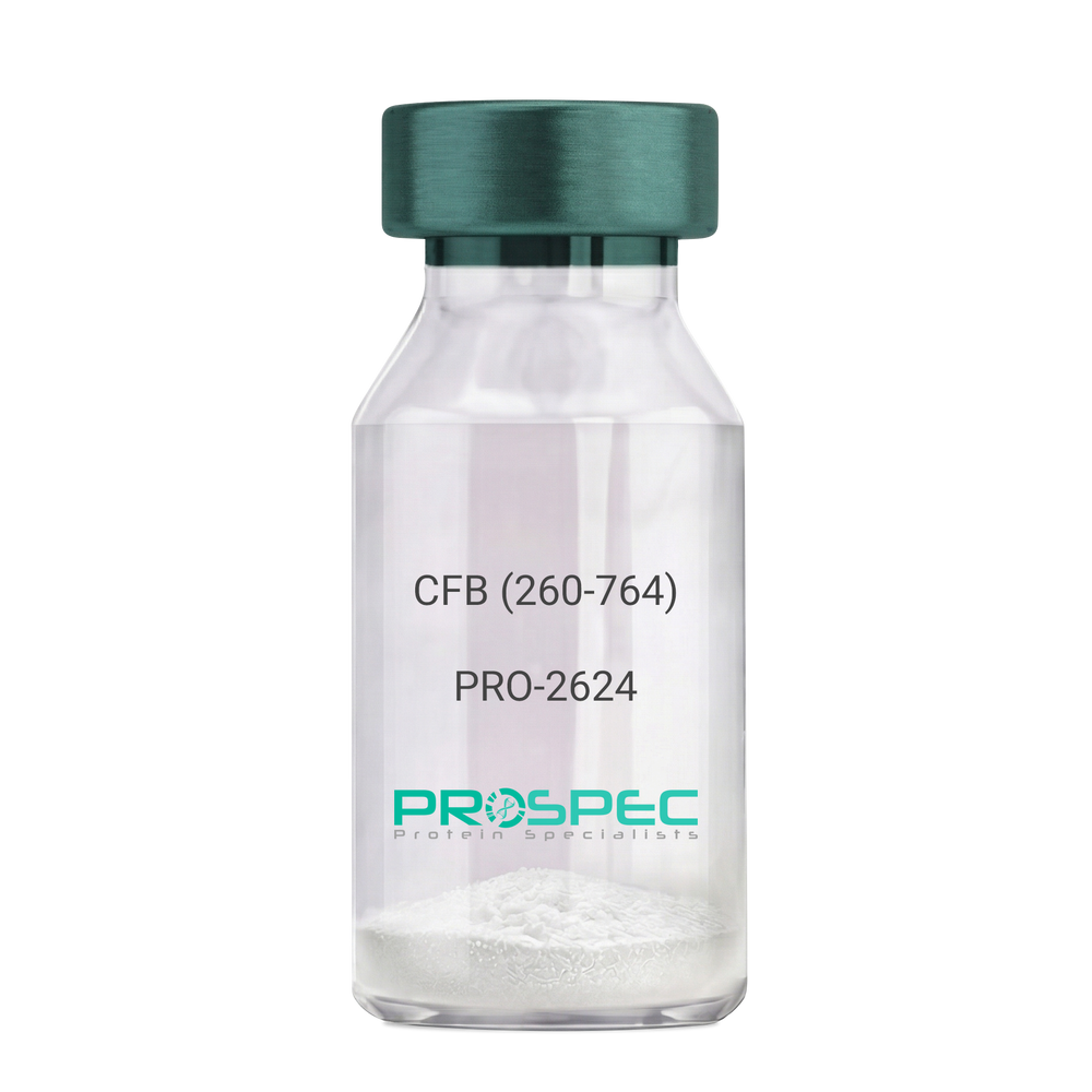
CFB (260-764) Human, Sf9Description:
Complement Factor B (260-764 a.a.) Human Recombinant, Sf9
CFB, C3/C5 convertase, Glycine-rich beta glycoprotein, GBG, PBF2, Properdin factor B, BF, BFD, AHUS4, ARMD14, CFAB, CFBD, FB, FBI12, GBG, H2-Bf.
Product # :
PRO-2624Price :
Quantity :
Shipping Method :
Shipped with Ice Packs
More Info
- description
- source
- formulation
- purity
- More Info
Description
CFB Human Recombinant produced in Sf9 Baculovirus cells is a single, glycosylated polypeptide chain containing 514 amino acids (260-764 a.a) and having a molecular mass of 58.1kDa.CFB is fused to an 9 amino acid His-tag at C-terminus & purified by proprietary chromatographic techniques.
Source
Sf9, Baculovirus cells.
Formulation
CFB protein solution (0.5mg/ml) containing Phosphate Buffered Saline (pH 7.4) and 20% glycerol.
Purity
Greater than 90.0% as determined by SDS-PAGE.
More Info
-
Introduction
Factor B is a protein, build from a single chain that circulates in the blood. Complement factor D cleaves and activates factor B, thus creating a catalytic subunit Bb and noncatalytic chain Ba. Subunit Bb is a serine protease that can bind to C3b and forms alternative pathway C3 convertase. Subunit Ba acts as an inhibitor for proliferation of lymphocytes, when Bb is part of the lymphocytes’ proliferation. The gene that codes for this protein is located on chromosome six.
-
Synonyms
CFB, C3/C5 convertase, Glycine-rich beta glycoprotein, GBG, PBF2, Properdin factor B, BF, BFD, AHUS4, ARMD14, CFAB, CFBD, FB, FBI12, GBG, H2-Bf.
-
Physical Appearance
Sterile Filtered colorless solution.
-
Stability
Store at 4°C if entire vial will be used within 2-4 weeks. Store, frozen at -20°C for longer periods of time. For long term storage it is recommended to add a carrier protein (0.1% HSA or BSA).Avoid multiple freeze-thaw cycles.
-
Amino Acid Sequence
ADPKIVLDPS GSMNIYLVLD GSDSIGASNF TGAKKCLVNL IEKVASYGVK PRYGLVTYAT
YPKIWVKVSE ADSSNADWVT KQLNEINYED HKLKSGTNTK KALQAVYSMM SWPDDVPPEG
WNRTRHVIIL MTDGLHNMGG DPITVIDEIR DLLYIGKDRK NPREDYLDVY VFGVGPLVNQ
VNINALASKK DNEQHVFKVK DMENLEDVFY QMIDESQSLS LCGMVWEHRK GTDYHKQPWQ
AKISVIRPSK GHESCMGAVV SEYFVLTAAH CFTVDDKEHS IKVSVGGEKR DLEIEVVLFH
PNYNINGKKE AGIPEFYDYD VALIKLKNKL KYGQTIRPIC LPCTEGTTRA LRLPPTTTCQ
QQKEELLPAQ DIKALFVSEE EKKLTRKEVY IKNGDKKGSC ERDAQYAPGY DKVKDISEVV
TPRFLCTGGV SPYADPNTCR GDSGGPLIVH KRSRFIQVGV ISWGVVDVCK NQKRQKQVPA HARDFHINLF QVLPWLKEKL QDEDLGFLHH HHHH
ProSpec's products are furnished for LABORATORY RESEARCH USE ONLY. The product may not be used as drugs, agricultural or pesticidal products, food additives or household chemicals.
- View Data Sheet
Name :
IL17F HumanDescription:
Interleukin 17F Human Recombinant
Cytokine ML-1, IL-17F, Interleukin-17F precursor, IL17F, ML1, ML-1.
Product # :
CYT-587Price :
Quantity :
Shipping Method :
Shipped at Room temp
More Info
- description
- source
- formulation
- purity
- More Info
Description
Interleukin-17F Human Recombinant produced in E.Coli is a homodimeric, cysteine linked, non-glycosylated polypeptide chain containing 2 x 134 amino acids and having a total molecular mass of 30.1 kDa.The IL-17F Human is purified by proprietary chromatographic techniques.
Source
Escherichia Coli.
Formulation
Lyophilized from a concentrated (1mg/ml) solution containing 10mM sodium citrate buffer pH-3.
Purity
Greater than 97.0% as determined by(a) Analysis by RP-HPLC.
(b) Analysis by SDS-PAGE.More Info
-
Introduction
IL-17F having an accession number of Q96PD4 is a cytokine that shares sequence similarity with IL17. IL-17F is expressed by activated T cells, and has been shown to stimulate the production of several other cytokines, including IL6, IL8, and CSF2/GM_CSF. IL-17F inhibits the angiogenesis of endothelial cells and induce endothelial cells to produce IL2, TGFB1/TGFB, and monocyte chemoattractant protein-1. IL-17F induces stromal cells to produce proinflammatory and hematopoietic cytokines. Intestinal IL17F gene expression is increased in active CD.
IL-17A & IL-17F alleles influence the susceptibility to and pathophysiological features of ulcerative colitis independently. IL-17F and MIF gene polymorphisms are significantly associated with the development of functional dyspepsia.
The initiation of IL-17F/IL-17R signaling pathway requires the receptor ubiquitination by TRAF6. IL-17F induces expression of IFN-gamma-inducible protein 10 (IP-10) by activating Raf1-mitogen-activated protein kinase 1/2-extracellular-regulated kinase 1/2-p90 ribosomal S6 kinase-cyclic AMP response element-binding protein signaling pathway. -
Synonyms
Cytokine ML-1, IL-17F, Interleukin-17F precursor, IL17F, ML1, ML-1.
-
Physical Appearance
Sterile Filtered White lyophilized (freeze-dried) powder.
-
Stability
Lyophilized Interleukin Human IL17F although stable at room temperature for 3 weeks, should be stored desiccated below -18°C. Upon reconstitution Human IL-17F should be stored at 4°C between 2-7 days and for future use below -18°C.For long term storage it is recommended to add a carrier protein (0.1% HSA or BSA).Please prevent freeze-thaw cycles.
-
Solubility
It is recommended to reconstitute the lyophilized Interleukin Human IL17F in sterile water not more than 1mg/ml, which can then be further diluted to other aqueous solutions.
-
Amino Acid Sequence
MRKIPKVGHT FFQKPESCPP VPGGSMKLDI GIINENQRVS MSRNIESRST SPWNYTVTWD PNRYPSEVVQ AQCRNLGCIN AQGKEDISMN SVPIQQETLV VRRKHQGCSV SFQLEKVLVT VGCTCVTPVI HHVQ.
ProSpec's products are furnished for LABORATORY RESEARCH USE ONLY. The product may not be used as drugs, agricultural or pesticidal products, food additives or household chemicals.
- View Data Sheet
Name :
ST6GAL1 HumanDescription:
ST6 Beta-Galactosamide Alpha-2,6-Sialyltranferase 1 Human Recombinant
ST6 Beta-Galactosamide Alpha-2,6-Sialyltranferase 1, ST6Gal I, SIAT1, CMP-N-Acetylneuraminate-Beta-Galactosamide-Alpha-2,6-Sialyltransferase 1, B-Cell Antigen CD75, Alpha 2,6-ST 1, EC 2.4.99.1, ST6GalI, CMP-N-Acetylneuraminate Beta-Galactosamide Alpha-2,6-Sialyltransferase, Sialyltransferase 1 (Beta-Galactoside Alpha-2,6-Sialyltransferase), Sialyltransferase 1 (Beta-Galactoside Alpha-2,6-Sialytransferase),Beta-Galactoside Alpha-2,6-Sialyltransferase 1, Sialyltransferase 1, ST6N, ST6GAL1.
Product # :
ENZ-893Price :
Quantity :
Shipping Method :
Shipped with Ice Packs
More Info
- description
- source
- formulation
- purity
- More Info
Description
ST6GAL1 Human Recombinant produced in E.Coli is a single, non-glycosylated polypeptide chain containing 403 amino acids (27-406 a.a) and having a molecular mass of 46kDa.ST6GAL1 is fused to a 23 amino acid His-tag at N-terminus & purified by proprietary chromatographic techniques.
Source
Escherichia Coli.
Formulation
ST6GAL1 protein solution (1mg/ml) containing 20mM Tris-HCl (pH 8.0) and 10% glycerol.
Purity
Greater than 90.0% as determined by SDS-PAGE.
More Info
-
Introduction
ST6GAL1 also known as ST6 Beta-Galactosamide Alpha-2,6-Sialyltranferase 1, is part of the glycosyltransferase family 29. ST6GAL1 is a type II membrane protein which catalyzes the transfer of sialic acid from CMP-sialic acid to galactose-containing substrates. Furthermore, ST6GAL1 is normally found in the Golgi however it can be proteolytically processed to a soluble form, ST6GAL1 is also involved in the generation of the cell-surface carbohydrate determinants as well differentiation antigens HB-6, CD75, and CD76.
-
Synonyms
ST6 Beta-Galactosamide Alpha-2,6-Sialyltranferase 1, ST6Gal I, SIAT1, CMP-N-Acetylneuraminate-Beta-Galactosamide-Alpha-2,6-Sialyltransferase 1, B-Cell Antigen CD75, Alpha 2,6-ST 1, EC 2.4.99.1, ST6GalI, CMP-N-Acetylneuraminate Beta-Galactosamide Alpha-2,6-Sialyltransferase, Sialyltransferase 1 (Beta-Galactoside Alpha-2,6-Sialyltransferase), Sialyltransferase 1 (Beta-Galactoside Alpha-2,6-Sialytransferase),Beta-Galactoside Alpha-2,6-Sialyltransferase 1, Sialyltransferase 1, ST6N, ST6GAL1.
-
Physical Appearance
Sterile Filtered colorless solution.
-
Stability
Store at 4°C if entire vial will be used within 2-4 weeks. Store, frozen at -20°C for longer periods of time. For long term storage it is recommended to add a carrier protein (0.1% HSA or BSA).Avoid multiple freeze-thaw cycles.
-
Amino Acid Sequence
MGSSHHHHHH SSGLVPRGSH MGSKEKKKGS YYDSFKLQTK EFQVLKSLGK LAMGSDSQSV SSSSTQDPHR GRQTLGSLRG LAKAKPEASF QVWNKDSSSK NLIPRLQKIW KNYLSMNKYK VSYKGPGPGI KFSAEALRCH LRDHVNVSMV EVTDFPFNTS EWEGYLPKES IRTKAGPWGR CAVVSSAGSL KSSQLGREID DHDAVLRFNG APTANFQQDV GTKTTIRLMN SQLVTTEKRF LKDSLYNEGI LIVWDPSVYH SDIPKWYQNP DYNFFNNYKT YRKLHPNQPF YILKPQMPWE LWDILQEISP EEIQPNPPSS GMLGIIIMMT LCDQVDIYEF LPSKRKTDVC YYYQKFFDSA CTMGAYHPLL YEKNLVKHLN QGTDEDIYLL GKATLPGFRT IHC
ProSpec's products are furnished for LABORATORY RESEARCH USE ONLY. The product may not be used as drugs, agricultural or pesticidal products, food additives or household chemicals.
- View Data Sheet
Name :
Lymphotactin HumanDescription:
Lymphotactin Human Recombinant (XCL1)
XCL1, Cytokine SCM-1, ATAC, Lymphotaxin, SCM-1-alpha, Small inducible cytokine C1, XC chemokine ligand 1, LTN, LPTN, SCM1, SCM-1, SCYC1, SCM-1a.
Product # :
CHM-314Price :
Quantity :
Shipping Method :
Shipped at Room temp
More Info
- description
- source
- formulation
- purity
- biological activity
- More Info
Description
Lymphotactin Human Recombinant produced in E.Coli is a single, non-glycosylated, polypeptide chain containing 92 amino acids and having a molecular mass of 10007 Dalton. The Lymphotactin is purified by proprietary chromatographic techniques.
Source
Escherichia Coli.
Formulation
The XCL1 was lyophilized from a concentrated (1mg/ml) solution in water containing no additives.
Purity
Greater than 99.0% as determined by:
(a) Analysis by RP-HPLC.
(b) Analysis by SDS-PAGE.Biological Activity
The Biological activity is calculated by its ability to chemoattract human T cells at 10-100ng/ml corresponding to a Specific Activity of 10,000-100,000IU/mg.More Info
-
Introduction
Chemokine (C motif) ligand (XCL1) is a small cytokine belonging to the XC chemokine family that is also known as lymphotactin. It is found in high levels in spleen, thymus, intestine and peripheral blood leukocytes, and at lower levels in lung, prostate gland and ovary. Cellular sources for XCL1 include activated thymic and peripheral blood CD8+ T cells. This chemokine attracts T cells. In humans, XCL1 is closely related to another chemokine called XCL2, whose geneis found at the same locus on chromosome 1. XCL1 induces it chemotactic function by binding to a chemokine receptor called XCR1.
-
Synonyms
XCL1, Cytokine SCM-1, ATAC, Lymphotaxin, SCM-1-alpha, Small inducible cytokine C1, XC chemokine ligand 1, LTN, LPTN, SCM1, SCM-1, SCYC1, SCM-1a.
-
Physical Appearance
Sterile Filtered White lyophilized (freeze-dried) powder.
-
Stability
Lyophilized Lymphotactin although stable at room temperature for 3 weeks, should be stored desiccated below -18°C. Upon reconstitution XCL1 should be stored at 4°C between 2-7 days and for future use below -18°C. For long term storage it is recommended to add a carrier protein (0.1% HSA or BSA). Please avoid freeze-thaw cycles.
-
Solubility
It is recommended to reconstitute the lyophilized Lymphotactin in sterile 18MΩ-cm H2O not less than 100µg/ml, which can then be further diluted to other aqueous solutions.
-
Amino Acid Sequence
The sequence of the first five N-terminal amino acids was determined and was found to be Gln-Ser-Glu-Val-Ser.
ProSpec's products are furnished for LABORATORY RESEARCH USE ONLY. The product may not be used as drugs, agricultural or pesticidal products, food additives or household chemicals.
- View Data Sheet
Name :
RELT HumanDescription:
RELT Human Recombinant
RELT, TNF Receptor, Receptor Expressed In Lymphoid Tissues, Tumor Necrosis Factor Receptor Superfamily Member 19L, RELT Tumor Necrosis Factor Receptor, TNFRSF19L, Tumor Necrosis Factor Receptor Superfamily, Member 19-Like, TRLT.
Product # :
CYT-1101Price :
Quantity :
Shipping Method :
Shipped with Ice Packs
More Info
- description
- source
- formulation
- purity
- More Info
Description
RELT produced in Sf9 Baculovirus cells is a single, glycosylated polypeptide chain containing 376 amino acids (26-162a.a.) and having a molecular mass of 41.4kDa. (Molecular size on SDS-PAGE will appear at approximately 40-57kDa).RELT is expressed with a 239 amino acid hIgG-His-tag at C-Terminus and purified by proprietary chromatographic techniques.
Source
Sf9, Baculovirus cells.
Formulation
RELT protein solution (0.5mg/ml) contains 10% glycerol & Phosphate Buffered Saline (pH 7.4).
Purity
Greater than 90% as determined by SDS-PAGE.
More Info
-
Introduction
RELTis expressed in hematopoietic tissues and peripheral blood leukocytesand is a part of thetumor necrosis factor receptor superfamily. RELT mediates activation of NF-kappa-B and takes part in T-cell activation.overexpression of RELT in HEK-293 cells induces p38 and JNK signalling and leads to apoptosis. it can also costimulate T-cell proliferation in the presence of CD3 signalling.
-
Synonyms
RELT, TNF Receptor, Receptor Expressed In Lymphoid Tissues, Tumor Necrosis Factor Receptor Superfamily Member 19L, RELT Tumor Necrosis Factor Receptor, TNFRSF19L, Tumor Necrosis Factor Receptor Superfamily, Member 19-Like, TRLT.
-
Physical Appearance
Sterile filtered colorless solution.
-
Stability
Store at 4°C if entire vial will be used within 2-4 weeks. Store, frozen at -20°C for longer periods of time. For long term storage it is recommended to add a carrier protein (0.1% HSA or BSA).Avoid multiple freeze-thaw cycles.
-
Amino Acid Sequence
STTLWQCPPG EEPDLDPGQG TLCRPCPPGT FSAAWGSSPC QPHARCSLWR RLEAQVGMAT
RDTLCGDCWP GWFGPWGVPR VPCQPCSWAPLGTHGCDEWG RRARRGVEVA AGASSGGETR QPGNGTRAGG PEETAAQVEP KSCDKTHTCP PCPAPELLGG PSVFLFPPKP KDTLMISRTP EVTCVVVDVS HEDPEVKFNW YVDGVEVHNA KTKPREEQYN STYRVVSVLT VLHQDWLNGK EYKCKVSNKA LPAPIEKTIS KAKGQPREPQ VYTLPPSRDE LTKNQVSLTC LVKGFYPSDI AVEWESNGQP ENNYKTTPPV LDSDGSFFLY SKLTVDKSRW QQGNVFSCSV MHEALHNHYT QKSLSLSPGK HHHHHH
ProSpec's products are furnished for LABORATORY RESEARCH USE ONLY. The product may not be used as drugs, agricultural or pesticidal products, food additives or household chemicals.
- View Data Sheet
Name :
SLAMF6 Human, sf9Description:
SLAMF6 Human Recombinant, sf9
CD352, KALI, KALIb, Ly108, NTB-A, NTBA, Activating NK receptor, SLAM family member 6.
Product # :
PRO-2348Price :
Quantity :
Shipping Method :
Shipped with Ice Packs
More Info
- description
- source
- formulation
- purity
- More Info
Description
SLAMF6 Human Recombinant produced in Insect cells is a single, glycosylated polypeptide chain containing 214 amino acids (22-226) and having a molecular mass of 24.1kDa (Molecular size on SDS-PAGE will appear at approximately 28-40kDa).SLAMF6 is fused to 6 amino acid His-Tag at C-terminus and purified by proprietary chromatographic techniques.
Source
Baculovirus sf9 cells.
Formulation
The SLAMF6 protein solution (0.5mg/ml) contains 1xPBS (pH 7.4) and 10% glycerol.
Purity
Greater than 95.0% as determined by SDS-PAGE.
More Info
-
Introduction
SLAM family member 6 (SLAMF6) is a member of the SLAM family of immune cell receptors. SLAMF6 is a unique receptor on T cells which, when triggered potentiates T cell expansion in a CD28-independent manner. SLAMF6 has a high expression on NK-, T-, and B cells. SLAMF6 exhibits homotypic interactions and can connect with adaptor molecules such as SAP to alter immune cell function.
-
Synonyms
CD352, KALI, KALIb, Ly108, NTB-A, NTBA, Activating NK receptor, SLAM family member 6.
-
Physical Appearance
Sterile Filtered clear solution.
-
Stability
Store at 4°C if entire vial will be used within 2-4 weeks. Store, frozen at -20°C for longer periods of time. For long term storage it is recommended to add a carrier protein (0.1% HSA or BSA).Avoid multiple freeze-thaw cycles.
-
Amino Acid Sequence
ADPQSSLTPL MVNGILGESV TLPLEFPAGE KVNFITWLFN ETSLAFIVPH ETKSPEIHVT NPKQGKRLNF TQSYSLQLSN LKMEDTGSYR AQISTKTSAK LSSYTLRILR QLRNIQVTNH SQLFQNMTCE LHLTCSVEDA DDNVSFRWEA LGNTLSSQPN LTVSWDPRIS SEQDYTCIAE NAVSNLSFSV SAQKLCEDVK IQYTDTKMHH HHHH.
ProSpec's products are furnished for LABORATORY RESEARCH USE ONLY. The product may not be used as drugs, agricultural or pesticidal products, food additives or household chemicals.
- View Data Sheet
Name :
POFUT1 HumanDescription:
Protein O-Fucosyltransferase 1 Human Recombinant
FUT12, O-Fuc-T, O-FucT-1, O-FUT, GDP-fucose protein O-fucosyltransferase 1, Peptide-O-fucosyltraferase 1,KIAA0180.
Product # :
ENZ-679Price :
Quantity :
Shipping Method :
Shipped with Ice Packs
More Info
- description
- source
- formulation
- purity
- More Info
Description
POFUT1 Human Recombinant produced in E. coli is a single polypeptide chain containing 385 amino acids (27-388) and having a molecular mass of 43.7 kDa. POFUT1 is fused to a 23 amino acid His-tag at N-terminus & purified by proprietary chromatographic techniques.
Source
Escherichia Coli.
Formulation
The POFUT1 solution (1mg/1ml) contains 20mM Tris-HCl buffer (pH 8.0), 2M Urea and 10% glycerol.
Purity
Greater than 85.0% as determined by SDS-PAGE.
More Info
-
Introduction
GDP-fucose protein O-fucosyltransferase 1 (POUFUT1), belongs to the glycosyltransferase O-Fuc family. POUFUT1 encodes a member of the glycosyltransferase O-Fuc family and Expressed mainly in pancreas, kidney, lung, heart, brain, liver, placenta and skeletal muscle.POUFUT1 adds O-fucose through an O-glycosidic linkage to Preserve serine or threonine residues in the epidermal growth factor-like repeats of a number of cell surface and emitted proteins. POUFUT1 is involved in ligand-induced receptor signaling. Alternative splicing of this gene results in 2 transcript variants encoding different isoforms.POFUT1 participates in Notch signaling, as Notch ligands can use as POFUT1 substrates.
-
Synonyms
FUT12, O-Fuc-T, O-FucT-1, O-FUT, GDP-fucose protein O-fucosyltransferase 1, Peptide-O-fucosyltraferase 1,KIAA0180.
-
Physical Appearance
Sterile Filtered colorless solution.
-
Stability
Store at 4°C if entire vial will be used within 2-4 weeks. Store, frozen at -20°C for longer periods of time. For long term storage it is recommended to add a carrier protein (0.1% HSA or BSA).Avoid multiple freeze-thaw cycles.
-
Amino Acid Sequence
MGSSHHHHHH SSGLVPRGSH MGSGSWDPAG YLLYCPCMGR FGNQADHFLG SLAFAKLLNR TLAVPPWIEY QHHKPPFTNL HVSYQKYFKL EPLQAYHRVI SLEDFMEKLA PTHWPPEKRV AYCFEVAAQR SPDKKTCPMK EGNPFGPFWD QFHVSFNKSE LFTGISFSAS YREQWSQRFS PKEHPVLALP GAPAQFPVLE EHRPLQKYMV WSDEMVKTGE AQIHAHLVRP YVGIHLRIGS DWKNACAMLK DGTAGSHFMA SPQCVGYSRS TAAPLTMTMC LPDLKEIQRA VKLWVRSLDA QSVYVATDSE SYVPELQQLF KGKVKVVSLK PEVAQVDLYI LGQADHFIGN CVSSFTAFVK RERDLQGRPS SFFGMDRPPK LRDEF.
ProSpec's products are furnished for LABORATORY RESEARCH USE ONLY. The product may not be used as drugs, agricultural or pesticidal products, food additives or household chemicals.
- View Data Sheet
Name :
LY6G6F HumanDescription:
Lymphocyte Antigen 6 Complex Locus G6F Human Recombinant
Lymphocyte Antigen 6 Complex Locus G6F, Lymphocyte Antigen 6 Complex Locus G6D, Chromosome 6 Open Reading Frame 21, C6orf21, LY6G6D, NG32, G6F.
Product # :
PRO-1782Price :
Quantity :
Shipping Method :
Shipped with Ice Packs
More Info
- description
- source
- formulation
- purity
- More Info
Description
LY6G6F Human Recombinant produced in E.coli is a single, non-glycosylated polypeptide chain containing 242 amino acids (17-235) and having a molecular mass of 26.2kDa.LY6G6F is fused to a 23 amino acid His-tag at N-terminus & purified by proprietary chromatographic techniques.
Source
Escherichia Coli.
Formulation
The LY6G6F solution contains 20mM Tris-HCl buffer (pH 8.0), 0.4M Urea and 10% glycerol.
Purity
Greater than 90% as determined by SDS-PAGE.
More Info
-
Introduction
The human Lymphocyte Antigen 6 Complex Locus G6F (LY6G6F) protein is a type I transmembrane protein belonging to the immunoglobin (Ig) superfamily, which contains cell-surface proteins involved in the immune system and cellular recognition. The LY6G6F protein has a role in the downstream signal transduction pathways involving GRB2 and GRB7.
-
Synonyms
Lymphocyte Antigen 6 Complex Locus G6F, Lymphocyte Antigen 6 Complex Locus G6D, Chromosome 6 Open Reading Frame 21, C6orf21, LY6G6D, NG32, G6F.
-
Physical Appearance
Sterile filtered colorless solution.
-
Stability
Store at 4°C if entire vial will be used within 2-4 weeks. Store, frozen at -20°C for longer periods of time. For long term storage it is recommended to add a carrier protein (0.1% HSA or BSA).Avoid multiple freeze-thaw cycles.
-
Amino Acid Sequence
MGSSHHHHHH SSGLVPRGSH MGSADNMQAI YVALGEAVEL PCPSPPTLHG DEHLSWFCSP AAGSFTTLVA QVQVGRPAPD PGKPGRESRL RLLGNYSLWL EGSKEEDAGR YWCAVLGQHH NYQNWRVYDV LVLKGSQLSA RAADGSPCNV LLCSVVPSRR MDSVTWQEGK GPVRGRVQSF WGSEAALLLV CPGEGLSEPR SRRPRIIRCL MTHNKGVSFS LAASIDASPA LCAPSTGWDM PW
ProSpec's products are furnished for LABORATORY RESEARCH USE ONLY. The product may not be used as drugs, agricultural or pesticidal products, food additives or household chemicals.
- View Data Sheet
Name :
IL 3 MouseDescription:
Interleukin-3 Mouse Recombinant
MCGF (Mast cell growth factor), Multi-CSF, HCGF, P-cell stimulation factor, IL-3, MGC79398, MGC79399.
Product # :
CYT-371Price :
Quantity :
Shipping Method :
Shipped at Room temp
More Info
- description
- source
- formulation
- purity
- biological activity
- More Info
Description
Interleukin-3 mouse Recombinant produced in E.Coli is a single, non-glycosylated polypeptide chain containing 135 amino acids and having a molecular mass of 15100 Dalton. The IL-3 is purified by proprietary chromatographic techniques.
Source
Escherichia Coli.
Formulation
Lyophilized from a concentrated (1mg/ml) solution in water containing no additives.
Purity
Greater than 97.0% as determined by:
(a) Analysis by SEC-HPLC.
(b) Analysis by SDS-PAGE.Biological Activity
The ED50 as determined by the dose-dependant stimulation of murine M-NFS-60 cells is < 0.05 ng/ml, corresponding to a Specific Activity of 20,000,000IU/mg.More Info
-
Introduction
Interleukin-3 is a pleiotropic cytokine produced primarily by activated T cells.
IL-3 is thought to function via specific cell surface receptors to stimulate the proliferation, differentiation and survival of haematopoietic cell lines. IL-3 has also been shown to affect the functional activity of a variety of other cell types including mast cells, eosinophils, megakaryocytes and basophils. -
Synonyms
MCGF (Mast cell growth factor), Multi-CSF, HCGF, P-cell stimulation factor, IL-3, MGC79398, MGC79399.
-
Physical Appearance
Sterile Filtered White lyophilized (freeze-dried) powder.
-
Stability
Lyophilized Interleukin-3 although stable at room temperature for 3 weeks, should be stored desiccated below -18°C. Upon reconstitution IL-3 should be stored at 4°C between 2-7 days and for future use below -18°C.For long term storage it is recommended to add a carrier protein (0.1% HSA or BSA).Please prevent freeze-thaw cycles.
-
Solubility
It is recommended to reconstitute the lyophilized Interleukin-3 in sterile 18MΩ-cm H2O not less than 100µg/ml, which can then be further diluted to other aqueous solutions.
-
Amino Acid Sequence
The sequence of the first five N-terminal amino acids was determined and was found to be Met-Asp-Thr-His-Arg.
-
Protein content
Protein quantitation was carried out by two independent methods1. UV spectroscopy at 280 nm using the absorbency value of 0.154 as the extinction coefficient for a 0.1% (1mg/ml) solution. This value is calculated by the PC GENE computer analysis program of protein sequences (IntelliGenetics). 2. Analysis by RP-HPLC, using a standard solution of IL3 as a Reference Standard.
ProSpec's products are furnished for LABORATORY RESEARCH USE ONLY. The product may not be used as drugs, agricultural or pesticidal products, food additives or household chemicals.
- View Data Sheet
Name :
IL 7 Human, HisDescription:
Interleukin-7 Human Recombinant, His Tag
Lymphopoietin 1 (LP-1), pre-B cell factor, IL-7.
Product # :
CYT-485Price :
Quantity :
Shipping Method :
Shipped with Ice Packs
More Info
- description
- source
- formulation
- purity
- More Info
Description
IL-7 Human Recombinant produced in E.Coli is single, a non-glycosylated, Polypeptide chain containing 152 amino acids fragment (26-177) and having a total molecular mass of 21.97 kDa with an amino-terminal hexahistidine tag. The IL-7 His-Tag protein is purified by proprietary chromatographic techniques.
Source
Escherichia Coli.
Formulation
Interleukin -7 His is supplied in 1x PBS and 50% glycerol.
Purity
Greater than 95.0% as determined by SDS-PAGE.
More Info
-
Introduction
Interleukin-7 is a potent lymphoid cell growth factor produced primarily by stromal cells.
IL-7 has been shown to support the proliferation and differentiation of pre B- and early T cells as well as displaying a biological effect on cells of NK and myeloid lineages. -
Synonyms
Lymphopoietin 1 (LP-1), pre-B cell factor, IL-7.
-
Physical Appearance
Sterile Filtered clear solution.
-
Stability
Store at 4°C if entire vial will be used within 2-4 weeks. Store, frozen at -20°C for longer periods of time. Please avoid freeze thaw cycles.
ProSpec's products are furnished for LABORATORY RESEARCH USE ONLY. The product may not be used as drugs, agricultural or pesticidal products, food additives or household chemicals.
- View Data Sheet
Name :
NME1 HumanDescription:
Non-Metastatic Cells 1 Human Recombinant
Nucleoside diphosphate kinase A, NDP kinase A, NDK A, Tumor metastatic process-associated protein, Metastasis inhibition factor nm23, nm23-H1, Granzyme A-activated DNase, GAAD, NME1, NDPKA, NM23, NB, AWD, NBS, NDPK-A.
Product # :
PRO-715Price :
Quantity :
Shipping Method :
Shipped with Ice Packs
More Info
- description
- source
- formulation
- purity
- More Info
Description
NME1 Human Recombinant produced in E.Coli is a single, non-glycosylated, polypeptide chain containing 152 amino acids (1-152 a.a.) and having a molecular mass of 17.1kDa.The NME1 is purified by proprietary chromatographic techniques.
Source
Escherichia Coli.
Formulation
The NME1 solution contains 20mM Tris-HCl buffer (pH7.5), 1mM DTT and 10% glycerol.
Purity
Greater than 95.0% as determined by SDS-PAGE.
More Info
-
Introduction
NDK (Nucleoside diphosphate kinase) exists as a hexamer composed of 'A' (encoded by NME1) and 'B' (encoded by NME2) isoforms. NME1 is involved in cell proliferation, differentiation and development, signal transduction, G protein-coupled receptor endocytosis, and gene expression. It also has tumor metastasis-suppressive capacity. NME1 has a key role in the synthesis of nucleoside triphosphates other than ATP. NME1 is essential for neural development including neural patterning and cell future determination.
The NME1 gene is expressed in various tumor types where its levels have been alternatively linked to reduced or increased metastatic potential.
Decrease in NME1 expression is notably connected to aggressive behavior in melanoma, breast, colon, and gastric carcinomas. In contrast, elevated levels of NME1 gene expression are noted in the advanced stage of thyroid carcinomas.
Somatic mutations of the NME1 gene are found in neuroblastoma. Increased NME1 in neuroblastoma is linked to features of the disease that are associated with aggressive tumors. -
Synonyms
Nucleoside diphosphate kinase A, NDP kinase A, NDK A, Tumor metastatic process-associated protein, Metastasis inhibition factor nm23, nm23-H1, Granzyme A-activated DNase, GAAD, NME1, NDPKA, NM23, NB, AWD, NBS, NDPK-A.
-
Physical Appearance
Sterile Filtered colorless solution.
-
Stability
Store at 4°C if entire vial will be used within 2-4 weeks. Store, frozen at -20°C for longer periods of time. For long term storage it is recommended to add a carrier protein (0.1% HSA or BSA).Avoid multiple freeze-thaw cycles.
-
Amino Acid Sequence
MANCERTFIA IKPDGVQRGL VGEIIKRFEQ KGFRLVGLKF MQASEDLLKE HYVDLKDRPF FAGLVKYMHS GPVVAMVWEG LNVVKTGRVM LGETNPADSK PGTIRGDFCI QVGRNIIHGS DSVESAEKEI GLWFHPEELV DYTSCAQNWI YE.
ProSpec's products are furnished for LABORATORY RESEARCH USE ONLY. The product may not be used as drugs, agricultural or pesticidal products, food additives or household chemicals.
- View Data Sheet
Name :
LGALS2 Mouse, ActiveDescription:
Galectin-2, BioActive Mouse Recombinant
Galectin-2, Gal-2, Lgals2, 2200008F12Rik, AI324147.
Product # :
CYT-1155Price :
Quantity :
Shipping Method :
Shipped with Ice Packs
More Info
- description
- source
- formulation
- purity
- biological activity
- More Info
Description
LGALS2 Mouse Recombinant produced in E.Coli is a single, non-glycosylated polypeptide chain containing 153 amino acids (1-130 a.a) and having a molecular mass of 17.3kDa.LGALS2 is fused to a 23 amino acid His-tag at N-terminus & purified by proprietary chromatographic techniques.
Source
E.coli.
Formulation
LGALS2 protein (1mg/ml) contains 10% glycerol, 0.1M NaCl, 1mM DTT and 20mM Tris-HCl buffer (pH 8.0).
Purity
Greater than 95% as determined by SDS-PAGE.
Biological Activity
Measured by its ability to agglutinate human red blood cells. The ED50 is ≥ 20ug/ml.
More Info
-
Introduction
Galectin-2 or LGALS2 is a protein, part of the galectin proteins family. The galectin proteins family holds galectin proteins family lectins that mediates adhesion between cells or cells to ECM. This family also take part in pre-mRNA splicing, apoptosis & tumor progression. Galectin-2 induces apoptosis in T cells that are activated & binds to lymphotoxin-a, also can implicatate on myocardial infarction. LGALS2 from human and mouse share about 65% amino acid sequence resemblance.
-
Synonyms
Galectin-2, Gal-2, Lgals2, 2200008F12Rik, AI324147.
-
Physical Appearance
Sterile Filtered colorless solution.
-
Stability
Store at 4°C if entire vial will be used within 2-4 weeks. Store, frozen at -20°C for longer periods of time. For long term storage it is recommended to add a carrier protein (0.1% HSA or BSA).Avoid multiple freeze-thaw cycles.
-
Amino Acid Sequence
MGSSHHHHHH SSGLVPRGSH MGSMSEKFEV KDLNMKPGMS LKIKGKIHND VDRFLINLGQ GKETLNLHFN PRFDESTIVC NTSEGGRWGQ EQRENHMCFS PGSEVKITIT FQDKDFKVTL PDGHQLTFPN RLGHNQLHYL SMGGLQISSF KLE
-
Background
What is the molecular weight/Mw of LGALS2 MOUSE, ACTIVE Protein?
LGALS2 MOUSE, ACTIVE Protein has a total Mw of 17.3kDa.
What is the source or expression system of LGALS2 MOUSE, ACTIVE Protein?
Escherichia Coli.
What is the Purity of LGALS2 MOUSE, ACTIVE Protein?
LGALS2 MOUSE, ACTIVE Protein is >95% pure as determined by SDS-PAGE.
What is the Biological Activity of LGALS2 MOUSE, ACTIVE Protein?
Measured by its ability to agglutinate human red blood cells. The ED50 is ≥ 20ug/ml.
What is the amino acid sequence of LGALS2 MOUSE, ACTIVE Protein?
MGSSHHHHHH SSGLVPRGSH MGSMSEKFEV KDLNMKPGMS LKIKGKIHND VDRFLINLGQ GKETLNLHFN PRFDESTIVC NTSEGGRWGQ EQRENHMCFS PGSEVKITIT FQDKDFKVTL PDGHQLTFPN RLGHNQLHYL SMGGLQISSF KLE.
What applications can LGALS2 MOUSE, ACTIVE Protein be used in?
LGALS2 MOUSE, ACTIVE Protein can probably be used in western blot, ELISA and Lateral Flow.
What is the endotoxin level for LGALS2 MOUSE, ACTIVE Protein?
The endotoxin level is minimal, LGALS2 MOUSE, ACTIVE Protein was purified using conventional chromatography techniques.
ProSpec's products are furnished for LABORATORY RESEARCH USE ONLY. The product may not be used as drugs, agricultural or pesticidal products, food additives or household chemicals.
- View Data Sheet
Name :
Periostin HumanDescription:
Periostin Human Recombinant
OSF-2, Periostin, Osteoblast Specific Factor 2, PN OSF-2, PDLPOSTN, POSTN, MGC119510, MGC119511, PN, RP11-412K4.1.
Product # :
CYT-452Price :
Quantity :
Shipping Method :
Shipped at Room temp
More Info
- description
- source
- formulation
- purity
- More Info
Description
The OSF2 His-Tagged Fusion Protein Human is produced in E. coli, and its molecular weight is 75 kDa protein containing 648 amino acid residues of the human OSF-2 and 23 additional amino acid residues - HisTag, Xa - cleavage site.
Source
Escherichia Coli.
Formulation
Filtered (0.4 µm) and lyophilized from 0.5 mg/ml in 0.05M Acetate buffer pH-4.
Purity
Greater than 90% as determined by SDS-PAGE.
More Info
-
Introduction
Periostin is a disulfide linked 90 kDa, 811 amino acid protein originally isolated as a osteoblast-specific factor that functions as a cell adhesion molecule for preosteoblasts and is thought to be involved in osteoblast recruitment, attachment and spreading. Additionally, periostin expression has previously been shown to be significantly increased by both transforming growth factor beta-1(TGFbeta1) and bone morphogenetic protein (BMP-2). OSF-2 has a typical signal sequence, followed by a cysteine-rich domain, a fourfold repeated domain and a C-terminal domain. The fourfold repeated domain of OSF-2 shows homology with the insect protein fasciclin
Periostin mRNA is expressed in the developing mouse embryonic and fetal heart, and that it is localized to the endocardial cushions that ultimately divide the primitive heart tube into a four-chambered heart. -
Synonyms
OSF-2, Periostin, Osteoblast Specific Factor 2, PN OSF-2, PDLPOSTN, POSTN, MGC119510, MGC119511, PN, RP11-412K4.1.
-
Physical Appearance
Filtered White lyophilized (freeze-dried) powder.
-
Stability
Store lyophilized protein at -20°C. Aliquot the product after reconstitution to avoid repeated freezing/thawing cycles. Reconstituted protein can be stored at 4°C for a limited period of time; it does not show any change after two weeks at 4°C.
-
Solubility
It is recommended to add 0.1M Acetate buffer pH4 to prepare a working stock solution of approximately 0.5 mg/ml and let the lyophilized pellet dissolve completely. For conversion into higher pH value, we recommend intensive dilution by relevant buffer to a concentration of 10µg/ml. In higher concentrations the solubility of this antigen is limited. Product is not sterile! Please filter the product by an appropriate sterile filter before using it in the cell culture.
-
Amino Acid Sequence
MGHHHHHHHH HHSSGHIEGR HMRNNHYDKI LAHSRIRGRD QGPNVCALQQ ILGTKKKYFS TCKNWYKKSI CGQKTTVLYE CCPGYMRMEG MKGCPAVLPI DHVYGTLGIV GATTTQRYSD ASKLREEIEG KGSFTYFAPS NEAWDNLDSD IRRGLESNVN VELLNALHSH MINKRMLTKD LKNGMIIPSM YNNLGLFINH YPNGVVTVNC ARIIHGNQIA TNGVVHVIDR VLTQIGTSIQ DFIEAEDDLS SFRAAAITSD ILEALGRDGH FTLFAPTNEA FEKLPRGVLE RFMGDKVASEALMKYHILNT LQCSESIMGG AVFETLEGNT IEIGCDGDSI TVNGIKMVNK KDIVTNNGVI HLIDQVLIPD SAKQVIELAG KQQTTFTDLV AQLGLASALR PDGEYTLLAP VNNAFSDDTL SMVQRLLKLI LQNHILKVKV GLNELYNGQI LETIGGKQLR VFVYRTAVCI ENSCMEKGSK QGRNGAIHIF REIIKPAEKS LHEKLKQDKR FSTFLSLLEA ADLKELLTQP GDWTLFVPTN DAFKGMTSEE KEILIRDKNA LQNIILYHLT PGVFIGKGFE PGVTNILKTT QGSKIFLKEV NDTLLVNELK SKESDIMTTN GVIHVVDKLL YPADTPVGND QLLEILNKLI KYIQIKFVRG STFKEIPVTV Y.
ProSpec's products are furnished for LABORATORY RESEARCH USE ONLY. The product may not be used as drugs, agricultural or pesticidal products, food additives or household chemicals.
- View Data Sheet
Name :
FASLG HumanDescription:
FAS Ligand Human Recombinant
Tumor necrosis factor ligand superfamily member 6, Apoptosis antigen ligand, APTL, CD95 ligand, CD95-L, Fas antigen ligand, Fas ligand, FasL, CD178, FASLG, APT1LG1, CD95L, TNFSF6, ALPS1B.
Product # :
CYT-031Price :
Quantity :
Shipping Method :
Shipped with Ice Packs
More Info
- description
- source
- formulation
- purity
- More Info
- sds-page
Description
FASLG Human Recombinant produced in E.Coli is a single, non-glycosylated polypeptide chain containing 173 amino acids (130-281 a.a.) and having a molecular mass of 19.6kDa.FASLG is fused to a 21 amino acid His-tag at N-terminus & purified by proprietary chromatographic techniques.
Source
Escherichia Coli.
Formulation
FASLG protein solution (1mg/ml) containing 20mM Tris-HCl buffer (pH 8.0), 0.4M urea and 10% glycerol.
Purity
Greater than 90% as determined by SDS-PAGE.
sds-page
More Info
-
Introduction
The type II transmembrane protein FASLG is a member of the tumor necrosis factor (TNF) superfamily. A fas ligand/receptor interaction has a significant part in the regulation of the immune system and the advancement of cancer. FASLG is expressed on the activated T cell surface as a nondisulfidelinked homotrimer. FASLG binding to Fas/CD95/TNFRSF6 on a nearby cell prompts apoptosis in the Fas expressing cell. FASLG is released from the cell surface by metalloproteinases as a soluble molecule that stays trimeric and is able to bind with Fas, but its capability to activate apoptosis is radically reduced. In addition, FASLG binds to DcR3 - a soluble trap receptor with no signal transduction capabilities. Flawed Fas-mediated apoptosis causes oncogenesis in addition to drug resistance in existing tumors. Constitutive expression of FASLG in a variety of tumors enables their immune evasion. Both mouse and human FASLG are active on mouse and human cells.
-
Synonyms
Tumor necrosis factor ligand superfamily member 6, Apoptosis antigen ligand, APTL, CD95 ligand, CD95-L, Fas antigen ligand, Fas ligand, FasL, CD178, FASLG, APT1LG1, CD95L, TNFSF6, ALPS1B.
-
Physical Appearance
Sterile Filtered colorless solution.
-
Stability
Store at 4°C if entire vial will be used within 2-4 weeks. Store, frozen at -20°C for longer periods of time. For long term storage it is recommended to add a carrier protein (0.1% HSA or BSA).Avoid multiple freeze-thaw cycles.
-
Amino Acid Sequence
MGSSHHHHHH SSGLVPRGSH MQIGHPSPPP EKKELRKVAH LTGKSNSRSM PLEWEDTYGI VLLSGVKYKK GGLVINETGL YFVYSKVYFR GQSCNNLPLS HKVYMRNSKY PQDLVMMEGK MMSYCTTGQM WARSSYLGAV FNLTSADHLY VNVSELSLVN FEESQTFFGL YKL.
-
Background
What is the molecular weight/Mw of FASL Protein?
FASL Protein has a total Mw of 19.6kDa.
What is the source or expression system of FASL Protein?
Escherichia Coli.
What is the Purity of FASL Protein?
FASL Protein is >90% pure as determined by SDS-PAGE.
What is the Biological Activity of FASL Protein?
The biological functionality of FASL Protein will be determined in the future.
What is the amino acid sequence of FASL Protein?
MGSSHHHHHH SSGLVPRGSH MQIGHPSPPP EKKELRKVAH LTGKSNSRSM PLEWEDTYGI VLLSGVKYKK GGLVINETGL YFVYSKVYFR GQSCNNLPLS HKVYMRNSKY PQDLVMMEGK MMSYCTTGQM WARSSYLGAV FNLTSADHLY VNVSELSLVN FEESQTFFGL YKL.
What applications can FASL Protein be used in?
FASL Protein can probably be used in western blot, ELISA and Lateral Flow.
What is the endotoxin level for FASL Protein?
The endotoxin level is minimal, FASL Protein was purified using conventional chromatography techniques.
ProSpec's products are furnished for LABORATORY RESEARCH USE ONLY. The product may not be used as drugs, agricultural or pesticidal products, food additives or household chemicals.
- View Data Sheet
Name :
BMPR1A Human, CHODescription:
Bone Morphogenetic Protein Receptor-1A Human Recombinant, CHO
BMPR-1A, BMP-R1A, BMPR1A, BMR1A, CD292, CD-292, Serine/threonine-protein kinase receptor R5, SKR5, ALK-3, ACVRLK3, EC 2.7.11.30, CD292 antigen.
Product # :
CYT-1094Price :
Quantity :
Shipping Method :
Shipped at Room temp
More Info
- description
- source
- formulation
- purity
- biological activity
- More Info
Description
Bone Morphogenetic Protein Receptor-1A Human Recombinant produced in CHO cells is a glycosylated homodimer chain containing 2x362 amino acids and having a total molecular mass of 80.8kDa. BMPR1A is purified by proprietary chromatographic techniques.
Source
CHO cells.
Formulation
The protein was lyophilized from a sterile (0.2µm) filtered solution containing PBS.
Purity
Greater than 95.0% as determined by SDS-PAGE.
Biological Activity
The ED50, as calculated by the Inhibition of human BMP-4-induced alkaline phosphatase production caused by ATDC5 cells is 120ng/ml corresponding to a specific activity of 8.3x10^3 units/mg.
More Info
-
Introduction
The bone morphogenetic protein (BMP) receptors are a family of transmembrane serine/threonine kinases that include the type I receptors BMPR1A and BMPR1B and the type II receptor BMPR2. These receptors are also closely related to the receptors, ACVR1 and ACVR2. The ligands of these receptors are members of the TGF-beta superfamily. TGF-betas transduce their signals through the formation of heteromeric complexes with 2 different types of serine (threonine) kinase receptors: type I receptors of about 50-55 kD and type II receptors of about 70-80 kD. Type II receptors bind ligands in the absence of type I receptors, but they require their respective type I receptors for signaling, whereas type I receptors require their respective type II receptors for ligand binding.
-
Synonyms
BMPR-1A, BMP-R1A, BMPR1A, BMR1A, CD292, CD-292, Serine/threonine-protein kinase receptor R5, SKR5, ALK-3, ACVRLK3, EC 2.7.11.30, CD292 antigen.
-
Physical Appearance
Sterile Filtered White lyophilized (freeze-dried) powder.
-
Stability
Lyophilized BMPR1A although stable at room temperature for 3 weeks, should be stored desiccated below -18°C. Upon reconstitution BMPR1A should be stored at 4°C between 2-7 days and for future use below -18°C. Please prevent freeze-thaw cycles.
-
Solubility
It is recommended to reconstitute the lyophilized BMPR1A in sterile 18MΩ-cm H2O not less than 100µg/ml, which can then be further diluted to other aqueous solutions.
-
Amino Acid Sequence
QNLDSMLHGT GMKSDSDQKK SENGVTLAPE DTLPFLKCYC SGHCPDDAIN NTCITNGHCF AIIEEDDQGE TTLASGCMKY EGSDFQCKDS PKAQLRRTIE CCRTNLCNQY LQPTLPPVVI GPFFDGSIRI EGRMDDKTHT CPPCPAPELL GGPSVFLFPP KPKDTLMISR TPEVTCVVVD VSHEDPEVKF NWYVDGVEVH NAKTKPREEQ YNSTYRVVSV LTVLHQDWLN GKEYKCKVSN KALPAPIEKT ISKAKGQPRE PQVYTLPPSR DELTKNQVSL TCLVKGFYPS DIAVEWESNG QPENNYKTTP PVLDSDGSFF LYSKLTVDKS RWQQGNVFSC SVMHEALHNH YTQKSLSLSP GK.
-
Background
Research Paper on Bone Morphogenetic Protein Receptor-1A Human Recombinant, CHO, Monomer, HEK
Abstract:
Welcome to the captivating world of Bone Morphogenetic Protein Receptor-1A Human Recombinant, CHO, Monomer (BMPR-1A HR) in Human Embryonic Kidney Cells (HEK). This research paper explores the vital role of BMPR-1A HR in cellular responses. As a key receptor in the transforming growth factor-beta (TGF-β) superfamily, BMPR-1A HR plays a significant part in guiding cellular differentiation and tissue development. Join us as we unravel the molecular mechanisms behind BMPR-1A HR signaling in HEK cells and delve into its interactions with key cytokines, including Tumor Necrosis Factor-alpha (TNF-α) and Tumor Necrosis Factor-alpha Superfamily Member 2 (TNFα SF2 or TNFSF2).
Introduction:
Welcome to the intriguing world of BMPR-1A HR! In this section, we introduce the remarkable BMPR-1A HR and its crucial role in shaping cellular responses. Together, let's explore how this receptor influences cellular behavior and contributes to tissue growth, fostering our understanding of its importance in biological processes.
BMPR-1A HR Signaling in HEK Cells:
Be amazed by the intricate dance of BMPR-1A HR signaling within HEK cells! Uncover the complex process of ligand-receptor binding, initiating both the canonical SMAD-dependent and non-canonical SMAD-independent pathways. This harmonious interplay regulates a wide range of cellular processes, including gene transcription, cell proliferation, and differentiation, forming the foundation of cellular communication.
Influential Role in Cellular Responses:
Marvel at the influential role of BMPR-1A HR as a critical mediator of cellular responses within HEK cells. Witness its ability to modulate cellular differentiation, driving the expression of key differentiation markers such as DIF. Our exploration will highlight the multifaceted nature of BMPR-1A HR, impacting diverse cellular pathways, including those involving TNF-α and TNFSF2, shaping a dynamic and interconnected cellular network.
Interplay with Key Cytokines:
Discover the intriguing interactions between BMPR-1A HR and key cytokines like TNF-α and TNFSF2. Explore how BMPR-1A HR influences their expression and activity, hinting at potential cross-talk between BMPR-1A HR and inflammatory pathways. This delicate balance fosters a harmonious cellular environment, where multiple players contribute to overall cellular responses.
Therapeutic Implications and Tissue Development:
Witness the potential therapeutic implications of BMPR-1A HR in tissue development. Together, we explore the exciting possibilities of utilizing BMPR-1A HR in regenerative medicine, offering hope for enhanced tissue development and repair. As we venture forth, we also address challenges, such as optimal dosage, innovative delivery methods, and safety considerations, ensuring a responsible and effective approach.
Conclusion:
As we conclude our exploration of BMPR-1A HR in HEK cells, we stand in awe of its role in mediating cellular responses and tissue development. Equipped with this knowledge, we look forward to a promising future, where BMPR-1A HR from CHO cells opens doors to innovative applications in regenerative medicine, contributing to improved human health and well-being.
What is the molecular weight/Mw of BMPR1A Protein?
BMPR1A Protein has a total Mw of 80.8kDa.
What is the source or expression system of BMPR1A Protein?
CHO cells.
What is the Purity of BMPR1A Protein?
BMPR1A Protein is >95% pure as determined by SDS-PAGE.
What is the Biological Activity of BMPR1A Protein?
The ED50, as calculated by the Inhibition of human BMP-4-induced alkaline phosphatase production caused by ATDC5 cells is 120ng/ml corresponding to a specific activity of 8.3x10^3 units/mg.
What is the amino acid sequence of BMPR1A Protein?
QNLDSMLHGT GMKSDSDQKK SENGVTLAPE DTLPFLKCYC SGHCPDDAIN NTCITNGHCF AIIEEDDQGE TTLASGCMKY EGSDFQCKDS PKAQLRRTIE CCRTNLCNQY LQPTLPPVVI GPFFDGSIRI EGRMDDKTHT CPPCPAPELL GGPSVFLFPP KPKDTLMISR TPEVTCVVVD VSHEDPEVKF NWYVDGVEVH NAKTKPREEQ YNSTYRVVSV LTVLHQDWLN GKEYKCKVSN KALPAPIEKT ISKAKGQPRE PQVYTLPPSR DELTKNQVSL TCLVKGFYPS DIAVEWESNG QPENNYKTTP PVLDSDGSFF LYSKLTVDKS RWQQGNVFSC SVMHEALHNH YTQKSLSLSP GK.
What applications can BMPR1A Protein be used in?
BMPR1A Protein can probably be used in western blot, ELISA and Lateral Flow.
What is the endotoxin level for BMPR1A Protein?
The endotoxin level is minimal, BMPR1A Protein was purified using conventional chromatography techniques.
ProSpec's products are furnished for LABORATORY RESEARCH USE ONLY. The product may not be used as drugs, agricultural or pesticidal products, food additives or household chemicals.
- View Data Sheet
Name :
LGALS3 HumanDescription:
Galectin-3 Human Recombinant
Galectin-3, GAL3, MAC2, CBP35, GALB, GALIG, LGALS2, LGALS3, Galactose-specific lectin 3, Mac-2 antigen, IgE-binding protein, 35 kDa lectin, Carbohydrate-binding protein 35, CBP 35, Laminin-binding protein, Lectin L-29, L-31, Galactoside-binding protein, GALBP.
Product # :
CYT-606Price :
Quantity :
Shipping Method :
Shipped at Room temp
More Info
- description
- source
- formulation
- purity
- More Info
Description
LGALS3 Human Recombinant produced in E.Coli is a single, non-glycosylated, polypeptide chain containing 250 amino acids and having a molecular mass of 26.2kDa. The LGALS3 is purified by proprietary chromatographic techniques.
Source
Escherichia Coli.
Formulation
The Galectin-3 protein was lyophilized from a sterile 0.2 micron filtered aqueous solution containing 10 mM sodium phosphate and 50mM sodium chloride, pH 7.5.
Purity
Greater than 95.0% as determined by analysis by SDS-PAGE.
More Info
-
Introduction
Galectin-3 mediates with the alpha-3, beta-1 integrin the stimulation by cspg4 of endothelial cells migration. Galectin-3 plays an necessary part during the acquisition of vasculogenic mimicry and angiogenic properties associated with melanoma progression. LGALS3 overexpression is highly expressed in early stages of papillary carcinoma, and its expression intensity declines during tumor progression. Serum levels of LGALS3 are high in patients with thyroid malignancy but there is considerable overlap in serum LGALS3 concentrations between those with benign and malignant nodular thyroid disease. LGLAS3 takes part as an immune regulator to inhibit T-cell immune responses and promote tumor growth, as a result providing a new mechanism for tumor immune tolerance.
-
Synonyms
Galectin-3, GAL3, MAC2, CBP35, GALB, GALIG, LGALS2, LGALS3, Galactose-specific lectin 3, Mac-2 antigen, IgE-binding protein, 35 kDa lectin, Carbohydrate-binding protein 35, CBP 35, Laminin-binding protein, Lectin L-29, L-31, Galactoside-binding protein, GALBP.
-
Physical Appearance
Sterile Filtered White lyophilized (freeze-dried) powder.
-
Stability
Lyophilized Galectin-3 Recombinant although stable at room temperature for 3 weeks, should be stored desiccated below -18°C. Upon reconstitution Galectin-3 should be stored at 4°C between 2-7 days and for future use below -18°C.For long term storage it is recommended to add a carrier protein (0.1% HSA or BSA).Please prevent freeze-thaw cycles.
-
Solubility
It is recommended to reconstitute the lyophilized LGALS3 in sterile 18MΩ-cm H2O not less than 100µg/ml, which can then be further diluted to other aqueous solutions.
-
Amino Acid Sequence
MADNFSLHDAL SGSGNPNPQG WPGAWGNQPA GAGGYPGASY PGAYPGQAPP GAYPGQAPPG AYPGAPGAYP GAPAPGVYPG PPSGPGAYPS SGQPSATGAY PATGPYGAPA GPLIVPYNLP LPGGVVPRML ITILGTVKPN ANRIALDFQR GNDVAFHFNP RFNENNRRVI VCNTKLDNNW GREERQSVFP FESGKPFKIQ VLVEPDHFKV AVNDAHLLQY NHRVKKLNEI SKLGISGDID LTSASYTMI.
-
Background
What is the molecular weight/Mw of LGALS3 HUMAN Protein?
LGALS3 HUMAN Protein has a total Mw of 26.2kDa.
What is the source or expression system of LGALS3 HUMAN Protein?
Escherichia Coli.
What is the Purity of LGALS3 HUMAN Protein?
LGALS3 HUMAN Protein is >95% pure as determined by SDS-PAGE.
What is the Biological Activity of LGALS3 HUMAN Protein?
The biological functionality of LGALS3 HUMAN Protein will be determined in the future.
What is the amino acid sequence of LGALS3 HUMAN Protein?
MADNFSLHDAL SGSGNPNPQG WPGAWGNQPA GAGGYPGASY PGAYPGQAPP GAYPGQAPPG AYPGAPGAYP GAPAPGVYPG PPSGPGAYPS SGQPSATGAY PATGPYGAPA GPLIVPYNLP LPGGVVPRML ITILGTVKPN ANRIALDFQR GNDVAFHFNP RFNENNRRVI VCNTKLDNNW GREERQSVFP FESGKPFKIQ VLVEPDHFKV AVNDAHLLQY NHRVKKLNEI SKLGISGDID LTSASYTMI.
What applications can LGALS3 HUMAN Protein be used in?
LGALS3 HUMAN Protein can probably be used in western blot, ELISA and Lateral Flow.
What is the endotoxin level for LGALS3 HUMAN Protein?
The endotoxin level is minimal, LGALS3 HUMAN Protein was purified using conventional chromatography techniques.
ProSpec's products are furnished for LABORATORY RESEARCH USE ONLY. The product may not be used as drugs, agricultural or pesticidal products, food additives or household chemicals.
- View Data Sheet
Name :
CDH1 Human, Sf9Description:
E-Cadherin Human Recombinant, Sf9
Cadherin 1, Type 1, E-Cadherin, Cadherin 1, Type 1, E-Cadherin (Epithelial), Epithelial Cadherin, CAM 120/80, Uvomorulin, CDHE, UVO, Calcium-Dependent Adhesion Protein, Epithelial, Cadherin 1, E-Cadherin (Epithelial), Cell-CAM 120/80, CD324 Antigen, Arc-1, CD324, ECAD, LCAM, Cadherin-1.
Product # :
PRO-2265Price :
Quantity :
Shipping Method :
Shipped with Ice Packs
More Info
- description
- source
- formulation
- purity
- More Info
Description
CDH1 produced in Sf9 Baculovirus cells is a single, glycosylated polypeptide chain containing 694 amino acids (24-709a.a.) and having a molecular mass of 76.6kDa. (Molecular size on SDS-PAGE will appear at approximately 70kDa). CDH1 is expressed with an 8 amino acid His tag at C-Terminus and purified by proprietary chromatographic techniques.
Source
Sf9, Baculovirus cells.
Formulation
CDH1 protein solution (0.5mg/ml) contains Phosphate Buffered Saline (pH 7.4) and 10% glycerol.
Purity
Greater than 90% as determined by SDS-PAGE.
More Info
-
Introduction
E-cadherin (uvomorulin, cell-CAM120/80) is a calcium dependent cell adhesion molecule expressed predominately in epithelial tissues. It plays an important role in the growth and development of cells via the mechanisms of control of tissue architecture and the maintenance of tissue integrity. Numerous studies have demonstrated that reduction and/or loss of Ecadherin expression in carcinomas correlates positively with the potential of these tumors for invasion and metastasis.
-
Synonyms
Cadherin 1, Type 1, E-Cadherin, Cadherin 1, Type 1, E-Cadherin (Epithelial), Epithelial Cadherin, CAM 120/80, Uvomorulin, CDHE, UVO, Calcium-Dependent Adhesion Protein, Epithelial, Cadherin 1, E-Cadherin (Epithelial), Cell-CAM 120/80, CD324 Antigen, Arc-1, CD324, ECAD, LCAM, Cadherin-1.
-
Physical Appearance
Sterile Filtered colorless solution.
-
Stability
Store at 4°C if entire vial will be used within 2-4 weeks. Store, frozen at -20°C for longer periods of time. For long term storage it is recommended to add a carrier protein (0.1% HSA or BSA).Avoid multiple freeze-thaw cycles.
-
Amino Acid Sequence
EPEPCHPGFD AESYTFTVPR RHLERGRVLG RVNFEDCTGR QRTAYFSLDT RFKVGTDGVI TVKRPLRFHN PQIHFLVYAW DSTYRKFSTK VTLNTVGHHH RPPPHQASVS GIQAELLTFP NSSPGLRRQK RDWVIPPISC PENEKGPFPK NLVQIKSNKD KEGKVFYSIT GQGADTPPVG VFIIERETGW LKVTEPLDRE RIATYTLFSH AVSSNGNAVE DPMEILITVT DQNDNKPEFT QEVFKGSVME GALPGTSVME VTATDADDDV NTYNAAIAYT ILSQDPELPD KNMFTINRNT GVISVVTTGL DRESFPTYTL VVQAADLQGE GLSTTATAVI TVTDTNDNPP IFNPTTYKGQ VPENEANVVI TTLKVTDADA PNTPAWEAVY TILNDDGGQF VVTTNPVNND GILKTAKGLD FEAKQQYILH VAVTNVVPFE VSLTTSTATV TVDVLDVNEA PIFVPPEKRV EVSEDFGVGQ EITSYTAQEP DTFMEQKITY RIWRDTANWL EINPDTGAIS TRAELDREDF EHVKNSTYTA LIIATDNGSP VATGTGTLLL ILSDVNDNAP IPEPRTIFFC ERNPKPQVIN IIDADLPPNT SPFTAELTHG ASANWTIQYN DPTQESIILK PKMALEVGDY KINLKLMDNQ NKDQVTTLEV SVCDCEGAAG VCRKAQPVEA GLQIPALEHH HHHH.
ProSpec's products are furnished for LABORATORY RESEARCH USE ONLY. The product may not be used as drugs, agricultural or pesticidal products, food additives or household chemicals.