Search results
1000 results found for “interleukin”
Name
Description
Product #
Price
Quantity
Shipping Method
- View Data Sheet
Name :
TROVE2 HumanDescription:
TROVE Domain Family Member 2 Human Recombinant
60 kDa SS-A/Ro ribonucleoprotein, 60 kDa ribonucleoprotein Ro, RoRNP, 60 kDa Ro protein, Ro 60 kDa autoantigen, TROVE domain family member 2, Sjoegren syndrome type A antigen, SS-A, Sjoegren syndrome antigen A2, TROVE2, RO60, SSA2, RO-60.
Product # :
PRO-329Price :
Quantity :
Shipping Method :
Shipped with Ice Packs
More Info
- description
- source
- formulation
- purity
- More Info
- Citations
Description
RO-60 Human Recombinant produced in E.Coli is a single, non-glycosylated, polypeptide chain having a molecular mass of 60kDa. RO60 is fused to a 6 amino acid His-Tag at C-terminus and purified by proprietary chromatographic techniques.
Source
Escherichia Coli.
Formulation
The protein solution contains 6M Urea, 500mM NaCl, 500mM imidazole and 10mM Tris pH-6.
Purity
Greater than 90.0% as determined by SDS-PAGE.
Citations
More Info
-
Introduction
Ro 60 kDa autoantigen is an RNA-binding protein that binds to several small cytoplasmic RNA molecules known as Y RNAs. SSA2 may stabilize these RNAs from degradation. Sera taken from patients with Systemic Lupus Erythematosus (SLE) often contains antibodies that react with the normal cellular SSA2 protein as if this antigen was foreign.
-
Synonyms
60 kDa SS-A/Ro ribonucleoprotein, 60 kDa ribonucleoprotein Ro, RoRNP, 60 kDa Ro protein, Ro 60 kDa autoantigen, TROVE domain family member 2, Sjoegren syndrome type A antigen, SS-A, Sjoegren syndrome antigen A2, TROVE2, RO60, SSA2, RO-60.
-
Physical Appearance
Sterile Filtered clear solution.
-
Stability
RO-60 although stable at 4°C for 3 weeks, should be stored below -18°C. Please prevent freeze-thaw cycles.
ProSpec's products are furnished for LABORATORY RESEARCH USE ONLY. The product may not be used as drugs, agricultural or pesticidal products, food additives or household chemicals.
- View Data Sheet
Name :
TNFSF8 Human, Sf9Description:
CD30 Ligand Human Recombinant, Sf9
Tumor Necrosis Factor Superfamily Member 8, Tumor Necrosis Factor (Ligand) Superfamily, Member 8, CD153 Antigen, CD30 Ligand, CD30LG, CD30-L, CD30L, Tumor Necrosis Factor (Ligand) Superfamily Member 8, Tumor Necrosis Factor Ligand 3A, CD30 Antigen Ligand, TNLG3A, CD153, Tumor necrosis factor ligand superfamily member 8, TNFSF8, CD30 ligand.
Product # :
CYT-954Price :
Quantity :
Shipping Method :
Shipped with Ice Packs
More Info
- description
- source
- formulation
- purity
- More Info
- Citations
Description
TNFSF8 produced in Sf9 Baculovirus cells is a single, glycosylated polypeptide chain containing 181 amino acids (63-234a.a.) and having a molecular mass of 20.7kDa (Molecular size on SDS-PAGE will appear at approximately 18-40kDa). TNFSF8 is expressed with a 6 amino acid His tag at C-Terminus and purified by proprietary chromatographic techniques.
Source
Sf9, Baculovirus cells.
Formulation
TNFSF8 protein solution (0.5mg/ml) contains Phosphate Buffered Saline (pH 7.4) and 10% glycerol.
Purity
Greater than 85% as determined by SDS-PAGE.
Citations
More Info
-
Introduction
CD30 Ligand (TNFSF8) is a cytokine which is a member of the tumor necrosis factor (TNF) ligand family. The TNFSF8 cytokine is a ligand for TNFRSF8/CD30, which is a cell surface antigen and a marker for Hodgkin lymphoma and related hematologic malignancies. The employment of the TNFSF8 cytokine expressed on B cell surface has an inhibitory role in modulating Ig class switch. TNFSF8 enhances cell proliferation of some lymphoma cell lines, while inducing cell death and reducing cell proliferation of other lymphoma cell lines. The pleiotropic biological activities of the TNFSF8 cytokine on different CD30+ lymphoma cell lines has a pathophysiologic role in Hodgkin's and some non-Hodgkin's lymphomas.
-
Synonyms
Tumor Necrosis Factor Superfamily Member 8, Tumor Necrosis Factor (Ligand) Superfamily, Member 8, CD153 Antigen, CD30 Ligand, CD30LG, CD30-L, CD30L, Tumor Necrosis Factor (Ligand) Superfamily Member 8, Tumor Necrosis Factor Ligand 3A, CD30 Antigen Ligand, TNLG3A, CD153, Tumor necrosis factor ligand superfamily member 8, TNFSF8, CD30 ligand.
-
Physical Appearance
Sterile filtered colorless solution.
-
Stability
Store at 4°C if entire vial will be used within 2-4 weeks. Store, frozen at -20°C for longer periods of time. For long term storage it is recommended to add a carrier protein (0.1% HSA or BSA).Avoid multiple freeze-thaw cycles.
-
Amino Acid Sequence
ADPQRTDSIP NSPDNVPLKG GNCSEDLLCI LKRAPFKKSW AYLQVAKHLN KTKLSWNKDG ILHGVRYQDG NLVIQFPGLY FIICQLQFLV QCPNNSVDLK LELLINKHIK KQALVTVCES GMQTKHVYQN LSQFLLDYLQ VNTTISVNVD TFQYIDTSTF PLENVLSIFL YSNSDHHHHH H.
ProSpec's products are furnished for LABORATORY RESEARCH USE ONLY. The product may not be used as drugs, agricultural or pesticidal products, food additives or household chemicals.
- View Data Sheet
Name :
APOA AntibodyDescription:
Apolipoprotein A, Mouse Anti Human
Apolipoprotein A-I, Apo-AI, ApoA-I, APOA1, MGC117399.
Product # :
ANT-779Price :
Quantity :
Shipping Method :
Shipped with Ice Packs
More Info
- description
- formulation
- purity
- More Info
- Citations
Description
Monoclonal antibody to human lipoprotein A antibodies.
Formulation
PBS, sodium chloride and 0.095 % NaN3 as a preservative.
Purity
Protein is >95% pure.
Citations
More Info
-
Introduction
APOA, also known as Apolipoprotein A is a human protein with a specific role in lipid metabolism being the main protein component of HDL in the plasma. APOA1 promotes cholesterol efflux from tissues to the liver for excretion. Moreover, APOA1 is a cofactor for LCAT, which is responsible for the formation of most plasma cholesteryl esters. In addition, APOA1 activates spermatozoa motility as part of the SPAP complex. The APOA1 gene is highly related with 2 other apolipoprotein genes on chromosome 11.
-
Synonyms
Apolipoprotein A-I, Apo-AI, ApoA-I, APOA1, MGC117399.
-
Physical Appearance
Sterile filtered colorless solution.
-
Stability
APOA Antibody although stable at 4°C for 1 week, should be stored below -18°C.
Please prevent freeze thaw cycles. -
Applications
Immunoassay.
-
Type
Mouse antibody Monoclonal.
-
Purification Method
Purified monoclonal IgG1 by protein A chromatography.
ProSpec's products are furnished for LABORATORY RESEARCH USE ONLY. The product may not be used as drugs, agricultural or pesticidal products, food additives or household chemicals.
- View Data Sheet
Name :
CRP Paired AntibodyDescription:
Anti Human C-Reactive Protein Paired Antibody Mouse
Product # :
ANT-783Price :
Quantity :
Shipping Method :
Shipped with Ice Packs
More Info
- description
- formulation
- purity
- More Info
- Citations
Description
CRP Paired monoclonal antibodies are used to develop rapid test for CRP rapid test. Please note that when ordering for example: 100µg paired antibody, you receive 50µg from each antibody (100µg in total).
Formulation
* CRP conjugation antibody in PBS, NaCl and 0.095% NaN3.
* CRP coating antibody in PBS, NaCl and 0.095% NaN3.
Purity
Greater than 95%.
Citations
More Info
-
Physical Appearance
2 vials of sterile Filtered clear colorless solution.
-
Stability
For periods up to 1 month CRP Paired Antibody should be stored at 4°C, for longer periods of time, store at -20°C. Prevent freeze thaw cycles.
-
Applications
Lateral flow immunoassay.
-
Background
CRP is an important protein that correlates with inflammatory disease and is synthesized by hepatocytes during the acute phase response by IL-1 and TNF Alpha and Beta. CRP levels increase dramatically and serve as a useful marker of inflammation in such conditions as bacterial infection, viral infections, rheumatoid arthritis, transplantation rejection, myocardial infarction, meningitis, septicemia, osteomyelitis and others. CRP is also correlated to Serum Amyloid A levels.
-
Type
Mouse antibody Monoclonal.
-
Purification Method
Purified monoclonal IgG1 by protein A chromatography.
ProSpec's products are furnished for LABORATORY RESEARCH USE ONLY. The product may not be used as drugs, agricultural or pesticidal products, food additives or household chemicals.
- View Data Sheet
Description:
Mouse Anti Norovirus Group-II Paired
Product # :
ANT-663Price :
Quantity :
Shipping Method :
Shipped with Ice Packs
More Info
- description
- formulation
- purity
- More Info
- Citations
Description
Paired Norovirus Group-II antibodies, capture and conjugating, target the viral nuclear protein. They were developed to detect Norovirus II antigen in stool rapid test. The capture antibody is used as a coating antibody, and the conjugating antibody is used as the conjugate to bind to colloid gold. Please note that when ordering for example: 100µg antibody we ship 50µg from each of the antibodies (100µg in total).
Formulation
* Norovirus Group II capture antibody in 1xPBS, pH 7.4.
* Norovirus Group II conjugating antibody in 1xPBS, pH 7.4.Purity
Greater than 90%.
Citations
More Info
-
Introduction
Noroviruses are categorized into two groups - group 1 and group 2. Norovirus is a widespread virus which can cause human gastroenteritis, an illness characterized with symptoms such as abdominal pain, diarrhea, vomiting and sickness. In America there are about 20 million causes of infection by Nororvirus, 800 ending in deaths. Worldwide, this virus infects around 267 million people and causes over 200,000 deaths per year. Even though having norovirus is unpleasant, it is seldom dangerous and typically ends in full recovery after few days. The cases resulting in deaths are primarily very young, elderly and immuno-suppressed individuals and people from less developed countries. Norovirus is extremely contagious and is spread from person to person, by infected food or water or polluted surfaces. Outbreaks usually happen from November to April, peaking in January.
-
Physical Appearance
2 vials of sterile filtered clear colorless solution.
-
Stability
Norovirus Group II antibody although stable at 4°C for 1 week, should be stored below -18°C. For long term storage it is recommended to add a carrier protein (0.1% HSA or BSA). Please prevent freeze-thaw cycles.
-
Applications
Lateral flow immunoassay.
-
Type
Mouse antibody Monoclonal.
-
Purification Method
Purified monoclonal IgG by protein A chromatography.
ProSpec's products are furnished for LABORATORY RESEARCH USE ONLY. The product may not be used as drugs, agricultural or pesticidal products, food additives or household chemicals.
- View Data Sheet
Name :
C5a ProteinDescription:
Complement C5a Human
Complement Component 5, C3 and PZP-Like Alpha-2-Macroglobulin Domain-Containing Protein 4, C5a Anaphylatoxin, Prepro-C5, CPAMD4, Anaphylatoxin C5a Analog, ECLZB, C5A, C5D, C5b, C5.
Product # :
PRO-2692Price :
Quantity :
Shipping Method :
Shipped with Ice Packs
More Info
- description
- source
- formulation
- purity
- More Info
- Citations
Description
Human Complement C5a produced in Human plasma having a molecular mass of 10.4 kDa.
Source
Human Plasma.
Formulation
C5a protein solution contains 120 mM NaCl and 10mM HEPES, pH 7.2.
Purity
Greater than 95.0% as determined by SDS-PAGE.
Citations
More Info
-
Introduction
Complement Component C5a (C5a) is involved in the complement system and it is encoded by the C5 gene in human. Complement C5 is cleaved into C5a and C5b. C5a has an imperative role in chemotaxis and C5b forms the 1st part of the complement membrane attack complex. Complement C5 is the 5th component of complement, which plays a central role in inflammatory and cell killing processes. C5a protein is composed of alpha and beta polypeptide chains, which are linked by a disulfide bridge. An activation peptide, C5a, which is an anaphylatoxin, which has potent spasmogenic and chemotactic activity, is derivative from the alpha polypeptide via cleavage with a convertase. The C5b macromolecular cleavage product forms a complex with the C6 complement component, and this complex is the basis for creation of the membrane attack complex, which includes supplementary complement components.
-
Synonyms
Complement Component 5, C3 and PZP-Like Alpha-2-Macroglobulin Domain-Containing Protein 4, C5a Anaphylatoxin, Prepro-C5, CPAMD4, Anaphylatoxin C5a Analog, ECLZB, C5A, C5D, C5b, C5.
-
Physical Appearance
Sterile filtered solution.
-
Stability
C5a Human is stable at 4°C if entire vial will be used within 2-4 weeks.Store, frozen below -20°C for longer periods of time.For long term storage it is recommended to add a carrier protein (0.1% HSA or BSA).Avoid multiple freeze-thaw cycles.
-
Human Virus Test
Plasma from each donor has been tested and found negative for antibody to HIV-1, HIV-2, HCV, HTLV-I &II, STS and HBSAG.
ProSpec's products are furnished for LABORATORY RESEARCH USE ONLY. The product may not be used as drugs, agricultural or pesticidal products, food additives or household chemicals.
- View Data Sheet
Name :
IDH1 AntibodyDescription:
Isocitrate Dehydrogenase-1, Mouse Anti Human
Isocitrate dehydrogenase [NADP] cytoplasmic, EC 1.1.1.42, Cytosolic NADP-isocitrate dehydrogenase, Oxalosuccinate decarboxylase, IDH, NADP(+)-specific ICDH, IDP, PICD.
Product # :
ANT-632Price :
Quantity :
Shipping Method :
Shipped with Ice Packs
More Info
- formulation
- More Info
- Citations
Formulation
1mg/ml containing PBS, pH-7.4, 10% Glycerol and 0.02% Sodium Azide.
Citations
More Info
-
Introduction
Isocitrate Dehydrogenase is an enzyme of the oxidoreductase class that catalyzes the conversion of isocitrate and NAD+ to yield 2-ketoglutarate, carbon dioxide, and NADH. It occurs in cell mitochondria. The enzyme requires Mg2+, Mn2+; it is activated by ADP, citrate, and Ca2+, and inhibited by NADH, NADPH, and ATP. The reaction is the key rate-limiting step of the citric acid (tricarboxylic) cycle.
-
Synonyms
Isocitrate dehydrogenase [NADP] cytoplasmic, EC 1.1.1.42, Cytosolic NADP-isocitrate dehydrogenase, Oxalosuccinate decarboxylase, IDH, NADP(+)-specific ICDH, IDP, PICD.
-
Physical Appearance
Sterile filtered colorless solution.
-
Immunogen
Anti-human IDH1 mAb, is derived from hybridization of mouse F0 myeloma cells with spleen cells from BALB/c mice immunized with a recombinant human IDH1 protein 1-414 amino acids purified from E. coli.
-
Ig Subclass
Mouse IgG2a heavy chain and k light chain.
-
Clone
PAT25H10AT.
-
Applications
The antibody has been tested by ELISA, Western blot analysis, Flow cytometry and ICC/IF to assure specificity and reactivity. Since application varies, however, each investigation should be titrated by the reagent to obtain optimal results.
-
Type
Mouse Anti Human Monoclonal.
-
Storage Procedures
For periods up to 1 month store at 4°C, for longer periods of time, store at -20°C. Prevent freeze thaw cycles.
-
Purification Method
IDH1 antibody was purified from mouse ascitic fluids by protein-A affinity chromatography.
ProSpec's products are furnished for LABORATORY RESEARCH USE ONLY. The product may not be used as drugs, agricultural or pesticidal products, food additives or household chemicals.
- View Data Sheet
Name :
EPO, Clone PAT1C12ATDescription:
Erythropoietin clone PAT1C12AT, Mouse Anti Human
EPO-a, EPO-alpha, Epoetin, EP, MGC138142.
Product # :
ANT-744Price :
Quantity :
Shipping Method :
Shipped with Ice Packs
More Info
- formulation
- More Info
- Citations
Formulation
1mg/ml containing PBS, pH-7.4, 10% Glycerol and 0.02% Sodium Azide.
Citations
More Info
-
Introduction
EPO is a member of the EPO/TPO family and encodes a secreted, glycosylated cytokine composed of 4 alpha helical bundles. EPO is found in the plasma and regulates red cell production by promoting erythroid differentiation and initiating hemoglobin synthesis. EPO also has neuroprotective activity against a variety of potential brain injuries and antiapoptotic functions in several tissue types.
-
Synonyms
EPO-a, EPO-alpha, Epoetin, EP, MGC138142.
-
Physical Appearance
Sterile filtered colorless solution.
-
Stability
For periods up to 1 month store at 4°C, for longer periods of time, store at -20°C. Prevent freeze thaw cycles
-
Ig Subclass
Mouse IgG1 heavy chain and k light chain.
-
Clone
PAT1C12AT.
-
Type
Mouse Anti Human Monoclonal.
-
Purification Method
EPO antibody was purified from mouse ascitic fluids by protein-A affinity chromatography.
ProSpec's products are furnished for LABORATORY RESEARCH USE ONLY. The product may not be used as drugs, agricultural or pesticidal products, food additives or household chemicals.
- View Data Sheet
Name :
TREM2 AntibodyDescription:
Triggering receptor expressed on myeloid cells 2, Mouse Anti Human
Triggering receptor expressed on myeloid cells 2, Triggering receptor expressed on monocytes 2, TREM-2, TREM2, Trem2a, Trem2b, Trem2c.
Product # :
ANT-429Price :
Quantity :
Shipping Method :
Shipped with Ice Packs
More Info
- formulation
- More Info
- Citations
Formulation
1mg/ml containing PBS, pH-7.4, & 0.1% Sodium Azide.
Citations
More Info
-
Introduction
TREM2 is a membrane protein that forms a receptor signaling complex with TYROBP. TREM2 may be involved in chronic inflammation by triggering the production of constitutive inflammatory cytokines. Defects in the TREM2 gene are a source of polycystic lipomembranous osteodysplasia with sclerosing leukoencephalopathy (PLOSL).
-
Synonyms
Triggering receptor expressed on myeloid cells 2, Triggering receptor expressed on monocytes 2, TREM-2, TREM2, Trem2a, Trem2b, Trem2c.
-
Immunogen
Anti-human TREM2 mAb, is derived from hybridization of mouse F0 myeloma cells with spleen cells from BALB/c mice immunized with recombinant human TREM2 amino acids 19-161 purified from E. coli.
-
Ig Subclass
Mouse IgG3 heavy chain and κ light chain.
-
Clone
P2B5AT.
-
Applications
TREM2 antibody has been tested by ELISA and Western blot analysis to assure specificity and reactivity. Since application varies, however, each investigation should be titrated by the reagent to obtain optimal results. Recommended dilution range for Western blot analysis is 1:500 ~ 1,000.Recommended starting dilution is 1:500.
-
Type
Mouse Anti Human Monoclonal.
-
Storage Procedures
For periods up to 1 month store at 4°C, for longer periods of time, store at -20°C. Prevent freeze thaw cycles.
-
Purification Method
TREM2 antibody was purified from mouse ascitic fluids by protein-G affinity chromatography.
ProSpec's products are furnished for LABORATORY RESEARCH USE ONLY. The product may not be used as drugs, agricultural or pesticidal products, food additives or household chemicals.
- View Data Sheet
Name :
MAPK3 AntibodyDescription:
Mitogen-Activated Protein Kinase 3, Mouse Anti Human
Mitogen-activated protein kinase 3, EC 2.7.11.24, Extracellular signal-regulated kinase 1, ERK-1, Insulin-stimulated MAP2 kinase, MAP kinase 1, MAPK 1, p44-ERK1, ERT2, p44-MAPK, Microtubule-associated protein 2 kinase, ERK1, PRKM3, P44ERK1, P44MAPK, HS44KDAP, HUMKER1A, MGC20180.
Product # :
ANT-023Price :
Quantity :
Shipping Method :
Shipped with Ice Packs
More Info
- formulation
- More Info
- Citations
Formulation
1mg/ml containing PBS, pH-7.4, & 0.1% Sodium Azide.
Citations
More Info
-
Introduction
Extracellular signal-regulated kinases (ERKs) or classical MAP kinases are widely expressed protein kinaseintracellular signalingmolecules which are involved in functions including the regulation of meiosis, mitosis, and postmitotic functions in differentiated cells. Many different stimuli, including growth factors, cytokines, virusinfection, ligands for heterotrimeric G protein-coupled receptors, transforming agents, and carcinogens, activate the ERK pathway. The term, "extracellular signal-regulated kinases", is sometimes used as a synonym for mitogen-activated protein kinase(MAPK), but has more recently been adopted for a specific subset of the mammalianMAPK family. In the MAPK/ERK pathway, Rasactivates c-Raf, followed by MEKand then MAPK1/2 (below). Ras is typically activated by growth hormones through receptor tyrosine kinasesand GRB2/SOS, but may also receive other signals. ERKs are known to activate many transcription factors and some downstream protein kinases. Disruption of th
-
Synonyms
Mitogen-activated protein kinase 3, EC 2.7.11.24, Extracellular signal-regulated kinase 1, ERK-1, Insulin-stimulated MAP2 kinase, MAP kinase 1, MAPK 1, p44-ERK1, ERT2, p44-MAPK, Microtubule-associated protein 2 kinase, ERK1, PRKM3, P44ERK1, P44MAPK, HS44KDAP, HUMKER1A, MGC20180.
-
Immunogen
Anti-human MAPK3 mAb, is derived from hybridization of mouse FO myeloma cells with spleen cells from BALB/c mice immunized with recombinant human MAPK3 amino acids 1-137 purified from E. coli.
-
Ig Subclass
Mouse IgG2b heavy chain and κ light chain.
-
Clone
PAT1A2AT.
-
Applications
MAPK3 antibody has been tested by ELISA, Western blot, and Immunofluorescence to assure specificity and reactivity. Since applications vary, however, each investigation should be titrated by a reagent to obtain optimal results. Recommended dilution range for Western blot analysis and Immunofluorescence is 1:250 ~ 500.
Recommended starting dilution is 1:500. -
Type
Mouse Anti Human Monoclonal.
-
Storage Procedures
For periods up to 1 month store at 4°C, for longer periods of time, store at -20°C. Prevent freeze thaw cycles.
-
Purification Method
MAPK3 antibody was purified from mouse ascitic fluids by protein-G affinity chromatography.
ProSpec's products are furnished for LABORATORY RESEARCH USE ONLY. The product may not be used as drugs, agricultural or pesticidal products, food additives or household chemicals.
- View Data Sheet
Name :
ING1 HumanDescription:
Inhibitor of Growth Family, Member 1 Human Recombinant
Inhibitor Of Growth Family, Member 1, Growth Inhibitory Protein ING1, Tumor Suppressor ING1, Growth Inhibitor ING1, Inhibitor Of Growth Protein 1, Inhibitor Of Growth 1, P24ING1c,P33ING1b, P47ING1a, P33ING1, P33, P47, ING1 .
Product # :
PRO-2130Price :
Quantity :
Shipping Method :
Shipped with Ice Packs
More Info
- description
- source
- formulation
- purity
- More Info
- Citations
Description
ING1 Human Recombinant produced in E.Coli is a single, non-glycosylated polypeptide chain containing 302 amino acids (1-279 a.a) and having a molecular mass of 34.3kDa.ING1 is fused to a 23 amino acid His-tag at N-terminus & purified by proprietary chromatographic techniques.
Source
Escherichia Coli.
Formulation
ING1 protein solution (1mg/ml) containing 20mM Tris-HCl buffer (pH 8.0) and 10% glycerol.
Purity
Greater than 90.0% as determined by SDS-PAGE.
Citations
More Info
-
Introduction
Inhibitor of Growth Family Member 1, also known as ING1 is a tumor suppressor protein which is able to induce cell growth arrest and apoptosis. ING1 is a nuclear protein which physically act together with the tumor suppressor protein TP53 and is a component of the p53 signaling pathway. In addition, Reduced expression and rearrangement of ING1 have been identified in various cancers. Multiple alternatively spliced transcript variants encoding distinct isoforms have been described for ING1.
-
Synonyms
Inhibitor Of Growth Family, Member 1, Growth Inhibitory Protein ING1, Tumor Suppressor ING1, Growth Inhibitor ING1, Inhibitor Of Growth Protein 1, Inhibitor Of Growth 1, P24ING1c,P33ING1b, P47ING1a, P33ING1, P33, P47, ING1 .
-
Physical Appearance
Sterile Filtered clear solution.
-
Stability
Store at 4°C if entire vial will be used within 2-4 weeks. Store, frozen at -20°C for longer periods of time. For long term storage it is recommended to add a carrier protein (0.1% HSA or BSA).Avoid multiple freeze-thaw cycles.
-
Amino Acid Sequence
MGSSHHHHHH SSGLVPRGSH MGSMLSPANG EQLHLVNYVE DYLDSIESLP FDLQRNVSLM REIDAKYQEI LKELDECYER FSRETDGAQK RRMLHCVQRA LIRSQELGDE KIQIVSQMVE LVENRTRQVD SHVELFEAQQ ELGDTAGNSG KAGADRPKGE AAAQADKPNS KRSRRQRNNE NRENASSNHD HDDGASGTPK EKKAKTSKKK KRSKAKAERE ASPADLPIDP NEPTYCLCNQ VSYGEMIGCD NDECPIEWFH FSCVGLNHKP KGKWYCPKCR GENEKTMDKA LEKSKKERAY NR.
ProSpec's products are furnished for LABORATORY RESEARCH USE ONLY. The product may not be used as drugs, agricultural or pesticidal products, food additives or household chemicals.
- View Data Sheet
Name :
LL-37Description:
LL-37
LL37, antibacterial protein LL-37, cathelicidin LL 37, camp.
Product # :
HOR-041Price :
Quantity :
Shipping Method :
Shipped at Room temp
More Info
- description
- formulation
- purity
- More Info
- Citations
Description
LL-37 Synthetic is a single, non-glycosylated polypeptide chain containing 37 amino acids, having a molecular mass of 4493 Dalton and a Molecular formula of C205H340N60O53.
Formulation
The protein was lyophilized with no additives.
Purity
Greater than 97.0% as determined by analysis by RP-HPLC.
Citations
More Info
-
Synonyms
LL37, antibacterial protein LL-37, cathelicidin LL 37, camp.
-
Physical Appearance
Sterile Filtered White lyophilized (freeze-dried) powder.
-
Stability
Lyophilized LL-37 although stable at room temperature for 3 weeks, should be stored desiccated below -18°C. Upon reconstitution LL-37 should be stored at 4°C between 2-7 days and for future use below -18°C.For long term storage it is recommended to add a carrier protein (0.1% HSA or BSA). Please prevent freeze-thaw cycles.
-
Solubility
It is recommended to reconstitute the lyophilized LL-37 in sterile 18MΩ-cm H2O not less than 100 µg/ml, which can then be further diluted to other aqueous solutions.
-
Amino Acid Sequence
H-Leu-Leu-Gly-Asp-Phe-Phe-Arg-Lys-Ser-Lys-Glu-Lys-Ile-Gly-Lys-Glu-Phe-Lys-Arg-Ile-Val-Gln-Arg-Ile-Lys-Asp-Phe-Leu-Arg-Asn-Leu-Val-Pro-Arg-Thr-Glu-Ser-OH.
-
Background
LL-37, a prominent member of the human cathelicidin family, has emerged as a pivotal host defense peptide with diverse biological functions. This research paper aims to provide a comprehensive analysis of LL-37, elucidating its biochemical properties, antimicrobial activity, immunomodulatory effects, and potential therapeutic applications.
LL-37, derived from the precursor protein hCAP18, plays a crucial role in innate immunity and host defense against microbial pathogens. Beyond its well-established antimicrobial properties, LL-37 exhibits various immunomodulatory effects, making it an intriguing target for therapeutic interventions (Lai & Gallo, 2009). This paper aims to delve into the complexities of LL-37, uncovering its multifaceted nature and potential clinical applications.
LL-37 is a cationic peptide characterized by a helical structure that facilitates its interaction with microbial membranes. Its amphipathic nature enables it to penetrate microbial membranes, leading to disruption and subsequent cell death (Zaiou, 2007). Additionally, LL-37 can undergo proteolytic processing to release smaller bioactive fragments with distinct functions (Bowdish et al., 2005).
LL-37's antimicrobial activity extends beyond direct microbial killing. It also exhibits immunomodulatory effects, stimulating the recruitment of immune cells and promoting the clearance of pathogens through phagocytosis (Scott et al., 2002). Furthermore, LL-37 can neutralize endotoxins, reducing inflammation caused by microbial products (Davidson et al., 2004).
LL-37 possesses immunomodulatory properties that influence various immune cells, including neutrophils, macrophages, dendritic cells, and lymphocytes (Nagaoka et al., 2001). It can promote the differentiation and maturation of immune cells, modulate cytokine production, and contribute to wound healing and tissue repair (van Harten et al., 2018).
The multifunctional nature of LL-37 renders it a promising candidate for various therapeutic applications. LL-37-based therapies are being explored in wound healing, infectious diseases, and immune-related disorders (Pena et al., 2014). Furthermore, LL-37 has shown potential as a vaccine adjuvant, enhancing the immune response to antigens (Howell et al., 2018).
LL-37's diverse roles in immunity and host defense warrant further research to unravel its precise mechanisms of action and potential applications in clinical medicine. As we deepen our understanding of LL-37's complexities, its therapeutic potential continues to expand, offering exciting prospects for the future.
ProSpec's products are furnished for LABORATORY RESEARCH USE ONLY. The product may not be used as drugs, agricultural or pesticidal products, food additives or household chemicals.
- View Data Sheet
Name :
Clusterin MouseDescription:
Apolipoprotein-J Mouse Recombinant
CLI, AAG4, KUB1, SGP2, SGP-2, SP-40, TRPM2, MGC24903, Clusterin, Apolipoprotein J, Apo-J.
Product # :
CYT-1172Price :
Quantity :
Shipping Method :
Shipped at Room temp
More Info
- description
- source
- formulation
- purity
- More Info
- Citations
Description
Clusterin Mouse Recombinant is a single, glycosylated polypeptide chain containing 433 amino acids (22-448a.a) and having a molecular mass of 50.2kDa (calculated). Clusterin is fused to a 6 a.a His tag at C-terminal.
Source
HEK293
Formulation
Clusterin filtered (0.4 µm) and lyophilized from 0.5mg/ml in 20 mM Tris buffer and 50 mM NaCl, pH 7.5.
Purity
Greater than 90.0% as determined by SDS-PAGE.
Citations
More Info
-
Introduction
Clusterin also known as Apolipoprotein J (APO-J) is a 75-80 kD disulfide-linked heterodimeric protein containing about 30% of N-linked carbohydrate rich in sialic acid but truncated forms targeted to the nucleus have also been identified.
The precursor polypeptide chain is cleaved proteolytically to remove the 22-mer secretory signal peptide and subsequently between residues 227/228 to generate the a and b chains. These are assembled in anti-parallel to give a heterodimeric molecule in which the cysteine-rich centers are linked by 5 disulfide bridges and are flanked by 2 predicted coiled-coil a-helices and 3 predicted amphipathic a-helices.
Across a broad range of species clusterin shows a high degree of sequence homology ranging from 70% -80%. It is nearly ubiquitously expressed in most mammalian tissues and can be found in plasma, milk, urine, cerebrospinal fluid and semen.
It is able to bind and form complexes with numerous partners such as immunoglobulins, lipids, heparin, bacteria, complement components, paraoxonase, beta amyloid, leptin and others. Clusterin has been ascribed a plethora of functions such as phagocyte recruitment, aggregation induction, complement attack prevention, apoptosis inhibition, membrane remodeling, lipid transport, hormone transport and/or scavenging, matrix metalloproteinase inhibition.
Clusterin is up/down regulated on the mRNA or protein level in many pathological and clinically relevant situations including cancer, organ regeneration, infection, Alzheimer disease, retinitis pigmentosa, myocardial infarction, renal tubular damage, autoimmunity and others. -
Synonyms
CLI, AAG4, KUB1, SGP2, SGP-2, SP-40, TRPM2, MGC24903, Clusterin, Apolipoprotein J, Apo-J.
-
Physical Appearance
Filtered White lyophilized (freeze-dried) powder.
-
Stability
Store lyophilized protein at -20°C. Aliquot the product after reconstitution to avoid repeated freezing/thawing cycles. Reconstituted protein can be stored at 4°C for a limited period of time; it does not show any change after two weeks at 4°C.
-
Solubility
It is recommended to add deionized water to prepare a working stock solution of approximately 0.5mg/ml and let the lyophilized pellet dissolve completely.
-
Amino Acid Sequence
EQEVSDNELQ ELSTQGSRYI NKEIQNAVQG VKHIKTLIEK TNAERKSLLN SLEEAKKKKE DALEDTRDSE MKLKAFPEVC NETMMALWEE CKPCLKHTCM KFYARVCRSG SGLVGQQLEE FLNQSSPFYF WMNGDRIDSL LESDRQQSQV LDAMQDSFAR ASGIIDTLFQ DRFFARELHD PHYFSPIGFP HKRPHFLYPK SRLVRSLMSP SHYGPPSFHN MFQPFFEMIH QAQQAMDVQL HSPAFQFPDV DFLREGEDDR TVCKEIRRNS TGCLKMKGQC EKCQEILSVD CSTNNPAQAN LRQELNDSLQ VAERLTEQYK ELLQSFQSKM LNTSSLLEQL NDQFNWVSQL ANLTQGEDKY YLRVSTVTTH SSDSEVPSRV TEVVVKLFDS DPITVVLPEE VSKDNPKFMD TVAEKALQEY RRKSRAEHHH HHH
-
Background
What is the molecular weight/Mw of CLUSTERIN Protein?
CLUSTERIN Protein has a total Mw of 50.2kDa.
What is the source or expression system of CLUSTERIN Protein?
HEK293
What is the Purity of CLUSTERIN Protein?
CLUSTERIN Protein is >90% pure as determined by SDS-PAGE.
What is the Biological Activity of CLUSTERIN Protein?
The biological functionality of CLUSTERIN Protein will be determined in the future.
What is the amino acid sequence of CLUSTERIN Protein?
EQEVSDNELQ ELSTQGSRYI NKEIQNAVQG VKHIKTLIEK TNAERKSLLN SLEEAKKKKE DALEDTRDSE MKLKAFPEVC NETMMALWEE CKPCLKHTCM KFYARVCRSG SGLVGQQLEE FLNQSSPFYF WMNGDRIDSL LESDRQQSQV LDAMQDSFAR ASGIIDTLFQ DRFFARELHD PHYFSPIGFP HKRPHFLYPK SRLVRSLMSP SHYGPPSFHN MFQPFFEMIH QAQQAMDVQL HSPAFQFPDV DFLREGEDDR TVCKEIRRNS TGCLKMKGQC EKCQEILSVD CSTNNPAQAN LRQELNDSLQ VAERLTEQYK ELLQSFQSKM LNTSSLLEQL NDQFNWVSQL ANLTQGEDKY YLRVSTVTTH SSDSEVPSRV TEVVVKLFDS DPITVVLPEE VSKDNPKFMD TVAEKALQEY RRKSRAEHHH HHH
What applications can CLUSTERIN Protein be used in?
CLUSTERIN Protein can probably be used in western blot, ELISA and Lateral Flow.
What is the endotoxin level for CLUSTERIN Protein?
The endotoxin level is minimal, CLUSTERIN Protein was purified using conventional chromatography techniques.
ProSpec's products are furnished for LABORATORY RESEARCH USE ONLY. The product may not be used as drugs, agricultural or pesticidal products, food additives or household chemicals.
- View Data Sheet
Name :
Insulin HumanDescription:
Insulin Human Recombinant
Product # :
CYT-270Price :
Quantity :
Shipping Method :
Shipped at Room temp
More Info
- description
- source
- formulation
- purity
- biological activity
- More Info
- Citations
Description
Insulin Human Recombinant produced in E.Coli is a two chain, non-glycosylated polypeptide chain containing 51 amino acids and having a molecular mass of 5807 Dalton. Insulin is purified by proprietary chromatographic techniques.
Source
Escherichia Coli.
Formulation
The protein was lyophilized from a concentrated (1mg/ml) solution with no additives.
Purity
Greater than 98.0% as determined by RP-HPLC analysis.
Biological Activity
The Biological Activity was determined to be 28 units/mg.Citations
More Info
-
Introduction
Insulin decreases blood glucose concentration. It increases cell permeability to monosaccharides, amino acids and fatty acids. It accelerates glycolysis, the pentose phosphate cycle, and glycogen synthesis in liver.
-
Physical Appearance
Sterile Filtered White lyophilized (freeze-dried) powder.
-
Stability
Lyophilized Insulin although stable at room temperature for 3 weeks, should be stored desiccated below -18°C. Upon reconstitution Insulin should be stored at 4°C between 2-7 days and for future use below -18°C.For long term storage it is recommended to add a carrier protein (0.1% HSA or BSA).Please prevent freeze-thaw cycles.
-
Solubility
It is recommended to reconstitute the lyophilized Insulin in sterile 0.005N HCl not more than 1 mg/ml.
ProSpec's products are furnished for LABORATORY RESEARCH USE ONLY. The product may not be used as drugs, agricultural or pesticidal products, food additives or household chemicals.
- View Data Sheet
Name :
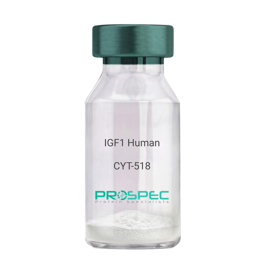
IGF1 Human Des1-3Description:
Insulin-Like Growth Factor 1 Des (1-3) Human Recombinant
Somatomedin C, IGF-I, IGFI, IGF1, IGF-IA, Mechano growth factor, MGF, Des(1-3), Des1-3, Des 1-3, Des (1-3), IGF-1 (4-70).
Product # :
CYT-518Price :
Quantity :
Shipping Method :
Shipped at Room temp
More Info
- description
- source
- formulation
- purity
- biological activity
- More Info
- Citations
Description
IGF-I Des(1-3) Human Recombinant produced in E.Coli is a single, non-glycosylated, polypeptide chain containing 67 amino acids (aa 4-70) and having a molecular mass of 7368.5 Dalton. IGF-1 Des1-3 is purified by proprietary chromatographic techniques.
Source
Escherichia Coli.
Formulation
The protein (1mg/ml) was lyophilized was lyophilized from a 0.2µm filtered concentrated solution in 20mM PBS, pH 7.0.
Purity
Greater than 97% as determined by:
(a) Analysis by RP-HPLC.
(b) Analysis by SDS-PAGE.Biological Activity
The ED50 as determined by a cell proliferation assay using FDC-P1 cells is less than 2.0 ng/ml, corresponding to a specific activity of > 5×10⁵ units/mg.Citations
More Info
-
Introduction
The somatomedins, or insulin-like growth factors (IGFs), comprise a family of peptides that play important roles in mammalian growth and development. IGF1 mediates many of the growth-promoting effects of growth hormone (GH; MIM 139250). Early studies showed that growth hormone did not directly stimulate the incorporation of sulfate into cartilage, but rather acted through a serum factor, termed 'sulfation factor,' which later became known as 'somatomedin' (Daughaday et al., 1972). Three main somatomedins have been characterized: somatomedin C (IGF1), somatomedin A (IGF2; MIM 147470), and somatomedin B (MIM 193190) (Rotwein, 1986; Rosenfeld, 2003).
-
Synonyms
Somatomedin C, IGF-I, IGFI, IGF1, IGF-IA, Mechano growth factor, MGF, Des(1-3), Des1-3, Des 1-3, Des (1-3), IGF-1 (4-70).
-
Physical Appearance
Sterile Filtered White lyophilized (freeze-dried) powder.
-
Stability
Lyophilized IGF-I des(1-3) although stable at room temperature for 3 weeks, should be stored desiccated below -18°C. Upon reconstitution IGF1 des-1-3 should be stored at 4°C between 2-7 days and for future use below -18°C.For long term storage it is recommended to add a carrier protein (0.1% HSA or BSA).Please prevent freeze-thaw cycles.
-
Solubility
It is recommended to reconstitute the IGF-I des(1-3) in sterile 18M-cm H2O not less than 100µg/ml, which can then be further diluted to other aqueous solutions.
-
Amino Acid Sequence
TLCGAELVDA LQFVCGDRGF YFNKPTGYGS SSRRAPQTGI VDECCFRSCD LRRLEMYCAP LKPAKSA.
-
Background
What is the molecular weight/Mw of IGF1 HUMAN DES1-3 Protein?
IGF1 HUMAN DES1-3 Protein has a total Mw of 7.3kDa.
What is the source or expression system of IGF1 HUMAN DES1-3 Protein?
Escherichia Coli.
What is the Purity of IGF1 HUMAN DES1-3 Protein?
IGF1 HUMAN DES1-3 Protein is >97% pure as determined by SDS-PAGE.
What is the Biological Activity of IGF1 HUMAN DES1-3 Protein?
The ED50 as determined by a cell proliferation assay using FDC-P1 cells is less than 2.0 ng/ml, corresponding to a specific activity of > 5×10⁵ units/mg.
What is the amino acid sequence of IGF1 HUMAN DES1-3 Protein?
TLCGAELVDA LQFVCGDRGF YFNKPTGYGS SSRRAPQTGI VDECCFRSCD LRRLEMYCAP LKPAKSA.
What applications can IGF1 HUMAN DES1-3 Protein be used in?
IGF1 HUMAN DES1-3 Protein can probably be used in western blot, ELISA and Lateral Flow.
What is the endotoxin level for IGF1 HUMAN DES1-3 Protein?
The endotoxin level is minimal, IGF1 HUMAN DES1-3 Protein was purified using conventional chromatography techniques.
ProSpec's products are furnished for LABORATORY RESEARCH USE ONLY. The product may not be used as drugs, agricultural or pesticidal products, food additives or household chemicals.
- View Data Sheet
Name :
CCR6 AntibodyDescription:
C-C chemokine receptor type 6, Mouse Anti Human
C-C chemokine receptor type 6, LARC receptor, GPR-CY4, Chemokine receptor-like 3, DRY6, G-protein coupled receptor 29, CD196, C-C CKR-6, CC-CKR-6, CCR-6, GPRCY4, CKR-L3, CCR6, CKRL3, CMKBR6, GPR29, STRL22, BN-1, CKR6, DCR2, DRY-6, GPRCY4, GPR-CY4.
Product # :
ANT-416Price :
Quantity :
Shipping Method :
Shipped with Ice Packs
More Info
- formulation
- More Info
- Citations
Formulation
1mg/ml containing PBS, pH-7.4, 0.02% Sodium Azide and 10% Glycerol.
Citations
More Info
-
Introduction
CCR6 belongs to the beta chemokine receptor family, which is predicted to be a 7 transmembrane protein similar to G protein-coupled receptors. CCR6 is preferentially expressed by immature dendritic cells and memory T cells. The ligand of CCR6 receptor is macrophage inflammatory protein 3 alpha (MIP-3 alpha). CCR6 receptor has been shown to be vital for B-lineage maturation and antigen-driven B-cell differentiation, and it may regulate the migration and recruitment of dentritic and T cells during inflammatory and immunological responses.
-
Synonyms
C-C chemokine receptor type 6, LARC receptor, GPR-CY4, Chemokine receptor-like 3, DRY6, G-protein coupled receptor 29, CD196, C-C CKR-6, CC-CKR-6, CCR-6, GPRCY4, CKR-L3, CCR6, CKRL3, CMKBR6, GPR29, STRL22, BN-1, CKR6, DCR2, DRY-6, GPRCY4, GPR-CY4.
-
Immunogen
Anti-human CCR6 mAb , is derived from hybridization of mouse F0myeloma cells with spleen cells from BALB/c mice immunized with recombinant human CCR6 amino acids 1-46 purified from E. coli.
-
Ig Subclass
Mouse IgG1 heavy chain and κ light chain.
-
Clone
P4C6AT.
-
Applications
CCR6 antibody has been tested by ELISA and Western blot analysis to assure specificity and reactivity. Since application varies, however, each investigation should be titrated by the reagent to obtain optimal results. Recommended dilution range for Western blot analysis is 1:500 ~ 1,000. Recommended starting dilution is 1:500.
-
Type
Mouse Anti Human Monoclonal.
-
Storage Procedures
For periods up to 1 month store at 4°C, for longer periods of time, store at -20°C. Prevent freeze thaw cycles.
-
Purification Method
CCR6 antibody was purified from mouse ascitic fluids by protein-G affinity chromatography.
ProSpec's products are furnished for LABORATORY RESEARCH USE ONLY. The product may not be used as drugs, agricultural or pesticidal products, food additives or household chemicals.
- View Data Sheet
Name :
Eotaxin MouseDescription:
Eotaxin Mouse Recombinant (CCL11)
Small inducible cytokine A11, CCL11, Eosinophil chemotactic protein, chemokine (C-C motif) ligand 11, SCYA11.
Product # :
CHM-308Price :
Quantity :
Shipping Method :
Shipped at Room temp
More Info
- description
- source
- formulation
- purity
- biological activity
- More Info
- Citations
Description
Eotaxin Mouse Recombinant produced in E.Coli is a single, non-glycosylated polypeptide chain containing 74 amino acids and having a molecular mass of 8403.2 Dalton. The CCL11 is purified by proprietary chromatographic techniques.
Source
Escherichia Coli.
Formulation
Lyophilized from a concentrated (1mg/ml) solution in water containing no additives.
Purity
Greater than 95.0% as determined by:
(a) Analysis by RP-HPLC.
(b) Analysis by SDS-PAGE.Biological Activity
The Biological activity was determined by measuring the dose dependent phosphorylation of ERK1 and ERK2 in CCR3 transfected 293 cells. Significant ERK phosphorylation is observed with >100 ng/ml (corresponding to a Specific Activity of 10,000IU/mg) of recombinant mouse eotaxin.Citations
More Info
-
Introduction
Chemokine (C-C motif) ligand 11 (CCL11) is a small cytokine belonging to the CC chemokine family that is also known as eotaxin. CCL11 selectively recruits eosinophils by inducing their chemotaxis, and therefore, is implicated in allergic responses. The effects of CCL11 are mediated by its binding to a G-protein-linked receptor known as a chemokine receptor. Chemokine receptors for which CCL11 is a ligand include CCR2, CCR3 and CCR5. The gene for human CCL11 (scya11) is encoded on three exons and is located on chromosome 17.
-
Synonyms
Small inducible cytokine A11, CCL11, Eosinophil chemotactic protein, chemokine (C-C motif) ligand 11, SCYA11.
-
Physical Appearance
Sterile Filtered White lyophilized (freeze-dried) powder.
-
Stability
Lyophilized Eotaxin although stable at room temperature for 3 weeks, should be stored desiccated below -18°C. Upon reconstitution CCL11 should be stored at 4°C between 2-7 days and for future use below -18°C. For long term storage it is recommended to add a carrier protein (0.1% HSA or BSA).Please prevent freeze-thaw cycles.
-
Solubility
It is recommended to reconstitute the lyophilized Eotaxin in sterile 18MΩ-cm H2O not less than 100µg/ml, which can then be further diluted to other aqueous solutions.
-
Amino Acid Sequence
The sequence of the first five N-terminal amino acids was determined and was found to be His-Pro-Gly-Ser-Ile.
-
Background
What is the molecular weight/Mw of EOTAXIN MOUSE Protein?
EOTAXIN MOUSE Protein has a total Mw of 8.4kDa.
What is the source or expression system of EOTAXIN MOUSE Protein?
Escherichia Coli.
What is the Purity of EOTAXIN MOUSE Protein?
EOTAXIN MOUSE Protein is >95% pure as determined by SDS-PAGE.
What is the Biological Activity of EOTAXIN MOUSE Protein?
The Biological activity was determined by measuring the dose dependent phosphorylation of ERK1 and ERK2 in CCR3 transfected 293 cells. Significant ERK phosphorylation is observed with >100 ng/ml (corresponding to a Specific Activity of 10,000IU/mg) of recombinant mouse eotaxin.
What is the amino acid sequence of EOTAXIN MOUSE Protein?
The sequence of the first five N-terminal amino acids was determined and was found to be His-Pro-Gly-Ser-Ile.
What applications can EOTAXIN MOUSE Protein be used in?
EOTAXIN MOUSE Protein can probably be used in western blot, ELISA and Lateral Flow.
What is the endotoxin level for EOTAXIN MOUSE Protein?
The endotoxin level is minimal, EOTAXIN MOUSE Protein was purified using conventional chromatography techniques.
Who supplies recombinant EOTAXIN MOUSE that is in stock and available to order?
ProSpec has the protein in stock and available for immediate delivery.
Where can I order bulk mg quantities of EOTAXIN MOUSE?
ProSpec supplies bulk mg quantities at affordable prices that meets research and academic budgets.
ProSpec's products are furnished for LABORATORY RESEARCH USE ONLY. The product may not be used as drugs, agricultural or pesticidal products, food additives or household chemicals.
- View Data Sheet
Name :
TNFRSF10A HumanDescription:
TRAIL Receptor-1 Human Recombinant
TNFRSF10A, TNF Receptor Superfamily Member 10a, Tumor Necrosis Factor Receptor Superfamily, Member 10a, TNF-Related Apoptosis-Inducing Ligand Receptor 1, Death Receptor 4, TRAIL Receptor 1, TRAIL-R1, TRAILR1, APO2, DR4, Tumor Necrosis Factor Receptor Superfamily Member 10a Variant 2, Tumor Necrosis Factor Receptor Superfamily Member 10A, Cytotoxic TRAIL Receptor, CD261 Antigen, TRAILR-1, CD261.
Product # :
CYT-1043Price :
Quantity :
Shipping Method :
Shipped with Ice Packs
More Info
- description
- source
- formulation
- purity
- More Info
- Citations
Description
TNFRSF10A produced in Sf9 Baculovirus cells is a single, glycosylated polypeptide chain containing 224 amino acids (24-239a.a.) and having a molecular mass of 23.9kDa. TNFRSF10A is expressed with an 6 amino acid His tag at C-Terminus and purified by proprietary chromatographic techniques.
Source
Sf9, Baculovirus cells.
Formulation
TNFRSF10A protein solution (0.5mg/ml) contains Phosphate Buffered Saline (pH 7.4) and 10% glycerol.
Purity
Greater than 90.0% as determined by SDS-PAGE.
Citations
More Info
-
Introduction
Human TNFRSF10A is a type 1 transmembrane protein in the TNF R superfamily. TNFRSF10A is not expressed in rodents. The trimeric ligand Trail binds TNFRSF10A and induces apoptosis, and recombinant, soluble forms of the receptor inhibit Trail-induced apoptosis. TNFRSF10A is expressed generally in damaged, infected, and malignant cells. TNFRSF10A functions in immune surveillance, inducing apoptosis in cancer cells but not normal cells.
-
Synonyms
TNFRSF10A, TNF Receptor Superfamily Member 10a, Tumor Necrosis Factor Receptor Superfamily, Member 10a, TNF-Related Apoptosis-Inducing Ligand Receptor 1, Death Receptor 4, TRAIL Receptor 1, TRAIL-R1, TRAILR1, APO2, DR4, Tumor Necrosis Factor Receptor Superfamily Member 10a Variant 2, Tumor Necrosis Factor Receptor Superfamily Member 10A, Cytotoxic TRAIL Receptor, CD261 Antigen, TRAILR-1, CD261.
-
Physical Appearance
Sterile Filtered colorless solution.
-
Stability
Store at 4°C if entire vial will be used within 2-4 weeks. Store, frozen at -20°C for longer periods of time. For long term storage it is recommended to add a carrier protein (0.1% HSA or BSA). Avoid multiple freeze-thaw cycles.
-
Amino Acid Sequence
ASGTEAAAAT PSKVWGSSAG RIEPRGGGRG ALPTSMGQHG PSARARAGRA PGPRPAREAS PRLRVHKTFK FVVVGVLLQV VPSSAATIKL HDQSIGTQQW EHSPLGELCP PGSHRSEHPG ACNRCTEGVG YTNASNNLFA CLPCTACKSD EEERSPCTTT RNTACQCKPG TFRNDNSAEM CRKCSRGCPR GMVKVKDCTP WSDIECVHKE SGNGHNLEHH HHHH
ProSpec's products are furnished for LABORATORY RESEARCH USE ONLY. The product may not be used as drugs, agricultural or pesticidal products, food additives or household chemicals.
- View Data Sheet
Name :
LAIR1 MouseDescription:
Leukocyte-Associated Ig-Like Receptor 1 Mouse Recombinant
Leukocyte-associated immunoglobulin-like receptor 1, LAIR-1, mLAIR1, CD305, LAIR1.
Product # :
PRO-2356Price :
Quantity :
Shipping Method :
Shipped with Ice Packs
More Info
- description
- source
- formulation
- purity
- More Info
- Citations
Description
LAIR1 Mouse Recombinant produced in Sf9 Baculovirus cells is a single, glycosylated polypeptide chain containing 132 amino acids (22-144 aa) and having a molecular mass of 15kDa (Migrates at 18-28kDa on SDS-PAGE under reducing conditions).LAIR1 is expressed with a 6 amino acid His tag at C-Terminus and purified by proprietary chromatographic techniques.
Source
Sf9, Baculovirus cells.
Formulation
LAIR1 protein solution (0.5mg/ml) contains Phosphate buffered saline (pH7.4) and 10% glycerol.
Purity
Greater than 90.0% as determined by SDS-PAGE.
Citations
More Info
-
Introduction
LAIR1 is a glycoprotein expressed on the surface of the majority of human peripheral blood mononuclear leukocytes including T cells, B cells, NK cells, macrophages and dendritic cells. LAIR-1 functions as an inhibitory receptor in NK cells, T cells and B cells. Inhibitory receptors control the immune response to prevent lysis of cells recognized as self. Lair1 consists of a leader sequence, extracellular domain, transmembrane domain, cytoplasmic region.LAIR1 is a member of both the immunoglobulin superfamily and the leukocyte-associated inhibitory receptor family. LAIR1 maps to a region of 19q13.4 labeled the leukocyte receptor cluster, which contains at least 29 genes encoding leukocyte-expressed receptors of the immunoglobulin superfamily.
-
Synonyms
Leukocyte-associated immunoglobulin-like receptor 1, LAIR-1, mLAIR1, CD305, LAIR1.
-
Physical Appearance
Sterile filtered colorless solution.
-
Stability
Store at 4°C if entire vial will be used within 2-4 weeks. Store, frozen at -20°C for longer periods of time. For long term storage it is recommended to add a carrier protein (0.1% HSA or BSA).Avoid multiple freeze-thaw cycles.
-
Amino Acid Sequence
ADPQEGSLPD ITIFPNSSLM ISQGTFVTVV CSYSDKHDLY NMVRLEKDGS TFMEKSTEPY KTEDEFEIGP VNETITGHYS CIYSKGITWS ERSKTLELKV IKENVIQTPA PGPTSDTSWL KTYSIYHHHH HH
ProSpec's products are furnished for LABORATORY RESEARCH USE ONLY. The product may not be used as drugs, agricultural or pesticidal products, food additives or household chemicals.
- View Data Sheet
Name :
MIF RatDescription:
Macrophage Migration Inhibitory Factor Rat Recombinant
Macrophage migration inhibitory factor, MIF, Glutathione-binding 13 kDa protein, L-dopachrome isomerase, L-dopachrome tautomerase, Phenylpyruvate tautomerase.
Product # :
CYT-193Price :
Quantity :
Shipping Method :
Shipped at Room temp
More Info
- description
- source
- formulation
- purity
- More Info
- Citations
Description
MIF Rat Recombinant produced in E.Coli is a single, non-glycosylated polypeptide chain containing 115 amino acids and having a molecular mass of 12.5kDa. The MIF is purified by proprietary chromatographic techniques.
Source
Escherichia Coli.
Formulation
Lyophilized from a 0.2µm filtered concentrated solution in 1×PBS, pH 7.4 and 5% trehalose.
Purity
Greater than 95.0% as determined by:
(a) Analysis by RP-HPLC.
(b) Analysis by SDS-PAGE.Citations
More Info
-
Introduction
The cytokine Macrophage migration inhibitory factor (MIF) has been identified to be secreted by the pituitary gland and the monocyte/macrophage and to play an important role in endotoxic shock. MIF has the unique property of being released from macrophages and T cells in response to physiological concentrations of glucocorticoids. The secretion of MIF is tightly regulated and decreases at high, anti-inflammatory steroid concentration.
-
Synonyms
Macrophage migration inhibitory factor, MIF, Glutathione-binding 13 kDa protein, L-dopachrome isomerase, L-dopachrome tautomerase, Phenylpyruvate tautomerase.
-
Physical Appearance
Sterile Filtered White lyophilized (freeze-dried) powder.
-
Stability
Lyophilized MIF although stable at room temperature for 3 weeks, should be stored desiccated below -18°C. Upon reconstitution MIF should be stored at 4°C between 2-7 days and for future use below -18°C. For long term storage it is recommended to add a carrier protein (0.1% HSA or BSA).Please prevent freeze-thaw cycles.
-
Solubility
It is recommended to reconstitute the lyophilized MIF in sterile 18MΩ-cm H2O not less than 100µg/ml, which can then be further diluted to other aqueous solutions.
-
Amino Acid Sequence
MPMFIVNTNV PRASVPEGFL SELTQQLAQA TGKPAQYIAV HVVPDQLMTF SGTSDPCALC SLHSIGKIGG AQNRNYSKLL CGLLSDRLHI SPDRVYINYY DMNAANVGWN GSTFA.
ProSpec's products are furnished for LABORATORY RESEARCH USE ONLY. The product may not be used as drugs, agricultural or pesticidal products, food additives or household chemicals.
- View Data Sheet
Name :
Flt3-Ligand Rhesus MacaqueDescription:
Flt3-Ligand Rhesus Macaque Recombinant
Fms-related tyrosine kinase 3 ligand, FLK2, STK1, CD135, Stem Cell Tyrosine Kinase 1, FLT3LG, Flt3.
Product # :
CYT-157Price :
Quantity :
Shipping Method :
Shipped at Room temp
More Info
- description
- source
- formulation
- purity
- biological activity
- More Info
- Citations
Description
Flt3 Ligand Rhesus Macaque Recombinant produced in E.Coli is a single, non-glycosylated, polypeptide chain containing 159 amino acids and having a molecular mass of 18.0kDa.The Flt3 Ligand Rhesus Macaque is purified by proprietary chromatographic techniques.
Source
Escherichia Coli.
Formulation
Lyophilized from a 0.2µm filtered concentrated solution in PBS, pH 7.4.
Purity
Greater than 97.0% as determined by SDS-PAGE.
Biological Activity
The ED50 as calculated by the dose-dependant stimulation of the proliferation of human AML5 cells is less than 1.0ng/ml, corresponding to a specific activity of 1,000,000IU/mg.Citations
More Info
-
Introduction
FLT3 ligand is a receptor for the fl cytokine has a tyrosine-protein kinase activity & a growth factor that regulates proliferation of early hematopoietic cells. Flt3-Ligand synergizes with other CSFs and interleukins to induce growth and differentiation.
-
Synonyms
Fms-related tyrosine kinase 3 ligand, FLK2, STK1, CD135, Stem Cell Tyrosine Kinase 1, FLT3LG, Flt3.
-
Physical Appearance
Sterile Filtered White lyophilized (freeze-dried) powder.
-
Stability
Lyophilized Flt3 Ligand Rhesus Macaque although stable at room temperature for 3 weeks, should be stored desiccated below -18°C. Upon reconstitution Flt3 Ligand Rhesus Macaque should be stored at 4°C between 2-7 days and for future use below -18°C.Please prevent freeze-thaw cycles.
-
Solubility
It is recommended to reconstitute the lyophilized Flt3 Ligand in sterile 18MΩ-cm H2O not less than 100µg/ml, which can then be further diluted to other aqueous solutions.
-
Amino Acid Sequence
TQDCSFQHSP ISSDFAVKIR ELSDYLLQDY PVTVPSNLQD EELCGALWRL VLAQRWMERL KTVAGSKMQG LLERVNTEIH FVTKCAFQHP PSCLRFVQTN ISRLLQETSE QLVALKPWIT RQNFSRCLEL QCQPDSSTLP PPRSPGALEA TALTAPQRP
-
Background
What is the molecular weight/Mw of FLT3-LIGAND RHESUS MACAQUE Protein?
FLT3-LIGAND RHESUS MACAQUE Protein has a total Mw of 18kDa.
What is the source or expression system of FLT3-LIGAND RHESUS MACAQUE Protein?
Escherichia Coli.
What is the Purity of FLT3-LIGAND RHESUS MACAQUE Protein?
FLT3-LIGAND RHESUS MACAQUE Protein is >97% pure as determined by SDS-PAGE.
What is the Biological Activity of FLT3-LIGAND RHESUS MACAQUE Protein?
The ED50 as calculated by the dose-dependant stimulation of the proliferation of human AML5 cells is less than 1.0ng/ml, corresponding to a specific activity of 1,000,000IU/mg.
What is the amino acid sequence of FLT3-LIGAND RHESUS MACAQUE Protein?
TQDCSFQHSP ISSDFAVKIR ELSDYLLQDY PVTVPSNLQD EELCGALWRL VLAQRWMERL KTVAGSKMQG LLERVNTEIH FVTKCAFQHP PSCLRFVQTN ISRLLQETSE QLVALKPWIT RQNFSRCLEL QCQPDSSTLP PPRSPGALEA TALTAPQRP
What applications can FLT3-LIGAND RHESUS MACAQUE Protein be used in?
FLT3-LIGAND RHESUS MACAQUE Protein can probably be used in western blot, ELISA and Lateral Flow.
What is the endotoxin level for FLT3-LIGAND RHESUS MACAQUE Protein?
The endotoxin level is minimal, FLT3-LIGAND RHESUS MACAQUE Protein was purified using conventional chromatography techniques.
ProSpec's products are furnished for LABORATORY RESEARCH USE ONLY. The product may not be used as drugs, agricultural or pesticidal products, food additives or household chemicals.
- View Data Sheet
Name :
GDF10 HumanDescription:
Growth differentiation factor 10 Human Recombinant
Bone morphogenetic protein 3b, BMP-3b, Growth/differentiation factor 10, GDF-10, Bone-inducing protein, BIP, GDF10, BMP3B.
Product # :
CYT-659Price :
Quantity :
Shipping Method :
Shipped with Ice Packs
More Info
- description
- source
- formulation
- purity
- More Info
- sds-page
- Citations
Description
GDF10 Human Recombinant produced in E.Coli is a single, non-glycosylated, polypeptide chain containing 111 amino acids (369-478 a.a.) and having a total molecular mass of 12.5 kDa. GDF10 is purified by proprietary chromatographic techniques.
Source
Escherichia Coli.
Formulation
The GDF10 solution (1mg/ml) contains 10mM Sodium citrate (pH 3.5), 1mM DTT, 40% glycerol and 0.1M NaCl.
Purity
Greater than 95.0% as determined by SDS-PAGE.
sds-page
Citations
More Info
-
Introduction
GDF10 is a member of the BMP family and the TGF-beta superfamily. GDF10 is expressed in femur, brain, lung, skeletal, muscle, pancreas and testis, and has a role in head formation and possibly multiple roles in skeletal morphogenesis. In humans, GDF10 mRNA is found in the cochlea and lung of fetuses, and in testis, retina, pineal gland, and other neural tissues of adults. The BMP family members are regulators of cell growth and differentiation in both embryonic and adult tissues. These proteins are characterized by a polybasic proteolytic processing site which is cleaved to produce a mature protein containing 7 conserved cysteine residues.
-
Synonyms
Bone morphogenetic protein 3b, BMP-3b, Growth/differentiation factor 10, GDF-10, Bone-inducing protein, BIP, GDF10, BMP3B.
-
Physical Appearance
Sterile Filtered colorless solution.
-
Stability
Store at 4°C if entire vial will be used within 2-4 weeks. Store, frozen at -20°C for longer periods of time. For long term storage it is recommended to add a carrier protein (0.1% HSA or BSA).Avoid multiple freeze-thaw cycles.
-
Amino Acid Sequence
MQWDEPRVCS RRYLKVDFAD IGWNEWIISP KSFDAYYCAG ACEFPMPKIV RPSNHATIQS IVRAVGIIPG IPEPCCVPDK MNSLGVLFLD ENRNVVLKVY PNMSVDTCAC R.
-
Background
What is the molecular weight/Mw of GDF10 HUMAN Protein?
GDF10 HUMAN Protein has a total Mw of 12.5kDa.
What is the source or expression system of GDF10 HUMAN Protein?
Escherichia Coli.
What is the Purity of GDF10 HUMAN Protein?
GDF10 HUMAN Protein is >95% pure as determined by SDS-PAGE.
What is the Biological Activity of GDF10 HUMAN Protein?
The biological functionality of GDF10 HUMAN Protein will be determined in the future.
What is the amino acid sequence of GDF10 HUMAN Protein?
MQWDEPRVCS RRYLKVDFAD IGWNEWIISP KSFDAYYCAG ACEFPMPKIV RPSNHATIQS IVRAVGIIPG IPEPCCVPDK MNSLGVLFLD ENRNVVLKVY PNMSVDTCAC R.
What applications can GDF10 HUMAN Protein be used in?
GDF10 HUMAN Protein can probably be used in western blot, ELISA and Lateral Flow.
What is the endotoxin level for GDF10 HUMAN Protein?
The endotoxin level is minimal, GDF10 HUMAN Protein was purified using conventional chromatography techniques.
ProSpec's products are furnished for LABORATORY RESEARCH USE ONLY. The product may not be used as drugs, agricultural or pesticidal products, food additives or household chemicals.
- View Data Sheet
Name :
BD 2 MouseDescription:
Beta Defensin-2 Mouse Recombinant
Beta-defensin 2, BD-2, mBD-2, Defensin beta 2, Defb2, MGC129140, MGC129141.
Product # :
CYT-035Price :
Quantity :
Shipping Method :
Shipped at Room temp
More Info
- description
- source
- formulation
- purity
- biological activity
- More Info
- Citations
Description
Beta Defensin-2 Mouse Recombinant produced in E.Coli is a single, non-glycosylated polypeptide chain containing 51 amino acids and having a molecular mass of 5.5kDa. The BD-2 is purified by proprietary chromatographic techniques.
Source
Escherichia Coli.
Formulation
The Mouse BD-2 was lyophilized from a 0.2µm filtered concentrated solution in PBS, pH7.4.
Purity
Greater than 98.0% as determined by:
(a) Analysis by RP-HPLC.
(b) Analysis by SDS-PAGE.Biological Activity
Determined by the ability to chemoattract immature human dendritic cells at a concentration of 10-100ng/ml.Citations
More Info
-
Introduction
The Defensin family are highly similar in their protein sequence and are microbicidal & cytotoxic peptides made by neutrophils. Beta Defensin-1 is an antimicrobial peptide having the resistance of epithelial surfaces to microbial colonization. Beta Defensin-1 has close proximity to Defensin Alpha-1 and has been implicated in the pathogenesis of cystic fibrosis.
Skin of patients having atopic dermatitis patients and mycosis fungoides (non-lesional and lesional) show lower human Beta Defensin-1 mRNA expression and higher human Beta Defensin-2 and human Beta Defensin-3 mRNA expression.
BBeta Defensin is highly expressed by epithelial cells.
Beta-defensin 1 may play a role in the pathogenesis of severe sepsis.
Variation in human Beta Defensin-1 contributes to asthma diagnosis, with apparent gender-specific effects. Human Beta Defensin-3 is a dimer, while Human BD-1 and Human BD-2 are monomeric. The expression of Human BD1 is correlated with induction profiles in gingival keratinocytes.
The level of expression of human DEFB1 mRNA is lower than that of human BD3 and human BD-2 in reconstructed epidermis.
Human BD1 is down-regulated in human prostatic and renal carcinomas. -
Synonyms
Beta-defensin 2, BD-2, mBD-2, Defensin beta 2, Defb2, MGC129140, MGC129141.
-
Physical Appearance
Sterile Filtered White lyophilized (freeze-dried) powder.
-
Stability
Lyophilized Beta Defensin-2 Recombinant although stable at room temperature for 3 weeks, should be stored desiccated below -18°C. Upon reconstitution BD-2 should be stored at 4°C between 2-7 days and for future use below -18°C.Please prevent freeze-thaw cycles.
-
Solubility
It is recommended to reconstitute the lyophilized Beta Defensin-2 in sterile 18M-cm H2O not less than 100µg/ml, which can then be further diluted to other aqueous solutions.
-
Amino Acid Sequence
AVGSLKSIGY EAELDHCHTN GGYCVRAICP PSARRPGSCF PEKNPCCKYM K.
-
Background
What is the molecular weight/Mw of BD2 Protein?
BD2 Protein has a total Mw of 5.5kDa.
What is the source or expression system of BD2 Protein?
Escherichia Coli.
What is the Purity of BD2 Protein?
BD2 Protein is >98% pure as determined by SDS-PAGE.
What is the Biological Activity of BD2 Protein?
Determined by the ability to chemoattract immature human dendritic cells at a concentration of 10-100ng/ml.
What is the amino acid sequence of BD2 Protein?
AVGSLKSIGY EAELDHCHTN GGYCVRAICP PSARRPGSCF PEKNPCCKYM K.
What applications can BD2 Protein be used in?
BD2 Protein can probably be used in western blot, ELISA and Lateral Flow.
What is the endotoxin level for BD2 Protein?
The endotoxin level is minimal, BD2 Protein was purified using conventional chromatography techniques.
ProSpec's products are furnished for LABORATORY RESEARCH USE ONLY. The product may not be used as drugs, agricultural or pesticidal products, food additives or household chemicals.
- View Data Sheet
Name :
GDF15 HumanDescription:
Growth and Differentiation Factor 15 Human Recombinant
GDF-15, MIC1, MIC-1, NAG-1, PDF, PLAB, PTGFB, Growth/differentiation factor 15, Placental bone morphogenetic protein, Placental TGF-beta, Macrophage inhibitory cytokine 1, Prostate differentiation factor, NSAID-activated gene 1 protein, NSAID-regulated gene 1 protein, NRG-1, GDF15.
Product # :
CYT-335Price :
Quantity :
Shipping Method :
Shipped at Room temp
More Info
- description
- source
- formulation
- purity
- More Info
- Citations
Description
GDF15 Human Recombinant produced in E.Coli is a homodimeric, non-glycosylated, Polypeptide chain containing 2x113 amino acids and having a molecular mass of 24.8 kDa. The GDF15 is purified by proprietary chromatographic techniques.
Source
Escherichia Coli.
Formulation
GDF15 is lyophilized from a sterile filtered solution containing 0.1% Trifluoroacetic Acid (TFA).
Purity
Greater than 95.0% as determined by SDS-PAGE.
Citations
More Info
-
Introduction
GDF15 is part of the TGF-Beta superfamily that is involved in regulating inflammatory and apoptotic pathways in injured tissues and throughout disease processes. GDF15 is most abundant in the liver. Its expression in liver can be considerably up-regulated in during injury of organs such as liver, kidney, heart and lung. GDF-15 promotes proliferation or growth arrest and differentiation due to differences in cellular differentiation. GDF15 prevents apoptosis in cerebellar granule neurons by activating Akt and inhibiting endogenously active ERK. GDF15 is a novel autocrine/endocrine factor that antagonizes the hypertrophic response and loss of ventricular performance.
-
Synonyms
GDF-15, MIC1, MIC-1, NAG-1, PDF, PLAB, PTGFB, Growth/differentiation factor 15, Placental bone morphogenetic protein, Placental TGF-beta, Macrophage inhibitory cytokine 1, Prostate differentiation factor, NSAID-activated gene 1 protein, NSAID-regulated gene 1 protein, NRG-1, GDF15.
-
Physical Appearance
Sterile Filtered White lyophilized (freeze-dried) powder.
-
Stability
Lyophilized GDF15 although stable at room temperature for 3 weeks, should be stored desiccated below -18°C. Upon reconstitution GDF15 should be stored at 4°C between 2-7 days and for future use below -18°C.For long term storage it is recommended to add a carrier protein (0.1% HSA or BSA).Please prevent freeze-thaw cycles.
-
Solubility
It is recommended to reconstitute the lyophilized GDF15 in sterile 18M-cm H2O at a concentration of 100µg/ml, which can then be further diluted to other aqueous solutions.
-
Amino Acid Sequence
MARNGDHCPL GPGRCCRLHT VRASLEDLGW ADWVLSPREV QVTMCIGACP SQFRAANMHA QIKTSLHRLK PDTVPAPCCV PASYNPMVLI QKTDTGVSLQ TYDDLLAKDC HCI.
-
Background
What is the molecular weight/Mw of GDF15 HUMAN Protein?
GDF15 HUMAN Protein has a total Mw of 24.8kDa.
What is the source or expression system of GDF15 HUMAN Protein?
Escherichia Coli.
What is the Purity of GDF15 HUMAN Protein?
GDF15 HUMAN Protein is >95% pure as determined by SDS-PAGE.
What is the Biological Activity of GDF15 HUMAN Protein?
The biological functionality of GDF15 HUMAN Protein will be determined in the future.
What is the amino acid sequence of GDF15 HUMAN Protein?
MARNGDHCPL GPGRCCRLHT VRASLEDLGW ADWVLSPREV QVTMCIGACP SQFRAANMHA QIKTSLHRLK PDTVPAPCCV PASYNPMVLI QKTDTGVSLQ TYDDLLAKDC HCI.
What applications can GDF15 HUMAN Protein be used in?
GDF15 HUMAN Protein can probably be used in western blot, ELISA and Lateral Flow.
What is the endotoxin level for GDF15 HUMAN Protein?
The endotoxin level is minimal, GDF15 HUMAN Protein was purified using conventional chromatography techniques.
ProSpec's products are furnished for LABORATORY RESEARCH USE ONLY. The product may not be used as drugs, agricultural or pesticidal products, food additives or household chemicals.