Skip to content

High-quality research proteins.

  • 1-800-805-6531

  • Mon - Sat 8.00-18.00, Sun - Closed

  • info@prospecbio.com

  • PO Box 6591 East Brunswick NJ 08816

  • Products
    • Cytokines
    • Growth factors
    • Chemokines
    • Cd antigens
    • Neurotrophins
    • Hormones
    • Enzymes
    • Viral antigens
    • Recombinant proteins
    • Natural proteins
    • Monoclonal antibodies
    • Polyclonal antibodies
    • Thermal Packaging
  • About
    • Company
    • Founder
    • Contribution
  • Customers
    • Eastern Asia
    • Eastern Europe
    • North America
    • South America
    • South East Asia
    • Southern Asia
    • Western Europe
  • Ordering
    • Ordering Method
    • Ordering - Credit Card
    • Ordering - PO
    • Shipping Fees
    • FAQs
  • Services
    • DNA Cloning
    • Protein Expression
    • Monoclonal Antibody
    • Fermentation
    • Technology License
  • Contact us
Log in

Country/region

  • USD $ | Albania
  • USD $ | Andorra
  • USD $ | Argentina
  • USD $ | Armenia
  • USD $ | Australia
  • USD $ | Austria
  • USD $ | Azerbaijan
  • USD $ | Belarus
  • USD $ | Belgium
  • USD $ | Bolivia
  • USD $ | Brazil
  • USD $ | Bulgaria
  • USD $ | Cambodia
  • USD $ | Canada
  • USD $ | Chile
  • USD $ | China
  • USD $ | Colombia
  • USD $ | Costa Rica
  • USD $ | Croatia
  • USD $ | Cyprus
  • USD $ | Czechia
  • USD $ | Denmark
  • USD $ | Estonia
  • USD $ | Finland
  • USD $ | France
  • USD $ | Georgia
  • USD $ | Germany
  • USD $ | Greece
  • USD $ | Hong Kong SAR
  • USD $ | Hungary
  • USD $ | Iceland
  • USD $ | India
  • USD $ | Ireland
  • USD $ | Israel
  • USD $ | Italy
  • USD $ | Japan
  • USD $ | Kazakhstan
  • USD $ | Latvia
  • USD $ | Liechtenstein
  • USD $ | Lithuania
  • USD $ | Madagascar
  • USD $ | Malta
  • USD $ | Mexico
  • USD $ | Moldova
  • USD $ | Monaco
  • USD $ | Netherlands
  • USD $ | New Zealand
  • USD $ | North Macedonia
  • USD $ | Norway
  • USD $ | Panama
  • USD $ | Paraguay
  • USD $ | Peru
  • USD $ | Philippines
  • USD $ | Poland
  • USD $ | Portugal
  • USD $ | Romania
  • USD $ | Singapore
  • USD $ | Slovakia
  • USD $ | Slovenia
  • USD $ | South Africa
  • USD $ | South Korea
  • USD $ | Spain
  • USD $ | Sri Lanka
  • USD $ | Sweden
  • USD $ | Switzerland
  • USD $ | Taiwan
  • USD $ | Thailand
  • USD $ | Türkiye
  • USD $ | Ukraine
  • USD $ | United Kingdom
  • USD $ | United States
  • USD $ | Uruguay
  • USD $ | Vatican City
  • USD $ | Venezuela
  • USD $ | Vietnam
  • Facebook
  • Instagram
logoProSpec - Recombinant Protein Manufacturer for academic and R&D industry
Become a distributor
Account Log in
Wishlist
Cart Cart(0)
  • Products
    +
    • Cytokines
    • Growth factors
    • Chemokines
    • Cd antigens
    • Neurotrophins
    • Hormones
    • Enzymes
    • Viral antigens
    • Recombinant proteins
    • Natural proteins
    • Monoclonal antibodies
    • Polyclonal antibodies
    • Thermal Packaging
  • About
    +
    • Company
    • Founder
    • Contribution
  • Customers
    +
    • Eastern Asia
    • Eastern Europe
    • North America
    • South America
    • South East Asia
    • Southern Asia
    • Western Europe
  • Ordering
    +
    • Ordering Method
    • Ordering - Credit Card
    • Ordering - PO
    • Shipping Fees
    • FAQs
  • Services
    +
    • DNA Cloning
    • Protein Expression
    • Monoclonal Antibody
    • Fermentation
    • Technology License
  • Contact us

Item added to your cart

View cart
View All
  • Cytokines
  • Growth factors
  • Chemokines
  • Cd antigens
  • Neurotrophins
  • Hormones
  • Enzymes
  • Viral antigens
  • Recombinant proteins
  • Natural proteins
  • Monoclonal antibodies
  • Polyclonal antibodies
  • Thermal Packaging
  • Tumor Necrosis Factor

    Tumor Necrosis Factor

    View All
  • 4-1BB

    4-1BB

    View All
  • Adiponectin

    Adiponectin

    View All
  • AIF1

    AIF1

    View All
  • Other Cytokines

    Other Cytokines

    View All
  • AITRL

    AITRL

    View All
  • Angiopoietin

    Angiopoietin

    View All
  • Apolipoprotein

    Apolipoprotein

    View All
  • B-Cell Activating Factor

    B-Cell Activating Factor

    View All
  • Beta Defensin

    Beta Defensin

    View All
  • Bone Morphogenetic Protein

    Bone Morphogenetic Protein

    View All
  • B type Natriuretic Peptide

    B type Natriuretic Peptide

    View All
  • BST

    BST

    View All
  • Betacellulin

    Betacellulin

    View All
  • Cardiotrophin

    Cardiotrophin

    View All
  • Activin

    Activin

    View All
  • Fibroblast Growth Factor

    Fibroblast Growth Factor

    View All
  • Other Growth Factors

    Other Growth Factors

    View All
  • CSF

    CSF

    View All
  • CTGF

    CTGF

    View All
  • IGFBP

    IGFBP

    View All
  • VEGF Protein

    VEGF Protein

    View All
  • EGF

    EGF

    View All
  • Epigen

    Epigen

    View All
  • Erythropoietin

    Erythropoietin

    View All
  • Insulin-Like Growth Factor

    Insulin-Like Growth Factor

    View All
  • Growth Hormone

    Growth Hormone

    View All
  • HDGF

    HDGF

    View All
  • Hepatocyte Growth Factor

    Hepatocyte Growth Factor

    View All
  • Insulin

    Insulin

    View All
  • BCA-1/ BLC (CXCL13)

    BCA-1/ BLC (CXCL13)

    View All
  • BRAK (CXCL14)

    BRAK (CXCL14)

    View All
  • C-10 (CCL6)

    C-10 (CCL6)

    View All
  • Other Chemokines

    Other Chemokines

    View All
  • MEC (CCL28)

    MEC (CCL28)

    View All
  • LD78-beta (CCL3L1)

    LD78-beta (CCL3L1)

    View All
  • CTACK (CCL27)

    CTACK (CCL27)

    View All
  • CXCL16

    CXCL16

    View All
  • CXCL17

    CXCL17

    View All
  • Platelet Factor-4 (CXCL4)

    Platelet Factor-4 (CXCL4)

    View All
  • ENA-78 (CXCL5)

    ENA-78 (CXCL5)

    View All
  • Eotaxin (CCL11,24,26)

    Eotaxin (CCL11,24,26)

    View All
  • Exodus-2 (CCL21)

    Exodus-2 (CCL21)

    View All
  • Fractalkine (CX3CL1)

    Fractalkine (CX3CL1)

    View All
  • CXCL6

    CXCL6

    View All
  • Other CD Antigens

    Other CD Antigens

    View All
  • CD14

    CD14

    View All
  • CD164

    CD164

    View All
  • CD1

    CD1

    View All
  • CD2

    CD2

    View All
  • CD200

    CD200

    View All
  • CD207

    CD207

    View All
  • CD226

    CD226

    View All
  • CD23

    CD23

    View All
  • CD244

    CD244

    View All
  • CD247

    CD247

    View All
  • CD27

    CD27

    View All
  • CD274

    CD274

    View All
  • CD300

    CD300

    View All
  • CD33

    CD33

    View All
  • Other Neurotrophins

    Other Neurotrophins

    View All
  • Beta-NGF

    Beta-NGF

    View All
  • BDNF

    BDNF

    View All
  • CDNF

    CDNF

    View All
  • CNTF

    CNTF

    View All
  • GDNF

    GDNF

    View All
  • Glia Maturation Factor

    Glia Maturation Factor

    View All
  • MANF

    MANF

    View All
  • Midkine

    Midkine

    View All
  • Neuroglobin

    Neuroglobin

    View All
  • Neurotrophic factors

    Neurotrophic factors

    View All
  • Neuregulin

    Neuregulin

    View All
  • Neuropilin

    Neuropilin

    View All
  • Pigment Epithelium-Derived Factor

    Pigment Epithelium-Derived Factor

    View All
  • Pleiotrophin

    Pleiotrophin

    View All
  • Peptide Hormones

    Peptide Hormones

    View All
  • Other Hormones

    Other Hormones

    View All
  • HCG

    HCG

    View All
  • Endothelin

    Endothelin

    View All
  • Exendin

    Exendin

    View All
  • FSH

    FSH

    View All
  • Glucagon

    Glucagon

    View All
  • GHRP

    GHRP

    View All
  • GLP

    GLP

    View All
  • Thyrostimulin

    Thyrostimulin

    View All
  • Inhibin A

    Inhibin A

    View All
  • LHRH

    LHRH

    View All
  • Melanotan

    Melanotan

    View All
  • Procalcitonin

    Procalcitonin

    View All
  • PTH

    PTH

    View All
  • Peroxiredoxin

    Peroxiredoxin

    View All
  • Peptidase

    Peptidase

    View All
  • Synthetase

    Synthetase

    View All
  • Transferase

    Transferase

    View All
  • Hydrolase

    Hydrolase

    View All
  • Dehydrogenase

    Dehydrogenase

    View All
  • ACE2

    ACE2

    View All
  • Esterase

    Esterase

    View All
  • Protein Kinases

    Protein Kinases

    View All
  • Other Enzymes

    Other Enzymes

    View All
  • Phosphatase

    Phosphatase

    View All
  • Deaminase

    Deaminase

    View All
  • Oxygenase

    Oxygenase

    View All
  • Adenylate Kinase

    Adenylate Kinase

    View All
  • Lyase

    Lyase

    View All
  • Chagas

    Chagas

    View All
  • SARS

    SARS

    View All
  • Influenza

    Influenza

    View All
  • ASFV

    ASFV

    View All
  • Astrovirus

    Astrovirus

    View All
  • Borrelia

    Borrelia

    View All
  • Helicobacter Pylori

    Helicobacter Pylori

    View All
  • Chikungunya

    Chikungunya

    View All
  • Chlamydia

    Chlamydia

    View All
  • Cytomegalo

    Cytomegalo

    View All
  • Dengue

    Dengue

    View All
  • EBV

    EBV

    View All
  • Ebola

    Ebola

    View All
  • Feline Leukemia Virus

    Feline Leukemia Virus

    View All
  • Hepatitis A

    Hepatitis A

    View All
  • Other

    Other

    View All
  • Actin

    Actin

    View All
  • ADAM

    ADAM

    View All
  • Complement Component

    Complement Component

    View All
  • Ag85

    Ag85

    View All
  • Eukaryotic Translation Initiation Factor

    Eukaryotic Translation Initiation Factor

    View All
  • Anterior Gradient Protein

    Anterior Gradient Protein

    View All
  • Heat Shock Protein

    Heat Shock Protein

    View All
  • Alpha-2-HS-Glycoprotein

    Alpha-2-HS-Glycoprotein

    View All
  • Hemoglobin

    Hemoglobin

    View All
  • Allergy

    Allergy

    View All
  • Anaplasma

    Anaplasma

    View All
  • Angiogenin

    Angiogenin

    View All
  • Ankyrin Repeat Domain

    Ankyrin Repeat Domain

    View All
  • Annexin

    Annexin

    View All
  • Other Natural Proteins

    Other Natural Proteins

    View All
  • Aprotinin

    Aprotinin

    View All
  • Transferrin

    Transferrin

    View All
  • Avidin

    Avidin

    View All
  • Anti Coagulation Factors

    Anti Coagulation Factors

    View All
  • Natural Albumin

    Natural Albumin

    View All
  • Natural Coagulation Factors

    Natural Coagulation Factors

    View All
  • Fibronectin

    Fibronectin

    View All
  • Thrombin

    Thrombin

    View All
  • Anti Human Enzyme

    Anti Human Enzyme

    View All
  • Other Monoclonal Antibodies

    Other Monoclonal Antibodies

    View All
  • Anti Human Cytokine

    Anti Human Cytokine

    View All
  • Anti Human Heat Shock Protein

    Anti Human Heat Shock Protein

    View All
  • Anti Mouse Lymphocyte

    Anti Mouse Lymphocyte

    View All
  • Anti Human Chemokine

    Anti Human Chemokine

    View All
  • Anti Human Lymphocyte

    Anti Human Lymphocyte

    View All
  • Anti Viral Monoclonal

    Anti Viral Monoclonal

    View All
  • Anti Mouse Cytokine

    Anti Mouse Cytokine

    View All
  • Anti-GST Monoclonal

    Anti-GST Monoclonal

    View All
  • Other Polyclonal Antibodies

    Other Polyclonal Antibodies

    View All
  • Anti Viral

    Anti Viral

    View All
  • Anti-GST Monoclonal

    Anti-GST Monoclonal

    View All
Back

Search results

1000 results found for “ectodysplasin”

Name

Description

Product #

Price

Quantity

Shipping Method

  • View Data Sheet

    Name :

    VHL PAT82B10AT Antibody

    Description:

    Von Hippel-Lindau Protein, Clone PAT82B10AT, Mouse Anti Human

    Von Hippel-Lindau disease tumor suppressor, pVHL, Protein G7, VHL, RCA1, VHL1, HRCA1. 

    Product # :

    ANT-722

    Price :

    Quantity :

    Shipping Method :

    Ice Icon

    Shipped with Ice Packs

    Add To Cart

    More Info

    • formulation
    • More Info

    Formulation

    1mg/ml containing PBS, pH-7.4, & 0.1% Sodium Azide.

    More Info

    • Introduction

      Von Hippel-Lindau disease is a dominant inherited syndrome characterized by the predisposition to develop various kinds of benign and malignant tumors, including clear cell renal carcinomas, pheochromocytomas and hemangioblastomas of the central nervous system and retina. VHL syndrome is caused by germline mutation in the VHL tumor suppressor, and VHL tumors are associated with loss or mutation of the remaining wild-type allele. VHL has two domains: a roughly 100-residue NH2-terminal domain rich in b sheet (b-domain) and a smaller a-helical domain (a-domain), held together by two linkers and a polar interface. VHL protein is also involved in the degradation of hypoxia-inducible factor (HIF).

    • Synonyms

      Von Hippel-Lindau disease tumor suppressor, pVHL, Protein G7, VHL, RCA1, VHL1, HRCA1.

    • Immunogen

      Anti-human VHL mAb, is derived from hybridization of mouse F0 myeloma cells with spleen cells from BALB/c mice immunized with a recombinant human VHL protein 1-154 amino acids purified from E. coli.

    • Ig Subclass

      Mouse IgG2b heavy chain and k light chain.

    • Clone

      PAT82B10AT.

    • Applications

      VHL antibody has been tested by ELISA and Western blot analysis to assure specificity and reactivity. Since application varies, however, each investigation should be titrated by the reagent to obtain optimal results.

    • Type

      Mouse Anti Human Monoclonal.

    • Storage Procedures

      For periods up to 1 month store at 4°C, for longer periods of time, store at -20°C. Prevent freeze thaw cycles.

    • Purification Method

      VHL antibody was purified from mouse ascitic fluids by protein-G affinity chromatography.

    ProSpec's products are furnished for LABORATORY RESEARCH USE ONLY. The product may not be used as drugs, agricultural or pesticidal products, food additives or household chemicals.

    Anti Vhl
  • View Data Sheet

    Name :

    SCGB1A1 Human, His

    Description:

    Uteroglobin Human Recombinant, His Tag

    Uteroglobin, Clara cell phospholipid-binding protein, CCPBP, Clara cells 10 kDa secretory protein, CC10, Secretoglobin family 1A member 1, Urinary protein 1, UP-1, UP1, Urine protein 1, SCGB1A1, CCSP, UGB, CC16.

    Product # :

    CYT-752

    Price :

    Quantity :

    Shipping Method :

    Room Temp Icon

    Shipped at Room temp

    Add To Cart

    More Info

    • description
    • source
    • formulation
    • purity
    • More Info

    Description

    Uteroglobin Human Recombinant produced in E.Coli is a single, non-glycosylated, polypeptide chain containing 80 amino acids and including a 10 a.a N-terminal His tag. The total molecular mass is 9.2kDa (calculated).

    Source

    Escherichia Coli.

    Formulation

    Filtered (0.4 µm) and lyophilized from 0.5 mg/ml in 0.05M phosphate buffer and 0.075M NaCl, pH 7.4.

    Purity

    Greater than 90.0% as determined by SDS-PAGE.

    More Info

    • Introduction

      Uteroglobin (SCGB1A1) which belongs to the Secretoglobin (SCGBs) superfamily, is a multifunctional protein that exerts anti-inflammatory and anti-tumorigenic effects by binding small hydrophobic molecules such as phospholipids and prostaglandins. Uteroglobin is involved in numerous functions including anti-inflammation, inhibition of phospholipase A2 and the sequestering of hydrophobic ligands. SCGB1A1 is expressed by Clara cells, the non-ciliated, non-mucous secretory cells predominant in lung bronchioles, and by other epithelia which communicate with the external environment. On top of sequestering pro-inflammatory mediators and carcinogens, Uteroglobin is implicated in the inhibition of cell migration and invasion, platelet aggregation, and T cell differentiation. SCGB1A1 gene defects are associated with a susceptibility to asthma.

    • Synonyms

      Uteroglobin, Clara cell phospholipid-binding protein, CCPBP, Clara cells 10 kDa secretory protein, CC10, Secretoglobin family 1A member 1, Urinary protein 1, UP-1, UP1, Urine protein 1, SCGB1A1, CCSP, UGB, CC16.

    • Physical Appearance

      Filtered White lyophilized (freeze-dried) powder.

    • Stability

      Store lyophilized protein at -20°C. Aliquot the product after reconstitution to avoid repeated freezing/thawing cycles. Reconstituted protein can be stored at 4°C for a limited period of time; it does not show any change after two weeks at 4°C.

    • Solubility

      It is recommended to add deionized water to prepare a working stock solution of approximately 0.5mg/ml and let the lyophilized pellet dissolve completely. SCGB1A1 is not sterile! Please filter the product by an appropriate sterile filter before using it in the cell culture.

    • Amino Acid Sequence

      MKHHHHHHASEICPSFQRVI ETLLMDTPSS YEAAMELFSP DQDMREAGAQ LKKLVDTLPQ KPRESIIKLM EKIAQSSLCN.

    ProSpec's products are furnished for LABORATORY RESEARCH USE ONLY. The product may not be used as drugs, agricultural or pesticidal products, food additives or household chemicals.

    Scgb1A1 Human His
  • View Data Sheet

    Name :

    CTF2P Mouse

    Description:

    Neuropoietin Mouse Recombinant

    Cardiotrophin-2, CT-2, Neuropoietin, Np, Ctf2, Gm494.

    Product # :

    CYT-1128

    Price :

    Quantity :

    Shipping Method :

    Room Temp Icon

    Shipped at Room temp

    Add To Cart

    More Info

    • description
    • source
    • formulation
    • purity
    • biological activity
    • More Info

    Description

    Neuropoietin Mouse Recombinant produced in E.Coli is a non-glycosylated polypeptide chain containing 182 amino acids and having a molecular mass of approximately 19.7kDa.CTF2P is purified by proprietary chromatographic techniques.

    Source

    Escherichia Coli.

    Formulation

    Lyophilized from a 0.2μm filtered concentrated solution in 20mM Tris-HCl, pH 8.0, 0.5mM DTT and 500mM NaCl.

    Purity

    Greater than 98.0% as determined by:
    (a) Analysis by RP-HPLC.
    (b) Analysis by SDS-PAGE.

    Biological Activity

    The ED50 as determined by a cell proliferation assay using human TF-1 cells is < 200 ng/ml, corresponding to a specific activity of > 5000 IU/mg.

    More Info

    • Introduction

      CTF2P, aka Neuropoietinis a part of the IL-6 family of cytokines. CTF2P is the outcome of a gene duplication event involving cardiotrophin-1 (CT-1) and it helps to define a subfamily within the IL-6 family that includes CT-1, CLC and CTNF. CTF2P Increases the platelet count associated with splenomegaly and takes part in neuronal precursor development and maturation.

    • Synonyms

      Cardiotrophin-2, CT-2, Neuropoietin, Np, Ctf2, Gm494.

    • Physical Appearance

      Sterile Filtered White lyophilized (freeze-dried) powder.

    • Stability

      Lyophilized CTF2P although stable at room temperature for 3 weeks, should be stored desiccated below -18°C. Upon reconstitution Neuropoietin should be stored at 4°C between 2-7 days and for future use below -18°C. Please prevent freeze-thaw cycles.

    • Solubility

      It is recommended to reconstitute the lyophilized Neuropoietin in sterile PBS not less than 100µg/ml, which can then be further diluted to other aqueous solutions.

    • Amino Acid Sequence

      APISPSEPIG QAYSLALYMQ KNTSALLQTY LQHQGSPFSD PGFSAPELQL STLPSAAVSF KTWHAMEDAE RLSRAQGAFL ALTQHLQLVG DDQSYLNPGS PILLAQLGAA RLRAQGLLGN MAAIMTALGL PIPPEEDTLG FVPFGASAFE RKCRGYIVTR EYGHWTDRAV RDLALLKAKY SA.

    • Background

      What is the molecular weight/Mw of CTF2P Protein?
      CTF2P Protein has a total Mw of 19.7kDa.

      What is the source or expression system of CTF2P Protein?
      Escherichia Coli.

      What is the Purity of CTF2P Protein?
      CTF2P Protein is >98% pure as determined by SDS-PAGE.

      What is the Biological Activity of CTF2P Protein?
      The ED50 as determined by a cell proliferation assay using human TF-1 cells is < 200 ng/ml, corresponding to a specific activity of > 5000 IU/mg.

      What is the amino acid sequence of CTF2P Protein?
      APISPSEPIG QAYSLALYMQ KNTSALLQTY LQHQGSPFSD PGFSAPELQL STLPSAAVSF KTWHAMEDAE RLSRAQGAFL ALTQHLQLVG DDQSYLNPGS PILLAQLGAA RLRAQGLLGN MAAIMTALGL PIPPEEDTLG FVPFGASAFE RKCRGYIVTR EYGHWTDRAV RDLALLKAKY SA.

      What applications can CTF2P Protein be used in?
      CTF2P Protein can probably be used in western blot, ELISA and Lateral Flow.

      What is the endotoxin level for CTF2P Protein?
      The endotoxin level is minimal, CTF2P Protein was purified using conventional chromatography techniques.

    ProSpec's products are furnished for LABORATORY RESEARCH USE ONLY. The product may not be used as drugs, agricultural or pesticidal products, food additives or household chemicals.

    Neuropoietin Mouse
  • View Data Sheet

    Name :

    Leptin qA Mouse, PEG

    Description:

    Leptin Quadruple Antagonist Pegylated Mouse Recombinant

    Product # :

    CYT-1244

    Price :

    Quantity :

    Shipping Method :

    Room Temp Icon

    Shipped at Room temp

    Add To Cart

    More Info

    • description
    • source
    • formulation
    • purity
    • biological activity
    • More Info

    Description

    Leptin Antagonist Quadruple Mutant Mouse Recombinant is a single non-glycosilated polypeptide chain containing 146 amino acids and additional Ala at N-terminus. The Mouse Leptin antagonist was mutated, resulting in D23L/L39A/D40A/F41A mutant. The Mouse Leptin antagonist is bound to 20 kDa mono-PEG at N-terminus, resulting in 35.6 kDa. The Mouse Leptin Quadruple anatagonist Pegylated runs as a 55 kDa due to enlarged hydrodymanic volume. Leptin Antagonist Quadruple Mutant Mouse Recombinant was purified by proprietary chromatographic techniques.

    Source

    Escherichia coli.

    Formulation

    The Mouse Leptin Quadruple anatagonist was lyophilized from a concentrated (0.65mg/ml) solution with 0.003mM NaHCO3.

    Purity

    Greater than 95.0% as determined by:

    (a) Gel filtration analysis.

    (b) Analysis by SDS-PAGE.

    Biological Activity

    Pegylated Mouse Leptin Quadruple Antagonist inhibits leptin-induced proliferation of BAF/3 cells stably transfected with the long form of human leptin receptor.  Pegylated recombinant mouse leptin antagonist in vitro activity is 6-8 fold lower than the non-pegylated recombinant super mouse leptin antagonist but in vivo it has profound weight gain effect, resulting mainly from increased food intake. Its in vivo activity compared to that of PEG-MLA is 9-27 fold higher.

    More Info

    • Physical Appearance

      White lyophilized (freeze-dried) powder.

    • Stability

      Lyophilized Leptin Antagonist Quadruple Mutant Mouse Recombinant although stable at room temperature for several weeks, should be stored desiccated below -18°C. Upon reconstitution at > 0.1 mg/ml and up to 2mM and filter sterilization LEP mutant can be stored at 4°C or even room temperature for several weeks making it suitable for long term infusion studies using osmotic pumps. At lower concentration addition of a carrier protein (0.1% HSA or BSA) is suggested. Please prevent freeze-thaw cycles.

    • Solubility

      It is recommended to reconstitute the lyophilized Leptin Antagonist Quadruple Mutant Mouse Recombinant in sterile water or sterile 0.4% NaHCO3adjusted to pH 8-9, not less than 100µg/ml, which can then be further diluted with other aqueous solutions.

    • Background

      Leptin produced mainly by adipocytes. Leptin mostly regulate long-term energy balance. Leptin is encoded by the LEP gene. Leptin receptors are expressed by various brain and peripheral cell types. leptin levels influence satiety, appetite and triggers behaviors which lead to energy savings. High leptin levels are interpreted by the brain that energy reserves are high, whereas low leptin levels means that energy reserves are low, in the process adapting the organism to starvation through a variety of metabolic, neurobiochemical, endocrine and behavioral change.

    • Protein content

      Protein quantization was carried out by UV spectroscopy at 280 nm using the absorbency value of 0.2 as the extinction coefficient for a 0.1% (1mg/ml) solution at pH 8.0. This value is calculated by the PC GENE computer analysis program of protein sequences (IntelliGenetics).

    ProSpec's products are furnished for LABORATORY RESEARCH USE ONLY. The product may not be used as drugs, agricultural or pesticidal products, food additives or household chemicals.

    Leptin Mouse Ta Peg
  • View Data Sheet

    Name :

    Y.Enterocolitica (O:9) YopD

    Description:

    Yersinia Enterocolitica (O:9) YopD Recombinant

    Product # :

    PRO-2276

    Price :

    Quantity :

    Shipping Method :

    Ice Icon

    Shipped with Ice Packs

    Add To Cart

    More Info

    • description
    • source
    • formulation
    • purity
    • More Info

    Description

    Recombinant Yersinia Enterocolitica (O:9) YopD produced in SF9 is a glycosylated, polypeptide chain having a calculated molecular mass of 34,603 Dalton. Y.Enterocolitica (O:9) YopD is expressed with a 10xHis tag at N-terminus and purified by proprietary chromatographic techniques.

    Source

    Escherichia Coli.

    Formulation

    Y.Enterocolitica (O:9) YopD is supplied in 20mM HEPES buffer pH-7.6, 250mM NaCl and 20% glycerol.

    Purity

    Greater than 80.0% as determined by SDS-PAGE.

    More Info

    • Introduction

      Yersinia enterocolitica is a Gram-negative bacillus-shaped bacterium, which is a member of the Enterobacteriaceae family. Y.Enterocolitica is motile at temperatures between 22-29°C, however becomes non-motile at normal human body temperature. Y. Enterocolitica infection causes the yersiniosis disease, which is an animal-borne disease occurring in humans, as well as in a various groups of animals such as cattle, deer, pigs, and birds. Yersinia enterocolitica is a heterogeneous group of strains, which are conventionally classified by bio-typing into six bio-groups on the basis of phenotypic characteristics, and by serotyping into more than 57 “O” serogroups, on the basis of their O (lipopolysaccharide or LPS) surface antigen. Five of the six biogroups (1B and 2–5) are considered as pathogens. Nevertheless, only a few of these serogroups have been linked with disease in either humans or animals. Strains which belong to serogroups O:3 (biogroup 4), O:5,27 (biogroups 2 and 3), O:8 (biogroup 1B), and O:9 (biogroup 2) are most frequently isolated worldwide from human samples. Still, the main Y. enterocolitica serogroup in many European countries is serogroup O:3 followed by O:9, whereas the serogroup O:8 is mostly detected in the United States.

    • Physical Appearance

      Sterile Filtered clear solution.

    • Stability

      Store at 4°C if entire vial will be used within 2-4 weeks. Store, frozen at -20°C for longer periods of time. Avoid multiple freeze-thaw cycles.

    • Immunological Functions

      1. Binds IgG- and IgM- and IgA-type human antibodies.2. Immunodot test with positive/negative sera panels.

    ProSpec's products are furnished for LABORATORY RESEARCH USE ONLY. The product may not be used as drugs, agricultural or pesticidal products, food additives or household chemicals.

    Yenterocolitica O 9 Yopd
  • View Data Sheet

    Name :

    CXCL13 Mouse

    Description:

    BCA-1/BLC Mouse Recombinant (CXCL13)

    C-X-C motif chemokine 13, B lymphocyte chemoattractant, CXC chemokine BLC, Small-inducible cytokine B13, Cxcl13, Blc, Scyb13.

    Product # :

    CHM-030

    Price :

    Quantity :

    Shipping Method :

    Room Temp Icon

    Shipped at Room temp

    Add To Cart

    More Info

    • description
    • source
    • formulation
    • purity
    • biological activity
    • More Info

    Description

    CXCL13 Mouse Recombinant (22-109) produced in E.Coli is a single, non-glycosylated, polypeptide chain containing 88 amino acids and having a molecular mass of 10kDa.The BCA-1 is purified by proprietary chromatographic techniques.

    Source

    Escherichia Coli.

    Formulation

    The BCA1 protein was lyophilized from a 0.2µm filtered solution in Acetonitrile and TFA.

    Purity

    Greater than 97.0% as determined by SDS-PAGE.

    Biological Activity

    The ED50, as measured by its ability to chemoattract human CXCR5-transfected mouse BaF3 cells, is less than 2µg/ml.

    More Info

    • Introduction

      BCA-1 is a CXC chemokine that is highly expressed in thesecondary lymphoid organs, such as follicles of the spleen, lymph nodes, and Peyer's patches. CXCL13 promotes the migration of B lymphocytes (compared to T cells and macrophages), by stimulating calcium influx into, and chemotaxis of, cells expressing Burkitt's lymphoma receptor 1 (BLR1). BCA1 therefore function in the homing of B lymphocytes to follicles. Human BCA-1 shares a 64% amino acid sequence similarity with the mouse protein and 23 - 34% amino acid sequence identity with other known CXC chemokines. Recombinant or chemically synthesized BCA1 is a potent chemoattractant for B lymphocytes but not T lymphocytes, monocytes or neutrophils. BLR1, a G protein-coupled receptor originally isolated from Burkitt’s lymphoma cells, has now been shown to be the specific receptor for BCA1. Among cells of the hematopoietic lineages, the expression of BLR-1, now designated CXCR-5, is restricted to B lymphocytes and a subpopulation of T helper memory cells.

    • Synonyms

      C-X-C motif chemokine 13, B lymphocyte chemoattractant, CXC chemokine BLC, Small-inducible cytokine B13, Cxcl13, Blc, Scyb13.

    • Physical Appearance

      Sterile Filtered White lyophilized (freeze-dried) powder.

    • Stability

      Lyophilized BCA1 although stable at room temperature for 3 weeks, should be stored desiccated below -18°C. Upon reconstitution BCA1 should be stored at 4°C between 2-7 days and for future use below -18°C.For long term storage it is recommended to add a carrier protein (0.1% HSA or BSA).Please prevent freeze-thaw cycles.

    • Solubility

      It is recommended to reconstitute the lyophilized CXCL13 in sterile 18MΩ-cm H2O not less than 100µg/ml, which can then be further diluted to other aqueous solutions.

    • Amino Acid Sequence

      ILEAHYTNLK CRCSGVISTV VGLNIIDRIQ VTPPGNGCPK TEVVIWTKMK KVICVNPRAK WLQRLLRHVQ SKSLSSTPQA PVSKRRAA.

    • Background

      What is the molecular weight/Mw of CXCL13 MOUSE Protein?
      CXCL13 MOUSE Protein has a total Mw of 10kDa.

      What is the source or expression system of CXCL13 MOUSE Protein?
      Escherichia Coli.

      What is the Purity of CXCL13 MOUSE Protein?
      CXCL13 MOUSE Protein is >97% pure as determined by SDS-PAGE.

      What is the Biological Activity of CXCL13 MOUSE Protein?
      The ED50, as measured by its ability to chemoattract human CXCR5-transfected mouse BaF3 cells, is less than 2µg/ml.

      What is the amino acid sequence of CXCL13 MOUSE Protein?
      ILEAHYTNLK CRCSGVISTV VGLNIIDRIQ VTPPGNGCPK TEVVIWTKMK KVICVNPRAK WLQRLLRHVQ SKSLSSTPQA PVSKRRAA.

      What applications can CXCL13 MOUSE Protein be used in?
      CXCL13 MOUSE Protein can probably be used in western blot, ELISA and Lateral Flow.

      What is the endotoxin level for CXCL13 MOUSE Protein?
      The endotoxin level is minimal, CXCL13 MOUSE Protein was purified using conventional chromatography techniques.


    ProSpec's products are furnished for LABORATORY RESEARCH USE ONLY. The product may not be used as drugs, agricultural or pesticidal products, food additives or household chemicals.

    Bca 1 Mouse
  • View Data Sheet

    Name :

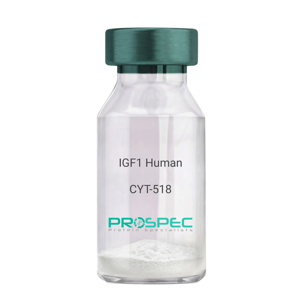
    IGF1 Human Des1-3

    Description:

    Insulin-Like Growth Factor 1 Des (1-3) Human Recombinant

    Somatomedin C, IGF-I, IGFI, IGF1, IGF-IA, Mechano growth factor, MGF, Des(1-3), Des1-3, Des 1-3, Des (1-3), IGF-1 (4-70).

    Product # :

    CYT-518

    Price :

    Quantity :

    Shipping Method :

    Room Temp Icon

    Shipped at Room temp

    Add To Cart

    More Info

    • description
    • source
    • formulation
    • purity
    • biological activity
    • More Info

    Description

    IGF-I Des(1-3) Human Recombinant produced in E.Coli is a single, non-glycosylated, polypeptide chain containing 67 amino acids (aa 4-70) and having a molecular mass of 7368.5 Dalton. IGF-1 Des1-3 is purified by proprietary chromatographic techniques.

    Source

    Escherichia Coli.

    Formulation

    The protein (1mg/ml) was lyophilized was lyophilized from a 0.2µm filtered concentrated solution in 20mM PBS, pH 7.0.

    Purity

    Greater than 97% as determined by:
    (a) Analysis by RP-HPLC.
    (b) Analysis by SDS-PAGE.

    Biological Activity

    The ED50 as determined by a cell proliferation assay using FDC-P1 cells is less than 2.0 ng/ml, corresponding to a specific activity of > 5×10⁵ units/mg.

    More Info

    • Introduction

      The somatomedins, or insulin-like growth factors (IGFs), comprise a family of peptides that play important roles in mammalian growth and development. IGF1 mediates many of the growth-promoting effects of growth hormone (GH; MIM 139250). Early studies showed that growth hormone did not directly stimulate the incorporation of sulfate into cartilage, but rather acted through a serum factor, termed 'sulfation factor,' which later became known as 'somatomedin' (Daughaday et al., 1972). Three main somatomedins have been characterized: somatomedin C (IGF1), somatomedin A (IGF2; MIM 147470), and somatomedin B (MIM 193190) (Rotwein, 1986; Rosenfeld, 2003).

    • Synonyms

      Somatomedin C, IGF-I, IGFI, IGF1, IGF-IA, Mechano growth factor, MGF, Des(1-3), Des1-3, Des 1-3, Des (1-3), IGF-1 (4-70).

    • Physical Appearance

      Sterile Filtered White lyophilized (freeze-dried) powder.

    • Stability

      Lyophilized IGF-I des(1-3) although stable at room temperature for 3 weeks, should be stored desiccated below -18°C. Upon reconstitution IGF1 des-1-3 should be stored at 4°C between 2-7 days and for future use below -18°C.For long term storage it is recommended to add a carrier protein (0.1% HSA or BSA).Please prevent freeze-thaw cycles.

    • Solubility

      It is recommended to reconstitute the IGF-I des(1-3) in sterile 18M-cm H2O not less than 100µg/ml, which can then be further diluted to other aqueous solutions.

    • Amino Acid Sequence

      TLCGAELVDA LQFVCGDRGF YFNKPTGYGS SSRRAPQTGI VDECCFRSCD LRRLEMYCAP LKPAKSA.

    • Background

      What is the molecular weight/Mw of IGF1 HUMAN DES1-3 Protein?
      IGF1 HUMAN DES1-3 Protein has a total Mw of 7.3kDa.

      What is the source or expression system of IGF1 HUMAN DES1-3 Protein?
      Escherichia Coli.

      What is the Purity of IGF1 HUMAN DES1-3 Protein?
      IGF1 HUMAN DES1-3 Protein is >97% pure as determined by SDS-PAGE.

      What is the Biological Activity of IGF1 HUMAN DES1-3 Protein?
      The ED50 as determined by a cell proliferation assay using FDC-P1 cells is less than 2.0 ng/ml, corresponding to a specific activity of > 5×10⁵ units/mg.

      What is the amino acid sequence of IGF1 HUMAN DES1-3 Protein?
      TLCGAELVDA LQFVCGDRGF YFNKPTGYGS SSRRAPQTGI VDECCFRSCD LRRLEMYCAP LKPAKSA.

      What applications can IGF1 HUMAN DES1-3 Protein be used in?
      IGF1 HUMAN DES1-3 Protein can probably be used in western blot, ELISA and Lateral Flow.

      What is the endotoxin level for IGF1 HUMAN DES1-3 Protein?
      The endotoxin level is minimal, IGF1 HUMAN DES1-3 Protein was purified using conventional chromatography techniques.


    ProSpec's products are furnished for LABORATORY RESEARCH USE ONLY. The product may not be used as drugs, agricultural or pesticidal products, food additives or household chemicals.

    Igf I Des1 3 Human
  • View Data Sheet

    Name :

    VEGF Equine

    Description:

    Vascular Endothelial Growth Factor Equine Recombinant

    VEGF-A, VPF, glioma-derived endothelial cell mitogen.

    Product # :

    CYT-1200

    Price :

    Quantity :

    Shipping Method :

    Room Temp Icon

    Shipped at Room temp

    Add To Cart

    More Info

    • description
    • source
    • formulation
    • purity
    • biological activity
    • More Info

    Description

    VEGF Equine Recombinant produced in E.Coli is a single, non-glycosylated, polypeptide chain containing 2 x 165 amino acids and having a total molecular mass of 38.6 kDa.The VEGF is purified by proprietary chromatographic techniques.

    Source

    Escherichia Coli.

    Formulation

    Lyophilized from a sterile (0.2 micron) filtered aqueous solution containing 10 mM Sodium Phosphate, pH 7.5.

    Purity

    Greater than 95.0% as determined by SDS-PAGE.

    Biological Activity

    The ED50 as determined by HUVEC Proliferation is ≤ 10 ng/mL, corresponding to a specific activity of ≥ 1.0 x 10^5 units/mg.

    More Info

    • Introduction

      Vascular endothelial growth factor is an important signaling protein involved in both vasculogenesis and angiogenesis. As its name implies, VEGF activity has been mostly studied on cells of the vascular endothelium, although it does have effects on a number of other cell types (e.g. stimulation monocyte/macrophage migration, neurons, cancer cells, kidney epithelial cells ).VEGF mediates increased vascular permeability, induces angiogenesis, vasculogenesis and endothelial cell growth, promotes cell migration, and inhibits apoptosis. In vitro, VEGF has been shown to stimulate endothelial cell mitogenesis and cell migration. VEGF is also a vasodilator and increases microvascular permeability and was originally referred to as vascular permeability factor. Elevated levels of this protein is linked to POEMS syndrome, also known as Crow-Fukase syndrome. Mutations in this gene have been associated with proliferative and nonproliferative diabetic retinopathy.

    • Synonyms

      VEGF-A, VPF, glioma-derived endothelial cell mitogen.

    • Physical Appearance

      Sterile Filtered White lyophilized (freeze-dried) powder.

    • Stability

      Lyophilized VEGF although stable at room temperature for 3 weeks, should be stored desiccated below -18°C. Upon reconstitution VEGF Human should be stored at 4°C between 2-7 days and for future use below -18°C.For long term storage it is recommended to add a carrier protein (0.1% HSA or BSA).Please prevent freeze-thaw cycles.

    • Solubility

      It is recommended to reconstitute the lyophilized VEGF in sterile water at a concentration of 0.1 mg/ml, which can then be further diluted to other aqueous solutions.

    • Amino Acid Sequence

      MAPMAEGEHK THEVVKFMDV YQRSYCRPIE TLVDIFQEYP DEIEYIFKPS CVPLMRCGGC CNDEGLECVP TAEFNITMQI MRIKPHQSQH IGEMSFLQHS KCECRPKKDK ARQENPCGPC SERRKHLFVQ DPQTCKCSCK NTDSRCKARQ LELNERTCRC DKPRR

    ProSpec's products are furnished for LABORATORY RESEARCH USE ONLY. The product may not be used as drugs, agricultural or pesticidal products, food additives or household chemicals.

    Vegf Equine
  • View Data Sheet

    Name :

    B.Microti p32

    Description:

    Babesia Microti p32 Recombinant

    Product # :

    PRO-2269

    Price :

    Quantity :

    Shipping Method :

    Ice Icon

    Shipped with Ice Packs

    Add To Cart

    More Info

    • description
    • source
    • formulation
    • purity
    • More Info

    Description

    Recombinant Babesia Microti p32 produced in SF9 is a glycosylated, polypeptide chain having a calculated molecular mass of 35,808 Dalton. B.Microti p32 is expressed with a 10xHis tag at N-terminus and purified by proprietary chromatographic techniques.

    Source

    Sf9 insect cells.

    Formulation

    B.Microti p32 is supplied in 20mM HEPES buffer pH-7.6, 250mM NaCl and 20% glycerol.

    Purity

    Greater than 80.0% as determined by SDS-PAGE.

    More Info

    • Introduction

      Babesiosis is a disease caused by apicomplexan parasites of the Babesia genus. The Babesia microti life cycle involves 2 hosts, which include a rodent, mainly the white-footed mouse (Peromyscus leucopus) and a tick in the Ixodes genus. During a blood meal, a Babesia-infected tick introduces sporozoites into the mouse host. Sporozoites pass into the erythrocytes and undergo asexual reproduction (budding). In the blood, some parasites differentiate into male and female gametes, though these cannot be distinguished by light microscopy. The definitive host is the tick. Once ingested by an proper tick, gametes unite and undergo a sporogonic cycle resulting in sporozoites. Transovarial transmission (aka vertical or hereditary transmission) has been detected for "large" Babesia species but not for the "small" Babesia, such as B. microti. Humans enter the cycle when bitten by the infected ticks. Thus during a blood meal, a Babesia-infected tick introduces sporozoites into the human host. Sporozoites then enter erythrocytes and undergo asexual replication (budding). Multiplication of the blood-stage parasites is responsible for the clinical manifestations of the disease. Humans typically are dead-end hosts. However, human-to-human transmission is well acknowledged to occur via contaminated blood transfusions.

    • Physical Appearance

      Sterile Filtered clear solution.

    • Stability

      Store at 4°C if entire vial will be used within 2-4 weeks. Store, frozen at -20°C for longer periods of time. Avoid multiple freeze-thaw cycles.

    ProSpec's products are furnished for LABORATORY RESEARCH USE ONLY. The product may not be used as drugs, agricultural or pesticidal products, food additives or household chemicals.

    Bmicroti P32
  • View Data Sheet

    Name :

    Y.Enterocolitica (O:9) YopN

    Description:

    Yersinia Enterocolitica (O:9) YopN Recombinant

    Product # :

    PRO-2274

    Price :

    Quantity :

    Shipping Method :

    Ice Icon

    Shipped with Ice Packs

    Add To Cart

    More Info

    • description
    • source
    • formulation
    • purity
    • More Info

    Description

    Recombinant Yersinia Enterocolitica (O:9) YopN produced in E.coli is a non-glycosylated, polypeptide chain having a calculated molecular mass of 34,296 Dalton. Y.Enterocolitica (O:9) YopN is expressed with a 10xHis tag at N-terminus and purified by proprietary chromatographic techniques.

    Source

    Escherichia Coli.

    Formulation

    Y.Enterocolitica (O:9) YopN is supplied in 20mM HEPES buffer pH-7.6, 250mM NaCl and 20% glycerol.

    Purity

    Greater than 80.0% as determined by SDS-PAGE.

    More Info

    • Introduction

      Yersinia enterocolitica is a Gram-negative bacillus-shaped bacterium, which is a member of the Enterobacteriaceae family. Y.Enterocolitica is motile at temperatures between 22-29°C, however becomes non-motile at normal human body temperature. Y. Enterocolitica infection causes the yersiniosis disease, which is an animal-borne disease occurring in humans, as well as in a various groups of animals such as cattle, deer, pigs, and birds. Yersinia enterocolitica is a heterogeneous group of strains, which are conventionally classified by bio-typing into six bio-groups on the basis of phenotypic characteristics, and by serotyping into more than 57 “O” serogroups, on the basis of their O (lipopolysaccharide or LPS) surface antigen. Five of the six biogroups (1B and 2–5) are considered as pathogens. Nevertheless, only a few of these serogroups have been linked with disease in either humans or animals. Strains which belong to serogroups O:3 (biogroup 4), O:5,27 (biogroups 2 and 3), O:8 (biogroup 1B), and O:9 (biogroup 2) are most frequently isolated worldwide from human samples. Still, the main Y. enterocolitica serogroup in many European countries is serogroup O:3 followed by O:9, whereas the serogroup O:8 is mostly detected in the United States.

    • Physical Appearance

      Sterile Filtered clear solution.

    • Stability

      Store at 4°C if entire vial will be used within 2-4 weeks. Store, frozen at -20°C for longer periods of time. Avoid multiple freeze-thaw cycles.

    • Immunological Functions

      1. Binds IgG- and IgM- and IgA-type human antibodies.2. Immunodot test with positive/negative sera panels.

    ProSpec's products are furnished for LABORATORY RESEARCH USE ONLY. The product may not be used as drugs, agricultural or pesticidal products, food additives or household chemicals.

    Yenterocolitica O 9 Yopn
  • View Data Sheet

    Name :

    PLA2G10 Human

    Description:

    Secreted Phospholipase A2-X Human Recombinant

    Group 10 secretory phospholipase A2, EC 3.1.1.4, Group X secretory phospholipase A2, Phosphatidylcholine 2-acylhydrolase GX, GX sPLA2, sPLA2-X, SPLA2, GXPLA2, MGC119918, MGC119919, MGC133367, PLA2G10.

    Product # :

    ENZ-329

    Price :

    Quantity :

    Shipping Method :

    Room Temp Icon

    Shipped at Room temp

    Add To Cart

    More Info

    • description
    • source
    • formulation
    • purity
    • More Info

    Description

    Secreted Phospholipase A2-X Human Recombinant is manufactured with N-terminal fusion HisTag. PLA2G10 His-Tagged Fusion Protein, is 15.5 kDa containing 123 amino acid residues of the human secreted phospholipase A2-X and 16 additional amino acid residues - HisTag (underlined).

    Source

    Escherichia Coli.

    Formulation

    PLA2G10 filtered (0.4 µm) and lyophilized from 0.5mg/ml solution in 20mM Tris and 50mM NaCl, pH 7.5.

    Purity

    Greater than 95% as determined by SDS PAGE.

    More Info

    • Introduction

      Phospholipase A2 (PLA2) catalyzes the hydrolysis of the sn-2 position of membrane glycerophospholipids to liberate arachidonic acid (AA), a precursor of eicosanoids including prostaglandins and leukotrienes. The same reaction also produces lysophosholipids, which represent another class of lipid mediators.
      The secretory PLA2 (sPLA2) family, in which 10 isozymes have been identified, consists of low molecular weight, Ca2+-requiring secretory enzymes that have been implicated in a number of biological processes, such as modification of eicosanoid generation, inflammation, and host defense.
      This enzyme has been proposed to hydrolyze phosphatidylcholine (PC) in lipoproteins to liberate lyso-PC and free fatty acids in the arterial wall, thereby facilitating the accumulation of bioactive lipids and modified lipoproteins in atherosclerotic foci.
      In mice, sPLA2 expression significantly influences HDL particle size and composition and demonstrate that an induction of sPLA2 is required for the decrease in plasma HDL cholesterol in response to inflammatory stimuli. Instillation of bacteria into the bronchi was associated with surfactant degradation and a decrease in large:small ratio of surfactant aggregates in rats.

    • Synonyms

      Group 10 secretory phospholipase A2, EC 3.1.1.4, Group X secretory phospholipase A2, Phosphatidylcholine 2-acylhydrolase GX, GX sPLA2, sPLA2-X, SPLA2, GXPLA2, MGC119918, MGC119919, MGC133367, PLA2G10.

    • Physical Appearance

      Sterile Filtered lyophilized (freeze-dried) powder.

    • Stability

      Store lyophilized protein at -20°C. Aliquot the product after reconstitution to avoid repeated freezing/thawing cycles. Reconstituted protein can be stored at 4°C for a limited period of time; it does not show any change after two weeks at 4°C.

    • Solubility

      It is recommended to add deionized water to prepare a working stock solution of approximately 0.5mg/ml and let the lyophilized pellet dissolve completely.

    • Amino Acid Sequence

      MRGSHHHHHH GMASHMGILE LAGTVGCVGP RTPIAYMKYG CFCGLGGHGQ PRDAIDWCCH GHDCCYTRAE EAGCSPKTER YSWQCVNQSV LCGPAENKCQ ELLCKCDQEI ANCLAQTEYN LKYLFYPQFL CEPDSPKCD

    ProSpec's products are furnished for LABORATORY RESEARCH USE ONLY. The product may not be used as drugs, agricultural or pesticidal products, food additives or household chemicals.

    Pla2G10 Human
  • View Data Sheet

    Name :

    PNPLA2 Antibody

    Description:

    Patatin-like phospholipase domain-containing protein 2, Mouse Anti Human

    Patatin-like phospholipase domain-containing protein 2, Adipose triglyceride lipase, Desnutrin Transport-secretion protein 2, TTS2.2, Calcium-independent phospholipase A2, IPLA2-zeta Pigment epithelium-derived factor, TTS2, PNPLA2, ATGL, PEDF-R, FP17548, TTS-2.2, DKFZp667M109, 1110001C14Rik.

    Product # :

    ANT-684

    Price :

    Quantity :

    Shipping Method :

    Ice Icon

    Shipped with Ice Packs

    Add To Cart

    More Info

    • formulation
    • More Info

    Formulation

    1mg/ml containing PBS, pH-7.4, 10% Glycerol and 0.02% Sodium Azide.

    More Info

    • Introduction

      PNPLA2 is an enzyme which catalyzes the first step in the hydrolysis of triglycerides in adipose tissue. Mutations in the PNPLA2 gene are linked to neutral lipid storage disease with myopathy.

    • Synonyms

      Patatin-like phospholipase domain-containing protein 2, Adipose triglyceride lipase, Desnutrin Transport-secretion protein 2, TTS2.2, Calcium-independent phospholipase A2, IPLA2-zeta Pigment epithelium-derived factor, TTS2, PNPLA2, ATGL, PEDF-R, FP17548, TTS-2.2, DKFZp667M109, 1110001C14Rik.

    • Physical Appearance

      Sterile filtered colorless solution.

    • Immunogen

      Anti-human PNPLA2 mAb, is derived from hybridization of mouse F0 myeloma cells with spleen cells from BALB/c mice immunized with a recombinant human PNPLA2 protein 30-504 amino acids purified from E. coli.

    • Ig Subclass

      Mouse IgG2b heavy chain and k light chain.

    • Clone

      PAT18E6AT.

    • Applications

      PNPLA2 antibody has been tested by ELISA, Western blot analysis, Flow cytometry and ICC/IF to assure specificity and reactivity. Since application varies, however, each investigation should be titrated by the reagent to obtain optimal results.

    • Type

      Mouse Anti Human Monoclonal.

    • Storage Procedures

      For periods up to 1 month store at 4°C, for longer periods of time, store at -20°C. Prevent freeze thaw cycles.

    • Purification Method

      PNPLA2 antibody was purified from mouse ascitic fluids by protein-A affinity chromatography.

    ProSpec's products are furnished for LABORATORY RESEARCH USE ONLY. The product may not be used as drugs, agricultural or pesticidal products, food additives or household chemicals.

    Pnpla2 Pat18E6At Antibody
  • View Data Sheet

    Name :

    Platelet Factor 4 Bovine

    Description:

    Platelet Factor-4 (CXCL4) Bovine Recombinant

    CXCL4, PF-4, PF4, Iroplact, Oncostatin-A, SCYB4, MGC138298.

    Product # :

    CHM-039

    Price :

    Quantity :

    Shipping Method :

    Room Temp Icon

    Shipped at Room temp

    Add To Cart

    More Info

    • description
    • source
    • formulation
    • purity
    • biological activity
    • More Info

    Description

    Platelet Factor-4 (CXCL4) Bovine Recombinant produced in E.Coli is a non-glycosylated polypeptide chain containing 88 amino acid and having a molecular mass of approximately 9.5kDa.PF4 is purified by proprietary chromatographic techniques.

    Source

    Escherichia Coli.

    Formulation

    Lyophilized from a 0.2μm filtered concentrated solution in 20 mM PB and 500mM NaCl, pH 7.0.

    Purity

    Greater than 95.0% as determined by:
    (a) Analysis by RP-HPLC.
    (b) Analysis by SDS-PAGE.

    Biological Activity

    The biological activity determined by a chemotaxis bioassay using human neutrophils is 10-100ng/ml.

    More Info

    • Introduction

      Platelet factor-4 is a 70-amino acid protein that is released from the alpha-granules of activated platelets.PF4’s major physiologic role appears to be neutralization of molecules on the endothelial surface of blood vessels, thereby inhibiting local antithrombin III activity and promoting coagulation. As a strong chemoattractant for neutrophils and fibroblasts, PF4 probably has a role in inflammation and wound repair. Oncostatin-A is a member of the CXC chemokine family. Furthermore, it is used as an inhibitor in the angiogenesis during tumor therapy.

    • Synonyms

      CXCL4, PF-4, PF4, Iroplact, Oncostatin-A, SCYB4, MGC138298.

    • Physical Appearance

      Sterile Filtered White lyophilized (freeze-dried) powder.

    • Stability

      Lyophilized CXCL4 although stable at room temperature for 3 weeks, should be stored desiccated below -18°C. Upon reconstitution Platelet Factor-4 should be stored at 4°C between 2-7 days and for future use below -18°C. Please prevent freeze-thaw cycles.

    • Solubility

      It is recommended to reconstitute the lyophilized Platelet Factor-4 in sterile 18MΩ-cm H2O not less than 100µg/ml, which can then be further diluted to other aqueous solutions.

    • Amino Acid Sequence

      ESSFPATFVP LPADSEGGED EDLQCVCLKT TSGINPRHIS SLEVIGAGTH CPSPQLLATK KTGRKICLDQ QRPLYKKILK KLLDGDES.

    • Background

      What is the molecular weight/Mw of PLATELET FACTOR 4 BOVINE Protein?
      PLATELET FACTOR 4 BOVINE Protein has a total Mw of 9.5kDa.

      What is the source or expression system of PLATELET FACTOR 4 BOVINE Protein?
      Escherichia Coli.

      What is the Purity of PLATELET FACTOR 4 BOVINE Protein?
      PLATELET FACTOR 4 BOVINE Protein is > 95% pure as determined by SDS-PAGE.

      What is the Biological Activity of PLATELET FACTOR 4 BOVINE Protein?
      The biological activity determined by a chemotaxis bioassay using human neutrophils is 10-100ng/ml.

      What is the amino acid sequence of PLATELET FACTOR 4 BOVINE Protein?
      ESSFPATFVP LPADSEGGED EDLQCVCLKT TSGINPRHIS SLEVIGAGTH CPSPQLLATK KTGRKICLDQ QRPLYKKILK KLLDGDES.

      What applications can PLATELET FACTOR 4 BOVINE Protein be used in?
      PLATELET FACTOR 4 BOVINE Protein can probably be used in western blot, ELISA and Lateral Flow.

      What is the endotoxin level for PLATELET FACTOR 4 BOVINE Protein?
      The endotoxin level is minimal, PLATELET FACTOR 4 BOVINE Protein was purified using conventional chromatography techniques.


    ProSpec's products are furnished for LABORATORY RESEARCH USE ONLY. The product may not be used as drugs, agricultural or pesticidal products, food additives or household chemicals.

    Cxcl4 Bovine
  • View Data Sheet

    Name :

    NUP210 Human

    Description:

    Nucleopurin 210kDa Recombinant Human

    Nucleoporin 210kDa, KIAA0906, GP210, Nuclear envelope pore membrane protein POM 210, Nuclear pore protein gp210, nuclear pore membrane glycoprotein 210.

    Product # :

    PRO-109

    Price :

    Quantity :

    Shipping Method :

    Ice Icon

    Shipped with Ice Packs

    Add To Cart

    More Info

    • description
    • source
    • formulation
    • purity
    • More Info

    Description

    Recombinant NUP210 protein is a construct carrying multiple concatenated copies of the short cytoplasmic gp210 C-terminus (which contains the autoreactive epitopes) and having a molecular mass of 27kDa (pH 9.8). NUP210 protein is fused to a hexa-histidine purification tag.

    Source

    Sf9 insect cells.

    Formulation

    NUP210 is supplied in 20mM HEPES buffer pH-7.9 and 6M Urea.

    Purity

    Greater than 95% as determined by SDS-PAGE.

    More Info

    • Introduction

      The NUP210 complex is a immense structure which spreads through the nuclear envelope, forming a doorway which regulates the flow of macromolecules between the nucleus and the cytoplasm. Nucleoporins are the core components of the nuclear pore complex in eukaryotic cells. NUP210 is a membrane-spanning glycoprotein and is a major component of the nuclear pore complex.

    • Synonyms

      Nucleoporin 210kDa, KIAA0906, GP210, Nuclear envelope pore membrane protein POM 210, Nuclear pore protein gp210, nuclear pore membrane glycoprotein 210.

    • Stability

      Store at 4°C if entire vial will be used within 2-4 weeks. Store, frozen at -20°C for longer periods of time. Avoid multiple freeze-thaw cycles.

    • Immunological Functions

      1. Binds IgG-type human auto-antibodies. 2. Standard ELISA test (checkerboard analysis of positive/negative sera panels); immunodot test.

    • coating concentration

      0.4-1.0 µg/ml (depending on the type of ELISA plate and coating buffer). Suitable for labeling of functional groups.

    • Applications

      Western blot with PBC sera or monoclonal anti-hexa-His-tag antibody.

    ProSpec's products are furnished for LABORATORY RESEARCH USE ONLY. The product may not be used as drugs, agricultural or pesticidal products, food additives or household chemicals.

    Nup210 Human
  • View Data Sheet

    Name :

    FGFR3 Human

    Description:

    Fibroblast Growth Factor Receptor 3 Fc Chimera Human Recombinant

    Achondroplasia, Thanatophoric Dwarfism, CD333, ACH, CEK2, JTK4, HSFGFR3EX, FGFR3.

    Product # :

    PKA-232

    Price :

    Quantity :

    Shipping Method :

    Room Temp Icon

    Shipped at Room temp

    Add To Cart

    More Info

    • description
    • source
    • formulation
    • purity
    • More Info

    Description

    Soluble FGFR3 Human recombinant fused using a Xa cleavage site with the Fc part of human IgG1 produced in Fc Chimera is a heterodimeric, glycosylated, Polypeptide chain containing 593aa and having a molecular mass of 170kDa.

    Source

    Insect Cells.

    Formulation

    FGFR3 was lyophilized from a concentrated (1 mg/ml) sterile solution containing PBS.

    Purity

    Greater than 90.0% as determined by:(a) Analysis by silver stain.(b) Analysis by SDS-PAGE.

    More Info

    • Introduction

      Fibroblast Growth Factors (FGFs) comprise a family of at least eighteen structurally realted proteins that are involved in a multitude of physiological and pathological cellular processes, including cell growth, differentation, angiogenesis, wound healing and tumorgenesis. The biological activities of the FGFs are mediated by a family if type I transmembrane tyrosine kinases which undergo dimerization and autophosphorylation after ligand binding. Four distinct genes encoding closely related FGF receptors, FGFR-1to -4 are known. Multiple forms of FGFR-1 to -3 are generated by alternative splicing of the mRNAs. A frequent splicing event involving FGFR-1 and -2 results in receptors containing all three Ig domains, referred to as the alpha isoform, or only IgII and IgIII, referred to as the ? isoform. Only the alpha isoform has been identified for FGFR-3 and FGFR-4. Additional splicing events for FGFR-1 to -3, involving the C-terminal half of the IgIII domain encoded by two mutually exclusive alternative exons, generate FGF receptors with alternative IgIII domains (IIIb and IIIc). A IIIa isoform which is a secreted FGF binding protein containing only the N-terminal half of the IgIII domain plus some intron sequences has also been reported for FGFR-1. Mutations in FGFR-1 to -3 have been found in patients with birth defects involving craniosynostosis.

    • Synonyms

      Achondroplasia, Thanatophoric Dwarfism, CD333, ACH, CEK2, JTK4, HSFGFR3EX, FGFR3.

    • Physical Appearance

      Sterile Filtered White lyophilized (freeze-dried) powder.

    • Stability

      Lyophilized FGFR3 although stable at room temperature for 3 weeks, should be stored desiccated below -18oC. Upon reconstitution FGFR3 should be stored at 4oC between 2-7 days and for future use below -18oC.For long term storage it is recommended to add a carrier protein (0.1% HSA or BSA).Please prevent freeze-thaw cycles.

    • Solubility

      It is recommended to reconstitute the lyophilized FGFR-3 in sterile PBS not less than 50µg/ml, which can then be further diluted to other aqueous solutions.

    • Amino Acid Sequence

      ESLGTEQRVV GRAAEVPGPE PGQQEQLVFG SGDAVELSCP PPGGGPMGPT VWVKDGTGLV PSERVLVGPQ RLQVLNASHE DSGAYSCRQR LTQRVLCHFS VRVTDAPSSG DDEDGEDEAE DTGVDTGAPY WTRPERMDKK LLAVPAANTV RFRCPAAGNP TPSISWLKNG REFRGEHRIG GIKLRHQQWS LVMESVVPSD RGNYTCVVEN KFGSIRQTYT LDVLERSPHR PILQAGLPAN QTAVLGSDVE FHCKVYSDAQ PHIQWLKHVE VNGSKVGPDG TPYVTVLKTA GANTTDKELE VLSLHNVTFE DAGEYTCLAG NSIGFSHHSA WLVVLPAEEE LVEADEAGDP RRASIEGRGD PEEPKSCDKT HTCPPCPAPE LLGGPSVFLF PPKPKDTLMI SRTPEVTCVV VDVSHEDPEV KFNWYVDGVE VHNAKTKPRE EQYNSTYRVV SVTVLHQDWL NGKEYKCKVS NKALPAPIEK TISKAKGQPR EPQVYTLPPS RDELTKNQVS LTCLVKGFYP SDIAVEWESN GQPENNYKTT PPVLDSDGSF FLYSKLTVDK SRWQQGNVFS CSVMHEALHN HYTQKSLSLS PGK.

    ProSpec's products are furnished for LABORATORY RESEARCH USE ONLY. The product may not be used as drugs, agricultural or pesticidal products, food additives or household chemicals.

    Fgfr3 Human
  • View Data Sheet

    Name :

    G CSF Human, PEG

    Description:

    Granulocyte-Colony Stimulating Factor Pegylated Human Recombinant

    CSF-3, MGI-1G, GM-CSF beta, Pluripoietin, Filgrastim, Lenograstim, G-CSF, MGC45931, GCSF.

    Product # :

    CYT-018

    Price :

    Quantity :

    Shipping Method :

    Ice Icon

    Shipped with Ice Packs

    Add To Cart

    More Info

    • description
    • source
    • formulation
    • purity
    • biological activity
    • More Info

    Description

    Granulocyte Colony Stimulating Factor Human Recombinant produced in E.coli is a single, non-glycosylated, polypeptide chain containing 175 amino acids and having a molecular mass of 18.8kDa. The Pegylated G-CSF is produced by attaching a 20kDa methoxypolyethylene glycol propionaldehyde (mPEG-ALD) to the N-terminal amino acid of G-CSF giving a total molecular mass of 38.8kDa. G-CSF is purified by proprietary chromatographic techniques.

    Source

    Escherichia Coli.

    Formulation

    G-CSF is supplied in solution (0.69mg/ml) containing 10mM Acetate Buffer (pH 4.0), and 0.004% Polysorbate 80.

    Purity

    Greater than 95.0% as determined by SEC-HPLC.

    Biological Activity

    The ED50, calculated by the dose-dependent proliferation of murine NFS-60 indicator cells is less than 0.1 ng/ml, corresponding to a Specific Activity of 10,000,000IU/mg.

    More Info

    • Introduction

      GCSF is a cytokine that controls the production, differentiation, and function of granulocytes. The active protein is found extracellularly. Three transcript variants encoding three different isoforms have been found for this gene. Granulocyte/macrophage colony-stimulating factors are cytokines that act in hematopoiesis by controlling the production, differentiation, and function of 2 related white cell populations of the blood, the granulocytes and the monocytes-macrophages. This csf induces granulocytes.

    • Synonyms

      CSF-3, MGI-1G, GM-CSF beta, Pluripoietin, Filgrastim, Lenograstim, G-CSF, MGC45931, GCSF.

    • Physical Appearance

      Colorless, clear and transparent solution.

    • Stability

      G-CSF PEG should be stored refrigerated at 2° to 8°C. Vials should be kept in theirpackaging to protect from light until the time of use. Shaking and freezing should be avoided.

    • Background

      What is the molecular weight/Mw of G CSF HUMAN, PEG Protein?
      G CSF HUMAN, PEG Protein has a total Mw of 18.8kDa.

      What is the source or expression system of G CSF HUMAN, PEG Protein?
      Escherichia Coli.

      What is the Purity of G CSF HUMAN, PEG Protein?
      G CSF HUMAN, PEG Protein is >95% pure as determined by SDS-PAGE.

      What is the Biological Activity of G CSF HUMAN, PEG Protein?
      The ED50, calculated by the dose-dependent proliferation of murine NFS-60 indicator cells is less than 0.1 ng/ml, corresponding to a Specific Activity of 10,000,000IU/mg.

      What is the amino acid sequence of G CSF HUMAN, PEG Protein?
      G CSF HUMAN, PEG Protein is composed from 175 amino acids.

      What applications can G CSF HUMAN, PEG Protein be used in?
      G CSF HUMAN, PEG Protein can probably be used in western blot, ELISA and Lateral Flow.

      What is the endotoxin level for G CSF HUMAN, PEG Protein?
      The endotoxin level is minimal, G CSF HUMAN, PEG Protein was purified using conventional chromatography techniques.


    ProSpec's products are furnished for LABORATORY RESEARCH USE ONLY. The product may not be used as drugs, agricultural or pesticidal products, food additives or household chemicals.

    G Csf Human Pegylated
  • View Data Sheet

    Name :

    SQSTM1 Human

    Description:

    Sequestosome 1 Human Recombinant

    A170, OSIL, p60, p62, p62B, PDB3, ZIP3, EBIAP, ORCA, OSIL, SQSTM1.

    Product # :

    PRO-806

    Price :

    Quantity :

    Shipping Method :

    Ice Icon

    Shipped with Ice Packs

    Add To Cart

    More Info

    • description
    • source
    • formulation
    • purity
    • More Info

    Description

    SQSTM1 Human Recombinant produced in E.Coli is a single, non-glycosylated, polypeptide chain containing 364 amino acids (1-356 a.a.) and having a molecular mass of 39.7 kDa. SQSTM1 protein is fused to an 8 amino acid His-Tag at C-terminus and purified by standard chromatography.

    Source

    Escherichia Coli.

    Formulation

    SQSTM1 Human 0.5mg/ml solution containing 20mM Tris-HCl pH-8, 1mM DTT and 10% glycerol.

    Purity

    Greater than 85% as determined by SDS-PAGE.

    More Info

    • Introduction

      SQSTM1 is an adapter protein which binds ubiquitin and mediates the activation of NFKB1 by TNF-a, NGF and IL-1. SQSTM1 is involved in titin/TTN downstream signaling in muscle cells. SQSTM1 controls signaling cascades through ubiquitination. SQSTM1 participates in cell differentiation, apoptosis, immune response and regulation of K(+) channels. Mutations in UBA domain of the SQSTM1 protein cause Paget’s disease because the UBA is essential for aggregate sequestration and cell survival.

    • Synonyms

      A170, OSIL, p60, p62, p62B, PDB3, ZIP3, EBIAP, ORCA, OSIL, SQSTM1.

    • Physical Appearance

      Sterile filtered colorless solution.

    • Stability

      Store at 4°C if entire vial will be used within 2-4 weeks. Store, frozen at -20°C for longer periods of time. For long term storage it is recommended to add a carrier protein (0.1% HSA or BSA).Avoid multiple freeze-thaw cycles.

    • Amino Acid Sequence

      MAMSYVKDDI FRIYIKEKKE CRRDHRPPCA QEAPRNMVHP NVICDGCNGP VVGTRYKCSV CPDYDLCSVC EGKGLHRGHT KLAFPSPFGH LSEGFSHSRW LRKVKHGHFG WPGWEMGPPG NWSPRPPRAG EARPGPTAES ASGPSEDPSV NFLKNVGESV AAALSPLGIE VDIDVEHGGK RSRLTPVSPE SSSTEEKSSS QPSSCCSDPS KPGGNVEGAT QSLAEQMRKI ALESEGRPEE QMESDNCSGG DDDWTHLSSK EVDPSTGELQ SLQMPESEGP SSLDPSQEGP TGLKEAALYP HLPPEADPRL IESLSQMLSM GFSDEGGWLT RLLQTKNYDI GAALDTIQYS KHPPPLLEHH HHHH.

    ProSpec's products are furnished for LABORATORY RESEARCH USE ONLY. The product may not be used as drugs, agricultural or pesticidal products, food additives or household chemicals.

    Sqstm1 Human
  • View Data Sheet

    Name :

    Adiponectin (108-244) Human

    Description:

    Adiponectin (108-244 a.a.) Human Recombinant

    Acrp30, AdipoQ, GBP-28, APM-1, ACDC.

    Product # :

    CYT-073

    Price :

    Quantity :

    Shipping Method :

    Ice Icon

    Shipped with Ice Packs

    Add To Cart

    More Info

    • description
    • source
    • formulation
    • purity
    • More Info
    • sds-page

    Description

    Acrp30 Human Recombinant produced in E.Coli is a single, non-glycosylated polypeptide chain containing 158 amino acids (108-244 a.a.) and having a molecular mass of 18.1kDa.Acrp30 is fused to a 21 amino acid His-tag at N-terminus & purified by proprietary chromatographic techniques.

    Source

    Escherichia Coli.

    Formulation

    Acrp30 protein solution (1mg/ml) contains 20mM Tris-HCl buffer (pH 8.0), 10% glycerol and 0.4M Urea.

    Purity

    Greater than 80.0% as determined by SDS-PAGE.

    sds-page

    Adiponectin-sds-page - Product image 1

    More Info

    • Introduction

      Adiponectin is an adipocyte specific secreted protein that circulates in the plasma. It is induced during adipocyte differentiation and its secretion is stimulated by insulin. Mouse adiponectin shares about 83% amino acid identity with that human. Adiponectin plays a role in various physiological processes such as energy homeostasis and obesity. Adiponectin is reduced in obese humans, and decreased level is associated with insulin resistance and hyperinsulinemia.

    • Synonyms

      Acrp30, AdipoQ, GBP-28, APM-1, ACDC.

    • Stability

      Store at 4°C if entire vial will be used within 2-4 weeks. Store, frozen at -20°C for longer periods of time. For long term storage it is recommended to add a carrier protein (0.1% HSA or BSA).Avoid multiple freeze-thaw cycles.

    • Amino Acid Sequence

      MGSSHHHHHH SSGLVPRGSH MAYVYRSAFS VGLETYVTIP NMPIRFTKIF YNQQNHYDGS TGKFHCNIPG LYYFAYHITV YMKDVKVSLF KKDKAMLFTY DQYQENNVDQ ASGSVLLHLE VGDQVWLQVY GEGERNGLYA DNDNDSTFTG FLLYHDTN.

    • Background

      What is the molecular weight / Mw of ADIPONECTIN Protein?
      ADIPONECTIN Protein has a total Mw of 18.1kDa.

      What is the source or expression system of ADIPONECTIN Protein?
      Escherichia Coli.

      What is the Purity of ADIPONECTIN Protein?
      ADIPONECTIN Protein is >80% pure as determined by SDS-PAGE.

      What is the Biological Activity of ADIPONECTIN Protein?
      The biological functionality of ADIPONECTIN Protein will be determined in the future.

      What is the amino acid sequence of ADIPONECTIN Protein? MGSSHHHHHH SSGLVPRGSH MAYVYRSAFS VGLETYVTIP NMPIRFTKIF YNQQNHYDGS TGKFHCNIPG LYYFAYHITV YMKDVKVSLF KKDKAMLFTY DQYQENNVDQ ASGSVLLHLE VGDQVWLQVY GEGERNGLYA DNDNDSTFTG FLLYHDTN..

      What applications can ADIPONECTIN Protein be used in?
      ADIPONECTIN Protein can probably be used in western blot, ELISA and Lateral Flow.

      What is the endotoxin level for ADIPONECTIN Protein?
      The endotoxin level is minimal, ADIPONECTIN Protein was purified using conventional chromatography techniques

    ProSpec's products are furnished for LABORATORY RESEARCH USE ONLY. The product may not be used as drugs, agricultural or pesticidal products, food additives or household chemicals.

    Acrp30 108 244 Human
  • View Data Sheet

    Name :

    EBI3 Human, His

    Description:

    Epstein Barr Virus Induced 3 Human Recombinant, His Tag

    Interleukin-27 subunit beta, IL-27 subunit beta, IL-27B, Epstein-Barr virus-induced gene 3 protein, EBV-induced gene 3 protein, EBI3, IL27B.

    Product # :

    CYT-668

    Price :

    Quantity :

    Shipping Method :

    Ice Icon

    Shipped with Ice Packs

    Add To Cart

    More Info

    • description
    • source
    • formulation
    • purity
    • More Info

    Description

    EBI3 Human Recombinant produced in E.Coli is a single, non-glycosylated, Polypeptide chain containing 209 amino acids fragment (21-229) having a molecular weight of 34kDa and fused with a 4.5kDa amino-terminal hexahistidine tag. The EBI3 is purified by proprietary chromatographic techniques.

    Source

    Escherichia Coli.

    Formulation

    EBI3 protein is supplied in 1xPBS, 50% glycerol.

    Purity

    Greater than 95.0% as determined by SDS-PAGE.

    More Info

    • Introduction

      EBI3 has an induced expression in B lymphocytes in reaction to Epstein-Barr virus infection. EBI3 encodes a secreted glycoprotein belonging to the hematopoietin receptor family, and heterodimerizes with a 28 kDa protein to form iIL-27. EBI3 drives rapid clonal expansion of naive cd4(+) t-cells. EBI3 strongly synergizes with IL-12 to activate IFN-gamma production of naive cd4(+) t-cells. EBI3 mediates its biologic effects through the cytokine receptor wsx-1/tccr.

    • Synonyms

      Interleukin-27 subunit beta, IL-27 subunit beta, IL-27B, Epstein-Barr virus-induced gene 3 protein, EBV-induced gene 3 protein, EBI3, IL27B.

    • Physical Appearance

      Sterile Filtered clear solution.

    • Stability

      Store at 4°C if entire vial will be used within 2-4 weeks. Store, frozen at -20°C for longer periods of time. Please avoid freeze thaw cycles.

    • Background

      What is the molecular weight/Mw of EBI3 Protein?
      EBI3 Protein has a total Mw of 34kDa.

      What is the source or expression system of EBI3 Protein?
      Escherichia Coli.

      What is the Purity of EBI3 Protein?
      EBI3 Protein is >95% pure as determined by SDS-PAGE.

      What is the Biological Activity of EBI3 Protein?
      The biological functionality of EBI3 Protein will be determined in the future.

      What is the amino acid sequence of EBI3 Protein?
      EBI3 Protein is composed from 209 amino acids.

      What applications can EBI3 Protein be used in?
      EBI3 Protein can probably be used in western blot, ELISA and Lateral Flow.

      What is the endotoxin level for EBI3 Protein?
      The endotoxin level is minimal, EBI3 Protein was purified using conventional chromatography techniques.

    ProSpec's products are furnished for LABORATORY RESEARCH USE ONLY. The product may not be used as drugs, agricultural or pesticidal products, food additives or household chemicals.

    Ebi3 Human His
  • View Data Sheet

    Name :

    Der P1

    Description:

    Der P1 Protein Recombinant

    Peptidase 1, Major mite fecal allergen Der p 1, Allergen Der p I, Der p 1, DERP1, Der-P1.

    Product # :

    ALR-003

    Price :

    Quantity :

    Shipping Method :

    Ice Icon

    Shipped with Ice Packs

    Add To Cart

    More Info

    • description
    • source
    • formulation
    • purity
    • More Info

    Description

    The E.Coli derived recombinant protein contains the Dermatophagoides pteronyssinus Dust Mite Der P1 protein (a.a. 20-320) and fused to a 6 His Tag at C-terminus, having a total Mw of 34.5kDa, pI 5.6.

    Source

    Escherichia Coli.

    Formulation

    60mM NaCl and 50mM Tris-HCl pH 8.0.

    Purity

    Protein is >95% pure as determined by 10% SDS-PAGE (coomassie staining).

    More Info

    • Introduction

      DERP1 is a thiol protease, with a preference for substrates with a large hydrophobic side chain in the P2 position, or with basic residues. DERP1 is a C1 peptidase family member. DERP1 has extensive endopeptidase specificity. DERP1 is N-glycosylated. N-glycanase treatment does not completely remove carbohydrates, suggesting that the protein contains additional glycosylation sites. DERP1 causes an allergic reaction in humans. Common symptoms of mite allergy are bronchial asthma, allergic rhinitis and conjunctivitis. DERP1 binds to IgE in 80% of patients with house dust allergy.

    • Synonyms

      Peptidase 1, Major mite fecal allergen Der p 1, Allergen Der p I, Der p 1, DERP1, Der-P1.

    • Physical Appearance

      Sterile filtered colorless solution.

    • Stability

      Der-P1 although stable at 4°C for 1 week, should be stored below -18°C. Please prevent freeze thaw cycles.

    • Amino Acid Sequence

      MSIKTFEEYKKAFNKSYATFEDEEAARKNFLESVKYVQSNGGAINHLSDLSLDEFKNRFLMSAEAFEHLKTQFDLNAETNACSINGNAPAEIDLRQMRTVTPIRMQGGCGSAWAFSGVAATESAYLAYRNQSLDLAEQELVDCASQHGCHGDTIPRGIEYIQHNGVVQESYYRYVAREQSCRRPNAQRFGISNYCQIYPPNVNKIREALAQTHSAIAVIIGIKDLDAFRHYDGRTIIQRDNGYQPNYHAVNIVGYSNAQGVDYWIVRNSWDTNWGDNGYGYFAANIDLMMIEEYPYVVILHHHHHH.

    • Purification Method

      Purified by proprietary chromatographic technique.

    ProSpec's products are furnished for LABORATORY RESEARCH USE ONLY. The product may not be used as drugs, agricultural or pesticidal products, food additives or household chemicals.

    Der P1
  • View Data Sheet

    Name :

    T.pallidum p47 (Partial)

    Description:

    Treponema pallidum p47 (Partial) Recombinant

    Product # :

    TRP-249

    Price :

    Quantity :

    Shipping Method :

    Ice Icon

    Shipped with Ice Packs

    Add To Cart

    More Info

    • description
    • source
    • formulation
    • purity
    • More Info

    Description

    The E.Coli derived recombinant protein is fused at the C- terminus with a 6xHis Tag and contains the T.Pallidum p47 immunodominant regions.

    Source

    Escherichia Coli.

    Formulation

    70mM Tris-HCl pH8.0, 50mM NaCl, 50% Glycerol and 1.5M urea.

    Purity

    Treponema Pallidum protein is >95% pure as determined by 10% PAGE (coomassie staining).

    More Info

    • Introduction

      Treponema pallidum is a gram-negative spirochaete bacterium and is considered to be metabolically crippled. There are at least four known subspecies: T. pallidum pallidum, T. pallidum pertenue, T. pallidum carateum and T. pallidum endemicum. The helical structure of T. pallidum pallidum allows it to move in a corkscrew motion through viscous mediums such as mucus. Treponema pallidum subsp. pallidum has one of the smallest bacterial genomes at 1.14 million base pairs (Mb) and has limited metabolic capabilities, reflecting its adaptation through genome reduction to the rich environment of mammalian tissue.

    • Stability

      Treponema Pallidum although stable at 4°C for 1 week, should be stored below -18°C. Please prevent freeze thaw cycles.

    • Applications

      Treponema Pallidum protein is suitable for ELISA and Western blots, excellent antigen for detection of T.Pallidum with minimal specificity problems.

    • Specificity

      Immunoreactive with sera of T.Pallidum infected individuals.

    • Purification Method

      Treponema Pallidum protein was purified by proprietary chromatographic technique.

    ProSpec's products are furnished for LABORATORY RESEARCH USE ONLY. The product may not be used as drugs, agricultural or pesticidal products, food additives or household chemicals.

    Tpallidum P47 Partial
  • View Data Sheet

    Name :

    T.pallidum TmpA

    Description:

    Treponema pallidum TmpA Recombinant

    Product # :

    TRP-250

    Price :

    Quantity :

    Shipping Method :

    Ice Icon

    Shipped with Ice Packs

    Add To Cart

    More Info

    • description
    • source
    • formulation
    • purity
    • More Info

    Description

    The E.Coli derived recombinant protein contains the T.Pallidum TmpA immunodominant regions. The protein contains beta- galactosidase (114 kDa) fused at the N- terminus.

    Source

    Escherichia Coli.

    Formulation

    8M urea, 20mM Tris-HCl pH-8 & 10mM B-ME.

    Purity

    Treponema Pallidum protein is >90% pure as determined by 10% PAGE (coomassie staining).

    More Info

    • Introduction

      Treponema pallidum is a gram-negative spirochaete bacterium and is considered to be metabolically crippled. There are at least four known subspecies: T. pallidum pallidum, T. pallidum pertenue, T. pallidum carateum and T. pallidum endemicum. The helical structure of T. pallidum pallidum allows it to move in a corkscrew motion through viscous mediums such as mucus. Treponema pallidum sub sp. pallidum has one of the smallest bacterial genomes at 1.14 million base pairs (Mb) and has limited metabolic capabilities, reflecting its adaptation through genome reduction to the rich environment of mammalian tissue.

    • Stability

      Treponema Pallidum protein although stable at 4°C for 1 week, should be stored below -18°C. Please prevent freeze thaw cycles.

    • Applications

      Treponema Pallidum protein binds to murine anti-TmpA protein monoclonal antibodies and Treponema pallidum converted human serum polyclonal antibodies in ELISA and Western Elisa. Dot Blots and Lateral Flow immunochromatographic diagnostics test.

    • Specificity

      Immunoreactive with sera of T.Pallidum infected individuals.

    • Purification Method

      Treponema Pallidum protein was purified by proprietary chromatographic technique.

    ProSpec's products are furnished for LABORATORY RESEARCH USE ONLY. The product may not be used as drugs, agricultural or pesticidal products, food additives or household chemicals.

    Tpallidum Tmpa
  • View Data Sheet

    Name :

    U. Urealyticum

    Description:

    Ureaplasma Urealyticum Parvum Recombinant

    U. urealyticum, UreD, MB Antigen.

    Product # :

    PRO-670

    Price :

    Quantity :

    Shipping Method :

    Ice Icon

    Shipped with Ice Packs

    Add To Cart

    More Info

    • description
    • source
    • formulation
    • purity
    • More Info

    Description

    MB antigen of Ureaplasma parvum serovar fused with GST, is expressed in E.Coli, having a Mw of 40kDa, and recognized by the specific IgM anf IgG to Ureaplasma parvum.

    Source

    E.Coli

    Formulation

    MB Antigen (2.2 mg/ml) is formulated in 1x PBS and 1mM EDTA pH 7.4 and 0.01% Sodium azide.

    Purity

    Greater than 95% as determined by 10% PAGE (Coomassie staining).

    More Info

    • Introduction

      Ureaplasma urealyticum parvum is a pathogenic mycoplasma localized in genito-urinary tracts, Ureaplasma urealyticum parvum is one of the smallest free living organisms and the only organism expressing human immuno-globulin A1 protease to decompose human protective antibody. Ureaplasma parvum is realted with various diseases, causing male urethritis, pyelonephritis, chorioamnionitis, pelvic inflammation, infertility via placental inflammation and abnormal outcomes during the early infection of pregnancy via amniotic sac, other diseases include supperative arthritis, surgical infections, neonatal meningitis and neonatal disease. Lower respiratory infection is also very common for preterm infants and low birth weight baby. Which usually lead to bronco pulmonary dysplasia and congenital pneumonia, meningitis, and even death. MB antigen is considered as a major antigen of Ureaplasma parvum, it is preent in different clinical isolates.

    • Synonyms

      U. urealyticum, UreD, MB Antigen.

    • Physical Appearance

      Sterile filtered colorless solution.

    • Stability

      Upon arrival, Store at -20°C. Please prevent freeze-thaw cycles.

    • Applications

      Can be used for lateral flow product, ELISA assay and vaccine development.

    • Purification Method

      The recombinant fusion protein was purified by GSH affinity chromatography technique.

    ProSpec's products are furnished for LABORATORY RESEARCH USE ONLY. The product may not be used as drugs, agricultural or pesticidal products, food additives or household chemicals.

    Mb Antigen
  • View Data Sheet

    Name :

    Anaplasma OmpA

    Description:

    Anaplasma Phagocytophilum OmpA Recombinant

    Product # :

    PRO-2676

    Price :

    Quantity :

    Shipping Method :

    Ice Icon

    Shipped with Ice Packs

    Add To Cart

    More Info

    • description
    • source
    • formulation
    • purity
    • More Info

    Description

    Anaplasma OmpA produced in E.Coli is a single, non-glycosylated polypeptide chain having a molecular mass of 24kDa. Anaplasma OmpA is expressed with a 10xHis tag and purified by proprietary chromatographic techniques.

    Source

    Escherichia Coli.

    Formulation

    Anaplasma OmpA is supplied in 20mM HEPES buffer pH-8.0, 200mM NaCl and 20% glycerol.

    Purity

    Greater than 95.0% as determined by SDS-PAGE.

    More Info

    • Introduction

      Anaplasma phagocytophilum is a tick-transmitted obligate intracellular bacterium leading to human granulocytic anaplasmosis. Its outer membrane protein A is a peptidoglycan-associated lipoprotein which plays a significant role in the pathogenesis of the disease. OmpA is a part of the porin superfamily sharing a beta-barrel structure.

    • Physical Appearance

      Sterile Filtered clear solution.

    • Stability

      Store at 4°C if entire vial will be used within 2-4 weeks. Store, frozen at -20°C for longer periods of time. Avoid multiple freeze-thaw cycles.

    • Immunological Functions

      1. Binds IgG-and IgM-type human antibodies.2. immunodot analysis with positive/negative samples.

    • Applications

      Tested by LAL (Limulus Amoebocyte Lysate) chromogenic endotoxin assay.

    ProSpec's products are furnished for LABORATORY RESEARCH USE ONLY. The product may not be used as drugs, agricultural or pesticidal products, food additives or household chemicals.

    Anaplasma Ompa
  • 1
  • …
  • 36
  • 37
  • 38
  • 39
  • 40
  • …
  • 42
Your browser does not support the video tag.

Products

  • Cytokines
  • Growth Factors
  • Chemokines
  • CD Antigens
  • Neurotrophins
  • Hormones
  • Enzymes
  • Viral Antigens
  • Recombinant Proteins
  • Natural Proteins
  • Monoclonal Antibodies
  • Polyclonal Antibodies
  • Thermal Packaging

About

  • Company
  • Founder
  • Contribution
  • Contact Us
  • My Account

Ordering

  • Ordering Method
  • Ordering - Credit Card
  • Ordering PO
  • Shipping Fees
  • FAQ's

Legal

  • Terms & Conditions
  • Privacy
  • Site Map
  • info@prospecbio.com
  • Facebook
  • Instagram
  • Linkedin Profile

© 2026 ProSpec-Tany TechnoGene Ltd. All Rights Reserved.

  • Choosing a selection results in a full page refresh.
  • Opens in a new window.