Search results
1000 results found for “beta-ngf”
Name
Description
Product #
Price
Quantity
Shipping Method
- View Data Sheet
Name :
FLAG Antibody, IgG2bDescription:
FLAG Peptide Mouse Antibody, IgG2b
Product # :
ANT-222Price :
Quantity :
Shipping Method :
Shipped at Room temp
More Info
- description
- formulation
- More Info
Description
Monoclonal antibodies are produced by immunizing mouse with synthetic peptide (DYKDDDDK), coupled to KLH.
Formulation
1×PBS & 50% glycerol.
More Info
-
Introduction
A fusion tag called FLAG consists of eight amino acids (AspTyrLysAspAspAspAspLys) including an enterokinase-cleavage site. FLAG is specifically designed for immunoaffinity chromatography. It allows elution under non-denaturing conditions. Several antibodies against this peptide have been developed. One antibody denoted as M1 binds the peptide in the presence of bivalent metal cations preferably Ca(+). Elution is effected by chelating agents. Another strategy is competitive elution with excess of free Flag Peptide. The FLAG peptide purifies and detects recombinant fusion proteins. FLAG peptide is useful in Western blotting, immunocytochemistry, immunoprecipitation, flow cytometry, protein purification, and in the study of protein-protein interactions, cell ultrastructure, and protein localization. Flag peptide is a hydrophilic tag which significantly improves the detection and purification of recombinant fusion proteins.
-
Ig Subclass
Mouse IgG2b.
-
Clone
PFLAGSHG.
-
Applications
Western Blot, Immunoprecipitation.
-
Titer
Western Blotting: 0.5µg/ml.
-
Type
Mouse Antibody Monoclonal.
ProSpec's products are furnished for LABORATORY RESEARCH USE ONLY. The product may not be used as drugs, agricultural or pesticidal products, food additives or household chemicals.
- View Data Sheet
Name :
BNP HumanDescription:
B-type Natriuretic Peptide Human
NPPB, Natriuretic Peptide Precursor B, BNP, B-type Natriuretic Peptide.
Product # :
CYT-369Price :
Quantity :
Shipping Method :
Shipped at Room temp
More Info
- description
- formulation
- purity
- More Info
Description
B-type Natriuretic Peptide Human is a polypeptide chain containing 32 amino acids and having a molecular mass of 3464 Dalton. The molecular formula is:C143H244N50O42S4.
Formulation
The protein was lyophilized without additives.
Purity
Greater than 95.0% as determined by RP-HPLC.
More Info
-
Introduction
Natriuretic Peptide Precursor B acts as a cardiac hormone with a variety of biological actions including natriuresis, diuresis, vasorelaxation, and inhibition of renin and aldosterone secretion. It is thought to play a key role in cardiovascular homeostasis. Helps restore the body's salt and water balance. Improves heart function.
-
Synonyms
NPPB, Natriuretic Peptide Precursor B, BNP, B-type Natriuretic Peptide.
-
Physical Appearance
Sterile Filtered White lyophilized (freeze-dried) powder.
-
Stability
Lyophilized B-type Natriuretic Peptide although stable at room temperature for 3 weeks, should be stored desiccated below -18°C. Upon reconstitution B-type Natriuretic Peptide should be stored at 4°C between 2-7 days and for future use below -18°C.For long term storage it is recommended to add a carrier protein (0.1% HSA or BSA).Please prevent freeze-thaw cycles.
-
Solubility
It is recommended to reconstitute the lyophilized B-type Natriuretic Peptide in sterile 18MΩ-cm H2O not less than 100µg/ml, which can then be further diluted to other aqueous solutions.
-
Amino Acid Sequence
SPKMVQGSGCFGRKMDRISSSSGLGCKVLRRH.
-
Background
What is the molecular weight / Mw of BNP Human?
BNP Human has a total Mw of 3.4kDa.
What is the source or expression system of BNP Human?
Synthetic.
What is the Purity of BNP Human?
BNP Human is >95% pure as determined by SDS-PAGE.
What is the Biological Activity of BNP Human?
The biological functionality of BNP Human will be determined in the future.
What is the amino acid sequence of BNP Human?
SPKMVQGSGCFGRKMDRISSSSGLGCKVLRRH.
What applications can BNP Human Protein be used in?
BNP Human can probably be used in western blot, ELISA and Lateral Flow.
What is the endotoxin level for BNP Human?
The endotoxin level is minimal, BNP Human was purified using conventional chromatography techniques.
ProSpec's products are furnished for LABORATORY RESEARCH USE ONLY. The product may not be used as drugs, agricultural or pesticidal products, food additives or household chemicals.
- View Data Sheet
Name :
PPARG AntibodyDescription:
Peroxisome Proliferator Activated Receptor Gamma, Mouse Anti Human
Peroxisome proliferator-activated receptor gamma, PPAR-gamma, PPARG, NR1C3, PPARG1, PPARG2.
Product # :
ANT-746Price :
Quantity :
Shipping Method :
Shipped with Ice Packs
More Info
- formulation
- More Info
Formulation
1mg/ml containing PBS, pH-7.4, 10% Glycerol and 0.02% Sodium Azide.
More Info
-
Introduction
Peroxisome proliferators are non-genotoxic carcinogens which are purported to exert their effect on cells through their interaction with members of the nuclear hormone receptor family termed peroxisome proliferators activated receptors (PPARs). Nuclear hormone receptors are ligand-dependent intracellular proteins that stimulate transcription of specific genes by binding to specific DNA sequences following activation by the appropriate ligand. Studies indicate that PPARs are activated by peroxisome proliferators such as clofibric acid, nafenopin, and WY-14,643, as well as by some fatty acids.
-
Synonyms
Peroxisome proliferator-activated receptor gamma, PPAR-gamma, PPARG, NR1C3, PPARG1, PPARG2.
-
Physical Appearance
Sterile filtered colorless solution.
-
Immunogen
Anti-human PPARG mAb, is derived from hybridization of mouse F0 myeloma cells with spleen cells from BALB/c mice immunized with a recombinant human PPARG protein 209-477 amino acids purified from E. coli.
-
Ig Subclass
Mouse IgG1 heavy chain and k light chain.
-
Clone
PAT4F5AT
-
Applications
The antibody has been tested by ELISA, Western blot analysis, Flow cytometry and ICC/IF to assure specificity and reactivity. Since application varies, however, each investigation should be titrated by the reagent to obtain optimal results.
-
Type
Mouse Anti Human Monoclonal.
-
Storage Procedures
For periods up to 1 month store at 4°C, for longer periods of time, store at -20°C. Prevent freeze thaw cycles.
-
Purification Method
PPARG antibody was purified from mouse ascitic fluids by protein-A affinity chromatography.
ProSpec's products are furnished for LABORATORY RESEARCH USE ONLY. The product may not be used as drugs, agricultural or pesticidal products, food additives or household chemicals.
- View Data Sheet
Name :
MOGDescription:
Myelin Oligodendrocyte Glycoprotein
Myelin Oligodendrocyte Glycoprotein, MOG.
Product # :
PRO-371Price :
Quantity :
Shipping Method :
Shipped at Room temp
More Info
- description
- formulation
- purity
- More Info
Description
Myelin Oligodendrocyte Glycoprotein is a single, non-glycosylated polypeptide chain containing 21 amino acids and having a molecular mass of 2581 Dalton, the molecular formula: C118H177N35O29S.
Formulation
The protein was lyophilized with no additives.
Purity
Greater than 98.0% as determined by RP-HPLC.
More Info
-
Introduction
MOG is a transmembrane protein expressed on the surface of oligodendrocyte cell and on the outermost surface of myelin sheaths. MOG comprises about 0.1% of total CNS myelin protein. The MOG gene is a member of the immunoglobulin gene superfamily and is found within the MHC. The MOG gene is found on chromosome 6p21.3-p22. Myelin Oligodendrocyte Glycoprotein is a glycoprotein thought to be significant in the process of myelinization of nerves in the central nervous system (CNS). MOG peptide (35-55) is highly encephalitogenic and can induce strong T and B cell responses. A single injection of this peptide produces a relapsing- remitting neurologic disease with extensive plaque-like demyelination. Because of the clinical, histophathologic, and immunologic similarities with multiple sclerosis (MS), the MOG induced demyelinating encephalomyelitis may serve as a model for investigating MS.
-
Synonyms
Myelin Oligodendrocyte Glycoprotein, MOG.
-
Physical Appearance
Sterile Filtered White lyophilized (freeze-dried) powder.
-
Stability
Lyophilized MOG although stable at room temperature for 3 weeks, should be stored desiccated below -18°C. Upon reconstitution MOG should be stored at 4°C between 2-7 days and for future use below -18°C.For long term storage it is recommended to add a carrier protein (0.1% HSA or BSA).Please prevent freeze-thaw cycles.
-
Solubility
It is recommended to reconstitute the lyophilized MOG in sterile 18MΩ-cm H2O not less than 100µg/ml, which can then be further diluted to other aqueous solutions.
-
Amino Acid Sequence
H-Met-Glu-Val-Gly-Trp-Tyr-Arg-Ser-Pro-Phe-Ser-Arg-Val-Val-His-Leu-Tyr-Arg-Asn-Gly-Lys-OH.
ProSpec's products are furnished for LABORATORY RESEARCH USE ONLY. The product may not be used as drugs, agricultural or pesticidal products, food additives or household chemicals.
- View Data Sheet
Name :
HBeAgDescription:
Hepatitis B Virus e-Antigen Recombinant
Product # :
HBV-272Price :
Quantity :
Shipping Method :
Shipped with Ice Packs
More Info
- description
- source
- formulation
- purity
- More Info
Description
Recombinant hepatitis B virus “e” antigen is fused to a His Tag and having a molecular mass of approximately 18kDa.
Source
Escherichia Coli.
Formulation
20mM Tris-Hcl, 0.5M NaCl and 0.8M imidazole.
Purity
Protein is >95%
More Info
-
Introduction
Hepatitis B virus is the main cause for human liver disease, chronic infection frequently causes liver cancer and cirrhosis. The HBV core gene codes 2 distinct protein products, a 21.5-kDa protein being assembled to form nucleocapsid particles designated HBcAg, which wraps the viral DNA as well as the viral polymerase and RNase H, and a precore protein, designated as HBeAg, which is directly to the endoplasmic reticulum, processed at N- and C-terminally and secreted as non-particulate e-anitgen (HBeAg). The pre-core protein contains an extra 29 N-terminal amino acids, serving as a signal peptide to direct the nascent polypeptide into secretory pathway. After the secretion, mature HBeAg is deleted at the residue 149 C-terminally and retains 10 precore residues N-terminally. HBcAg and HBeAg are distinctly recognized by antibodies but highly cross-reactive at the T-cell level.
The e antigen is found in the circulation, it is found during the active HBV infection, positive result indicates the risk for contagiousness, and is also used as indicator for the effectiveness of HBV treatment. Positive anti-HBeAg specifies an active stage of acute HBV infection that is in its final stages, the risk for contagiousness is dramatically reduced. -
Stability
HBeAg although stable at 4°C for 1 week, should be stored below -18°C. Please prevent freeze thaw cycles.
-
Amino Acid Sequence
s klclgwlwgm didpykefga svellsflps dffpsirdll dtasalyrea lespehcsph htalrqailc
wgelmnlatw vgsnledpas relvvsyvnv nmglkirqll wfhiscltfg retvleylvs fgvwirtpta
yrppnapils tlpettvv
-
Purification Method
Purified by proprietary chromatographic technique.
ProSpec's products are furnished for LABORATORY RESEARCH USE ONLY. The product may not be used as drugs, agricultural or pesticidal products, food additives or household chemicals.
- View Data Sheet
Name :
SARS MERS, Sf9Description:
SARS MERS Spike Recombinant
Middle East respiratory syndrome coronavirus, Human betacoronavirus 2c EMC/2012, MERS-CoV, MERS, MERSCoV SP, Spike glycoprotein, S glycoprotein, S, Spike protein, E2, Peplomer protein
Product # :
SARS-051Price :
Quantity :
Shipping Method :
Shipped with Ice Packs
More Info
- description
- source
- formulation
- purity
- More Info
Description
SARS MERS Recombinant produced in Sf9 Baculovirus cells is a single, glycosylated polypeptide chain containing 1285 amino acids (18-1296aa) and having a molecular mass of 141.6kDa.SARS MERS is fused to a 6 amino acid His-tag at C-terminus & purified by proprietary chromatographic techniques.
Source
Sf9, Baculovirus cells.
Formulation
The SARS MERS solution (0.25mg/ml) contains 10% glycerol and Phosphate-Buffered Saline (pH 7.4).
Purity
Greater than 85.0% as determined by SDS-PAGE.
More Info
-
Introduction
Since April 2012, cases of the Middle East Respiratory Syndrome Coronavirus (MERS-CoV) have been identified in various countries. Coronaviruses are the cause of the common cold, SARS (severe acute respiratory syndrome) and other severe illnesses with high mortality rates, all are classified into coronavirus family. MERS-CoV is a new type of SARS found in the coronavirus family causing severe pneumonia with sudden and serious respiratory illness with high mortality rates as well. Since January 27th 2015, the WHO has reported 956 human cases, including 351 deaths. More cases of the new coronavirus strain are expected. Like in other coronaviruses, large surface spike glycoprotein is a central structural protein of this virus; it is located above the virion surface to bind and enter into the target cell. Spike protein has 2 domains- S1 and S2. The S1 domain is responsible for cellular tropism and interaction with target cell, while the S2 domain is responsible for membrane fusion. The C-terminal of S1 domain contains a receptor binding domain, and is also a potential target for vaccine development and an antigen for diagnosis.
-
Synonyms
Middle East respiratory syndrome coronavirus, Human betacoronavirus 2c EMC/2012, MERS-CoV, MERS, MERSCoV SP, Spike glycoprotein, S glycoprotein, S, Spike protein, E2, Peplomer protein
-
Physical Appearance
Sterile filtered colorless solution.
-
Stability
Store at 4°C if entire vial will be used within 2-4 weeks.Store, frozen at -20°C for longer periods of time.For long term storage it is recommended to add a carrier protein (0.1% HSA or BSA).Avoid multiple freeze-thaw cycles.
-
Amino Acid Sequence
YVDVGPDSVK SACIEVDIQQ TFFDKTWPRP IDVSKADGII YPQGRTYSNI TITYQGLFPY QGDHGDMYVY SAGHATGTTP QKLFVANYSQ DVKQFANGFV VRIGAAANST GTVIISPSTS ATIRKIYPAF MLGSSVGNFS DGKMGRFFNH TLVLLPDGCG TLLRAFYCIL EPRSGNHCPA GNSYTSFATY HTPATDCSDG NYNRNASLNS FKEYFNLRNC TFMYTYNITE DEILEWFGIT QTAQGVHLFS SRYVDLYGGN MFQFATLPVY DTIKYYSIIP HSIRSIQSDR KAWAAFYVYK LQPLTFLLDF SVDGYIRRAI DCGFNDLSQL HCSYESFDVE SGVYSVSSFE AKPSGSVVEQ AEGVECDFSP LLSGTPPQVY NFKRLVFTNC NYNLTKLLSL FSVNDFTCSQ ISPAAIASNC YSSLILDYFS YPLSMKSDLS VSSAGPISQF NYKQSFSNPT CLILATVPHN LTTITKPLKY SYINKCSRLL SDDRTEVPQL VNANQYSPCV SIVPSTVWED GDYYRKQLSP LEGGGWLVAS GSTVAMTEQL QMGFGITVQY GTDTNSVCPK LEFANDTKIA SQLGNCVEYS LYGVSGRGVF QNCTAVGVRQ QRFVYDAYQN LVGYYSDDGN YYCLRACVSV PVSVIYDKET KTHATLFGSV ACEHISSTMS QYSRSTRSML KRRDSTYGPL QTPVGCVLGL VNSSLFVEDC KLPLGQSLCA LPDTPSTLTP RSVRSVPGEM RLASIAFNHP IQVDQLNSSY FKLSIPTNFS FGVTQEYIQT TIQKVTVDCK QYVCNGFQKC EQLLREYGQF CSKINQALHG ANLRQDDSVR NLFASVKSSQ SSPIIPGFGG DFNLTLLEPV SISTGSRSAR SAIEDLLFDK VTIADPGYMQ GYDDCMQQGP ASARDLICAQ YVAGYKVLPP LMDVNMEAAY TSSLLGSIAG VGWTAGLSSF AAIPFAQSIF YRLNGVGITQ QVLSENQKLI ANKFNQALGA MQTGFTTTNE AFQKVQDAVN NNAQALSKLA SELSNTFGAI SASIGDIIQR LDVLEQDAQI DRLINGRLTT LNAFVAQQLV RSESAALSAQ LAKDKVNECV KAQSKRSGFC GQGTHIVSFV VNAPNGLYFM HVGYYPSNHI EVVSAYGLCD AANPTNCIAP VNGYFIKTNN TRIVDEWSYT GSSFYAPEPI TSLNTKYVAP QVTYQNISTN LPPPLLGNST GIDFQDELDE FFKNVSTSIP NFGSLTQINT TLLDLTYEML SLQQVVKALN ESYIDLKELG NYTYYNKWPH HHHHH
ProSpec's products are furnished for LABORATORY RESEARCH USE ONLY. The product may not be used as drugs, agricultural or pesticidal products, food additives or household chemicals.
- View Data Sheet
Name :
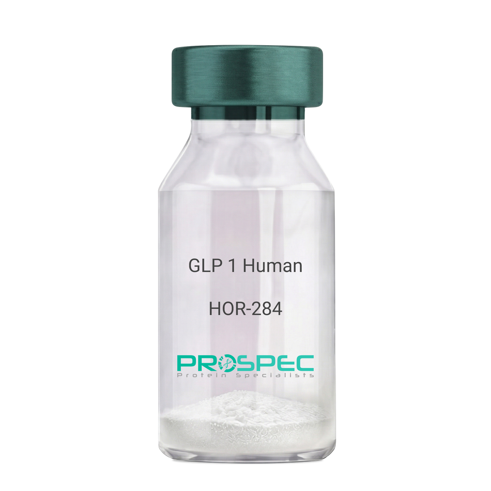
GLP 1 HumanDescription:
Human Glucagon Like Peptide-1
GLP1, Glucagon Like Peptide-1, Incretin hormone.
Product # :
HOR-284Price :
Quantity :
Shipping Method :
Shipped at Room temp
More Info
- description
- formulation
- purity
- More Info
Description
Glucagon Like Peptide-1 is a single, non-glycosylated, polypeptide chain containing 30 amino acids and having a molecular mass of 3297.7 Dalton.The GLP-1 is purified by proprietary chromatographic techniques.
Formulation
The GLP-1 peptide was lyophilized with no additives.
Purity
Greater than 96.0% as determined by RP-HPLC.
More Info
-
Introduction
Glucagon-like peptide-1 (GLP-1) is derived from the transcription product of the proglucagon gene. The major source of GLP-1 in the body is the intestinal L cell that secretes GLP-1 as a guthormone. The biologically active forms of GLP-1 are: GLP-1-(7-37) and GLP-1-(7-36)NH2.
GLP-1 secretion by L cells is dependent on the presence of nutrients in the lumen of the small intestine. The secretagogues (agents that causes or stimulates secretion) of this hormone include major nutrients like carbohydrate, proteinand lipid. Once in the circulation, GLP-1 has a half life of less than 2 minutes, due to rapid degradation by the enzyme dipeptidyl peptidase-4.
GLP-1 possesses several physiological properties that make it a subject of intensive investigation as a potential treatment of diabetes mellitus. The known physiological functions of GLP-1 include: Increases insulin secretion from the pancreas in a glucose-dependent manner, decreases glucagon secretion from the pancreas, increases beta cells mass and insulin gene expression, inhibits acid secretion and gastric emptying in the stomach, decreases food intake by increasing satiety. -
Synonyms
GLP1, Glucagon Like Peptide-1, Incretin hormone.
-
Physical Appearance
Sterile Filtered White lyophilized (freeze-dried) powder.
-
Stability
Lyophilized Glucagon Like Peptide-1 although stable at room temperature for 3 weeks, should be stored desiccated below -18°C. Upon reconstitution GLP-1 should be stored at 4°C between 2-7 days and for future use below -18°C. For long term storage it is recommended to add a carrier protein (0.1% HSA or BSA).Please prevent freeze-thaw cycles.
-
Solubility
It is recommended to reconstitute the lyophilized Glucagon Like Peptide-1 in sterile H2O at 0.5mg/ml, which can then be further diluted to other aqueous solutions.
-
Amino Acid Sequence
H-His-Ala-Glu-Gly-Thr-Phe-Thr-Ser-Asp-Val-Ser-Ser-Tyr-Leu-Glu-Gly-Gln-Ala-Ala-Lys-Glu-Phe-Ile-Ala-Trp-Leu-Val-Lys-Gly-Arg-NH2.
ProSpec's products are furnished for LABORATORY RESEARCH USE ONLY. The product may not be used as drugs, agricultural or pesticidal products, food additives or household chemicals.
- View Data Sheet
Name :
HEV ORF2 (403-461 a.a.)Description:
Hepatitis E Virus ORF2 (403-461 a.a.) Recombinant
Product # :
HEV-272Price :
Quantity :
Shipping Method :
Shipped with Ice Packs
More Info
- description
- formulation
- purity
- More Info
Description
The e.coli derived HEV protein is fused with beta-galactosidase at the N-Terminus and contains the HEV immunodominant regions from ORF2 403-461 a.a.
Formulation
20mM Tris-HCl pH-8, 8M urea and 10mM B-ME.
Purity
HEV ORF2 Protein is >95% pure as determined by 10% PAGE (coomassie staining).
More Info
-
Introduction
Hepatitis E virus (HEV), the major etiologic agent of enterically transmitted non-A, non-B hepatitis worldwide, is a spherical, non-enveloped, single stranded RNA virus that is approximately 32 to 34 nm in diameter. HEV belongs to a genus of HEV-like viruses (unassigned genus). HEV has a single-stranded polyadenylated RNA genome of approximately 8 kb. Based on its physicochemical properties it is presumed to be a calici-like virus.
-
Stability
HEV ORF2 protein although stable at 4°C for 1 week, should be stored below -18°C. Please prevent freeze thaw cycles.
-
Applications
HEV ORF2 antigen is suitable for ELISA and Western blots, excellent antigen for detection of HEV with minimal specificity problems.
-
Specificity
Immunoreactive with sera HEV-infected individuals.
-
Purification Method
HEV ORF2 was purified by proprietary chromatographic technique.
ProSpec's products are furnished for LABORATORY RESEARCH USE ONLY. The product may not be used as drugs, agricultural or pesticidal products, food additives or household chemicals.
- View Data Sheet
Name :
HEV ORF3Description:
Hepatitis E Virus ORF3 Recombinant
Product # :
HEV-273Price :
Quantity :
Shipping Method :
Shipped with Ice Packs
More Info
- description
- formulation
- purity
- More Info
Description
The e.coli derived HEV protein is fused with beta-galactosidase at the N-Terminus and contains the HEV immunodominant ORF3 92-123 a.a.
Formulation
20mM Tris-HCl, pH-8, 10mM B-ME and 8M urea.
Purity
Greater than 95.0% as determined by Analysis by SDS-PAGE.
More Info
-
Introduction
Hepatitis E virus (HEV), the major etiologic agent of enterically transmitted non-A, non-B hepatitis worldwide, is a spherical, non-enveloped, single stranded RNA virus that is approximately 32 to 34 nm in diameter. HEV belongs to a genus of HEV-like viruses (unassigned genus). HEV has a single-stranded polyadenylated RNA genome of approximately 8 kb. Based on its physicochemical properties it is presumed to be a calici-like virus.
-
Stability
HEV ORF3 protein although stable at 4°C for 1 week, should be stored below -18°C. Please prevent freeze thaw cycles.
-
Specificity
Immunoreactive with sera HEV-infected individuals.
-
Purification Method
HEV ORF3 protein was purified by proprietary chromatographic technique.
ProSpec's products are furnished for LABORATORY RESEARCH USE ONLY. The product may not be used as drugs, agricultural or pesticidal products, food additives or household chemicals.
- View Data Sheet
Name :
IL 27 HumanDescription:
Interleukin-27 Human Recombinant
Interleukin-30, IL-30, IL-27/p28, p28, Interleukin-27, Interleukin-27/p28, IL-27, Interleukin-27 subunit alpha, IL-27 subunit alpha, IL27-A, Il27, Il27a, IL-27p28.
Product # :
CYT-048Price :
Quantity :
Shipping Method :
Shipped at Room temp
More Info
- description
- source
- formulation
- purity
- biological activity
- More Info
Description
IL-27 Human Recombinant produced in HEK cells is a glycosylated non-covalently heterodimer that consists of 2 chains, IL27A (IL27p28) having a Mw of 25kDa and IL27B (EBI3) having a Mw of 30kDa. Each chain migrates separately on SDS-PAGE. The IL-27 protein is purified by proprietary chromatographic techniques
Source
HEK.
Formulation
The IL-27 was lyophilized from 1mg/ml in 1xPBS.
Purity
Greater than 90% as obsereved by SDS-PAGE.
Biological Activity
The specific activity was determined by the dose dependent inhibition of HIV replication in human CD4+ T cells and in human monocyte-derived macrophages and is typically 4-12ng/ml.More Info
-
Introduction
Interleukin-27 protein is related to IL-12A & is one of the subunits of a heterodimeric cytokine complex. IL-27 interacts with EBV induced gene 3 also called EBI3, and forms a complex that drives rapid expansion of CD4 (+) T cells. IL-27 complex is synergizes with IL-12 in order to trigger the cytokine production of IFN-Gamma of CD4 (+) T cells. The biological effect of IL-27 is mediated by class-I cytokine receptor (WSX1/TCRR). The pro-inflammatory activity of IL-27 is mediated through the growing expression of key molecules involved in the MHC class-I & MHC class-II pathways. Both MHC class-I and MHC-class-II expression are increased in endothelial cells after Interleukin-27 stimulation which suggests that it may play an important role in conferring the immune function on vascular endothelium IL-27p28 subunit can be induced by IFN-beta and during LPS-induced maturation of dendritic cells in type-I IFN-dependent manner through IFN regulatory factor-1 activation. Interleukin-27 regulates Interleukin-12 responsiveness of CD4+ T cells through Stat1-dependent and -independent mechanisms. IL-17 & IL-23 play an important IL-17 role in inflammation. Interleukin-27 possesses potent anti-angiogenic activity that plays an important role in its antitumor and antimetastatic activities. EBV induced gene 3 plays a role, independently from IL-27, in regulating anti-viral or anti-tumoral immune responses. Interleukin-27 is a potent inhibitor of HIV-1 replication in macrophages, CD4+ T cells, peripheral blood mononuclear cells. Interleukin-27 triggers STAT activation and gene transcription.
-
Synonyms
Interleukin-30, IL-30, IL-27/p28, p28, Interleukin-27, Interleukin-27/p28, IL-27, Interleukin-27 subunit alpha, IL-27 subunit alpha, IL27-A, Il27, Il27a, IL-27p28.
-
Physical Appearance
Sterile Filtered White lyophilized (freeze-dried) powder.
-
Stability
Lyophilized IL-27 although stable at room temperature for 3 weeks, should be stored desiccated below -18°C. Upon reconstitution IL27 should be stored at 4°C between 2-7 days and for future use below -18°C.For long term storage it is recommended to add a carrier protein (0.1% HSA or BSA).Please prevent freeze-thaw cycles.
-
Solubility
It is recommended to reconstitute the lyophilized IL-27 in sterile PBS containing 0.1% endotoxin-free recombinant HSA.
-
Amino Acid Sequence
IL-27A (215aa):
FPRPPGRPQL SLQELRREFT VSLHLARKLL AEVRGQAHRF AESHLPGVNL YLLPLGEQLP DVSLTFQAWR RLSDPERLCF ISTTLQPFHA LLGGLGTQGR WTNMERMQLW AMRLDLRDLQ RHLRFQVLAA GFNLPEEEEE EEEEEEEERK GLLPGALGSA LQGPAQVSWP QLLSTYRLLH SLELVLSRAV RELLLLSKAG HSVWPLGFPT LSPQP.
IL-27B (209aa):
RKGPPAALTL PRVQCRASRY PIAVDCSWTL PPAPNSTSPV SFIATYRLGM AARGHSWPCL QQTPTSTSCT ITDVQLFSMA PYVLNVTAVH PWGSSSSFVP FITEHIIKPD PPEGVRLSPL AERQLQVQWE PPGSWPFPEI FSLKYWIRYK RQGAARFHRV GPIEATSFIL RAVRPRARYY VQVAAQDLTD YGELSDWSLP ATATMSLGK.
ProSpec's products are furnished for LABORATORY RESEARCH USE ONLY. The product may not be used as drugs, agricultural or pesticidal products, food additives or household chemicals.
- View Data Sheet
Name :
IL 6 Rhesus MacaqueDescription:
Interleukin-6 Rhesus Macaque Recombinant
IFN-b2, B cell differentiation factor, BCDF, BSF-2, HPGF, HSF, MGI-2, B-cell stimulatory factor 2, IFN beta-2, Hybridoma growth factor, CTL differentiation factor, CDF, IL-6, HGF.
Product # :
CYT-174Price :
Quantity :
Shipping Method :
Shipped at Room temp
More Info
- description
- source
- formulation
- purity
- biological activity
- More Info
Description
IL 6 Rhesus Macaque Recombinant produced in E.Coli is a single, non-glycosylated, polypeptide chain containing 186 amino acids and having a molecular mass of 21.1kDa.The IL 6 Rhesus Macaque is purified by proprietary chromatographic techniques.
Source
Escherichia Coli.
Formulation
Lyophilized from a 0.2µm filtered concentrated solution in PBS, pH 7.4.
Purity
Greater than 97.0% as determined by SDS-PAGE and HPLC analyses.
Biological Activity
Fully biologically active when compared to standard. The ED50 determined by a cell proliferation assay using IL-6-dependent murine 7TD1 cells is less than 0.1ng/ml, corresponding to a specific activity of > 1.0 × 10,000,000 IU/mg.More Info
-
Introduction
Il-6 is a cytokine with a wide variety of biological functions: it plays an essential role in the final differentiation of b-cells into ig-secreting cells, it induces myeloma and plasmacytoma growth, it induces nerve cells differentiation, in hepatocytes it induces acute phase reactants.
-
Synonyms
IFN-b2, B cell differentiation factor, BCDF, BSF-2, HPGF, HSF, MGI-2, B-cell stimulatory factor 2, IFN beta-2, Hybridoma growth factor, CTL differentiation factor, CDF, IL-6, HGF.
-
Physical Appearance
Sterile Filtered White lyophilized (freeze-dried) powder.
-
Stability
Lyophilized IL-6 although stable at room temperature for 3 weeks, should be stored desiccated below -18°C. Upon reconstitution IL-6 should be stored at 4°C between 2-7 days and for future use below -18°C.Please prevent freeze-thaw cycles.
-
Solubility
It is recommended to reconstitute the lyophilized IL-6 in sterile 18M-cm H2O not less than 100µg/ml, which can then be further diluted to other aqueous solutions.
-
Amino Acid Sequence
MAPVLPGEDS KNVAAPHSQP LTSSERIDKH IRYILDGISA LRKETCNRSN MCESSKEALA ENNLNLPKMA EKDGCFQSGF NEDTCLVKII TGLLEFEVYL EYLQNRFESS EEQARAVQMS TKVLIQFLQK KAKNLDAITT PEPTTNASLL TKLQAQNQWL QDMTTHLILR SFKEFLQSNL RALRQM
-
Background
Research Paper on Interleukin-6 Rhesus Macaque Recombinant
Abstract:
Interleukin-6 (IL-6) Rhesus Macaque Recombinant holds a significant role in the realm of immunological exploration. This research paper delves into the molecular intricacies and implications of IL-6 Rhesus Macaque Recombinant in immune modulation. Through an investigation of its functions, synonyms including DIF, TNFA, and TNFSF2, and potential applications, we gain valuable insights into its contribution to understanding immune responses.
Introduction:
IL-6 Rhesus Macaque Recombinant is a key player in immunological studies. This paper aims to provide a comprehensive understanding of its molecular attributes and its impact on immune mechanisms.
Molecular Structure and Insights:
Examining the molecular composition of IL-6 Rhesus Macaque Recombinant, we uncover its role in immune signaling. Its interactions and functions contribute to the fine-tuning of immune responses.
Exploring Immune Dynamics:
IL-6's influence on immune cell activation and inflammation is well-acknowledged. IL-6 Rhesus Macaque Recombinant provides a tool to delve deeper into these immune processes, enriching our comprehension of cytokine-mediated functions.
Synonyms and Network Connections:
Understanding IL-6's synonyms, such as DIF, TNFA, and TNFSF2, adds depth to our understanding of immune signaling networks. IL-6 Rhesus Macaque Recombinant helps uncover the complexity of these interconnected pathways.
Potential Applications in Research and Beyond:
Beyond research settings, IL-6 Rhesus Macaque Recombinant holds potential in deciphering immune-related diseases. Its significance extends to possible therapeutic interventions and diagnostic applications.
Clinical Implications and Future Prospects:
The clinical relevance of IL-6 Rhesus Macaque Recombinant is underlined by its role in diseases characterized by altered IL-6 signaling. Exploring its therapeutic potential paves the way for novel strategies in disease management.
Conclusion:
In the realm of immunology, IL-6 Rhesus Macaque Recombinant stands as a pivotal tool. Its molecular insights, pivotal functions, and potential implications position it as a valuable asset for advancing our understanding of immune regulation.
ProSpec's products are furnished for LABORATORY RESEARCH USE ONLY. The product may not be used as drugs, agricultural or pesticidal products, food additives or household chemicals.
- View Data Sheet
Name :
HLA-DOB HumanDescription:
Major Histocompatibility Complex Class II DO Beta Human Recombinant
HLA class II histocompatibility antigen, DO beta chain, MHC class II antigen DOB, HLA-DOB, Major histocompatibility complex, class II, DO beta, DOB.
Product # :
PRO-1967Price :
Quantity :
Shipping Method :
Shipped with Ice Packs
More Info
- description
- source
- formulation
- purity
- More Info
Description
HLA-DOB Human Recombinant produced in E. coli is a single polypeptide chain containing 222 amino acids (27-224) and having a molecular mass of 25.2 kDa.HLA-DOB is fused to a 24 amino acid His-tag at N-terminus & purified by proprietary chromatographic techniques.
Source
E.coli.
Formulation
The HLA-DOB solution (1mg/1ml) contains 20mM Tris-HCl buffer (pH 8.0), 0.4M Urea and 10% glycerol.
Purity
Greater than 95% as determined by SDS-PAGE.
More Info
-
Introduction
Major Histocompatibility Complex Class II DO Beta (HLA-DOB) is a part of the HLA class II beta chain paralogues. This class II beta chain is a heterodimer which is located in intracellular vesicles and consists of an alpha (DOA) and a beta chain (DOB), both anchored in the membrane. HLA-DOB which interacts with the HLA-DM molecule in B-cells is a significant modulator in the HLA class II restricted antigen presentation pathway. Class II molecules are expressed in antigen presenting cells such as B lymphocytes, dendritic cells and macrophages.
-
Synonyms
HLA class II histocompatibility antigen, DO beta chain, MHC class II antigen DOB, HLA-DOB, Major histocompatibility complex, class II, DO beta, DOB.
-
Physical Appearance
Sterile Filtered colorless solution.
-
Stability
Store at 4°C if entire vial will be used within 2-4 weeks. Store, frozen at -20°C for longer periods of time. For long term storage it is recommended to add a carrier protein (0.1% HSA or BSA).Avoid multiple freeze-thaw cycles.
-
Amino Acid Sequence
MGSSHHHHHH SSGLVPRGSH MGSGTDSPED FVIQAKADCY FTNGTEKVQF VVRFIFNLEE YVRFDSDVGM FVALTKLGQP DAEQWNSRLD LLERSRQAVD GVCRHNYRLG APFTVGRKVQ PEVTVYPERT PLLHQHNLLH CSVTGFYPGD IKIKWFLNGQ EERAGVMSTG PIRNGDWTFQ TVVMLEMTPE LGHVYTCLVD HSSLLSPVSV EWRAQSEYSW RK.
ProSpec's products are furnished for LABORATORY RESEARCH USE ONLY. The product may not be used as drugs, agricultural or pesticidal products, food additives or household chemicals.
- View Data Sheet
Name :
HCV NS5 Genotype-3aDescription:
Hepatitis C Virus NS5 Genotype-3a Recombinant
Product # :
HCV-232Price :
Quantity :
Shipping Method :
Shipped with Ice Packs
More Info
- description
- formulation
- purity
- More Info
Description
The E.coli derived recombinant protein contains the HCV NS5 Genotype 3a immunodominant regions, amino acids 2212-2313. The protein is fused to a GST tag at N-terminus.
Formulation
1.5M urea, 25 mM Tris-HCl, pH-8, 0.2% Triton-X & 50% Glycerol.
Purity
HCV NS5 Genotype-3a protein is >95% pure as determined by 10% PAGE (coomassie staining).
More Info
-
Introduction
HCV is a small 50nm, enveloped, single-stranded, positive sense RNAvirus in the family Flaviviridae.
HCV has a high rate of replication with approximately one trillion particles produced each day in an infected individual. Due to lack of proofreading by the HCV RNA polymerase, the HCV has an exceptionally high mutation rate, a factor that may help it elude the host's immune response. Hepatitis C virus is classified into six genotypes(1-6) with several subtypes within each genotype. The preponderance and distribution of HCV genotypes varies globally. Genotype is clinically important in determining potential response to interferon-based therapy and the required duration of such therapy. Genotypes 1 and 4 are less responsive to interferon-based treatment than are the other genotypes (2, 3, 5 and 6). -
Stability
HCV NS5 Genotype-3a although stable at 4°C for 1 week, should be stored below -18°C. Please prevent freeze thaw cycles.
-
Applications
HCV NS5 Genotype-3 antigen is suitable for ELISA and Western blots, excellent antigen for detection of HCV with minimal specificity problems.
-
Specificity
Immunoreactive with sera of HCV-infected individuals.
-
Purification Method
HCV NS5 Genotype-3a protein was purified by proprietary chromatographic technique.
ProSpec's products are furnished for LABORATORY RESEARCH USE ONLY. The product may not be used as drugs, agricultural or pesticidal products, food additives or household chemicals.
- View Data Sheet
Name :
IFNG RatDescription:
IFN-Gamma Rat Recombinant
Immune IFN, type II IFN, T cell IFN, MAF, IFNG, IFG, IFI, IFN-gamma.
Product # :
CYT-359Price :
Quantity :
Shipping Method :
Shipped at Room temp
More Info
- description
- source
- formulation
- purity
- biological activity
- More Info
Description
IFN-gamma Rat Recombinant produced in E.Coli is a single, non-glycosylated, polypeptide chain containing 135 amino acids and having a molecular mass of 15609 Dalton.The IFN-gamma is purified by proprietary chromatographic techniques.
Source
Escherichia Coli.
Formulation
Lyophilized from a 0.2μm filtered concentrated (1mg/ml) solution in PBS, pH 7.4.
Purity
Greater than 95.0% as determined by:
(a) Analysis by RP-HPLC.
(b) Analysis by SDS-PAGE.Biological Activity
The specific activity as determined by the cytopathic affect inhibition assay with murine L929 cells chalenged with EMC virus was < 0.1 ng/ml, corresponding to a specific activity of 10,000,000units/mg.More Info
-
Introduction
IFN-gamma produced by lymphocytes activated by specific antigens or mitogens.
IFN-gamma, in addition to having antiviral activity, has important immunoregulatory functions, it is a potent activator of macrophages, and has antiproliferative effects on transformed cells and it can potentiate the antiviral and antitumor effects of the type I IFNs. -
Synonyms
Immune IFN, type II IFN, T cell IFN, MAF, IFNG, IFG, IFI, IFN-gamma.
-
Physical Appearance
Sterile Filtered White lyophilized (freeze-dried) powder.
-
Stability
Lyophilized IFN-gamma although stable at room temperature for 3 weeks, should be stored desiccated below -18°C. Upon reconstitution IFN-gamma should be stored at 4°C between 2-7 days and for future use below -18°C.For long term storage it is recommended to add a carrier protein (0.1% HSA or BSA).Please prevent freeze-thaw cycles.
-
Solubility
It is recommended to reconstitute the lyophilized IFN-gamma in sterile 18MΩ-cm H2O not less than 100µg/ml, which can then be further diluted to other aqueous solutions.
-
Amino Acid Sequence
The sequence of the first five N-terminal amino acids was determined and was found to be Met-Gln-Gly-Tyr-Leu.
-
Background
What is the molecular weight/Mw of IFNG RAT Protein?
IFNG RAT Protein has a total Mw of 15.6kDa.
What is the source or expression system of IFNG RAT Protein?
Escherichia Coli.
What is the Purity of IFNG RAT Protein?
IFNG RAT Protein is >95% pure as determined by SDS-PAGE.
What is the Biological Activity of IFNG RAT Protein?
The specific activity as determined by the cytopathic affect inhibition assay with murine L929 cells chalenged with EMC virus was < 0.1 ng/ml, corresponding to a specific activity of 10,000,000units/mg.
What is the amino acid sequence of IFNG RAT Protein?
he sequence of the first five N-terminal amino acids was determined and was found to be Met-Gln-Gly-Tyr-Leu.
What applications can IFNG RAT Protein be used in?
IFNG RAT Protein can probably be used in western blot, ELISA and Lateral Flow.
What is the endotoxin level for IFNG RAT Protein?
The endotoxin level is minimal, IFNG RAT Protein was purified using conventional chromatography techniques.
ProSpec's products are furnished for LABORATORY RESEARCH USE ONLY. The product may not be used as drugs, agricultural or pesticidal products, food additives or household chemicals.
- View Data Sheet
Name :
Borrelia NapADescription:
Borrelia Burgdorferi Neutrophil Activating Protein A Recombinant
Product # :
BOR-014Price :
Quantity :
Shipping Method :
Shipped with Ice Packs
More Info
- description
- source
- formulation
- purity
- More Info
Description
Recombinant Borrelia Burgdorferi NapA produced in E.coli is a non-glycosylated, full-length polypeptide chain having a calculated molecular mass of 21 kDa. Borrelia NapA is expressed with a -6x His tag at N-terminus and purified by proprietary chromatographic techniques.
Source
Escherichia Coli.
Formulation
Borrelia NapA is supplied in 16mM HEPES buffer pH-7.8, 400mM NaCl and 20% glycerol.
Purity
Greater than 95.0% as determined by SDS-PAGE.
More Info
-
Introduction
Borrelia belongs to a genus of bacteria of the spirochete phylum. Borrelia causes borreliosis, which is a zoonotic, vector-borne disease transmitted mainly by ticks and some by lice, depending on the species. Of the 36 known species of Borrelia, 12 are distinguished to cause Lyme disease or borreliosis and are transmitted by ticks. The main Borrelia species causing Lyme disease are Borrelia burgdorferi, Borrelia afzelii, and Borrelia garinii. The Borrelia genus members have a linear chromosome which is about 900 kbp in length as well as an excess of both linear and circular plasmids in the 5-220 kbp size range. The plasmids are atypical, as compared to most bacterial plasmids, since they contain many paralogous sequences, a large number of pseudogenes and, in some cases, essential genes. Moreover, a number of the plasmids have features suggesting that they are prophages.
-
Physical Appearance
Sterile Filtered clear solution.
-
Stability
Store at 4°C if entire vial will be used within 2-4 weeks. Store, frozen at -20°C for longer periods of time. Avoid multiple freeze-thaw cycles.
ProSpec's products are furnished for LABORATORY RESEARCH USE ONLY. The product may not be used as drugs, agricultural or pesticidal products, food additives or household chemicals.
- View Data Sheet
Name :
FLT1 Human, HisDescription:
Vascular Endothelial Growth Factor receptor-1 Human Recombinant, His Tag
FLT-1, FLT1, Tyrosine-protein kinase receptor FLT, Flt-1, Tyrosine-protein kinase FRT, Fms-like tyrosine kinase 1, VEGFR-1.
Product # :
PKA-354Price :
Quantity :
Shipping Method :
Shipped with Ice Packs
More Info
- description
- source
- formulation
- purity
- More Info
Description
FLT1 Human Recombinant produced in E.Coli is single, a non-glycosylated, Polypeptide chain containing 298 amino acids fragment (31-328) corresponding to the IgG like domains 1-3 from the mature soluble FLT1 protein, having a total molecular mass of 43kDa and fused with a 4.5kDa amino-terminal hexahistidine tag. The FLT1 His is purified by proprietary chromatographic techniques.
Source
Escherichia Coli.
Formulation
FLT1 His-Tag protein is supplied in 1x PBS and 50% glycerol.
Purity
Greater than 95.0% as determined by SDS-PAGE.
More Info
-
Introduction
Endothelial cells express three different vascular endothelial growth factor (VEGF) receptors, belonging to the family of receptor tyrosine kinases (RTKs). They are named VEGFR-1 (Flt-1), VEGFR-2 (KDR/Flk-1), VEGFR-3 (Flt-4). Their expression is almost exclusively restricted to endothelial cells, but VEGFR-1 can also be found on monocytes, dendritic cells and on trophoblast cells. The flt-1 gene was first described in 1990. The receptor contains seven immunoglobulin-like extracellular domains, a single transmembrane region and an intracellular splited tyrosine kinase domain. Compared to VEGFR-2 the Flt-1 receptor has a higher affinity for VEGF but a weaker signaling activity. VEGFR-1 thus leads not to proliferation of endothelial cells, but mediates signals for differentiation. Interestingly a naturally occuring soluble variant of VEGFR-1 (sVEGFR-1) was found in HUVE supernatants in 1996, which is generated by alternative splicing of the flt-1 mRNA. The biological functions of sVEGFR-1 still are not clear, but it seems to be an endogenous regulator of angiogenesis, binding VEGF with the same affinity as the full-length receptor.
-
Synonyms
FLT-1, FLT1, Tyrosine-protein kinase receptor FLT, Flt-1, Tyrosine-protein kinase FRT, Fms-like tyrosine kinase 1, VEGFR-1.
-
Physical Appearance
Sterile Filtered clear solution.
-
Stability
Store at 4°C if entire vial will be used within 2-4 weeks. Store, frozen at -20°C for longer periods of time. Please avoid freeze thaw cycles.
ProSpec's products are furnished for LABORATORY RESEARCH USE ONLY. The product may not be used as drugs, agricultural or pesticidal products, food additives or household chemicals.
- View Data Sheet
Name :
LGMN MouseDescription:
Legumain Mouse Recombinant
Legumain, Asparaginyl endopeptidase, Protease, cysteine 1.
Product # :
ENZ-933Price :
Quantity :
Shipping Method :
Shipped with Ice Packs
More Info
- description
- source
- formulation
- purity
- More Info
- sds-page
Description
LGMN produced in Sf9 Baculovirus cells is a single, glycosylated polypeptide chain containing 426 amino acids (18-435a.a.) and having a molecular mass of 48.6kDa. LGMN is expressed with an 8 amino acid His tag at C-Terminus and purified by proprietary chromatographic techniques.
Source
Sf9, Baculovirus cells.
Formulation
LGMN protein solution (0.5mg/ml) contains Phosphate Buffered Saline (pH 7.4) and 10% glycerol.
Purity
Greater than 90.0% as determined by SDS-PAGE.
sds-page
More Info
-
Introduction
Legumain, also known as LGMN is a lysosomal cysteine protease which is found in all mouse tissues, however it was mainly abundant in the kidney as well as placenta. LGMN plays an essential role in the endosomal/lysosomal degradation system as the Legumain deficiency causes the accumulation of pro cathepsins B, H & L, another group of lysosomal cysteine proteases. Furthermore, over expression of LGMN in tumors is important for invasion/metastasis.
-
Synonyms
Legumain, Asparaginyl endopeptidase, Protease, cysteine 1.
-
Physical Appearance
Sterile filtered colorless solution.
-
Stability
Store at 4°C if entire vial will be used within 2-4 weeks.Store, frozen at -20°C for longer periods of time. For long term storage it is recommended to add a carrier protein (0.1% HSA or BSA).Avoid multiple freeze-thaw cycles.
-
Amino Acid Sequence
VPVGVDDPED GGKHWVVIVA GSNGWYNYRH QADACHAYQI IHRNGIPDEQ IIVMMYDDIA NSEENPTPGV VINRPNGTDV YKGVLKDYTG EDVTPENFLA VLRGDAEAVK GKGSGKVLKS GPRDHVFIYF TDHGATGILV FPNDDLHVKD LNKTIRYMYE HKMYQKMVFY IEACESGSMM NHLPDDINVY ATTAANPKES SYACYYDEER GTYLGDWYSV NWMEDSDVED LTKETLHKQY HLVKSHTNTS HVMQYGNKSI STMKVMQFQG MKHRASSPIS LPPVTHLDLT PSPDVPLTIL KRKLLRTNDV KESQNLIGQI QQFLDARHVI EKSVHKIVSL LAGFGETAER HLSERTMLTA HDCYQEAVTH FRTHCFNWHS VTYEHALRYL YVLANLCEAP YPIDRIEMAM DKVCLSHYLE HHHHHH.
ProSpec's products are furnished for LABORATORY RESEARCH USE ONLY. The product may not be used as drugs, agricultural or pesticidal products, food additives or household chemicals.
- View Data Sheet
Name :
CSNK2B HumanDescription:
Casein Kinase 2 beta Human Recombinant
PKCK2 Beta, PKCK2B, CK2N, CSK2B, MGC138222, MGC138224, G5A, Phosvitin, Casein Kinase 2 beta, Casein Kinase 2B, Casein Kinase 2 beta, Casein kinase 2 beta polypeptide, Casein kinase II subunit beta, CK II beta, CK2B, CSNK 2B, CSNK2B, G5A
Product # :
PKA-129Price :
Quantity :
Shipping Method :
Shipped with Ice Packs
More Info
- description
- source
- formulation
- purity
- More Info
Description
CSNK2B Human Recombinant produced in E.Coli is a single, non-glycosylated polypeptide chain containing 215 amino acids (1-215 a.a) and having a molecular mass of 24.9 kDa.CSNK2B is purified by proprietary chromatographic techniques.
Source
Escherichia Coli.
Formulation
CSNK2B protein solution (1mg/ml) in 0.2M NaCl, 20mM Tris-HCl buffer (pH 8.0), 1mM EDTA, 1mM DTT and 10% glycerol.
Purity
Greater than 95.0% as determined by SDS-PAGE.
More Info
-
Introduction
Casein Kinase 2 beta (CSNK2B) is a ubiquitous Ser/Thr kinase expressed in all eukaryotes. CSNK2Bis atetramer comprised of 2 catalytic kinase domains, alpha subunits and 2 identical regulatory beta subunits.CSNK2Btakes part incell cycle control, DNA repair,regulation of the circadian rhythm and other cellularprocesses. The beta subunit itself confers stability to the CK2 alpha subunitand is involved in activity and substrate specificity but does not have kinase activity.
-
Synonyms
PKCK2 Beta, PKCK2B, CK2N, CSK2B, MGC138222, MGC138224, G5A, Phosvitin, Casein Kinase 2 beta, Casein Kinase 2B, Casein Kinase 2 beta, Casein kinase 2 beta polypeptide, Casein kinase II subunit beta, CK II beta, CK2B, CSNK 2B, CSNK2B, G5A
-
Physical Appearance
Sterile Filtered colorless solution.
-
Stability
Store at 4°C if entire vial will be used within 2-4 weeks.Store, frozen at -20°C for longer periods of time.For long term storage it is recommended to add a carrier protein (0.1% HSA or BSA).Avoid multiple freeze-thaw cycles.
-
Amino Acid Sequence
MSSSEEVSWI SWFCGLRGNE FFCEVDEDYI QDKFNLTGLN EQVPHYRQAL DMILDLEPDE ELEDNPNQSD LIEQAAEMLY GLIHARYILT NRGIAQMLEK YQQGDFGYCP RVYCENQPML PIGLSDIPGE AMVKLYCPKC MDVYTPKSSR HHHTDGAYFG TGFPHMLFMV HPEYRPKRPA NQFVPRLYGF KIHPMAYQLQ LQAASNFKSP VKTIR
ProSpec's products are furnished for LABORATORY RESEARCH USE ONLY. The product may not be used as drugs, agricultural or pesticidal products, food additives or household chemicals.
- View Data Sheet
Name :
HSP27 Human, HisDescription:
Heat Shock Protein 27 Human Recombinant, His Tag
HSPB1, CMT2F, HMN2B, HSP27, HSP28, HSP25, Heat shock protein beta-1, Heat shock 27 kDa protein, Stress-responsive protein 27, SRP27, HS.76067, DKFZp586P1322.
Product # :
HSP-029Price :
Quantity :
Shipping Method :
Shipped with Ice Packs
More Info
- description
- source
- formulation
- purity
- More Info
Description
Recombinant Human HSP-27 produced in E.Coli is a single, non-glycosylated polypeptide chain containing 225 amino acids (1-205 a.a.) and having a molecular mass of 24.9kDa. Hsp27 is fused to a 20 amino acid his tag at N-terminus and purified by conventional chromatography.
Source
Escherichia Coli.
Formulation
The HSP27 protein solution contains 20mM Tris-Hcl buffer, 1mM DTT and 10% glycerol.
Purity
Greater than 95.0% as determined by SDS-PAGE.
More Info
-
Introduction
HSP27 (also known as the Estrogen-Regulated 24K protein, and hsp 28) is a member of the mammalian small heat shock protein family. Hsp27 is expressed constitutively in many tissues and its expression is increased to high levels after various types of stress including elevated temperatures, toxic metals, drugs and oxidants. Also, Hsp27 is phosphorylated in vivo on three phosphorylation sites (Ser15, Ser78 and Ser82) by protein kinases including MAPKAP kinase 2 and the stress-activated protein kinase SAPK2 (p38).
-
Synonyms
HSPB1, CMT2F, HMN2B, HSP27, HSP28, HSP25, Heat shock protein beta-1, Heat shock 27 kDa protein, Stress-responsive protein 27, SRP27, HS.76067, DKFZp586P1322.
-
Physical Appearance
Sterile Filtered colorless solution.
-
Stability
Store at 4°C if entire vial will be used within 2-4 weeks. Store, frozen at -20°C for longer periods of time. For long term storage it is recommended to add a carrier protein (0.1% HSA or BSA).Avoid multiple freeze-thaw cycles.
-
Amino Acid Sequence
MGSSHHHHHH SSGLVPRGSH MTERRVPFSL LRGPSWDPFR DWYPHSRLFD QAFGLPRLPE EWSQWLGGSS WPGYVRPLPP AAIESPAVAA PAYSRALSRQ LSSGVSEIRH TADRWRVSLD VNHFAPDELT VKTKDGVVEI TGKHEERQDE HGYISRCFTR KYTLPPGVDP TQVSSSLSPE GTLTVEAPMP KLATQSNEIT IPVTFESRAQ LGGPEAAKSD ETAAK.
ProSpec's products are furnished for LABORATORY RESEARCH USE ONLY. The product may not be used as drugs, agricultural or pesticidal products, food additives or household chemicals.
- View Data Sheet
Name :
PDPN P56F7AT AntibodyDescription:
Podoplanin clone P56F7AT, Mouse Anti Human
Podoplanin, Glycoprotein 36, PA2.26 antigen, T1A, GP36, GP40, Gp38, OTS8, T1A2, HT1A-1, PA2.26, T1-alpha, PDPN.
Product # :
ANT-641Price :
Quantity :
Shipping Method :
Shipped with Ice Packs
More Info
- formulation
- More Info
Formulation
1mg/ml containing PBS, pH-7.4, 10% Glycerol and 0.02% Sodium Azide.
More Info
-
Introduction
Podoplanin is a small mucin-like type-1 transmembrane protein, typically expressed in various specialized cell types throughout the body. Podoplanin is a type-I integral membrane glycoprotein with diverse distribution in human tissues. PDPN physiological function is related to its mucin-type character. The homologous protein in other species has been described as a differentiation antigen and influenza-virus receptor.
PDPN is expressed in lymphatic progenitor cells and afterwards during mouse development in lymphatic endothelial cells. Podoplanin is a specific marker for lymph vessel endothelial cells. Over-expression of podoplanin significantly elevates endothelial cell adhesion, migration, and tube formation. Inhibition of Podoplanin expression decreases cell adhesion in human dermal lymphatic endothelial cells. Podoplanin is used as a specific marker for lymphatic endothelium in histopathology.
Podoplanin expression is increased in nearly all human colon, rectum, and small intestine tumors. AGGRUS may serve as a diagnostic marker that distinguishes seminomas, the majority of which over express the protein, from embryonal carcinoma in testicular germ cell tumors. -
Synonyms
Podoplanin, Glycoprotein 36, PA2.26 antigen, T1A, GP36, GP40, Gp38, OTS8, T1A2, HT1A-1, PA2.26, T1-alpha, PDPN.
-
Physical Appearance
Sterile filtered colorless solution.
-
Immunogen
Anti-human PDPN mAb, is derived from hybridization of mouse F0 myeloma cells with spleen cells from BALB/c mice immunized with recombinant human PDPN amino acids 99-207 purified from E. coli.
-
Ig Subclass
Mouse IgG1 heavy chain and k light chain.
-
Clone
P56F7AT.
-
Applications
PDPN antibody has been tested by ELISA, Western blot analysis, Flow cytometry and ICC/IF to assure specificity and reactivity. Since application varies, however, each investigation should be titrated by the reagent to obtain optimal results.
-
Type
Mouse Anti Human Monoclonal.
-
Storage Procedures
For periods up to 1 month store at 4°C, for longer periods of time, store at -20°C. Prevent freeze thaw cycles.
-
Purification Method
PDPN antibody was purified from mouse ascitic fluids by protein-A affinity chromatography.
ProSpec's products are furnished for LABORATORY RESEARCH USE ONLY. The product may not be used as drugs, agricultural or pesticidal products, food additives or household chemicals.
- View Data Sheet
Name :
UNG AntibodyDescription:
Uracil DNA Glycosilase, Mouse Anti Human
Uracil DNA Glycosilase, Uracil DNA Glycosylase, UNG.
Product # :
ANT-374Price :
Quantity :
Shipping Method :
Shipped with Ice Packs
More Info
- formulation
- More Info
Formulation
1mg/ml containing PBS, pH-7.4, & 0.1% Sodium Azide.
More Info
-
Introduction
E.Coli Uracil DNA Glycosilase (UNG) catalyses the release of free Uracil from Uracil-containing DNA. UNG efficiently hydrolyzes uracil from signle-stranded or double-stranded DNA, but not from oligomers (6 fewer bases).
-
Synonyms
Uracil DNA Glycosilase, Uracil DNA Glycosylase, UNG.
-
Physical Appearance
Sterile Filtered clear solution.
-
Immunogen
Anti-human UNG mAb is derived from hybridization of mouse SP2/O myeloma cells with spleen cells from BALB/c mice immunized with recombinant human UNG amino acids 1-313 purified from E. coli.
-
Ig Subclass
Mouse IgG2b heavy chain and κ light chain.
-
Clone
Pk1C12AT.
-
Applications
UNG antibody has been tested by ELISA and Western blot analysis to assure specificity and reactivity. Since application varies, however, each investigation should be titrated by the reagent to obtain optimal results. Recommended dilution range for Western blot analysis is 1:1,000 ~ 2,000. Recommended starting dilution is 1:1,000.
-
Type
Mouse Anti Human Monoclonal.
-
Storage Procedures
For periods up to 1 month store at 4°C, for longer periods of time, store at -20°C. Prevent freeze thaw cycles.
-
Purification Method
UNG antibody was purified from mouse ascitic fluids by protein-G affinity chromatography.
ProSpec's products are furnished for LABORATORY RESEARCH USE ONLY. The product may not be used as drugs, agricultural or pesticidal products, food additives or household chemicals.
- View Data Sheet
Name :
NUDT21 HumanDescription:
Nudix Type Motif 21 Human Recombinant
Cleavage and polyadenylation specificity factor subunit 5, Cleavage and polyadenylation specificity factor 25 kDa subunit, CFIm25, CPSF 25 kDa subunit, Nucleoside diphosphate-linked moiety X motif 21, Nudix motif 21, Pre-mRNA cleavage factor Im 25 kDa subunit, NUDT21, CFIM25, CPSF25, CPSF5, DKFZp686H1588.
Product # :
ENZ-080Price :
Quantity :
Shipping Method :
Shipped with Ice Packs
More Info
- description
- source
- formulation
- purity
- More Info
Description
NUDT21 Human Recombinant fused with a 20 amino acid His tag at N-terminus produced in E.Coli is a single, non-glycosylated, polypeptide chain containing 247 amino acids (1-227 a.a.) and having a molecular mass of 28.3kDa. The NUDT21 is purified by proprietary chromatographic techniques.
Source
Escherichia Coli.
Formulation
The NUDT21 solution (1 mg/ml) contains 20mM Tris-HCl buffer (pH8.0), 0.2M NaCl, 2mM DTT and 20% glycerol.
Purity
Greater than 90.0% as determined by SDS-PAGE.
More Info
-
Introduction
NUDT21 belongs to the Nudix hydrolase family of pyrophosphatases. NUDT21 localizes to the paraspeckles and forms a heterodimer with CPSF6 or CPSF7 to comprise the CFIm (mammalian cleavage factor I) complex. NUDT21 being the smaller subunit of the complex, is present in all heterodimer combinations. NUDT21 has a vital role in pre-mRNA 3' cleavage and polyadenylation processing.
-
Synonyms
Cleavage and polyadenylation specificity factor subunit 5, Cleavage and polyadenylation specificity factor 25 kDa subunit, CFIm25, CPSF 25 kDa subunit, Nucleoside diphosphate-linked moiety X motif 21, Nudix motif 21, Pre-mRNA cleavage factor Im 25 kDa subunit, NUDT21, CFIM25, CPSF25, CPSF5, DKFZp686H1588.
-
Physical Appearance
Sterile Filtered colorless solution.
-
Stability
Store at 4°C if entire vial will be used within 2-4 weeks. Store, frozen at -20°C for longer periods of time. For long term storage it is recommended to add a carrier protein (0.1% HSA or BSA).Avoid multiple freeze-thaw cycles.
-
Amino Acid Sequence
MGSSHHHHHH SSGLVPRGSH MSVVPPNRSQ TGWPRGVTQF GNKYIQQTKP LTLERTINLY PLTNYTFGTK EPLYEKDSSV AARFQRMREE FDKIGMRRTV EGVLIVHEHR LPHVLLLQLG TTFFKLPGGE LNPGEDEVEG LKRLMTEILG RQDGVLQDWV IDDCIGNWWR PNFEPPQYPY IPAHITKPKE HKKLFLVQLQ EKALFAVPKN YKLVAAPLFE LYDNAPGYGP IISSLPQLLS RFNFIYN.
ProSpec's products are furnished for LABORATORY RESEARCH USE ONLY. The product may not be used as drugs, agricultural or pesticidal products, food additives or household chemicals.
- View Data Sheet
Name :
BTF3L4 HumanDescription:
Basic Transcription Factor 3-Like 4 Human Recombinant
Basic transcription factor 3-like 4, BTF3L4.
Product # :
PRO-1870Price :
Quantity :
Shipping Method :
Shipped with Ice Packs
More Info
- description
- source
- formulation
- purity
- More Info
Description
BTF3L4 Human Recombinant produced in E.Coli is a single, non-glycosylated polypeptide chain containing 101 amino acids (1-78 a.a) and having a molecular mass of 11.3kDa. BTF3L4 is fused to a 23 amino acid His-tag at N-terminus & purified by proprietary chromatographic techniques.
Source
Escherichia Coli.
Formulation
BTF3L4 protein solution (1mg/ml) containing 20mM Tris-HCl buffer (pH 8.0), 10% glycerol and 0.4M Urea.
Purity
Greater than 85.0% as determined by SDS-PAGE.
More Info
-
Introduction
BTF3L4 is a part of the NAC-beta family which contains 1 NAC-A/B (NAC-alpha/beta) domain and required for the initiation of transcription. BTF3L4 binds to polypeptide chains as they emerge from the ribosome and prevents their interaction with the SRP, which usually targets nascent secretory peptides to the ER. BTF3 is also a general transcription factor which forms a stable complex with RNA polymerase II.
-
Synonyms
Basic transcription factor 3-like 4, BTF3L4.
-
Physical Appearance
Sterile Filtered colorless solution.
-
Stability
Store at 4°C if entire vial will be used within 2-4 weeks. Store, frozen at -20°C for longer periods of time. For long term storage it is recommended to add a carrier protein (0.1% HSA or BSA).Avoid multiple freeze-thaw cycles.
-
Amino Acid Sequence
MGSSHHHHHH SSGLVPRGSH MGSMNQEKLA KLQAQVRIGG KGTARRKKKV VHRTATADDK KLQSSLKKLA VNNIAGIEES WTVKHQNQKT LMRKMMMFQI L.
ProSpec's products are furnished for LABORATORY RESEARCH USE ONLY. The product may not be used as drugs, agricultural or pesticidal products, food additives or household chemicals.
- View Data Sheet
Name :
ULBP2 HumanDescription:
UL16 Binding Protein 2 Human Recombinant
UL16 binding protein 2, retinoic acid early transcript 1 H, NKG2D ligand 2, Retinoic acid early transcript 1H, ALCAN-alpha, N2DL-2, RAET1H.
Product # :
PRO-1134Price :
Quantity :
Shipping Method :
Shipped with Ice Packs
More Info
- description
- source
- formulation
- purity
- More Info
Description
ULBP2 Human Recombinant produced in E. coli is a single polypeptide chain containing 216 amino acids (26-216) and having a molecular mass of 24.3 kDa.ULBP2 is fused to a 25 amino acid His-tag at N-terminus & purified by proprietary chromatographic techniques.
Source
E.coli.
Formulation
The ULBP2 solution (1mg/ml) contains 20mM Tris-HCl buffer (pH 8.0), 2M urea, 0.2M NaCl, 2mM DTT and 30% glycerol.
Purity
Greater than 85% as determined by SDS-PAGE.
More Info
-
Introduction
ULBP2 is a member of the MHC class I family. ULBP2 is ligand for the NKG2D receptor, composed with at least ULBP1 and ULBP3. ULBPs promote multiple signaling pathways in primary NK cells, triggering the production of cytokines and chemokines. Binding of ULBPs ligands to NKG2D encourages calcium mobilization and activation of the JAK2, STAT5, ERK and PI3K kinase/Akt signal transduction pathway. In CMV infected cells, ULBP2 cooperates with soluble CMV glycoprotein UL16. This cooperation is blocked with the NKG2D receptor, providing a mechanism in which CMV infected cells can escape the immune system. Additionally, UL16 causes ULBP2 to be held in the ER and cis-Golgi apparatus so that it does not reach the cell surface.
-
Synonyms
UL16 binding protein 2, retinoic acid early transcript 1 H, NKG2D ligand 2, Retinoic acid early transcript 1H, ALCAN-alpha, N2DL-2, RAET1H.
-
Physical Appearance
Sterile Filtered colorless solution.
-
Stability
Store at 4°C if entire vial will be used within 2-4 weeks. Store, frozen at -20°C for longer periods of time. For long term storage it is recommended to add a carrier protein (0.1% HSA or BSA).Avoid multiple freeze-thaw cycles.
-
Amino Acid Sequence
MGSSHHHHHH SSGLVPRGSH MGSHMGRADP HSLCYDITVI PKFRPGPRWC AVQGQVDEKT FLHYDCGNKT VTPVSPLGKK LNVTTAWKAQ NPVLREVVDI LTEQLRDIQL ENYTPKEPLT LQARMSCEQK AEGHSSGSWQ FSFDGQIFLL FDSEKRMWTT VHPGARKMKE KWENDKVVAM SFHYFSMGDC IGWLEDFLMG MDSTLEPSAG APLAMS
ProSpec's products are furnished for LABORATORY RESEARCH USE ONLY. The product may not be used as drugs, agricultural or pesticidal products, food additives or household chemicals.