Search results
1000 results found for “Syntaxin”
Name
Description
Product #
Price
Quantity
Shipping Method
- View Data Sheet
Name :
CLTB HumanDescription:
Clathrin, Light Chain B Human Recombinant
Clathrin light chain B, Lcb, CLTB.
Product # :
PRO-1172Price :
Quantity :
Shipping Method :
Shipped with Ice Packs
More Info
- description
- source
- formulation
- purity
- More Info
Description
CLTB Human Recombinant produced in E.Coli is a single, non-glycosylated polypeptide chain containing 234 amino acids (1-211 a.a) and having a molecular mass of 25.6kDa (Molecular size on SDS-PAGE will appear higher).CLTB is fused to a 23 amino acid His-tag at N-terminus & purified by proprietary chromatographic techniques.
Source
Escherichia Coli.
Formulation
CLTB protein solution (1mg/ml) containing 20mM Tris-HCl buffer, pH8.0, 10% glycerol, 0.2mM PMSF and 100mM NaCl.
Purity
Greater than 85.0% as determined by SDS-PAGE.
More Info
-
Introduction
Clathrin light chain B (CLTB) is a member of the clathrin light chain family. Clathrin is a protein that has a key role in the formation of coated vesicles. CLTB functions as the key structural component of the lattice-type cytoplasmic face of coated pits and vesicles which capture certain macromolecules during receptor-mediated endocytosis. CLTB forms a triskelion form comprised of 3 clathrin heavy chains and 3 light chains. When the triskelia interact they form a polyhedral pattern which encircles the vesicle. CME (Clathrin-mediated endocytosis) controls numerous cellular physiological processes such as the internalization of growth factors and receptors, entry of pathogens, and synaptic transmission.
-
Synonyms
Clathrin light chain B, Lcb, CLTB.
-
Physical Appearance
Sterile Filtered colorless solution.
-
Stability
Store at 4°C if entire vial will be used within 2-4 weeks. Store, frozen at -20°C for longer periods of time. For long term storage it is recommended to add a carrier protein (0.1% HSA or BSA).Avoid multiple freeze-thaw cycles.
-
Amino Acid Sequence
MGSSHHHHHH SSGLVPRGSH MGSMADDFGF FSSSESGAPE AAEEDPAAAF LAQQESEIAG IENDEGFGAP AGSHAAPAQP GPTSGAGSED MGTTVNGDVF QEANGPADGY AAIAQADRLT QEPESIRKWR EEQRKRLQEL DAASKVTEQE WREKAKKDLE EWNQRQSEQV EKNKINNRAS
EEAFVKESKE ETPGTEWEKV AQLCDFNPKS SKQCKDVSRL RSVLMSLKQT PLSR.
ProSpec's products are furnished for LABORATORY RESEARCH USE ONLY. The product may not be used as drugs, agricultural or pesticidal products, food additives or household chemicals.
- View Data Sheet
Name :
GSTA1 MouseDescription:
Glutathione S-Transferase Alpha 1 Mouse Recombinant
Glutathione S-transferase A1, GST class-alpha member 1, Glutathione S-transferase Ya , Glutathione S-transferase Ya1.
Product # :
ENZ-873Price :
Quantity :
Shipping Method :
Shipped with Ice Packs
More Info
- description
- source
- formulation
- purity
- biological activity
- More Info
Description
GSTA1 Mouse Recombinant produced in E.Coli is a single, non-glycosylated polypeptide chain containing 246 amino acids (1-223 a.a) and having a molecular mass of 28kDa.GSTA1 is fused to a 23 amino acid His-tag at N-terminus & purified by proprietary chromatographic techniques.
Source
Escherichia Coli.
Formulation
GSTA1 protein solution (1mg/ml) containing Phosphate buffered saline (pH7.4), 10% glycerol and 1mM DTT.
Purity
Greater than 95.0% as determined by SDS-PAGE.
Biological Activity
The specific activity is defined as the amount of enzyme that conjugate 1.0 pmole of 1-chloro-2,4-dinitrobenzene (CDNB) with reduced glutathione per minute at pH 6.5 at 25C and is > 4,000 pmol/min/ug.
More Info
-
Introduction
Membrane-bound & Cytosolic forms of GST are encoded by 2 separate supergene families. These enzymes function in the detoxification of electrophilic compounds, including carcinogens, therapeutic drugs, environmental toxins and products of oxidative stress, by conjugation with glutathione. There are 8 different classes of soluble cytoplasmic mammalian GST: alpha, kappa, mu, omega, pi, sigma, theta and zeta. The GSTA1 is found in a cluster mapped to chromosome 6, and is highly expressed in the liver. GSTA1 protects the cells from reactive oxygen species.
-
Synonyms
Glutathione S-transferase A1, GST class-alpha member 1, Glutathione S-transferase Ya , Glutathione S-transferase Ya1.
-
Physical Appearance
Sterile Filtered colorless solution.
-
Stability
Store at 4°C if entire vial will be used within 2-4 weeks. Store, frozen at -20°C for longer periods of time. For long term storage it is recommended to add a carrier protein (0.1% HSA or BSA).Avoid multiple freeze-thaw cycles.
-
Amino Acid Sequence
MGSSHHHHHH SSGLVPRGSH MGSMAGKPVL HYFNARGRME CIRWLLAAAG VEFEEKFIQS PEDLEKLKKD GNLMFDQVPM VEIDGMKLAQ TRAILNYIAT KYDLYGKDMK ERALIDMYSE GILDLTEMIG QLVLCPPDQR EAKTALAKDR TKNRYLPAFE KVLKSHGQDY LVGNRLTRVD IHLLEVLLYV EEFDASLLTP FPLLKAFKSR ISSLPNVKKF LQPGSQRKPP MDAKQIQEAR KAFKIQ.
ProSpec's products are furnished for LABORATORY RESEARCH USE ONLY. The product may not be used as drugs, agricultural or pesticidal products, food additives or household chemicals.
- View Data Sheet
Name :
MAGOH HumanDescription:
Mago-Nashi Homolog Human Recombinant
MAGOH1, MAGOHA, Protein mago nashi homolog, MAGOH, proliferation-associated (Drosophila).
Product # :
PRO-810Price :
Quantity :
Shipping Method :
Shipped with Ice Packs
More Info
- description
- source
- formulation
- purity
- More Info
Description
MAGOH Human Recombinant produced in E.Coli is a single, non-glycosylated, polypeptide chain containing 154 amino acids (1-146 a.a.) and having a molecular mass of 18.2 kDa. MAGOH protein is fused to a 8 amino acid His-Tag at C-terminus and purified by standard chromatography.
Source
Escherichia Coli.
Formulation
MAGOH Human solution containing 20mM Tris-HCl pH-8, 2mM DTT, 0.1M NaCl and 20% glycerol.
Purity
Greater than 90% as determined by SDS-PAGE.
More Info
-
Introduction
Drosophila that have mutations in their MAGOH gene produce progeny with defects in germplasm assembly and germline development. MAGOH, the human homolog of Drosophila mago nashi, is necessary for embryo development. In mammals, mRNA expression is not limited to the germ plasm, but is expressed ubiquitously in adult tissues and is induced by serum stimulation of quiescent fibroblast.
-
Synonyms
MAGOH1, MAGOHA, Protein mago nashi homolog, MAGOH, proliferation-associated (Drosophila).
-
Physical Appearance
Sterile filtered colorless solution.
-
Stability
Store at 4°C if entire vial will be used within 2-4 weeks. Store, frozen at -20°C for longer periods of time. For long term storage it is recommended to add a carrier protein (0.1% HSA or BSA).Avoid multiple freeze-thaw cycles.
-
Amino Acid Sequence
MESDFYLRYY VGHKGKFGHE FLEFEFRPDG KLRYANNSNY KNDVMIRKEA YVHKSVMEEL KRIIDDSEIT KEDDALWPPP DRVGRQELEI VIGDEHISFT TSKIGSLIDV NQSKDPEGLR VFYYLVQDLK CLVFSLIGLH FKIKPILEHH HHHH.
ProSpec's products are furnished for LABORATORY RESEARCH USE ONLY. The product may not be used as drugs, agricultural or pesticidal products, food additives or household chemicals.
- View Data Sheet
Name :
CNTFR HumanDescription:
Ciliary Neurotrophic Factor Receptor Human Recombinant
Ciliary Neurotrophic Factor Receptor, CNTF Receptor Subunit Alpha, CNTFR-Alpha, Ciliary Neurotrophic Factor Receptor Subunit Alpha, Ciliary neurotrophic factor receptor subunit alpha, CNTF receptor subunit alpha, CNTFR-alpha.
Product # :
CYT-883Price :
Quantity :
Shipping Method :
Shipped with Ice Packs
More Info
- description
- source
- formulation
- purity
- More Info
- sds-page
Description
CNTFR Human Recombinant produced in E.Coli is a single, non-glycosylated polypeptide chain containing 341 amino acids (23-342 a.a) and having a molecular mass of 38.1kDa. CNTFR is fused to a 21 amino acid His-tag at N-terminus & purified by proprietary chromatographic techniques.
Source
Escherichia Coli.
Formulation
CNTFR protein solution (1mg/ml) containing 20mM Tris-HCl buffer (pH 8.0) and 10% glycerol.
Purity
Greater than 85.0% as determined by SDS-PAGE.
sds-page
More Info
-
Introduction
Ciliary Neurotrophic Factor Receptor, also known as CNTFR is a member of the type I cytokine receptor family. CNTFR binds to CNTF. The alpha subunit provides the receptor specificity. Sole nucleotide polymorphisms in CNTFR has been associated with variations in muscle strength, in addition to early onset of eating disorders.
-
Synonyms
Ciliary Neurotrophic Factor Receptor, CNTF Receptor Subunit Alpha, CNTFR-Alpha, Ciliary Neurotrophic Factor Receptor Subunit Alpha, Ciliary neurotrophic factor receptor subunit alpha, CNTF receptor subunit alpha, CNTFR-alpha.
-
Physical Appearance
Sterile Filtered colorless clear solution.
-
Stability
Store at 4°C if entire vial will be used within 2-4 weeks. Store, frozen at -20°C for longer periods of time. For long term storage it is recommended to add a carrier protein (0.1% HSA or BSA).Avoid multiple freeze-thaw cycles.
-
Amino Acid Sequence
MGSSHHHHHH SSGLVPRGSH MQRHSPQEAP HVQYERLGSD VTLPCGTANW DAAVTWRVNG TDLAPDLLNG SQLVLHGLEL GHSGLYACFH RDSWHLRHQV LLHVGLPPRE PVLSCRSNTY PKGFYCSWHL PTPTYIPNTF NVTVLHGSKI MVCEKDPALK NRCHIRYMHL FSTIKYKVSI SVSNALGHNA TAITFDEFTI VKPDPPENVV ARPVPSNPRR LEVTWQTPST WPDPESFPLK FFLRYRPLIL DQWQHVELSD GTAHTITDAY AGKEYIIQVA AKDNEIGTWS DWSVAAHATP WTEEPRHLTT EAQAAETTTS TTSSLAPPPT TKICDPGELG S.
-
Background
Unveiling the Potential of Human Recombinant Ciliary Neurotrophic Factor Receptor: Insights and Applications
Abstract:
The Ciliary Neurotrophic Factor Receptor (CNTFR) holds a crucial role in mediating the effects of ciliary neurotrophic factor (CNTF) on neuronal survival and growth. This paper discusses the significance of Human Recombinant CNTFR, its production methods, and its potential applications in neurobiology and therapeutic interventions. The review sheds light on the pivotal role of CNTFR in neuroprotection and neuroregeneration research.
Introduction:
CNTFR, a transmembrane protein, plays a pivotal role in transmitting CNTF-mediated signals to the cell. The availability of Human Recombinant CNTFR allows researchers to investigate its role in neuronal function and develop targeted therapies for neurodegenerative disorders. CNTFR's involvement in modulating neuronal health and promoting regeneration makes it an essential component in neurobiology.
Role in CNTF Signaling:
CNTFR forms a receptor complex with other proteins, including gp130 and LIFRβ, to bind CNTF and initiate downstream signaling pathways. Activation of intracellular signaling cascades, such as JAK/STAT and MAPK, contributes to the neuroprotective and growth-promoting effects of CNTF.
Production Methods:
Human Recombinant CNTFR is produced through gene expression in suitable host cells, often employing bacterial or mammalian systems. Ensuring proper folding and post-translational modifications is essential to maintain its functionality and binding affinity for CNTF.
Therapeutic Applications:
The availability of Human Recombinant CNTFR offers potential therapeutic applications in neurodegenerative diseases like amyotrophic lateral sclerosis (ALS), multiple sclerosis, and retinal degeneration. Manipulating CNTFR-mediated signaling presents opportunities to enhance neuronal survival and regeneration, ultimately improving patient outcomes.
Challenges and Future Directions:
While promising, challenges include optimizing the interaction between CNTFR and CNTF, ensuring efficient delivery to target tissues, and understanding potential off-target effects. Ongoing research is essential to unravel the complete mechanisms of CNTFR-mediated signaling and its implications for therapy.
Conclusion:
Human Recombinant Ciliary Neurotrophic Factor Receptor serves as a crucial tool in advancing our understanding of neuroprotection and regeneration. Its potential to modulate CNTF-mediated effects opens avenues for innovative therapeutic strategies targeting neurodegenerative disorders, exemplifying the intersection of molecular biology and clinical application.
What is the molecular weight/Mw of CNTF Protein?
CNTF Protein has a total Mw of 38.1kDa.
What is the source or expression system of CNTF Protein?
Escherichia Coli.
What is the Purity of CNTF Protein?
CNTF Protein is >85% pure as determined by SDS-PAGE.
What is the Biological Activity of CNTF Protein?
The biological functionality of CNTF Protein will be determined in the future.
What is the amino acid sequence of CNTF Protein?
MGSSHHHHHH SSGLVPRGSH MQRHSPQEAP HVQYERLGSD VTLPCGTANW DAAVTWRVNG TDLAPDLLNG SQLVLHGLEL GHSGLYACFH RDSWHLRHQV LLHVGLPPRE PVLSCRSNTY PKGFYCSWHL PTPTYIPNTF NVTVLHGSKI MVCEKDPALK NRCHIRYMHL FSTIKYKVSI SVSNALGHNA TAITFDEFTI VKPDPPENVV ARPVPSNPRR LEVTWQTPST WPDPESFPLK FFLRYRPLIL DQWQHVELSD GTAHTITDAY AGKEYIIQVA AKDNEIGTWS DWSVAAHATP WTEEPRHLTT EAQAAETTTS TTSSLAPPPT TKICDPGELG S.
What applications can CNTF Protein be used in?
CNTF Protein can probably be used in western blot, ELISA and Lateral Flow.
What is the endotoxin level for CNTF Protein?
The endotoxin level is minimal, CNTF Protein was purified using conventional chromatography tech
ProSpec's products are furnished for LABORATORY RESEARCH USE ONLY. The product may not be used as drugs, agricultural or pesticidal products, food additives or household chemicals.
- View Data Sheet
Name :
MAPKAPK3 HumanDescription:
Mitogen-Activated Protein Kinase-Activated Protein Kinase 3 Human Recombinant
3PK, MAPKAP-K3, MAPKAP3, MAPKAPK-3, MK-3, MAP kinase-activated protein kinase 3, MAPK-activated protein kinase 3, MAPKAP kinase 3,MAPKAPK-3, MK-3,MAPKAPK3.
Product # :
PKA-045Price :
Quantity :
Shipping Method :
Shipped with Ice Packs
More Info
- description
- source
- formulation
- purity
- More Info
Description
MAPKAPK3 Human Recombinant produced in E.Coli is a single, non-glycosylated polypeptide chain containing 405 amino acids (1-382 a.a) and having a molecular mass of 45.4kDa. MAPKAPK3 is fused to a 23 amino acid His-tag at N-terminus & purified by proprietary chromatographic techniques.
Source
E.coli.
Formulation
MAPKAPK3 protein solution (1mg/ml) containing 20mM Tris-HCl buffer (pH 8.0), 0.2M NaCl, 20% glycerol and 1mM DTT.
Purity
Greater than 95% as determined by SDS-PAGE.
More Info
-
Introduction
MAP kinase-activated protein kinase 3 (MAPKAPK3) is involved in inflammatory Reaction by regulating tumor necrosis factor (TNF) and IL6 production post-transcriptionally. MAPKAPK3 phosphorylates AU-rich elements (AREs)-binding proteins, like TTP/ZFP36, leading to control of stability and translation of TNF and IL6 mRNAs. Phosphorylation of TTP/ZFP36 (a major post-transcriptional regulator of TNF), promotes its binding to 14-3-3 proteins and reduces its ARE mRNA affinity resulting in inhibition of dependent degradation of ARE-containing transcript. MAPKAPK3 is activated by growth inducers and stress stimulation of cells.
-
Synonyms
3PK, MAPKAP-K3, MAPKAP3, MAPKAPK-3, MK-3, MAP kinase-activated protein kinase 3, MAPK-activated protein kinase 3, MAPKAP kinase 3,MAPKAPK-3, MK-3,MAPKAPK3.
-
Physical Appearance
Sterile Filtered colorless solution.
-
Stability
Store at 4°C if entire vial will be used within 2-4 weeks. Store, frozen at -20°C for longer periods of time. For long term storage it is recommended to add a carrier protein (0.1% HSA or BSA).Avoid multiple freeze-thaw cycles.
-
Amino Acid Sequence
MGSSHHHHHH SSGLVPRGSH MGSMDGETAE EQGGPVPPPV APGGPGLGGA PGGRREPKKY AVTDDYQLSK QVLGLGVNGK VLECFHRRTG QKCALKLLYD SPKARQEVDH HWQASGGPHI VCILDVYENM HHGKRCLLII MECMEGGELF SRIQERGDQA FTEREAAEIM RDIGTAIQFL HSHNIAHRDV KPENLLYTSK EKDAVLKLTD FGFAKETTQN ALQTPCYTPY YVAPEVLGPE KYDKSCDMWS LGVIMYILLC GFPPFYSNTG QAISPGMKRR IRLGQYGFPN PEWSEVSEDA KQLIRLLLKT DPTERLTITQ FMNHPWINQS MVVPQTPLHT ARVLQEDKDH WDEVKEEMTS ALATMRVDYD QVKIKDLKTS NNRLLNKRRK KQAGSSSASQ GCNNQ.
ProSpec's products are furnished for LABORATORY RESEARCH USE ONLY. The product may not be used as drugs, agricultural or pesticidal products, food additives or household chemicals.
- View Data Sheet
Name :
COPS7A HumanDescription:
COP9 Signalosome Subunit 7A Human Recombinant
COP9 Signalosome Subunit 7A, COPS7A, Dermal Papilla-Derived Protein 10, CSN7A, SGN7a, JAB1-Containing Signalosome Subunit 7a, COP9 Complex Subunit 7a, COP9 Constitutive Photomorphogenic Homolog Subunit 7A, COP9 Signalosome Complex Subunit 7a, DERP10, Signalosome Subunit 7a.
Product # :
PRO-1947Price :
Quantity :
Shipping Method :
Shipped with Ice Packs
More Info
- description
- source
- formulation
- purity
- More Info
Description
COPS7A Human Recombinant produced in E.coli is a single, non-glycosylated polypeptide chain containing 298 amino acids (1-275) and having a molecular mass of 32.7 kDa.COPS7A is fused to a 23 amino acid His-tag at N-terminus & purified by proprietary chromatographic techniques.
Source
Escherichia Coli.
Formulation
The COPS7A solution (0.5mg/ml) contains 20mM Tris-HCl buffer (pH 8.0), 0.4M Urea and 10% glycerol.
Purity
Greater than 80.0% as determined by SDS-PAGE.
More Info
-
Introduction
COP9 Signalosome Subunit 7A (COPS7A) is a component of the COP9 signalosome complex (CSN), a complex involved in a variety of cellular and developmental processes. The CSN complex is a vital regulator of the ubiquitin (Ubl) conjugation pathway by mediating the deneddylation of the cullin subunits of SCF-type E3 ligase complexes, thus leading to decrease in the Ubl ligase activity of SCF-type complexes such as SCF, CSA or DDB2. This complex is also involved in phosphorylation of p53/TP53, JUN, I-kappa-B-alpha/NFKBIA, ITPK1 and IRF8/ICSBP, probably through its association with CK2 and PKD kinases. CSN-dependent phosphorylation of TP53 and JUN stimulates and protects degradation by the Ubl system, respectively.
-
Synonyms
COP9 Signalosome Subunit 7A, COPS7A, Dermal Papilla-Derived Protein 10, CSN7A, SGN7a, JAB1-Containing Signalosome Subunit 7a, COP9 Complex Subunit 7a, COP9 Constitutive Photomorphogenic Homolog Subunit 7A, COP9 Signalosome Complex Subunit 7a, DERP10, Signalosome Subunit 7a.
-
Physical Appearance
Sterile Filtered clear solution.
-
Stability
Store at 4°C if entire vial will be used within 2-4 weeks. Store, frozen at -20°C for longer periods of time. For long term storage it is recommended to add a carrier protein (0.1% HSA or BSA).Avoid multiple freeze-thaw cycles.
-
Amino Acid Sequence
MGSSHHHHHH SSGLVPRGSH MGSMSAEVKV TGQNQEQFLL LAKSAKGAAL ATLIHQVLEA PGVYVFGELL DMPNVRELAE SDFASTFRLL TVFAYGTYAD YLAEARNLPP LTEAQKNKLR HLSVVTLAAK VKCIPYAVLL EALALRNVRQ LEDLVIEAVY ADVLRGSLDQ RNQRLEVDYS IGRDIQRQDL SAIARTLQEW CVGCEVVLSG IEEQVSRANQ HKEQQLGLKQ QIESEVANLK KTIKVTTAAA AAATSQDPEQ HLTELREPAP GTNQRQPSKK ASKGKGLRGS AKIWSKSN.
ProSpec's products are furnished for LABORATORY RESEARCH USE ONLY. The product may not be used as drugs, agricultural or pesticidal products, food additives or household chemicals.
- View Data Sheet
Name :
HARS HumanDescription:
Histidyl-tRNA Synthetase Human Recombinant
Histidyl-tRNA synthetase, EC 6.1.1.21, Histidine-tRNA ligase, HisRS, HRS, FLJ20491, JO-1, HARS.
Product # :
ENZ-268Price :
Quantity :
Shipping Method :
Shipped with Ice Packs
More Info
- description
- source
- formulation
- purity
- More Info
Description
Histidyl-tRNA Synthetase Human Recombinant produced in E.Coli is a single, non-glycosylated, polypeptide chain having a molecular mass of 55 kDa.
Source
Escherichia Coli.
Formulation
The protein solution contains 500mM NaCl and 10mM Tris (pH 8.0) and 6M Urea.
Purity
Greater than 95.0% as determined by Analysis by SDS-PAGE.
More Info
-
Introduction
Aminoacyl-tRNA synthetases are a class of enzymes that charge tRNAs with their cognate amino acids. The protein encoded by this gene is a cytoplasmic enzyme which belongs to the class II family of aminoacyl-tRNA synthetases. The enzyme is responsible for the synthesis of histidyl-transfer RNA, which is essential for the incorporation of histidine into proteins. The gene is located in a head-to-head orientation with HARSL on chromosome five, where the homologous genes share a bidirectional promoter. The gene product is a frequent target of autoantibodies in the human autoimmune disease polymyositis/dermatomyositis.
-
Synonyms
Histidyl-tRNA synthetase, EC 6.1.1.21, Histidine-tRNA ligase, HisRS, HRS, FLJ20491, JO-1, HARS.
-
Physical Appearance
Sterile Filtered clear solution.
-
Stability
Histidyl-tRNA Synthetase although stable at 4°C for 3 weeks, should be stored below -18°C.Please prevent freeze-thaw cycles.
-
Applications
Western Blot: Strongly reactive with human anti Histidyl-tRNA Synthetase antisera.
ProSpec's products are furnished for LABORATORY RESEARCH USE ONLY. The product may not be used as drugs, agricultural or pesticidal products, food additives or household chemicals.
- View Data Sheet
Name :
VPS25 HumanDescription:
Vacuolar Protein Sorting 25 Human Recombinant
Vacuolar protein sorting 25 homolog (S. cerevisiae), vacuolar protein-sorting-associated protein 25, ELL-associated protein of 20 kDa, ESCRT-II complex subunit VPS25, Dermal papilla-derived protein 9, DERP9, EAP20, FAP20, MGC10540.
Product # :
PRO-1054Price :
Quantity :
Shipping Method :
Shipped with Ice Packs
More Info
- description
- source
- formulation
- purity
- More Info
Description
VPS25 Human Recombinant produced in E. coli is a single polypeptide chain containing 200 amino acids (1-176) and having a molecular mass of 23.3kDa.VPS25 is fused to a 24 amino acid His-tag at N-terminus & purified by proprietary chromatographic techniques.
Source
E.coli.
Formulation
The VPS25 solution (0.5mg/ml) contains 20mM Tris-HCl buffer (pH 8.0), 100mM Nacl, and 20% glycerol.
Purity
Greater than 85% as determined by SDS-PAGE.
More Info
-
Introduction
VPS25 contains the ESCRT-II complex (endosomal sorting complex required for transport II), that is needed for sorting of endosomal cargo proteins into MVBs and multivesicular body (MVB) formation. The MVB pathway facilitates transport of transmembrane proteins into the lumen of the lysosome for degradation. The ESCRT-II complex takes part in the recruitment of the ESCRT-III complex which is involved in transcription regulation, probably through its collaboration with ELL.
-
Synonyms
Vacuolar protein sorting 25 homolog (S. cerevisiae), vacuolar protein-sorting-associated protein 25, ELL-associated protein of 20 kDa, ESCRT-II complex subunit VPS25, Dermal papilla-derived protein 9, DERP9, EAP20, FAP20, MGC10540.
-
Physical Appearance
Sterile Filtered colorless solution.
-
Stability
Store at 4°C if entire vial will be used within 2-4 weeks. Store, frozen at -20°C for longer periods of time. For long term storage it is recommended to add a carrier protein (0.1% HSA or BSA).Avoid multiple freeze-thaw cycles.
-
Amino Acid Sequence
MGSSHHHHHH SSGLVPRGSH MGSHMAMSFE WPWQYRFPPF FTLQPNVDTR QKQLAAWCSL VLSFCRLHKQ SSMTVMEAQE SPLFNNVKLQ RKLPVESIQI VLEELRKKGN LEWLDKSKSS FLIMWRRPEE WGKLIYQWVS RSGQNNSVFT LYELTNGEDT EDEEFHGLDE ATLLRALQAL QQEHKAEIIT VSDGRGVKFF
ProSpec's products are furnished for LABORATORY RESEARCH USE ONLY. The product may not be used as drugs, agricultural or pesticidal products, food additives or household chemicals.
- View Data Sheet
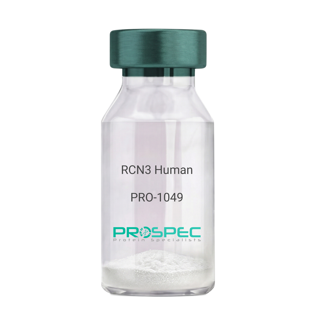
Name :
RCN3 HumanDescription:
Reticulocalbin 3 Human Recombinant
Reticulocalbin-3, EF-hand calcium-binding protein RLP49, RCN3, RLP49.
Product # :
PRO-1049Price :
Quantity :
Shipping Method :
Shipped with Ice Packs
More Info
- description
- source
- formulation
- purity
- More Info
Description
RCN3 Human Recombinant produced in E.Coli is a single, non-glycosylated polypeptide chain containing 333 amino acids (21-328 a.a) and having a molecular mass of 37.9kDa.RCN3 is fused to a 25 amino acid His-tag at N-terminus & purified by proprietary chromatographic techniques.
Source
Escherichia Coli.
Formulation
RCN3 protein solution (1mg/ml) containing 20mM Tris-HCl buffer (pH 8.0), 1mM DTT, 10% glycerol and 0.1M NaCl.
Purity
Greater than 95.0% as determined by SDS-PAGE.
More Info
-
Introduction
Reticulocalbin 3 (RCN3) belongs to the CREC (cab45/reticulocalbin/ ERC45/calumenin) family. RCN3 contains 5 Arg-Xaa-Xaa-Arg motifs, which function as target sequences of SPCs (subtilisin-like proprotein convertases), which is a family of serine endoproteases that proteolytically activate proproteins. The synthesis of PACE4 is induced by association and coexpression with RCN3.
-
Synonyms
Reticulocalbin-3, EF-hand calcium-binding protein RLP49, RCN3, RLP49.
-
Physical Appearance
Sterile Filtered colorless solution.
-
Stability
Store at 4°C if entire vial will be used within 2-4 weeks. Store, frozen at -20°C for longer periods of time. For long term storage it is recommended to add a carrier protein (0.1% HSA or BSA).Avoid multiple freeze-thaw cycles.
-
Amino Acid Sequence
MGSSHHHHHH SSGLVPRGSH MGSHMKPSPD AGPHGQGRVH QAAPLSDAPH DDAHGNFQYD HEAFLGREVA KEFDQLTPEE SQARLGRIVD RMDRAGDGDG WVSLAELRAW IAHTQQRHIR DSVSAAWDTY DTDRDGRVGW EELRNATYGH YAPGEEFHDV EDAETYKKML ARDERRFRVA DQDGDSMATR EELTAFLHPE EFPHMRDIVI AETLEDLDRN KDGYVQVEEY IADLYSAEPG EEEPAWVQTE RQQFRDFRDL NKDGHLDGSE VGHWVLPPAQ DQPLVEANHL LHESDTDKDG RLSKAEILGN WNMFVGSQAT NYGEDLTRHH DEL.
ProSpec's products are furnished for LABORATORY RESEARCH USE ONLY. The product may not be used as drugs, agricultural or pesticidal products, food additives or household chemicals.
- View Data Sheet
Name :
LGALS3 Mouse, ActiveDescription:
Galectin-3 Mouse Recombinant, BioActive
Lectin galactose binding soluble 3, Lectin, galactose binding, soluble 3, CBP35, GAL3, GALBP, GALIG, LGALS2, MAC2.
Product # :
CYT-1151Price :
Quantity :
Shipping Method :
Shipped with Ice Packs
More Info
- description
- source
- formulation
- purity
- biological activity
- More Info
- sds-page
Description
LGALS3 Mouse Recombinant produced in E.Coli is a single, non-glycosylated polypeptide chain containing 287 amino acids ( 1-264 a.a) and having a molecular mass of 29.8kDa.LGALS3 is fused to a 23 amino acid His-tag at N-terminus & purified by proprietary chromatographic techniques.
Source
E.coli.
Formulation
LGALS3 protein (0.5mg/ml) contains 20mM Tris-HCl buffer (pH 8.0), 0.15M NaCl, 50% glycerol,1mM DTT and 2mM EDTA.
Purity
Greater than 95% as determined by SDS-PAGE.
Biological Activity
Measured by its ability to agglutinate human red blood cells. The ED50 for this effect is ≥ 25ug/ml.
sds-page
More Info
-
Introduction
Galectin 3, or LGALS3, is a protein which belongs to the animal lectins family, that binds betagalactoside residues selectively. LGALS3 is originated and leaves cells through ectocytosis. The protein is capable of inhibition apoptosis and the development of cancer. Galectin 3 is found in epithelial tissues in organisms, it can be located in dendritic cells, Kupffer cells, macrophages etc. LGALS3 levels elevated when inflammation is generating, cell proliferation, trans-activation by viral proteins and cell differentiation.
-
Synonyms
Lectin galactose binding soluble 3, Lectin, galactose binding, soluble 3, CBP35, GAL3, GALBP, GALIG, LGALS2, MAC2.
-
Physical Appearance
Sterile Filtered colorless solution.
-
Stability
Store at 4°C if entire vial will be used within 2-4 weeks. Store, frozen at -20°C for longer periods of time. For long term storage it is recommended to add a carrier protein (0.1% HSA or BSA).Avoid multiple freeze-thaw cycles.
-
Amino Acid Sequence
MGSSHHHHHH SSGLVPRGSH MGSMADSFSL NDALAGSGNP NPQGYPGAWG NQPGAGGYPG AAYPGAYPGQ APPGAYPGQA PPGAYPGQAP PSAYPGPTAP GAYPGPTAPG AYPGSTAPGA FPGQPGAPGA YPSAPGGYPA AGPYGVPAGP LTVPYDLPLP GGVMPRMLIT IMGTVKPNAN RIVLDFRRGN DVAFHFNPRF NENNRRVIVC NTKQDNNWGK EERQSAFPFE SGKPFKIQVL VEADHFKVAV NDAHLLQYNH RMKNLREISQ LGISGDITLT SANHAMI.
-
Background
What is the molecular weight/Mw of LGALS3 MOUSE Protein?
LGALS3 MOUSE Protein has a total Mw of 29.8kDa.
What is the source or expression system of LGALS3 MOUSE Protein?
Escherichia Coli.
What is the Purity of LGALS3 MOUSE Protein?
LGALS3 MOUSE Protein is >95% pure as determined by SDS-PAGE.
What is the Biological Activity of LGALS3 MOUSE Protein?
Measured by its ability to agglutinate human red blood cells. The ED50 for this effect is ≥ 25ug/ml.
What is the amino acid sequence of LGALS3 MOUSE Protein?
MGSSHHHHHH SSGLVPRGSH MGSMADSFSL NDALAGSGNP NPQGYPGAWG NQPGAGGYPG AAYPGAYPGQ APPGAYPGQA PPGAYPGQAP PSAYPGPTAP GAYPGPTAPG AYPGSTAPGA FPGQPGAPGA YPSAPGGYPA AGPYGVPAGP LTVPYDLPLP GGVMPRMLIT IMGTVKPNAN RIVLDFRRGN DVAFHFNPRF NENNRRVIVC NTKQDNNWGK EERQSAFPFE SGKPFKIQVL VEADHFKVAV NDAHLLQYNH RMKNLREISQ LGISGDITLT SANHAMI.
What applications can LGALS3 MOUSE Protein be used in?
LGALS3 MOUSE Protein can probably be used in western blot, ELISA and Lateral Flow.
ProSpec's products are furnished for LABORATORY RESEARCH USE ONLY. The product may not be used as drugs, agricultural or pesticidal products, food additives or household chemicals.
- View Data Sheet
Name :
KRT8 HumanDescription:
Cytokeratin 8 Human Recombinant
Keratin type II cytoskeletal 8, Cytokeratin-8, CK-8, Keratin-8, K8, KRT8, CYK8, KO, CK8, K2C8, CARD2.
Product # :
PRO-347Price :
Quantity :
Shipping Method :
Shipped at Room temp
More Info
- description
- source
- formulation
- purity
- More Info
Description
Cytokeratin 8 Human Recombinant produced in E.Coli is a single, non-glycosylated polypeptide chain having a molecular mass of 53,532 Dalton. The KRT8 is purified by proprietary chromatographic techniques.
Source
Escherichia Coli.
Formulation
The protein (1mg/ml) was lyophilized after from a sterile solution containing 30mM Tris-HCl pH-8, 9.5M urea, 2mM DTT, 2mM EDTA and 10mM methylammonium chloride.
Purity
Greater than 95.0% as determined by:
(a) Analysis by RP-HPLC.
(b) Analysis by SDS-PAGE.More Info
-
Introduction
Keratin 8 and 18 (K8/18) are the major components of intermediate filament (IF) proteins of simple or single-layered epithelia.
-
Synonyms
Keratin type II cytoskeletal 8, Cytokeratin-8, CK-8, Keratin-8, K8, KRT8, CYK8, KO, CK8, K2C8, CARD2.
-
Physical Appearance
Sterile Filtered White lyophilized (freeze-dried) powder.
-
Stability
Lyophilized KRT8 although stable at room temperature for 3 weeks, should be stored at 2-8°C. Upon reconstitution KRT8 should be stored at 4°C between 2-7 days and for future use below -18°C. For long term storage it is recommended to add a carrier protein (0.1% HSA or BSA). Please prevent freeze-thaw cycles.
-
Solubility
It is recommended to reconstitute the lyophilized KRT8 in sterile 18MΩ-cm H2O not less than 100µg/ml, which can then be further diluted to other aqueous solutions.
-
Reconstitution to filaments
Performed by mixing equimolar amounts of cytokeratins of type I and type II at concentrations of approx. 0.5 mg/ml, both dissolved in 9.5 M urea buffer (see above). Protofilaments and filament complexes are obtained by dialyzing the resulting polypeptide solution stepwise to a concentration of 4 M urea and then to low salt condition (50 mM NaCl, 2 mM dithiothreitol, 10 mM Tris-HCl, pH 7.4). For immunization purposes, the solution can be further dialyzed against PBS (phosphate buffered saline, e.g. Dulbecco's PBS).
ProSpec's products are furnished for LABORATORY RESEARCH USE ONLY. The product may not be used as drugs, agricultural or pesticidal products, food additives or household chemicals.
- View Data Sheet
Name :
CXCL8 Human, GSTDescription:
Interleukin-8 (1-72) (CXCL8) Human Recombinant, GST Tag
Interleukin-8, IL-8, C-X-C motif chemokine 8, IL8, CXCL8
Product # :
CHM-047Price :
Quantity :
Shipping Method :
Shipped with Ice Packs
More Info
- description
- source
- formulation
- purity
- More Info
Description
Recombinant Human Interleukin-8 produced in E. coli containing 72 amino acids.Recombinant Human Interleukin-8 is fused to GST tag at its N-terminus and purified by proprietary chromatographic technique.
Source
Escherichia Coli.
Formulation
IL8 GST solution contains 25mM Tris-Base/ 25mM K2CO3.
Purity
Protein is >95% pure as determined by 10% PAGE (coomassie staining).
More Info
-
Introduction
Interleukin-8 (IL-8) is a chemokine produced by macrophages and other cell types such as epithelial cells. It is also synthesized by endothelial cells, which store IL-8 in their storage vesicles, the Weibel-Palade bodies. When first encountering an antigen, the primary cells to encounter it are the macrophages who phagocytose the particle. Upon processing, they release chemokines to signal other immune cells to come in to the site of inflammation. IL-8 is one such chemokine. It serves as a chemical signal that attracts neutrophils at the site of inflammation, and therefore is also known as Neutrophil Chemotactic Factor.
-
Synonyms
Interleukin-8, IL-8, C-X-C motif chemokine 8, IL8, CXCL8
-
Physical Appearance
Sterile Filtered clear solution.
-
Stability
Store at 4°C if entire vial will be used within 2-4 weeks.Store, frozen at -20°C for longer periods of time.For long term storage it is recommended to add a carrier protein (0.1% HSA or BSA).Avoid multiple freeze-thaw cycles.
-
Applications
Immunoassay.
-
Background
What is the source or expression system of CXCL8 HUMAN, GST Protein?
Escherichia Coli.
What is the Purity of CXCL8 HUMAN, GST Protein?
CXCL8 HUMAN, GST Protein is >95% pure as determined by SDS-PAGE.
What is the Biological Activity of CXCL8 HUMAN, GST Protein?
The biological functionality of CXCL8 HUMAN, GST Protein will be determined in the future.
What is the amino acid sequence of CXCL8 HUMAN, GST Protein?
CXCL8 HUMAN, GST Protein is composed from 72 amino acids.
What applications can CXCL8 HUMAN, GST Protein be used in?
CXCL8 HUMAN, GST Protein can probably be used in western blot, ELISA and Lateral Flow.
What is the endotoxin level for CXCL8 HUMAN, GST Protein?
The endotoxin level is minimal, CXCL8 HUMAN, GST Protein was purified using conventional chromatography techniques.
ProSpec's products are furnished for LABORATORY RESEARCH USE ONLY. The product may not be used as drugs, agricultural or pesticidal products, food additives or household chemicals.
- View Data Sheet
Name :
BAFFR Human, HEKDescription:
BAFF (BLyS) Receptor Human Recombinant, HEK
TNFRSF13C, CD268, BAFF-R, MGC138235, B cell-activating factor receptor.
Product # :
CYT-1224Price :
Quantity :
Shipping Method :
Shipped with Ice Packs
More Info
- description
- source
- formulation
- purity
- biological activity
- More Info
Description
BAFFR Human Recombinant is a single, glycosylated, polypeptide chain (1-78 a.a) containing a total of 314 amino acids and having a molecular mass of 34.4 kDa. BAFFR is fused to 233 a.a hIgG-Tag at C-terminus and is purified by proprietary chromatographic techniques.
Source
HEK293 Cells.
Formulation
The BAFFR solution (1mg/ml) contains 10% Glycerol and Phosphate-Buffered Saline (pH 7.4).
Purity
Greater than 95.0% as determined by SDS-PAGE.
Biological Activity
The ED50 is ≤0.7 ug/ml, measured by its ability in a functional ELISA with BAFF Human.
More Info
-
Synonyms
TNFRSF13C, CD268, BAFF-R, MGC138235, B cell-activating factor receptor.
-
Physical Appearance
Sterile Filtered colorless solution.
-
Stability
Store at 4°C if entire vial will be used within 2-4 weeks. Store, frozen at -20°C for longer periods of time. For long term storage it is recommended to add a carrier protein (0.1% HSA or BSA).Avoid multiple freeze-thaw cycles.
-
Amino Acid Sequence
DGSMRRGPRS LRGRDAPAPT PCVPAECFDL LVRHCVACGL LRTPRPKPAG ASSPAPRTAL QPQESVGAGA GEAALPLPGL LLEPKSCDKT HTCPPCPAPE LLGGPSVFLF PPKPKDTLMI SRTPEVTCVV VDVSHEDPEV KFNWYVDGVE VHNAKTKPRE EQYNSTYRVV SVLTVLHQDW LNGKEYKCKV SNKALPAPIE KTISKAKGQP REPQVYTLPP SRDELTKNQV SLTCLVKGFY PSDIAVEWES NGQPENNYKT TPPVLDSDGS FFLYSKLTVD KSRWQQGNVF SCSVMHEALH NHYTQKSLSL SPGK.
-
Background
B-cell Activating Factor (BAFF) and its corresponding receptor, BAFF-R, are integral components of the immune system, orchestrating crucial processes in B-cell survival, maturation, and differentiation. As we delve into the intricate world of immunology, the study of BAFF and its receptor has unveiled essential pathways that govern the immune responses in health and disease. This research investigates the multifaceted role of BAFF Receptor Protein, shedding light on its structural complexities, signaling mechanisms, and its pivotal contributions to immune regulation. By exploring the interactions between BAFF and its receptor, scientists aim to decipher the delicate balance that underlies immune homeostasis and explore potential therapeutic avenues.
Structural Architecture of BAFF Receptor Protein:
BAFF Receptor, a transmembrane protein predominantly expressed on B cells, belongs to the tumor necrosis factor receptor (TNFR) superfamily. Its intricate structure involves various domains, each playing a unique role in ligand binding, receptor activation, and downstream signaling. Understanding the structural intricacies of BAFF Receptor is paramount to unraveling the molecular events that govern B-cell fate decisions and immune responses.
Physiological Significance in B-Cell Biology:
BAFF Receptor, upon binding with its ligand BAFF, initiates a cascade of events critical for B-cell survival and function. This interaction promotes B-cell maturation, prevents premature apoptosis, and influences the formation of immune synapses. Additionally, BAFF Receptor signaling is tightly regulated to prevent excessive B-cell activation, ensuring immune tolerance and preventing autoimmune responses. Disruptions in these pathways can lead to autoimmune disorders, underscoring the crucial role of BAFF Receptor in maintaining immune equilibrium.
Regulation of Immune Responses:
BAFF Receptor signaling not only affects B-cell development but also has broader implications for immune responses. By modulating antibody production, B-cell activation, and immune memory, BAFF Receptor plays a vital role in shaping adaptive immunity. Its dysregulation has been implicated in various autoimmune conditions, making it an attractive target for therapeutic interventions aimed at restoring immune balance.
BAFF Receptor as a Therapeutic Target:
The intricate involvement of BAFF Receptor in autoimmune diseases, such as rheumatoid arthritis and systemic lupus erythematosus, has positioned it as a promising therapeutic target. Researchers are exploring monoclonal antibodies and other targeted therapies that aim to modulate BAFF Receptor signaling, providing a new frontier in autoimmune disease management. Additionally, understanding the BAFF-BAFF Receptor axis offers potential insights into the development of vaccines and immunotherapies, fostering innovative approaches in the fight against infectious diseases and malignancies.
BAFF Receptor Protein, as a key player in immune regulation, embodies the complexities of immunology. Its interactions with BAFF orchestrate fundamental processes in B-cell biology and adaptive immunity. As scientists unravel the intricate signaling pathways and structural nuances of BAFF Receptor, they pave the way for novel therapeutic strategies and innovative treatments for autoimmune disorders and beyond. This research not only deepens our understanding of immune regulation but also holds the promise of transformative advancements in immunotherapy, ultimately shaping the future of immune-related healthcare.
What is the molecular weight/Mw of BAFF-R Protein?
BAFF-R Protein has a total Mw of 34.4kDa.
What is the source or expression system of BAFF-R Protein?
HEK293 Cells.
What is the Purity of BAFF-R Protein?
BAFF-R Protein is >95% pure as determined by SDS-PAGE.
What is the Biological Activity of BAFF-R Protein?
The ED50 is ≤0.7 ug/ml, measured by its ability in a functional ELISA with BAFF Human.
What is the amino acid sequence of BAFF-R Protein?
DGSMRRGPRS LRGRDAPAPT PCVPAECFDL LVRHCVACGL LRTPRPKPAG ASSPAPRTAL QPQESVGAGA GEAALPLPGL LLEPKSCDKT HTCPPCPAPE LLGGPSVFLF PPKPKDTLMI SRTPEVTCVV VDVSHEDPEV KFNWYVDGVE VHNAKTKPRE EQYNSTYRVV SVLTVLHQDW LNGKEYKCKV SNKALPAPIE KTISKAKGQP REPQVYTLPP SRDELTKNQV SLTCLVKGFY PSDIAVEWES NGQPENNYKT TPPVLDSDGS FFLYSKLTVD KSRWQQGNVF SCSVMHEALH NHYTQKSLSL SPGK.
What applications can BAFF-R Protein be used in?
BAFF-R Protein can probably be used in western blot, ELISA and Lateral Flow.
What is the endotoxin level for BAFF-R Protein?
The endotoxin level is minimal, BAFF-R Protein was purified using conventional chromatography techniques.
ProSpec's products are furnished for LABORATORY RESEARCH USE ONLY. The product may not be used as drugs, agricultural or pesticidal products, food additives or household chemicals.
- View Data Sheet
Name :
PRDX1 MouseDescription:
Peroxiredoxin-1 Mouse Recombinant
Peroxiredoxin-1, Macrophage 23 kDa stress protein, Osteoblast-specific factor 3, OSF-3, Thioredoxin peroxidase 2, Thioredoxin-dependent peroxide reductase 2.
Product # :
ENZ-951Price :
Quantity :
Shipping Method :
Shipped with Ice Packs
More Info
- description
- source
- formulation
- purity
- biological activity
- More Info
Description
PRDX1 Mouse Recombinant produced in Sf9 Baculovirus cells is a single, glycosylated polypeptide chain containing 207 amino acids (1-199a.a.) and having a molecular mass of 23.2kDa (Molecular size on SDS-PAGE will appear at approximately 18-28kDa). PRDX1 is expressed with an 8 amino acid His tag at C-Terminus and purified by proprietary chromatographic techniques.
Source
Sf9, Baculovirus cells.
Formulation
PRDX1 protein solution (0.5mg/ml) containing Phosphate Buffered Saline (pH 7.4) and 10% glycerol.
Purity
Greater than 90.0% as determined by SDS-PAGE.
Biological Activity
Specific activity is >2,500 pmol/min/ug. Enzymatic activity is defined as the amount of hydroperoxide that 1ug of enzyme can reduce at 25°C for minute.More Info
-
Introduction
PRDX1 is part of the peroxiredoxin family of antioxidant enzymes, which reduce hydrogen peroxide and alkyl hydroperoxides. PRDX1 is an important protector of red blood cells against reactive oxygen species and in tumor prevention.
PRDX1 is antioxidant protective in cells, and contributes to the antiviral activity of CD8(+) T-cells. PRDX1 has a proliferative effect and is involved in cancer development or progression.
Peroxiredoxin-1 is plays a role in redox regulation of the cell. Peroxiredoxin decreases peroxides with reducing equivalents provided through the thioredoxin system but not from glutaredoxin. Peroxiredoxin is involved in eliminating peroxides generated during metabolism. Peroxiredoxin participates in the signaling cascades of growth factors and TNF-alpha by regulating the intracellular concentrations of h(2)o(2). -
Synonyms
Peroxiredoxin-1, Macrophage 23 kDa stress protein, Osteoblast-specific factor 3, OSF-3, Thioredoxin peroxidase 2, Thioredoxin-dependent peroxide reductase 2.
-
Physical Appearance
Sterile Filtered colorless solution.
-
Stability
Store at 4°C if entire vial will be used within 2-4 weeks. Store, frozen at -20°C for longer periods of time. For long term storage it is recommended to add a carrier protein (0.1% HSA or BSA).Avoid multiple freeze-thaw cycles.
-
Amino Acid Sequence
MSSGNAKIGY PAPNFKATAV MPDGQFKDIS LSEYKGKYVV FFFYPLDFTF VCPTEIIAFS DRADEFKKLN CQVIGASVDS HFCHLAWINT PKKQGGLGPM NIPLISDPKR TIAQDYGVLK ADEGISFRGL FIIDDKGILR QITINDLPVG RSVDEIIRLV QAFQFTDKHG EVCPAGWKPG SDTIKPDVNK SKEYFSKQKL EHHHHHH.
ProSpec's products are furnished for LABORATORY RESEARCH USE ONLY. The product may not be used as drugs, agricultural or pesticidal products, food additives or household chemicals.
- View Data Sheet
Name :
AGA Human, sf9Description:
Aspartylglucosaminidase Human Recombinant, sf9
Aspartylglucosaminidase, Glycosylasparaginase, N4-(N-Acetyl-Beta-Glucosaminyl)-L-Asparagine Amidase, N(4)-(Beta-N-Acetylglucosaminyl)-L-Asparaginase , EC 3.5.1.26, Aspartylglucosylamine Deaspartylase, EC 3.5.1, ASRG, AGU, GA.
Product # :
ENZ-990Price :
Quantity :
Shipping Method :
Shipped with Ice Packs
More Info
- description
- source
- formulation
- purity
- More Info
Description
AGA produced in Sf9 Baculovirus cells is a single, glycosylated polypeptide chain containing 332 amino acids (24-346 a.a.) and having a molecular mass of 35.7kDa (Molecular size on SDS-PAGE will appear at approximately 18-57kDa). AGA is expressed with a 6 amino acid His tag at C-Terminus and purified by proprietary chromatographic techniques.
Source
Sf9, Baculovirus cells.
Formulation
AGA protein solution (0.25mg/ml) contains Phosphate Buffered Saline (pH 7.4) and 10% glycerol.
Purity
Greater than 90.0% as determined by SDS-PAGE.
More Info
-
Introduction
Aspartylglucosaminidase, also known as AGA, takes part in the catabolism of Nlinked oligosaccharides of glycoproteins. AGA is a protein coding gene which cleaves asparagine from N-acetylglucosamines in the lysosomal breakdown of glycoproteins.
-
Synonyms
Aspartylglucosaminidase, Glycosylasparaginase, N4-(N-Acetyl-Beta-Glucosaminyl)-L-Asparagine Amidase, N(4)-(Beta-N-Acetylglucosaminyl)-L-Asparaginase , EC 3.5.1.26, Aspartylglucosylamine Deaspartylase, EC 3.5.1, ASRG, AGU, GA.
-
Physical Appearance
Sterile Filtered colorless solution.
-
Stability
Store at 4°C if entire vial will be used within 2-4 weeks. Store, frozen at -20°C for longer periods of time. For long term storage it is recommended to add a carrier protein (0.1% HSA or BSA). Avoid multiple freeze-thaw cycles.
-
Amino Acid Sequence
ADPSSPLPLV VNTWPFKNAT EAAWRALASG GSALDAVESG CAMCEREQCD GSVGFGGSPD ELGETTLDAM IMDGTTMDVG AVGDLRRIKN AIGVARKVLE HTTHTLLVGE SATTFAQSMG FINEDLSTTA SQALHSDWLA RNCQPNYWRN VIPDPSKYCG PYKPPGILKQ DIPIHKETED DRGHDTIGMV VIHKTGHIAA GTSTNGIKFK IHGRVGDSPI PGAGAYADDT AGAAAATGNG DILMRFLPSY QAVEYMRRGE DPTIACQKVI SRIQKHFPEF FGAVICANVT GSYGAACNKL STFTQFSFMV YNSEKNQPTE EKVDCIHHHH HH.
ProSpec's products are furnished for LABORATORY RESEARCH USE ONLY. The product may not be used as drugs, agricultural or pesticidal products, food additives or household chemicals.
- View Data Sheet
Name :
DNAJC15 HumanDescription:
DnaJ (Hsp40) Homolog, Subfamily C, Member 15 Human Recombinant
DnaJ homolog subfamily C member 15, Cell growth-inhibiting gene 22 protein, Methylation-controlled J protein, MCJ, DNAJC15, DNAJD1, GIG22, HSD18.
Product # :
HSP-057Price :
Quantity :
Shipping Method :
Shipped with Ice Packs
More Info
- description
- source
- formulation
- purity
- More Info
Description
DNAJC15 Human Recombinant produced in E.Coli is a single, non-glycosylated polypeptide chain containing 116 amino acids (58-150 a.a.) and having a molecular mass of 12.8 kDa.DNAJC15 is fused to a 23 amino acid His-tag at N-terminus & purified by proprietary chromatographic techniques.
Source
E.coli.
Formulation
The DNAJC15 solution (0.25mg/ml) contains 20mM Tris-HCl buffer (pH8.0), 0.15M NaCl, 20% glycerol and 1mM DTT.
Purity
Greater than 90% as determined by SDS-PAGE.
More Info
-
Introduction
DNAJC15 which is expressed ubiquitously and located on the membrane contains 1 J domain. In many advanced cases of ovarian adenocarcinoma DNAJC15 is absent or down-regulated, due to hypermethylation and allelic loss. Loss of expression correlates with increased resistance to antineoplastic drugs, such as cisplatin. DNAJC15is a crucial component of the TIM23 translocase complex and stimulates the ATPase activity of HSPA9.
-
Synonyms
DnaJ homolog subfamily C member 15, Cell growth-inhibiting gene 22 protein, Methylation-controlled J protein, MCJ, DNAJC15, DNAJD1, GIG22, HSD18.
-
Physical Appearance
Sterile Filtered colorless solution.
-
Stability
Store at 4°C if entire vial will be used within 2-4 weeks. Store, frozen at -20°C for longer periods of time. For long term storage it is recommended to add a carrier protein (0.1% HSA or BSA).Avoid multiple freeze-thaw cycles.
-
Amino Acid Sequence
MGSSHHHHHH SSGLVPRGSH MGSFRIWKPL EQVITETAKK ISTPSFSSYY KGGFEQKMSR REAGLILGVS PSAGKAKIRT AHRRVMILNH PDKGGSPYVA AKINEAKDLL ETTTKH.
ProSpec's products are furnished for LABORATORY RESEARCH USE ONLY. The product may not be used as drugs, agricultural or pesticidal products, food additives or household chemicals.
- View Data Sheet
Name :
DUSP6 HumanDescription:
Dual Specificity Phosphatase 6 Human Recombinant
Dual specificity protein phosphatase 6, Dual specificity protein phosphatase PYST1, Mitogen-activated protein kinase phosphatase 3, MAP kinase phosphatase 3, MKP-3, DUSP6, MKP3, PYST1, Dual Specificity Phosphatase 6, Dual specificity phosphatase 6 isoform a.
Product # :
ENZ-817Price :
Quantity :
Shipping Method :
Shipped with Ice Packs
More Info
- description
- source
- formulation
- purity
- More Info
Description
DUSP6 Human Recombinant produced in E.Coli is a single, non-glycosylated polypeptide chain containing 401 amino acids (1-381 a.a) and having a molecular mass of 44.4kDa.DUSP6 is fused to a 20 amino acid His-tag at N-terminus & purified by proprietary chromatographic techniques.
Source
Escherichia Coli.
Formulation
DUSP6 protein solution (0.25mg/ml) containing 20mM Tris-HCl buffer (pH 8.0), 0.15M NaCl, 10% glycerol and 1mM DTT.
Purity
Greater than 85.0% as determined by SDS-PAGE.
More Info
-
Introduction
Dual Specificity Phosphatase 6, also known as DUSP6 belongs to the dual specificity protein phosphatase subfamily. DUSP6 is a Protein coding gene which inactivates MAP kinases. Various members of the family of dual specificity phosphatases show diverse tissue distribution, subcellular localization, and different modes of inducibility of their expression by extracellular stimuli.
-
Synonyms
Dual specificity protein phosphatase 6, Dual specificity protein phosphatase PYST1, Mitogen-activated protein kinase phosphatase 3, MAP kinase phosphatase 3, MKP-3, DUSP6, MKP3, PYST1, Dual Specificity Phosphatase 6, Dual specificity phosphatase 6 isoform a.
-
Physical Appearance
Sterile Filtered colorless solution.
-
Stability
Store at 4°C if entire vial will be used within 2-4 weeks. Store, frozen at -20°C for longer periods of time. For long term storage it is recommended to add a carrier protein (0.1% HSA or BSA).Avoid multiple freeze-thaw cycles.
-
Amino Acid Sequence
MGSSHHHHHH SSGLVPRGSH MIDTLRPVPF ASEMAISKTV AWLNEQLELG NERLLLMDCR PQELYESSHI ESAINVAIPG IMLRRLQKGN LPVRALFTRG EDRDRFTRRC GTDTVVLYDE SSSDWNENTG GESVLGLLLK KLKDEGCRAF YLEGGFSKFQ AEFSLHCETN LDGSCSSSSP PLPVLGLGGL RISSDSSSDI ESDLDRDPNS ATDSDGSPLS NSQPSFPVEI LPFLYLGCAK DSTNLDVLEE FGIKYILNVT PNLPNLFENA GEFKYKQIPI SDHWSQNLSQ FFPEAISFID EARGKNCGVL VHCLAGISRS VTVTVAYLMQ KLNLSMNDAY DIVKMKKSNI SPNFNFMGQL LDFERTLGLS SPCDNRVPAQ QLYFTTPSNQ NVYQVDSLQS T.
ProSpec's products are furnished for LABORATORY RESEARCH USE ONLY. The product may not be used as drugs, agricultural or pesticidal products, food additives or household chemicals.
- View Data Sheet
Name :
BASP1 HumanDescription:
Brain Abundant Membrane Attached Signal Protein 1 Human Recombinant
CAP-23, CAP23, NAP-22, NAP22, Brain acid soluble protein 1, BASP1, BASP1 Human, 22 kDa neuronal tissue-enriched acidic protein, Neuronal axonal membrane protein NAP-22.
Product # :
PRO-1355Price :
Quantity :
Shipping Method :
Shipped with Ice Packs
More Info
- description
- source
- formulation
- purity
- More Info
Description
BASP1 Human Recombinant produced in E.coli is a single, non-glycosylated polypeptide chain containing 250 amino acids (1-227) and having a molecular mass of 25 kDa (Molecular size on SDS-PAGE will appear higher). BASP1 is fused to a 23 amino acid His-tag at N-terminus & purified by proprietary chromatographic techniques.
Source
Escherichia Coli.
Formulation
The BASP1 solution (0.25mg/ml) contains 20mM Tris-HCl buffer (pH 8.0), 0.2M NaCl and 20% glycerol.
Purity
Greater than 95.0% as determined by SDS-PAGE.
More Info
-
Introduction
Brain Abundant Membrane Attached Signal Protein 1 (BASP1) is a membrane bound protein with numerous transient phosphorylation positions and PEST motifs. Preservation of proteins with PEST sequences amongst diverse species supports their functional significance. PEST sequences take place in proteins with high turnover rates. Immunological attributes of this protein are species specific. BASP1 undergoes N-terminal myristoylation.
-
Synonyms
CAP-23, CAP23, NAP-22, NAP22, Brain acid soluble protein 1, BASP1, BASP1 Human, 22 kDa neuronal tissue-enriched acidic protein, Neuronal axonal membrane protein NAP-22.
-
Physical Appearance
Sterile Filtered colorless solution.
-
Stability
Store at 4°C if entire vial will be used within 2-4 weeks. Store, frozen at -20°C for longer periods of time. For long term storage it is recommended to add a carrier protein (0.1% HSA or BSA).Avoid multiple freeze-thaw cycles.
-
Amino Acid Sequence
MGSSHHHHHH SSGLVPRGSH MGSMGGKLSK KKKGYNVNDE KAKEKDKKAE GAATEEEGTP KESEPQAAAE PAEAKEGKEK PDQDAEGKAE EKEGEKDAAA AKEEAPKAEP EKTEGAAEAK AEPPKAPEQE QAAPGPAAGG EAPKAAEAAA APAESAAPAA GEEPSKEEGE PKKTEAPAAP AAQETKSDGA PASDSKPGSS EAAPSSKETP AATEAPSSTP KAQGPAASAE EPKPVEAPAA NSDQTVTVKE.
ProSpec's products are furnished for LABORATORY RESEARCH USE ONLY. The product may not be used as drugs, agricultural or pesticidal products, food additives or household chemicals.
- View Data Sheet
Name :
CA10 HumanDescription:
Carbonic Anhydrase X Human Recombinant
Carbonic anhydrase-related protein 10, Carbonic anhydrase-related protein X, CA-RP X, CARP X, carbonic anhydrase X, Cerebral protein 15, hucep-15, epididymis secretory sperm binding protein, UNQ533/PRO1076.
Product # :
ENZ-1189Price :
Quantity :
Shipping Method :
Shipped with Ice Packs
More Info
- description
- source
- formulation
- purity
- biological activity
- More Info
Description
CA10 Human Recombinant is a single, glycosylated polypeptide chain containing 317 amino acids (22-328a.a) and having a molecular mass of 36.3kDa (calculated). CA10 is fused to a 6 amino acid His-tag at C-terminus and is purified by proprietary chromatographic techniques.
Source
HEK293 Cells.
Formulation
CA10 protein solution (0.5mg/ml) is filtered in Phosphate-Buffered Saline pH 7.4 and 10% (w/v) glycerol.
Purity
Greater than 95.0% as determined by SDS-PAGE.
Biological Activity
Specific activity is > 150 pmol/min/ug, and is defined as the amount of enzyme that hydrolyze 1pmole of pnitrophenyl acetate to p-nitrophenol per minute at pH8.0 at 37℃.
More Info
-
Synonyms
Carbonic anhydrase-related protein 10, Carbonic anhydrase-related protein X, CA-RP X, CARP X, carbonic anhydrase X, Cerebral protein 15, hucep-15, epididymis secretory sperm binding protein, UNQ533/PRO1076.
-
Physical Appearance
Sterile Filtered colorless solution.
-
Stability
Store at 4°C if entire vial will be used within 2-4 weeks. Store, frozen at -20°C for longer periods of time. Avoid multiple freeze-thaw cycles.
-
Amino Acid Sequence
DGSMQQNSPK IHEGWWAYKE VVQGSFVPVP SFWGLVNSAW NLCSVGKRQS PVNIETSHMI FDPFLTPLRI NTGGRKVSGT MYNTGRHVSL RLDKEHLVNI SGGPMTYSHR LEEIRLHFGS EDSQGSEHLL NGQAFSGEVQ LIHYNHELYT NVTEAAKSPN GLVVVSIFIK VSDSSNPFLN RMLNRDTITR ITYKNDAYLL QGLNIEELYP ETSSFITYDG SMTIPPCYET ASWIIMNKPV YITRMQMHSL RLLSQNQPSQ IFLSMSDNFR PVQPLNNRCI RTNINFSLQG KDCPNNRAQK LQYRVNEWLL KHHHHHH
-
Background
Carbonic anhydrases (CAs) are a family of enzymes that play a crucial role in regulating pH balance and carbon dioxide transport in various tissues and organs. Carbonic anhydrase X (CA10) is a less-studied member of this family, and this research aims to explore its structure, function, and implications in metabolism and disease. Understanding the molecular mechanisms and regulatory roles of CA10 can provide valuable insights into its potential as a therapeutic target for various disorders.
Structure and Expression of Carbonic Anhydrase X:
CA10, also known as mitochondrial carbonic anhydrase, is a membrane-associated protein predominantly found in the mitochondria of various tissues, including the liver, kidney, and brain. It possesses the characteristic zinc-binding catalytic domain found in other CAs. However, CA10 has distinct features, including a unique N-terminal mitochondrial targeting sequence, suggesting its specific role within mitochondria.
Role of Carbonic Anhydrase X in Metabolism:
CA10 is involved in the regulation of pH and bicarbonate concentrations within the mitochondrial matrix, impacting mitochondrial metabolism. It catalyzes the reversible hydration of carbon dioxide to bicarbonate, facilitating the exchange of carbon dioxide between the mitochondria and the cytoplasm. This process is vital for maintaining acid-base homeostasis and efficient energy production through oxidative phosphorylation.
Implications of Carbonic Anhydrase X in Disease:
Emerging evidence suggests that CA10 may be implicated in various pathological conditions. Alterations in CA10 expression or activity have been associated with metabolic disorders, including obesity and diabetes. Furthermore, dysregulation of mitochondrial function and pH homeostasis, in which CA10 plays a role, have been linked to neurodegenerative diseases, cancer, and cardiovascular disorders. Elucidating the precise contributions of CA10 in these pathologies is an area of active investigation.
Therapeutic Potential of Carbonic Anhydrase X:
The unique properties and expression patterns of CA10 make it an intriguing target for therapeutic interventions. Modulating CA10 activity or expression could have implications in metabolic disorders, where the manipulation of mitochondrial function and pH regulation could offer therapeutic benefits. Developing selective inhibitors or activators of CA10 could be explored to regulate its enzymatic activity and modulate mitochondrial metabolism.
Challenges and Future Directions:
Although CA10 shows promise as a therapeutic target, several challenges remain. The elucidation of the precise regulatory mechanisms and signaling pathways involving CA10 within mitochondria is necessary for a comprehensive understanding of its function. Additionally, the development of specific modulators that selectively target CA10 without affecting other CAs or disrupting physiological processes is a critical consideration.
Conclusion:
The study of CA10 protein provides valuable insights into its distinct role in mitochondrial metabolism and disease pathogenesis. Understanding the molecular mechanisms and functional implications of CA10 opens avenues for the development of targeted therapies for metabolic disorders, neurodegenerative diseases, cancer, and cardiovascular disorders. Further research on CA10, its interactions, and its modulation in pathological conditions will contribute to the development of novel therapeutic interventions to improve patient outcomes.
ProSpec's products are furnished for LABORATORY RESEARCH USE ONLY. The product may not be used as drugs, agricultural or pesticidal products, food additives or household chemicals.
- View Data Sheet
Name :
PGC HumanDescription:
Progastricsin-C Human Recombinant
Progastricsin (Pepsinogen C), Pepsinogen C, EC 3.4.23.3, Pepsinogen Group II, Preprogastricsin, EC 3.4.23, Pepsin C, PGII, PEPC.
Product # :
ENZ-966Price :
Quantity :
Shipping Method :
Shipped with Ice Packs
More Info
- description
- source
- formulation
- purity
- More Info
Description
PGC produced in Sf9 Baculovirus cells is a single, glycosylated polypeptide chain containing 380 amino acids (17-388 a.a.) and having a molecular mass of 41.6kDa (Molecular size on SDS-PAGE will appear at approximately 40-57kDa). PGC is expressed with an 8 amino acid His tag at C-Terminus and purified by proprietary chromatographic techniques.
Source
Sf9, Baculovirus cells.
Formulation
PGC protein solution (0.25mg/ml) contains Phosphate Buffered Saline (pH 7.4) and 10% glycerol.
Purity
Greater than 95.0% as determined by SDS-PAGE.
More Info
-
Introduction
Progastricsin-C (PGC) is an aspartic proteinase which is synthesized in the gastric mucosa as inactive precursors. PGC is a part of the peptidase family A1 and contains a prosegment which is responsible for stabilizing the inactive form and preventing the entrance of the substrate to the active site. PGC is used as a biomarker for various gastric diseases including Helicobacter pylori related gastritis. PGC is also hydrolyzes various proteins.
-
Synonyms
Progastricsin (Pepsinogen C), Pepsinogen C, EC 3.4.23.3, Pepsinogen Group II, Preprogastricsin, EC 3.4.23, Pepsin C, PGII, PEPC.
-
Physical Appearance
Sterile Filtered colorless solution.
-
Stability
Store at 4°C if entire vial will be used within 2-4 weeks. Store, frozen at -20°C for longer periods of time. For long term storage it is recommended to add a carrier protein (0.1% HSA or BSA). Avoid multiple freeze-thaw cycles.
-
Amino Acid Sequence
AVVKVPLKKF KSIRETMKEK GLLGEFLRTH KYDPAWKYRF GDLSVTYEPM AYMDAAYFGE ISIGTPPQNF LVLFDTGSSN LWVPSVYCQS QACTSHSRFN PSESSTYSTN GQTFSLQYGS GSLTGFFGYD TLTVQSIQVP NQEFGLSENE PGTNFVYAQF DGIMGLAYPA LSVDEATTAM QGMVQEGALT SPVFSVYLSN QQGSSGGAVV FGGVDSSLYT GQIYWAPVTQ ELYWQIGIEE FLIGGQASGW CSEGCQAIVD TGTSLLTVPQ QYMSALLQAT GAQEDEYGQF LVNCNSIQNL PSLTFIINGV EFPLPPSSYI LSNNGYCTVG VEPTYLSSQN GQPLWILGDV FLRSYYSVYD LGNNRVGFAT AALEHHHHHH.
ProSpec's products are furnished for LABORATORY RESEARCH USE ONLY. The product may not be used as drugs, agricultural or pesticidal products, food additives or household chemicals.
- View Data Sheet
Name :
Fibronectin RecombinantDescription:
Fibronectin Human Recombinant
Product # :
PRO-2621Price :
Quantity :
Shipping Method :
Shipped at Room temp
More Info
- description
- source
- formulation
- purity
- biological activity
- More Info
Description
Fibronectin Human Recombinant produced in E.Coli is a single, non-glycosylated polypeptide chain containing 574 amino acids and having a molecular mass of 62.6kDa. The Fibronectin is purified by proprietary chromatographic techniques.
Source
Escherichia Coli.
Formulation
Lyophilized from a 0.2μm filtered concentrated solution in 20 mM Tris-HCl, pH 8.0, 150 mM NaCl, with 5 % Trehalose and 0.02 % Tween-20.
Purity
Greater than 95.0% as determined by:
(a) Analysis by RP-HPLC.
(b) Analysis by SDS-PAGE.Biological Activity
Was measured by its ability to support cell attachment and spreading when used as a substratum for cell culture. The recommended concentration in this application for this effect is typically 1-5 μg/cm2. Fibronectin can also be added to the media to support cell spreading at a concentration of 0.5-50 μg/ml. Optimal concentrations will need to be determined for individual user applications.
More Info
-
Introduction
Fibronectin takes part in several cellular processes, including tissue repair, embryogenesis, blood clotting, and cell migration/adhesion.Plasma fibronectin level is elevated in severe coronary artery disease. Increased plasma fibronectin levels are related with venous thromboembolism (VTE) particularly in males, and extend the probable association between biomarkers and risk factors for arterial atherothrombosis and VTE. Fibronectin consists in 2 main forms: 1) as an insoluble glycoprotein dimer that serves as a linker in the etracellular matrix and 2) as a soluble disulphide linked dimer found in the plasma. The plasma form is produced by hepatocytes, and the ECM form is synthesized by fibroblasts, chondrocytes, endothelial cells, macrophages, as well as certain epithelial cells. Fibronectin alos takes part as a general cell adhesion molecule by anchoring cells to collagen or proteoglycan substrates. Fibronectin organizes cellular interaction with the ECM by binding to different components of the extracellular matrix and to membrane-bound Fibronectin receptors on cell surfaces.
-
Physical Appearance
Sterile Filtered White lyophilized (freeze-dried) powder.
-
Stability
Lyophilized Fibronectin although stable at room temperature for 3 weeks, should be stored desiccated below -18°C. Upon reconstitution Fibronectin should be stored at 4°C between 2-7 days and for future use below -18°C. Please prevent freeze-thaw cycles.
-
Solubility
It is recommended to reconstitute the lyophilized Fibronectin in sterile 18MΩ-cm H2O not less than 100µg/ml, which can then be further diluted to other aqueous solutions.
ProSpec's products are furnished for LABORATORY RESEARCH USE ONLY. The product may not be used as drugs, agricultural or pesticidal products, food additives or household chemicals.
- View Data Sheet
Name :
CCNB1 HumanDescription:
Cyclin-B1 Human Recombinant
G2/mitotic-specific cyclin-B1, cyclin B1, CCNB.
Product # :
PKA-037Price :
Quantity :
Shipping Method :
Shipped with Ice Packs
More Info
- description
- source
- formulation
- purity
- More Info
Description
CCNB1 Human Recombinant produced in E. coli is a single polypeptide chain containing 457 amino acids (1-433) and having a molecular mass of 50.9 kDa.CCNB1 is fused to a 24 amino acid His-tag at N-terminus & purified by proprietary chromatographic techniques.
Source
E.coli.
Formulation
The CCNB1 solution (0.25mg/1ml) contains 20mM Tris-HCl buffer (pH 8.0), 150mM NaCl and 20% glycerol.
Purity
Greater than 85% as determined by SDS-PAGE.
More Info
-
Introduction
Cyclin B1 (CCNB1) is a regulatory protein involved in mitosis. CCNB1 creates a complex with p34(cdc2) to form the maturation-promoting factor (MPF). CCNB1 is vital for the control of the cell cycle at the G2/M (mitosis) transition. CCNB1 builds up steadily during the G2 and is immediately destroyed at mitosis. The 2 alternative transcripts produce a constitutively expressed transcript and a cell cycle-regulated transcript which is expressed predominantly during G2/M phase. These transcripts are a result of alternate transcription initiation sites.
-
Synonyms
G2/mitotic-specific cyclin-B1, cyclin B1, CCNB.
-
Physical Appearance
Sterile Filtered colorless solution.
-
Stability
Store at 4°C if entire vial will be used within 2-4 weeks. Store, frozen at -20°C for longer periods of time. For long term storage it is recommended to add a carrier protein (0.1% HSA or BSA).Avoid multiple freeze-thaw cycles.
-
Amino Acid Sequence
MGSSHHHHHH SSGLVPRGSH MGSHMALRVT RNSKINAENK AKINMAGAKR VPTAPAATSK PGLRPRTALG DIGNKVSEQL QAKMPMKKEA KPSATGKVID KKLPKPLEKV PMLVPVPVSE PVPEPEPEPE PEPVKEEKLS PEPILVDTAS PSPMETSGCA PAEEDLCQAF SDVILAVNDV DAEDGADPNL CSEYVKDIYA YLRQLEEEQA VRPKYLLGRE VTGNMRAILI DWLVQVQMKF RLLQETMYMT VSIIDRFMQN NCVPKKMLQL VGVTAMFIAS KYEEMYPPEI GDFAFVTDNT YTKHQIRQME MKILRALNFG LGRPLPLHFL RRASKIGEVD VEQHTLAKYL MELTMLDYDM VHFPPSQIAA GAFCLALKIL DNGEWTPTLQ HYLSYTEESL LPVMQHLAKN VVMVNQGLTK HMTVKNKYAT SKHAKISTLP QLNSALVQDL AKAVAKV.
ProSpec's products are furnished for LABORATORY RESEARCH USE ONLY. The product may not be used as drugs, agricultural or pesticidal products, food additives or household chemicals.
- View Data Sheet
Name :
PKNOX1 HumanDescription:
PBX/Knotted 1 Homeobox 1 Human Recombinant
pkonx1c, PREP1, Homeobox protein PKNOX1, Homeobox protein PREP-1, PBX/knotted homeobox 1, PKNOX1.
Product # :
PRO-1979Price :
Quantity :
Shipping Method :
Shipped with Ice Packs
More Info
- description
- source
- formulation
- purity
- More Info
Description
PKNOX1 Human Recombinant produced in E.Coli is a single, non-glycosylated polypeptide chain containing 456 amino acids (1-436 a.a) and having a molecular mass of 49.7kDa. PKNOX1 is fused to a 20 amino acid His-tag at N-terminus & purified by proprietary chromatographic techniques.
Source
Escherichia Coli.
Formulation
PKNOX1 protein solution (0.25mg/ml) containing 20mM Tris-HCl buffer (pH 8.0), 0.1M NaCl and 10% glycerol.
Purity
Greater than 80.0% as determined by SDS-PAGE.
More Info
-
Introduction
PBX/Knotted 1 Homeobox 1 (PKNOX1) is a DNA-binding factor which takes part in the regulation of the transcriptional activity of AP-1 containing promoters. PKNOX1, which is also known as UEF3, PRP-1 and p64, is a Hmeobox protein. PKNOX1 forms stable complexes containing PBX proteins which synergize with AP-1 binding factors.
-
Synonyms
pkonx1c, PREP1, Homeobox protein PKNOX1, Homeobox protein PREP-1, PBX/knotted homeobox 1, PKNOX1.
-
Physical Appearance
Sterile Filtered colorless solution.
-
Stability
Store at 4°C if entire vial will be used within 2-4 weeks. Store, frozen at -20°C for longer periods of time. For long term storage it is recommended to add a carrier protein (0.1% HSA or BSA).Avoid multiple freeze-thaw cycles.
-
Amino Acid Sequence
MGSSHHHHHH SSGLVPRGSH MMATQTLSID SYQDGQQMQV VTELKTEQDP NCSEPDAEGV SPPPVESQTP MDVDKQAIYR HPLFPLLALL FEKCEQSTQG SEGTTSASFD VDIENFVRKQ EKEGKPFFCE DPETDNLMVK AIQVLRIHLL ELEKVNELCK DFCSRYIACL KTKMNSETLL SGEPGSPYSP VQSQQIQSAI TGTISPQGIV VPASALQQGN VAMATVAGGT VYQPVTVVTP QGQVVTQTLS PGTIRIQNSQ LQLQLNQDLS ILHQDDGSSK NKRGVLPKHA TNVMRSWLFQ HIGHPYPTED EKKQIAAQTN LTLLQVNNWF INARRRILQP MLDSSCSETP KTKKKTAQNR PVQRFWPDSI ASGVAQPPPS ELTMSEGAVV TITTPVNMNV DSLQSLSSDG ATLAVQQVMM AGQSEDESVD STEEDAGALA PAHISGLVLE NSDSLQ.
ProSpec's products are furnished for LABORATORY RESEARCH USE ONLY. The product may not be used as drugs, agricultural or pesticidal products, food additives or household chemicals.
- View Data Sheet
Name :
GLO1 MouseDescription:
Glyoxalase-I Mouse Recombinant
Lactoylglutathione lyase, Aldoketomutase, Glyoxalase I, Glx I, Ketone-aldehyde mutase, Methylglyoxalase, S-D-lactoylglutathione methylglyoxal lyase.
Product # :
ENZ-953Price :
Quantity :
Shipping Method :
Shipped with Ice Packs
More Info
- description
- source
- formulation
- purity
- biological activity
- More Info
Description
GLO1 Mouse Recombinant produced in Sf9 Baculovirus cells is a single, glycosylated polypeptide chain containing 192 amino acids (1-184a.a.) and having a molecular mass of 21.8kDa (Molecular size on SDS-PAGE will appear at approximately 28-40kDa). GLO1 is expressed with an 8 amino acid His tag at C-Terminus and purified by proprietary chromatographic techniques.
Source
Sf9, Baculovirus cells.
Formulation
GLO1 protein solution (0.5mg/ml) containing Phosphate Buffered Saline (pH 7.4) and 10% glycerol.
Purity
Greater than 95.0% as determined by SDS-PAGE.
Biological Activity
Specific activity is > 210 units/mg, and is defined as the amount of enzyme that will form 1.0 µmol of S-lactoylglutathione from methylglyoxal and reduced glutathione per minute at pH 6.5 at 25C.More Info
-
Introduction
GLO1 is involved in the catalysis and formation of S-lactoyl-glutathione from methylglyoxal condensation and reduced glutatione. GLO1 is linked to HLA and is localized to 6p21.3-p21.1, between HLA and the centromere. GLO1 enzyme is ubundantly expressed and present in numerous tumor cell lines, in which its concentration is often upregulated ubiquitisly. GLO1 is a major susceptible gene for autism in an ethnic Chinese population from Taiwan. GLO1 might be involved in the pathophysiology of mood disorders. GLO1 plays a role in the pathophysiology of mood disorders. Overexpression of GLO1 is associated with kidney tumor.
-
Synonyms
Lactoylglutathione lyase, Aldoketomutase, Glyoxalase I, Glx I, Ketone-aldehyde mutase, Methylglyoxalase, S-D-lactoylglutathione methylglyoxal lyase.
-
Physical Appearance
Sterile Filtered colorless solution.
-
Stability
Store at 4°C if entire vial will be used within 2-4 weeks. Store, frozen at -20°C for longer periods of time. For long term storage it is recommended to add a carrier protein (0.1% HSA or BSA).Avoid multiple freeze-thaw cycles.
-
Amino Acid Sequence
MAEPQPASSG LTDETAFSCC SDPDPSTKDF LLQQTMLRIK DPKKSLDFYT RVLGLTLLQK LDFPAMKFSL YFLAYEDKND IPKDKSEKTA WTFSRKATLE LTHNWGTEDD ETQSYHNGNS DPRGFGHIGI AVPDVYSACK RFEELGVKFV KKPDDGKMKG LAFIQDPDGY WIEILNPNKI ATIILEHHHH HH.
ProSpec's products are furnished for LABORATORY RESEARCH USE ONLY. The product may not be used as drugs, agricultural or pesticidal products, food additives or household chemicals.