Skip to content

High-quality research proteins.

  • 1-800-805-6531

  • Mon - Sat 8.00-18.00, Sun - Closed

  • info@prospecbio.com

  • PO Box 6591 East Brunswick NJ 08816

  • Products
    • Cytokines
    • Growth factors
    • Chemokines
    • Cd antigens
    • Neurotrophins
    • Hormones
    • Enzymes
    • Viral antigens
    • Recombinant proteins
    • Natural proteins
    • Monoclonal antibodies
    • Polyclonal antibodies
    • Thermal Packaging
  • About
    • Company
    • Founder
    • Contribution
  • Customers
    • Eastern Asia
    • Eastern Europe
    • North America
    • South America
    • South East Asia
    • Southern Asia
    • Western Europe
  • Ordering
    • Ordering Method
    • Ordering - Credit Card
    • Ordering - PO
    • Shipping Fees
    • FAQs
  • Services
    • DNA Cloning
    • Protein Expression
    • Monoclonal Antibody
    • Fermentation
    • Technology License
  • Contact us
Log in

Country/region

  • USD $ | Albania
  • USD $ | Andorra
  • USD $ | Argentina
  • USD $ | Armenia
  • USD $ | Australia
  • USD $ | Austria
  • USD $ | Azerbaijan
  • USD $ | Belarus
  • USD $ | Belgium
  • USD $ | Bolivia
  • USD $ | Brazil
  • USD $ | Bulgaria
  • USD $ | Cambodia
  • USD $ | Canada
  • USD $ | Chile
  • USD $ | China
  • USD $ | Colombia
  • USD $ | Costa Rica
  • USD $ | Croatia
  • USD $ | Cyprus
  • USD $ | Czechia
  • USD $ | Denmark
  • USD $ | Estonia
  • USD $ | Finland
  • USD $ | France
  • USD $ | Georgia
  • USD $ | Germany
  • USD $ | Greece
  • USD $ | Hong Kong SAR
  • USD $ | Hungary
  • USD $ | Iceland
  • USD $ | India
  • USD $ | Ireland
  • USD $ | Israel
  • USD $ | Italy
  • USD $ | Japan
  • USD $ | Kazakhstan
  • USD $ | Latvia
  • USD $ | Liechtenstein
  • USD $ | Lithuania
  • USD $ | Madagascar
  • USD $ | Malta
  • USD $ | Mexico
  • USD $ | Moldova
  • USD $ | Monaco
  • USD $ | Netherlands
  • USD $ | New Zealand
  • USD $ | North Macedonia
  • USD $ | Norway
  • USD $ | Panama
  • USD $ | Paraguay
  • USD $ | Peru
  • USD $ | Philippines
  • USD $ | Poland
  • USD $ | Portugal
  • USD $ | Romania
  • USD $ | Singapore
  • USD $ | Slovakia
  • USD $ | Slovenia
  • USD $ | South Africa
  • USD $ | South Korea
  • USD $ | Spain
  • USD $ | Sri Lanka
  • USD $ | Sweden
  • USD $ | Switzerland
  • USD $ | Taiwan
  • USD $ | Thailand
  • USD $ | Türkiye
  • USD $ | Ukraine
  • USD $ | United Kingdom
  • USD $ | United States
  • USD $ | Uruguay
  • USD $ | Vatican City
  • USD $ | Venezuela
  • USD $ | Vietnam
  • Facebook
  • Instagram
logoProSpec - Recombinant Protein Manufacturer for academic and R&D industry
Become a distributor
Account Log in
Wishlist
Cart Cart(0)
  • Products
    +
    • Cytokines
    • Growth factors
    • Chemokines
    • Cd antigens
    • Neurotrophins
    • Hormones
    • Enzymes
    • Viral antigens
    • Recombinant proteins
    • Natural proteins
    • Monoclonal antibodies
    • Polyclonal antibodies
    • Thermal Packaging
  • About
    +
    • Company
    • Founder
    • Contribution
  • Customers
    +
    • Eastern Asia
    • Eastern Europe
    • North America
    • South America
    • South East Asia
    • Southern Asia
    • Western Europe
  • Ordering
    +
    • Ordering Method
    • Ordering - Credit Card
    • Ordering - PO
    • Shipping Fees
    • FAQs
  • Services
    +
    • DNA Cloning
    • Protein Expression
    • Monoclonal Antibody
    • Fermentation
    • Technology License
  • Contact us

Item added to your cart

View cart
View All
  • Cytokines
  • Growth factors
  • Chemokines
  • Cd antigens
  • Neurotrophins
  • Hormones
  • Enzymes
  • Viral antigens
  • Recombinant proteins
  • Natural proteins
  • Monoclonal antibodies
  • Polyclonal antibodies
  • Thermal Packaging
  • Tumor Necrosis Factor

    Tumor Necrosis Factor

    View All
  • 4-1BB

    4-1BB

    View All
  • Adiponectin

    Adiponectin

    View All
  • AIF1

    AIF1

    View All
  • Other Cytokines

    Other Cytokines

    View All
  • AITRL

    AITRL

    View All
  • Angiopoietin

    Angiopoietin

    View All
  • Apolipoprotein

    Apolipoprotein

    View All
  • B-Cell Activating Factor

    B-Cell Activating Factor

    View All
  • Beta Defensin

    Beta Defensin

    View All
  • Bone Morphogenetic Protein

    Bone Morphogenetic Protein

    View All
  • B type Natriuretic Peptide

    B type Natriuretic Peptide

    View All
  • BST

    BST

    View All
  • Betacellulin

    Betacellulin

    View All
  • Cardiotrophin

    Cardiotrophin

    View All
  • Activin

    Activin

    View All
  • Fibroblast Growth Factor

    Fibroblast Growth Factor

    View All
  • Other Growth Factors

    Other Growth Factors

    View All
  • CSF

    CSF

    View All
  • CTGF

    CTGF

    View All
  • IGFBP

    IGFBP

    View All
  • VEGF Protein

    VEGF Protein

    View All
  • EGF

    EGF

    View All
  • Epigen

    Epigen

    View All
  • Erythropoietin

    Erythropoietin

    View All
  • Insulin-Like Growth Factor

    Insulin-Like Growth Factor

    View All
  • Growth Hormone

    Growth Hormone

    View All
  • HDGF

    HDGF

    View All
  • Hepatocyte Growth Factor

    Hepatocyte Growth Factor

    View All
  • Insulin

    Insulin

    View All
  • BCA-1/ BLC (CXCL13)

    BCA-1/ BLC (CXCL13)

    View All
  • BRAK (CXCL14)

    BRAK (CXCL14)

    View All
  • C-10 (CCL6)

    C-10 (CCL6)

    View All
  • Other Chemokines

    Other Chemokines

    View All
  • MEC (CCL28)

    MEC (CCL28)

    View All
  • LD78-beta (CCL3L1)

    LD78-beta (CCL3L1)

    View All
  • CTACK (CCL27)

    CTACK (CCL27)

    View All
  • CXCL16

    CXCL16

    View All
  • CXCL17

    CXCL17

    View All
  • Platelet Factor-4 (CXCL4)

    Platelet Factor-4 (CXCL4)

    View All
  • ENA-78 (CXCL5)

    ENA-78 (CXCL5)

    View All
  • Eotaxin (CCL11,24,26)

    Eotaxin (CCL11,24,26)

    View All
  • Exodus-2 (CCL21)

    Exodus-2 (CCL21)

    View All
  • Fractalkine (CX3CL1)

    Fractalkine (CX3CL1)

    View All
  • CXCL6

    CXCL6

    View All
  • Other CD Antigens

    Other CD Antigens

    View All
  • CD14

    CD14

    View All
  • CD164

    CD164

    View All
  • CD1

    CD1

    View All
  • CD2

    CD2

    View All
  • CD200

    CD200

    View All
  • CD207

    CD207

    View All
  • CD226

    CD226

    View All
  • CD23

    CD23

    View All
  • CD244

    CD244

    View All
  • CD247

    CD247

    View All
  • CD27

    CD27

    View All
  • CD274

    CD274

    View All
  • CD300

    CD300

    View All
  • CD33

    CD33

    View All
  • Other Neurotrophins

    Other Neurotrophins

    View All
  • Beta-NGF

    Beta-NGF

    View All
  • BDNF

    BDNF

    View All
  • CDNF

    CDNF

    View All
  • CNTF

    CNTF

    View All
  • GDNF

    GDNF

    View All
  • Glia Maturation Factor

    Glia Maturation Factor

    View All
  • MANF

    MANF

    View All
  • Midkine

    Midkine

    View All
  • Neuroglobin

    Neuroglobin

    View All
  • Neurotrophic factors

    Neurotrophic factors

    View All
  • Neuregulin

    Neuregulin

    View All
  • Neuropilin

    Neuropilin

    View All
  • Pigment Epithelium-Derived Factor

    Pigment Epithelium-Derived Factor

    View All
  • Pleiotrophin

    Pleiotrophin

    View All
  • Peptide Hormones

    Peptide Hormones

    View All
  • Other Hormones

    Other Hormones

    View All
  • HCG

    HCG

    View All
  • Endothelin

    Endothelin

    View All
  • Exendin

    Exendin

    View All
  • FSH

    FSH

    View All
  • Glucagon

    Glucagon

    View All
  • GHRP

    GHRP

    View All
  • GLP

    GLP

    View All
  • Thyrostimulin

    Thyrostimulin

    View All
  • Inhibin A

    Inhibin A

    View All
  • LHRH

    LHRH

    View All
  • Melanotan

    Melanotan

    View All
  • Procalcitonin

    Procalcitonin

    View All
  • PTH

    PTH

    View All
  • Peroxiredoxin

    Peroxiredoxin

    View All
  • Peptidase

    Peptidase

    View All
  • Synthetase

    Synthetase

    View All
  • Transferase

    Transferase

    View All
  • Hydrolase

    Hydrolase

    View All
  • Dehydrogenase

    Dehydrogenase

    View All
  • ACE2

    ACE2

    View All
  • Esterase

    Esterase

    View All
  • Protein Kinases

    Protein Kinases

    View All
  • Other Enzymes

    Other Enzymes

    View All
  • Phosphatase

    Phosphatase

    View All
  • Deaminase

    Deaminase

    View All
  • Oxygenase

    Oxygenase

    View All
  • Adenylate Kinase

    Adenylate Kinase

    View All
  • Lyase

    Lyase

    View All
  • Chagas

    Chagas

    View All
  • SARS

    SARS

    View All
  • Influenza

    Influenza

    View All
  • ASFV

    ASFV

    View All
  • Astrovirus

    Astrovirus

    View All
  • Borrelia

    Borrelia

    View All
  • Helicobacter Pylori

    Helicobacter Pylori

    View All
  • Chikungunya

    Chikungunya

    View All
  • Chlamydia

    Chlamydia

    View All
  • Cytomegalo

    Cytomegalo

    View All
  • Dengue

    Dengue

    View All
  • EBV

    EBV

    View All
  • Ebola

    Ebola

    View All
  • Feline Leukemia Virus

    Feline Leukemia Virus

    View All
  • Hepatitis A

    Hepatitis A

    View All
  • Other

    Other

    View All
  • Actin

    Actin

    View All
  • ADAM

    ADAM

    View All
  • Complement Component

    Complement Component

    View All
  • Ag85

    Ag85

    View All
  • Eukaryotic Translation Initiation Factor

    Eukaryotic Translation Initiation Factor

    View All
  • Anterior Gradient Protein

    Anterior Gradient Protein

    View All
  • Heat Shock Protein

    Heat Shock Protein

    View All
  • Alpha-2-HS-Glycoprotein

    Alpha-2-HS-Glycoprotein

    View All
  • Hemoglobin

    Hemoglobin

    View All
  • Allergy

    Allergy

    View All
  • Anaplasma

    Anaplasma

    View All
  • Angiogenin

    Angiogenin

    View All
  • Ankyrin Repeat Domain

    Ankyrin Repeat Domain

    View All
  • Annexin

    Annexin

    View All
  • Other Natural Proteins

    Other Natural Proteins

    View All
  • Aprotinin

    Aprotinin

    View All
  • Transferrin

    Transferrin

    View All
  • Avidin

    Avidin

    View All
  • Anti Coagulation Factors

    Anti Coagulation Factors

    View All
  • Natural Albumin

    Natural Albumin

    View All
  • Natural Coagulation Factors

    Natural Coagulation Factors

    View All
  • Fibronectin

    Fibronectin

    View All
  • Thrombin

    Thrombin

    View All
  • Anti Human Enzyme

    Anti Human Enzyme

    View All
  • Other Monoclonal Antibodies

    Other Monoclonal Antibodies

    View All
  • Anti Human Cytokine

    Anti Human Cytokine

    View All
  • Anti Human Heat Shock Protein

    Anti Human Heat Shock Protein

    View All
  • Anti Mouse Lymphocyte

    Anti Mouse Lymphocyte

    View All
  • Anti Human Chemokine

    Anti Human Chemokine

    View All
  • Anti Human Lymphocyte

    Anti Human Lymphocyte

    View All
  • Anti Viral Monoclonal

    Anti Viral Monoclonal

    View All
  • Anti Mouse Cytokine

    Anti Mouse Cytokine

    View All
  • Anti-GST Monoclonal

    Anti-GST Monoclonal

    View All
  • Other Polyclonal Antibodies

    Other Polyclonal Antibodies

    View All
  • Anti Viral

    Anti Viral

    View All
  • Anti-GST Monoclonal

    Anti-GST Monoclonal

    View All
Back

Search results

1000 results found for “HDGF”

Name

Description

Product #

Price

Quantity

Shipping Method

  • View Data Sheet

    Name :

    PFN1 Rat

    Description:

    Profilin-1 Rat Recombinant

    Profilin-1, Profilin I.

    Product # :

    PRO-2231

    Price :

    Quantity :

    Shipping Method :

    Ice Icon

    Shipped with Ice Packs

    Add To Cart

    More Info

    • description
    • source
    • formulation
    • purity
    • More Info

    Description

    PFN1 Rat Recombinant produced in E.Coli is a single, non-glycosylated polypeptide chain containing 164 amino acids (1-140 a.a) and having a molecular mass of 17.5kDa. PFN1 is fused to a 24 amino acid His-tag at N-terminus & purified by proprietary chromatographic techniques.

    Source

    Escherichia Coli.

    Formulation

    PFN1 protein solution (1mg/ml) containing Phosphate Buffered Saline (pH 7.4) and 10% glycerol 1mM DTT.

    Purity

    Greater than 95.0% as determined by SDS-PAGE.

    More Info

    • Introduction

      Profilin-1 also known as Pfn1 is a ubiquitous actin monomer-binding protein which is a member of the profilin family. Pfn1 significantly enhances skin wound healing in-vitro as well as in-vivo which is mediated by purinergic receptors. Furthermore, Pfn1 is also active in endothelial cell migration and vessel sprouting. Pfn1 is considered to regulate actin polymerization in response to extracellular signals.

    • Synonyms

      Profilin-1, Profilin I.

    • Physical Appearance

      Sterile Filtered colorless solution.

    • Stability

      Store at 4°C if entire vial will be used within 2-4 weeks. Store, frozen at -20°C for longer periods of time. For long term storage it is recommended to add a carrier protein (0.1% HSA or BSA).Avoid multiple freeze-thaw cycles.

    • Amino Acid Sequence

      MGSSHHHHHH SSGLVPRGSH MGSHMAGWNA YIDSLMADGT CQDAAIVGYK DSPSVWAAVP GKTFVSITPA EVGVLVGKDR SSFFVNGLTL GGQKCSVIRD SLLQDGEFTM DLRTKSTGGA PTFNVTVTMT AKTLVLLMGK EGVHGGLINK KCYEMASHLR RSQY.

    ProSpec's products are furnished for LABORATORY RESEARCH USE ONLY. The product may not be used as drugs, agricultural or pesticidal products, food additives or household chemicals.

    Pfn1 Rat
  • View Data Sheet

    Name :

    JMJD6 Human

    Description:

    Jumonji Domain Containing 6 Human Recombinant

    Jumonji Domain Containing 6, Phosphatidylserine Receptor, Jumonji Domain-Containing Protein 6, Histone Arginine Demethylase JMJD6, Peptide-Lysine 5-Dioxygenase JMJD6, JmjC Domain-Containing Protein 6, Lysyl-Hydroxylase JMJD6, PTDSR, PSR, Bifunctional Arginine Demethylase And Lysyl-Hydroxylase JMJD6, Protein PTDSR, EC 1.14.11.-, EC 1.14.11, KIAA0585, PTDSR1, Bifunctional arginine demethylase and lysyl-hydroxylase JMJD6 (EC:1.14.11.-).

    Product # :

    PRO-2183

    Price :

    Quantity :

    Shipping Method :

    Ice Icon

    Shipped with Ice Packs

    Add To Cart

    More Info

    • description
    • source
    • formulation
    • purity
    • More Info

    Description

    JMJD6 Human Recombinant produced in E.Coli is a single, non-glycosylated polypeptide chain containing 437 amino acids (1-414 a.a) and having a molecular mass of 50.0kDa. JMJD6 is fused to a 23 amino acid His-tag at N-terminus & purified by proprietary chromatographic techniques.

    Source

    Escherichia Coli.

    Formulation

    JMJD6 protein solution (0.5mg/ml) containing Phosphate buffered saline (pH7.4), 30% glycerol and 1mM DTT.

    Purity

    Greater than 90% as determined by SDS-PAGE.

    More Info

    • Introduction

      Jumonji Domain Containing 6, also known as JMJD6 performs as a lysyl-hydroxylase which catalyzes 5-hydroxylation on specific lysine residues of target proteins for instance U2AF2/U2AF65 and LUC7L2, as a regulator of RNA splicing by mediating 5-hydroxylation of U2AF2/U2AF65, affecting the pre-mRNA splicing activity of U2AF2/U2AF65. Additionally to peptidyl-lysine 5-dioxygenase activity, JMJD6 performs as an RNA hydroxylase, as proposed by its ability to bind single strand RNA. Furthermore, it acts as an arginine demethylase which demethylates histone H3 at 'Arg-2' (H3R2me) as well as histone H4 at 'Arg-3' (H4R3me), thus taking a part in histone code.

    • Synonyms

      Jumonji Domain Containing 6, Phosphatidylserine Receptor, Jumonji Domain-Containing Protein 6, Histone Arginine Demethylase JMJD6, Peptide-Lysine 5-Dioxygenase JMJD6, JmjC Domain-Containing Protein 6, Lysyl-Hydroxylase JMJD6, PTDSR, PSR, Bifunctional Arginine Demethylase And Lysyl-Hydroxylase JMJD6, Protein PTDSR, EC 1.14.11.-, EC 1.14.11, KIAA0585, PTDSR1, Bifunctional arginine demethylase and lysyl-hydroxylase JMJD6 (EC:1.14.11.-).

    • Physical Appearance

      Sterile filtered colorless solution.

    • Stability

      Store at 4°C if entire vial will be used within 2-4 weeks. Store, frozen at -20°C for longer periods of time. For long term storage it is recommended to add a carrier protein (0.1% HSA or BSA).Avoid multiple freeze-thaw cycles.

    • Amino Acid Sequence

      MGSSHHHHHH SSGLVPRGSH MGSMNHKSKK RIREAKRSAR PELKDSLDWT RHNYYESFSL SPAAVADNVE RADALQLSVE EFVERYERPY KPVVLLNAQE GWSAQEKWTL ERLKRKYRNQ KFKCGEDNDG YSVKMKMKYY IEYMESTRDD SPLYIFDSSY GEHPKRRKLL EDYKVPKFFT DDLFQYAGEK RRPPYRWFVM GPPRSGTGIH IDPLGTSAWN ALVQGHKRWC LFPTSTPREL IKVTRDEGGN QQDEAITWFN VIYPRTQLPT WPPEFKPLEI LQKPGETVFV PGGWWHVVLN LDTTIAITQN FASSTNFPVV WHKTVRGRPK LSRKWYRILK QEHPELAVLA DSVDLQESTG IASDSSSDSS SSSSSSSSDS DSECESGSEG DGTVHRRKKR RTCSMVGNGD TTSQDDCVSK ERSSSRIRDT CGGRAHP.

    ProSpec's products are furnished for LABORATORY RESEARCH USE ONLY. The product may not be used as drugs, agricultural or pesticidal products, food additives or household chemicals.

    Jmjd6 Human
  • View Data Sheet

    Name :

    PLA2G7 Human, HEK

    Description:

    Secreted Phospholipase A2-VII Human Recombinant, HEK

    Platelet-activating factor acetylhydrolase, PAF acetylhydrolase, PAF 2-acylhydrolase, LDL-associated phospholipase A2, LDL-PLA(2), 2-acetyl-1-alkylglycerophosphocholine esterase, 1-alkyl-2-acetylglycerophosphocholine esterase, PLA2G7, PAFAH, LP-PLA2, LDL-PLA2.

    Product # :

    ENZ-736

    Price :

    Quantity :

    Shipping Method :

    Ice Icon

    Shipped with Ice Packs

    Add To Cart

    More Info

    • description
    • source
    • formulation
    • purity
    • More Info

    Description

    Recombinant Human PLA2G7 produced in HEK293 cells is a polypeptide chain (22-441 a.a), fused to an 8 amino acid His-tag at C-terminus, containing a total of 428 amino acids. PLA2G7 is purified by proprietary chromatographic techniques.

    Source

    HEK293 cells.

    Formulation

    The PLA2G7 is supplied as a 0.2µm filtered solution in 20mM HAc-NaCl, 150mM NaCl and 10% Glycerol, pH 4.5.

    Purity

    Greater than 95% as determined by SEC-HPLC and SDS-PAGE.

    More Info

    • Introduction

      PLA2G7 is a secreted enzyme which catalyzes the degradation of platelet-activating factor to biologically inactive products. The PLA2G7 enzyme is produced by inflammatory cells and hydrolyzes oxidised phospholipids in LDL. In the blood, PLA2G7 goes mainly with LDL and less than 20% is coupled with HDL.
      PLA2G7 is implicated in the development of atherosclerosis and is also a marker for cardiac disease. PLA2G7 might have a major physiologic effect in the presence of inflammatory bodily responses.
      PLA2G7 alters the action of PAF (platelet-activating factor) by hydrolyzing the sn-2 ester bond to yield the biologically inactive lyso-PAF. PLA2G7 has specificity for substrates with a short residue at the sn-2 position. PLA2G7 is inactive against long-chain phospholipids.
      PLA2G7 gene defects are the source of platelet-activating factor acetylhydrolase deficiency, which is a trait that is present in 27% of the Japanese population.

    • Synonyms

      Platelet-activating factor acetylhydrolase, PAF acetylhydrolase, PAF 2-acylhydrolase, LDL-associated phospholipase A2, LDL-PLA(2), 2-acetyl-1-alkylglycerophosphocholine esterase, 1-alkyl-2-acetylglycerophosphocholine esterase, PLA2G7, PAFAH, LP-PLA2, LDL-PLA2.

    • Physical Appearance

      Sterile Filtered colorless solution.

    • Stability

      Store at 4°C if entire vial will be used within 2-4 weeks. Store, frozen at -20°C for longer periods of time.Avoid multiple freeze-thaw cycles.

    • Amino Acid Sequence

      FDWQYINPVAHMKSSAWVNKIQVLMAAASFGQTKIPRGNGPYSVGCTDLMFDHTNKGTFLRLYYPS
      QDNDRLDTLWIPNKEYFWGLSKFLGTHWLMGNILRLLFGSMTTPANWNSPLRPGEKYPLVVFSHGL
      GAFRTLYSAIGIDLASHGFIVAAVEHRDRSASATYYFKDQSAAEIGDKSWLYLRTLKQEEETHIRN
      EQVRQRAKECSQALSLILDIDHGKPVKNALDLKFDMEQLKDSIDREKIAVIGHSFGGATVIQTLSE
      DQRFRCGIALDAWMFPLGDEVYSRIPQPLFFINSEYFQYPANIIKMKKCYSPDKERKMITIRGSVH
      QNFADFTFATGKIIGHMLKLKGDIDSNAAIDLSNKASLAFLQKHLGLHKDFDQWDCLIEGDDENLI
      PGTNINTTNQHIMLQNSSGIEKYNVDHHHHHH.

    ProSpec's products are furnished for LABORATORY RESEARCH USE ONLY. The product may not be used as drugs, agricultural or pesticidal products, food additives or household chemicals.

    Pla2G7 Human Hek
  • View Data Sheet

    Name :

    LGALS7 Mouse

    Description:

    Galectin-7 Mouse Recombinant

    Galectin-7, Gal-7, HKL-14, PI7, p53-induced gene 1 protein, LGALS7, PIG1, LGALS7B, GAL7, LGALS7A.

    Product # :

    CYT-184

    Price :

    Quantity :

    Shipping Method :

    Ice Icon

    Shipped with Ice Packs

    Add To Cart

    More Info

    • description
    • source
    • formulation
    • purity
    • More Info
    • SDS-PAGE

    Description

    LGALS7 mouse Recombinant produced E. coli is a single polypeptide chain containing 159 amino acids (1-136) and having a molecular mass of 17.6kDa.LGALS7 is fused to a 23 amino acid His-tag at N-terminus & purified by proprietary chromatographic techniques.

    Source

    Escherichia Coli.

    Formulation

    The LGALS7 solution (1mg/ml) contains 20mM Tris-HCl buffer (pH 8.0), 0.1M NaCl, 20% glycerol and 2mM DTT.

    Purity

    Greater than 95.0% as determined by SDS-PAGE.

    SDS-PAGE

    LGALS7 Mouse-SDS-PAGE - Product image 1

    More Info

    • Introduction

      Galectins are a family of animal lectins with an affinity for beta-galactosides. This family has at least 14 identified members. Galectins share similarities in the CRD (the carbohydrate recognition domain). Galectins are synthesized as cytosolic proteins. Though localized principally in the cytoplasm and lacking a classical signal peptide, galectins can also be stimulated to secretion by non-classical pathways or alternatively targeted to the nucleus. Galectins are involved in modulating cell-cell and cell-matrix interactions. Human Galectin-7 belongs to the prototypical Galectins containing a single CRD, which is initially identified in human epidermis as a monomer. The Galectin-7 expression is induced by tumor suppressor protein p53 and associated with apoptosis. Galectin-7 is a pro-apoptotic protein which functions intracellularlly upstream of JNK activation and mitochondrial cytochrome c release. The correlation of Galectin-7 with the UV-induced apoptosis of keratinocytes presents a critical mechanism in the maintenance of epidermal homeostasis. Human Galectin-7 is localized in both nucleus and cytoplasm.

    • Synonyms

      Galectin-7, Gal-7, HKL-14, PI7, p53-induced gene 1 protein, LGALS7, PIG1, LGALS7B, GAL7, LGALS7A.

    • Physical Appearance

      Sterile Filtered colorless solution.

    • Stability

      Store at 4°C if entire vial will be used within 2-4 weeks. Store, frozen at -20°C for longer periods of time. For long term storage it is recommended to add a carrier protein (0.1% HSA or BSA).Avoid multiple freeze-thaw cycles.

    • Amino Acid Sequence

      MGSSHHHHHH SSGLVPRGSH MGSMSATQHK TSLPQGVRVG TVMRIRGMVP DQAGRFHVNL LCGEEQGADA ALHFNPRLDT SEVVFNTKEQ GKWGREERGT GIPFERGQPF EVLLIATEEG FKAVVGDDEY LHFHHRMPPA RVRLVEVGGD VQLHSVKIF.

    • Background

      What is the molecular weight/Mw of LGALS7 MOUSE Protein?
      LGALS7 MOUSE Protein has a total Mw of 17.6kDa.

      What is the source or expression system of LGALS7 MOUSE Protein?
      Escherichia Coli.

      What is the Purity of LGALS7 MOUSE Protein?
      LGALS7 MOUSE Protein is >95% pure as determined by SDS-PAGE.

      What is the Biological Activity of LGALS7 MOUSE Protein?
      The biological functionality of LGALS7 MOUSE Protein will be determined in the future.

      What is the amino acid sequence of LGALS7 MOUSE Protein?
      MGSSHHHHHH SSGLVPRGSH MGSMSATQHK TSLPQGVRVG TVMRIRGMVP DQAGRFHVNL LCGEEQGADA ALHFNPRLDT SEVVFNTKEQ GKWGREERGT GIPFERGQPF EVLLIATEEG FKAVVGDDEY LHFHHRMPPA RVRLVEVGGD VQLHSVKIF.

      What applications can LGALS7 MOUSE Protein be used in?
      LGALS7 MOUSE Protein can probably be used in western blot, ELISA and Lateral Flow.

      What is the endotoxin level for LGALS7 MOUSE Protein?
      The endotoxin level is minimal, LGALS7 MOUSE Protein was purified using conventional chromatography techniques.


    ProSpec's products are furnished for LABORATORY RESEARCH USE ONLY. The product may not be used as drugs, agricultural or pesticidal products, food additives or household chemicals.

    Lgals7 Mouse
  • View Data Sheet

    Name :

    UBE2F Human

    Description:

    Ubiquitin-Conjugating Enzyme E2F Human Recombinant

    Ubiquitin-conjugating enzyme E2F (putative), NEDD8 carrier protein UBE2F, NEDD8 protein ligase UBE2F, NEDD8-conjugating enzyme 2, NEDD8-conjugating enzyme UBE2F, NCE2, EC 6.3.2.-.

    Product # :

    ENZ-625

    Price :

    Quantity :

    Shipping Method :

    Ice Icon

    Shipped with Ice Packs

    Add To Cart

    More Info

    • description
    • source
    • formulation
    • purity
    • More Info

    Description

    UBE2F Human Recombinant produced in E. coli is a single polypeptide chain containing 209 amino acids (1-185) and having a molecular mass of 23.7 kDa.UBE2F is fused to a 24 amino acid His-tag at N-terminus & purified by proprietary chromatographic techniques.

    Source

    E.coli.

    Formulation

    The UBE2F solution (1mg/1ml) contains 20mM Tris-HCl buffer (pH 8.0), 200mM NaCl, 1mM DTT and 30% glycerol.

    Purity

    Greater than 95% as determined by SDS-PAGE.

    More Info

    • Introduction

      Ubiquitin-conjugating enzyme E2 F (UBE2F) is a member of the ubiquitin-conjugating enzyme family and UBE2F subfamily. Ubiquitin-conjugating enzymes or sometimes as ubiquitin-carrier enzymes, run the second step in the ubiquitination reaction which targets a protein for degradation using the proteasome. The UBE2F receives the ubiquitin-like protein NEDD8 from the UBA3-NAE1 E1 complex and catalyzes its covalent attachment to other proteins.
      RBX2-UBE2F complex neddylates specific target proteins, for example CUL5 as can be seen from the specific interaction with the E3 ubiquitin ligase RBX2, but not RBX1.

    • Synonyms

      Ubiquitin-conjugating enzyme E2F (putative), NEDD8 carrier protein UBE2F, NEDD8 protein ligase UBE2F, NEDD8-conjugating enzyme 2, NEDD8-conjugating enzyme UBE2F, NCE2, EC 6.3.2.-.

    • Physical Appearance

      Sterile Filtered colorless solution.

    • Stability

      Store at 4°C if entire vial will be used within 2-4 weeks. Store, frozen at -20°C for longer periods of time. For long term storage it is recommended to add a carrier protein (0.1% HSA or BSA).Avoid multiple freeze-thaw cycles.

    • Amino Acid Sequence

      MGSSHHHHHH SSGLVPRGSH MGSHMLTLAS KLKRDDGLKG SRTAATASDS TRRVSVRDKL LVKEVAELEA NLPCTCKVHF PDPNKLHCFQ LTVTPDEGYY QGGKFQFETE VPDAYNMVPP KVKCLTKIWH PNITETGEIC LSLLREHSID GTGWAPTRTL KDVVWGLNSL FTDLLNFDDP LNIEAAEHHL RDKEDFRNKV DDYIKRYAR

    ProSpec's products are furnished for LABORATORY RESEARCH USE ONLY. The product may not be used as drugs, agricultural or pesticidal products, food additives or household chemicals.

    Ube2F Human
  • View Data Sheet

    Name :

    PSMF1 Human

    Description:

    Proteasome Inhibitor Subunit 1 Human Recombinant

    Proteasome inhibitor PI31 subunit, hPI31, PSMF1, PI31.

    Product # :

    ENZ-224

    Price :

    Quantity :

    Shipping Method :

    Ice Icon

    Shipped with Ice Packs

    Add To Cart

    More Info

    • description
    • source
    • formulation
    • purity
    • More Info

    Description

    PSMF1 Human Recombinant produced in E.coli is a single, non-glycosylated polypeptide chain containing 291 amino acids (1-271) and having a molecular mass of 31.9kDa.PSMF1 is fused to a 20 amino acid His-tag at N-terminus & purified by proprietary chromatographic techniques.

    Source

    Escherichia Coli.

    Formulation

    The PSMF1 solution (0.5mg/ml) contains 20mM Tris-HCl buffer (pH8.0), 20% glycerol, 1mM DTT and 0.1mM PMSF.

    Purity

    Greater than 85.0% as determined by SDS-PAGE.

    More Info

    • Introduction

      PSMF1 is a protein which hinders the activation of the proteasome by the 11S and 19S regulators. The 26S proteasome is a multicatalytic proteinase complex with an extremely ordered structure composed of 2 complexes, a 20S core and a 19S regulator. The 20S core is comprised of 4 rings of 28 non-identical subunits; 2 rings are composed of 7 alpha subunits and 2 rings are composed of 7 beta subunits.

    • Synonyms

      Proteasome inhibitor PI31 subunit, hPI31, PSMF1, PI31.

    • Physical Appearance

      Sterile Filtered colorless solution.

    • Stability

      Store at 4°C if entire vial will be used within 2-4 weeks. Store, frozen at -20°C for longer periods of time. For long term storage it is recommended to add a carrier protein (0.1% HSA or BSA).Avoid multiple freeze-thaw cycles.

    • Amino Acid Sequence

      MGSSHHHHHH SSGLVPRGSH MAGLEVLFAS AAPAITCRQD ALVCFLHWEV VTHGYCGLGV GDQPGPNDKK SELLPAGWNN NKDLYVLRYE YKDGSRKLLV KAITVESSMI LNVLEYGSQQ VADLTLNLDD YIDAEHLGDF HRTYKNSEEL RSRIVSGIIT PIHEQWEKAN VSSPHREFPP ATAREVDPLR IPPHHPHTSR QPPWCDPLGP FVVGGEDLDP FGPRRGGMIV DPLRSGFPRA LIDPSSGLPN RLPPGAVPPG ARFDPFGPIG TSPPGPNPDH LPPPGYDDMY L.

    ProSpec's products are furnished for LABORATORY RESEARCH USE ONLY. The product may not be used as drugs, agricultural or pesticidal products, food additives or household chemicals.

    Psmf1 Human
  • View Data Sheet

    Name :

    LZTFL1 Human

    Description:

    Leucine Zipper Transcription Factor-Like 1 Human Recombinant

    Leucine Zipper Transcription Factor-Like 1, BBS17, Leucine Zipper Transcription Factor-Like Protein 1, Leucine zipper transcription factor-like protein 1.

    Product # :

    PRO-2157

    Price :

    Quantity :

    Shipping Method :

    Ice Icon

    Shipped with Ice Packs

    Add To Cart

    More Info

    • description
    • source
    • formulation
    • purity
    • More Info

    Description

    LZTFL1 Human Recombinant produced in E.Coli is a single, non-glycosylated polypeptide chain containing 322 amino acids (1-299 a.a) and having a molecular mass of 37.0kDa. LZTFL1 is fused to a 23 amino acid His-tag at N-terminus & purified by proprietary chromatographic techniques.

    Source

    Escherichia Coli.

    Formulation

    LZTFL1 protein solution (1mg/ml) containing PBS buffer (pH 7.4), 10% glycerol and 1mM DTT.

    Purity

    Greater than 90% as determined by SDS-PAGE.

    More Info

    • Introduction

      Leucine Zipper Transcription Factor-Like 1, also known as LZTFL1 is a ubiquitously expressed protein which localizes to the cytoplasm. LZTFL1 interacts with Bardet-Biedl Syndrome proteins (BBS) and during its interaction with BBS protein complexes, regulates protein trafficking to the ciliary membrane. Nonsense mutations in LZTFL1 cause a form of Bardet-Biedl Syndrome; a ciliopathy characterized partly by polydactyly, obesity, cognitive impairment, hypogonadism, and kidney failure. LZTFL1 also acts as a tumor suppressor; possibly by interacting with E-cadherin as well as the actin cytoskeleton and in this manner regulating the transition of epithelial cells to mesenchymal cells.

    • Synonyms

      Leucine Zipper Transcription Factor-Like 1, BBS17, Leucine Zipper Transcription Factor-Like Protein 1, Leucine zipper transcription factor-like protein 1.

    • Physical Appearance

      Sterile filtered colorless solution.

    • Stability

      Store at 4°C if entire vial will be used within 2-4 weeks. Store, frozen at -20°C for longer periods of time. For long term storage it is recommended to add a carrier protein (0.1% HSA or BSA).Avoid multiple freeze-thaw cycles.

    • Amino Acid Sequence

      MGSSHHHHHH SSGLVPRGSH MGSMAELGLN EHHQNEVINY MRFARSKRGL RLKTVDSCFQ DLKESRLVED TFTIDEVSEV LNGLQAVVHS EVESELINTA YTNVLLLRQL FAQAEKWYLK LQTDISELEN RELLEQVAEF EKAEITSSNK KPILDVTKPK LAPLNEGGTA ELLNKEILRL QEENEKLKSR LKTIEIQATN ALDEKSKLEK ALQDLQLDQG NQKDFIKAQD LSNLENTVAA LKSEFQKTLN DKTENQKSLE ENLATAKHDL LRVQEQLHMA EKELEKKFQQ TAAYRNMKEI LTKKNDQIKD LRKRLAQYEP ED.

    ProSpec's products are furnished for LABORATORY RESEARCH USE ONLY. The product may not be used as drugs, agricultural or pesticidal products, food additives or household chemicals.

    Lztfl1 Human
  • View Data Sheet

    Name :

    CNTF Human, His Active

    Description:

    Ciliary Neurotrophic Factor Human Recombinant, His Tag Active

    Ciliary neurotrophic factor, CNTF, HCNTF.

    Product # :

    CYT-909

    Price :

    Quantity :

    Shipping Method :

    Ice Icon

    Shipped with Ice Packs

    Add To Cart

    More Info

    • description
    • source
    • formulation
    • purity
    • biological activity
    • More Info

    Description

    CNTF Human Recombinant produced in E.Coli is a single, non-glycosylated polypeptide chain containing 220 amino acids (1-200 a.a) and having a molecular mass of 25kDa. CNTF is fused to a 20 amino acid His-tag at N-terminus & purified by proprietary chromatographic techniques.

    Source

    Escherichia Coli.

    Formulation

    CNTF protein solution (1mg/ml) containing 20 mM Tris-HCl buffer (pH 8.5), 1 mM DTT,30% Glycerol and 0.2M NaCl.

    Purity

    Greater than 95.0% as determined by SDS-PAGE.

    Biological Activity

    Measured in a cell proliferation assay using TF-1 human erythroleukemic cell. The ED50 for this effects is less or equal to 1 ug/ml.

    More Info

    • Introduction

      CNTF is a polypeptide hormone whose actions appear to be restricted to the nervous system where it promotes neurotransmitter synthesis and neurite outgrowth in certain neuronal populations. The protein is a potent survival factor for neurons and oligodendrocytes and may be relevant in reducing tissue destruction during inflammatory attacks. A mutation in this gene, which results in aberrant splicing, leads to ciliary neurotrophic factor deficiency, but this phenotype is not causally related to neurologic disease. In addition to the predominant monocistronic transcript originating from this locus, the gene is also co-transcribed with the upstream ZFP91 gene. Co-transcription from the two loci results in a transcript that contains a complete coding region for the zinc finger protein but lacks a complete coding region for ciliary neurotrophic factor.
      CNTF is a survival factor for various neuronal cell types. Seems to prevent the degeneration of motor axons after axotomy.

    • Synonyms

      Ciliary neurotrophic factor, CNTF, HCNTF.

    • Physical Appearance

      Sterile Filtered clear solution.

    • Stability

      Store at 4°C if entire vial will be used within 2-4 weeks. Store, frozen at -20°C for longer periods of time. For long term storage it is recommended to add a carrier protein (0.1% HSA or BSA).Avoid multiple freeze-thaw cycles.

    • Amino Acid Sequence

      MGSSHHHHHH SSGLVPRGSH MAFTEHSPLT PHRRDLCSRS IWLARKIRSD LTALTESYVK HQGLNKNINL DSADGMPVAS TDQWSELTEA ERLQENLQAY RTFHVLLARL LEDQQVHFTP TEGDFHQAIH TLLLQVAAFA YQIEELMILL EYKIPRNEAD GMPINVGDGG LFEKKLWGLK VLQELSQWTV RSIHDLRFIS SHQTGIPARG SHYIANNKKM.

    • Background

      What is the molecular weight/Mw of CNTF Protein?
      CNTF Protein has a total Mw of 25kDa.

      What is the source or expression system of CNTF Protein?
      Escherichia Coli.

      What is the Purity of CNTF Protein?
      CNTF Protein is >95% pure as determined by SDS-PAGE.

      What is the Biological Activity of CNTF Protein?
      Measured in a cell proliferation assay using TF-1 human erythroleukemic cell. The ED50 for this effects is less or equal to 1 ug/ml.

      What is the amino acid sequence of CNTF Protein?
      MGSSHHHHHH SSGLVPRGSH MAFTEHSPLT PHRRDLCSRS IWLARKIRSD LTALTESYVK HQGLNKNINL DSADGMPVAS TDQWSELTEA ERLQENLQAY RTFHVLLARL LEDQQVHFTP TEGDFHQAIH TLLLQVAAFA YQIEELMILL EYKIPRNEAD GMPINVGDGG LFEKKLWGLK VLQELSQWTV RSIHDLRFIS SHQTGIPARG SHYIANNKKM.

      What applications can CNTF Protein be used in?
      CNTF Protein can probably be used in western blot, ELISA and Lateral Flow.

      What is the endotoxin level for CNTF Protein?
      The endotoxin level is minimal, CNTF Protein was purified using conventional chromatography techniques.

    ProSpec's products are furnished for LABORATORY RESEARCH USE ONLY. The product may not be used as drugs, agricultural or pesticidal products, food additives or household chemicals.

    Cntf Human His Active
  • View Data Sheet

    Name :

    COMP Human, HEK

    Description:

    Cartilage Oligomeric Matrix Protein Human Recombinant, HEK

    Cartilage Oligomeric Matrix Protein (pseudoachondroplasia epiphyseal dysplasia 1 multiple), MED, THBS5, TSP5, EDM1, PSACH, EPD1, Thrombospondin-5.

    Product # :

    PRO-132

    Price :

    Quantity :

    Shipping Method :

    Room Temp Icon

    Shipped at Room temp

    Add To Cart

    More Info

    • description
    • source
    • formulation
    • More Info

    Description

    COMP HEK Protein is a 82.4 kDa protein containing 750 aa fused to a 13 aa N-Terminal FLAG-tag.

    Source

    HEK293

    Formulation

    COMP HEK Human was filtered (0.4µm) and lyophilized from 0.5 mg/ml supplied in 20mM TRIS and 50mM NaCl, pH 7.5

    More Info

    • Introduction

      COMP is a non-collagenous glycoprotein and is belongs to the thrombospondin family of extracellular proteins. COMP is a calcium-binding protein of high molecular weight (>500kDa) found in the extracellular matrix of articular, nasal and tracheal cartilage. COMP is not only cartilage-derived but is common in other tissues, such as synovium and tendon. Intact COMP is pentameric, with five equal subunits and the carboxy-terminal globular domain of native COMP binds to collagens I, II, and IX. COMP molecules are vital for conserving the properties and integrity of collagen network. Moreover COMP has a storage and delivery function for hydrophobic cellsignaling molecules such as vitamin D. Mutations of the COMP gene cause Pseudoachondroplasia and some forms of multiple epiphyseal dysplasia which implicates that it is vital that COMP develops and functions normally. It is a well-known fact that serum levels of COMP offer essential data about metabolic changes taking place in the cartil

    • Synonyms

      Cartilage Oligomeric Matrix Protein (pseudoachondroplasia epiphyseal dysplasia 1 multiple), MED, THBS5, TSP5, EDM1, PSACH, EPD1, Thrombospondin-5.

    • Stability

      Store lyophilized COMP HEK Human at -20°C. Aliquot the product after reconstitution to avoid repeated freezing/thawing cycles. Reconstituted COMP HEK can be stored at 4°C for a limited period of time; it does not show any change after two weeks at 4°C.

    • Solubility

      It is recommended to add deionized water to prepare a working stock solution of approximately 0.5mg/ml and let the lyophilized pellet dissolve completely. Product is not sterile! Please filter the product by an appropriate sterile filter before using it on cell culture.

    • Amino Acid Sequence

      HVDYKDDDDK PAGQGQSPLG SDLGPQMLRE LQETNAALQD VRELLRQQVR EITFLKNTVM ECDACGMQQS VRTGLPSVRP LLHCAPGFCF PGVACIQTES GARCGPCPAG FTGNGSHCTD VNECNAHPCF PRVRCINTSP GFRCEACPPG YSGPTHQGVG LAFAKANKQV CTDINECETG QHNCVPNSVC INTRGSFQCG PCQPGFVGDQ ASGCQRRAQR FCPDGSPSEC HEHADCVLER DGSRSCVCAV GWAGNGILCG RDTDLDGFPD EKLRCPERQC RKDNCVTVPN SGQEDVDRDG IGDACDPDAD GDGVPNEKDN CPLVRNPDQR NTDEDKWGDA CDNCRSQKND DQKDTDQDGR GDACDDDIDG DRIRNQADNC PRVPNSDQKD SDGDGIGDAC DNCPQKSNPD QADVDHDFVG DACDSDQDQD GDGHQDSRDN CPTVPNSAQE DSDHDGQGDA CDDDDDNDGV PDSRDNCRLV PNPGQEDADR DGVGDVCQDD FDADKVVDKI DVCPENAEVT LTDFRAFQTV VLDPEGDAQI DPNWVVLNQG REIVQTMNSD PGLAVGYTAF NGVDFEGTFH VNTVTDDDYA GFIFGYQDSS SFYVVMWKQM EQTYWQANPF RAVAEPGIQL KAVKSSTGPG EQLRNALWHT GDTESQVRLL WKDPRNVGWK DKKSYRWFLQ HRPQVGYIRV RFYEGPELVA DSNVVLDTTM RGGRLGVFCF SQENIIWANL RYRCNDTIPE DYETHQLRQA

    ProSpec's products are furnished for LABORATORY RESEARCH USE ONLY. The product may not be used as drugs, agricultural or pesticidal products, food additives or household chemicals.

    Comp Hek Human
  • View Data Sheet

    Name :

    Haptoglobin (19-347) Human

    Description:

    Haptoglobin (19-347 a.a) Human Recombinant

    Haptoglobin, Zonulin, Haptoglobin alpha chain, Haptoglobin beta chain, Haptoglobin isoform 2 preproprotein, BP, HP2ALPHA2, HPA1S.

    Product # :

    PRO-1897

    Price :

    Quantity :

    Shipping Method :

    Ice Icon

    Shipped with Ice Packs

    Add To Cart

    More Info

    • description
    • source
    • formulation
    • purity
    • More Info

    Description

    Haptoglobin Human Recombinant produced in E. coli is a single polypeptide chain containing 352 amino acids (19-347) and having a molecular mass of 39.0 kDa. Haptoglobin is fused to a 23 amino acid His-tag at N-terminus & purified by proprietary chromatographic techniques.

    Source

    Escherichia Coli.

    Formulation

    The Haptoglobin solution (0.5mg/1ml) contains 20mM Tris-HCl buffer (pH 8.0), 10% glycerol and 0.4M Urea.

    Purity

    Greater than 80.0% as determined by SDS-PAGE.

    More Info

    • Introduction

      Haptoglogin participates in intestinal permeability, permitting intercellular tight junction disassembly, and controlling the equilibrium among tolerance and immunity to non-self antigens. Haptoglobin has antibacterial activity and takes part in modulating many aspects of the acute phase response. Haptoglobin is processed to yield both alpha and beta chains, which later unite as a tetramer to produce haptoglobin. Haptoglobin binds free hemoglobin (Hb) secreted from erythrocytes In blood plasma with high affinity and thus inhibits its oxidative activity. Further, the haptoglobin-hemoglobin complex is being removed by the reticuloendothelial system.

    • Synonyms

      Haptoglobin, Zonulin, Haptoglobin alpha chain, Haptoglobin beta chain, Haptoglobin isoform 2 preproprotein, BP, HP2ALPHA2, HPA1S.

    • Physical Appearance

      Sterile Filtered colorless solution.

    • Stability

      Store at 4°C if entire vial will be used within 2-4 weeks. Store, frozen at -20°C for longer periods of time. For long term storage it is recommended to add a carrier protein (0.1% HSA or BSA).Avoid multiple freeze-thaw cycles.

    • Amino Acid Sequence

      MGSSHHHHHH SSGLVPRGSH MGSVDSGNDV TDIADDGCPK PPEIAHGYVE HSVRYQCKNY YKLRTEGDGV YTLNNEKQWI NKAVGDKLPE CEAVCGKPKN PANPVQRILG GHLDAKGSFP WQAKMVSHHN LTTGATLINE QWLLTTAKNL FLNHSENATA KDIAPTLTLY VGKKQLVEIE KVVLHPNYSQ VDIGLIKLKQ KVSVNERVMP ICLPSKDYAE VGRVGYVSGW GRNANFKFTD HLKYVMLPVA DQDQCIRHYE GSTVPEKKTP KSPVGVQPIL NEHTFCAGMS KYQEDTCYGD AGSAFAVHDL EEDTWYATGI LSFDKSCAVA EYGVYVKVTS IQDWVQKTIA EN.

    ProSpec's products are furnished for LABORATORY RESEARCH USE ONLY. The product may not be used as drugs, agricultural or pesticidal products, food additives or household chemicals.

    Haptoglobin 19 347 Human
  • View Data Sheet

    Name :

    VEGF Equine

    Description:

    Vascular Endothelial Growth Factor Equine Recombinant

    VEGF-A, VPF, glioma-derived endothelial cell mitogen.

    Product # :

    CYT-1200

    Price :

    Quantity :

    Shipping Method :

    Room Temp Icon

    Shipped at Room temp

    Add To Cart

    More Info

    • description
    • source
    • formulation
    • purity
    • biological activity
    • More Info

    Description

    VEGF Equine Recombinant produced in E.Coli is a single, non-glycosylated, polypeptide chain containing 2 x 165 amino acids and having a total molecular mass of 38.6 kDa.The VEGF is purified by proprietary chromatographic techniques.

    Source

    Escherichia Coli.

    Formulation

    Lyophilized from a sterile (0.2 micron) filtered aqueous solution containing 10 mM Sodium Phosphate, pH 7.5.

    Purity

    Greater than 95.0% as determined by SDS-PAGE.

    Biological Activity

    The ED50 as determined by HUVEC Proliferation is ≤ 10 ng/mL, corresponding to a specific activity of ≥ 1.0 x 10^5 units/mg.

    More Info

    • Introduction

      Vascular endothelial growth factor is an important signaling protein involved in both vasculogenesis and angiogenesis. As its name implies, VEGF activity has been mostly studied on cells of the vascular endothelium, although it does have effects on a number of other cell types (e.g. stimulation monocyte/macrophage migration, neurons, cancer cells, kidney epithelial cells ).VEGF mediates increased vascular permeability, induces angiogenesis, vasculogenesis and endothelial cell growth, promotes cell migration, and inhibits apoptosis. In vitro, VEGF has been shown to stimulate endothelial cell mitogenesis and cell migration. VEGF is also a vasodilator and increases microvascular permeability and was originally referred to as vascular permeability factor. Elevated levels of this protein is linked to POEMS syndrome, also known as Crow-Fukase syndrome. Mutations in this gene have been associated with proliferative and nonproliferative diabetic retinopathy.

    • Synonyms

      VEGF-A, VPF, glioma-derived endothelial cell mitogen.

    • Physical Appearance

      Sterile Filtered White lyophilized (freeze-dried) powder.

    • Stability

      Lyophilized VEGF although stable at room temperature for 3 weeks, should be stored desiccated below -18°C. Upon reconstitution VEGF Human should be stored at 4°C between 2-7 days and for future use below -18°C.For long term storage it is recommended to add a carrier protein (0.1% HSA or BSA).Please prevent freeze-thaw cycles.

    • Solubility

      It is recommended to reconstitute the lyophilized VEGF in sterile water at a concentration of 0.1 mg/ml, which can then be further diluted to other aqueous solutions.

    • Amino Acid Sequence

      MAPMAEGEHK THEVVKFMDV YQRSYCRPIE TLVDIFQEYP DEIEYIFKPS CVPLMRCGGC CNDEGLECVP TAEFNITMQI MRIKPHQSQH IGEMSFLQHS KCECRPKKDK ARQENPCGPC SERRKHLFVQ DPQTCKCSCK NTDSRCKARQ LELNERTCRC DKPRR

    ProSpec's products are furnished for LABORATORY RESEARCH USE ONLY. The product may not be used as drugs, agricultural or pesticidal products, food additives or household chemicals.

    Vegf Equine
  • View Data Sheet

    Name :

    TNF alpha human

    Description:

    Tumor Necrosis Factor-Alpha Human Recombinant

    TNF-alpha, Tumor necrosis factor ligand superfamily member 2, TNF-a, Cachectin, DIF, TNFA, TNFSF2.

    Product # :

    CYT-223

    Price :

    Quantity :

    Shipping Method :

    Room Temp Icon

    Shipped at Room temp

    Add To Cart

    More Info

    • description
    • source
    • formulation
    • purity
    • biological activity
    • More Info

    Description

    Tumor Necrosis Factor-a Human Recombinant produced in E.Coli is a single, non-glycosylated, polypeptide chain containing 158 amino acids (157 a.a. of the mature human TNF-alpha and an N-terminal methionine) and having a molecular mass of 17.5kDa. The TNF-alpha is purified by standard chromatographic techniques.

    Source

    Escherichia Coli.

    Formulation

    TNF-a Human was lyophilized from a concentrated 1mg/ml solution containing 20mM PB, pH-7.2, and 100mM NaCl.

    Purity

    Greater than 95.0% as determined by:
    (a) Analysis by RP-HPLC.
    (b) Analysis by SDS-PAGE.

    Biological Activity

    The Specific Activity is >5.0×10⁷ IU/mg as determined by the cytolysis of murine L929 cells in the presence of Actinomycin D.

    More Info

    • Introduction

      Tumor necrosis factor is a cytokine involved in systemic inflammation and is a member of a group of cytokines that all stimulate the acute phase reaction. TNF is mainly secreted by macrophages.
      TNF causes apoptotic cell death, cellular proliferation, differentiation, inflammation, tumorigenesis and viral replication, TNF is also involved in lipid metabolism, and coagulation. TNF's primary role is in the regulation of immune cells.
      Dysregulation and, in particular, overproduction of TNF have been implicated in a variety of human diseases- autoimmune diseases, INS resistance, and cancer.

    • Synonyms

      TNF-alpha, Tumor necrosis factor ligand superfamily member 2, TNF-a, Cachectin, DIF, TNFA, TNFSF2.

    • Physical Appearance

      Sterile Filtered White lyophilized (freeze-dried) powder.

    • Stability

      Lyophilized Tumor Necrosis Factor-a although stable at room temperature for 3 weeks, should be stored desiccated below -18°C. Upon reconstitution TNF-a should be stored at 4°C between 2-7 days and for future use below -18°C.For long term storage it is recommended to add a carrier protein (0.1% HSA or BSA).Please prevent freeze-thaw cycles.

    • Solubility

      It is recommended to reconstitute the lyophilized Tumor Necrosis Factor-alpha in sterile 18MΩ-cm H2O not less than 100µg/ml, which can then be further diluted to other aqueous solutions.

    • Amino Acid Sequence

      MVRSSSRTPS DKPVAHVVAN PQAEGQLQWL NRRANALLAN GVELRDNQLV VPSEGLYLIY SQVLFKGQGC PSTHVLLTHT ISRIAVSYQT KVNLLSAIKS PCQRETPEGA E AKPWYEPIY LGGVFQLEKG DRLSAEINRP DYLDFAESGQ VYFGIIAL.

    • Background

      TNF Alpha Human: An Overview of Its Role and Importance in Immunology

      TNF alpha human, also known as Tumor Necrosis Factor-alpha, is a critical cytokine in the immune system. Macrophages mainly produce this protein, and it plays a key role in inflammation and the acute phase reaction.

      This protein is involved in various cellular functions, including cell death, differentiation, proliferation, and immune regulation.

      Production and Properties

      Tumor Necrosis Factor-alpha is produced recombinantly in E. coli and consists of a single, non-glycosylated polypeptide chain. It includes 157 amino acids of the mature human TNF-alpha and an N-terminal methionine, resulting in a molecular mass of approximately 17.5 kDa.

      Furthermore, the protein is purified through standard chromatographic techniques to ensure high purity and biological activity.

      Solubility and Usage

      The lyophilized form of TNF appears as a sterile, white powder. It is recommended to reconstitute this powder in sterile water to achieve a solution of no less than 100µg/ml.

      This solution can then be further diluted for various experimental applications. TNF alpha is used extensively in research, particularly for studying its effects on cell signaling and immune response.

      Storage and Stability

      For long-term storage, TNF should be kept desiccated below -18°C. Once reconstituted, it should be used within a week if stored at 4°C or kept below -18°C for future use. Avoiding freeze-thaw cycles is crucial to maintain the protein's functionality.

      Biological Role and Implications

      TNF alpha human is involved in the regulation of immune cells and is known for its role in inflammatory processes.

      Dysregulation of TNF alpha production is linked to various diseases, such as autoimmune disorders, insulin resistance, and cancer. It is also a target for therapeutic interventions, particularly in conditions like rheumatoid arthritis and inflammatory bowel disease.

      Mechanism of Action

      TNF alpha can induce fever, apoptotic cell death, and can inhibit tumorigenesis and viral replication. Moreover, it is a potent mediator of the acute phase reaction, which influences the activity of various cells involved in systemic inflammation.

      Research and Clinical Importance

      Scientific research on TNF has provided insights into its complex role in disease mechanisms. Its interaction with receptors such as TNFRSF1A underscores its multifaceted effects across different organ systems, from liver function to brain activity.

      Ongoing studies continue to explore its therapeutic potential, especially how it can be modulated to treat diseases without harmful side effects.

      In essence, TNF alpha human is a versatile and powerful component of the immune system, important for both health and disease. Understanding its pathways and functions helps scientists develop better treatments for various inflammatory and autoimmune diseases.


    ProSpec's products are furnished for LABORATORY RESEARCH USE ONLY. The product may not be used as drugs, agricultural or pesticidal products, food additives or household chemicals.

    Tnf Alpha Human
  • View Data Sheet

    Name :

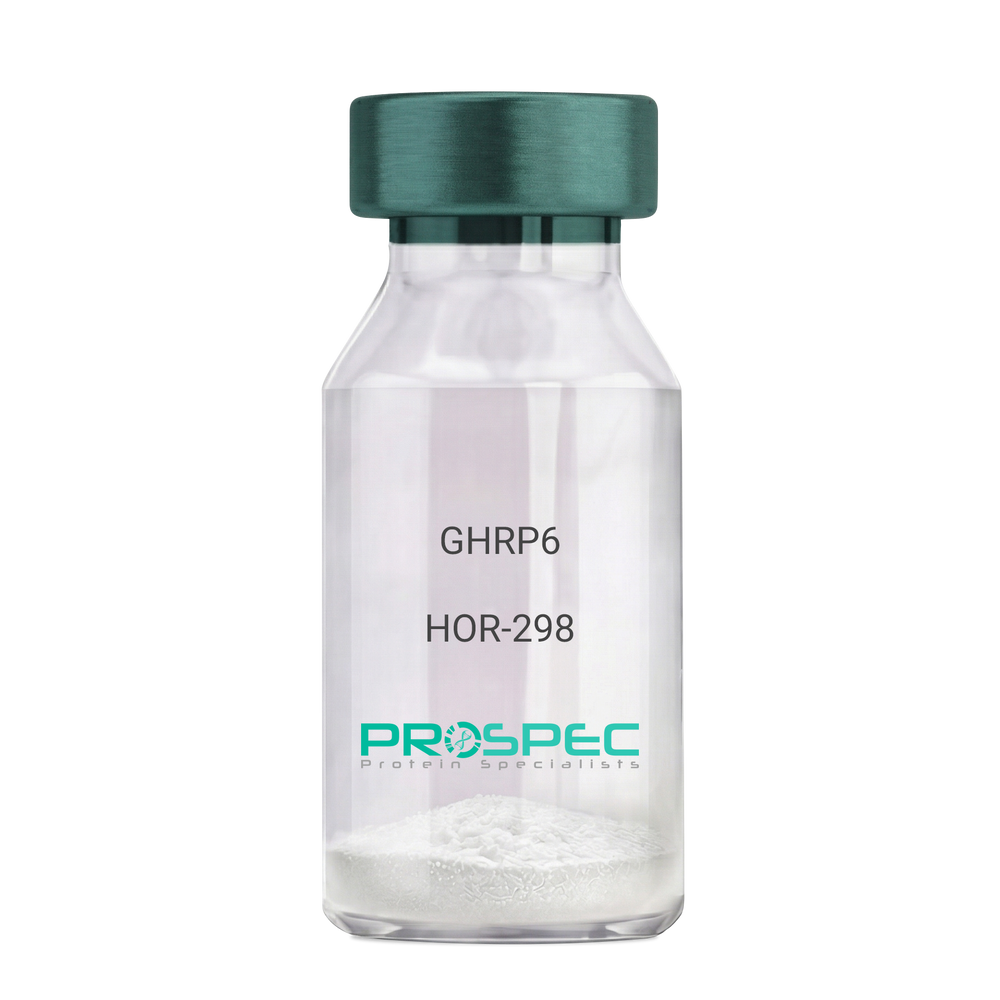
    GHRP6

    Description:

    Growth Hormone Releasing Peptide-6

    GHRP-6, GHRP6

    Product # :

    HOR-298

    Price :

    Quantity :

    Shipping Method :

    Room Temp Icon

    Shipped at Room temp

    Add To Cart

    More Info

    • description
    • formulation
    • purity
    • More Info

    Description

    Growth Hormone Releasing Peptide-6 Synthetic is a single, non-glycosylated polypeptide chain containing 6 amino acids, having a molecular mass of 873 Dalton and a Molecular formula of C46H56N12O6.

    Formulation

    The GHRP-6 peptide was lyophilized with no additives.

    Purity

    Greater than 98.0% as determined by analysis by RP-HPLC.

    More Info

    • Introduction

      GH-releasing peptides (GHRPs) are synthetic peptides that like GHRH act directly on pituitary somatotrophs to stimulate GH release. Growth hormone (GH) release is stimulated by a variety of synthetic secretagogues, of which growth hormone-releasing hexapeptide (GHRP-6) has been most thoroughly studied; it is thought to have actions at both pituitary and hypothalamic site.

    • Synonyms

      GHRP-6, GHRP6

    • Physical Appearance

      Sterile Filtered White lyophilized (freeze-dried) powder.

    • Stability

      Lyophilized Growth Hormone Releasing Peptide-6 although stable at room temperature for 3 weeks, should be stored desiccated below -18°C. Upon reconstitution GHRP-6 should be stored at 4°C between 2-7 days and for future use below -18°C. For long term storage it is recommended to add a carrier protein (0.1% HSA or BSA).Please prevent freeze-thaw cycles.

    • Solubility

      It is recommended to reconstitute the lyophilized GHRP-6 in sterile 18MΩ-cm H2O not less than 100 µg/ml, which can then be further diluted to other aqueous solutions.

    • Amino Acid Sequence

      His-D-Trp-Ala-Trp-D-Phe-Lys-NH2.

    ProSpec's products are furnished for LABORATORY RESEARCH USE ONLY. The product may not be used as drugs, agricultural or pesticidal products, food additives or household chemicals.

    Ghrp 6
  • View Data Sheet

    Name :

    NAIF1 Human

    Description:

    Nuclear Apoptosis Inducing Factor 1 Human Recombinant

    bA379C10.2, C9orf90, RP11-379C10.2, Nuclear apoptosis-inducing factor 1, NAIF1.

    Product # :

    PRO-1978

    Price :

    Quantity :

    Shipping Method :

    Ice Icon

    Shipped with Ice Packs

    Add To Cart

    More Info

    • description
    • source
    • formulation
    • purity
    • More Info

    Description

    NAIF1 Human Recombinant produced in E.Coli is a single, non-glycosylated polypeptide chain containing 350 amino acids (1-327 a.a) and having a molecular mass of 37.6kDa. NAIF1 is fused to a 23 amino acid His-tag at N-terminus & purified by proprietary chromatographic techniques.

    Source

    Escherichia Coli.

    Formulation

    NAIF1 protein solution (0.25mg/ml) containing 20mM Tris-HCl buffer (pH 8.0), 0.15M NaCl, 30% glycerol and 1mM DTT.

    Purity

    Greater than 90.0% as determined by SDS-PAGE.

    More Info

    • Introduction

      Nuclear Apoptosis Inducing Factor 1 (NAIF1) is a part of the NAIF1 family. NAIF1 Interacts with HARBI1 and Induces apoptosis.

    • Synonyms

      bA379C10.2, C9orf90, RP11-379C10.2, Nuclear apoptosis-inducing factor 1, NAIF1.

    • Physical Appearance

      Sterile Filtered colorless solution.

    • Stability

      Store at 4°C if entire vial will be used within 2-4 weeks. Store, frozen at -20°C for longer periods of time. For long term storage it is recommended to add a carrier protein (0.1% HSA or BSA).Avoid multiple freeze-thaw cycles.

    • Amino Acid Sequence

      MGSSHHHHHH SSGLVPRGSH MGSMAVPAKK RKMNFSEREV EIIVEELELK KHLLVNHFNA GVPLAAKSAA WHGILRRVNA VATCRRELPE VKKKWSDLKT EVRRKVAQVR AAVEGGEAPG PTEEDGAGGP GTGGGSGGGG PAVAPVLLTP MQQRICNLLG EATIISLPST TEIHPVALGP SATAAAATVT LTQIPTETTY HTLEEGVVEY CTAEAPPPLP PETPVDMMAQ HADTSVKPQA LKSRIALNSA KLIQEQRVTN LHVKEIAQHL EQQNDLLQMI RRSQEVQACA QERQAQAMEG TQAALSVLIQ VLRPMIKDFR RYLQSNTANP APASDPGQVA QNGQPDSIIQ.

    ProSpec's products are furnished for LABORATORY RESEARCH USE ONLY. The product may not be used as drugs, agricultural or pesticidal products, food additives or household chemicals.

    Naif1 Human
  • View Data Sheet

    Name :

    DHODH Human

    Description:

    Dihydroorotate Dehydrogenase Human Recombinant

    Dihydroorotate dehydrogenase (quinone), Dihydroorotate oxidase, human complement of yeast URA1, DHOdehase, POADS, EC 1.3.5.2, EC 1.3.3.1.

    Product # :

    ENZ-642

    Price :

    Quantity :

    Shipping Method :

    Ice Icon

    Shipped with Ice Packs

    Add To Cart

    More Info

    • description
    • source
    • formulation
    • purity
    • More Info

    Description

    DHODH Human Recombinant produced in E. coli is a single polypeptide chain containing 390 amino acids (31-395) and having a molecular mass of 42.3 kDa.DHODH is fused to a 25 amino acid His-tag at N-terminus & purified by proprietary chromatographic techniques.

    Source

    E.coli.

    Formulation

    The DHODH solution (0.5mg/1ml) contains 20mM Tris-HCl buffer (pH 8.0), 100mM NaCl, 1mM DTT and 20% glycerol.

    Purity

    Greater than 90% as determined by SDS-PAGE.

    More Info

    • Introduction

      Dihydroorotate dehydrogenase quinone (DHODH) is a member of the dihydroorotate dehydrogenase family. DHODH is a mitochondrial protein found on the outer surface of the inner mitochondrial membrane. DHODH catalyzes the 4th enzymatic step, the ubiquinone-mediated oxidation of dihydroorotate to orotate (with quinone as electron acceptor), in de novo pyrimidine biosynthesis. DHODH gene defects cause the postaxial acrofacial dysostosis (POADS), also known as Miller syndrome.

    • Synonyms

      Dihydroorotate dehydrogenase (quinone), Dihydroorotate oxidase, human complement of yeast URA1, DHOdehase, POADS, EC 1.3.5.2, EC 1.3.3.1.

    • Physical Appearance

      Sterile Filtered colorless solution.

    • Stability

      Store at 4°C if entire vial will be used within 2-4 weeks. Store, frozen at -20°C for longer periods of time. For long term storage it is recommended to add a carrier protein (0.1% HSA or BSA).Avoid multiple freeze-thaw cycles.

    • Amino Acid Sequence

      GSSHHHHHH SSGLVPRGSH MGSHMTGDER FYAEHLMPTL QGLLDPESAH RLAVRFTSLG LLPRARFQDS DMLEVRVLGH KFRNPVGIAA GFDKHGEAVD GLYKMGFGFV EIGSVTPKPQ EGNPRPRVFR LPEDQAVINR YGFNSHGLSV VEHRLRARQQ KQAKLTEDGL PLGVNLGKNK TSVDAAEDYA EGVRVLGPLA DYLVVNVSSP NTAGLRSLQG KAELRRLLTK VLQERDGLRR VHRPAVLVKI APDLTSQDKE DIASVVKELG IDGLIVTNTT VSRPAGLQGA LRSETGGLSG KPLRDLSTQT IREMYALTQG RVPIIGVGGV SSGQDALEKI RAGASLVQLY TALTFWGPPV VGKVKRELEA LLKEQGFGGV TDAIGADHRR.

    ProSpec's products are furnished for LABORATORY RESEARCH USE ONLY. The product may not be used as drugs, agricultural or pesticidal products, food additives or household chemicals.

    Dhodh Human
  • View Data Sheet

    Name :

    LDHB Mouse

    Description:

    Lactate Dehydrogenase B Mouse Recombinant

    L-lactate dehydrogenase B chain, LDH-B, LDH heart subunit, LDH-H, Ldh-2, Ldh2, Ldhb.

    Product # :

    ENZ-1052

    Price :

    Quantity :

    Shipping Method :

    Ice Icon

    Shipped with Ice Packs

    Add To Cart

    More Info

    • description
    • source
    • formulation
    • purity
    • biological activity
    • More Info

    Description

    LDHB Mouse Recombinant produced in E.Coli is a single, non-glycosylated polypeptide chain containing 357 amino acids (1-334 a.a) and having a molecular mass of 39kDa.LDHB is fused to a 23 amino acid His-tag at N-terminus & purified by proprietary chromatographic techniques.

    Source

    Escherichia Coli.

    Formulation

    LDHB protein solution (1mg/ml) containing 20mM Tris-HCl (pH8.0), 1mM DTT and 10% glycerol.

    Purity

    Greater than 95.0% as determined by SDS-PAGE.

    Biological Activity

    Specific activity is > 150 units/mg, in which one unit will 1.0 umole of pyruvate to L-lactate and beta-NAD per minute at pH 7.5.

    More Info

    • Introduction

      Lactate dehydrogenase (LDH) is an enzyme(EC1.1.1.27) present in a wide variety of organisms, including plants and animals.
      A tetrameric enzyme that catalyses the interconversion of pyruvateand lactate with concomitant interconversion of NADH and NAD+. At high concentrations of pyruvate, the enzyme exhibits feedback inhibition and the rate of conversion of pyruvate to lactate is decreased. In vertebrates, genes for three different subunits (LDH-A, LDH-B and LDH-C) exist.

    • Synonyms

      L-lactate dehydrogenase B chain, LDH-B, LDH heart subunit, LDH-H, Ldh-2, Ldh2, Ldhb.

    • Physical Appearance

      Sterile filtered colorless solution.

    • Stability

      Store at 4°C if entire vial will be used within 2-4 weeks. Store, frozen at -20°C for longer periods of time. For long term storage, it is recommended to add a carrier protein (0.1% HSA or BSA).Avoid multiple freeze-thaw cycles.

    • Amino Acid Sequence

      MGSSHHHHHH SSGLVPRGSH MGSMATLKEK LIASVADDEA AVPNNKITVV GVGQVGMACA ISILGKSLAD ELALVDVLED KLKGEMMDLQ HGSLFLQTPK IVADKDYSVT ANSKIVVVTA GVRQQEGESR LNLVQRNVNV FKFIIPQIVK YSPDCTIIVV SNPVDILTYV TWKLSGLPKH RVIGSGCNLD SARFRYLMAE KLGIHPSSCH GWILGEHGDS SVAVWSGVNV AGVSLQELNP EMGTDNDSEN WKEVHKMVVD SAYEVIKLKG YTNWAIGLSV ADLIESMLKN LSRIHPVSTM VKGMYGIENE VFLSLPCILN ARGLTSVINQ KLKDDEVAQL RKSADTLWDI QKDLKDL.

    ProSpec's products are furnished for LABORATORY RESEARCH USE ONLY. The product may not be used as drugs, agricultural or pesticidal products, food additives or household chemicals.

    Ldhb Mouse
  • View Data Sheet

    Name :

    LFA 3 Human

    Description:

    Lymphocyte Function Associated Antigen-3 Human Recombinant , Fusion Protein

    CD58, LFA-3, Ag3, Surface glycoprotein LFA-3.

    Product # :

    CYT-423

    Price :

    Quantity :

    Shipping Method :

    Room Temp Icon

    Shipped at Room temp

    Add To Cart

    More Info

    • description
    • source
    • formulation
    • purity
    • More Info

    Description

    Lymphocyte Function-Associated Antigen-3 Fusion Protein Recombinant Human is produced by recombinant DNA technology in a Chinese Hamster Ovary (CHO) mammalian cell expression system. The molecular weight is 91.4 kDa. Recombinant LFA3 is purified by proprietary chromatographic techniques.

    Source

    Chinese Hamster Ovary.

    Formulation

    Each mg of CD58 contains 0.8mg sucrose, 0.3mg glycine, 0.25mg sodium citrate dihydrate, and 4µg citric acid monohydrate.

    Purity

    Greater than 98.0% as determined by(a) Analysis by RP-HPLC.
    (b) Analysis by SDS-PAGE.

    More Info

    • Introduction

      LFA-3 is ligand of the t-lymphocyte cd2 glycoprotein. This interaction is important in mediating thymocyte interactions with thymic epithelial cells, antigen-independent and dependent interactions of t-lymphocytes with target cells and antigen- presenting cells and the t-lymphocyte rosetting with erythrocytes. In addition, the lfa-3/cd2 interaction may prime response by both the cd2+ and lfa-3+ cells.

    • Synonyms

      CD58, LFA-3, Ag3, Surface glycoprotein LFA-3.

    • Physical Appearance

      Sterile Filtered White lyophilized (freeze-dried) powder.

    • Stability

      Lyophilized LFA3 Human although stable at room temperature for 3 weeks, should be stored desiccated below -18°C. Upon reconstitution Human LFA-3 should be stored at 4°C between 2-7 days and for future use below -18°C. For long term storage it is recommended to add a carrier protein (0.1% HSA or BSA).Please prevent freeze-thaw cycles.

    • Solubility

      It is recommended to reconstitute the lyophilized LFA-3 Human in sterile 18MΩ-cm H2O not less than 100µg/ml, which can then be further diluted to other aqueous solutions.

    ProSpec's products are furnished for LABORATORY RESEARCH USE ONLY. The product may not be used as drugs, agricultural or pesticidal products, food additives or household chemicals.

    Lfa 3 Human
  • View Data Sheet

    Name :

    Protein G

    Description:

    Protein G Recombinant

    Product # :

    PRO-402

    Price :

    Quantity :

    Shipping Method :

    Room Temp Icon

    Shipped at Room temp

    Add To Cart

    More Info

    • description
    • source
    • formulation
    • purity
    • More Info
    • sds-page

    Description

    The Protein G is a single, non-glycosylated protein contains 200 amino acids having a molecular mass of 21.8kDa. The Protein-G migrates on SDS-PAGE around 32kDa.

    Source

    Escherichia Coli.

    Formulation

    Lyophilized white powder containing no additives.

    Purity

    >96% as determined by SDS-PAGE and RP-HPLC.

    sds-page

    Protein-G sds-page - Product image 1

    More Info

    • Physical Appearance

      Sterile Filtered White lyophilized (freeze-dried) powder.

    • Stability

      Lyophilized Recombinant Protein G although stable at room temperature for 3 weeks, should be stored desiccated below -18°C. Upon reconstitution Protein G should be stored at 4°C between 2-7 days and for future use below -18°C.Please prevent freeze-thaw cycles.

    • Solubility

      Reconstitution with deionized water or PBS.

    • Amino Acid Sequence

      LPKTDTYKLILNGKTLKGETTTEAVDAATAEKVFKQYANDNGVDGEWTYDDAT KTFTVTEKPEVIDASELTPAVTTYKLVINGKTLKGETTTEAVDAATAEKVFK QYANDNGVDGEWTYDDATKTFTVTEKPEVIDASELTPAVTTYKLVINGKTL KGETTTKAVDAETAEKAFKQYANDNGVDGVWTYDDATKTFTVTE.

    ProSpec's products are furnished for LABORATORY RESEARCH USE ONLY. The product may not be used as drugs, agricultural or pesticidal products, food additives or household chemicals.

    Protein G
  • View Data Sheet

    Name :

    PPARG Human (1-477)

    Description:

    Peroxisome Proliferator Activated Receptor Gamma Human Recombinant, (1-477 a.a)

    Peroxisome proliferator-activated receptor gamma, PPAR-gamma, PPARG, NR1C3, PPARG1, PPARG2.

    Product # :

    PKA-334

    Price :

    Quantity :

    Shipping Method :

    Ice Icon

    Shipped with Ice Packs

    Add To Cart

    More Info

    • description
    • source
    • formulation
    • More Info

    Description

    PPARG Human Recombinant encoding amino acids 1-477 expressed in E.coli, is fused to a GST tag and it is antibody reactive.The PPARG is purified by proprietary chromatographic techniques.

    Source

    Escherichia Coli.

    Formulation

    PPARG at 0.1mg/ml in 50mM Tris-HCl and 10mM L-glutathione (reduced).

    More Info

    • Introduction

      Peroxisome proliferators are non-genotoxic carcinogens which are purported to exert their effect on cells through their interaction with members of the nuclear hormone receptor family termed peroxisome proliferators activated receptors (PPARs). Nuclear hormone receptors are ligand-dependent intracellular proteins that stimulate transcription of specific genes by binding to specific DNA sequences following activation by the appropriate ligand. Studies indicate that PPARs are activated by peroxisome proliferators such as clofibric acid, nafenopin, and WY-14,643, as well as by some fatty acids.

    • Synonyms

      Peroxisome proliferator-activated receptor gamma, PPAR-gamma, PPARG, NR1C3, PPARG1, PPARG2.

    • Physical Appearance

      Sterile Filtered clear solution.

    • Stability

      Store vial at -20°C to -80°C. When stored at the recommended temperature, this protein is stable for 12 months. Avoid multiple freeze-thaw cycles.

    • Applications

      ELISA.
      Inhibition Assays.
      Western Blotting.

    ProSpec's products are furnished for LABORATORY RESEARCH USE ONLY. The product may not be used as drugs, agricultural or pesticidal products, food additives or household chemicals.

    Pparg Human 477 Aa
  • View Data Sheet

    Name :

    FCGR1A Antibody

    Description:

    CD64, Mouse Anti Human

    Fc Fragment Of IgG High Affinity Ia Receptor (CD64), High Affinity Immunoglobulin Gamma Fc Receptor I, Fc-Gamma Receptor I A1, FcgammaRIa, CD64 Antigen, IgG Fc Receptor I, Fc-Gamma RI, CD64, CD64A, IGFR1, FCRI.

    Product # :

    ANT-550

    Price :

    Quantity :

    Shipping Method :

    Ice Icon

    Shipped with Ice Packs

    Add To Cart

    More Info

    • formulation
    • More Info

    Formulation

    1mg/ml containing PBS, pH-7.4, 10% Glycerol and 0.02% Sodium Azide.

    More Info

    • Introduction

      FCGR1A is a member of to the immunoglobulin superfamily. FCGR1A protein acts as a high affinity receptor for the Fc region of immunoglobulins gamma and has a vital part in both innate and adaptive immune response.

    • Synonyms

      Fc Fragment Of IgG High Affinity Ia Receptor (CD64), High Affinity Immunoglobulin Gamma Fc Receptor I, Fc-Gamma Receptor I A1, FcgammaRIa, CD64 Antigen, IgG Fc Receptor I, Fc-Gamma RI, CD64, CD64A, IGFR1, FCRI.

    • Physical Appearance

      Sterile filtered colorless solution.

    • Immunogen

      Anti-human FCGR1A mAb, is derived from hybridization of mouse F0 myeloma cells with spleen cells from BALB/c mice immunized with a recombinant human FCGR1A protein 16-292 amino acids purified from E. coli.

    • Ig Subclass

      Mouse IgG2b heavy chain and k light chain.

    • Clone

      PAT11D10AT.

    • Applications

      FCGR1A antibody has been tested by ELISA, Western blot analysis to assure specificity and reactivity. Since application varies, however, each investigation should be titrated by the reagent to obtain optimal results. Recommended starting dilution is 1:2000.

    • Type

      Mouse Anti Human Monoclonal.

    • Storage Procedures

      For periods up to 1 month store at 4°C, for longer periods of time, store at -20°C. Prevent freeze thaw cycles.

    • Purification Method

      FCGR1A antibody was purified from mouse ascitic fluids by protein-A affinity chromatography.

    ProSpec's products are furnished for LABORATORY RESEARCH USE ONLY. The product may not be used as drugs, agricultural or pesticidal products, food additives or household chemicals.

    Fcgr1A Antibody
  • View Data Sheet

    Name :

    Heparanase 1 Clone HP3/17

    Description:

    Heparanase 1 (HPA1), Monoclonal Anti-Human Antibody

    Product # :

    ANT-154

    Price :

    Quantity :

    Shipping Method :

    Ice Icon

    Shipped with Ice Packs

    Add To Cart

    More Info

    • source
    • formulation
    • purity
    • More Info

    Source

    Mab HP3/17 is a Protein G affinity purified monoclonal antibody raised against a polypeptide from the 50 kDa subunit of Heparanase.

    Formulation

    Each vial contains 50, 100 or 150 μg in 14, 28 or 42 μl respectively, of 0.22 micron filtered solution of 20 mM Sodium Phosphate; 150 mM NaCl; pH 7.2, containing 0.01% Thimerosal.

    Purity

    >98% on SDS-PAGE when loaded 50 μg/lane.

    More Info

    • Introduction

      Heparanase is an endo-β-D-glucuronidase, which degrades heparan sulfate side chains of heparan sulfate proteoglycans (HSPGs) in the extracellular matrix. Heparanase plays an important role in ECM degradation, facilitating the migration and extravasation of tumor cells and inflammatory leukocytes (1,2,3). Upon degradation, heparanase releases growth factors and cytokines that stimulate cell proliferation and chemotaxis (4,5). Heparanase is a heterodimer comprised of a 50 kDa subunit harboring the active site and a 8 kDa subunit. It is produced as a latent 65 kDa precursor and proteolytically processed to its active form (1,6). Heparanase is highly expressed in myeloid leukocytes (i.e. neutrophils) in platelets and in human placenta. Human heparanase was found to be upregulated in various types of primary tumors, correlating in some cases with increased tumor invasiveness and vascularity and with poor prospective survival (7,8).

    • Stability

      Store at 4°C. Stable for six months from the date of shipment. For extended storage, freeze in working aliquots at -20°C. Avoid repeated freeze-thaw cycles.

    • Ig Subclass

      Mouse IgG2Bκ

    • Applications

      Western blot
      Immunohistochemistry

    • Data Sheet

      To view the FULL VERSION click Monoclonal Anti-Human Heparanase 1:

    • Patent Protected Countries

      Anti-heparanase antibodies and their uses, including HP3/17 and its uses, are protected by US. Patents No. 6,177,545; 6,531,129, additional US patent applications and patents and patent applications worldwide.

    • Specificity

      HP3/17 reacts with the 50 kDa subunit and with the 65 kDa precursor of human or mouse Heparanase by Western blotting and immunohistochemistry.Recommended dilution range for Western blot analysis: 1:4000.Recommended dilution range for immunohistochemistry: 1:40.

    ProSpec's products are furnished for LABORATORY RESEARCH USE ONLY. The product may not be used as drugs, agricultural or pesticidal products, food additives or household chemicals.

    Heparanase 1 Clone Hp3 17 Antibody
  • View Data Sheet

    Name :

    VEGF Antibody

    Description:

    Vascular Endothelial Cell Growth Factor, Mouse Anti-Human

    Vascular endothelial growth factor A, VEGF-A, Vascular permeability factor, VPF, VEGF, MGC70609.

    Product # :

    ANT-125

    Price :

    Quantity :

    Shipping Method :

    Room Temp Icon

    Shipped at Room temp

    Add To Cart

    More Info

    • More Info

    More Info

    • Introduction

      Vascular endothelial growth factoris an important signaling proteininvolved in both vasculogenesisand angiogenesis. As its name implies, VEGF activity has been mostly studied on cells of the vascular endothelium, although it does have effects on a number of other cell types (e.g. stimulation monocyte/macrophagemigration, neurons, cancer cells, kidney epithelial cells ).VEGF mediates increased vascular permeability, induces angiogenesis, vasculogenesis and endothelial cell growth, promotes cell migration, and inhibits apoptosis. In vitro, VEGF has been shown to stimulate endothelial cell mitogenesisand cell migration. VEGF is also a vasodilator and increases microvascular permeability and was originally referred to as vascular permeability factor.
      Elevated levels of this protein is linked to POEMS syndrome, also known as Crow-Fukase syndrome. Mutations in this gene have been associated with proliferative and nonproliferative diabetic retinopathy.

    • Synonyms

      Vascular endothelial growth factor A, VEGF-A, Vascular permeability factor, VPF, VEGF, MGC70609.

    • Solubility

      Reconstitute with sterile H20. Mix gently, wash the sides of the vial and wait 30-60 seconds before use.

    • Immunogen

      r.HumanVEGF.

    • Ig Subclass

      Mouse IgM.

    • Clone

      NYRhVEGF.

    • Applications

      Direct ELISA, Western Blot, Immuneprecipitation.

    • Titer

      By direct ELISA, 1:10,000 dilution will yield 0.15 O.D using alkaline phosphatase conjugated rabbit anti-mouse Ig (Jackson Laboratories).

    • Shipping Conditions

      Antibody is shipped lyophilized at ambient temperature.

    • Type

      Mouse Anti Human Monoclonal.

    • Storage Procedures

      In lyophilized form, for long periods, store at 4oC in a dry environment. After reconstitution, if not intended for use within a month, aliquot and store at -20oC.

    • Purification Method

      Boric acid precipitation Protein concentration1mg/ml in PBS (after reconstitution).

    ProSpec's products are furnished for LABORATORY RESEARCH USE ONLY. The product may not be used as drugs, agricultural or pesticidal products, food additives or household chemicals.

    Vegf Antibody
  • View Data Sheet

    Name :

    IGF1 Bovine

    Description:

    IGF-1 Bovine Recombinant

    Somatomedin C, IGF-I, IGFI, IGF1, IGF-IA MGF.

    Product # :

    CYT-1261

    Price :

    Quantity :

    Shipping Method :

    Room Temp Icon

    Shipped at Room temp

    Add To Cart

    More Info

    • description
    • source
    • formulation
    • purity
    • biological activity
    • More Info

    Description

    IGF1 Bovine Recombinant produced in E.Coli is a single, non-glycosylated, polypeptide chain containing 70 amino acids and having a molecular mass of 7.6kDa. IGF- I is purified by proprietary chromatographic techniques.

    Source

    Escherichia Coli.

    Formulation

    The IGF1 Bovine was lyophilized from a 0.2µm filtered concentrated solution in PBS, pH 7.0.

    Purity

    Greater than 97.0% as determined by:
    (a) Analysis by RP-HPLC.
    (b) Analysis by SDS-PAGE.

    Biological Activity

    The biological activity was determined by the cell proliferation assay using serum free human MCF-7 cells in <2ng/ml, corresponding to a Specific Activity of >5.0 x 10⁵IU/mg.

    More Info

    • Introduction

      The somatomedins, IGFs, comprise a family of peptides that play important roles in mammalian growth and development. IGF1 mediates many of the growth-promoting effects of GH. Early studies showed that GH did not directly stimulate the incorporation of sulfate into cartilage, but rather acted through a serum factor, termed 'sulfation factor,' which later became known as 'somatomedin. Three main somatomedins have been characterized: somatomedin C (IGF1), somatomedin A (IGF2; MIM 147470), and somatomedin B.

    • Synonyms

      Somatomedin C, IGF-I, IGFI, IGF1, IGF-IA MGF.

    • Physical Appearance

      Sterile Filtered White lyophilized (freeze-dried) powder.

    • Stability

      Lyophilized IGF-1 Bovine although stable at room temperature for 3 weeks, should be stored desiccated below -18°C. Upon reconstitution bovine IGF1 should be stored at 4°C between 2-7 days and for future use below -18°C. For long term storage it is recommended to add a carrier protein (0.1% HSA or BSA). Please prevent freeze-thaw cycles.

    • Solubility

      It is recommended to reconstitute the lyophilized bovine IGF-1 in sterile 18MΩ-cm H2O at a concentration of 100µg/ml, which can then be further diluted to other aqueous solutions.

    • Amino Acid Sequence

      GPETLCGAEL VDALQFVCGD RGFYFNKPTG YGSSSRRAPQ TGIVDECCFR SCDLRRLEMY CAPLKPAKSA.

    ProSpec's products are furnished for LABORATORY RESEARCH USE ONLY. The product may not be used as drugs, agricultural or pesticidal products, food additives or household chemicals.

    Igf1 Bovine
  • View Data Sheet

    Name :

    NDRG1 Antibody

    Description:

    N-Myc Downstream Regulated 1, Mouse Anti Human

    Protein NDRG1, N-myc downstream-regulated gene 1 protein, Differentiation-related gene 1 protein, Reducing agents and tunicamycin-responsive protein, Nickel-specific induction protein Cap43, DRG-1, RTP, Rit42, NDRG1, CAP43, DRG1, GC4, NDR1, NMSL, TDD5, CMT4D, HMSNL, TARG1, PROXY1.

    Product # :

    ANT-624

    Price :

    Quantity :

    Shipping Method :

    Ice Icon

    Shipped with Ice Packs

    Add To Cart

    More Info

    • formulation
    • More Info

    Formulation

    1mg/ml containing PBS, pH-7.4, 10% Glycerol and 0.02% Sodium Azide.

    More Info

    • Introduction

      NDRG1 is a cytoplasmic protein that is involved in stress responses, hormone responses, cell growth, and differentiation. NDRG1 is one of 4 members of the NDRG ?/?-hydrolase family. NDRG1 is classified in databases as a tumor suppressor and heavy metal-response protein. NDRG1’s functions include cell-cycle regulation, cellular differentiation, apoptosis, hypoxia response and metal-ion sensing. NDRG1 is also essential for p53-mediated caspase activation and apoptosis. The NDRG1 is a Rab4a effector that is involved in vesicular recycling of E-cadherin. NDRG1 is ubiquitous; it is expressed most notably in placental membranes and prostate, kidney, small intestine, and ovary tissues. NDRG1 has reduced expression in adenocarcinomas compared to normal tissues.
      NDRG1 gene mutations are reported to be the cause for hereditary motor and sensory neuropathy-Lom (HMSNL), which is a severe autosomal recessive form of Charcot- Marie-Tooth (CMT) disease. In addition, decreased NDRG1 expression in glioma is linked to tumor progression. On the other hand, overexpression of NDRG1 is connected to malignant status of esophageal cancer. NDRG1 may also have a role in portal vein invasion and intrahepatic metastasis in human hepatocellular carcinoma.

    • Synonyms

      Protein NDRG1, N-myc downstream-regulated gene 1 protein, Differentiation-related gene 1 protein, Reducing agents and tunicamycin-responsive protein, Nickel-specific induction protein Cap43, DRG-1, RTP, Rit42, NDRG1, CAP43, DRG1, GC4, NDR1, NMSL, TDD5, CMT4D, HMSNL, TARG1, PROXY1.

    • Physical Appearance

      Sterile filtered colorless solution.

    • Immunogen

      Anti-human NDRG1 mAb, is derived from hybridization of mouse F0 myeloma cells with spleen cells from BALB/c mice immunized with a recombinant human NDRG1 protein 1-394 amino acids purified from E. coli.

    • Ig Subclass

      Mouse IgG1 heavy chain and k light chain.

    • Clone

      PAT11G4AT.

    • Applications

      The antibody has been tested by ELISA, Western blot analysis, Flow cytometry and ICC/IF to assure specificity and reactivity. Since application varies, however, each investigation should be titrated by the reagent to obtain optimal results.

    • Type

      Mouse Anti Human Monoclonal.

    • Storage Procedures

      For periods up to 1 month store at 4°C, for longer periods of time, store at -20°C. Prevent freeze thaw cycles.

    • Purification Method

      NDRG1 antibody was purified from mouse ascitic fluids by protein-A affinity chromatography.

    ProSpec's products are furnished for LABORATORY RESEARCH USE ONLY. The product may not be used as drugs, agricultural or pesticidal products, food additives or household chemicals.

    Ndrg1 Antibody
  • 1
  • …
  • 36
  • 37
  • 38
  • 39
  • 40
  • …
  • 42
Your browser does not support the video tag.

Products

  • Cytokines
  • Growth Factors
  • Chemokines
  • CD Antigens
  • Neurotrophins
  • Hormones
  • Enzymes
  • Viral Antigens
  • Recombinant Proteins
  • Natural Proteins
  • Monoclonal Antibodies
  • Polyclonal Antibodies
  • Thermal Packaging

About

  • Company
  • Founder
  • Contribution
  • Contact Us
  • My Account

Ordering

  • Ordering Method
  • Ordering - Credit Card
  • Ordering PO
  • Shipping Fees
  • FAQ's

Legal

  • Terms & Conditions
  • Privacy
  • Site Map
  • info@prospecbio.com
  • Facebook
  • Instagram
  • Linkedin Profile

© 2026 ProSpec-Tany TechnoGene Ltd. All Rights Reserved.

  • Choosing a selection results in a full page refresh.
  • Opens in a new window.