Search results
1000 results found for “Glycophorin”
Name
Description
Product #
Price
Quantity
Shipping Method
- View Data Sheet
Name :
RCHY1 HumanDescription:
Ring Finger & CHY Zinc Finger Domain Containing 1 Human Recombinant
ARNIP, CHIMP, hARNIP, PIRH2, PIRH2E, PIRH2F, PRO1996, RNF199, ZNF363, RING finger and CHY zinc finger domain-containing protein 1, Androgen receptor N-terminal-interacting protein, CH-rich-interacting match with PLAG1, E3 ubiquitin-protein ligase Pirh2, RING finger protein 199, Zinc finger protein 363, p53-induced RING-H2 protein, hPirh2, RCHY1.
Product # :
PRO-1536Price :
Quantity :
Shipping Method :
Shipped with Ice Packs
More Info
- description
- source
- formulation
- purity
- More Info
Description
RCHY1 Human Recombinant produced in E. coli is a single polypeptide chain containing 284 amino acids (1-261) and having a molecular mass of 32.5kDa. RCHY1 is fused to a 23 amino acid His-tag at N-terminus & purified by proprietary chromatographic techniques.
Source
Escherichia Coli.
Formulation
The RCHY1 solution (0.5mg/ml) contains 20mM Tris-HCl buffer (pH 8.0), 0.4M UREA and 10% glycerol.
Purity
Greater than 85% as determined by SDS-PAGE.
More Info
-
Introduction
Ring Finger & CHY Zinc Finger Domain Containing 1 (RCHY1) acts as an ubiquitin ligase. RCHY1 mediates E3-dependent ubiquitination and proteasomal degradation of target proteins which among them are TP53, HDAC1 and CDKN1B, consequently regulating their levels and cell cycle progression. RCHY1 is also increases AR transcription factor activity.
-
Synonyms
ARNIP, CHIMP, hARNIP, PIRH2, PIRH2E, PIRH2F, PRO1996, RNF199, ZNF363, RING finger and CHY zinc finger domain-containing protein 1, Androgen receptor N-terminal-interacting protein, CH-rich-interacting match with PLAG1, E3 ubiquitin-protein ligase Pirh2, RING finger protein 199, Zinc finger protein 363, p53-induced RING-H2 protein, hPirh2, RCHY1.
-
Physical Appearance
Sterile Filtered clear solution.
-
Stability
Store at 4°C if entire vial will be used within 2-4 weeks. Store frozen at -20°C for longer periods of time. For long term storage it is recommended to add a carrier protein (0.1% HSA or BSA).Avoid multiple freeze-thaw cycles.
-
Amino Acid Sequence
MGSSHHHHHH SSGLVPRGSH MGSMAATARE DGASGQERGQ RGCEHYDRGC LLKAPCCDKL YTCRLCHDNN EDHQLDRFKV KEVQCINCEK IQHAQQTCEE CSTLFGEYYC DICHLFDKDK KQYHCENCGI CRIGPKEDFF HCLKCNLCLA MNLQGRHKCI ENVSRQNCPI CLEDIHTSRV VAHVLPCGHL LHRTCYEEML KEGYRCPLCM HSALDMTRYW RQLDDEVAQT PMPSEYQNMT VDILCNDCNG RSTVQFHILG MKCKICESYN TAQAGGRRIS LDQQ.
ProSpec's products are furnished for LABORATORY RESEARCH USE ONLY. The product may not be used as drugs, agricultural or pesticidal products, food additives or household chemicals.
- View Data Sheet
Name :
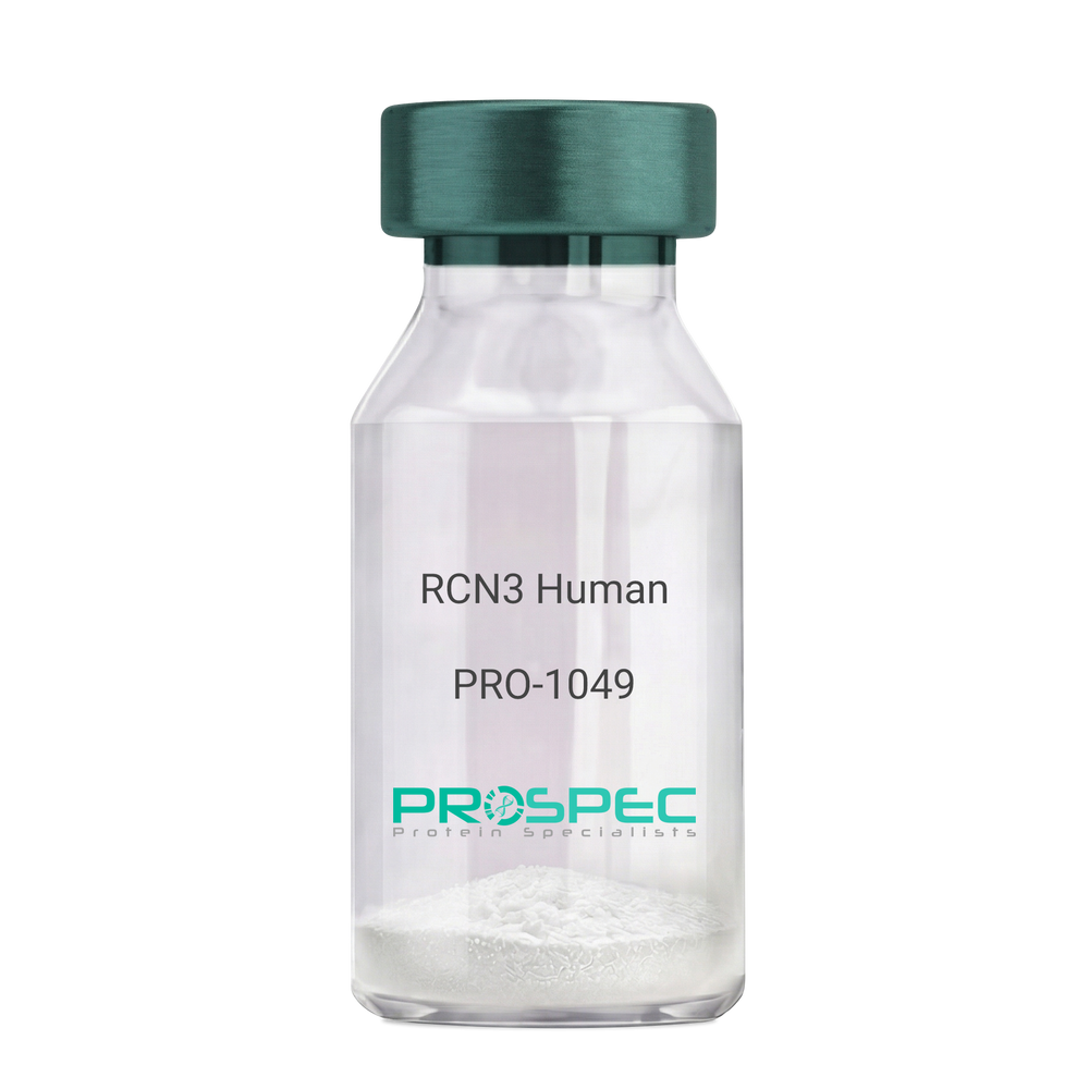
RCN3 HumanDescription:
Reticulocalbin 3 Human Recombinant
Reticulocalbin-3, EF-hand calcium-binding protein RLP49, RCN3, RLP49.
Product # :
PRO-1049Price :
Quantity :
Shipping Method :
Shipped with Ice Packs
More Info
- description
- source
- formulation
- purity
- More Info
Description
RCN3 Human Recombinant produced in E.Coli is a single, non-glycosylated polypeptide chain containing 333 amino acids (21-328 a.a) and having a molecular mass of 37.9kDa.RCN3 is fused to a 25 amino acid His-tag at N-terminus & purified by proprietary chromatographic techniques.
Source
Escherichia Coli.
Formulation
RCN3 protein solution (1mg/ml) containing 20mM Tris-HCl buffer (pH 8.0), 1mM DTT, 10% glycerol and 0.1M NaCl.
Purity
Greater than 95.0% as determined by SDS-PAGE.
More Info
-
Introduction
Reticulocalbin 3 (RCN3) belongs to the CREC (cab45/reticulocalbin/ ERC45/calumenin) family. RCN3 contains 5 Arg-Xaa-Xaa-Arg motifs, which function as target sequences of SPCs (subtilisin-like proprotein convertases), which is a family of serine endoproteases that proteolytically activate proproteins. The synthesis of PACE4 is induced by association and coexpression with RCN3.
-
Synonyms
Reticulocalbin-3, EF-hand calcium-binding protein RLP49, RCN3, RLP49.
-
Physical Appearance
Sterile Filtered colorless solution.
-
Stability
Store at 4°C if entire vial will be used within 2-4 weeks. Store, frozen at -20°C for longer periods of time. For long term storage it is recommended to add a carrier protein (0.1% HSA or BSA).Avoid multiple freeze-thaw cycles.
-
Amino Acid Sequence
MGSSHHHHHH SSGLVPRGSH MGSHMKPSPD AGPHGQGRVH QAAPLSDAPH DDAHGNFQYD HEAFLGREVA KEFDQLTPEE SQARLGRIVD RMDRAGDGDG WVSLAELRAW IAHTQQRHIR DSVSAAWDTY DTDRDGRVGW EELRNATYGH YAPGEEFHDV EDAETYKKML ARDERRFRVA DQDGDSMATR EELTAFLHPE EFPHMRDIVI AETLEDLDRN KDGYVQVEEY IADLYSAEPG EEEPAWVQTE RQQFRDFRDL NKDGHLDGSE VGHWVLPPAQ DQPLVEANHL LHESDTDKDG RLSKAEILGN WNMFVGSQAT NYGEDLTRHH DEL.
ProSpec's products are furnished for LABORATORY RESEARCH USE ONLY. The product may not be used as drugs, agricultural or pesticidal products, food additives or household chemicals.
- View Data Sheet
Name :
YWHAH Human, HisDescription:
Tyr-3/Trp-5 Monooxygenase Activation Protein ETA Human Recombinant, His Tag
14-3-3 ETA, YWHAH, YWHA1, Protein AS1, Tyr-3/Trp-5 Monooxygenase Activation Protein ETA.
Product # :
PKA-347Price :
Quantity :
Shipping Method :
Shipped with Ice Packs
More Info
- description
- source
- formulation
- purity
- More Info
Description
YWHAH Human Recombinant produced in E.Coli is a single, non-glycosylated, polypeptide chain containing 266 amino acids and having a molecular mass of 30.3 kDa. YWHAH is fused to an N-Terminal His Tag and purified by proprietary chromatographic techniques.
Source
Escherichia Coli.
Formulation
YWHAH solution containing 20mM Tris pH-8.
Purity
Greater than 95.0% as determined by SDS-PAGE.
More Info
-
Introduction
YWHAH belongs to the 14-3-3 family of proteins which mediate signal transduction by binding to phosphoserine-containing proteins. 14-3-3 ETA is found in plants and mammals, and there is 99% identity to the mouse, rat and bovine orthologs. YWHAH gene contains a 7 base pair repeat sequence in its 5' UTR, and changes in the number of this repeat has been associated with early-onset schizophrenia.
14-3-3 eta is specific to the site of joint inflammation.
14-3-3 proteins are colocalized with Lewy bodies in Parkinson disease, though there is no specific staining for the 14-3-3 eta subunit.
There are 3 different isoforms types of 14-3-3: Beta, Gamma and ETA that are DAL-1/Protein 4.1B-binding proteins. -
Synonyms
14-3-3 ETA, YWHAH, YWHA1, Protein AS1, Tyr-3/Trp-5 Monooxygenase Activation Protein ETA.
-
Physical Appearance
Sterile filtered colorless solution.
-
Stability
Store at 4°C if entire vial will be used within 2-4 weeks. Store, frozen at -20°C for longer periods of time. For long term storage it is recommended to add a carrier protein (0.1% HSA or BSA).Avoid multiple freeze-thaw cycles.
-
Amino Acid Sequence
MGSSHHHHHH SSGLVPRGSH MGDREQLLQR ARLAEQAERY DDMASAMKAV TELNEPLSNE DRNLLSVAYK NVVGARRSSW RVISSIEQKT MADGNEKKLE KVKAYREKIE KELETVCNDV LSLLDKFLIK NCNDFQYESK VFYLKMKGDY YRYLAEVASG EKKNSVVEAS EAAYKEAFEI SKEQMQPTHP IRLGLALNFS VFYYEIQNAP EQACLLAKQA FDDAIAELDT LNEDSYKDST LIMQLLRDNL TLWTSDQQDE EAGEGN.
ProSpec's products are furnished for LABORATORY RESEARCH USE ONLY. The product may not be used as drugs, agricultural or pesticidal products, food additives or household chemicals.
- View Data Sheet
Name :
CD40 Human, HEKDescription:
CD40 Human Recombinant, HEK
CD40 Molecule, TNF Receptor Superfamily Member 5, TNFRSF5, Tumor Necrosis Factor Receptor Superfamily, Member 5, Bp50, B-Cell Surface Antigen CD40, CD40L Receptor, CDW40, B Cell Surface Antigen CD40, B Cell-Associated Molecule, CD40 Antigen (TNF Receptor Superfamily Member 5), CD40 Type II Isoform, Nerve Growth Factor Receptor-Related B-Lymphocyte Activation Molecule, p50, Tumor Necrosis Factor Receptor Superfamily Member 5, CDw40, CD40 Antigen
Product # :
PRO-2719Price :
Quantity :
Shipping Method :
Shipped with Ice Packs
More Info
- description
- source
- formulation
- purity
- More Info
Description
CD40 Human Recombinant produced in HEK cells is a single, glycosylated, polypeptide chain containing 412 amino acids (21-193 a.a.) and having a molecular mass of 46.1kDa.CD40 is expressed with a 239 amino acid hIgG-His-Tag at C-Terminus and purified by proprietary chromatographic techniques.
Source
HEK 293.
Formulation
CD40 protein solution (1mg/ml) contains 10% glycerol and Phosphate-Buffered Saline (pH 7.4).
Purity
Greater than 95.0% as determined by SDS-PAGE.
More Info
-
Introduction
CD40 belongs to the TNF-receptor super family. CD40 has been found to be vital in mediating a wide range of immune and inflammatory responses including T cell-dependent immunoglobulin class switching, memory B cell development, and germinal center formation. AT-hook transcription factor AKNA is accounted to coordinately regulate the expression of CD40 and its ligand, which is significant for homotypic cell interactions. Adaptor protein TNFR2 interacts with CD40 and functions as a mediator of the signal transduction. The interaction of CD40 and its ligand is found to be essential for amyloid-beta-induced microglial activation, and therefore is considered to be an early event in Alzheimer disease pathogenesis.
-
Synonyms
CD40 Molecule, TNF Receptor Superfamily Member 5, TNFRSF5, Tumor Necrosis Factor Receptor Superfamily, Member 5, Bp50, B-Cell Surface Antigen CD40, CD40L Receptor, CDW40, B Cell Surface Antigen CD40, B Cell-Associated Molecule, CD40 Antigen (TNF Receptor Superfamily Member 5), CD40 Type II Isoform, Nerve Growth Factor Receptor-Related B-Lymphocyte Activation Molecule, p50, Tumor Necrosis Factor Receptor Superfamily Member 5, CDw40, CD40 Antigen
-
Physical Appearance
Sterile filtered colorless solution.
-
Stability
Store at 4°C if entire vial will be used within 2-4 weeks.Store, frozen at -20°C for longer periods of time.For long term storage it is recommended to add a carrier protein (0.1% HSA or BSA).Avoid multiple freeze-thaw cycles.
-
Amino Acid Sequence
EPPTACREKQ YLINSQCCSL CQPGQKLVSD CTEFTETECL PCGESEFLDT WNRETHCHQH KYCDPNLGLR VQQKGTSETD TICTCEEGWH CTSEACESCV LHRSCSPGFG VKQIATGVSD TICEPCPVGF FSNVSSAFEK CHPWTSCETK DLVVQQAGTN KTDVVCGPQD RLRLEPKSCD KTHTCPPCPA PELLGGPSVF LFPPKPKDTL MISRTPEVTC VVVDVSHEDP EVKFNWYVDG VEVHNAKTKP REEQYNSTYR VVSVLTVLHQ DWLNGKEYKC KVSNKALPAP IEKTISKAKG QPREPQVYTL PPSRDELTKN QVSLTCLVKG FYPSDIAVEW ESNGQPENNY KTTPPVLDSD GSFFLYSKLT VDKSRWQQGN VFSCSVMHEA LHNHYTQKSL SLSPGKHHHH HH
ProSpec's products are furnished for LABORATORY RESEARCH USE ONLY. The product may not be used as drugs, agricultural or pesticidal products, food additives or household chemicals.
- View Data Sheet
Name :
RPL34 HumanDescription:
Ribosomal Protein L34 Human Recombinant
Ribosomal Protein L34, 60S Ribosomal Protein L34, Leukemia-Associated Protein, L34.
Product # :
PRO-1704Price :
Quantity :
Shipping Method :
Shipped with Ice Packs
More Info
- description
- source
- formulation
- purity
- More Info
Description
RPL34 Human Recombinant produced in E.coli is a single, non-glycosylated polypeptide chain containing 140 amino acids (1-117) and having a molecular mass of 15.7kDa.RPL34 is fused to a 23 amino acid His-tag at N-terminus & purified by proprietary chromatographic techniques.
Source
Escherichia Coli.
Formulation
The RPL34 solution (1mg/ml) contains 20mM Tris-HCl buffer (pH 8.0), 0.4M Urea and 10% glycerol.
Purity
Greater than 85.0% as determined by SDS-PAGE.
More Info
-
Introduction
Ribosomes, the organelles which catalyze protein synthesis, contain a small 40S subunit and a large 60S subunit. Together these subunits are composed of 4 RNA species and nearly 80 structurally different proteins. RPL34 is a member of the L34E family of ribosomal proteins and it encodes a ribosomal protein which is a component of the 60S subunit. RPL34 is found in the cytoplasm and has been mapped to 4q though it was initially believed it is located at 17q21. Transcript variants which are a result of alternative splicing, alternative transcription initiation sites, and/or alternative polyadenylation exist; all encode the same protein. As is typical for genes encoding ribosomal proteins, there are several processed pseudogenes of this gene spread all over the genome.
-
Synonyms
Ribosomal Protein L34, 60S Ribosomal Protein L34, Leukemia-Associated Protein, L34.
-
Physical Appearance
Sterile Filtered clear solution.
-
Stability
Store at 4°C if entire vial will be used within 2-4 weeks. Store, frozen at -20°C for longer periods of time. For long term storage it is recommended to add a carrier protein (0.1% HSA or BSA).Avoid multiple freeze-thaw cycles.
-
Amino Acid Sequence
MGSSHHHHHH SSGLVPRGSH MGSMVQRLTY RRRLSYNTAS NKTRLSRTPG NRIVYLYTKK VGKAPKSACG VCPGRLRGVR AVRPKVLMRL SKTKKHVSRA YGGSMCAKCV RDRIKRAFLI EEQKIVVKVL KAQAQSQKAK
ProSpec's products are furnished for LABORATORY RESEARCH USE ONLY. The product may not be used as drugs, agricultural or pesticidal products, food additives or household chemicals.
- View Data Sheet
Name :
TSH ProteinDescription:
Thyroid Stimulating Hormone Human Recombinant
Glycoprotein hormones alpha chain, Anterior pituitary glycoprotein hormones common subunit alpha, Follitropin alpha chain, Follicle-stimulating hormone alpha chain, FSH-alpha, Lutropin alpha chain, Luteinizing hormone alpha chain, LSH-alpha, Thyrotropin alpha chain, Thyroid-stimulating hormone alpha chain, TSH-alpha, Choriogonadotropin alpha chain, Chorionic gonadotrophin alpha subunit, CG-alpha, Thyrotropin subunit beta, Thyroid-stimulating hormone subunit beta, TSH-beta, TSH-B, Thyrotropin beta chain, Thyrotropin alfa.
Product # :
HOR-050Price :
Quantity :
Shipping Method :
Shipped at Room temp
More Info
- description
- source
- formulation
- purity
- biological activity
- More Info
Description
Thyroid Stimulating Hormone Human Recombinant produced in HEK 293 cells.
Source
HEK 293 cells.
Formulation
Lyophilized from a concentrated 50mM ammonium bicarbonate.
Purity
Greater than 95.0% as determined by SDS-PAGE.
Biological Activity
The protein is biologically active using Siemens Centaur CP is standardized against WHO 3rd IS 81/565.
More Info
-
Synonyms
Glycoprotein hormones alpha chain, Anterior pituitary glycoprotein hormones common subunit alpha, Follitropin alpha chain, Follicle-stimulating hormone alpha chain, FSH-alpha, Lutropin alpha chain, Luteinizing hormone alpha chain, LSH-alpha, Thyrotropin alpha chain, Thyroid-stimulating hormone alpha chain, TSH-alpha, Choriogonadotropin alpha chain, Chorionic gonadotrophin alpha subunit, CG-alpha, Thyrotropin subunit beta, Thyroid-stimulating hormone subunit beta, TSH-beta, TSH-B, Thyrotropin beta chain, Thyrotropin alfa.
-
Physical Appearance
Sterile Filtered White lyophilized (freeze-dried) powder.
-
Stability
Lyophilized Recombinant TSH although stable at room temperature for 3 weeks, should be stored desiccated below -18°C. Upon reconstitution Recombinant TSH should be stored at 4°C between 2-7 days and for future use below -18°C. For long term storage it is recommended to add a carrier protein (0.1% HSA or BSA). Please prevent freeze-thaw cycles.
-
Solubility
It is recommended to reconstitute the lyophilized recombinant TSH in sterile 10 mM Sodium Phosphate, 150 mM Sodium Chloride, 1 mg/ml BSA, 0.1% Sodium Azide (optional), pH 7.4.
-
Background
Thyroid-stimulating hormone (TSH), also known as thyrotropin, is a glycoprotein hormone produced by the anterior pituitary gland. Its primary function is to regulate thyroid gland activity by stimulating the synthesis and secretion of thyroid hormones, thyroxine (T4) and triiodothyronine (T3). Recombinant human TSH (rhTSH) has emerged as a valuable tool in clinical practice, particularly in the management of thyroid disorders and in diagnostic procedures involving the thyroid gland. This paper aims to provide an overview of research on rhTSH, including its mechanism of action, activity, and therapeutic applications.
The mechanism of action of rhTSH involves binding to the TSH receptor (TSHR) on the surface of thyroid follicular cells. This interaction activates intracellular signalling pathways, including cyclic adenosine monophosphate (cAMP) production and protein kinase A (PKA) activation. These pathways stimulate various cellular processes within thyroid follicular cells, such as iodine uptake, thyroid hormone synthesis, and secretion.
What is the source or expression system of TSH PROTEIN Protein?
HEK 293 cells.
What is the Purity of TSH PROTEIN Protein?
TSH PROTEIN Protein is >95% pure as determined by SDS-PAGE.
What is the Biological Activity of TSH PROTEIN Protein?
The protein is biologically active using Siemens Centaur CP is standardized against WHO 3rd IS 81/565.
What applications can TSH PROTEIN Protein be used in?
TSH PROTEIN Protein can probably be used in western blot, ELISA and Lateral Flow.
What is the endotoxin level for TSH PROTEIN Protein?
The endotoxin level is minimal, TSH PROTEIN Protein was purified using conventional chromatography techniques.
ProSpec's products are furnished for LABORATORY RESEARCH USE ONLY. The product may not be used as drugs, agricultural or pesticidal products, food additives or household chemicals.
- View Data Sheet
Description:
Vasoactive Intestinal Peptide
Product # :
HOR-043Price :
Quantity :
Shipping Method :
Shipped at Room temp
More Info
- description
- formulation
- purity
- More Info
Description
Vasoactive Intestinal Peptide Synthetic is a single, non-glycosylated polypeptide chain containing 28 amino acids, having a molecular mass of 3325 Dalton and a Molecular formula of C147H238N44O42S .
Formulation
The protein was lyophilized with no additives.
Purity
Greater than 97.0% as determined by analysis by RP-HPLC.
More Info
-
Physical Appearance
Sterile Filtered White lyophilized (freeze-dried) powder.
-
Stability
Lyophilized Vasoactive Intestinal Peptide although stable at room temperature for 3 weeks, should be stored desiccated below -18°C. Upon reconstitution Vasoactive Intestinal Peptide should be stored at 4°C between 2-7 days and for future use below -18°C. For long term storage it is recommended to add a carrier protein (0.1% HSA or BSA). Please prevent freeze-thaw cycles.
-
Solubility
It is recommended to reconstitute the lyophilized Vasoactive Intestinal Peptide in sterile 18MΩ-cm H2O not less than 100 µg/ml, which can then be further diluted to other aqueous solutions.
-
Amino Acid Sequence
H-His-Ser-Asp-Ala-Val-Phe-Thr-Asp-Asn-Tyr-Thr-Arg-Leu-Arg-Lys-Gln-Met-Ala-Val-Lys-Lys-Tyr-Leu-Asn-Ser-Ile-Leu-Asn-NH2.
-
Background
Vasoactive Intestinal Peptide (VIP), a prominent member of the glucagon-secretin peptide superfamily, plays a multifaceted role as a neuropeptide and neurotransmitter. This research paper aims to provide an extensive analysis of VIP, exploring its biochemical properties, diverse physiological functions, and potential therapeutic applications in various disease conditions.
Vasoactive Intestinal Peptide (VIP), originally identified for its potent vasodilatory properties in the gastrointestinal system, has since been recognized for its wide-ranging physiological effects throughout the body. As a neuropeptide and neurotransmitter.
VIP, a 28-amino acid peptide, exhibits a diverse range of biological activities due to its interactions with multiple G protein-coupled receptors (GPCRs) from the VIP/PACAP receptor family (Harmar et al., 2012). These receptors are expressed in various tissues and cells, enabling VIP to exert its pleiotropic effects.
VIP is involved in the regulation of numerous physiological processes. It functions as a potent vasodilator, modulates smooth muscle activity in the gastrointestinal tract, and participates in neurotransmission in the central and peripheral nervous systems (Lelievre et al., 2008). Additionally, VIP has immunomodulatory properties, influencing immune cell functions and inflammatory responses (Delgado et al., 2004).
VIP serves as a neurotransmitter in the central nervous system, where it contributes to various neuronal functions, including learning, memory, and sensory perception (Deng et al., 2015). Moreover, VIP has demonstrated neuroprotective effects in neurodegenerative disorders, making it a potential therapeutic target for neuroprotection and neuroregeneration (Delgado et al., 2002).
VIP's diverse physiological functions offer potential therapeutic applications in various disease contexts. Research has explored its potential in the treatment of inflammatory diseases, neurodegenerative disorders, and gastrointestinal disorders (Delgado et al., 2013). Furthermore, VIP-based therapies are being investigated for their neuroprotective potential in brain injuries and stroke.
As the research on VIP continues to progress, further investigation is warranted to uncover the precise mechanisms of action and potential clinical applications in medicine. VIP stands as a remarkable example of a neuropeptide with extensive physiological roles and therapeutic potential in various disease conditions.
What is the molecular weight/Mw of VASOACTIVE INTESTINAL PEPTIDE Protein?
VASOACTIVE INTESTINAL PEPTIDE Protein has a total Mw of 3.32kDa.
What is the Purity of VASOACTIVE INTESTINAL PEPTIDE Protein?
VASOACTIVE INTESTINAL PEPTIDE Protein is >97% pure as determined by SDS-PAGE.
What is the Biological Activity of VASOACTIVE INTESTINAL PEPTIDE Protein?
The biological functionality of VASOACTIVE INTESTINAL PEPTIDE Protein will be determined in the future.
What is the amino acid sequence of VASOACTIVE INTESTINAL PEPTIDE Protein?
H-His-Ser-Asp-Ala-Val-Phe-Thr-Asp-Asn-Tyr-Thr-Arg-Leu-Arg-Lys-Gln-Met-Ala-Val-Lys-Lys-Tyr-Leu-Asn-Ser-Ile-Leu-Asn-NH2.
What applications can VASOACTIVE INTESTINAL PEPTIDE Protein be used in?
VASOACTIVE INTESTINAL PEPTIDE Protein can probably be used in western blot, ELISA and Lateral Flow.
What is the endotoxin level for VASOACTIVE INTESTINAL PEPTIDE Protein?
The endotoxin level is minimal, VASOACTIVE INTESTINAL PEPTIDE Protein was purified using conventional chromatography techniques.
ProSpec's products are furnished for LABORATORY RESEARCH USE ONLY. The product may not be used as drugs, agricultural or pesticidal products, food additives or household chemicals.
- View Data Sheet
Name :
Omp PyloriDescription:
Helicobacter Pylori Outer Membrane Protein Recombinant
Product # :
PRO-369Price :
Quantity :
Shipping Method :
Shipped with Ice Packs
More Info
- description
- source
- formulation
- purity
- More Info
Description
Omp Pylori recombinant antigen is produced in E. coli expressing the H. pylori outer membrane protein having the Mw of 23 kDa. Omp Pylori recombinant antigen is well recognized by specific IgG and IgM from H. pylori infected patients.
Source
E.Coli
Formulation
The Omp Pylori recombinant protein is formulated in 1xPBS pH 7.4.
Purity
Greater than 95% pure as determined by 12% PAGE (Coomassie staining).
More Info
-
Introduction
Helicobacter pylori, a Gram-negative, microaerophilic bacterium that populates in the stomach, predominantly at the antrum. Helicobacter pylori causes a chronic inflammation of the mucoid lining of stomach and is highly related to the growth of duodenal and gastric ulcers and stomach cancer. Over 50% of the world's population harbor H. pylori in their upper gastrointestinal tract, infection is more prevalent in developing countries. Thus far, no ideal target H. pylori antigen has been developed for the diagnostic purpose. 23 kDa H. pylori outer membrane protein was identified with a good sensitivity and coverage in the diagnosis of H. pylori infection.
-
Physical Appearance
Sterile filtered liquid formulation.
-
Stability
Omp Pylori although stable at 4°C for 1 week, should be stored below -18°C. Please prevent freeze thaw cycles.
-
Amino Acid Sequence
MLVTKLAPDFKAPAVLGNNEVDEHFELSKNLGKNGAILFFWP
KDFTFVCPTEIIAFDKRVKDFQEKGFNVIGVSIDSEQVHFAWK
NTPVEKGGIGQVTFPMVADITKSISRDYDVLFEEAIALRGAFLI
DKNMKVRHAVINDLPLGRNADEMLRMVDALLHFEEHGEVCP
AGWRKGDKGMKATHQGVAEYLKENSIKL. -
Purification Method
Purified by proprietary chromatographic technique.
ProSpec's products are furnished for LABORATORY RESEARCH USE ONLY. The product may not be used as drugs, agricultural or pesticidal products, food additives or household chemicals.
- View Data Sheet
Name :
OmpA S.EnteritidisDescription:
Salmonella Enteritidis Outer Membrane Protein-A Recombinant
Outer Membrane Protein-A, OmpA.
Product # :
PRO-1918Price :
Quantity :
Shipping Method :
Shipped with Ice Packs
More Info
- description
- source
- formulation
- purity
- More Info
Description
The Recombinant Salmonella Enteritidis Outer Membrane Protein A, E.Coli derived, 330 amino acids, contains the ompA immunodominant regions. The protein is fused to a His tag at C-terminal and purified by standard chromatography techniques.
Source
Escherichia Coli.
Formulation
PBS and 25MmM Arginine.
Purity
Protein is >95% pure as determined by 10% PAGE (coomassie staining).
More Info
-
Introduction
The OmpA protein is one of the main outer-membrane proteins of a large array of Gram-negative bacteria such as A.salmonicida, Shigella dysenteriae and E.coli.OmpA’s major physiological functions include maintenance of the structural integrity and morphology of the cells and porin activity, as well as a role in conjugation and bacteriophage binding.Achromogenic atypical Aeromonas salmonicida is the causative agent of goldfish ulcer disease.Virulence of this bacterium is associated with the production of a paracrystalline outer membrane A-layer protein.The species specific structural gene for the monomeric form of A-protein was cloned into a pET-3d plasmid in order to express and produce a recombinant form of the protein in E.coli BL21(DE3). The induced protein was isolated from inclusion bodies by a simple solubilization-renaturation procedure and purified by ion exchange chromatography on Q-Sepharose to over 95% pure monomeric protein.Recombinant A-protein was compared by biochemical, immunological and molecular methods with the A-protein isolated from atypical A.salmonicida bacterial cells by the glycine and the membrane extraction methods.
-
Synonyms
Outer Membrane Protein-A, OmpA.
-
Physical Appearance
Sterile Filtered clear solution.
-
Stability
OmpA S.Enteritidis Recombinant although stable at 4°C for 1 week, should be stored below -18°C. Please prevent freeze thaw cycles.
-
Applications
Immunoassay. Outer membrane protein A (ompA) of S. Enteritidis is a protein directly exposing to outsides of this organism, In hens, the production of antibodies against outer membrane protein A (ompA) during the infection has been demonstrated by inoculating both the complete bacterium and expressed protein produced from ompA DNA vaccine. Vaccination by ompA protein to hens is a poteintail tool to control S. enteritidis contaminated eggs into market, and prevent human foodborne disease from eggs.
ProSpec's products are furnished for LABORATORY RESEARCH USE ONLY. The product may not be used as drugs, agricultural or pesticidal products, food additives or household chemicals.
- View Data Sheet
Name :
PCOLCE HumanDescription:
Procollagen C-Endopeptidase Enhancer Human Recombinant
Procollagen C-Endopeptidase Enhancer, Procollagen C-Proteinase Enhancer 1, Type I Procollagen COOH-Terminal Proteinase Enhancer, Type 1 Procollagen C-Proteinase Enhancer Protein, Procollagen COOH-Terminal Proteinase Enhancer 1, PCPE-1, PCPE1, Procollagen, Type 1, COOH-Terminal Proteinase Enhancer, Procollagen C-Endopeptidase Enhancer 1, COOH-Terminal Proteinase Enhancer, Procollagen, Type 1, PCPE, Procollagen C-endopeptidase enhancer 1.
Product # :
ENZ-863Price :
Quantity :
Shipping Method :
Shipped with Ice Packs
More Info
- description
- source
- formulation
- purity
- More Info
Description
PCOLCE Human Recombinant produced in E.Coli is a single, non-glycosylated polypeptide chain containing 447 amino acids (26-449 a.a) and having a molecular mass of 47.9kDa. PCOLCE is fused to a 23 amino acid His-tag at N-terminus & purified by proprietary chromatographic techniques.
Source
Escherichia Coli.
Formulation
PCOLCE protein solution (1mg/ml) containing 20mM Tris-HCl buffer (pH 8.0) and 10% glycerol.
Purity
Greater than 85.0% as determined by SDS-PAGE.
More Info
-
Introduction
Procollagen C-endopeptidase enhancer 1, also known as PCOLCE binds to the C-terminal propeptide of type I procollagen and enhances procollagen C-proteinase activity. In addition, C-terminal processed part of PCPE (CT-PCPE) has a metalloproteinase inhibitory activity. Among the diseases which are associated with PCOLCE: bone fracture & oculopharyngeal muscular dystrophy.
-
Synonyms
Procollagen C-Endopeptidase Enhancer, Procollagen C-Proteinase Enhancer 1, Type I Procollagen COOH-Terminal Proteinase Enhancer, Type 1 Procollagen C-Proteinase Enhancer Protein, Procollagen COOH-Terminal Proteinase Enhancer 1, PCPE-1, PCPE1, Procollagen, Type 1, COOH-Terminal Proteinase Enhancer, Procollagen C-Endopeptidase Enhancer 1, COOH-Terminal Proteinase Enhancer, Procollagen, Type 1, PCPE, Procollagen C-endopeptidase enhancer 1.
-
Physical Appearance
Sterile filtered colorless solution.
-
Stability
Store at 4°C if entire vial will be used within 2-4 weeks. Store, frozen at -20°C for longer periods of time. For long term storage it is recommended to add a carrier protein (0.1% HSA or BSA).Avoid multiple freeze-thaw cycles.
-
Amino Acid Sequence
MGSSHHHHHH SSGLVPRGSH MGSQTPNYTR PVFLCGGDVK GESGYVASEG FPNLYPPNKE CIWTITVPEG QTVSLSFRVF DLELHPACRY DALEVFAGSG TSGQRLGRFC GTFRPAPLVA PGNQVTLRMT TDEGTGGRGF LLWYSGRATS GTEHQFCGGR LEKAQGTLTT PNWPESDYPP GISCSWHIIA PPDQVIALTF EKFDLEPDTY CRYDSVSVFN GAVSDDSRRL GKFCGDAVPG SISSEGNELL VQFVSDLSVT ADGFSASYKT LPRGTAKEGQ GPGPKRGTEP KVKLPPKSQP PEKTEESPSA PDAPTCPKQC RRTGTLQSNF CASSLVVTAT VKSMVREPGE GLAVTVSLIG AYKTGGLDLP SPPTGASLKF YVPCKQCPPM KKGVSYLLMG QVEENRGPVL PPESFVVLHR PNQDQILTNL SKRKCPSQPV RAAASQD.
ProSpec's products are furnished for LABORATORY RESEARCH USE ONLY. The product may not be used as drugs, agricultural or pesticidal products, food additives or household chemicals.
- View Data Sheet
Name :
Adiponectin Human, Sf9Description:
Adiponectin Human Recombinant, Sf9
Adiponectin, C1Q And Collagen Domain Containing, Adipose Most Abundant Gene Transcript 1 Protein, Adipocyte Complement-Related 30 KDa Protein, 30 KDa Adipocyte Complement-Related Protein, ACRP30, APM-1, GBP28, ACDC, APM1, Adipocyte, C1q And Collagen Domain-Containing Protein, Adipocyte, C1Q And Collagen Domain Containing, Adipose Most Abundant Gene Transcript 1, Adipose Specific Collagen-Like Factor, Gelatin-Binding Protein 28, Gelatin-Binding Protein, Adiponectin Precursor, Adiponectin, ADIPQTL1, ADPN, 30 kDa adipocyte complement-related protein, dipocyte complement-related 30 kDa protein, ACRP30, Adipocyte, C1q and collagen domain-containing protein, Adipose most abundant gene transcript 1 protein, apM-1, Gelatin-binding protein.
Product # :
CYT-1002Price :
Quantity :
Shipping Method :
Shipped with Ice Packs
More Info
- description
- source
- formulation
- purity
- More Info
- sds-page
Description
Acrp30 Human Recombinant produced in Sf9 Baculovirus cells is a single, glycosylated polypeptide chain containing 146 amino acids (106-242a.a.) and having a molecular mass of 16.9kDa. (Molecular size on SDS-PAGE will appear at approximately 13.5-18kDa). Acrp30 is expressed with a 6 amino acid His tag at C-Terminus and purified by proprietary chromatographic techniques.
Source
Sf9, Baculovirus cells.
Formulation
Acrp30 protein solution (0.5mg/ml) contains Phosphate Buffered Saline (pH 7.4) and 10% glycerol.
Purity
Greater than 95.0% as determined by SDS-PAGE.
sds-page
More Info
-
Introduction
The adipose tissue exclusively expresses and secretes Adiponectin (Acrp30). Acrp30 is involved in various physiological processes such as energy homeostasis, insulin sensitivity, hormonal processes, fatty acid metabolism and obesity.
Adiponectin circulates in the plasma. Decreased levels of Adiponectin are associated with insulin resistance and hyperinsulinemia, as seen in people with obesity insulin resistance, and diabetes type 2, whose plasma levels of adiponectin are reduced.
The modular structure of Acrp30 is comprised of N-terminal collagenous domain followed by a C-terminal globular domain.
Acrp30 also acts as a significant negative regulator in hematopoiesis and immune systems; it may be involved in ending inflammatory responses through its inhibitory functions. Adiponectin inhibits endothelial NF-kappa-b signaling through a cAMP-dependent pathway, it also inhibits TNF-alpha- induced expression of endothelial adhesion molecules. -
Synonyms
Adiponectin, C1Q And Collagen Domain Containing, Adipose Most Abundant Gene Transcript 1 Protein, Adipocyte Complement-Related 30 KDa Protein, 30 KDa Adipocyte Complement-Related Protein, ACRP30, APM-1, GBP28, ACDC, APM1, Adipocyte, C1q And Collagen Domain-Containing Protein, Adipocyte, C1Q And Collagen Domain Containing, Adipose Most Abundant Gene Transcript 1, Adipose Specific Collagen-Like Factor, Gelatin-Binding Protein 28, Gelatin-Binding Protein, Adiponectin Precursor, Adiponectin, ADIPQTL1, ADPN, 30 kDa adipocyte complement-related protein, dipocyte complement-related 30 kDa protein, ACRP30, Adipocyte, C1q and collagen domain-containing protein, Adipose most abundant gene transcript 1 protein, apM-1, Gelatin-binding protein.
-
Physical Appearance
Sterile Filtered clear solution.
-
Stability
Store at 4°C if entire vial will be used within 2-4 weeks. Store, frozen at -20°C for longer periods of time. For long term storage it is recommended to add a carrier protein (0.1% HSA or BSA).Avoid multiple freeze-thaw cycles.
-
Amino Acid Sequence
ADPEGAYVYR SAFSVGLETY VTIPNMPIRF TKIFYNQQNH YDGSTGKFHC NIPGLYYFAY HITVYMKDVK VSLFKKDKAM LFTYDQYQEN NVDQASGSVL LHLEVGDQVW LQVYGEGERN GLYADNDNDS TFTGFLLYHD HHHHHH.
-
Background
What is the molecular weight/Mw of ADIPONECTIN Protein?
ADIPONECTIN Protein has a total Mw of 16.9kDa.
What is the source or expression system of ADIPONECTIN Protein?
Sf9, Baculovirus cells.
What is the Purity of ADIPONECTIN Protein?
ADIPONECTIN Protein is >95% pure as determined by SDS-PAGE.
What is the Biological Activity of ADIPONECTIN Protein?
The biological functionality of ADIPONECTIN Protein will be determined in the future.
What is the amino acid sequence of ADIPONECTIN Protein?
ADPEGAYVYR SAFSVGLETY VTIPNMPIRF TKIFYNQQNH YDGSTGKFHC NIPGLYYFAY HITVYMKDVK VSLFKKDKAM LFTYDQYQEN NVDQASGSVL LHLEVGDQVW LQVYGEGERN GLYADNDNDS TFTGFLLYHD HHHHHH.
What applications can ADIPONECTIN Protein be used in?
ADIPONECTIN Protein can probably be used in western blot, ELISA and Lateral Flow.
What is the endotoxin level for ADIPONECTIN Protein?
The endotoxin level is minimal, ADIPONECTIN Protein was purified using conventional chromatography techniques.
ProSpec's products are furnished for LABORATORY RESEARCH USE ONLY. The product may not be used as drugs, agricultural or pesticidal products, food additives or household chemicals.
- View Data Sheet
Name :
BMP3 HumanDescription:
Bone Morphogenetic protein-3 Human Recombinant
Bone Morphogenetic Protein 3, Osteogenin, Bone Morphogenetic Protein 3 (Osteogenic), Bone Morphogenetic Protein 3A, BMP-3A, BMP-3, Bone Morphogenetic Protein-3, BMP3A, BMP3.
Product # :
CYT-937Price :
Quantity :
Shipping Method :
Shipped at Room temp
More Info
- description
- source
- formulation
- purity
- biological activity
- More Info
Description
BMP3 Human Recombinant produced in E.coli is a non-glycosylated disulfide linked homodimer containing 2 chains of 110 amino acids and having a molecular mass of 24.8kDa.The BMP-3 is purified by proprietary chromatographic techniques.
Source
Escherichia Coli.
Formulation
BMP-3 protein was lyophilized from a 0.2µm filtered concentrated solution in 30% Acetonitrile and 0.1% TFA.
Purity
Greater than 95.0% as determined by: (a) Analysis by HPLC. (b) Analysis by SDS-PAGE.
Biological Activity
The ED50 as determined by its ability to inhibit BMP-2-induced activity in murine MC3T3- E1 cells.More Info
-
Introduction
Bone Morphogenetic Protein 3 (BMP3) is one of the BMPs, some of which are members of the TGF-beta superfamily (BMP2-7). There are more than 13 BMPs, which are involved in inducing cartilage and bone formation, embryogenesis and morphogenesis of various tissues and organs. In addition, BMPs regulate the growth, differentiation, chemotaxis, and apoptosis of various cell types. Akin to most other TGF-beta family proteins, BMPs are extremely conserved across animal species. At the amino acid sequence level, mature human and rat BMP3 are 98% identical.
-
Synonyms
Bone Morphogenetic Protein 3, Osteogenin, Bone Morphogenetic Protein 3 (Osteogenic), Bone Morphogenetic Protein 3A, BMP-3A, BMP-3, Bone Morphogenetic Protein-3, BMP3A, BMP3.
-
Physical Appearance
Sterile Filtered White lyophilized (freeze-dried) powder.
-
Stability
Lyophilized BMP3 although stable at room temperature for 3 weeks, should be stored desiccated below -18°C. Upon reconstitution BMP-3 should be stored at 4°C between 2-7 days and for future use below -18°C. For long term storage it is recommended to add a carrier protein (0.1% HSA or BSA). Please prevent freeze-thaw cycles.
-
Solubility
It is recommended to reconstitute the lyophilized BMP3 in sterile 4mM HCl not less than 100µg/ml, which can then be further diluted to other aqueous solutions.
-
Amino Acid Sequence
QWIEPRNCAR RYLKVDFADI GWSEWIISPK SFDAYYCSGA CQFPMPKSLK PSNHATIQSI VRAVGVVPGI PEPCCVPEKM SSLSILFFDE NKNVVLKVYP NMTVESCACR.
-
Background
Bone Morphogenetic Protein-3 Human Recombinant: Unveiling the Potential of a Key Regulator in Tissue Regeneration
Abstract:
Bone Morphogenetic Protein-3 (BMP-3) human recombinant is a critical member of the bone morphogenetic protein family, known for its role in tissue development, repair, and regeneration. This research paper provides a comprehensive analysis of BMP-3, including its characteristics, signaling pathways, and potential therapeutic applications. Additionally, innovative methodologies for the production and optimization of BMP-3 human recombinant are proposed, shedding light on its future implications in the field of regenerative medicine.
Introduction:
Tissue regeneration is a complex biological process requiring precise molecular cues. BMP-3, a crucial member of the BMP family, plays a significant role in tissue development and regeneration. This paper explores the unique features of BMP-3 and presents novel approaches for its production and optimization, aiming to unlock its therapeutic potential in various regenerative contexts.
Characteristics and Signaling Pathways:
BMP-3 is a secreted protein that binds to cell surface receptors, initiating intracellular signaling cascades. It influences cell differentiation, proliferation, and extracellular matrix synthesis through both Smad-dependent and Smad-independent signaling pathways. BMP-3 signaling regulates critical processes involved in tissue regeneration, including chondrogenesis and osteogenesis.
Production of BMP-3 Human Recombinant:
Efficient production methodologies are essential for harnessing the therapeutic potential of BMP-3 human recombinant. Recombinant protein expression systems, such as Escherichia coli or mammalian cells, have been utilized to produce functional BMP-3. Optimization strategies, including codon optimization, signal peptide engineering, and protein folding optimization, have been employed to enhance the yield and activity of BMP-3 recombinant protein.
Potential Therapeutic Applications:
BMP-3 human recombinant holds significant promise in the field of regenerative medicine. It plays a crucial role in bone and cartilage regeneration, making it a potential candidate for the treatment of skeletal disorders and tissue injuries. Additionally, BMP-3 signaling influences tissue remodeling and wound healing, suggesting its broader therapeutic applications in other regenerative processes.
Conclusion:
BMP-3 human recombinant represents a key regulator in tissue regeneration, with immense potential in regenerative medicine. Optimizing production methodologies and further unraveling its signaling mechanisms will enhance its therapeutic applications. With its implications in bone and cartilage regeneration and its role in tissue remodeling, BMP-3 human recombinant emerges as a promising tool for promoting tissue repair and regeneration.
What is the molecular weight/Mw of BMP3 Protein?
BMP3 Protein has a total Mw of 24.8kDa.
What is the source or expression system of BMP3 Protein?
Escherichia Coli.
What is the Purity of BMP3 Protein?
BMP3 Protein is >95% pure as determined by SDS-PAGE.
What is the Biological Activity of BMP3 Protein?
The ED50 as determined by its ability to inhibit BMP-2-induced activity in murine MC3T3- E1 cells.
What is the amino acid sequence of BMP3 Protein?
QWIEPRNCAR RYLKVDFADI GWSEWIISPK SFDAYYCSGA CQFPMPKSLK PSNHATIQSI VRAVGVVPGI PEPCCVPEKM SSLSILFFDE NKNVVLKVYP NMTVESCACR.
What applications can BMP3 Protein be used in?
BMP3 Protein can probably be used in western blot, ELISA and Lateral Flow.
What is the endotoxin level for BMP3 Protein?
The endotoxin level is minimal, BMP3 Protein was purified using conventional chromatography techniques.
ProSpec's products are furnished for LABORATORY RESEARCH USE ONLY. The product may not be used as drugs, agricultural or pesticidal products, food additives or household chemicals.
- View Data Sheet
Name :
FCER1A Human, HEKDescription:
Fc-Epsilon RI-Alpha Human Recombinant, HEK
FCERIA, FCERA, Fc epsilon receptor Ia, Fcepsilon RI-alpha, Fc epsilon RI alpha chain, FcERI, High affinity immunoglobulin epsilon receptor subunit alpha isoform1, IgE Fc receptor subunit alpha, FCER1A, FCE1A.
Product # :
PRO-2779Price :
Quantity :
Shipping Method :
Shipped with Ice Packs
More Info
- description
- source
- formulation
- purity
- biological activity
- More Info
Description
FCER1A Human Recombinant produced in HEK293 Cells is a single, glycosylated polypeptide chain containing 186 amino acids (26-205aa) and having a molecular mass of 21.8kDa. FCER1A is fused to a 6 His-tag at C-terminus & purified by proprietary chromatographic techniques.
Source
HEK293.
Formulation
FCER1A protein solution (1mg/ml) containing 10% Glycerol and Phosphate-Buffered Saline (pH 7.4).
Purity
Greater than 90% as determined by SDS-PAGE.
Biological Activity
The ED50 range ≤ 0.01 ug/ml and is measured by its binding ability in a functional ELISA with Human IgE.
More Info
-
Synonyms
FCERIA, FCERA, Fc epsilon receptor Ia, Fcepsilon RI-alpha, Fc epsilon RI alpha chain, FcERI, High affinity immunoglobulin epsilon receptor subunit alpha isoform1, IgE Fc receptor subunit alpha, FCER1A, FCE1A.
-
Physical Appearance
Sterile Filtered clear solution.
-
Stability
Store at 4°C if entire vial will be used within 2-4 weeks. Store, frozen at -20°C for longer periods of time. For long term storage it is recommended to add a carrier protein (0.1% HSA or BSA). Avoid multiple freeze-thaw cycles.
-
Amino Acid Sequence
VPQKPKVSLN PPWNRIFKGE NVTLTCNGNN FFEVSSTKWF HNGSLSEETN SSLNIVNAKF EDSGEYKCQH QQVNESEPVY LEVFSDWLLL QASAEVVMEG QPLFLRCHGW RNWDVYKVIY YKDGEALKYW YENHNISITN ATVEDSGTYY CTGKVWQLDY ESEPLNITVI KAPREKYWLQ HHHHHH
-
Background
The FCER1A gene encodes the alpha subunit of the high-affinity immunoglobulin E (IgE) receptor, known as FCER1A. This receptor is primarily expressed on mast cells and basophils, and its activation plays a pivotal role in allergic and inflammatory responses. This research aims to explore the significance of FCER1A and its potential implications in allergic disorders and immune-mediated diseases. By investigating the functions and regulation of FCER1A, we can gain insights into its role in immune responses and identify potential therapeutic targets.
The FCER1A receptor is responsible for the binding of IgE antibodies, initiating a cascade of signaling events upon allergen exposure. Crosslinking of IgE-bound FCER1A leads to the release of various inflammatory mediators, such as histamine, cytokines, and leukotrienes, which contribute to the clinical manifestations of allergic reactions. Understanding the molecular mechanisms underlying FCER1A activation and downstream signaling is crucial for comprehending allergic diseases.
In addition to its role in allergies, FCER1A has been implicated in immune-mediated inflammatory diseases, including asthma, atopic dermatitis, and autoimmune conditions. Dysregulation of FCER1A expression and signaling pathways can lead to exaggerated immune responses and chronic inflammation. Investigating the genetic and epigenetic factors influencing FCER1A expression and the interplay between FCER1A and other immune molecules can provide valuable insights into disease pathogenesis.
This research will delve into the molecular mechanisms governing FCER1A expression, activation, and downstream signaling pathways. The paper will explore the regulatory effects of FCER1A on mast cell and basophil activation, the release of inflammatory mediators, and the recruitment of other immune cells. Additionally, it will examine the impact of FCER1A dysregulation in allergic and immune-mediated diseases and discuss the potential of FCER1A as a therapeutic target for intervention.
The study will also investigate the diagnostic and prognostic value of FCER1A in various allergic and immune disorders. Understanding the expression patterns and alterations of FCER1A in different diseases and patient populations may aid in disease stratification, treatment selection, and monitoring of treatment response.
By unraveling the molecular mechanisms underlying FCER1A's functions in allergic and inflammatory responses, this research aims to contribute to our understanding of immune-mediated diseases. Furthermore, it highlights the potential of FCER1A as a target for therapeutic interventions and emphasizes the need for further investigations to develop novel treatments and improve patient outcomes.
ProSpec's products are furnished for LABORATORY RESEARCH USE ONLY. The product may not be used as drugs, agricultural or pesticidal products, food additives or household chemicals.
- View Data Sheet
Name :
KLRC1 HumanDescription:
Killer Cell Lectin-Like Receptor Subfamily C, Member 1 Human Recombinant
NKG2-A/NKG2-B type II integral membrane protein isoform NKG2-A, KLRC1, Killer Cell Lectin-Like Receptor Subfamily C, Member 1, KLRC1, CD159A, NKG2, NKG2A, CD159 antigen-like family member A, NK cell receptor A, NKG2-A/B-activating NK receptor.
Product # :
PRO-2440Price :
Quantity :
Shipping Method :
Shipped with Ice Packs
More Info
- description
- source
- formulation
- purity
- More Info
Description
KLRC1 Human Recombinant produced in Sf9 Baculovirus cells is a single, glycosylated polypeptide chain containing 149 amino acids (94-233a.a.) and having a molecular mass of 17.1kDa. KLRC1 is expressed with a 9 amino acids His tag at C-Terminus and purified by proprietary chromatographic techniques.
Source
Sf9, Baculovirus cells.
Formulation
KLRC1 protein solution (0.25mg/ml) contains Phosphate Buffered Saline (pH 7.4) and 10% glycerol.
Purity
Greater than 90.0% as determined by SDS-PAGE.
More Info
-
Introduction
Killer Cell Lectin-Like Receptor Subfamily C, Member 1 (KLRC1) is a part of the killer cell lectin-like receptor family. KLRC1 functions as a receptor for the detection of MHC class I HLA-E molecules by NK cells and some cytotoxic T-cells. KLRC1 is also connects with CD94 and is expressed on NK cells and some activated T cell.
-
Synonyms
NKG2-A/NKG2-B type II integral membrane protein isoform NKG2-A, KLRC1, Killer Cell Lectin-Like Receptor Subfamily C, Member 1, KLRC1, CD159A, NKG2, NKG2A, CD159 antigen-like family member A, NK cell receptor A, NKG2-A/B-activating NK receptor.
-
Physical Appearance
Sterile Filtered colorless solution.
-
Stability
Store at 4°C if entire vial will be used within 2-4 weeks. Store, frozen at -20°C for longer periods of time. For long term storage it is recommended to add a carrier protein (0.1% HSA or BSA).Avoid multiple freeze-thaw cycles.
-
Amino Acid Sequence
ADPPSTLIQR HNNSSLNTRT QKARHCGHCP EEWITYSNSC YYIGKERRTW EESLLACTSK NSSLLSIDNE EEMKFLSIIS PSSWIGVFRN SSHHPWVTMN GLAFKHEIKD SDNAELNCAV LQVNRLKSAQ CGSSIIYHCK HKLHHHHHH
ProSpec's products are furnished for LABORATORY RESEARCH USE ONLY. The product may not be used as drugs, agricultural or pesticidal products, food additives or household chemicals.
- View Data Sheet
Name :
LBH HumanDescription:
Limb Bud And Heart Development Human Recombinant
Protein LBH, hLBH, Limb Bud And Heart Development Homolog.
Product # :
PRO-423Price :
Quantity :
Shipping Method :
Shipped with Ice Packs
More Info
- description
- source
- formulation
- purity
- More Info
Description
LBH Human Recombinant produced in E.Coli is a single, non-glycosylated polypeptide chain containing 128 amino acids (1-105 a.a) and having a molecular mass of 14.6kDa.LBH is fused to a 23 amino acid His-tag at N-terminus & purified by proprietary chromatographic techniques.
Source
Escherichia Coli.
Formulation
LBH protein solution (0.25mg/ml) containing 20mM Tris-HCl(pH 8.0), 20% glycerol, 0.15M NaCl and 1mM DTT.
Purity
Greater than 90.0% as determined by SDS-PAGE.
More Info
-
Introduction
Limb Bud And Heart Development, (LBH) belongs to the LBH family.LBH is highly expressed in the heart, and expressed at low levels in placenta, lung, skeletal muscle, kidney and liver. In addition, LBH protein is a transcriptional activator which may act in mitogen-activated protein kinase signaling pathway. Among the diseases associated with LBH are celiac disease, and rheumatoid arthritis.
-
Synonyms
Protein LBH, hLBH, Limb Bud And Heart Development Homolog.
-
Physical Appearance
Sterile Filtered clear solution.
-
Stability
Store at 4°C if entire vial will be used within 2-4 weeks. Store, frozen at -20°C for longer periods of time. For long term storage it is recommended to add a carrier protein (0.1% HSA or BSA).Avoid multiple freeze-thaw cycles.
-
Amino Acid Sequence
MGSSHHHHHH SSGLVPRGSH MGSMSIYFPI HCPDYLRSAK MTEVMMNTQP MEEIGLSPRK DGLSYQIFPD PSDFDRCCKL KDRLPSIVVE PTEGEVESGE LRWPPEEFLV QEDEQDNCEE TAKENKEQ
ProSpec's products are furnished for LABORATORY RESEARCH USE ONLY. The product may not be used as drugs, agricultural or pesticidal products, food additives or household chemicals.
- View Data Sheet
Name :
GP1BB HumanDescription:
Glycoprotein Ib Platelet Subunit Beta Human Recombinant
GPIBB, glycoprotein Ib platelet subunit beta, BDPLT1,GP-Ib beta, BS, CD42C ,GPIbbeta, Antigen CD42b-beta, GPIb-beta, GPIbB, Platelet glycoprotein Ib beta chain.
Product # :
PRO-2774Price :
Quantity :
Shipping Method :
Shipped with Ice Packs
More Info
- description
- source
- formulation
- purity
- More Info
Description
GP1BB Human Recombinant is a single, glycosylated, polypeptide chain (26-147 a.a) containing a total of 131 amino acids, having a molecular mass of 14.0 kDa. GP1BB is fused to a 6 amino acid His-tag at C-terminus and is purified by proprietary chromatographic techniques.
Source
HEK293 Cells.
Formulation
The GP1BB solution (0.25mg/ml) contains 10% Glycerol and Phosphate-Buffered Saline (pH 7.4).
Purity
Greater than 95.0% as determined by SDS-PAGE.
More Info
-
Synonyms
GPIBB, glycoprotein Ib platelet subunit beta, BDPLT1,GP-Ib beta, BS, CD42C ,GPIbbeta, Antigen CD42b-beta, GPIb-beta, GPIbB, Platelet glycoprotein Ib beta chain.
-
Physical Appearance
Sterile Filtered colorless solution.
-
Stability
Store at 4°C if entire vial will be used within 2-4 weeks. Store, frozen at -20°C for longer periods of time. For long term storage it is recommended to add a carrier protein (0.1% HSA or BSA). Avoid multiple freeze-thaw cycles.
-
Amino Acid Sequence
DGSCPAPCSC AGTLVDCGRR GLTWASLPTA FPVDTTELVL TGNNLTALPP GLLDALPALR TAHLGANPWR CDCRLVPLRA WLAGRPERAP YRDLRCVAPP ALRGRLLPYL AEDELRAACA PGPLCHHHHH H.
-
Background
Glycoprotein 1b beta (GP1BB) is a crucial component of the platelet glycoprotein Ib-IX-V complex, playing a pivotal role in platelet function and hemostasis. This research aims to explore the structure, function, and implications of GP1BB protein in platelet adhesion, aggregation, and the development of thrombotic disorders. Understanding the molecular mechanisms and regulatory roles of GP1BB can provide valuable insights into its potential as a therapeutic target for thrombotic diseases.
Structure and Expression of GP1BB Protein:
GP1BB is a transmembrane protein consisting of an extracellular domain, a single transmembrane region, and a short cytoplasmic tail. It is predominantly expressed on the surface of platelets, where it interacts with von Willebrand factor (VWF) and other components of the glycoprotein Ib-IX-V complex. GP1BB is crucial for mediating platelet adhesion to damaged endothelium and the formation of stable platelet aggregates at the site of injury.
GP1BB and Platelet Adhesion:
GP1BB interacts with VWF, which is exposed upon vascular injury. This interaction facilitates the initial attachment of platelets to the damaged endothelium. GP1BB also participates in the formation of high-affinity bonds between platelets and VWF, contributing to the stabilization of platelet adhesion and the initiation of platelet aggregation. Dysregulation of GP1BB-mediated platelet adhesion can lead to abnormal clot formation and thrombotic disorders.
Implications of GP1BB in Thrombotic Disorders:
Defects or mutations in the GP1BB gene can result in Bernard-Soulier syndrome (BSS), a rare inherited bleeding disorder characterized by giant platelets, thrombocytopenia, and impaired platelet adhesion. BSS patients often experience excessive bleeding and bruising due to defective GP1BB function. On the other hand, abnormal activation or overexpression of GP1BB has been associated with increased risk of thrombotic events, such as myocardial infarction and stroke.
Therapeutic Strategies Targeting GP1BB:
Given its crucial role in platelet function and thrombotic disorders, GP1BB represents a potential target for therapeutic interventions. Strategies aimed at modulating GP1BB expression or function have been explored. This includes the development of monoclonal antibodies or small molecule inhibitors that selectively target GP1BB and interfere with its interaction with VWF or other binding partners. Such approaches aim to prevent excessive platelet adhesion and aggregation, reducing the risk of thrombotic events.
Challenges and Future Directions:
While targeting GP1BB shows promise, several challenges need to be addressed. The development of specific inhibitors or modulators of GP1BB that do not interfere with its physiological functions is crucial. Additionally, understanding the complex interplay between GP1BB and other platelet receptors and signaling pathways is essential for the development of effective therapeutic strategies.
Conclusion:
The study of GP1BB protein provides valuable insights into its central role in platelet adhesion, aggregation, and thrombotic disorders. Understanding the molecular mechanisms and functional implications of GP1BB opens avenues for the development of targeted therapies for thrombotic diseases. Further research on GP1BB protein, its interactions, and its modulation in pathological conditions will contribute to the development of novel therapeutic interventions to mitigate the risk of thrombotic events and improve patient outcomes.
ProSpec's products are furnished for LABORATORY RESEARCH USE ONLY. The product may not be used as drugs, agricultural or pesticidal products, food additives or household chemicals.
- View Data Sheet
Name :
Leptin Human, PEGDescription:
Leptin Human Recombinant, PEG
OB Protein, Obesity Protein, OBS, Obesity factor.
Product # :
CYT-1108Price :
Quantity :
Shipping Method :
Shipped at Room temp
More Info
- description
- source
- formulation
- purity
- biological activity
- More Info
Description
Pegylated Leptin Human Recombinant produced in E.Coli is a single non-glycosilated polypeptide chain containing 146 amino acids, an additional Ala at N-terminus and one molecule of PEG 20 kDa at its N-terminus acids and having a molecular weight of 35.6kDa. However due to enlarged hydrodymanic volume it runs on the SDS-PAGE as 48 kDa protein and in gel-filtration on Superdex 200 as over 100 kDa protein. Pegylated Leptin Human Recombinant was purified by proprietary chromatographic techniques.
Source
Escherichia Coli.
Formulation
Lyophilized from a concentrated (1mg/ml) solution with 0.0045mM NaHCO3.
Purity
Greater than 98.0% as determined by:
(a) Gel filtration analysis.
(b) Analysis by SDS-PAGE.Biological Activity
Biological Activity is < than 0.1% as determined by inducing proliferation of BAF/3 cells stably transfected with the long form of human leptin receptor. It’s in vitro activity is 5-7 fold lower than the non-pegylated recombinant human leptin but in vivo it has profound weight reducing effect, resulting mainly from reduced food intake.
More Info
-
Introduction
Leptin takes an important part in the regulation of energy balance and body weight control.After entering the circulation, Leptin binds LEPRwhich results in the activation of several major signalling pathways. In the hypothalamus Leptin acts as an appetite-regulating factor that induces a decrease in food intake and an increase in energy consumption and also regulates bone mass and secretion of hypothalamo-pituitary-adrenal hormones. In the periphery, increases basal metabolism, regulates pancreatic beta-cell function and insulin secretion and affects innate and adaptive immunity.
-
Synonyms
OB Protein, Obesity Protein, OBS, Obesity factor.
-
Physical Appearance
Sterile Filtered White lyophilized (freeze-dried) powder.
-
Stability
Lyophilized Pegylated leptin although stable at room temperature for 3 weeks, should be stored desiccated below -18°C. Upon reconstitution leptin N82K should be stored at 4°C between 2-7 days and for future use below -18°C. For long term storage it is recommended to add a carrier protein (0.1% HSA or BSA). Please prevent freeze-thaw cycles.
-
Solubility
It is recommended to reconstitute the lyophilized Pegylated leptin in sterile water or 0.4% NaHCO3 adjusted to pH 8-9, not less than 100µg/ml, which can then be further diluted to other aqueous solutions.
-
Protein content
Protein quantitation was carried out by UV spectroscopy at 280 nm using the absorbency value of 0.87 as the extinction coefficient for a 0.1% (1mg/ml) solution at pH 8.0. This value is calculated by the PC GENE computer analysis program of protein sequences (IntelliGenetics).
ProSpec's products are furnished for LABORATORY RESEARCH USE ONLY. The product may not be used as drugs, agricultural or pesticidal products, food additives or household chemicals.
- View Data Sheet
Name :
EGFR Human, CHODescription:
Epidermal Growth Factor Receptor, CHO Human Recombinant
Epidermal Growth Factor Receptor, Receptor Tyrosine-Protein Kinase ErbB-1, Erb-B2 Receptor Tyrosine Kinase, Proto-Oncogene C-ErbB-1, EC 2.7.10.1, ERBB1, ERBB, HER1, Epidermal Growth Factor Receptor (Avian Erythroblastic Leukemia Viral (V-Erb-B) Oncogene Homolog), Erythroblastic Leukemia Viral (V-Erb-B) Oncogene Homolog (Avian), Avian Erythroblastic Leukemia Viral (V-Erb-B) Oncogene Homolog, Cell Proliferation-Inducing Protein 61, Cell Growth Inhibiting Protein 40, EC 2.7.10, NISBD2, PIG61, MENA.
Product # :
PKA-086Price :
Quantity :
Shipping Method :
Shipped with Ice Packs
More Info
- description
- source
- formulation
- purity
- More Info
Description
EGFR produced in CHO cells is a single, glycosylated polypeptide chain containing 860 amino acids (25-645 a.a.) and having a molecular mass of 95.5 kDa (Migrates at 100-150 on SDS-PAGE under reducing conditions). EGFR is expressed with a 239 amino acid hIgG-His tag at C-Terminus and purified by proprietary chromatographic techniques.
Source
Chinese Hamster Ovary cells.
Formulation
EGFR protein solution (0.25mg/ml) contains Phosphate Buffered Saline (pH 7.4) and 10% glycerol.
Purity
Greater than 90.0% as determined by SDS-PAGE.
More Info
-
Introduction
The epidermal growth factor receptor (EGF R) subfamily of receptor tyrosine kinases comprises four members: EGF R (also known as HER1, ErbB1 or ErbB), ErbB2 (Neu, HER-2), ErbB3 (HER-3), and ErbB4 (HER-4). All family members are type I transmembrane glycoprotein that has an extracellular domain which contains two cysteine-rich domains separated by a spacer region that is involved in ligand-binding, and a cytoplasmic domain which has a membrane-proximal tyrosine kinase domain and a C-terminal tail with multiple tyrosine autophosphorylation sites. The human EGF R gene encodes a 1210 amino acid (aa) residue precursor with a 24 aa putative signal peptide, a 621 aa extracellular domain, a 23 aa transmembrane domain, and a 542 aa cytoplasmic domain. EGF R has been shown to bind a subset of the EGF family ligands, including EGF, amphiregulin, TGFa , betacellulin, epiregulin, heparin-binding EGF and neuregulin-2 in the absence of a co-receptor. Ligand binding induces EGF R homodimerization as well as heterdimerization with ErbB2, resulting in kinase activation, tyrosine phosphorylation and cell signaling. EGF R can also be recruited to form heterodimers with the ligand-activated ErbB3 or ErbB4. EGF R signaling has been shown to regulate multiple biological functions including cell proliferation, differentiation, motility and apoptosis. In addition, EGF R signaling has also been shown to play a role in carcinogenesis.
-
Synonyms
Epidermal Growth Factor Receptor, Receptor Tyrosine-Protein Kinase ErbB-1, Erb-B2 Receptor Tyrosine Kinase, Proto-Oncogene C-ErbB-1, EC 2.7.10.1, ERBB1, ERBB, HER1, Epidermal Growth Factor Receptor (Avian Erythroblastic Leukemia Viral (V-Erb-B) Oncogene Homolog), Erythroblastic Leukemia Viral (V-Erb-B) Oncogene Homolog (Avian), Avian Erythroblastic Leukemia Viral (V-Erb-B) Oncogene Homolog, Cell Proliferation-Inducing Protein 61, Cell Growth Inhibiting Protein 40, EC 2.7.10, NISBD2, PIG61, MENA.
-
Physical Appearance
Sterile Filtered colorless solution.
-
Stability
Store at 4°C if entire vial will be used within 2-4 weeks. Store, frozen at -20°C for longer periods of time. For long term storage it is recommended to add a carrier protein (0.1% HSA or BSA). Avoid multiple freeze-thaw cycles.
-
Amino Acid Sequence
LEEKKVCQGT SNKLTQLGTF EDHFLSLQRM FNNCEVVLGN LEITYVQRNY DLSFLKTIQE VAGYVLIALN TVERIPLENL QIIRGNMYYE NSYALAVLSN YDANKTGLKE LPMRNLQEIL HGAVRFSNNP ALCNVESIQW RDIVSSDFLS NMSMDFQNHL GSCQKCDPSC PNGSCWGAGE ENCQKLTKII CAQQCSGRCR GKSPSDCCHN QCAAGCTGPR ESDCLVCRKF RDEATCKDTC PPLMLYNPTT YQMDVNPEGK YSFGATCVKK CPRNYVVTDH GSCVRACGAD SYEMEEDGVR KCKKCEGPCR KVCNGIGIGE FKDSLSINAT NIKHFKNCTS ISGDLHILPV AFRGDSFTHT PPLDPQELDI LKTVKEITGF LLIQAWPENR TDLHAFENLE IIRGRTKQHG QFSLAVVSLN ITSLGLRSLK EISDGDVIIS GNKNLCYANT INWKKLFGTS GQKTKIISNR GENSCKATGQ VCHALCSPEG CWGPEPRDCV SCRNVSRGRE CVDKCNLLEG EPREFVENSE CIQCHPECLP QAMNITCTGR GPDNCIQCAH YIDGPHCVKT CPAGVMGENN TLVWKYADAG HVCHLCHPNC TYGCTGPGLE GCPTNGPKIP SRSPKSCDKT HTCPPCPAPE LLGGPSVFLF PPKPKDTLMI SRTPEVTCVV VDVSHEDPEV KFNWYVDGVE VHNAKTKPRE EQYNSTYRVV SVLTVLHQDW LNGKEYKCKV SNKALPAPIE KTISKAKGQP REPQVYTLPP SRDELTKNQV SLTCLVKGFY PSDIAVEWES NGQPENNYKT TPPVLDSDGS FFLYSKLTVD KSRWQQGNVF SCSVMHEALH NHYTQKSLSL SPGKHHHHHH.
ProSpec's products are furnished for LABORATORY RESEARCH USE ONLY. The product may not be used as drugs, agricultural or pesticidal products, food additives or household chemicals.
- View Data Sheet
Name :
LFA 3 HumanDescription:
Lymphocyte Function Associated Antigen-3 Human Recombinant , Fusion Protein
CD58, LFA-3, Ag3, Surface glycoprotein LFA-3.
Product # :
CYT-423Price :
Quantity :
Shipping Method :
Shipped at Room temp
More Info
- description
- source
- formulation
- purity
- More Info
Description
Lymphocyte Function-Associated Antigen-3 Fusion Protein Recombinant Human is produced by recombinant DNA technology in a Chinese Hamster Ovary (CHO) mammalian cell expression system. The molecular weight is 91.4 kDa. Recombinant LFA3 is purified by proprietary chromatographic techniques.
Source
Chinese Hamster Ovary.
Formulation
Each mg of CD58 contains 0.8mg sucrose, 0.3mg glycine, 0.25mg sodium citrate dihydrate, and 4µg citric acid monohydrate.
Purity
Greater than 98.0% as determined by(a) Analysis by RP-HPLC.
(b) Analysis by SDS-PAGE.More Info
-
Introduction
LFA-3 is ligand of the t-lymphocyte cd2 glycoprotein. This interaction is important in mediating thymocyte interactions with thymic epithelial cells, antigen-independent and dependent interactions of t-lymphocytes with target cells and antigen- presenting cells and the t-lymphocyte rosetting with erythrocytes. In addition, the lfa-3/cd2 interaction may prime response by both the cd2+ and lfa-3+ cells.
-
Synonyms
CD58, LFA-3, Ag3, Surface glycoprotein LFA-3.
-
Physical Appearance
Sterile Filtered White lyophilized (freeze-dried) powder.
-
Stability
Lyophilized LFA3 Human although stable at room temperature for 3 weeks, should be stored desiccated below -18°C. Upon reconstitution Human LFA-3 should be stored at 4°C between 2-7 days and for future use below -18°C. For long term storage it is recommended to add a carrier protein (0.1% HSA or BSA).Please prevent freeze-thaw cycles.
-
Solubility
It is recommended to reconstitute the lyophilized LFA-3 Human in sterile 18MΩ-cm H2O not less than 100µg/ml, which can then be further diluted to other aqueous solutions.
ProSpec's products are furnished for LABORATORY RESEARCH USE ONLY. The product may not be used as drugs, agricultural or pesticidal products, food additives or household chemicals.
- View Data Sheet
Name :
Mouse OX40LDescription:
OX40 Ligand Mouse Recombinant
Tumor necrosis factor ligand superfamily member 4, OX40 ligand, OX40L, OX-40L, CD252, Tnfsf4, Ox40l, Txgp1l, gp3, OX4, Ath-1, Ath1, CD134L, gp34, Tnlg2b, Txgp1l
Product # :
CYT-1191Price :
Quantity :
Shipping Method :
Shipped with Ice Packs
More Info
- description
- source
- formulation
- purity
- biological activity
- More Info
Description
OX40L Mouse produced in Sf9 Baculovirus cells is a single, glycosylated polypeptide chain containing 157 amino acids (51-198 aa) and having a molecular mass of 17.7kDa.OX40L is fused to a 6 amino acid His tag at C-terminus and purified by proprietary chromatographic techniques.
Source
Sf9, Baculovirus cells.
Formulation
The OX40L solution (1mg/ml) contains 10% Glycerol and Phosphate-Buffered Saline (pH 7.4).
Purity
Greater than 90.0% as determined by SDS-PAGE.
Biological Activity
Determined by its binding ability in a functional ELISA with Human TNFRSF4 (CAT# cyt-1039).
More Info
-
Introduction
OX40 Ligandis a part of the tumor necrosis factor (TNF)receptor family. OX40L receptor is OX40 which is expressed predominantly on activated CD4+ T cells.OX40 Ligandis expressed on various antigen-presenting cells such asmacrophages, DC2sand activated Blymphocytes. Expression of OX40 is momentary following engagement of T cell receptors. The ligation of OX40-OX40L stimulates proliferation anddifferentiation of activated B cells,increases immunoglobulin secretion and is also a sourceof survival signal for T cells and enables the development of memory T cells.
-
Synonyms
Tumor necrosis factor ligand superfamily member 4, OX40 ligand, OX40L, OX-40L, CD252, Tnfsf4, Ox40l, Txgp1l, gp3, OX4, Ath-1, Ath1, CD134L, gp34, Tnlg2b, Txgp1l
-
Physical Appearance
Sterile filtered colorless solution.
-
Stability
Store at 4°C if entire vial will be used within 2-4 weeks.Store, frozen at -20°C for longer periods of time.For long term storage it is recommended to add a carrier protein (0.1% HSA or BSA).Avoid multiple freeze-thaw cycles.
-
Amino Acid Sequence
ADPSSSPAKD PPIQRLRGAV TRCEDGQLFI SSYKNEYQTM EVQNNSVVIK CDGLYIIYLK GSFFQEVKID LHFREDHNPI SIPMLNDGRR IVFTVVASLA FKDKVYLTVN APDTLCEHLQ INDGELIVVQ LTPGYCAPEG SYHSTVNQVP LHHHHHH
ProSpec's products are furnished for LABORATORY RESEARCH USE ONLY. The product may not be used as drugs, agricultural or pesticidal products, food additives or household chemicals.
- View Data Sheet
Name :
VEGF Human, HEKDescription:
Vascular Endothelial Growth Factor Human Recombinant, HEK
Vascular endothelial growth factor A, VEGF-A, Vascular permeability factor, VPF, VEGF, MGC70609.
Product # :
CYT-225Price :
Quantity :
Shipping Method :
Shipped at Room temp
More Info
- description
- source
- formulation
- purity
- biological activity
- More Info
Description
Vascular Endothelial Growth Factor Human Recombinant produced in HEK293 cells is a double, glycosylated, polypeptide chain containing 165 amino acids (27-191) and having a molecular mass of 40 kDa. The VEGF is purified by proprietary chromatographic techniques.
Source
HEK293 (Human Embryonic Kidney cell line).
Formulation
The protein was lyophilized from a 0.2µM filtered solution of 20mM PB, 150mM NaCl, pH 7.2.
Purity
Greater than 95.0% as determined by SDS-PAGE.
Biological Activity
Determined by the dose-dependent stimulation of the proliferation of human umbilical vein endothelial cells (HUVEC) using a concentration range of 4.0ng/ml, corresponding to a specific activity of 2.5 x 10⁵ Units/mg.More Info
-
Introduction
Vascular endothelial growth factor is an important signaling protein involved in both vasculogenesis and angiogenesis. As its name implies, VEGF activity has been mostly studied on cells of the vascular endothelium, although it does have effects on a number of other cell types (e.g. stimulation monocyte/ macrophagemigration, neurons, cancer cells, kidney epithelial cells ).VEGF mediates increased vascular permeability, induces angiogenesis, vasculogenesis and endothelial cell growth, promotes cell migration, and inhibits apoptosis. In vitro, VEGF has been shown to stimulate endothelial cell mitogenesisand cell migration. VEGF is also a vasodilator and increases microvascular permeability and was originally referred to as vascular permeability factor.
Elevated levels of this protein are linked to POEMS syndrome, also known as Crow-Fukase syndrome. Mutations in this gene have been associated with proliferative and nonproliferative diabetic retinopathy. -
Synonyms
Vascular endothelial growth factor A, VEGF-A, Vascular permeability factor, VPF, VEGF, MGC70609.
-
Physical Appearance
Sterile Filtered White lyophilized (freeze-dried) powder.
-
Stability
Lyophilized Vascular Endothelial Growth Factor HEK although stable at room temperature for 3 weeks, should be stored desiccated below -18°C. Upon reconstitution VEGF HEK should be stored at 4°C between 2-7 days and for future use below -18°C. For long term storage it is recommended to add a carrier protein (0.1% HSA or BSA). Please prevent freeze-thaw cycles.
-
Solubility
It is recommended to reconstitute the lyophilized Vascular Endothelial Growth Factor-HEK in sterile 18M-cm H2O not less than 100µg/ml, which can then be further diluted to other aqueous solutions.
-
Amino Acid Sequence
APMAEGGGQN HHEVVKFMDV YQRSYCHPIE TLVDIFQEYP DEIEYIFKPS CVPLMRCGGC CNDEGLECVP TEESNITMQI MRIKPHQGQH IGEMSFLQHN KCECRPKKDR ARQENPCGPC SERRKHLFVQ DPQTCKCSCK NTDSRCKARQ LELNERTCRC DKPRR.
ProSpec's products are furnished for LABORATORY RESEARCH USE ONLY. The product may not be used as drugs, agricultural or pesticidal products, food additives or household chemicals.
- View Data Sheet
Name :
Activin B Human ActiveDescription:
Activin-B Human Recombinant, Active
Inhibin beta B (activin AB beta polypeptide), Inhibin, beta-2, Activin beta-B chain, MGC157939.
Product # :
CYT-057Price :
Quantity :
Shipping Method :
Shipped at Room temp
More Info
- description
- source
- formulation
- purity
- biological activity
- More Info
Description
Activin B human Recombinant produced in Nicotiana benthamiana plant is a beta-B single chain (aa 293-406) containing 123 amino acids (molecular formula C615H910N178O177S12). Activin B is fused to a 10-His-tag at the N-terminal having the total molecular mass of 14kDa and purified by standard chromatographic techniques.
Source
Nicotiana benthamiana plant
Formulation
Lyophilized from 1mg/ml solution in 0.05M Tris-HCl buffer pH 7.4.
Purity
Greater than 97.0% as determined by Analysis by SDS-PAGE.
Biological Activity
The biological activity of Activin B is measured by its ability to inhibit mouse plasmacytoma cell line (MPC-11) cells proliferation. EC50 <5ng/ml is required to stimulate a half-maximal response at cytokine saturation. Note: Since applications vary, each investigator should titrate the reagent to obtain optimal results.More Info
-
Synonyms
Inhibin beta B (activin AB beta polypeptide), Inhibin, beta-2, Activin beta-B chain, MGC157939.
-
Physical Appearance
Sterile Filtered White lyophilized (freeze-dried) powder.
-
Stability
Lyophilized Activin B although stable at room temperature for 3 weeks, should be stored desiccated below -18°C. Upon reconstitution Activin B should be stored at 4°C between 2-7 days and for future use below -18°C. For long term storage it is recommended to add a carrier protein (0.1% HSA or BSA). Please prevent freeze-thaw cycles.
-
Solubility
It is recommended to reconstitute the lyophilized Activin B in sterile 18M-cm H2O not less than 100µg/ml, which can then be further diluted to other aqueous solutions.
-
Amino Acid Sequence
HHHHHHHHHH GLECDGRTNL CCRQQFFIDF RLIGWNDWII APTGYYGNYC EGSCPAYLAG VPGSASSFHT AVVNQYRMRG LNPGTVNSCC IPTKLSTMSM LYFDDEYNIV KRDVPNMIVE ECG.
-
Background
An Investigation into the Functional Roles and Therapeutic Potential of Activin-B Human Recombinant, Active
1. Abstract
Activin-B Human Recombinant, Active, also referred to as beta-2, Activin beta-B chain, or MGC157939, is a crucial component of the Transforming Growth Factor-beta (TGF-beta) superfamily. The multifaceted nature of this protein implicates it in numerous physiological processes. This paper delves into the bioactivity of Activin-B, exploring its role in cellular proliferation, differentiation, apoptosis, and its potential for therapeutic applications, especially in the realms of regenerative medicine, reproductive health, and cancer therapy.
2. Introduction
The TGF-beta superfamily, of which Activin-B is a member, is renowned for its far-reaching implications in cell and developmental biology. This superfamily boasts members that control cell growth, differentiation, and apoptosis, thus playing vital roles in organogenesis, bone growth, and reproductive functions. This research paper aims to shed light on the characteristics and potential therapeutic applications of Activin-B.
3. Structure and Synthesis of Activin-B
Activin-B is a dimeric protein, composed of two identical beta-B chains. This homodimer undergoes multiple stages of synthesis, starting as a precursor protein, which then experiences proteolytic processing to eventually form the mature peptide. It is this coordinated activity of various enzymes and molecular chaperones that ensure the accurate biosynthesis of Activin-B.
4. Biological Functions of Activin-B
Activin-B's roles extend from embryogenesis and organogenesis to the modulation of reproductive functions. Its influence over cellular proliferation, differentiation, and apoptosis has significant repercussions in physiological and pathological scenarios. Its regulatory functions also encompass immunomodulation and wound healing, underpinning its extensive biological reach.
5. Activin-B in Regenerative Medicine
Regenerative medicine's primary focus is the repair and regeneration of tissues, and it is here that the potential of Activin-B shines. The protein's capacity to regulate cellular processes positions it as a possible agent in tissue repair, making it an intriguing research topic for therapeutic applications in regenerative medicine.
6. Activin-B and Reproductive Health
Activin-B’s role in reproductive health is undeniable, having been implicated in follicular development, ovulation, and pregnancy maintenance. Its potent influence on reproductive functions indicates the possibility of its use in the treatment of reproductive disorders, providing a potential pathway for further therapeutic development.
7. Activin-B in Cancer
Recent research has connected the deregulation of Activin-B to various types of cancer. Deciphering the mechanisms through which Activin-B affects cancer cell proliferation and survival could open up new avenues for targeted cancer therapy. This critical linkage emphasizes the need for comprehensive studies on Activin-B's role in oncogenesis.
8. Conclusion and Future Perspectives
Our understanding of Activin-B's biological functions has grown immensely, but many mysteries remain. The continued exploration of the molecular mechanisms through which Activin-B operates will undoubtedly yield more insights into its potential therapeutic uses, guiding the development of new treatments for a myriad of diseases.
What is the molecular weight / Mw of Activin B Protein?
Activin A Protein has a total Mw of 14 kDa.What is the source or expression system of Activin B Protein?
Nicotinia
What is the Purity of Activin B Protein?
Activin B Protein is >97% pure as determined by SDS-PAGE.
What is the Biological Activity of Activin B Protein?
The biological activity of Activin B is measured by its ability to inhibit mouse plasmacytoma cell line (MPC-11) cells proliferation. EC50 <5ng/ml is required to stimulate a half-maximal response at cytokine saturation. Note: Since applications vary, each investigator should titrate the reagent to obtain optimal results.
What is the endotoxin level for Activin B Protein?
The endotoxin level is minimal, ACTIVIN B Protein was purified using conventional chromatography techniques.
What is the amino acid sequence of ACTIVIN B Protein?
HHHHHHHHHH GLECDGRTNL CCRQQFFIDF RLIGWNDWII APTGYYGNYC EGSCPAYLAG VPGSASSFHT AVVNQYRMRG LNPGTVNSCC IPTKLSTMSM LYFDDEYNIV KRDVPNMIVE ECG
What applications can ACTIVIN B Protein be used in?
ACTIVIN A Protein can probably be used in western blot, ELISA and Lateral Flow.
ProSpec's products are furnished for LABORATORY RESEARCH USE ONLY. The product may not be used as drugs, agricultural or pesticidal products, food additives or household chemicals.
- View Data Sheet
Name :
TMOD3 HumanDescription:
Tropomodulin 3 Human Recombinant
Tropomodulin-3, Ubiquitous tropomodulin, U-Tmod, TMOD3, UTMOD.
Product # :
PRO-1165Price :
Quantity :
Shipping Method :
Shipped with Ice Packs
More Info
- description
- source
- formulation
- purity
- More Info
Description
TMOD3 Human Recombinant produced in E.Coli is a single, non-glycosylated polypeptide chain containing 376 amino acids (1-352 a.a) and having a molecular mass of 42kDa.TMOD3 is fused to a 24 amino acid His-tag at N-terminus & purified by proprietary chromatographic techniques.
Source
Escherichia Coli.
Formulation
TMOD3 protein solution (0.25mg/ml) containing 20mM Tris-HCl buffer (pH 8.0), 0.15M NaCl and 10% glycerol.
Purity
Greater than 80.0% as determined by SDS-PAGE.
More Info
-
Introduction
Tropomodulin 3 (TMOD3) is a member of the tropomodulin family. TMOD3 prevents the elongation and depolymerization of the actin filaments at the pointed end. Actin cytoskeleton regulation by filament capping proteins is essential to many dynamic cellular functions. TMOD3 functions as a negative regulator of cell migration; nevertheless the processes behind its cellular functions are unknown. The Tmod/TM complex influences the formation of the short actin protofilament, which sequentially outlines the geometry of the membrane skeleton.
-
Synonyms
Tropomodulin-3, Ubiquitous tropomodulin, U-Tmod, TMOD3, UTMOD.
-
Physical Appearance
Sterile Filtered colorless solution.
-
Stability
Store at 4°C if entire vial will be used within 2-4 weeks. Store, frozen at -20°C for longer periods of time. For long term storage it is recommended to add a carrier protein (0.1% HSA or BSA).Avoid multiple freeze-thaw cycles.
-
Amino Acid Sequence
MGSSHHHHHH SSGLVPRGSH MGSHMALPFR KDLEKYKDLD EDELLGNLSE TELKQLETVL DDLDPENALL PAGFRQKNQT SKSTTGPFDR EHLLSYLEKE ALEHKDREDY VPYTGEKKGK IFIPKQKPVQ TFTEEKVSLD PELEEALTSA SDTELCDLAA ILGMHNLITN TKFCNIMGSS NGVDQEHFSN VVKGEKILPV FDEPPNPTNV EESLKRTKEN DAHLVEVNLN NIKNIPIPTL KDFAKALETN THVKCFSLAA TRSNDPVATA FAEMLKVNKT LKSLNVESNF ITGVGILALI DALRDNETLA ELKIDNQRQQ LGTAVELEMA KMLEENTNIL KFGYQFTQQG PRTRAANAIT KNNDLVRKRR VEGDHQ.
ProSpec's products are furnished for LABORATORY RESEARCH USE ONLY. The product may not be used as drugs, agricultural or pesticidal products, food additives or household chemicals.
- View Data Sheet
Name :
TNFR Human, Sf9Description:
Tumor Necrosis Factor Receptor, sf9 Human Recombinant
Tumor Necrosis Factor Receptor Superfamily Member 9, Tumor Necrosis Factor Receptor Superfamily, Member 9, T-Cell Antigen 4-1BB Homolog, 4-1BB Ligand Receptor, T-Cell Antigen ILA, CD137 Antigen, CDw137, CD137, ILA, Interleukin-Activated Receptor, Homolog Of Mouse Ly63,Induced By Lymphocyte Activation (ILA), Homolog Of Mouse 4-1BB, Receptor Protein 4-1BB, T Cell Antigen ILA, 4-1BB.
Product # :
CYT-951Price :
Quantity :
Shipping Method :
Shipped with Ice Packs
More Info
- description
- source
- formulation
- purity
- More Info
Description
TNFR produced in Sf9 Baculovirus cells is a single, glycosylated polypeptide chain containing 411 amino acids (18-186 a.a.) and having a molecular mass of 45.3kDa. (Migrates at 40-57kDa on SDS-PAGE under reducing conditions). TNFR is expressed with a 239 amino acid hIgG-His-tag at C-Terminus and purified by proprietary chromatographic techniques.
Source
Sf9, Baculovirus cells.
Formulation
TNFR protein solution (0.5mg/ml) contains Phosphate Buffered Saline (pH 7.4) and 10% glycerol.
Purity
Greater than 90.0% as determined by SDS-PAGE.
More Info
-
Introduction
TNFR1 belongs to the TNF-receptor superfamily. TNFR1 is a receptor for TNFSF2/TNF-alpha and homotrimeric TNFSF1/lymphotoxin-alpha.
There are 2 types of soluble TNF receptors: sTNFR-I and sTNFR-II, which act to neutralize the biological activities of TNF alpha and TNF beta. The levels of these soluble receptors seem to increase as a result of shedding of the extracellular domains of the membrane bound receptors. TNF-a, TNFR1 and TNFR2 have roles in cellular differentiation. TNFR1 and TNFR2 function in cell type-specific renal injury.
TNFR1 is capable of signaling both cell survival and apoptosis. TNFR1-induced apoptosis requires 2 sequential signaling complexes. TNFR1 is capable of activating NF-kappaB, mediate apoptosis, and function as a regulator of inflammation. Oxidative stress promotes TNFR1 and TNFR2 self-interaction, ligand-independent and enhanced ligand-dependent TNF signaling. TNFR1 contributes to the induction of non-cytocidal TNF effects including anti-viral state and activation of the acid sphingomyelinase. Human TNFR1 has a major region which controls cell surface expression. High levels of soluble TNF receptors are found in the amniotic fluid of pregnant women.
Germline mutations of the extracellular domains of TNFR1 are linked to the autosomal dominant periodic fever syndrome. The impaired receptor clearance is believed to be a mechanism of the disease. Familial hibernian fever (FHF) is caused by defects in TNFRSF1A gene. -
Synonyms
Tumor Necrosis Factor Receptor Superfamily Member 9, Tumor Necrosis Factor Receptor Superfamily, Member 9, T-Cell Antigen 4-1BB Homolog, 4-1BB Ligand Receptor, T-Cell Antigen ILA, CD137 Antigen, CDw137, CD137, ILA, Interleukin-Activated Receptor, Homolog Of Mouse Ly63,Induced By Lymphocyte Activation (ILA), Homolog Of Mouse 4-1BB, Receptor Protein 4-1BB, T Cell Antigen ILA, 4-1BB.
-
Physical Appearance
Sterile Filtered colorless solution.
-
Stability
Store at 4°C if entire vial will be used within 2-4 weeks. Store, frozen at -20°C for longer periods of time. For long term storage it is recommended to add a carrier protein (0.1% HSA or BSA). Avoid multiple freeze-thaw cycles.
-
Amino Acid Sequence
ADLFERTRSL QDPCSNCPAG TFCDNNRNQI CSPCPPNSFS SAGGQRTCDI CRQCKGVFRT RKECSSTSNA ECDCTPGFHC LGAGCSMCEQ DCKQGQELTK KGCKDCCFGT FNDQKRGICR PWTNCSLDGK SVLVNGTKER DVVCGPSPAD LSPGASSVTP PAPAREPGHS PQLEPKSCDK THTCPPCPAP ELLGGPSVFL FPPKPKDTLM ISRTPEVTCV VVDVSHEDPE VKFNWYVDGV EVHNAKTKPR EEQYNSTYRV VSVLTVLHQD WLNGKEYKCK VSNKALPAPI EKTISKAKGQ PREPQVYTLP PSRDELTKNQ VSLTCLVKGF YPSDIAVEWE SNGQPENNYK TTPPVLDSDG SFFLYSKLTV DKSRWQQGNV FSCSVMHEAL HNHYTQKSLS LSPGKHHHHH H.
ProSpec's products are furnished for LABORATORY RESEARCH USE ONLY. The product may not be used as drugs, agricultural or pesticidal products, food additives or household chemicals.