Search results
1000 results found for “fibronectin type iii domain containing”
Name
Description
Product #
Price
Quantity
Shipping Method
- View Data Sheet
Name :
CRADD HumanDescription:
Caspase and RIP Adapter with Death Domain Human Recombinant
RAIDD, MGC9163, CRADD, Death domain-containing protein CRADD, Caspase and RIP adapter with death domain, RIP-associated protein with a death domain, CASP2 and RIPK1 domain containing adaptor with death domain.
Product # :
PRO-465Price :
Quantity :
Shipping Method :
Shipped with Ice Packs
More Info
- description
- source
- formulation
- purity
- More Info
- Citations
Description
CRADD Human Recombinant produced in E.Coli is a single, non-glycosylated polypeptide chain containing 219 amino acids (1-199) and having a molecular mass of 24.9 kDa. CRADD is fused to a 20 amino acids His-Tag at N-terminus.
Source
Escherichia Coli.
Formulation
The CRADD protein solution (1mg/ml) contains 20mM Tris-HCl pH-8 and 20% glycerol.
Purity
Greater than 95.0% as determined by SDS-PAGE.
Citations
More Info
-
Introduction
CRADD is a 22kDa, widely-expressed cytosolic adaptor/signaling protein that induces cell apoptosis/cell death in numerous tissues. CRADD is a death domain (CARD) that recruits, caspase 2/ICH1 to the cell death signal transduction complex that includes TNFR1A, RIPK1/RIP kinase, and numbers of other CARD domain-containing proteins.
-
Synonyms
RAIDD, MGC9163, CRADD, Death domain-containing protein CRADD, Caspase and RIP adapter with death domain, RIP-associated protein with a death domain, CASP2 and RIPK1 domain containing adaptor with death domain.
-
Physical Appearance
Sterile Filtered colorless solution.
-
Stability
Store at 4°C if entire vial will be used within 2-4 weeks. Store, frozen at -20°C for longer periods of time. For long term storage it is recommended to add a carrier protein (0.1% HSA or BSA).Avoid multiple freeze-thaw cycles.
-
Amino Acid Sequence
MGSSHHHHHH SSGLVPRGSH MEARDKQVLR SLRLELGAEV LVEGLVLQYL YQEGILTENH IQEINAQTTG LRKTMLMLDI LPSRGPKAFD TFLDSLQEFP WVREKLKKAR EEAMTDLPAG DRLTGIPSHI LNSSPSDRQI NQLAQRLGPE WEPMVLSLGL SQTDIYRCKA NHPHNVQSQV VEAFIRWRQR FGKQATFQSL HNGLRAVEVD PSLLLHMLE.
ProSpec's products are furnished for LABORATORY RESEARCH USE ONLY. The product may not be used as drugs, agricultural or pesticidal products, food additives or household chemicals.
- View Data Sheet
Name :
TFRC NativeDescription:
Transferrin Receptor Human
Transferrin receptor protein 1, TR, TfR, TfR1, Trfr, T9, p90, CD_antigen: CD71, Transferrin receptor, serum form, sTfR, TFRC, CD71
Product # :
PRO-2742Price :
Quantity :
Shipping Method :
Shipped with Ice Packs
More Info
- description
- source
- formulation
- More Info
- Citations
Description
Human Transferrin Receptor Protein produced in human serum tissue having a molecular mass of 85kDa.
Source
Human serum.
Formulation
TFRC Native solution (0.2µm filtered) contains 250mM TRIS-HCl buffer, 0.15M NaCl, 0.09% NaN3, pH 8.0.
Citations
More Info
-
Introduction
Transferrin receptor protein 1 (TFRC) is required for iron delivery from transferring to cells. The TFRC protein is a transmembrane glycoprotein comprised of 2 disulfide-linked monomers joined by 2 disulfide bonds. Each monomer will bind one holo-transferrin molecule producing an iron-Tf-TfR complex which enters the cell by endocytosis.
-
Synonyms
Transferrin receptor protein 1, TR, TfR, TfR1, Trfr, T9, p90, CD_antigen: CD71, Transferrin receptor, serum form, sTfR, TFRC, CD71
-
Physical Appearance
Sterile Filtered brown solution.
-
Stability
Store at 4°C if entire vial will be used within 2-4 weeks. Store, frozen at -20°C for longer periods of time. For long term storage it is recommended to add a carrier protein (0.1% HSA or BSA). Avoid multiple freeze-thaw cycles.
-
Human Virus Test
Starting material donor has been tested and certified negative for antibodies to HIV-1, HIV-2, HCV, HBSAG, Parvovirus B19, Syphilis and HIV/HBV/HCV (PCR).
ProSpec's products are furnished for LABORATORY RESEARCH USE ONLY. The product may not be used as drugs, agricultural or pesticidal products, food additives or household chemicals.
- View Data Sheet
Name :
CXCL11 Human (63-87 a.a.)Description:
ITAC (63-87 a.a.) Human Recombinant
ITAC, I-TAC, CXCL-11, CXCL11.
Product # :
CHM-049Price :
Quantity :
Shipping Method :
Shipped at Room temp
More Info
- description
- source
- formulation
- purity
- More Info
- Citations
Description
The I-TAC Human is created as a recombinant protein with a 4kda N-terminal fusion of His Tag. The I-TAC His-Tagged Fusion Protein, produced in E. coli, is a 9kDa protein containing 25 amino acid residues of the I-TACHuman, 63-87 amino acids.
Source
Escherichia Coli.
Formulation
Each mg was lyophilized with 1xPBS, 0.4% SDS and 4mM DTT.
Purity
Greater than 90% as determined by SDS-PAGE.
Citations
More Info
-
Synonyms
ITAC, I-TAC, CXCL-11, CXCL11.
-
Physical Appearance
Filtered White lyophilized (freeze-dried) powder.
-
Stability
Store lyophilized I-TAC at -20°C. Aliquot the product after reconstitution to avoid repeated freezing/thawing cycles. Reconstituted protein can be stored at 4°C for a limited period of time; it does not show any change after two weeks at 4°C.
-
Solubility
It is recommended to add deionized water to prepare a working stock solution of approximately 0.5mg/ml and let the lyophilized pellet dissolve completely. Product is not sterile! Please filter the product by an appropriate sterile filter before using it on cell culture.
-
Background
I-TAC is a small cytokine belongs to the CXC chemokinen family which also called inducible T-cell alpha chemoattractant and IP-9. I-TAC is expressed mainly in peripheral blood leukocytes, liver and pancreas with moderate levels in spleen, thymus and lung and low levels in small intestine, placenta and prostate. IFN-g and IFN-b induces strongly gene expression of I-TAC. The I-TAC chemokine elicits its effects on its target cells by interacting with the cell surface chemokine receptor CXCR3, with a higher affinity than do the other ligands for this receptor, CXCL9 and CXCL10.
What is the molecular weight/Mw of CXCL11 HUMAN (63-87 A.A.) Protein?
CXCL11 HUMAN (63-87 A.A.) Protein has a total Mw of 9kDa.
What is the source or expression system of CXCL11 HUMAN (63-87 A.A.) Protein?
Escherichia Coli.
What is the Purity of CXCL11 HUMAN (63-87 A.A.) Protein?
CXCL11 HUMAN (63-87 A.A.) Protein is >90% pure as determined by SDS-PAGE.
What is the Biological Activity of CXCL11 HUMAN (63-87 A.A.) Protein?
The biological functionality of CXCL11 HUMAN (63-87 A.A.) Protein will be determined in the future.
What is the amino acid sequence of CXCL11 HUMAN (63-87 A.A.) Protein?
CXCL11 HUMAN (63-87 A.A.) Protein is composed from 25 amino acids.
What applications can CXCL11 HUMAN (63-87 A.A.) Protein be used in?
CXCL11 HUMAN (63-87 A.A.) Protein can probably be used in western blot, ELISA and Lateral Flow.
What is the endotoxin level for CXCL11 HUMAN (63-87 A.A.) Protein?
The endotoxin level is minimal, CXCL11 HUMAN (63-87 A.A.) Protein was purified using conventional chromatography techniques.
Who supplies recombinant CXCL11 HUMAN (63-87 A.A.) that is in stock and available to order?
ProSpec has the protein in stock and available for immediate delivery.
Where can I order bulk mg quantities of CXCL11 HUMAN (63-87 A.A.)?
ProSpec supplies bulk mg quantities at affordable prices that meets research and academic budgets.
ProSpec's products are furnished for LABORATORY RESEARCH USE ONLY. The product may not be used as drugs, agricultural or pesticidal products, food additives or household chemicals.
- View Data Sheet
Name :
L-Selectin Human, Sf9Description:
L-Selectin Human Recombinant, Sf9
Selectin L, Leukocyte-Endothelial Cell Adhesion Molecule 1, CD62 Antigen-Like Family Member L, Leukocyte Surface Antigen Leu-8, Lymphocyte Adhesion Molecule 1, Lymph Node Homing Receptor, Gp90-MEL, LECAM1, LYAM1, LNHR, TQ1, Leukocyte Adhesion Molecule, Pln Homing Receptor, CD62L Antigen, L-Selectin, CD62L, PLNHR, LAM-1, LAM1, LEU8, LSEL, L-selectin.
Product # :
PRO-2487Price :
Quantity :
Shipping Method :
Shipped with Ice Packs
More Info
- description
- source
- formulation
- purity
- More Info
- Citations
Description
L-Selectin produced in Sf9 Baculovirus cells is a single, glycosylated polypeptide chain containing 303 amino acids (52-345a.a.) and having a molecular mass of 34.1kDa. (Molecular size on SDS-PAGE will appear at approximately 40-57kDa).L-Selectin is expressed with a 6 amino acid His tag at C-Terminus and purified by proprietary chromatographic techniques.
Source
Sf9, Baculovirus cells.
Formulation
L-Selectin protein solution (0.25mg/ml) contains Phosphate Buffered Saline (pH 7.4) and 10% glycerol.
Purity
Greater than 90% as determined by SDS-PAGE.
Citations
More Info
-
Introduction
L-Selectin belongs to a family of divalent cation-dependent carbohydrate-binding glycoproteins or adhesion molecules. The L-Selectin molecule is composed of various domains: one homologous to lectins, one to epidermal growth factor, and two to the consensus repeat units found in C3/C4 binding proteins.
L-selectin is expressed constitutively on lymphocytes, monocytes and granulocytes and interacts specifically with carbohydrate groups on activated endothelial cells. L-Selectin may be shed by proteolytic cleavage and circulating levels in biological fluids may be used as an indicator of various pathological conditions. L-Selectin is cleaved by ADAM17.
L-selectin works as a "homing receptor" for leukocytes to enter secondary lymphoid tissues via the high endothelial venules. Ligands present on endothelial cells will attach to leukocytes expressing L-selectin, which causes the leukocytes to become localized at that juncture. The receptor is also located on the cell surfaces of "naive" T cells, which have not yet encountered their particular antigen. This surface expression is lost following the cells activation. -
Synonyms
Selectin L, Leukocyte-Endothelial Cell Adhesion Molecule 1, CD62 Antigen-Like Family Member L, Leukocyte Surface Antigen Leu-8, Lymphocyte Adhesion Molecule 1, Lymph Node Homing Receptor, Gp90-MEL, LECAM1, LYAM1, LNHR, TQ1, Leukocyte Adhesion Molecule, Pln Homing Receptor, CD62L Antigen, L-Selectin, CD62L, PLNHR, LAM-1, LAM1, LEU8, LSEL, L-selectin.
-
Physical Appearance
Sterile Filtered clear solution.
-
Stability
Store at 4°C if entire vial will be used within 2-4 weeks. Store, frozen at -20°C for longer periods of time. For long term storage it is recommended to add a carrier protein (0.1% HSA or BSA).Avoid multiple freeze-thaw cycles.
-
Amino Acid Sequence
ADPWTYHYSE KPMNWQRARR FCRDNYTDLV AIQNKAEIEY LEKTLPFSRS YYWIGIRKIG GIWTWVGTNK SLTEEAENWG DGEPNNKKNK EDCVEIYIKR NKDAGKWNDD ACHKLKAALC YTASCQPWSC SGHGECVEII NNYTCNCDVG YYGPQCQFVI QCEPLEAPEL GTMDCTHPLG NFSFSSQCAF SCSEGTNLTG IEETTCGPFG NWSSPEPTCQ VIQCEPLSAP DLGIMNCSHP LASFSFTSAC TFICSEGTEL IGKKKTICES SGIWSNPSPI CQKLDKSFSM IKEGDYNHHH HHH.
ProSpec's products are furnished for LABORATORY RESEARCH USE ONLY. The product may not be used as drugs, agricultural or pesticidal products, food additives or household chemicals.
- View Data Sheet
Name :
FGF 9 RatDescription:
Fibroblast Growth Factor-9 Rat Recombinant
GAF (Glia-activating factor), HBGF-9, MGC119914, MGC119915, FGF-9.
Product # :
CYT-558Price :
Quantity :
Shipping Method :
Shipped at Room temp
More Info
- description
- source
- formulation
- purity
- biological activity
- More Info
- Citations
Description
Rat FGF9 Recombinant produced in E.Coli is a single, non-glycosylated, polypeptide chain containing 207 amino acids and having a molecular mass of 23.3kDa.The FGF-9 Mouse Recombinant is purified by proprietary chromatographic techniques.
Source
Escherichia Coli.
Formulation
The FGF-9 was lyophilized from a concentrated (1mg/ml) sterile solution containing 10mM NaP, pH-7.5 &, 75mM Ammonium Sulfate.
Purity
Greater than 95.0% as determined by:
(a) Analysis by RP-HPLC.
(b) Analysis by SDS-PAGE.Biological Activity
The ED50, calculated by the dose-dependant proliferation of BAF3 cells expressing FGF receptors (measured by 3H-thymidine uptake) is <0.5 ng/ml, corresponding to a specific activity of 2,000,000 Units/mg.Citations
More Info
-
Introduction
Rat and mouse FGF-9 show a very high homology to human FGF-9. The transcripts for FGF-9 have been found in brain and in kidney tissue. Fibroblast Growth Factor-9 is a member of the fibroblast growth factor (FGF) family. FGF family members possess broad mitogenic and cell survival activities, and are involved in a variety of biological processes, including embryonic development, cell growth, morphogenesis, tissue repair, tumor growth and invasion. FGF9 was isolated as a secreted factor that exhibits a growth-stimulating effect on cultured glial cells. In nervous system, this protein is produced mainly by neurons and may be important for glial cell development. Expression of the mouse homolog of this gene was found to be dependent on Sonic hedgehog (Shh) signaling. Mice lacking the homolog gene displayed a male-to-female sex reversal phenotype, which suggested a role in testicular embryogenesis Fibroblast Growth Factor 9 may have a role in glial cell growth and differentiation during development, gliosis during repair and regeneration of brain tissue after damage, differentiation and survival of neuronal cells, and growth stimulation of glial tumors.
-
Synonyms
GAF (Glia-activating factor), HBGF-9, MGC119914, MGC119915, FGF-9.
-
Physical Appearance
Sterile Filtered White lyophilized (freeze-dried) powder.
-
Stability
Lyophilized Rat Fibroblast Growth Factor-9 although stable at room temperature for 3 weeks, should be stored desiccated below -18°C. Upon reconstitution FGF9 Rat Recombinant should be stored at 4°C between 2-7 days and for future use below -18°C.For long term storage it is recommended to add a carrier protein (0.1% HSA or BSA).Please prevent freeze-thaw cycles.
-
Solubility
It is recommended to reconstitute the lyophilized Rat FGF-9 in sterile 18MΩ-cm H2O not less than 100µg/ml, which can then be further diluted to other aqueous solutions.
-
Amino Acid Sequence
MPLGEVGSYFG VQDAVPFGNV PVLPVDSPVL LNDHLGQSEA GGLPRGPAVT DLDHLKGILR RRQLYCRTGF HLEIFPNGTI QGTRKDHSRF GILEFISIAV GLVSIRGVDS GLYLGMNEKG ELYGSEKLTQ ECVFREQFEE NWYNTYSSNL YKHVDTGRRY YVALNKDGTP REGTRTKRHQ KFTHFLPRPV DPDKVPELYK DILSQS.
-
Background
What is the molecular weight/Mw of FGF9 Protein?
FGF9 Protein has a total Mw of 23.3kDa.
What is the source or expression system of FGF9 Protein?
Escherichia Coli.
What is the Purity of FGF9 Protein?
FGF9 Protein is >95% pure as determined by SDS-PAGE.
What is the Biological Activity of FGF9 Protein?
The ED50, calculated by the dose-dependant proliferation of BAF3 cells expressing FGF receptors (measured by 3H-thymidine uptake) is <0.5 ng/ml, corresponding to a specific activity of 2,000,000 Units/mg.
What is the amino acid sequence of FGF9 Protein?
MPLGEVGSYFG VQDAVPFGNV PVLPVDSPVL LNDHLGQSEA GGLPRGPAVT DLDHLKGILR RRQLYCRTGF HLEIFPNGTI QGTRKDHSRF GILEFISIAV GLVSIRGVDS GLYLGMNEKG ELYGSEKLTQ ECVFREQFEE NWYNTYSSNL YKHVDTGRRY YVALNKDGTP REGTRTKRHQ KFTHFLPRPV DPDKVPELYK DILSQS.
What applications can FGF9 Protein be used in?
FGF9 Protein can probably be used in western blot, ELISA and Lateral Flow.
What is the endotoxin level for FGF9 Protein?
The endotoxin level is minimal, FGF9 Protein was purified using conventional chromatography techniques.
ProSpec's products are furnished for LABORATORY RESEARCH USE ONLY. The product may not be used as drugs, agricultural or pesticidal products, food additives or household chemicals.
- View Data Sheet
Name :
PPP3CA HumanDescription:
Protein Phosphatase 3, Catalytic subunit, Alpha Isozyme Human Recombinant
Serine/threonine-protein phosphatase 2B catalytic subunit alpha isoform, CAM-PRP catalytic subunit, Calmodulin-dependent calcineurin A subunit alpha isoform, PPP3CA, CALNA, CAN, CALN, CCN1, CNA1, PPP2B, CALNA1.
Product # :
ENZ-051Price :
Quantity :
Shipping Method :
Shipped with Ice Packs
More Info
- description
- source
- formulation
- purity
- More Info
- Citations
Description
PPP3CA Human Recombinant fused with a 23 amino acid His tag at N-terminus produced in E.Coli is a single, non-glycosylated, polypeptide chain containing 534 amino acids (1-511 a.a.) and having a molecular mass of 60kDa. The PPP3CA is purified by proprietary chromatographic techniques.
Source
Escherichia Coli.
Formulation
The PPP3CA solution (0.25 mg/ml) contains 20mM Tris-HCl buffer (pH8.0), 0.2M NaCl, 5mM DTT, 1mM EDTA and 20% glycerol.
Purity
Greater than 85.0% as determined by SDS-PAGE.
Citations
More Info
-
Introduction
PPP3CA (aka Calcineurin A) is a major soluble calmodulin binding protein in the brain and a Ca2+/calmodulin dependent serine/threonine protein phosphatase, with a relatively limited substrate specificity. PPP3CA activates the T cells of the immune system and can be blocked by drugs. PPP3CA activates NFATc (a transcription factor) by dephosphorylating it. The activated NFATc is subsequently translocated into the nucleus, where it upregulates the expression of interleukin 2.
-
Synonyms
Serine/threonine-protein phosphatase 2B catalytic subunit alpha isoform, CAM-PRP catalytic subunit, Calmodulin-dependent calcineurin A subunit alpha isoform, PPP3CA, CALNA, CAN, CALN, CCN1, CNA1, PPP2B, CALNA1.
-
Physical Appearance
Sterile Filtered colorless solution.
-
Stability
Store at 4°C if entire vial will be used within 2-4 weeks. Store, frozen at -20°C for longer periods of time. For long term storage it is recommended to add a carrier protein (0.1% HSA or BSA).Avoid multiple freeze-thaw cycles.
-
Amino Acid Sequence
MGSSHHHHHH SSGLVPRGSH TGSMSEPKAI DPKLSTTDRV VKAVPFPPSH RLTAKEVFDN DGKPRVDILK AHLMKEGRLE ESVALRIITE GASILRQEKN LLDIDAPVTV CGDIHGQFFD LMKLFEVGGS PANTRYLFLG DYVDRGYFSI ECVLYLWALK ILYPKTLFLL RGNHECRHLT EYFTFKQECK IKYSERVYDA CMDAFDCLPL AALMNQQFLC VHGGLSPEIN TLDDIRKLDR FKEPPAYGPM CDILWSDPLE DFGNEKTQEH FTHNTVRGCS YFYSYPAVCE FLQHNNLLSI LRAHEAQDAG YRMYRKSQTT GFPSLITIFS APNYLDVYNN KAAVLKYENN VMNIRQFNCS PHPYWLPNFM DVFTWSLPFV GEKVTEMLVN VLNICSDDEL GSEEDGFDGA TAAARKEVIR NKIRAIGKMA RVFSVLREES ESVLTLKGLT PTGMLPSGVL SGGKQTLQSA IKGFSPQHKI TSFEEAKGLD RINERMPPRR DAMPSDANLN SINKALTSET NGTDSNGSNS SNIQ.
ProSpec's products are furnished for LABORATORY RESEARCH USE ONLY. The product may not be used as drugs, agricultural or pesticidal products, food additives or household chemicals.
- View Data Sheet
Name :
OSM Human, 209 a.aDescription:
Oncostatin M Human Recombinant (209 a.a.)
OSM, MGC20461, Oncostatin M.
Product # :
CYT-639Price :
Quantity :
Shipping Method :
Shipped at Room temp
More Info
- description
- source
- formulation
- purity
- biological activity
- More Info
- Citations
Description
Oncostatin-M (209 a.a.) Human Recombinant produced in E.Coli is a single, non-glycosylated polypeptide chain containing 209 amino acids and having a molecular mass of 23.9kDa. The Oncostatin-M (209 a.a.) is purified by proprietary chromatographic techniques.
Source
Escherichia Coli.
Formulation
Oncostatin-M (209 a.a.) was lyophilized from a concentrated (1mg/ml) solution containing 1x PBS pH-7.4.
Purity
Greater than 97.0% as determined by(a) Analysis by RP-HPLC.
(b) Analysis by SDS-PAGE.Biological Activity
The ED50 as determined by the dose-dependant stimulation of Human TF-1 cells is < 2 ng/ml, corresponding to a Specific Activity of 500,000 IU/mg.Citations
More Info
-
Introduction
Oncostatin M is a member of a cytokine family that includes leukemia-inhibitory factor, granulocyte colony-stimulating factor, and interleukin 6. This gene encodes a growth regulator which inhibits the proliferation of a number of tumor cell lines. It regulates cytokine production, including IL-6, G-CSF and GM-CSF from endothelial cells.
-
Synonyms
OSM, MGC20461, Oncostatin M.
-
Physical Appearance
Sterile Filtered White lyophilized (freeze-dried) powder.
-
Stability
Lyophilized Oncostatin-M (209 a.a.) although stable at room temperature for 3 weeks, should be stored desiccated below -18°C. Upon reconstitution Oncostatin-M (209 a.a.) should be stored at 4°C between 2-7 days and for future use below -18°C.For long term storage it is recommended to add a carrier protein (0.1% HSA or BSA).Please prevent freeze-thaw cycles.
-
Solubility
It is recommended to reconstitute the lyophilized Oncostatin-M (209 a.a.) in sterile 18MΩ-cm H2O not less than 100µg/ml, which can then be further diluted to other aqueous solutions.
-
Amino Acid Sequence
AAIGSCSKEYRVLLGQLQKQTDLMQDTSRLLDPYIRIQGLDVPKLREHCRERPG
AFPSEETLRGLGRRGFLQTLNATLGCVLHRLADLEQRLPKAQDLERSGLNIEDL
EKLQMARPNILGLRNNIYCMAQLLDNSDTAEPTKAGRGASQPPTPTPASDAFQ
RKLEGCRFLHGYHRFMHSVGRVFKWGESPNRSRRHSPHQALRKGVRR.
ProSpec's products are furnished for LABORATORY RESEARCH USE ONLY. The product may not be used as drugs, agricultural or pesticidal products, food additives or household chemicals.
- View Data Sheet
Name :
KLRF1 HumanDescription:
Killer Cell lectin-like Receptor Subfamily F Member 1 Human Recombinant
NKp80, KLRF1, C-type lectin domain family 5 member C, CLEC5C, ML, Killer cell lectin-like receptor F1, Killer cell lectin-like receptor subfamily F member 1,Killer cell lectin-like receptor subfamily F member 1 isoform 1, Lectin-like receptor F1, Activating coreceptor NKp80, CLEC5CMGC119908, MGC119907, MGC119909.
Product # :
PRO-2753Price :
Quantity :
Shipping Method :
Shipped with Ice Packs
More Info
- description
- source
- formulation
- purity
- biological activity
- More Info
- Citations
Description
KLRF1 Human Recombinant produced in HEK293 Cells is a single, glycosylated polypeptide chain containing 414 amino acids (60-231a.a) and having a molecular mass of 47.1kDa.KLRF1 is fused to a 242 amino acid hIgG-His-Tag at C-terminus & purified by proprietary chromatographic techniques.
Source
HEK293 Cells.
Formulation
The KLRF1 solution (0.25mg/1ml) contains phosphate buffered saline (pH7.4) and 10% glycerol.
Purity
Greater than 85.0% as determined by SDS-PAGE.
Biological Activity
Measured by the ability of the immobilized protein to support the adhesion of U937 human histiocytic lymphoma cells. When cells are added to human KLRF1 coated plates 2 ug/ml. This effect is more to 60%.
Citations
More Info
-
Introduction
Killer cell lectin-like receptor subfamily F member 1, aka KLRF1, is a member of the C-type lectin family. KLRF1, an activating homodimeric C-type lectin-like receptor (CTLR), is expressed on nearly all natural killer (NK) cells and stimulates their cytoxicity and cytokine release. Killer cell lectin-like receptor subfamily F member 1 works through interaction with its ligand, AICL (activation-induced C-type lectin), which is selectively expressed on myeloid cells. AICL interaction regulates the immune responses at sites of inflammation by stimulating the release of proinflammatory cytokines. Diseases associated with KLRF1 include Developmental And Epileptic Encephalopathy 76.
-
Synonyms
NKp80, KLRF1, C-type lectin domain family 5 member C, CLEC5C, ML, Killer cell lectin-like receptor F1, Killer cell lectin-like receptor subfamily F member 1,Killer cell lectin-like receptor subfamily F member 1 isoform 1, Lectin-like receptor F1, Activating coreceptor NKp80, CLEC5CMGC119908, MGC119907, MGC119909.
-
Physical Appearance
Sterile filtered colorless solution.
-
Stability
Store at 4°C if entire vial will be used within 2-4 weeks.Store, frozen at -20°C for longer periods of time.For long term storage it is recommended to add a carrier protein (0.1% HSA or BSA).Avoid multiple freeze-thaw cycles.
-
Amino Acid Sequence
DGSLLVSQGV LLKCQKGSCS NATQYEDTGD LKVNNGTRRN ISNKDLCASR SADQTVLCQS EWLKYQGKCY WFSNEMKSWS DSYVYCLERK SHLLIIHDQL EMAFIQKNLR QLNYVWIGLN FTSLKMTWTW VDGSPIDSKI FFIKGPAKEN SCAAIKESKI FSETCSSVFK WICQYLEPKS CDRTHTCPPC PAPELLGGPS VFLFPPKPKD TLMISRTPEV TCVVVDVSHE DPEVKFNWYV DGVEVHNAKT KPREEQYNST YRVVSVLTVL HQDWLNGKEY KCKVSNKALP APIEKTISKA KGQPREPQVY TLPPSRDELT KNQVSLTCLV KGFYPSDIAV EWESNGQPEN NYKTTPPVLD SDGSFFLYSK LTVDKSRWQQ GNVFSCSVMH EALHNHYTQK SLSLSPGKHH HHHH
ProSpec's products are furnished for LABORATORY RESEARCH USE ONLY. The product may not be used as drugs, agricultural or pesticidal products, food additives or household chemicals.
- View Data Sheet
Name :
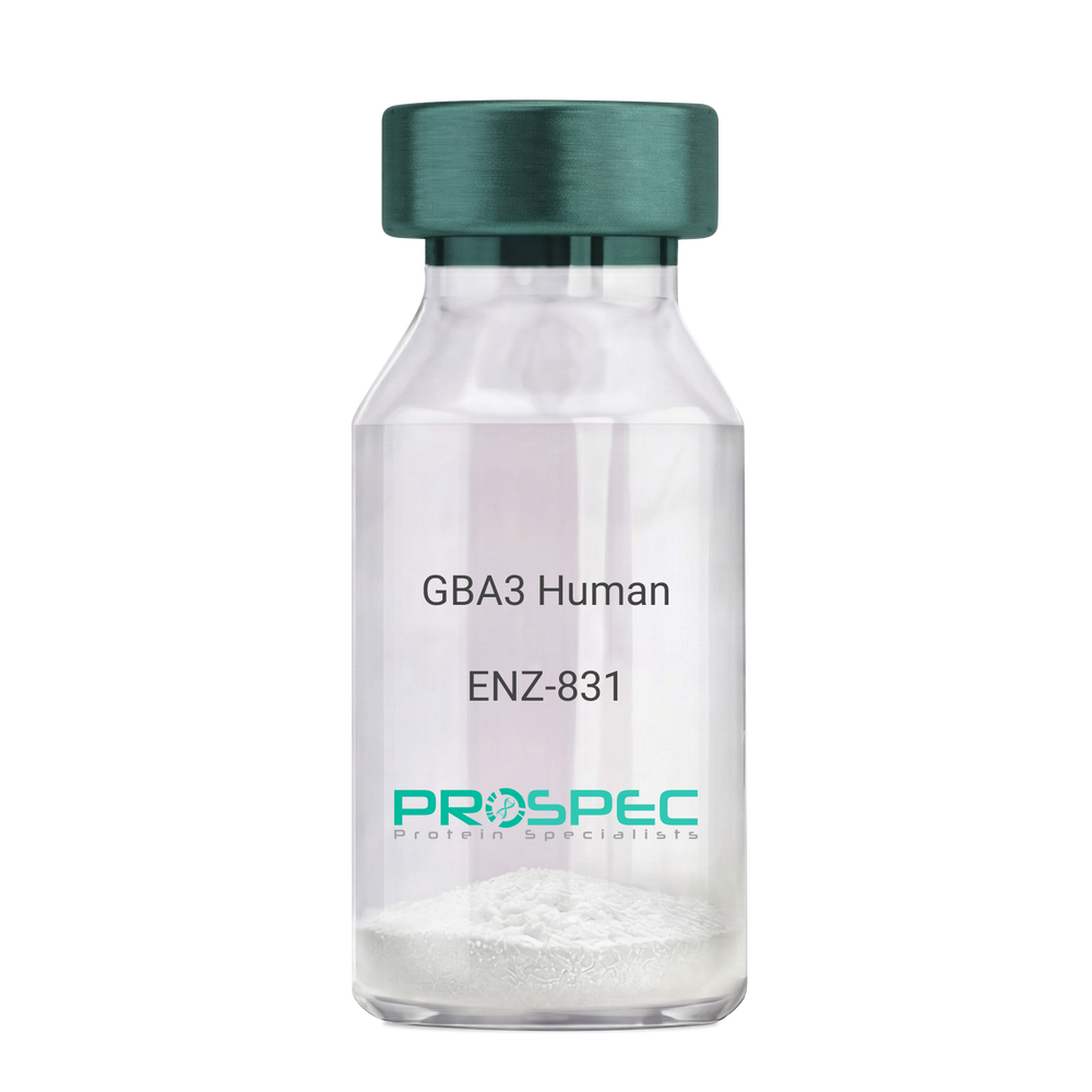
GBA3 HumanDescription:
Glucosidase Beta Acid 3 Human Recombinant
Cytosolic beta-glucosidase, Cytosolic beta-glucosidase-like protein 1, GBA3, CBG, CBGL1, Glucosidase Beta Acid 3, GH1, Glycoside Hydrolase Family 1, GLUC, KLRP.
Product # :
ENZ-831Price :
Quantity :
Shipping Method :
Shipped with Ice Packs
More Info
- description
- source
- formulation
- purity
- More Info
- Citations
Description
GBA3 produced in E.Coli is a single, non-glycosylated polypeptide chain containing 492 amino acids (1-469 a.a.) and having a molecular mass of 56.1kDa.GBA3 is fused to a 23 amino acid His-tag at N-terminus & purified by proprietary chromatographic techniques.
Source
E.coli.
Formulation
GBA3 protein solution (0.25mg/ml) containing Phosphate Buffered Saline (pH7.4), 20% glycerol and 1mM DTT.
Purity
Greater than 85% as determined by SDS-PAGE.
Citations
More Info
-
Introduction
Glucosidase Beta Acid 3, also known as GBA3, hydrolyzes various types of glycosides. GBA3 takes part in a nonlysosomal catabolic pathway of glycosylceramide. GBA3 is a protein coding gene which acts as beta-glycosylceramidase.
-
Synonyms
Cytosolic beta-glucosidase, Cytosolic beta-glucosidase-like protein 1, GBA3, CBG, CBGL1, Glucosidase Beta Acid 3, GH1, Glycoside Hydrolase Family 1, GLUC, KLRP.
-
Physical Appearance
Sterile Filtered colorless solution.
-
Stability
Store at 4°C if entire vial will be used within 2-4 weeks. Store, frozen at -20°C for longer periods of time. For long term storage it is recommended to add a carrier protein (0.1% HSA or BSA).Avoid multiple freeze-thaw cycles.
-
Amino Acid Sequence
MGSSHHHHHH SSGLVPRGSH MGSMAFPAGF GWAAATAAYQ VEGGWDADGK GPCVWDTFTH QGGERVFKNQ TGDVACGSYT LWEEDLKCIK QLGLTHYRFS LSWSRLLPDG TTGFINQKGI DYYNKIIDDL LKNGVTPIVT LYHFDLPQTL EDQGGWLSEA IIESFDKYAQ FCFSTFGDRV KQWITINEAN VLSVMSYDLG MFPPGIPHFG TGGYQAAHNL IKAHARSWHS YDSLFRKKQK GMVSLSLFAV WLEPADPNSV SDQEAAKRAI TFHLDLFAKP IFIDGDYPEV VKSQIASMSQ KQGYPSSRLP EFTEEEKKMI KGTADFFAVQ YYTTRLIKYQ ENKKGELGIL QDAEIEFFPD PSWKNVDWIY VVPWGVCKLL KYIKDTYNNP VIYITENGFP QSDPAPLDDT QRWEYFRQTF QELFKAIQLD KVNLQVYCAW SLLDNFEWNQ GYSSRFGLFH VDFEDPARPR VPYTSAKEYA KIIRNNGLEA HL.
ProSpec's products are furnished for LABORATORY RESEARCH USE ONLY. The product may not be used as drugs, agricultural or pesticidal products, food additives or household chemicals.
- View Data Sheet
Name :
PI3Kb HumanDescription:
Phosphoinositide 3-kinase beta p110β/p85α Human Recombinant
Phosphoinositide 3-kinase beta p110b/p85a, PI3Kb, PI3Kb.
Product # :
PKA-333Price :
Quantity :
Shipping Method :
Shipped with Ice Packs
More Info
- description
- source
- formulation
- purity
- biological activity
- More Info
- Citations
Description
Phosphoinositide 3-kinase beta Human Recombinant is a glycosilated protein having a molecular weight as follows: p85a chain 83.5 kDa, p110b chain 124.3 kDa.
Source
Sf9 insect cells.
Formulation
0.9 mg/ml solution in PBS and 2mM MgCl2.
Purity
Greater than 90.0% as determined by SDS Page.
Biological Activity
The specific activity was found to be 3,000 units/mg (1 unit is defined as 1 picomole phosphate transferred to PIP2 per minute).
Citations
More Info
-
Introduction
The PI3Kb isoform can be activated by insulin via the insulin receptor to initiate a cascade of events that control cell growth and metabolism. The activation of
PI3Kb is mediated by the p85 regulatory subunit binding to tyrosine phosphorylated insulin receptor substrate (IRS) proteins (e.g. IRS-1 and IRS-2).
It was also shown that PI3Kb is involved in apoptosis in human colon carcinoma cells. Injection of neutralizing antibodies specific to p110b in WiDr,
HCT116 and CO 115 adenocarcinoma cells inhibited de novo DNA synthesis.
PI3Kb is the major PI3K isoform required for apoptotic cell and Fc-g receptor mediated phagocytosis shown for primary mouse macrophages and the Jurkat human leukemia T cell line.
It was shown by several research groups that the catalytic subunit of PI3Kb can be activated by Gbg subunits of G-protein coupled receptors. -
Synonyms
Phosphoinositide 3-kinase beta p110b/p85a, PI3Kb, PI3Kb.
-
Physical Appearance
Sterile filtered liquid formualtion.
-
Stability
Store at 4°C if entire vial will be used within 2-4 weeks. Store, frozen at -20°C for longer periods of time. For long term storage it is recommended to add a carrier protein (0.1% HSA or BSA).Avoid multiple freeze-thaw cycles.
ProSpec's products are furnished for LABORATORY RESEARCH USE ONLY. The product may not be used as drugs, agricultural or pesticidal products, food additives or household chemicals.
- View Data Sheet
Name :
TNNI1 Paired AntibodyDescription:
Mouse Anti Human Troponin I Type 1 Paired
DKFZp451O223, SSTNI, TNN1, Troponin I, slow skeletal muscle ,Troponin I, slow-twitch isoform.
Product # :
ANT-724Price :
Quantity :
Shipping Method :
Shipped with Ice Packs
More Info
- description
- formulation
- purity
- More Info
- Citations
Description
TNNI1 conjugation antibody and TNNI1 coating antibody are used to develop rapid test for TNNI1 rapid test.
Please note that when ordering for example: 100µg antibody we ship 50µg from each of the antibodies (100µg in total).
Formulation
* TNNI1 conjugation in 50mM Na-citrate, pH 6.0, 0.9% NaCl and 0.095 % NaN3.
* TNNI1 coating antibody in 50mM Na-citrate, pH 6.0 0.9% NaCl and 0.095 % NaN3.
Purity
Greater than 95%.
Citations
More Info
-
Introduction
Troponin I, (TNNI1) is a member of the troponin I family. Troponin complex has 3 subunits, TNNI1 known as the inhibitory Subunit which prevents the actin-myosin interactions and thus mediating striated muscle relaxation. TNNI1 combines with tropomyosin and regulates calcium sensitivity of striated muscles by structural modifications in actin-myosin complexes.
-
Synonyms
DKFZp451O223, SSTNI, TNN1, Troponin I, slow skeletal muscle ,Troponin I, slow-twitch isoform.
-
Physical Appearance
2 vials of sterile Filtered clear colorless solution.
-
Applications
Lateral flow immunoassay.
-
Type
Mouse Anti Human Monoclonal.
-
Purification Method
Purified monoclonal IgG by protein A chromatography.
ProSpec's products are furnished for LABORATORY RESEARCH USE ONLY. The product may not be used as drugs, agricultural or pesticidal products, food additives or household chemicals.
- View Data Sheet
Name :
OPG HumanDescription:
Osteoprotegerin Human Recombinant
TNFRSF11B, OPG, OCIF, Osteoclastogenesis inhibitory factor, Osteoprotegerin, TR1, MGC29565.
Product # :
CYT-177Price :
Quantity :
Shipping Method :
Shipped at Room temp
More Info
- description
- source
- formulation
- purity
- biological activity
- More Info
- Citations
Description
Recombinant Human Osteoprotegerin produced in E.coli cells is a single, non-glycosylated, polypeptide chain containing 174 amino acids and having a molecular mass of 20kDa. The OPG is purified by proprietary chromatographic techniques.
Source
Escherichia Coli.
Formulation
The OPG was lyophilized from a 0.2µm filtered concentrated (0.5mg/ml) solution in PBS, pH 7.4.
Purity
Greater than 95.0% as determined by:
(a) Analysis by RP-HPLC.
(b) Analysis by SDS-PAGE.Biological Activity
The activity is determined by its ability to neutralize the stimulation of U937 cells treated with 10ng/ml of soluble RANKL corresponding to a specific activity of 100,000IU/mg.Citations
More Info
-
Introduction
Osteoprotegerin acts as decoy receptor for rankl and thereby neutralizes its function in osteoclastogenesis. OPG inhibits the activation of osteoclasts and promotes osteoclast apoptosis in vitro. Bone homeostasis seems to depend on the local rankl/opg ratio. Osteoprotegerin may also play a role in preventing arterial calcification. May act as decoy receptor for trail and protect against apoptosis. Trail binding blocks the inhibition of osteoclastogenesis.
-
Synonyms
TNFRSF11B, OPG, OCIF, Osteoclastogenesis inhibitory factor, Osteoprotegerin, TR1, MGC29565.
-
Physical Appearance
Sterile Filtered White lyophilized (freeze-dried) powder.
-
Stability
Lyophilized Osteoprotegerin although stable at room temperature for 3 weeks, should be stored desiccated below -18°C. Upon reconstitution OCIF should be stored at 4°C between 2-7 days and for future use below -18°C. For long term storage it is recommended to add a carrier protein (0.1% HSA or BSA).Please prevent freeze-thaw cycles.
-
Solubility
It is recommended to reconstitute the lyophilized Osteoprotegerin in sterile 18MΩ-cm H2O not less than 100µg/ml, which can then be further diluted to other aqueous solutions.
-
Amino Acid Sequence
METFPPKYLH YDEETSHQLL CDKCPPGTYL KQHCTAKWKT VCAPCPDHYY TDSWHTSDEC LYCSPVCKEL QYVKQECNRT HNRVCECKEG RYLEIEFCLK HRSCPPGFGV VQAGTPERNT VCKRCPDGFF SNETSSKAPC RKHTNCSVFG LLLTQKGNAT HDNICSGNSE STQK.
ProSpec's products are furnished for LABORATORY RESEARCH USE ONLY. The product may not be used as drugs, agricultural or pesticidal products, food additives or household chemicals.
- View Data Sheet
Name :
NTRK3 MouseDescription:
Neurotrophic Receptor Tyrosine Kinase 3 Mouse Recombinant
NT-3 growth factor receptor, GP145-TrkC, Trk-C, Neurotrophic tyrosine kinase receptor type 3, TrkC tyrosine kinase, Ntrk3, TrkC, AW125844, Ntrk3_tv3
Product # :
CYT-1163Price :
Quantity :
Shipping Method :
Shipped with Ice Packs
More Info
- description
- source
- formulation
- purity
- More Info
- Citations
Description
NTRK3 Mouse Recombinant produced in Baculovirus is a single glycosylated polypeptide chain containing 404 amino acids (32-429 aa) and having a molecular mass of 45.4kDa.NTRK3 is fused to a 6 amino acid His tag at C-terminus and purified by proprietary chromatographic techniques.
Source
Sf9, Baculovirus cells.
Formulation
NTRK3 protein (0.25mg/ml) contains 10% glycerol and Phosphate-Buffered Saline (pH 7.4).
Purity
Greater than 90.0% as determined by SDS-PAGE.
Citations
More Info
-
Introduction
Neurotrophic Receptor Tyrosine Kinase 3 (NTRK3)belongs to the receptor tyrosine kinases family and is a high affinity catalytic receptor of neurotrophin NT-3. NTRK3 mediates multiple effects of this neurotrophic factor, which includes neuronal differentiation and survival. NTRK3 inducesvarious pleiotorpic responses in malignant cells, including enhanced tumor cell invasiveness and chemotoxis.Increased NTRK3 expression was observed in neuroblastoma, medulloblastoma, and in neuroectodermal brain tumors.
-
Synonyms
NT-3 growth factor receptor, GP145-TrkC, Trk-C, Neurotrophic tyrosine kinase receptor type 3, TrkC tyrosine kinase, Ntrk3, TrkC, AW125844, Ntrk3_tv3
-
Physical Appearance
Sterile Filtered colorless solution.
-
Stability
Store at 4°C if entire vial will be used within 2-4 weeks. Store, frozen at -20°C for longer periods of time. For long term storage it is recommended to add a carrier protein (0.1% HSA or BSA).Avoid multiple freeze-thaw cycles.
-
Amino Acid Sequence
CPANCVCSKT EINCRRPDDG NLFPLLEGQD SGNSNGNASI NITDISRNIT SIHIENWRGL HTLNAVDMEL YTGLQKLTIK NSGLRNIQPR AFAKNPHLRY INLSSNRLTT LSWQLFQTLS LRELRLEQNF FNCSCDIRWM QLWQEQGEAR LDSQSLYCIS ADGSQLPLFR MNISQCDLPE ISVSHVNLTV REGDNAVITC NGSGSPLPDV DWIVTGLQSI NTHQTNLNWT NVHAINLTLV NVTSEDNGFT LTCIAENVVG MSNASVALTV YYPPRVVSLV EPEVRLEHCI EFVVRGNPTP TLHWLYNGQP LRESKIIHMD YYQEGEVSEG CLLFNKPTHY NNGNYTLIAK NALGTANQTI NGHFLKEPFP ESTDFFDFES DASPTPPITV THKPEEDTHH HHHH
ProSpec's products are furnished for LABORATORY RESEARCH USE ONLY. The product may not be used as drugs, agricultural or pesticidal products, food additives or household chemicals.
- View Data Sheet
Name :
SDC4 HumanDescription:
Syndecan-4 Human Recombinant
SDC-4, SYND4, SYND-4, Amphiglycan, Ryudocan core protein, Syndecan-4.
Product # :
PRO-778Price :
Quantity :
Shipping Method :
Shipped with Ice Packs
More Info
- description
- source
- formulation
- purity
- More Info
- Citations
Description
SDC4 Human Recombinant produced in E.Coli is a single, non-glycosylated, polypeptide chain containing 128 amino acids (19-145a.a.) and having a molecular mass of 13.9 kDa. SDC4 is purified by proprietary chromatographic techniques.
Source
Escherichia Coli.
Formulation
Syndecan-4 (1mg/ml) protein solution containing 20mM Tris-HCl pH-8, and 10% glycerol.
Purity
Greater than 85% as determined by SDS-PAGE.
Citations
More Info
-
Introduction
Syndecan-4 is a type I integral membrane heparan sulfate proteoglycan (HSPG), which was originally isolated from cloned rat microvascular endothelial cells as an antithrombin-binding molecule, and is now known to be a member of the syndecan family. Syndecan-4 binds to basic fibroblast growth factor (bFGF), midkine, and tissue factor pathway inhibitor via its heparan sulfate chains, and is thought to be involved in various biologic functions such as signaling of bFGF, anticoagulation, and focal adhesion formation. A previous study demonstrated that syndecan-4 is expressed in various tissues, and its level of expression in the kidney is stronger than those of other syndecan family members. Thus, syndecan-4 is thought to play certain roles in maintaining renal function. Moreover, it has been reported that proteoglycans, especially sulfated proteoglycans, are involved in organogenesis of the kidney.
-
Synonyms
SDC-4, SYND4, SYND-4, Amphiglycan, Ryudocan core protein, Syndecan-4.
-
Physical Appearance
Sterile filtered colorless solution.
-
Stability
Store at 4°C if entire vial will be used within 2-4 weeks. Store, frozen at -20°C for longer periods of time. For long term storage it is recommended to add a carrier protein (0.1% HSA or BSA). Please prevent freeze thaw cycles.
-
Amino Acid Sequence
MESIRETEVI DPQDLLEGRY FSGALPDDED VVGPGQESDD FELSGSGDLD DLEDSMIGPE VVHPLVPLDN HIPERAGSGS QVPTEPKKLE ENEVIPKRIS PVEESEDVSN KVSMSSTVQG SNIFERTE.
ProSpec's products are furnished for LABORATORY RESEARCH USE ONLY. The product may not be used as drugs, agricultural or pesticidal products, food additives or household chemicals.
- View Data Sheet
Name :
SNRPD3 HumanDescription:
Small Nuclear Ribonucleoprotein Polypeptide D3 Human Recombinant
Small nuclear ribonucleoprotein D3 polypeptide 18kDa, Sm-D3, snRNP core protein D3.
Product # :
PRO-945Price :
Quantity :
Shipping Method :
Shipped with Ice Packs
More Info
- description
- source
- formulation
- purity
- More Info
- Citations
Description
SNRPD3 Human Recombinant produced in E.coli is a single, non-glycosylated polypeptide chain containing 146 amino acids (1-126) and having a molecular mass of 16.0 kDa.The SNRPD3 is fused to a 20 amino acid His-Tag at N-terminus and purified by proprietary chromatographic techniques.
Source
E.coli.
Formulation
The SNRPD3 solution (0.25mg/ml) contains 20mM Tris-HCl buffer (pH 8.0), 0.5M NaCl, 2mM DTT, 0.1mM PMSF and 40% glycerol.
Purity
Greater than 85% as determined by SDS-PAGE.
Citations
More Info
-
Introduction
SNRPD3 is a small nuclear ribonucleoprotein (snRNPs) that contains the spliceosome in eukaryotes. SNRPD3 is essential for pre-mRNA splicing and small nuclear ribonucleoprotein biogenesis. Alternative splicing happens in this locus and two transcript variants encoding the same protein were branded.
-
Synonyms
Small nuclear ribonucleoprotein D3 polypeptide 18kDa, Sm-D3, snRNP core protein D3.
-
Physical Appearance
Sterile Filtered colorless solution.
-
Stability
Store at 4°C if entire vial will be used within 2-4 weeks. Store, frozen at -20°C for longer periods of time. For long term storage it is recommended to add a carrier protein (0.1% HSA or BSA).Avoid multiple freeze-thaw cycles.
-
Amino Acid Sequence
MGSSHHHHHH SSGLVPRGSH MSIGVPIKVL HEAEGHIVTC ETNTGEVYRG KLIEAEDNMN CQMSNITVTY RDGRVAQLEQ VYIRGSKIRF LILPDMLKNA PMLKSMKNKN QGSGAGRGKA AILKAQVAAR GRGRGMGRGN IFQKRR
ProSpec's products are furnished for LABORATORY RESEARCH USE ONLY. The product may not be used as drugs, agricultural or pesticidal products, food additives or household chemicals.
- View Data Sheet
Name :
FGFBP HumanDescription:
Fibroblast Growth Factor Binding Protein 1 Human Recombinant
Fibroblast Growth Factor Binding Protein 1, FGFBP, HBP17, 17 KDa Heparin-Binding Growth Factor-Binding Protein, 17 KDa HBGF-Binding Protein, FGF-Binding Protein 1, FGF-BP1, FGFBP-1, FGF-BP, Heparin-Binding Growth Factor Binding Protein, Fibroblast Growth Factor-Binding Protein 1, Fibroblast growth factor-binding protein 1.
Product # :
CYT-860Price :
Quantity :
Shipping Method :
Shipped with Ice Packs
More Info
- description
- source
- formulation
- purity
- More Info
- sds-page
- Citations
Description
FGFBP Human Recombinant produced in E.Coli is a single, non-glycosylated polypeptide chain containing 234 amino acids (24-234 a.a) and having a molecular mass of 26.2kDa. FGFBP is fused to a 23 amino acid His-tag at N-terminus & purified by proprietary chromatographic techniques.
Source
Escherichia Coli.
Formulation
FGFBP protein solution (0.25mg/ml) containing Phosphate buffered saline (pH7.4), 10% glycerol and 1mM DTT.
Purity
Greater than 85.0% as determined by SDS-PAGE.
sds-page
Citations
More Info
-
Introduction
Fibroblast Growth Factor Binding Protein 1, also known as FGFBP1 is a secreted fibroblast growth factor carrier protein. FGFBP1 plays a vital part in cell proliferation, differentiation and migration by binding to fibroblast growth factors and potentiating their biological effects on target cells. In addition, FGFBP1 also takes part in tumor growth as an angiogenic switch molecule, furthermore an expression of FGFBP1 has been associated with more than a few types of cancer as well as pancreatic and colorectal adenocarcinoma.
-
Synonyms
Fibroblast Growth Factor Binding Protein 1, FGFBP, HBP17, 17 KDa Heparin-Binding Growth Factor-Binding Protein, 17 KDa HBGF-Binding Protein, FGF-Binding Protein 1, FGF-BP1, FGFBP-1, FGF-BP, Heparin-Binding Growth Factor Binding Protein, Fibroblast Growth Factor-Binding Protein 1, Fibroblast growth factor-binding protein 1.
-
Physical Appearance
Sterile Filtered colorless clear solution.
-
Stability
Store at 4°C if entire vial will be used within 2-4 weeks. Store, frozen at -20°C for longer periods of time. For long term storage it is recommended to add a carrier protein (0.1% HSA or BSA).Please avoid freeze thaw cycles.
-
Amino Acid Sequence
MGSSHHHHHH SSGLVPRGSH MGSKKKVKNG LHSKVVSEQK DTLGNTQIKQ KSRPGNKGKF VTKDQANCRW AATEQEEGIS LKVECTQLDH EFSCVFAGNP TSCLKLKDER VYWKQVARNL RSQKDICRYS KTAVKTRVCR KDFPESSLKL VSSTLFGNTK PRKEKTEMSP REHIKGKETT PSSLAVTQTM ATKAPECVED PDMANQRKTA LEFCGETWSS LCTFFLSIVQ DTSC.
-
Background
What is the molecular weight/Mw of CYT-860 Protein?
CYT-860 Protein has a total Mw of 26.2kDa.
What is the source or expression system of CYT-860 Protein?
Escherichia Coli.
What is the Purity of CYT-860 Protein?
CYT-860 Protein is >85% pure as determined by SDS-PAGE.
What is the Biological Activity of CYT-860 Protein?
The biological functionality of CYT-860 Protein will be determined in the future.
What is the amino acid sequence of CYT-860 Protein?
MGSSHHHHHH SSGLVPRGSH MGSKKKVKNG LHSKVVSEQK DTLGNTQIKQ KSRPGNKGKF VTKDQANCRW AATEQEEGIS LKVECTQLDH EFSCVFAGNP TSCLKLKDER VYWKQVARNL RSQKDICRYS KTAVKTRVCR KDFPESSLKL VSSTLFGNTK PRKEKTEMSP REHIKGKETT PSSLAVTQTM ATKAPECVED PDMANQRKTA LEFCGETWSS LCTFFLSIVQ DTSC.
What applications can CYT-860 Protein be used in?
CYT-860 Protein can probably be used in western blot, ELISA and Lateral Flow.
What is the endotoxin level for CYT-860 Protein?
The endotoxin level is minimal, CYT-860 Protein was purified using conventional chromatography techniques.
ProSpec's products are furnished for LABORATORY RESEARCH USE ONLY. The product may not be used as drugs, agricultural or pesticidal products, food additives or household chemicals.
- View Data Sheet
Name :
FSTL1 Human, HEKDescription:
Follistatin Like 1 Human Recombinant, HEK
Follistatin-related protein 1, Follistatin-like protein 1, FSTL1, FRP, Follistatin Like 1, FSL1, MIR198.
Product # :
CYT-1027Price :
Quantity :
Shipping Method :
Shipped at Room temp
More Info
- description
- source
- formulation
- purity
- More Info
- Citations
Description
FSTL1 Human Recombinant produced in HEK293 cells is a single, glycosylated polypeptide chain (a.a 21-308) containing 296 amino acids including a 8 a.a C-terminal His tag. The total molecular mass is 33.8kDa (calculated).
Source
HEK293 cells.
Formulation
FSTL1 filtered (0.4 µm) and lyophilized from 0.5mg/ml in phosphate buffered saline and 5 % (w/v) trehalose, pH 7.4.
Purity
Greater than 95.0% as determined by SDS-PAGE.
Citations
More Info
-
Introduction
FSTL1 protein resembles follistatin, an activin-binding protein. FSTL1 is an autoantigen associated with rheumatoid arthritis and it holds an FS section, a follistatin-like sequence having 10 conserved cysteine residues.
-
Synonyms
Follistatin-related protein 1, Follistatin-like protein 1, FSTL1, FRP, Follistatin Like 1, FSL1, MIR198.
-
Physical Appearance
Filtered White lyophilized (freeze-dried) powder.
-
Stability
Store lyophilized protein at -20°C. Aliquot the product after reconstitution to avoid repeated freezing/thawing cycles. Reconstituted protein can be stored at 4°C for a limited period of time; it does not show any change after one week at 4°C.
-
Solubility
It is recommended to add deionized water to prepare a working stock solution of approximately 0.5mg/ml and let the lyophilized pellet dissolve completely. FSTL1 is not sterile! Please filter the product by an appropriate sterile filter before using it in the cell culture.
-
Amino Acid Sequence
EEELRSKSKI CANVFCGAGR ECAVTEKGEP TCLCIEQCKP HKRPVCGSNG KTYLNHCELH RDACLTGSKI QVDYDGHCKE KKSVSPSASP VVCYQSNRDE LRRRIIQWLE AEIIPDGWFS KGSNYSEILD KYFKNFDNGD SRLDSSEFLK FVEQNETAIN ITTYPDQENN KLLRGLCVDA LIELSDENAD WKLSFQEFLK CLNPSFNPPE KKCALEDETY ADGAETEVDC NRCVCACGNW VCTAMTCDGK NQKGAQTQTE EEMTRYVQEL QKHQETAEKT KRVSTKEIHH HHHHHH.
-
Background
What is the molecular weight/Mw of FSTL1 HUMAN, HEK Protein?
FSTL1 HUMAN, HEK Protein has a total Mw of 33.8kDa.
What is the source or expression system of FSTL1 HUMAN, HEK Protein?
HEK293 cells.
What is the Purity of FSTL1 HUMAN, HEK Protein?
FSTL1 HUMAN, HEK Protein is >95% pure as determined by SDS-PAGE.
What is the Biological Activity of FSTL1 HUMAN, HEK Protein?
The biological functionality of FSTL1 HUMAN, HEK Protein will be determined in the future.
What is the amino acid sequence of FSTL1 HUMAN, HEK Protein?
EEELRSKSKI CANVFCGAGR ECAVTEKGEP TCLCIEQCKP HKRPVCGSNG KTYLNHCELH RDACLTGSKI QVDYDGHCKE KKSVSPSASP VVCYQSNRDE LRRRIIQWLE AEIIPDGWFS KGSNYSEILD KYFKNFDNGD SRLDSSEFLK FVEQNETAIN ITTYPDQENN KLLRGLCVDA LIELSDENAD WKLSFQEFLK CLNPSFNPPE KKCALEDETY ADGAETEVDC NRCVCACGNW VCTAMTCDGK NQKGAQTQTE EEMTRYVQEL QKHQETAEKT KRVSTKEIHH HHHHHH.
What applications can FSTL1 HUMAN, HEK Protein be used in?
FSTL1 HUMAN, HEK Protein can probably be used in western blot, ELISA and Lateral Flow.
What is the endotoxin level for FSTL1 HUMAN, HEK Protein?
The endotoxin level is minimal, FSTL1 HUMAN, HEK Protein was purified using conventional chromatography techniques.
ProSpec's products are furnished for LABORATORY RESEARCH USE ONLY. The product may not be used as drugs, agricultural or pesticidal products, food additives or household chemicals.
- View Data Sheet
Name :
VCAM1 MouseDescription:
Vascular cell adhesion molecule 1 Mouse Recombinant
V-CAM 1, VCAM-1, CD106, Vcam1, Vcam-1, Vascular cell adhesion protein 1.
Product # :
PRO-2257Price :
Quantity :
Shipping Method :
Shipped with Ice Packs
More Info
- description
- source
- formulation
- purity
- More Info
- Citations
Description
VCAM1 Mouse Recombinant produced in Sf9 Baculovirus cells is a single, glycosylated polypeptide chain containing 682 amino acids (25-698 a.a) and having a molecular mass of 75.4kDa (Migrates at 70-100kDa on SDS-PAGE under reducing conditions). VCAM1 is fused to an 8 amino acid His-tag at C-terminus & purified by proprietary chromatographic techniques.
Source
Sf9, Baculovirus cells.
Formulation
VCAM1 protein solution (0.5mg/ml) containing Phosphate Buffered Saline (pH 7.4) and 10% glycerol.
Purity
Greater than 95.0% as determined by SDS-PAGE.
Citations
More Info
-
Introduction
VCAM1 is a member of the Ig superfamily. It is a cell surface sialoglycoprotein expressed by cytokine activated endothelium. VCAM-1 contains 6 or 7 immunoglobulin domains, and is expressed on both large and small vessels only after the endothelial cells are stimulated by cytokines. The protein has a number of functions including the regulation of leukocyte migration, leukocyte endothelial cell adhesion and signal transduction and may play a role in a number of inflammatory diseases (artherosclerosis and rheumatoid arthritis).
-
Synonyms
V-CAM 1, VCAM-1, CD106, Vcam1, Vcam-1, Vascular cell adhesion protein 1.
-
Physical Appearance
Sterile Filtered colorless solution.
-
Stability
Store at 4°C if entire vial will be used within 2-4 weeks. Store, frozen at -20°C for longer periods of time. For long term storage it is recommended to add a carrier protein (0.1% HSA or BSA).Avoid multiple freeze-thaw cycles.
-
Amino Acid Sequence
FKIEISPEYK TIAQIGDSMA LTCSTTGCES PLFSWRTQID SPLNAKVRTE GSKSVLTMEP VSFENEHSYL CTATCGSGKL ERSIHVDIYS FPKDPEIQFS GPLEVGKPVT VKCLAPDIYP VYRLEIDLFK GDQLMNRQEF SSEEMTKSLE TKSLEVTFTP VIEDIGKALV CRAKLHIDQI DSTLKERETV KELQVYISPR NTTISVHPST RLQEGGAVTM TCSSEGLPAP EIFWGRKLDN EVLQLLSGNA TLTLIAMRME DSGVYVCEGV NLIGRDKAEV ELVVQEKPFI VDISPGSQVA AQVGDSVVLT CAAIGCDSPS FSWRTQTDSP LNGVVRNEGA KSTLVLSSVG FEDEHSYLCA VTCLQRTLEK RTQVEVYSFP EDPVIKMSGP LVHGRPVTVN CTVPNVYPFD HLEIELLKGE TTLMKKYFLE EMGIKSLETK ILETTFIPTI EDTGKSLVCL ARLHSGEMES EPKQRQSVQP LYVNVAPKET TIWVSPSPIL EEGSPVNLTC SSDGIPAPKI LWSRQLNNGE LQPLSENTTL TFMSTKRDDS GIYVCEGINE AGISRKSVEL IIQVSPKDIQ LTVFPSKSVK EGDTVIISCT CGNVPETWII LKKKAKTGDM VLKSVDGSYT IRQAQLQDAG IYECESKTEV GSQLRSLTLD VKGKEHNKNY FSPELEHHHH HH.
ProSpec's products are furnished for LABORATORY RESEARCH USE ONLY. The product may not be used as drugs, agricultural or pesticidal products, food additives or household chemicals.
- View Data Sheet
Name :
RAGE Human, Sf9Description:
Advanced Glycosylation End Product-Specific Receptor Human Recombinant, Sf9
Advanced Glycosylation End-Product Specific Receptor, Advanced Glycosylation End Product-Specific Receptor, RAGE, Receptor for Advanced Glycation End-Products Variant 20, Receptor for Advanced Glycosylation End Products, Receptor for Advanced Glycation End-Products, RAGE Isoform NtRAGE-Delta, RAGE Isoform SRAGE-Delta, SCARJ1.
Product # :
PRO-2645Price :
Quantity :
Shipping Method :
Shipped with Ice Packs
More Info
- description
- source
- formulation
- purity
- biological activity
- More Info
- Citations
Description
RAGE Human Recombinant produced in Sf9 Baculovirus cells is a single, glycosylated polypeptide chain containing 561 amino acids (24-342 a.a) and having a molecular mass of 61.2kDa. RAGE is fused to a 242 amino acid hIgG-His-tag at C-terminus & purified by proprietary chromatographic techniques.
Source
Sf9, Baculovirus cells.
Formulation
The RAGE solution (0.25mg/1ml) contains Phosphate-Buffered Saline (pH 7.4) and 10% glycerol.
Purity
Greater than 90.0% as determined by SDS-PAGE.
Biological Activity
Measured by the ability of the binding activity in a functional ELISA. The ED50 range ≤15ug/ml.
Citations
More Info
-
Introduction
Advanced Glycosylation End Product-Specific Receptor or RAGE is an immunoglobulin, part of the immunoglobulin transmembrane proteins family. RAGE moderates interactions between advanced glycosylation end products, also known as AGE. These signaling has a crucial part in the regulation of TNFalpha,oxidative stress, endothelial dysfunction in type 2 diabetesproduction and expression. RAGE helps the translocation of ABPP (amyloid-beta peptide) through the membrane of cells to the intracellular area in the cortical neurons.
-
Synonyms
Advanced Glycosylation End-Product Specific Receptor, Advanced Glycosylation End Product-Specific Receptor, RAGE, Receptor for Advanced Glycation End-Products Variant 20, Receptor for Advanced Glycosylation End Products, Receptor for Advanced Glycation End-Products, RAGE Isoform NtRAGE-Delta, RAGE Isoform SRAGE-Delta, SCARJ1.
-
Physical Appearance
Sterile Filtered colorless solution.
-
Stability
Store at 4°C if entire vial will be used within 2-4 weeks. Store, frozen at -20°C for longer periods of time. For long term storage it is recommended to add a carrier protein (0.1% HSA or BSA).Avoid multiple freeze-thaw cycles.
-
Amino Acid Sequence
ADLQNITARI GEPLVLKCKG APKKPPQRLE WKLNTGRTEA WKVLSPQGGG PWDSVARVLP NGSLFLPAVG IQDEGIFRCQ AMNRNGKETK SNYRVRVYQI PGKPEIVDSA SELTAGVPNK VGTCVSEGSY PAGTLSWHLD GKPLVPNEKG VSVKEQTRRH PETGLFTLQS ELMVTPARGG DPRPTFSCSF SPGLPRHRAL RTAPIQPRVW EPVPLEEVQL VVEPEGGAVA PGGTVTLTCE VPAQPSPQIH WMKDGVPLPL PPSPVLILPE IGPQDQGTYS CVATHSSHGP QESRAVSISI IEPGEEGPTA GSVGGSGLGT LALEPKSCDK THTCPPCPAP ELLGGPSVFL FPPKPKDTLM ISRTPEVTCV VVDVSHEDPE VKFNWYVDGV EVHNAKTKPR EEQYNSTYRV VSVLTVLHQD WLNGKEYKCK VSNKALPAPI EKTISKAKGQ PREPQVYTLP PSRDELTKNQ VSLTCLVKGF YPSDIAVEWE SNGQPENNYK TTPPVLDSDG SFFLYSKLTV DKSRWQQGNV FSCSVMHEA HNHYTQKSLS LSPGKHHHHH H.
ProSpec's products are furnished for LABORATORY RESEARCH USE ONLY. The product may not be used as drugs, agricultural or pesticidal products, food additives or household chemicals.
- View Data Sheet
Name :
HCV Core Genotype-3aDescription:
Hepatitis C Virus Core Genotype-3a Recombinant
Product # :
HCV-215Price :
Quantity :
Shipping Method :
Shipped with Ice Packs
More Info
- description
- source
- formulation
- purity
- More Info
- Citations
Description
The E.coli derived recombinant protein contains the HCV core nucleocapsid immunodominant regions, amino acids 2-119. The protein is fused to a GST tag at N-terminus.
Source
Escherichia Coli.
Formulation
(1mg/ml) 1.5M urea, 25mM Tris-HCl, pH 8.0, 0.2% Triton-X and 50% glycerol.
Purity
HCV Core Genotype-3a protein is >95% pure as determined by 10% PAGE (coomassie staining).
Citations
More Info
-
Introduction
HCV is a small 50nm, enveloped, single-stranded, positive sense RNAvirus in the family Flaviviridae.
HCV has a high rate of replication with approximately one trillion particles produced each day in an infected individual. Due to lack of proofreading by the HCV RNA polymerase, the HCV has an exceptionally high mutation rate, a factor that may help it elude the host's immune response. Hepatitis C virus is classified into six genotypes(1-6) with several subtypes within each genotype. The preponderance and distribution of HCV genotypes varies globally. Genotype is clinically important in determining potential response to interferon-based therapy and the required duration of such therapy. Genotypes 1 and 4 are less responsive to interferon-based treatment than are the other genotypes (2, 3, 5 and 6). -
Stability
HCV Core Genotype-3aalthough stable at 4°C for 1 week, should be stored below -18°C. Please prevent freeze thaw cycles.
-
Applications
HCV Core Genotype-3a antigen is suitable for ELISA and Western blots, excellent antigen for detection of HCV with minimal specificity problems.
-
Specificity
Immunoreactive with sera of HCV-infected individuals.
-
Purification Method
HCV Core Genotype-3a protein was purified by proprietary chromatographic technique.
ProSpec's products are furnished for LABORATORY RESEARCH USE ONLY. The product may not be used as drugs, agricultural or pesticidal products, food additives or household chemicals.
- View Data Sheet
Name :
CHCHD7 HumanDescription:
Coiled-Coil-Helix-Coiled-Coil-Helix Domain Containing 7 Human Recombinant
Coiled-Coil-Helix-Coiled-Coil-Helix Domain Containing 7, Coiled-Coil-Helix-Coiled-Coil-Helix Domain-Containing Protein 7, COX23 Cytochrome C Oxidase Assembly Homolog (S. Cerevisiae), COX23 Cytochrome C Oxidase Assembly Homolog , COX23, Coiled-coil-helix-coiled-coil-helix domain-containing protein 7.
Product # :
PRO-2083Price :
Quantity :
Shipping Method :
Shipped with Ice Packs
More Info
- description
- source
- formulation
- purity
- More Info
- Citations
Description
CHCHD7 Human Recombinant produced in E.Coli is a single, non-glycosylated polypeptide chain containing 120 amino acids (1-97 a.a) and having a molecular mass of 13.9kDa. CHCHD7 is fused to a 23 amino acid His-tag at N-terminus & purified by proprietary chromatographic techniques.
Source
E.coli.
Formulation
CHCHD7 protein solution (0.25mg/ml) containing 20mM Tris-HCl buffer (pH8.0), 10% glycerol and 0.1M NaCl.
Purity
Greater than 90% as determined by SDS-PAGE.
Citations
More Info
-
Introduction
Coiled-Coil-Helix-Coiled-Coil-Helix Domain Containing 7, also known as CHCHD7 is a newly recognized member of a multifamily of proteins which contains a conserved (coiled coil 1)-(helix 1)-(coiled coil 2)-(helix 2) domain. The biological function of CHCHD7 is unknown and the gene has up till now not been connected with neoplasia. In addition, CHCHD7 & PLAG1 are located head-to-head about 500 bp apart in 8q12. Molecular analyses of twenty seven tumors exposed CHCHD7-PLAG1 fusions in three tumors, two of them had t(6;8) and t(8;15) translocations as a singular anomalies and one a normal karyotype.
-
Synonyms
Coiled-Coil-Helix-Coiled-Coil-Helix Domain Containing 7, Coiled-Coil-Helix-Coiled-Coil-Helix Domain-Containing Protein 7, COX23 Cytochrome C Oxidase Assembly Homolog (S. Cerevisiae), COX23 Cytochrome C Oxidase Assembly Homolog , COX23, Coiled-coil-helix-coiled-coil-helix domain-containing protein 7.
-
Physical Appearance
Sterile Filtered colorless solution.
-
Stability
Store at 4°C if entire vial will be used within 2-4 weeks. Store, frozen at -20°C for longer periods of time. For long term storage it is recommended to add a carrier protein (0.1% HSA or BSA).Avoid multiple freeze-thaw cycles.
-
Amino Acid Sequence
MGSSHHHHHH SSGLVPRGSH MGSMHQTRTG KKTVRMPSVT QRLRDPDINP CLSESDASTR CLDENNYDRE RCSTYFLRYK NCRRFWNSIV MQRRKNGVKP FMPTAAERDE ILRAVGNMPY.
ProSpec's products are furnished for LABORATORY RESEARCH USE ONLY. The product may not be used as drugs, agricultural or pesticidal products, food additives or household chemicals.
- View Data Sheet
Name :
JAM2 antibodyDescription:
Mouse Anti Human Junctional Adhesion Molecule 2
Junctional adhesion molecule B, JAM-B, Junctional adhesion molecule 2, JAM-2, Vascular endothelial junction-associated molecule, VE-JAM, CD322.
Product # :
ANT-738Price :
Quantity :
Shipping Method :
Shipped with Ice Packs
More Info
- formulation
- More Info
- Citations
Formulation
1mg/ml containing PBS, pH-7.4, 10% Glycerol and 0.02% Sodium Azide.
Citations
More Info
-
Introduction
Junctional Adhesion Molecule 2 (JAM2) is a member of the immunoglobulin superfamily, and the junctional adhesion molecule (JAM) family. JAM2 is a type I membrane protein which is localized at the tight junctions of both epithelial and endothelial cells. JAM2 functions as an adhesive ligand for interacting with various immune cell types, and may have a role in lymphocyte homing to secondary lymphoid organs.
-
Synonyms
Junctional adhesion molecule B, JAM-B, Junctional adhesion molecule 2, JAM-2, Vascular endothelial junction-associated molecule, VE-JAM, CD322.
-
Physical Appearance
Sterile filtered colorless solution.
-
Immunogen
Anti-human JAM2 mAb, is derived from hybridization of mouse F0 myeloma cells with spleen cells from BALB/c mice immunized with a recombinant human JAM2 protein 21-238 amino acids purified from E. coli.
-
Ig Subclass
Mouse IgG2a heavy chain and k light chain.
-
Clone
PAT21D7AT.
-
Applications
JAM2 antibody has been tested by ELISA and Western blot analysis to assure specificity and reactivity. Since application varies, however, each investigation should be titrated by the reagent to obtain optimal results.
-
Type
Mouse Anti Human Monoclonal.
-
Storage Procedures
For periods up to 1 month store at 4°C, for longer periods of time, store at -20°C. Prevent freeze thaw cycles.
-
Purification Method
JAM2 antibody was purified from mouse ascitic fluids by protein-A affinity chromatography.
ProSpec's products are furnished for LABORATORY RESEARCH USE ONLY. The product may not be used as drugs, agricultural or pesticidal products, food additives or household chemicals.
- View Data Sheet
Name :
TREM2 AntibodyDescription:
Triggering receptor expressed on myeloid cells 2, Mouse Anti Human
Triggering receptor expressed on myeloid cells 2, Triggering receptor expressed on monocytes 2, TREM-2, TREM2, Trem2a, Trem2b, Trem2c.
Product # :
ANT-429Price :
Quantity :
Shipping Method :
Shipped with Ice Packs
More Info
- formulation
- More Info
- Citations
Formulation
1mg/ml containing PBS, pH-7.4, & 0.1% Sodium Azide.
Citations
More Info
-
Introduction
TREM2 is a membrane protein that forms a receptor signaling complex with TYROBP. TREM2 may be involved in chronic inflammation by triggering the production of constitutive inflammatory cytokines. Defects in the TREM2 gene are a source of polycystic lipomembranous osteodysplasia with sclerosing leukoencephalopathy (PLOSL).
-
Synonyms
Triggering receptor expressed on myeloid cells 2, Triggering receptor expressed on monocytes 2, TREM-2, TREM2, Trem2a, Trem2b, Trem2c.
-
Immunogen
Anti-human TREM2 mAb, is derived from hybridization of mouse F0 myeloma cells with spleen cells from BALB/c mice immunized with recombinant human TREM2 amino acids 19-161 purified from E. coli.
-
Ig Subclass
Mouse IgG3 heavy chain and κ light chain.
-
Clone
P2B5AT.
-
Applications
TREM2 antibody has been tested by ELISA and Western blot analysis to assure specificity and reactivity. Since application varies, however, each investigation should be titrated by the reagent to obtain optimal results. Recommended dilution range for Western blot analysis is 1:500 ~ 1,000.Recommended starting dilution is 1:500.
-
Type
Mouse Anti Human Monoclonal.
-
Storage Procedures
For periods up to 1 month store at 4°C, for longer periods of time, store at -20°C. Prevent freeze thaw cycles.
-
Purification Method
TREM2 antibody was purified from mouse ascitic fluids by protein-G affinity chromatography.
ProSpec's products are furnished for LABORATORY RESEARCH USE ONLY. The product may not be used as drugs, agricultural or pesticidal products, food additives or household chemicals.
- View Data Sheet
Name :
DRG1 HumanDescription:
Developmentally Regulated GTP Binding Protein 1 Human Recombinant
Developmentally-regulated GTP-binding protein 1, DRG-1, Neural precursor cell expressed developmentally down-regulated protein 3, NEDD-3, DRG1, NEDD3.
Product # :
PRO-885Price :
Quantity :
Shipping Method :
Shipped with Ice Packs
More Info
- description
- source
- formulation
- purity
- More Info
- Citations
Description
DRG1 Human Recombinant produced in E.Coli is a single, non-glycosylated polypeptide chain containing 387 amino acids (1-367 a.a.) and having a molecular mass of 42.7kDa.DRG1 is fused to a 20 amino acid His-tag at N-terminus & purified by proprietary chromatographic techniques.
Source
Escherichia Coli.
Formulation
DRG1 protein solution (0.5mg/ml) containing 20mM Tris-HCl buffer (pH 8.0), 1mM DTT, 30% glycerol and 1mM EDTA.
Purity
Greater than 85.0% as determined by SDS-PAGE.
Citations
More Info
-
Introduction
Developmentally-regulated GTP-binding protein 1 (DRG1) is a member of the GTP1/OBG family. DRG1 has a role in cell proliferation and differentiation, as well as in apoptosis, proposing a role in tumor formation and metastasis. Expression of the DRG1 was considerably reduced in breast tumor cells, particularly in patients with lymph node or bone metastasis as compared to those with localized breast cancer. The DRG1 protein is expressed at high levels in the heart, kidney and skeletal muscle and at lower levels in the brain, liver, placenta, lung, colon and spleen.
-
Synonyms
Developmentally-regulated GTP-binding protein 1, DRG-1, Neural precursor cell expressed developmentally down-regulated protein 3, NEDD-3, DRG1, NEDD3.
-
Physical Appearance
Sterile Filtered clear solution.
-
Stability
Store at 4°C if entire vial will be used within 2-4 weeks. Store, frozen at -20°C for longer periods of time. For long term storage it is recommended to add a carrier protein (0.1% HSA or BSA).Avoid multiple freeze-thaw cycles.
-
Amino Acid Sequence
MGSSHHHHHH SSGLVPRGSH MSSTLAKIAE IEAEMARTQK NKATAHHLGL LKARLAKLRR ELITPKGGGG GGPGEGFDVA KTGDARIGFV GFPSVGKSTL LSNLAGVYSE VAAYEFTTLT TVPGVIRYKG AKIQLLDLPG IIEGAKDGKG RGRQVIAVAR TCNLILIVLD VLKPLGHKKI IENELEGFGI RLNSKPPNIG FKKKDKGGIN LTATCPQSEL DAETVKSILA EYKIHNADVT LRSDATADDL IDVVEGNRVY IPCIYVLNKI DQISIEELDI IYKVPHCVPI SAHHRWNFDD LLEKIWDYLK LVRIYTKPKG QLPDYTSPVV LPYSRTTVED FCMKIHKNLI KEFKYALVWG LSVKHNPQKV GKDHTLEDED VIQIVKK.
ProSpec's products are furnished for LABORATORY RESEARCH USE ONLY. The product may not be used as drugs, agricultural or pesticidal products, food additives or household chemicals.