Search results
1000 results found for “Regulator of Calcineurin”
Name
Description
Product #
Price
Quantity
Shipping Method
- View Data Sheet
Name :
GNAI2 HumanDescription:
Guanine Nucleotide Binding Protein-G Alpha Inhibiting Activity Polypeptide 2 Human Recombinant
Guanine Nucleotide Binding Protein (G Protein), Alpha Inhibiting Activity Polypeptide 2, GTP-Binding Regulatory Protein Gi Alpha-2 Chain, Guanine Nucleotide-Binding Protein G(I), Alpha-2 Subunit, Adenylate Cyclase-Inhibiting G Alpha Protein, GNAI2B, H_LUCA16.1, GIP.
Product # :
PRO-1310Price :
Quantity :
Shipping Method :
Shipped with Ice Packs
More Info
- description
- source
- formulation
- purity
- More Info
Description
GNAI2 Human Recombinant produced in E. coli is a single polypeptide chain containing 375 amino acids (1-355) and having a molecular mass of 42.0 kDa.GNAI2 is fused to a 20 amino acid His-tag at N-terminus & purified by proprietary chromatographic techniques.
Source
E.coli.
Formulation
The GNAI2 solution contains 20mM Tris-HCl buffer (pH 8.0), 0.15M NaCl and 10% glycerol.
Purity
Greater than 90% as determined by SDS-PAGE.
More Info
-
Introduction
GNAI2 is an alpha subunit of guanine nucleotide binding proteins (G proteins) and holds a guanine nucleotide binding site. GNAI2 takes part in the hormonal regulation of adenylate cyclase. GNAI2 has varies transcript variants encoding different isoforms yet only two full-length isoforms were identified up to date.
-
Synonyms
Guanine Nucleotide Binding Protein (G Protein), Alpha Inhibiting Activity Polypeptide 2, GTP-Binding Regulatory Protein Gi Alpha-2 Chain, Guanine Nucleotide-Binding Protein G(I), Alpha-2 Subunit, Adenylate Cyclase-Inhibiting G Alpha Protein, GNAI2B, H_LUCA16.1, GIP.
-
Physical Appearance
Sterile Filtered colorless solution.
-
Stability
Store at 4°C if entire vial will be used within 2-4 weeks. Store, frozen at -20°C for longer periods of time. For long term storage it is recommended to add a carrier protein (0.1% HSA or BSA).Avoid multiple freeze-thaw cycles.
-
Amino Acid Sequence
MGSSHHHHHH SSGLVPRGSH MGCTVSAEDK AAAERSKMID KNLREDGEKA AREVKLLLLG AGESGKSTIV KQMKIIHEDG YSEEECRQYR AVVYSNTIQS IMAIVKAMGN LQIDFADPSR ADDARQLFAL SCTAEEQGVL PDDLSGVIRR LWADHGVQAC FGRSREYQLN DSAAYYLNDL ERIAQSDYIP TQQDVLRTRV KTTGIVETHF TFKDLHFKMF DVGGQRSERK KWIHCFEGVT AIIFCVALSA YDLVLAEDEE MNRMHESMKL FDSICNNKWF TDTSIILFLN KKDLFEEKIT HSPLTICFPE YTGANKYDEA ASYIQSKFED LNKRKDTKEI YTHFTCATDT KNVQFVFDAV TDVIIKNNLK DCGLF.
ProSpec's products are furnished for LABORATORY RESEARCH USE ONLY. The product may not be used as drugs, agricultural or pesticidal products, food additives or household chemicals.
- View Data Sheet
Name :
PI16 HumanDescription:
Peptidase Inhibitor 16 Human Recombinant
Peptidase inhibitor 16, PI-16, Cysteine-rich secretory protein 9, CRISP-9, PSP94-binding protein, PI16, CRISP9, PSPBP, MSMBBP, MGC45378, DKFZp586B1817.
Product # :
ENZ-113Price :
Quantity :
Shipping Method :
Shipped at Room temp
More Info
- description
- source
- formulation
- purity
- More Info
Description
The Peptidase Inhibitor 16 Human Recombinant is produced in HEK293 cells and fused with a C-terminal Flag Tag (11 amino acids). The PI16 Flag Tagged Fusion Protein is 45.7kDa protein containing a total of 426 amino acid residues and purified by proprietary chromatographic techniques.
Source
HEK293 (Human Embryonic Kidney cell line).
Formulation
PI16 was filtered (0.4µm) and lyophilized from 0.5mg/ml in 20mM Tris and 50mM NaCl, pH 7.5.
Purity
Greater than 95.0% as determined by SDS-PAGE.
More Info
-
Introduction
Peptidase Inhibitor 16 (PI16) which a member of the CRISP family, is a putative serine protease inhibitor. PI16 interacts with PSP94/MSMB. PI16 is expressed in the prostate, testis, ovary and intestine. It also concentrates in prostate cancer patient's sera. PI16 may serve as a marker following prostatectomy for prostate cancer.
-
Synonyms
Peptidase inhibitor 16, PI-16, Cysteine-rich secretory protein 9, CRISP-9, PSP94-binding protein, PI16, CRISP9, PSPBP, MSMBBP, MGC45378, DKFZp586B1817.
-
Physical Appearance
Filtered White lyophilized (freeze-dried) powder.
-
Stability
Store lyophilized PI16 Human recombinant at -20°C. Aliquot the product after reconstitution to avoid repeated freezing/thawing cycles. Reconstituted protein can be stored at 4°C for a limited period of time; it does not show any change after two weeks at 4°C.
-
Solubility
It is recommended to add deionized water to prepare a working stock solution of 0.5mg/ml and let the lyophilized pellet dissolve completely. Product is not sterile! Please filter the product by an appropriate sterile filter before using it in the cell culture.
-
Amino Acid Sequence
LTDEEKRLMV ELHNLYRAQV SPPASDMLHM RWDEELAAFA KAYARQCVWG HNKERGRRGE NLFAITDEGM DVPLAMEEWH HEREHYNLSA ATCSPGQMCG HYTQVVWAKT ERIGCGSHFC EKLQGVEETN IELLVCNYEP PGNVKGKRPY QEGTPCSQCP SGYHCKNSLC EPIGSPEDAQ DLPYLVTEAP SFRATEASDS RKMGTPSSLA TGIPAFLVTE VSGSLATKAL PAVETQAPTS LATKDPPSMA TEAPPCVTTE VPSILAAHSL PSLDEEPVTF PKSTHVPIPK SADKVTDKTK VPSRSPENSL DPKMSLTGAR ELLPHAQEEA EAEAELPPSS EVLASVFPAQ DKPGELQATL DHTGHTSSKS LPNFPNTSAT ANATGGRALA LQSSLPGAEG PDKPSVVSGL NSGPGAAADYKDDDDK.
ProSpec's products are furnished for LABORATORY RESEARCH USE ONLY. The product may not be used as drugs, agricultural or pesticidal products, food additives or household chemicals.
- View Data Sheet
Name :
KLK3 AntibodyDescription:
Kallikrein-3, Mouse Anti Human
Prostate-specific antigen, PSA, Gamma-seminoprotein, Seminin, Kallikrein-3, P-30 antigen, Semenogelase, KLK3, APS, hK3, KLK2A1.
Product # :
ANT-088Price :
Quantity :
Shipping Method :
Shipped with Ice Packs
More Info
- formulation
- More Info
Formulation
1mg/ml containing PBS, pH-7.4, & 0.1% Sodium Azide.
More Info
-
Introduction
Kallikrein-3 (KLK3) belongs to the kallikrein-related peptidase family. Kallikreins are a subgroup of serine proteases having various physiological functions. Numerous kallikreins are involved in carcinogenesis and some may be prospective cancer and other disease biomarkers. Kallikrein-3 is 1 of the 15 kallikrein subfamily members located in a cluster on chromosome 19 and is a protease present in seminal plasma. KLK3 hydrolyzes semenogelin-1 consequently leading to the liquefaction of the seminal coagulum. KLK3 is assumed to act normally in the liquefaction of seminal coagulum, probably by hydrolysis of the high molecular mass seminal vesicle protein. Serum level of the KLK3 protein, called PSA in the clinical setting, is beneficial in the diagnosis and monitoring of prostatic carcinoma.
-
Synonyms
Prostate-specific antigen, PSA, Gamma-seminoprotein, Seminin, Kallikrein-3, P-30 antigen, Semenogelase, KLK3, APS, hK3, KLK2A1.
-
Immunogen
Anti-human KLK3 mAb, is derived from hybridization of mouse F0 myeloma cells with spleen cells from BALB/c mice immunized with a recombinant human KLK3 amino acids 25-261 purified from E. coli.
-
Ig Subclass
Mouse IgG1 heavy chain and ? light chain.
-
Clone
P1D10AT.
-
Applications
KLK3 antibody has been tested by ELISA, Western blot analysis to assure specificity and reactivity. Since application varies, however, each investigation should be titrated by the reagent to obtain optimal results. Recommended dilution range for Western blot analysis is 1:500 ~ 1:5000.
-
Type
Mouse Anti Human Monoclonal.
-
Storage Procedures
For periods up to 1 month store at 4°C, for longer periods of time, store at -20°C. Prevent freeze thaw cycles.
-
Purification Method
KLK3 antibody was purified from mouse ascitic fluids by protein-G affinity chromatography.
ProSpec's products are furnished for LABORATORY RESEARCH USE ONLY. The product may not be used as drugs, agricultural or pesticidal products, food additives or household chemicals.
- View Data Sheet
Name :
Resistin Human, AntagonistDescription:
Resistin Antagonist Human Recombinant
Cysteine-rich secreted protein FIZZ3, Adipose tissue-specific secretory factor, ADSF, C/EBP-epsilon-regulated myeloid-specific secreted cysteine-rich protein, Cysteine-rich secreted protein A12-alpha-like 2, RSTN, XCP1, RETN1, MGC126603, MGC126609.
Product # :
CYT-1255Price :
Quantity :
Shipping Method :
Shipped at Room temp
More Info
- description
- source
- formulation
- purity
- biological activity
- More Info
Description
Resistin Human antagonist is a monomeric C7A mutant that does not form covalent dimers. Resistin Human antagonist is purified by proprietary chromatographic techniques.
Source
Escherichia coli.
Formulation
Resistin was lyophilized from a concentrated (1mg/ml) solution with 0.03% NaHCO3.
Purity
Greater than 98.0% as determined by:
(a) Analysis by Gel Filtration.
(b) Analysis by SDS-PAGE.
(c) Analysis by RP-HPLC.
Biological Activity
The biological activity was evidenced by resistin antagonist activity to inhibit resistin-induced Akt phosphorylation in two cell lines. It also reduced the weight (mainly the visceral fat) and normalized GTT and ITT inHFD-fed mice.
More Info
-
Synonyms
Cysteine-rich secreted protein FIZZ3, Adipose tissue-specific secretory factor, ADSF, C/EBP-epsilon-regulated myeloid-specific secreted cysteine-rich protein, Cysteine-rich secreted protein A12-alpha-like 2, RSTN, XCP1, RETN1, MGC126603, MGC126609.
-
Physical Appearance
Sterile Filtered White lyophilized (freeze-dried) powder.
-
Stability
Lyophilized Resistin although stable at room temperature for 3 weeks, should be stored desiccated below -18°C. Upon reconstitution Resistin Mouse should be stored at 4°C between 2-7 days and for future use below -18°C. For long term storage it is recommended to add a carrier protein (0.1% HSA or BSA). Please prevent freeze-thaw cycles.
-
Solubility
It is recommended to reconstitute the lyophilized Resistin in sterile 18MΩ-cm H2O at a concentration of 100µg/ml, which can then be further diluted to other aqueous solutions.
-
Amino Acid Sequence
The sequence of the first seven N-terminal amino acids was determined and was found to be Ala-Ser-Ser-Lys-Thr-Leu-Ala.
-
Background
Resistin, also known as adipose tissue-specific secretory factor (ADSF) is a cysteine-rich peptide derived from adipose tissue. Resistin takes part in the inflammatory response, glucose metabolism, and angiogenesis. Resistin blocks insulin stimulated uptake of glucose by adipocytes and promote glucose release by hepatocytes. As such,Resistin considered to participate in diet‑induced insulin-sensitivity. Resistin causes high levels of low-density lipoprotein (LDL), increasing the risk of heart disease.
ProSpec's products are furnished for LABORATORY RESEARCH USE ONLY. The product may not be used as drugs, agricultural or pesticidal products, food additives or household chemicals.
- View Data Sheet
Name :
GH Antagonist OvineDescription:
Growth Hormone Antagonist Ovine Recombinant
GH1, GH, GHN, GH-N, hGH-N, Pituitary growth hormone, Growth hormone 1, Somatotropin.
Product # :
CYT-215Price :
Quantity :
Shipping Method :
Shipped at Room temp
More Info
- description
- source
- formulation
- purity
- biological activity
- More Info
Description
Somatotropin Ovine Antagonist Recombinant G119R produced in E.Coli is a single, non-glycosylated polypeptide chain containing 191 amino acids and having a molecular mass of 22 kDa. The Somatotropin Ovine Antagonist Recombinant is purified by proprietary chromatographic techniques.
Source
Escherichia Coli.
Formulation
The protein was lyophilized from a concentrated (1mg/ml) solution with 0.0045M NaHCO3, pH 9.
Purity
Greater than 98.0% as determined by:
(a) Analysis by RP-HPLC
(b) Analysis by Gel Filtration. (c) Analysis by SDS-PAGE.Biological Activity
GH G119R acts as antagonist using an in vitro bioassay in PDF-P1 3B9 cells stably transfected with rabbit GH receptors. It is capable of forming a 1:1 complex with the recombinant ovine growth hormone receptor extracellular domain (ECD) and binds to this ECD with affinity similar the the wild type oGH.More Info
-
Introduction
GH is a member of the somatotropin/prolactin family of hormones which play an important role in growth control. The gene, along with four other related genes, is located at the growth hormone locus on chromosome 17 where they are interspersed in the same transcriptional orientation; an arrangement which is thought to have evolved by a series of gene duplications. The five genes share a remarkably high degree of sequence identity. Alternative splicing generates additional isoforms of each of the five growth hormones, leading to further diversity and potential for specialization. This particular family member is expressed in the pituitary but not in placental tissue as is the case for the other four genes in the growth hormone locus. Mutations in or deletions of the gene lead to growth hormone deficiency and short stature.
-
Synonyms
GH1, GH, GHN, GH-N, hGH-N, Pituitary growth hormone, Growth hormone 1, Somatotropin.
-
Physical Appearance
Sterile Filtered White lyophilized (freeze-dried) powder.
-
Stability
Lyophilized GH G119R although stable at room temperature for at least two weeks, should be stored desiccated below -18°C. Upon reconstitution and filter sterilization GH can be stored at 4°C, pH 9 for up to 4 weeks. For long term storage and more diluted solutions it is recommended to add a carrier protein (0.1% HSA or BSA). Please prevent freeze-thaw cycles.
-
Solubility
It is recommended to reconstitute the lyophilized GH G119R in 0.4% NaHCO3 or water adjusted to pH 9, not less than 100µg/ml, which can then be further diluted to other aqueous solutions, preferably in a presence of a carrier protein such as BSA or similar.
-
Amino Acid Sequence
The sequence of the first five N-terminal amino acids was determined and was found to be Ala-Thr-Phe-Pro-Ala.
-
Protein content
Protein quantitation was carried out by UV spectroscopy at 280 nm using the absorbency value of 0.63 as the extinction coefficient for a 0.1% (1mg/ml) solution. This value is calculated by the DNAman computer analysis program of protein sequences.
ProSpec's products are furnished for LABORATORY RESEARCH USE ONLY. The product may not be used as drugs, agricultural or pesticidal products, food additives or household chemicals.
- View Data Sheet
Name :
BAFFR Human, HEKDescription:
BAFF (BLyS) Receptor Human Recombinant, HEK
TNFRSF13C, CD268, BAFF-R, MGC138235, B cell-activating factor receptor.
Product # :
CYT-1224Price :
Quantity :
Shipping Method :
Shipped with Ice Packs
More Info
- description
- source
- formulation
- purity
- biological activity
- More Info
Description
BAFFR Human Recombinant is a single, glycosylated, polypeptide chain (1-78 a.a) containing a total of 314 amino acids and having a molecular mass of 34.4 kDa. BAFFR is fused to 233 a.a hIgG-Tag at C-terminus and is purified by proprietary chromatographic techniques.
Source
HEK293 Cells.
Formulation
The BAFFR solution (1mg/ml) contains 10% Glycerol and Phosphate-Buffered Saline (pH 7.4).
Purity
Greater than 95.0% as determined by SDS-PAGE.
Biological Activity
The ED50 is ≤0.7 ug/ml, measured by its ability in a functional ELISA with BAFF Human.
More Info
-
Synonyms
TNFRSF13C, CD268, BAFF-R, MGC138235, B cell-activating factor receptor.
-
Physical Appearance
Sterile Filtered colorless solution.
-
Stability
Store at 4°C if entire vial will be used within 2-4 weeks. Store, frozen at -20°C for longer periods of time. For long term storage it is recommended to add a carrier protein (0.1% HSA or BSA).Avoid multiple freeze-thaw cycles.
-
Amino Acid Sequence
DGSMRRGPRS LRGRDAPAPT PCVPAECFDL LVRHCVACGL LRTPRPKPAG ASSPAPRTAL QPQESVGAGA GEAALPLPGL LLEPKSCDKT HTCPPCPAPE LLGGPSVFLF PPKPKDTLMI SRTPEVTCVV VDVSHEDPEV KFNWYVDGVE VHNAKTKPRE EQYNSTYRVV SVLTVLHQDW LNGKEYKCKV SNKALPAPIE KTISKAKGQP REPQVYTLPP SRDELTKNQV SLTCLVKGFY PSDIAVEWES NGQPENNYKT TPPVLDSDGS FFLYSKLTVD KSRWQQGNVF SCSVMHEALH NHYTQKSLSL SPGK.
-
Background
B-cell Activating Factor (BAFF) and its corresponding receptor, BAFF-R, are integral components of the immune system, orchestrating crucial processes in B-cell survival, maturation, and differentiation. As we delve into the intricate world of immunology, the study of BAFF and its receptor has unveiled essential pathways that govern the immune responses in health and disease. This research investigates the multifaceted role of BAFF Receptor Protein, shedding light on its structural complexities, signaling mechanisms, and its pivotal contributions to immune regulation. By exploring the interactions between BAFF and its receptor, scientists aim to decipher the delicate balance that underlies immune homeostasis and explore potential therapeutic avenues.
Structural Architecture of BAFF Receptor Protein:
BAFF Receptor, a transmembrane protein predominantly expressed on B cells, belongs to the tumor necrosis factor receptor (TNFR) superfamily. Its intricate structure involves various domains, each playing a unique role in ligand binding, receptor activation, and downstream signaling. Understanding the structural intricacies of BAFF Receptor is paramount to unraveling the molecular events that govern B-cell fate decisions and immune responses.
Physiological Significance in B-Cell Biology:
BAFF Receptor, upon binding with its ligand BAFF, initiates a cascade of events critical for B-cell survival and function. This interaction promotes B-cell maturation, prevents premature apoptosis, and influences the formation of immune synapses. Additionally, BAFF Receptor signaling is tightly regulated to prevent excessive B-cell activation, ensuring immune tolerance and preventing autoimmune responses. Disruptions in these pathways can lead to autoimmune disorders, underscoring the crucial role of BAFF Receptor in maintaining immune equilibrium.
Regulation of Immune Responses:
BAFF Receptor signaling not only affects B-cell development but also has broader implications for immune responses. By modulating antibody production, B-cell activation, and immune memory, BAFF Receptor plays a vital role in shaping adaptive immunity. Its dysregulation has been implicated in various autoimmune conditions, making it an attractive target for therapeutic interventions aimed at restoring immune balance.
BAFF Receptor as a Therapeutic Target:
The intricate involvement of BAFF Receptor in autoimmune diseases, such as rheumatoid arthritis and systemic lupus erythematosus, has positioned it as a promising therapeutic target. Researchers are exploring monoclonal antibodies and other targeted therapies that aim to modulate BAFF Receptor signaling, providing a new frontier in autoimmune disease management. Additionally, understanding the BAFF-BAFF Receptor axis offers potential insights into the development of vaccines and immunotherapies, fostering innovative approaches in the fight against infectious diseases and malignancies.
BAFF Receptor Protein, as a key player in immune regulation, embodies the complexities of immunology. Its interactions with BAFF orchestrate fundamental processes in B-cell biology and adaptive immunity. As scientists unravel the intricate signaling pathways and structural nuances of BAFF Receptor, they pave the way for novel therapeutic strategies and innovative treatments for autoimmune disorders and beyond. This research not only deepens our understanding of immune regulation but also holds the promise of transformative advancements in immunotherapy, ultimately shaping the future of immune-related healthcare.
What is the molecular weight/Mw of BAFF-R Protein?
BAFF-R Protein has a total Mw of 34.4kDa.
What is the source or expression system of BAFF-R Protein?
HEK293 Cells.
What is the Purity of BAFF-R Protein?
BAFF-R Protein is >95% pure as determined by SDS-PAGE.
What is the Biological Activity of BAFF-R Protein?
The ED50 is ≤0.7 ug/ml, measured by its ability in a functional ELISA with BAFF Human.
What is the amino acid sequence of BAFF-R Protein?
DGSMRRGPRS LRGRDAPAPT PCVPAECFDL LVRHCVACGL LRTPRPKPAG ASSPAPRTAL QPQESVGAGA GEAALPLPGL LLEPKSCDKT HTCPPCPAPE LLGGPSVFLF PPKPKDTLMI SRTPEVTCVV VDVSHEDPEV KFNWYVDGVE VHNAKTKPRE EQYNSTYRVV SVLTVLHQDW LNGKEYKCKV SNKALPAPIE KTISKAKGQP REPQVYTLPP SRDELTKNQV SLTCLVKGFY PSDIAVEWES NGQPENNYKT TPPVLDSDGS FFLYSKLTVD KSRWQQGNVF SCSVMHEALH NHYTQKSLSL SPGK.
What applications can BAFF-R Protein be used in?
BAFF-R Protein can probably be used in western blot, ELISA and Lateral Flow.
What is the endotoxin level for BAFF-R Protein?
The endotoxin level is minimal, BAFF-R Protein was purified using conventional chromatography techniques.
ProSpec's products are furnished for LABORATORY RESEARCH USE ONLY. The product may not be used as drugs, agricultural or pesticidal products, food additives or household chemicals.
- View Data Sheet
Name :
FCER1A Human 201 a.aDescription:
IgE Receptor Subunit A Human Recombinant
Fc Fragment Of IgE, High Affinity I, Receptor For; Alpha Polypeptide, FCE1A, IgE Fc Receptor Subunit Alpha, FcERI, Fc-Epsilon RI-Alpha, Fc Epsilon RI Alpha-Chain, Fc IgE Receptor, Alpha Polypeptide, High Affinity Immunoglobulin Epsilon Receptor Alpha-Subunit, High Affinity Immunoglobulin Epsilon Receptor Subunit Alpha, Immunoglobulin E Receptor, High-Affinity, Of Mast Cells, Alpha Polypeptide.
Product # :
PRO-2357Price :
Quantity :
Shipping Method :
Shipped with Ice Packs
More Info
- description
- source
- formulation
- purity
- More Info
Description
FCER1A Human Recombinant produced in E.Coli is a non-glycosylated polypeptide chain containing 201 amino acids.FCER1Ais fused to a 6 amino acid His-tag at C-terminus & purified by proprietary chromatographic techniques.
Source
Escherichia Coli.
Formulation
1x PBS, 50mM Arginine and 0.05% NaN3.
Purity
Greater than 90.0% as determined by SDS-PAGE.
More Info
-
Introduction
Fc fragment of IgE, high affinity I, receptor for; alpha polypeptide (FCER1A) binds to the Fc region of immunoglobulins epsilon. FCER1A is a high affinity receptor. In addition FCER1A is responsible for starting the allergic response. Binding of allergen to receptor-bound IgE leads to cell activation and the release of mediators such as histamine which is responsible for the manifestations of allergy. This receptor is contains of an alpha subunit, a beta subunit, and two gamma subunits. FCER1A stands for the alpha subunit. Among the diseases associated with FCER1A are mast-cell leukemia, and allergic asthma.
-
Synonyms
Fc Fragment Of IgE, High Affinity I, Receptor For; Alpha Polypeptide, FCE1A, IgE Fc Receptor Subunit Alpha, FcERI, Fc-Epsilon RI-Alpha, Fc Epsilon RI Alpha-Chain, Fc IgE Receptor, Alpha Polypeptide, High Affinity Immunoglobulin Epsilon Receptor Alpha-Subunit, High Affinity Immunoglobulin Epsilon Receptor Subunit Alpha, Immunoglobulin E Receptor, High-Affinity, Of Mast Cells, Alpha Polypeptide.
-
Physical Appearance
Sterile Filtered colorless solution.
-
Stability
The Recombinant FCER1A protein although stable at 4°C for 1 week, should be stored below -18°C. For long term storage it is recommended to add a carrier protein (0.1% HSA or BSA). Please prevent freeze-thaw cycles.
-
Amino Acid Sequence
HMAPAMESPTL LCVALLFFAP DGVLAVPQKP KVSLNPPWNR IFKGENVTLT CNGNNFFEVS STKWFHNGSL SEETNSSLNI VNAKFEDSGE YKCQHQQVNE SEPVYLEVFS DWLLLQASAE VVMEGQPLFL RCHGWRNWDV YKVIYYKDGE ALKYWYENHN ISITNATVED SGTYYCTGKV WQLDYESEPL NITVIKAPLEHHHHHH.
ProSpec's products are furnished for LABORATORY RESEARCH USE ONLY. The product may not be used as drugs, agricultural or pesticidal products, food additives or household chemicals.
- View Data Sheet
Name :
Insulin Human, HisDescription:
Insulin Human Recombinant, His Tag
Insulin, Insulin-Dependent Diabetes Mellitus 2, Preproinsulin, Proinsulin, MODY10, IDDM1, IDDM2, IDDM, ILPR, IRDN.
Product # :
CYT-1076Price :
Quantity :
Shipping Method :
Shipped with Ice Packs
More Info
- description
- source
- formulation
- purity
- biological activity
- More Info
Description
Insulin Recombinant produced in E.Coli is a single, non-glycosylated polypeptide chain containing 109 amino acids (25-110 a.a) and having a molecular mass of 11.8kDa. Insulin is fused to a 23 amino acid His-tag at N-terminus & purified by proprietary chromatographic techniques.
Source
Escherichia Coli.
Formulation
Insulin protein solution (0.5mg/ml) containing Phosphate-Buffered Saline (pH 7.4) and 10% Glycerol.
Purity
Greater than 95.0% as determined by SDS-PAGE.
Biological Activity
Measured in a cell proliferation assay using MCF7 human breast cancer cell. The ED50 for this effect is less or equal to 4 ug/ml.
More Info
-
Introduction
Insulin decreases blood glucose concentration. it increases cell permeability to monosaccharides, amino acids and fatty acids. it accelerates glycolysis, the pentose phosphate cycle, and glycogen synthesis in liver.
-
Synonyms
Insulin, Insulin-Dependent Diabetes Mellitus 2, Preproinsulin, Proinsulin, MODY10, IDDM1, IDDM2, IDDM, ILPR, IRDN.
-
Physical Appearance
Sterile Filtered clear solution.
-
Stability
Store at 4°C if entire vial will be used within 2-4 weeks. Store, frozen at -20°C for longer periods of time. For long term storage it is recommended to add a carrier protein (0.1% HSA or BSA).Avoid multiple freeze-thaw cycles.
-
Amino Acid Sequence
MGSSHHHHHH SSGLVPRGSH MGSFVNQHLC GSHLVEALYL VCGERGFFYT PKTRREAEDL QVGQVELGGG PGAGSLQPLA LEGSLQKRGI VEQCCTSICS LYQLENYCN
ProSpec's products are furnished for LABORATORY RESEARCH USE ONLY. The product may not be used as drugs, agricultural or pesticidal products, food additives or household chemicals.
- View Data Sheet
Name :
FLRT3 Human, HEKDescription:
Fibronectin Leucine Rich Transmembrane Protein 3 Human Recombinant, HEK
Fibronectin Leucine Rich Transmembrane Protein 3, Fibronectin-Like Domain, Containing Leucine-Rich Transmembrane Protein 3, HH21, Leucine-Rich Repeat, Transmembrane Protein FLRT3, KIAA1469.
Product # :
PRO-2805Price :
Quantity :
Shipping Method :
Shipped with Ice Packs
More Info
- description
- source
- formulation
- purity
- biological activity
- More Info
Description
FLRT3 Human Recombinant is a single, glycosylated, polypeptide chain (29-528 a.a) containing a total of 506 amino acids and having a molecular mass of 57.3 kDa. FLRT3 is fused to a 6 a.a his-Tag at C-terminus and is purified by proprietary chromatographic techniques.
Source
HEK293 Cells.
Formulation
FLRT3 protein solution (0.25mg/ml) contains Phosphate Buffered Saline (pH 7.4) and 10% glycerol.
Purity
Greater than 95.0% as determined by SDS-PAGE.
Biological Activity
>40%. Measured by the ability of the immobilized protein to support the adhesion of Neuro-2a neuroblast cells. When cells are added to human FLRT3 coated plates 5 ug/ml.
More Info
-
Synonyms
Fibronectin Leucine Rich Transmembrane Protein 3, Fibronectin-Like Domain, Containing Leucine-Rich Transmembrane Protein 3, HH21, Leucine-Rich Repeat, Transmembrane Protein FLRT3, KIAA1469.
-
Physical Appearance
Sterile filtered colorless solution.
-
Stability
Store at 4°C if entire vial will be used within 2-4 weeks.Store, frozen at -20°C for longer periods of time. For long term storage it is recommended to add a carrier protein (0.1% HSA or BSA).Avoid multiple freeze-thaw cycles.
-
Amino Acid Sequence
KSCPSVCRCD AGFIYCNDRF LTSIPTGIPE DATTLYLQNN QINNAGIPSD LKNLLKVERI YLYHNSLDEF PTNLPKYVKE LHLQENNIRT ITYDSLSKIP YLEELHLDDN SVSAVSIEEG AFRDSNYLRL LFLSRNHLST IPWGLPRTIE ELRLDDNRIS TISSPSLQGL TSLKRLVLDG NLLNNHGLGD KVFFNLVNLT ELSLVRNSLT AAPVNLPGTN LRKLYLQDNH INRVPPNAFS
YLRQLYRLDM SNNNLSNLPQ GIFDDLDNIT QLILRNNPWY CGCKMKWVRD WLQSLPVKVN VRGLMCQAPE KVRGMAIKDL NAELFDCKDS GIVSTIQITT AIPNTVYPAQ GQWPAPVTKQ PDIKNPKLTK DHQTTGSPSR KTITITVKSV TSDTIHISWK LALPMTALRL SWLKLGHSPA FGSITETIVT GERSEYLVTA LEPDSPYKVC MVPMETSNLY LFDETPVCIE TETAPLRMYN
PTTTLNREQE KEPYKNPNLP HHHHHH. -
Background
Fibronectin leucine-rich transmembrane protein 3, commonly known as FLRT3, stands as a molecular architect in the intricate landscape of neural development. Its roles, initially discovered in the embryonic nervous system, have expanded to encompass various physiological and pathological processes in both the brain and beyond. This research endeavors to unravel the enigma of FLRT3 protein, exploring its structural intricacies, physiological functions, and its far-reaching implications in neurobiology, embryogenesis, and disease. By delving into FLRT3's multifaceted roles, scientists aim to decipher the underlying mechanisms that govern its diverse functions and explore potential therapeutic avenues in the realms of neuroscience and beyond.
Structural Complexities of FLRT3:
FLRT3 belongs to the FLRT family, characterized by extracellular leucine-rich repeats (LRRs) and a transmembrane domain. These structural motifs enable FLRT3 to participate in a myriad of interactions, including binding with cell adhesion molecules and guidance cues. Understanding the three-dimensional architecture of FLRT3 is fundamental for unraveling its molecular partnerships, biological activities, and its contributions to cell adhesion and signaling.
Physiological Functions in Neural Development:
In the developing nervous system, FLRT3 acts as a guidance molecule, steering growing axons and dendrites to their precise destinations. Through interactions with other cell surface receptors and ligands, FLRT3 modulates axon pathfinding, synapse formation, and neuronal migration. Its presence in growth cones and developing neural circuits underscores its significance in sculpting the intricate neural networks essential for proper brain function.
Beyond Neural Development:
Beyond its canonical roles in neurodevelopment, FLRT3 has emerged as a versatile player in various physiological processes. It participates in tissue morphogenesis, modulates cell adhesion, and influences immune responses. Recent studies have also implicated FLRT3 in cancer progression, highlighting its involvement in pathological conditions and making it a potential target for therapeutic interventions in cancer therapy.
FLRT3 as a Therapeutic Target:
The diverse roles of FLRT3 in neural development and diseases position it as an attractive target for therapeutic interventions. Modulating FLRT3 interactions offers novel avenues for neurological disorder treatments, including neurodevelopmental disorders and neurodegenerative diseases. Moreover, understanding FLRT3's involvement in cancer biology opens doors for innovative cancer therapies, making it a promising target for precision medicine approaches.
FLRT3, with its intricate structural features and diverse functional roles, stands as a linchpin in the realms of neuroscience, embryogenesis, and disease. Its multifaceted contributions to neural development, tissue morphogenesis, and disease pathogenesis underscore its significance in both health and pathology. As researchers continue to unravel FLRT3’s complexities, they not only deepen our understanding of fundamental biological processes but also pave the way for groundbreaking discoveries in neuroscience and therapeutic interventions, ultimately shaping the future landscape of medicine and scientific inquiry.
ProSpec's products are furnished for LABORATORY RESEARCH USE ONLY. The product may not be used as drugs, agricultural or pesticidal products, food additives or household chemicals.
- View Data Sheet
Name :
SPA, HisDescription:
Staphylococcal Protein-A Recombinant, His Tag
Immunoglobulin G-binding protein A, IgG-binding protein A, Staphylococcal protein A, SPA.
Product # :
PRO-1925Price :
Quantity :
Shipping Method :
Shipped at Room temp
More Info
- description
- source
- formulation
- purity
- More Info
Description
SPA Recombinant produced in E.Coli is a single non-glycosylated polypeptide chain fused with 6×His tag at C-terminus. SPA is comprised of 5 IgG-binding domains E-D-A-B-C aligned in series containing 306 amino acids and having a molecular mass of 34.7kDa containing little or no carbohydrate. Cell wall binding region, cell membrane binding region and albumin binding region were removed to ensure the highest specific IgG binding.
Source
Escherichia Coli.
Formulation
SPA protein was lyophilized with no additives.
Purity
Greater than 97.0% as determined by:
(a) Analysis by HPLC.
(b) Analysis by SDS-PAGE.More Info
-
Introduction
Protein A is a cell wall component produced by several strains of Staphylococcus aureus. The recombinant Protein A is genetically engineered protein and holds 5 IgG-binding regions of protein A. Recombinant Protein A functions basically the same as native Protein A and is perfect for purification of polyclonal or monoclonal IgG antibodies. Protein A binds to human IgG1, IgG2 and IgG4, mouse IgG2a, IgG2b and IgG3 and rat IgG2c. Protein A also binds to total IgG from rabbit, pig, dog, cat, and guinea pig.
-
Synonyms
Immunoglobulin G-binding protein A, IgG-binding protein A, Staphylococcal protein A, SPA.
-
Physical Appearance
Sterile Filtered White lyophilized (freeze-dried) powder.
-
Stability
Lyophilized SPA although stable at room temperature for 3 weeks, should be stored desiccated below -18°C. Upon reconstitution SPA should be stored at 4°C between 2-7 days and for future use below -18°C. For long term storage it is recommended to add a carrier protein (0.1% HSA or BSA). Please prevent freeze-thaw cycles.
-
Solubility
It is recommended to reconstitute the lyophilized SPA in sterile 18MΩ-cm H2O not less than 0.1mg/ml, which can then be further diluted to other aqueous solutions.
-
Amino Acid Sequence
MNAAQHDEAQ QNAFYQVLNM PNLNADQRNG FIQSLKDDPS QSANVLGEAQ KLNDSQAPKA DAQQNNFNKD QQSAFYEILN MPNLNEAQRN GFIQSLKDDP SQSTNVLGEA KKLNESQAPK ADNNFNKEQQ NAFYEILNMP NLNEEQRNGF IQSLKDDPSQ SANLLSEAKK LNESQAPKAD NKFNKEQQNA FYEILHLPNL NEEQRNGFIQ SLKDDPSQSA NLLAEAKKLN DAQAPKADNK FNKEQQNAFY EILHLPNLTE EQRNGFIQSL KDDPSVSKEI LAEAKKLNDA QAPKEEDSLE HHHHHH.
ProSpec's products are furnished for LABORATORY RESEARCH USE ONLY. The product may not be used as drugs, agricultural or pesticidal products, food additives or household chemicals.
- View Data Sheet
Name :
TesamorelinDescription:
Tesamorelin
Product # :
HOR-062Price :
Quantity :
Shipping Method :
Shipped at Room temp
More Info
- description
- formulation
- purity
- More Info
Description
Tesamorelin is a synthetic single, non-glycosylated polypeptide chain containing 44 amino acids, having a molecular mass of 5135.78 Dalton and a Molecular formula of C221H366N72O67S.
Formulation
The protein was lyophilized with no additives.
Purity
Greater than 97.0% as determined by analysis by RP-HPLC.
More Info
-
Physical Appearance
Sterile Filtered White lyophilized (freeze-dried) powder.
-
Stability
Lyophilized Tesamorelin although stable at room temperature for 3 weeks, should be stored desiccated below -18°C. Upon reconstitution Tesamorelin should be stored at 4°C between 2-7 days and for future use below -18°C.
For long term storage it is recommended to add a carrier protein (0.1% HSA or BSA).
Please prevent freeze-thaw cycles.
-
Solubility
It is recommended to reconstitute the lyophilized Tesamorelin in sterile 18MΩ-cm H2O not less than 100 µg/ml, which can then be further diluted to other aqueous solutions.
-
Amino Acid Sequence
trans-3-hexenoyl-Tyr-Ala-Asp-Ala-IlePhe-Thr-Asn-Ser-Tyr-Arg-Lys-Val-Leu-Gly-Gln-Leu-Ser-Ala-Arg-LysLeu-Leu-Gln-Asp-Ile-Met-Ser-Arg-Gln-Gln-Gly-Glu-Ser-Asn-Gln-GluArg-Gly-Ala-Arg-Ala-Arg-Leu-NH2.
-
Background
Tesamorelin is a growth hormone-releasing hormone (GHRH) analogue. Tesamorelin stimulates the pituitary gland to produce endogenous growth hormone, which targets visceral adipose tissue. Tesamorelin has evolved from a HIV treatment into an effective metabolic and regenerative therapy.
ProSpec's products are furnished for LABORATORY RESEARCH USE ONLY. The product may not be used as drugs, agricultural or pesticidal products, food additives or household chemicals.
- View Data Sheet
Name :
CDK1 Human, Sf9Description:
Cyclin-Dependent Kinase 1 Human Recombinant, Sf9
Cyclin-Dependent Kinase 1, CDC2, Cell Division Cycle 2, G1 To S And G2 To M, Cell Division Control Protein 2 Homolog, Cell Division Protein Kinase 1, P34 Protein Kinase, P34CDC2, CDC28A, Cell Cycle Controller CDC2, EC 2.7.11.22, EC 2.7.11.23, CDKN1, Cyclin-dependent kinase 1.
Product # :
PKA-065Price :
Quantity :
Shipping Method :
Shipped with Ice Packs
More Info
- description
- source
- formulation
- purity
- More Info
Description
CDK1 produced in Sf9 Baculovirus cells is a single, glycosylated polypeptide chain containing 305 amino acids (1-297a.a.) and having a molecular mass of 35.1kDa. CDK1 is expressed with an 8 amino acid His tag at C-Terminus and purified by proprietary chromatographic techniques.
Source
Sf9, Baculovirus cells.
Formulation
CDK1 protein solution (0.5mg/ml) contains Phosphate buffered saline (pH7.4), 30% glycerol, 2mM DTT and 0.1mM PMSF.
Purity
Greater than 95.0% as determined by SDS-PAGE.
More Info
-
Introduction
Cyclin-dependent kinase 1 (CDK1) plays a significant part in the control of the eukaryotic cell cycle through modulating the centrosome cycle in addition to mitotic onset; CDK1 promotes G2-M transition and regulates G1 progress and G1-S transition using association with multiple interphase cyclins. CDK1 is essential in higher cells for the entry into S-phase and mitosis.
-
Synonyms
Cyclin-Dependent Kinase 1, CDC2, Cell Division Cycle 2, G1 To S And G2 To M, Cell Division Control Protein 2 Homolog, Cell Division Protein Kinase 1, P34 Protein Kinase, P34CDC2, CDC28A, Cell Cycle Controller CDC2, EC 2.7.11.22, EC 2.7.11.23, CDKN1, Cyclin-dependent kinase 1.
-
Physical Appearance
Sterile filtered colorless solution.
-
Stability
Store at 4°C if entire vial will be used within 2-4 weeks. Store, frozen at -20°C for longer periods of time. For long term storage it is recommended to add a carrier protein (0.1% HSA or BSA).Avoid multiple freeze-thaw cycles.
-
Amino Acid Sequence
MEDYTKIEKI GEGTYGVVYK GRHKTTGQVV AMKKIRLESE EEGVPSTAIR EISLLKELRH PNIVSLQDVL MQDSRLYLIF EFLSMDLKKY LDSIPPGQYM DSSLVKSYLY QILQGIVFCH SRRVLHRDLK PQNLLIDDKG TIKLADFGLA RAFGIPIRVY THEVVTLWYR SPEVLLGSAR YSTPVDIWSI GTIFAELATK KPLFHGDSEI DQLFRIFRAL GTPNNEVWPE VESLQDYKNT FPKWKPGSLA SHVKNLDENG LDLLSKMLIY DPAKRISGKM ALNHPYFNDL DNQIKKMLEH HHHHH.
ProSpec's products are furnished for LABORATORY RESEARCH USE ONLY. The product may not be used as drugs, agricultural or pesticidal products, food additives or household chemicals.
- View Data Sheet
Name :
CDKN1A HumanDescription:
Cyclin-Dependent Kinase Inhibitor 1A Human Recombinant
Cyclin-Dependent Kinase Inhibitor 1A (P21, Cip1), Melanoma Differentiation Associated Protein 6, Wild-Type P53-Activated Fragment 1, CDK-Interaction Protein 1, DNA Synthesis Inhibitor, CDK-Interacting Protein 1, CDKN1, P21Cip1/Waf1, MDA6, P21CIP1, CAP20, WAF1, CIP1, PIC1, P21, SDI1.
Product # :
PKA-043Price :
Quantity :
Shipping Method :
Shipped with Ice Packs
More Info
- description
- source
- formulation
- purity
- More Info
Description
CDKN1A Human Recombinant produced in E. coli is a single polypeptide chain containing 184 amino acids (1-164) and having a molecular mass of 20.2 kDa.CDKN1A is fused to a 20 amino acid His-tag at N-terminus & purified by proprietary chromatographic techniques.
Source
Escherichia Coli.
Formulation
The CDKN1A solution contains 20mM Tris-HCl buffer (pH 8.0), 2M Urea, 0.1M NaCl and 10% glycerol.
Purity
Greater than 85.0% as determined by SDS-PAGE.
More Info
-
Introduction
CDKN1A holds a key part in the cellular response to DNA mutation. CDKN1A overexpression causes cell cycle to stop. When collaborating with CDK2 complexes, CDKN1A aids to prevent kinase activity and inhibits progression through G1/S. Nevertheless, CDKN1A can additionally increase assembly and activity in complexes of CDK4 or CDK6 and cyclin D. CDKN1A is a vital intermediary by which p53/TP53 facilitates its part as an inhibitor of cellular proliferation as a reaction to DNA impairment.
-
Synonyms
Cyclin-Dependent Kinase Inhibitor 1A (P21, Cip1), Melanoma Differentiation Associated Protein 6, Wild-Type P53-Activated Fragment 1, CDK-Interaction Protein 1, DNA Synthesis Inhibitor, CDK-Interacting Protein 1, CDKN1, P21Cip1/Waf1, MDA6, P21CIP1, CAP20, WAF1, CIP1, PIC1, P21, SDI1.
-
Physical Appearance
Sterile Filtered colorless solution.
-
Stability
Store at 4°C if entire vial will be used within 2-4 weeks. Store, frozen at -20°C for longer periods of time. For long term storage it is recommended to add a carrier protein (0.1% HSA or BSA).Avoid multiple freeze-thaw cycles.
-
Amino Acid Sequence
MGSSHHHHHH SSGLVPRGSH MSEPAGDVRQ NPCGSKACRR LFGPVDSEQL SRDCDALMAG CIQEARERWN FDFVTETPLE GDFAWERVRG LGLPKLYLPT GPRRGRDELG GGRRPGTSPA LLQGTAEEDH VDLSLSCTLV PRSGEQAEGS PGGPGDSQGR KRRQTSMTDF YHSKRRLIFS KRKP.
ProSpec's products are furnished for LABORATORY RESEARCH USE ONLY. The product may not be used as drugs, agricultural or pesticidal products, food additives or household chemicals.
- View Data Sheet
Name :
FGF23 HumanDescription:
Fibroblast Growth Factor-23 Human Recombinant
Tumor-derived hypophosphatemia-inducing factor, HYPF, ADHR, HPDR2, PHPTC, FGF23, FGF-23, Fibroblast Growth Factor-23.
Product # :
CYT-020Price :
Quantity :
Shipping Method :
Shipped at Room temp
More Info
- description
- source
- formulation
- purity
- biological activity
- More Info
Description
Fibroblast Growth Factor-23 Human Recombinant produced in E.Coli is a single, non-glycosylated polypeptide chain containing a total of 228 amino acids and having a molecular mass of 22.5kDa. The FGF-23 is and purified by chromatographic techniques.
Source
Escherichia Coli.
Formulation
The FGF-23 protein (0.5mg/ml) was lyophilized from a 0.2µm filtered concentrated solution in PBS, pH 7.4.
Purity
Greater than 95.0% as determined by SDS-PAGE.
Biological Activity
The biological activity of FGF-23 was measured in a cell proliferation assay using NIH/3T3 mouse embryonic fibroblasts. The ED50 for this effect is typically 0.05-0.5µg/ml in the presence of 5µg/ml of Recombinant Mouse Klotho and 10 µg/ml of HPR.
More Info
-
Introduction
FGF-23 is a member of the fibroblast growth factor (FGF) family. FGF family members possess broad mitogenic and cell survival activities and are involved in a variety of biological processes including embryonic development, cell growth, morphogenesis, tissue repair, tumor growth and invasion. FGF-23 inhibits renal tubular phosphate transport. The FGF-23 gene was identified by its mutations associated with autosomal dominant hypophosphatemic rickets (ADHR), an inherited phosphate wasting disorder. Abnormally high level expression of FGF-23 was found in oncogenic hypophosphatemic osteomalacia (OHO), a phenotypically similar disease caused by abnormal phosphate metabolism. FGF-23 mutations have also been shown to cause familial tumoral calcinosis with hyperphosphatemia.
-
Synonyms
Tumor-derived hypophosphatemia-inducing factor, HYPF, ADHR, HPDR2, PHPTC, FGF23, FGF-23, Fibroblast Growth Factor-23.
-
Physical Appearance
Sterile Filtered white lyophilized powder.
-
Stability
Lyophilized FGF-23 although stable at room temperature for 3 weeks, should be stored desiccated below -18°C. Upon reconstitution FGF-23 should be stored at 4°C between 2-7 days and for future use below -18°C.For long term storage it is recommended to add a carrier protein (0.1% HSA or BSA).Please prevent freeze-thaw cycles.
-
Solubility
It is recommended to reconstitute the lyophilized FGF-23 in sterile 18M-cm H2O not less than 100µg/ml, which can then be further diluted to other aqueous solutions.
-
Amino Acid Sequence
MYPNASPLLG SSWGGLIHLY TATARNSYHL QIHKNGHVDG APHQTIYSAL MIRSEDAGFV VITGVMSRRY LCMDFRGNIF GSHYFDPENC RFQHQTLENG YDVYHSPQYH FLVSLGRAKR AFLPGMNPPP YSQFLSRRNE IPLIHFNTPI PRRHTRSAED DSERDPLNVL KPRARMTPAP ASCSQELPSA EDNSPMASDP LGVVRGGRVN THAGGTGPEG CRPFAKFI.
-
Background
Before it was discovered in 2000, there was a hypothesis that a similar type of protein existed that performed many of the functions we see in FGF23. This was originally referred to as phosphatonin. Various effects were described and noted by researchers including inhibition of production and inhibition of secretion of parathyroid hormone. Derived from the bone Fibroblast Growth Factor 23 is a phosphaturic hormone. It increases phosphate excretion when acting on the kidney and also suppresses the biosynthesis of 1,25(OH)2D3.
Mechanism
Despite various research studies into the topic, many of the mechanisms for the regulations of FGF23 production remain a mystery to the scientific community. While we know that mutations in PHEX, ENPP1 and DMP1 result in the increased expression of FGF23, it is unclear why this occurs. This also means that currently, it is not possible to regulate the production of FGF23 either. We also do not know how signals from FGF23 regulate vitamin D metabolism. However, understanding these types of mechanisms could offer information needed to provide better treatment for deranged bone and mineral metabolism.Interactions
Molecular interactions involving FGF-23, vitamin D and klotho do provide the solution needed to regulate phosphate levels within the body. Furthermore, an interaction between Vitamin D and FGF3 can have an impact on renal phosphate balance. As well as this, when in the presence of klotho, FGF3 does actually increase bioactivity and begins to change systemic phosphate homeostasis.Function
Based on research it seems that the main function for FGF23 is the regulation of phosphate concentration in plasma. It seems to be secreted from the osteocytes due to elevated levels . When acting upon the kidneys, the hormone reduces the expression of NPT2. This is a sodium-phosphate cotransporter found in the proximal tube.
As such, it appears as though FGF23 is able to reduce the reabsorption, all the while maxing the excretion of phosphate. It has also been suggested that the hormone is able to suppress 1-alpha-hydroxylase. If this is the case, it can limit its potential to activate vitamin D and thus impair the ability for calcium absorption.Structure
FGF243 is located on the chromosome 12. It is composed of three exons. The crystal structure of FGF23 is completely different from the common conformation typically adopted by paracrine-acting FGFs. Instead, there is a conformation of the HPR region between beta strands 10 and 12. As well as this, there is a cleft between the other HPR-binding region, the beta1-beta12 loop and this one. This comes before a direct interaction between HPR sulfate and FGF23s backbone atoms. Due to this, endocrine function is benefitted and HPR-binding affinity is reduced for the lipangs.
Certain mutations that cause the protein to be completely resistant to proteolytic cleavage does trigger a surge in activity of the protein and the renal phosphate loss typically found in certain human diseases including hypophosphatemic rickets.
Studies have also revealed that FGF23 is overproduced in certain tumors including phosphaturic mesenchymal tumors. Furthermore a reduced level of activity for this protein is believed to lead to higher phosphate levels and familial tumor calcinosis clinical syndrome.What is the molecular weight/Mw of FGF23 HUMAN Protein?
FGF23 HUMAN Protein has a total Mw of 22.5kDa.
What is the source or expression system of FGF23 HUMAN Protein?
Escherichia Coli.
What is the Purity of FGF23 HUMAN Protein?
FGF23 HUMAN Protein is >95X% pure as determined by SDS-PAGE.
What is the Biological Activity of FGF23 HUMAN Protein?
The biological activity of FGF-23 was measured in a cell proliferation assay using NIH/3T3 mouse embryonic fibroblasts. The ED50 for this effect is typically 0.05-0.5µg/ml in the presence of 5µg/ml of Recombinant Mouse Klotho and 10 µg/ml of HPR.
What is the amino acid sequence of FGF23 HUMAN Protein?
MYPNASPLLG SSWGGLIHLY TATARNSYHL QIHKNGHVDG APHQTIYSAL MIRSEDAGFV VITGVMSRRY LCMDFRGNIF GSHYFDPENC RFQHQTLENG YDVYHSPQYH FLVSLGRAKR AFLPGMNPPP YSQFLSRRNE IPLIHFNTPI PRRHTRSAED DSERDPLNVL KPRARMTPAP ASCSQELPSA EDNSPMASDP LGVVRGGRVN THAGGTGPEG CRPFAKFI.
What applications can FGF23 HUMAN Protein be used in?
FGF23 HUMAN Protein can probably be used in western blot, ELISA and Lateral Flow.
What is the endotoxin level for FGF23 HUMAN Protein?
The endotoxin level is minimal, FGF23 HUMAN Protein was purified using conventional chromatography techniques.
ProSpec's products are furnished for LABORATORY RESEARCH USE ONLY. The product may not be used as drugs, agricultural or pesticidal products, food additives or household chemicals.
- View Data Sheet
Name :
Noggin Human, HEKDescription:
Noggin Human Recombinant, HEK
Noggin, Symphalangism 1 (Proximal), Synostoses (Multiple) Syndrome 1, SYNS1A, SYNS1, SYM1.
Product # :
CYT-977Price :
Quantity :
Shipping Method :
Shipped with Ice Packs
More Info
- description
- source
- formulation
- purity
- More Info
Description
Noggin produced in HEK293 cells is a polypeptide chain containing 211 amino acids (28-232a.a.) and having a molecular mass of 23.8kDa. (Molecular size on SDS-PAGE will appear at approximately 28-40kDa).Noggin is expressed with a 6 amino acid His tag at C-Terminus and purified by proprietary chromatographic techniques.
Source
HEK293 Cells.
Formulation
Recombinant Human Noggin hek293 derived protein is provided as a solution (0.25mg/ml) containing 50mM MES (pH 6.5) and 30% glycerol.
Purity
Greater than 90.0% as determined by SDS-PAGE.
More Info
-
Introduction
The secreted polypeptide noggin, encoded by the NOG gene, binds and inactivates members of the transforming growth factor-beta (TGF-beta) superfamily signaling proteins, such as bone morphogenetic protein-4 (BMP4). By diffusing through extracellular matrices more efficiently than members of the TGF-beta superfamily, noggin may have a principal role in creating morphogenic gradients. Noggin appears to have pleiotropic effect, both early in development as well as in later stages. It was originally isolated from Xenopus based on its ability to restore normal dorsal-ventral body axis in embryos that had been artificially ventralized by UV treatment. The results of the mouse knockout of noggin suggest that it is involved in numerous developmental processes, such as neural tube fusion and joint formation. Recently, several dominant human NOG mutations in unrelated families with proximal symphalangism (SYM1) and multiple synostoses syndrome (SYNS1) were identified; both SYM1 and SYNS1 have multiple joint fusion as their principal feature, and map to the same region (17q22) as NOG. All NOG mutations altered evolutionarily conserved amino acid residues. The amino acid sequence of human noggin is highly homologous to that of Xenopus, rat and mouse.
-
Synonyms
Noggin, Symphalangism 1 (Proximal), Synostoses (Multiple) Syndrome 1, SYNS1A, SYNS1, SYM1.
-
Physical Appearance
Sterile filtered colorless solution.
-
Stability
Store at 4°C if entire vial will be used within 2-4 weeks.Store, frozen at -20°C for longer periods of time. For long term storage it is recommended to add a carrier protein (0.1% HSA or BSA).Avoid multiple freeze-thaw cycles.
-
Amino Acid Sequence
QHYLHIRPAP SDNLPLVDLI EHPDPIFDPK EKDLNETLLR SLLGGHYDPG FMATSPPEDR PGGGGGAAGG AEDLAELDQL LRQRPSGAMP SEIKGLEFSE GLAQGKKQRL SKKLRRKLQM WLWSQTFCPV LYAWNDLGSR FWPRYVKVGS CFSKRSCSVP EGMVCKPSKS VHLTVLRWRC QRRGGQRCGW IPIQYPIISE CKCSCHHHHH H.
ProSpec's products are furnished for LABORATORY RESEARCH USE ONLY. The product may not be used as drugs, agricultural or pesticidal products, food additives or household chemicals.
- View Data Sheet
Name :
TNFRSF10A Human FcDescription:
TRAIL Receptor-1 Human Recombinant Fc
Tumor Necrosis Factor Receptor Superfamily Member 10a, TRAILR1, APO2, DR4, TNF-Related Apoptosis-Inducing Ligand Receptor 1, Death Receptor 4, TRAIL Receptor 1, TRAIL-R1, Tumor Necrosis Factor Receptor Superfamily Member 10a Variant 2, Tumor Necrosis Factor Receptor Superfamily Member 10A, Cytotoxic TRAIL Receptor, CD261 Antigen, TRAILR-1, CD261, TNFRSF10A.
Product # :
CYT-877Price :
Quantity :
Shipping Method :
Shipped at Room temp
More Info
- description
- source
- formulation
- purity
- biological activity
- More Info
Description
TRAIL R1 Fc Human Recombinant (24-240) produced in E.Coli is a non-glycosylated, disulfide-linked homodimer containing two polypeptide chains of 217 amino acids each and having a molecular mass of 41kDa [including tag] each chain. TRAILR1 is fused to a 236 amino acid Fc tag at C-terminal and purified by proprietary chromatographic techniques.
Source
Escherichia Coli.
Formulation
Lyophilized from a 0.2µm filtered solution in PBS.
Purity
Greater than 95.0% as determined by SDS-PAGE.
Biological Activity
The ED50, as measured by its ability to inhibit Trail-mediated cytotoxicity in mouse L-929 cells, is less than 1.25ng/ml.More Info
-
Introduction
Human TNFRSF10A is a type 1 transmembrane protein in the TNF R superfamily. TNFRSF10A is not expressed in rodents. The trimeric ligand Trail binds TNFRSF10A and induces apoptosis, and recombinant, soluble forms of the receptor inhibit Trail-induced apoptosis. TNFRSF10A is expressed generally in damaged, infected, and malignant cells. TNFRSF10A functions in immune surveillance, inducing apoptosis in cancer cells but not normal cells.
-
Synonyms
Tumor Necrosis Factor Receptor Superfamily Member 10a, TRAILR1, APO2, DR4, TNF-Related Apoptosis-Inducing Ligand Receptor 1, Death Receptor 4, TRAIL Receptor 1, TRAIL-R1, Tumor Necrosis Factor Receptor Superfamily Member 10a Variant 2, Tumor Necrosis Factor Receptor Superfamily Member 10A, Cytotoxic TRAIL Receptor, CD261 Antigen, TRAILR-1, CD261, TNFRSF10A.
-
Physical Appearance
Sterile Filtered White lyophilized (freeze-dried) powder.
-
Stability
Lyophilized TNFRSF10A although stable at room temperature for 3 weeks, should be stored desiccated below -18°C. Upon reconstitution TRAIL-R1 should be stored at 4°C between 2-7 days and for future use below -18°C.For long term storage it is recommended to add a carrier protein (0.1% HSA or BSA).Please prevent freeze-thaw cycles.
-
Solubility
It is recommended to reconstitute the lyophilized TNFRSF10A in sterile 18M-cm H2O not less than 100µg/ml, which can then be further diluted to other aqueous solutions.
-
Amino Acid Sequence
ASGTEAAAAT PSKVWGSSAG RIEPRGGGRG ALPTSMGQHG PSARARAGRA PGPRPAREAS PRLRVHKTFK FVVVGVLLQV VPSSAATIKL HDQSIGTQQW EHSPLGELCP PGSHRSERPG ACNRCTEGVG YTNASNNLFA CLPCTACKSD EEERSPCTTT RNTACQCKPG TFRNDNSAEM CRKCSTGCPR GMVKVKDCTP WSDIECVHKE SGNGHNIEGR MDPKSCDKTH TCPPCPAPEL LGGPSVFLFP PKPKDTLMIS RTPEVTCVVV DVSHEDPEVK FNWYVDGVEV HNAKTKPREE QYNSTYRVVS VLTVLHQDWL NGKEYKCKVS NKALPAPIEK TISKAKGQPR EPQVYTLPPS RDELTKNQVS LTCLVKGFYP SDIAVEWESN GQPENNYKTT PPVLDSDGSF FLYSKLTVDK SRWQQGNVFS CSVMHEALHN HYTQKSLSLS PGK.
ProSpec's products are furnished for LABORATORY RESEARCH USE ONLY. The product may not be used as drugs, agricultural or pesticidal products, food additives or household chemicals.
- View Data Sheet
Name :
Prolactin Ovine AntagonistDescription:
Prolactin Ovine Antagonsit Recombinant
Mammotropin, Luteotropic hormone, Luteotropin, PRL.
Product # :
CYT-311Price :
Quantity :
Shipping Method :
Shipped at Room temp
More Info
- description
- source
- formulation
- purity
- biological activity
- More Info
Description
Prolactin Ovine Antagonist Recombinant produced in E.Coli is a single, non-glycosylated polypeptide chain containing 199 amino acids and having a molecular mass of 23kDa. Ovine Prolactin Antagonist is purified by proprietary chromatographic techniques.
Source
Escherichia Coli.
Formulation
Ovine Prolactin was lyophilized from a concentrated (1mg/ml) solution with 0.02%-0.03% NaHCO3.
Purity
Greater than 99.0% as determined by SDS-PAGE.
Biological Activity
Ovine Prolactin Antagonist is devoid of agonistic activity and capable of inhibiting biological activity of Ovine Prolactin or other lactogenic hormones as evidenced by proliferation assay of Nb2 or other cells.More Info
-
Introduction
Prolactin is a lactogenic hormone secreted by the adenohypophysis .Besides its major action on lactation, in some species prolactin exerts effects on reproduction, maternal behavior, fat metabolism, immunomodulation and osmoregulation.Prolactin has been shown also to have cytokine-like activities and to have important immunoregulatory activities. It contributes to the development of lymphoid tissues and the maintenance of physiological immune function and also modulates a variety of T-cell immune responses. Prolactin has been reported to activate cellular proliferation in nonreproductive tissue, such as liver, spleen, and thymus. It induces significant proliferation in aortic smooth muscle cells and also enhances proliferation of these cells induced by PDGF . Prolactin also appears to be directly mitogenic for pancreatic beta cells. Prolactin is also mitogenic for cultured astrocytes.
-
Synonyms
Mammotropin, Luteotropic hormone, Luteotropin, PRL.
-
Physical Appearance
Sterile Filtered White lyophilized (freeze-dried) powder.
-
Stability
Lyophilized Ovine Prolactin Antagonist although stable at room temperature for 3 weeks, should be stored desiccated below -18°C. Upon reconstitution Ovine Prolactin Antagonist should be stored at 4°C between 2-7 days and for future use below -18°C.For long term storage it is recommended to add a carrier protein (0.1% HSA or BSA).Please prevent freeze-thaw cycles.
-
Solubility
It is recommended to reconstitute the lyophilized Ovine Prolactin Antagonist in sterile 18MΩ-cm H2O not less than 100µg/ml, which can then be further diluted to other aqueous solutions.
-
Amino Acid Sequence
The sequence of the first five N-terminal amino acids was determined and was found to be Ala-Thr-Pro-Val-Cys-Pro.
ProSpec's products are furnished for LABORATORY RESEARCH USE ONLY. The product may not be used as drugs, agricultural or pesticidal products, food additives or household chemicals.
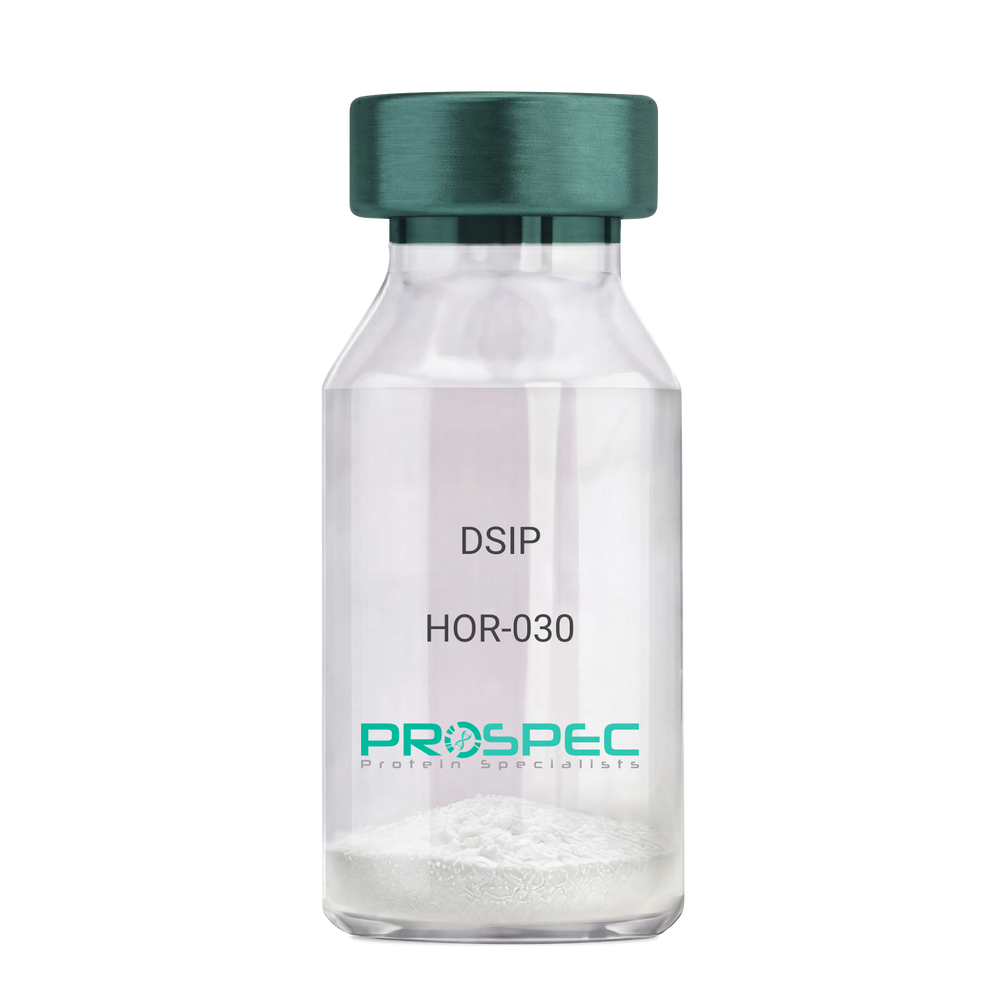
- View Data Sheet
Name :
DSIPDescription:
Delta Sleep Inducing Peptide
Product # :
HOR-030Price :
Quantity :
Shipping Method :
Shipped at Room temp
More Info
- description
- formulation
- purity
- More Info
Description
DSIP Synthetic is a single, non-glycosylated polypeptide chain containing 15 amino acids, having a molecular mass of 1419.55 Dalton and a Molecular formula of C62H98N16O22.
Formulation
The protein was lyophilized with no additives.
Purity
Greater than 97.0% as determined by analysis by RP-HPLC.
More Info
-
Physical Appearance
Sterile Filtered White lyophilized (freeze-dried) powder.
-
Stability
Lyophilized DSIP although stable at room temperature for 3 weeks, should be stored desiccated below -18°C. Upon reconstitution DSIP should be stored at 4°C between 2-7 days and for future use below -18°C. For long term storage it is recommended to add a carrier protein (0.1% HSA or BSA). Please prevent freeze-thaw cycles.
-
Solubility
It is recommended to reconstitute the lyophilized DSIP in sterile 18MΩ-cm H2O not less than 100 µg/ml, which can then be further diluted to other aqueous solutions.
-
Amino Acid Sequence
H-Gly-Glu-Pro-Pro-Pro-Gly-Lys-Pro-Ala-Asp-Asp-Ala-Gly-Leu-Val-OH
-
Background
Delta Sleep-Inducing Peptide (DSIP), also known as Sleep-Promoting Peptide, is a neuropeptide that has been the subject of extensive research due to its potential role in sleep regulation, stress response, and neuroprotection. This nonapeptide, first isolated from the cerebral venous blood of rabbits during sleep, has been shown to induce slow-wave sleep, modulate pain perception, and exhibit potential antioxidant and immunomodulatory properties.
DSIP's primary function is its interaction with the sleep regulatory system. By modulating the release of certain neurotransmitters, DSIP can influence sleep patterns, particularly promoting slow-wave sleep, the most restorative stage of sleep. Studies by Kovalzon et al. (2011) have demonstrated that DSIP can enhance sleep quality in rats, suggesting potential applications in sleep disorders and the promotion of healthy sleep patterns.
In addition to its sleep-inducing effects, DSIP has been shown to possess neuroprotective properties. Research by Zolotarev et al. (2014) found that DSIP could protect neurons from oxidative stress, suggesting potential applications in the treatment of neurodegenerative diseases such as Alzheimer's and Parkinson's disease.
Given its sleep-inducing and neuroprotective effects, DSIP has been proposed as a potential therapeutic agent for a variety of conditions, including sleep disorders, chronic pain, and neurodegenerative diseases. For instance, a study by Spong et al. (2016) found that DSIP could improve sleep quality in patients with chronic insomnia, indicating its potential as a therapeutic agent in the treatment of sleep disorders.
While research on DSIP is promising, it is important to note that most studies have been conducted in animals or in vitro. More research is needed to fully understand the potential effects and applications of DSIP in humans. However, the existing body of research suggests that DSIP could be a promising tool in the treatment of sleep disorders, chronic pain, and neurodegenerative diseases.
ProSpec's products are furnished for LABORATORY RESEARCH USE ONLY. The product may not be used as drugs, agricultural or pesticidal products, food additives or household chemicals.
- View Data Sheet
Name :
sRAGE MouseDescription:
Advanced Glycosylation End Product-Specific Receptor Mouse Recombinant
Advanced glycosylation end product-specific receptor, Receptor for advanced glycosylation end products, AGER, SRAGE, RAGE, MGC22357.
Product # :
PRO-2781Price :
Quantity :
Shipping Method :
Shipped at Room temp
More Info
- description
- source
- formulation
- purity
- More Info
Description
sRAGE Mouse Recombinant produced in HEK cells is a single, glycosylated, polypeptide chain containing 317 amino acids (Gly23–Asp333) and having a molecular mass of 34.0kDa. sRAGE Mouse is fused to a 6 a.a his tag at C-Terminus and is purified by proprietary chromatographic techniques.
Source
HEK 293.
Formulation
The filtered (0.4µm) concentrated protein solution was lyophilized with PBS, PH 7.4.
Purity
Greater than 95% as determined by SDS-PAGE.
More Info
-
Synonyms
Advanced glycosylation end product-specific receptor, Receptor for advanced glycosylation end products, AGER, SRAGE, RAGE, MGC22357.
-
Physical Appearance
Sterile Filtered White lyophilized (freeze-dried) powder.
-
Stability
Store lyophilized protein at -20°C. Aliquot the product after reconstitution to avoid repeated freezing/thawing cycles. Reconstituted protein can be stored at 4°C for a limited period of time.
-
Solubility
It is recommended to add deionized water to prepare a working stock solution of approximately 0.5mg/ml and let the lyophilized pellet dissolve completely.
-
Amino Acid Sequence
GQNITARIGE PLVLSCKGAP KKPPQQLEWK LNTGRTEAWK VLSPQGGPWD SVARILPNGS LLLPATGIVD EGTFRCRATN RRGKEVKSNY RVRVYQIPGK PEIVDPASEL TASVPNKVGT CVSEGSYPAG TLSWHLDGKL LIPDGKETLV KEETRRHPET GLFTLRSELT VIPTQGGTHP TFSCSFSLGL PRRRPLNTAP IQLRVREPGP PEGIQLLVEP EGGIVAPGGT VTLTCAISAQ PPPQVHWIKD GAPLPLAPSP VLLLPEVGHE DEGTYSCVAT HPSHGPQESP PVSIRVTETG DEGPAEGEGL DHHHHHH.
-
Background
The soluble receptor for advanced glycation end products, sRAGE, is a multifunctional protein known for its involvement in diverse physiological processes, including inflammation, aging, and chronic diseases. Research using mouse models has been instrumental in unraveling the complexities of sRAGE biology and its implications for health and disease. This study aims to provide a comprehensive exploration of sRAGE in mouse physiology, shedding light on its various functions and potential applications in understanding aging and disease mechanisms.
The primary objective of this research is to elucidate the impact of sRAGE in mouse models on aging processes. In vivo experiments utilizing genetically modified mice with altered sRAGE expression will be conducted to investigate how sRAGE influences the aging process, including effects on tissue homeostasis, oxidative stress, and longevity. Understanding these mechanisms is fundamental for deciphering the role of sRAGE in age-related diseases.
The second objective is to assess the clinical relevance of sRAGE in mouse models of chronic diseases. Mouse models of diseases such as diabetes, Alzheimer's disease, and cancer will be employed to explore how sRAGE modulation affects disease progression, inflammation, and tissue damage. These investigations may provide valuable insights into potential therapeutic strategies targeting sRAGE in various chronic diseases.
The third objective is to explore the broader implications of sRAGE in mouse physiology, including its effects on immunity, tissue repair, and metabolic regulation. Research will investigate its roles in immune cell function, wound healing, and glucose homeostasis. Understanding the multifaceted properties of sRAGE in mouse models may open new avenues for therapeutic interventions in various health and disease contexts.
By delving into the diverse functions of sRAGE in mouse physiology, this research aims to expand our knowledge of its physiological roles and clinical applications. The findings may contribute to the development of targeted interventions for age-related diseases and chronic conditions influenced by sRAGE.
ProSpec's products are furnished for LABORATORY RESEARCH USE ONLY. The product may not be used as drugs, agricultural or pesticidal products, food additives or household chemicals.
- View Data Sheet
Name :
CX3CL1 Human, Sf9Description:
Fractalkine (CX3CL1) Human Recombinant, Sf9
Fractalkine, CX3CL1, Neurotactin, CX3C membrane-anchored chemokine, Small inducible cytokine D1, NTN, NTT, CXC3, CXC3C, SCYD1, ABCD-3, C3Xkine.
Product # :
CHM-042Price :
Quantity :
Shipping Method :
Shipped with Ice Packs
More Info
- description
- source
- formulation
- purity
- More Info
- SDS-PAGE
Description
Fractalkine Human Recombinant produced in Sf9 Baculovirus cells is a single, glycosylated polypeptide chain containing 323 amino acids (25-339aa) and having a molecular mass of 34.3kDa.Fractalkine is fused to a 8 amino acid His tag at C-terminus and purified by proprietary chromatographic techniques.
Source
Sf9, Baculovirus cells.
Formulation
The Fractalkine solution (1 mg/ml) contains 10% Glycerol and Phosphate Buffered Saline (pH 7.4).
Purity
Greater than 90.0% as determined by SDS-PAGE.
SDS-PAGE
More Info
-
Introduction
Fractalkine soluble form is chemotactic for t-cells and monocytes, but not for neutrophils. Fractalkine membrane-bound form promotes adhesion of those leukocytes to endothelial cells. Fractalkine regulates leukocyte adhesion and migration processes at the endothelium and binds to CX3CR1. Fractalkine gene is located on human chromosome 16 along with some CC chemokines known as CCL17 and CCL22.
-
Synonyms
Fractalkine, CX3CL1, Neurotactin, CX3C membrane-anchored chemokine, Small inducible cytokine D1, NTN, NTT, CXC3, CXC3C, SCYD1, ABCD-3, C3Xkine.
-
Physical Appearance
Sterile Filtered colorless solution.
-
Stability
Store at 4°C if entire vial will be used within 2-4 weeks. Store, frozen at -20°C for longer periods of time. For long term storage it is recommended to add a carrier protein (0.1% HSA or BSA).Avoid multiple freeze-thaw cycles.
-
Amino Acid Sequence
QHHGVTKCNI TCSKMTSKIP VALLIHYQQN QASCGKRAII LETRQHRLFC ADPKEQWVKD
AMQHLDRQAA ALTRNGGTFE KQIGEVKPRT TPAAGGMDES VVLEPEATGE SSSLEPTPSS
QEAQRALGTS PELPTGVTGS SGTRLPPTPK AQDGGPVGTE LFRVPPVSTA ATWQSSAPHQ
PGPSLWAEAK TSEAPSTQDP STQASTASSP APEENAPSEG QRVWGQGQSP RPENSLEREE
MGPVPAHTDA FQDWGPGSMA HVSVVPVSSE GTPSREPVAS GSWTPKAEEP IHATMDPQRL GVLITPVPDA QAATRLEHHH HHH -
Background
What is the molecular weight/Mw of CX3CL1 HUMAN, SF9 Protein?
CX3CL1 HUMAN, SF9 Protein has a total Mw of 34.3kDa.
What is the source or expression system of CX3CL1 HUMAN, SF9 Protein?
Sf9, Baculovirus cells.
What is the Purity of CX3CL1 HUMAN, SF9 Protein?
CX3CL1 HUMAN, SF9 Protein is >90% pure as determined by SDS-PAGE.
What is the Biological Activity of CX3CL1 HUMAN, SF9 Protein?
The biological functionality of CX3CL1 HUMAN, SF9 Protein will be determined in the future.
What is the amino acid sequence of CX3CL1 HUMAN, SF9 Protein?
QHHGVTKCNI TCSKMTSKIP VALLIHYQQN QASCGKRAII LETRQHRLFC ADPKEQWVKD
AMQHLDRQAA ALTRNGGTFE KQIGEVKPRT TPAAGGMDES VVLEPEATGE SSSLEPTPSS
QEAQRALGTS PELPTGVTGS SGTRLPPTPK AQDGGPVGTE LFRVPPVSTA ATWQSSAPHQ
PGPSLWAEAK TSEAPSTQDP STQASTASSP APEENAPSEG QRVWGQGQSP RPENSLEREE
MGPVPAHTDA FQDWGPGSMA HVSVVPVSSE GTPSREPVAS GSWTPKAEEP IHATMDPQRL GVLITPVPDA QAATRLEHHH HHH
What applications can CX3CL1 HUMAN, SF9 Protein be used in?
CX3CL1 HUMAN, SF9 Protein can probably be used in western blot, ELISA and Lateral Flow.
What is the endotoxin level for CX3CL1 HUMAN, SF9 Protein?
The endotoxin level is minimal, CX3CL1 HUMAN, SF9 Protein was purified using conventional chromatography techniques.
ProSpec's products are furnished for LABORATORY RESEARCH USE ONLY. The product may not be used as drugs, agricultural or pesticidal products, food additives or household chemicals.
- View Data Sheet
Name :
REG3A AntibodyDescription:
Regenerating Islet-Derived 3 Alpha, Polyclonal Rabbit Anti-Human Antibody
Regenerating islet-derived protein 3 alpha, Reg III-alpha, Pancreatitis-associated protein 1, REG3A, HIP, PAP, PAP1, REG3, INGAP, PAP-H, PBCGF, REG-III.
Product # :
ANT-218Price :
Quantity :
Shipping Method :
Shipped at Room temp
More Info
- description
- formulation
- More Info
Description
The Antibody was raised in rabbits by immunization with the recombinant human REG3A. The amino acid sequence of the recombinant human REG3A is 100% homologus to the amino acid sequence of the human REG3A sequence. The immunization antigen is the 18.4 kDa protein containing 149 amino acid residues- His Tag.
Formulation
Sterile filtered and lyophilized from 1 mg/ml in 0.05M Phosphate buffer, 0.1M NaCl, pH-7.2.
More Info
-
Introduction
Pancreatitis-associated protein (PAP) is a secretory protein not normally expressed in healthy pancreas but highly induced during acute pancreatitis. While PAP has been shown to be anti-bacterial and antiapoptotic in vitro, its definitive biological function in vivo is not clear. Using antisepse oligonucleotides, inhibition of PAP expression significantly worsened pancreatitis in a rat model. During pancreatitis, PAP released by the pancreas could mediate lung inflammation through induction of hepatic TNF- alpha expression and subsequent increase in circulating TNF-alpha.
PAP is activated in primary liver cancers. In normal liver, the protein is undetectable in normal mature hepatocytes and found only in some ductular cells, representing potential hepatic progenitor cells. PAP can be considered hepatic cytokine that combines mitogenic and anti-apoptotic functions regarding hepatocytes, and consequently acts as a growth factor in vivo to enhance liver regeneration. In pancreatic cancor, PAP was overexpressed in 79% (30 of 38) of pancreatic ductal adenocarcinoma, 19% (7 of 36) of chronic pancreatitis, and 29% (2 of 7) of mucinous cystadenoma. PAP was found in malignant ductular structures in pancreatic carcinomas as well as in benign proliferating ductules and acinar cells in chronic pancreatitis. Elevation of PAP in patients with pancreatic cancer is not merely explainable by concomitant pancreatitis, but seems to be due to increased PAP production by the cancer cells and is also correlated to tumour load as expressed by the UICC stages.
Epithelial expression of PAP was induced under intestinal mucosal inflammation initiated by exposure to commensal bacteria or DSS as well as inflamed IBD colon. Increased serum level of PAP diagnosed ileal location in active Crohn disease with a sensitivity of 60%, a specificity of 94%, a positive predictive value of 84% and a negative predictive value of 81%. Elevated serum PAP (> 50 ng/mL) is significantly associated with disease activity and ileal location of Crohn disease. -
Synonyms
Regenerating islet-derived protein 3 alpha, Reg III-alpha, Pancreatitis-associated protein 1, REG3A, HIP, PAP, PAP1, REG3, INGAP, PAP-H, PBCGF, REG-III.
-
Physical Appearance
Sterile Filtered White lyophilized (freeze-dried) powder.
-
Stability
Store lyophilized antibody at -20°C. Aliquot the product after reconstitution to avoid repeated freezing/ thawing cycles and store frozen at -80°C. Reconstituted antibody can be stored at 4°C for a limited period of time; it does not show any change after two weeks at 4°C.
-
Solubility
Add 0.1 ml of deionized water and let the lyophilized pellet dissolve completely. Slight turbidity may occur after reconstitution, which does not affect activity of the antibody. In this case clarify the solution by centrifugation.
-
Immunogen
MRGSHHHHHH GMASHMEEPQ RELPSARIRC PKGSKAYGSH CYALFLSPKS WTDADLACQK RPSGNLVSVL SGAEGSFVSS LVKSIGNSYS YVWIGLHDPT QGTEPNGEGW EWSSSDVMNY FAWERNPSTI SSPGHCASLS RSTAFLRWKD YNCNVRLPYV CKFTD.
-
Species Reactivity
Human.
-
Titer
Defined by indirect ELISA, it is: >1: 100,000 for antibody concentration 1 mg/ml, 25 ng of antigen are coated per well, and is then defined at a point of maximal decrease of the titration curve.
-
Type
Polyclonal Rabbit Antibody.
-
Purification Method
Immunoaffinity chromatography on a column with immobilized recombinant human REG3A.
ProSpec's products are furnished for LABORATORY RESEARCH USE ONLY. The product may not be used as drugs, agricultural or pesticidal products, food additives or household chemicals.
- View Data Sheet
Name :
IL 12 p35 HumanDescription:
Interleukin-12 p35 Human Recombinant
NKSF1, CTL maturation factor (TCMF), Cytotoxic lymphocyte maturation factor 35 kDa subunit (CLMF p35), TSF, Edodekin-alpha, IL-12 p35, IL-12A, IL-12 subunit p35, NK cell stimulatory factor chain 1, SGT2, FLJ39002.
Product # :
CYT-487Price :
Quantity :
Shipping Method :
Shipped with Ice Packs
More Info
- description
- source
- formulation
- purity
- More Info
Description
Interleukin-12 p35 His Human Recombinant produced in E.Coli is single, a non-glycosylated, polypeptide chain containing 197 amino acids fragment (57-253) with an amino-terminal hexahistidine tag. The IL-12 p35 His is purified by proprietary chromatographic techniques.
Source
Escherichia Coli.
Formulation
Interleukin-12 p35 His-Tag is supplied in 1xPBS and 50% glycerol.
Purity
Greater than 95.0% as determined by SDS-PAGE.
More Info
-
Introduction
Active IL-12 is a p70 disulphide-linked dimer composed of p35 and p40 subunits. The protein is a pleiotropic cytokine produced primarily by antigen presenting cells and has multiple effects on T lymphocytes and natural killer cells in terms of stimulating cytotoxicity, proliferation, production of other cytokines and Th1 subset differentiation.
-
Synonyms
NKSF1, CTL maturation factor (TCMF), Cytotoxic lymphocyte maturation factor 35 kDa subunit (CLMF p35), TSF, Edodekin-alpha, IL-12 p35, IL-12A, IL-12 subunit p35, NK cell stimulatory factor chain 1, SGT2, FLJ39002.
-
Physical Appearance
Sterile Filtered clear solution.
-
Stability
Store at 4°C if entire vial will be used within 2-4 weeks. Store, frozen at -20°C for longer periods of time. Please avoid freeze thaw cycles.
ProSpec's products are furnished for LABORATORY RESEARCH USE ONLY. The product may not be used as drugs, agricultural or pesticidal products, food additives or household chemicals.
- View Data Sheet
Name :
IL 16 MouseDescription:
Interleukin-16 Mouse Recombinant
LCF, Lymphocyte Chemoattractant Factor, prIL-16, KIAA4048, mKIAA4048, Il16, IL-16, Interleukin-16.
Product # :
CYT-559Price :
Quantity :
Shipping Method :
Shipped at Room temp
More Info
- description
- source
- formulation
- purity
- More Info
Description
Interleukin-16 Mouse Recombinant produced in E.Coli is a single, non-glycosylated polypeptide chain containing 127 amino acids and having a molecular mass of 13.2 kDa. The Mouse IL-16 is purified by proprietary chromatographic techniques.
Source
Escherichia Coli.
Formulation
Murine IL-16 was lyophilized from 1mg/ml solution after extensive dialysis against 10mM sodium phosphate buffer, pH-7.5.
Purity
Greater than 90.0% as determined by(a) Analysis by RP-HPLC.
(b) Analysis SDS-PAGE.More Info
-
Introduction
IL-16 is a pleiotropic cytokine that functions as a chemoattractant, a modulator of T cell activation, and an inhibitor of HIV replication. The signaling process of IL-16 is mediated by CD4. The product of this gene undergoes proteolytic processing, which is found to yield two functional proteins. IL-16 functions exclusively attributed to the secreted C-terminal peptide, while the N-terminal product may play a role in cell cycle control. Caspase 3 is reported to be involved in the proteolytic processing of this protein. Two transcript variants encoding different isoforms have been found for this gene.
IL-16 stimulates a migratory response in cd4+ lymphocytes, monocytes, and eosinophils. Also induces t-lymphocyte expression of interleukin 2 receptor. ligand for cd4. -
Synonyms
LCF, Lymphocyte Chemoattractant Factor, prIL-16, KIAA4048, mKIAA4048, Il16, IL-16, Interleukin-16.
-
Physical Appearance
Sterile Filtered White lyophilized (freeze-dried) powder.
-
Stability
Lyophilized Mouse IL-16 although stable at room temperature for 3 weeks, should be stored desiccated below -18°C. Upon reconstitution mouse IL16 should be stored at 4°C between 2-7 days and for future use below -18°C.For long term storage it is recommended to add a carrier protein (0.1% HSA or BSA).Please prevent freeze-thaw cycles.
-
Solubility
It is recommended to reconstitute the lyophilized Murine IL16 in sterile 18MΩ-cm H2O not less than 100µg/ml, which can then be further diluted to other aqueous solutions.
-
Amino Acid Sequence
MHDLNSSTDS AASASAASDI SVESKEATVC TVTLEKTSAG LGFSLEGGKG SLHGDKPLTI NRIFKGDRTG EMVQPGDEIL QLAGTAVQGL TRFEAWNVIK ALPDGPVTIV IRRTSLQCKQ TTASADS.
ProSpec's products are furnished for LABORATORY RESEARCH USE ONLY. The product may not be used as drugs, agricultural or pesticidal products, food additives or household chemicals.
- View Data Sheet
Name :
LGALS9 HumanDescription:
Galectin-9 Human Recombinant
Lectin galactoside-binding soluble 9, Urate transporter/channel protein, LGALS9A, MGC125973, HUAT, Ecalectin, Galectin-9, MGC117375, MGC125974, HOM-HD-21, LGALS9.
Product # :
CYT-708Price :
Quantity :
Shipping Method :
Shipped with Ice Packs
More Info
- description
- source
- formulation
- purity
- More Info
- SDS-PAGE
Description
LGALS9 produced in E.Coli is a single, non-glycosylated polypeptide chain containing 168 amino acids (1-148 a.a.) and having a molecular mass of 18.5 kDa. Galectin-9 is fused to a 20 amino acid His-Tag at N-terminus and purified by proprietary chromatographic techniques.
Source
Escherichia Coli.
Formulation
The LGALS9 protein solution contains 20mM Tris-HCl, pH-8, 100mM NaCl and 20% glycerol.
Purity
Greater than 90.0% as determined by SDS-PAGE.
SDS-PAGE
More Info
-
Introduction
LGLAS9 binds galactosides and has high affinity for the Forssman pentasaccharide. LGLAS9 participates in thymocyte-epithelial interactions relevant to the biology of the thymus and Inhibits cell proliferation. LGLAS9 is a ligand for HAVCR2/TIM3. LGLAS9 Induces T-helper type 1 lymphocyte (Th1) death. LGLAS9 performs as an eosinophil chemoattractant LGLAS9 is an S-type lectin which is over-expressed in Hodgkin's disease tissue and takes part in the interaction between the H&RS cells with their surrounding cells.
-
Synonyms
Lectin galactoside-binding soluble 9, Urate transporter/channel protein, LGALS9A, MGC125973, HUAT, Ecalectin, Galectin-9, MGC117375, MGC125974, HOM-HD-21, LGALS9.
-
Physical Appearance
Sterile filtered colorless solution.
-
Stability
Store at 4°C if entire vial will be used within 2-4 weeks. Store, frozen at -20°C for longer periods of time. For long term storage it is recommended to add a carrier protein (0.1% HSA or BSA). Avoid multiple freeze-thaw cycles.
-
Amino Acid Sequence
MGSSHHHHHH SSGLVPRGSH MAFSGSQAPY LSPAVPFSGT IQGGLQDGLQ ITVNGTVLSS SGTRFAVNFQ TGFSGNDIAF HFNPRFEDGG YVVCNTRQNG SWGPEERKTH MPFQKGMPFD LCFLVQSSDF KVMVNGILFV QYFHRVPFHR VDTISVNGSV QLSYISFQ.
-
Background
What is the molecular weight/Mw of LGALS9 HUMAN Protein?
LGALS9 HUMAN Protein has a total Mw of 18.5kDa.
What is the source or expression system of LGALS9 HUMAN Protein?
Escherichia Coli.
What is the Purity of LGALS9 HUMAN Protein?
LGALS9 HUMAN Protein is >90% pure as determined by SDS-PAGE.
What is the Biological Activity of LGALS9 HUMAN Protein?
The biological functionality of LGALS9 HUMAN Protein will be determined in the future.
What is the amino acid sequence of LGALS9 HUMAN Protein?
MGSSHHHHHH SSGLVPRGSH MAFSGSQAPY LSPAVPFSGT IQGGLQDGLQ ITVNGTVLSS SGTRFAVNFQ TGFSGNDIAF HFNPRFEDGG YVVCNTRQNG SWGPEERKTH MPFQKGMPFD LCFLVQSSDF KVMVNGILFV QYFHRVPFHR VDTISVNGSV QLSYISFQ.
What applications can LGALS9 HUMAN Protein be used in?
LGALS9 HUMAN Protein can probably be used in western blot, ELISA and Lateral Flow.
What is the endotoxin level for LGALS9 HUMAN Protein?
The endotoxin level is minimal, LGALS9 HUMAN Protein was purified using conventional chromatography techniques.
ProSpec's products are furnished for LABORATORY RESEARCH USE ONLY. The product may not be used as drugs, agricultural or pesticidal products, food additives or household chemicals.