Search results
1000 results found for “Cysteine-Rich”
Name
Description
Product #
Price
Quantity
Shipping Method
- View Data Sheet
Name :
ICOSLG HumanDescription:
Inducible T-Cell Costimulator Ligand Human Recombinant
Inducible T-Cell Costimulator Ligand, B7-Related Protein 1, B7 Homolog 2, B7-Like Protein Gl50, B7 Homologue 2, B7RP-1, ICOSL, B7-H2, B7RP1, B7H2, Transmembrane Protein B7-H2 ICOS Ligand, Inducible T-Cell Co-Stimulator Ligand, CD275 Antigen, ICOS Ligand, KIAA0653, ICOS-L, CD275, LICOS, GL50, ICOS ligand, B7 homolog 2, B7-H2, B7-like protein Gl50, B7-related protein 1, B7RP-1.
Product # :
PRO-2431Price :
Quantity :
Shipping Method :
Shipped with Ice Packs
More Info
- description
- source
- formulation
- purity
- More Info
Description
ICOSLG produced in Sf9 Baculovirus cells is a single, glycosylated polypeptide chain containing 480 amino acids (19-256a.a.) and having a molecular mass of 53.7kDa (Molecular size on SDS-PAGE will appear at approximately 50-70kDa).ICOSLG is expressed with a 239 amino acid hIgG-His-tag at C-Terminus and purified by proprietary chromatographic techniques.
Source
Sf9, Baculovirus cells.
Formulation
ICOSLG protein solution (1mg/ml) contains Phosphate Buffered Saline (pH 7.4) and 10% glycerol.
Purity
Greater than 90.0% as determined by SDS-PAGE.
More Info
-
Introduction
Inducible T-Cell Costimulator Ligand also known as ICOSLG, is part of the B7 family of co-stimulatory molecules related to B7-1 and B7-2. ICOSLG is a transmembrane glycoprotein with extracellular IgV and IgC domains, in addition it binds to ICOS on activated T cells. The dependent signaling of ICOSLG takes part in a proliferative response.
-
Synonyms
Inducible T-Cell Costimulator Ligand, B7-Related Protein 1, B7 Homolog 2, B7-Like Protein Gl50, B7 Homologue 2, B7RP-1, ICOSL, B7-H2, B7RP1, B7H2, Transmembrane Protein B7-H2 ICOS Ligand, Inducible T-Cell Co-Stimulator Ligand, CD275 Antigen, ICOS Ligand, KIAA0653, ICOS-L, CD275, LICOS, GL50, ICOS ligand, B7 homolog 2, B7-H2, B7-like protein Gl50, B7-related protein 1, B7RP-1.
-
Physical Appearance
Sterile Filtered colorless solution.
-
Stability
Store at 4°C if entire vial will be used within 2-4 weeks. Store, frozen at -20°C for longer periods of time. For long term storage it is recommended to add a carrier protein (0.1% HSA or BSA).Avoid multiple freeze-thaw cycles.
-
Amino Acid Sequence
ADPDTQEKEV RAMVGSDVEL SCACPEGSRF DLNDVYVYWQ TSESKTVVTY HIPQNSSLEN VDSRYRNRAL MSPAGMLRGD FSLRLFNVTP QDEQKFHCLV LSQSLGFQEV LSVEVTLHVA ANFSVPVVSA PHSPSQDELT FTCTSINGYP RPNVYWINKT DNSLLDQALQ NDTVFLNMRG LYDVVSVLRI ARTPSVNIGC CIENVLLQQN LTVGSQTGND IGERDKITEN PVSTGEKNAA TLEPKSCDKT HTCPPCPAPE LLGGPSVFLF PPKPKDTLMI SRTPEVTCVV VDVSHEDPEV KFNWYVDGVE VHNAKTKPRE EQYNSTYRVV SVLTVLHQDW LNGKEYKCKV SNKALPAPIE KTISKAKGQP REPQVYTLPP SRDELTKNQV SLTCLVKGFY PSDIAVEWES NGQPENNYKT TPPVLDSDGS FFLYSKLTVD KSRWQQGNVF SCSVMHEALH NHYTQKSLSL SPGKHHHHHH.
ProSpec's products are furnished for LABORATORY RESEARCH USE ONLY. The product may not be used as drugs, agricultural or pesticidal products, food additives or household chemicals.
- View Data Sheet
Name :
His Tag AntibodyDescription:
Polyhistidine Tag, Mouse Antibody
Product # :
ANT-753Price :
Quantity :
Shipping Method :
Shipped with Ice Packs
More Info
- description
- formulation
- More Info
Description
Monoclonal antibodies are produced by immunizing mouse with synthesized polypeptide 6 x His. This antibody was purified via protein-A affinity chromatography.
Formulation
1mg/ml in PBS (pH 7.4) 0.02% Sodium Azide & 10% Glycerol
More Info
-
Introduction
His tag peptide also called polyhistidine-tag is chain of amino acids that are attached to proteins and consist of at least six histidine (6xHis, Hexa Histidine) residues, often at the N- or C-terminus of the protein. Polyhistidine-tags are used for affinity purification of genetically engineered proteins.
-
Physical Appearance
Sterile Filtered solution.
-
Ig Subclass
Kappa IgG1.
-
Clone
PAT1D6AT.
-
Applications
ELISA and W.B. The researcher should titrate to obtain optimal results.
-
Type
Mouse Antibody Monoclonal.
ProSpec's products are furnished for LABORATORY RESEARCH USE ONLY. The product may not be used as drugs, agricultural or pesticidal products, food additives or household chemicals.
- View Data Sheet
Name :
KRT18 AntibodyDescription:
Cytokeratin 18, Mouse Anti Human
Keratin type I cytoskeletal 18, Cytokeratin-18, CK-18, Keratin-18, K18, KRT18,CYK18,Cell proliferation-inducing gene 46 protein.
Product # :
ANT-025Price :
Quantity :
Shipping Method :
Shipped with Ice Packs
More Info
- formulation
- More Info
Formulation
1mg/ml containing PBS, pH-7.4, & 0.1% Sodium Azide.
More Info
-
Introduction
KRT18 encodes the type I intermediate filament chain keratin 18. Keratin 18, together with its filament partner keratin 8, are perhaps the most commonly found members of the intermediate filament gene family. They are expressed in single layer epithelial tissues of the body. Mutations in this gene have been linked to cryptogenic cirrhosis. Two transcript variants encoding the same protein have been found for this gene.
-
Synonyms
Keratin type I cytoskeletal 18, Cytokeratin-18, CK-18, Keratin-18, K18, KRT18,CYK18,Cell proliferation-inducing gene 46 protein.
-
Physical Appearance
Sterile filtered colorless solution.
-
Immunogen
Anti-human KRT18 mAb, is derived from hybridization of mouse FO myeloma cells with spleen cells from BALB/c mice immunized with recombinant human KRT18 amino acids 79-430 purified from E. coli.
-
Ig Subclass
Mouse IgG2b heavy chain and k light chain.
-
Clone
PAT2G3AT.
-
Applications
KRT18 antibody has been tested by ELISA, Western blot and Immunofluorescence analysis to assure specificity and reactivity. Since application varies, however, each investigation should be titrated by the reagent to obtain optimal results. Recommended dilution range for Western blot analysis and Immunofluorescence is 1:250 ~ 1000.
Recommended starting dilution is 1:500. -
Type
Mouse Anti Human Monoclonal.
-
Storage Procedures
For periods up to 1 month store at 4°C, for longer periods of time, store at -20°C. Prevent freeze thaw cycles.
-
Purification Method
KRT18 antibody was purified from mouse ascitic fluids by protein-G affinity chromatography.
ProSpec's products are furnished for LABORATORY RESEARCH USE ONLY. The product may not be used as drugs, agricultural or pesticidal products, food additives or household chemicals.
- View Data Sheet
Name :
NPPC HumanDescription:
Natriuretic Peptide C Human Recombinant
Natriuretic Peptide Precursor C, C-Type Natriuretic Peptide, CNP2, CNP.
Product # :
CYT-760Price :
Quantity :
Shipping Method :
Shipped with Ice Packs
More Info
- description
- source
- formulation
- purity
- More Info
Description
NPPC Human Recombinant produced in E.coli is a single, non-glycosylated polypeptide chain containing 126 amino acids (24-126) and having a molecular mass of 13.2kDa.NPPC is fused to a 23 amino acid His-tag at N-terminus & purified by proprietary chromatographic techniques.
Source
Escherichia Coli.
Formulation
The NPPC solution (0.25mg/ml) contains 20mM Tris-HCl buffer (pH 8.0), 0.15M NaCl and 10% glycerol.
Purity
Greater than 85% as determined by SDS-PAGE.
More Info
-
Introduction
NPPC is proteolytically managed to create a secreted hormone of the natriuretic peptide family. NPPC is vasoactive and natriuretic and controls the evolution and differentiation of cartilaginous growth plate chondrocytes.
-
Synonyms
Natriuretic Peptide Precursor C, C-Type Natriuretic Peptide, CNP2, CNP.
-
Physical Appearance
Sterile Filtered clear solution.
-
Stability
Store at 4°C if entire vial will be used within 2-4 weeks. Store, frozen at -20°C for longer periods of time. For long term storage it is recommended to add a carrier protein (0.1% HSA or BSA).Avoid multiple freeze-thaw cycles.
-
Amino Acid Sequence
MGSSHHHHHH SSGLVPRGSH MGSKPGAPPK VPRTPPAEEL AEPQAAGGGQ KKGDKAPGGG GANLKGDRSR LLRDLRVDTK SRAAWARLLQ EHPNARKYKG ANKKGLSKGC FGLKLDRIGS MSGLGC
ProSpec's products are furnished for LABORATORY RESEARCH USE ONLY. The product may not be used as drugs, agricultural or pesticidal products, food additives or household chemicals.
- View Data Sheet
Name :
SYT11 HumanDescription:
Synaptotagmin XI Human Recombinant
Synaptotagmin XI, SytXI, Synaptotagmin 12, KIAA0080, SYT12, Synaptotagmin-11.
Product # :
PRO-2301Price :
Quantity :
Shipping Method :
Shipped with Ice Packs
More Info
- description
- source
- formulation
- purity
- More Info
Description
SYT11 Human Recombinant produced in E.Coli is a single, non-glycosylated polypeptide chain containing 418 amino acids (37-431 a.a) and having a molecular mass of 47kDa. SYT11 is fused to a 23 amino acid His-tag at N-terminus & purified by proprietary chromatographic techniques.
Source
E.coli.
Formulation
SYT11 protein solution (0.25mg/ml) containing Phosphate Buffered Saline (pH7.4) and 50% glycerol.
Purity
Greater than 80% as determined by SDS-PAGE.
More Info
-
Introduction
Synaptotagmin-11 (SYT11) protein may be involved in Ca2+-dependent exocytosis of secretory vesicles through Ca2+ and phospholipid binding to the C2 domain or may function as Ca2+ sensors in the process of vesicular trafficking and exocytosis.
-
Synonyms
Synaptotagmin XI, SytXI, Synaptotagmin 12, KIAA0080, SYT12, Synaptotagmin-11.
-
Physical Appearance
Sterile Filtered colorless solution.
-
Stability
Store at 4°C if entire vial will be used within 2-4 weeks. Store, frozen at -20°C for longer periods of time. For long term storage it is recommended to add a carrier protein (0.1% HSA or BSA).Avoid multiple freeze-thaw cycles.
-
Amino Acid Sequence
MGSSHHHHHH SSGLVPRGSH MGSWSCCHQQ AEKKQKNPPY KFIHMLKGIS IYPETLSNKK KIIKVRRDKD GPGREGGRRN LLVDAAEAGL LSRDKDPRGP SSGSCIDQLP IKMDYGEELR SPITSLTPGE SKTTSPSSPE EDVMLGSLTF SVDYNFPKKA LVVTIQEAHG LPVMDDQTQG SDPYIKMTIL PDKRHRVKTR VLRKTLDPVF DETFTFYGIP YSQLQDLVLH FLVLSFDRFS RDDVIGEVMV PLAGVDPSTG KVQLTRDIIK RNIQKCISRG ELQVSLSYQP VAQRMTVVVL KARHLPKMDI TGLSGNPYVK VNVYYGRKRI AKKKTHVKKC TLNPIFNESF IYDIPTDLLP DISIEFLVID FDRTTKNEVV GRLILGAHSV TASGAEHWRE VCESPRKPVA KWHSLSEY.
ProSpec's products are furnished for LABORATORY RESEARCH USE ONLY. The product may not be used as drugs, agricultural or pesticidal products, food additives or household chemicals.
- View Data Sheet
Name :
PDGFRA HumanDescription:
Platelet-Derived Growth Factor Receptor, Alpha Human Recombinant
Platelet-derived growth factor receptor alpha polypeptide, PDGFR2, PDGF-R-alpha, CD140 antigen-like family member A, CD140a antigen, alpha-type platelet-derived growth factor receptor, RHEPDGFRA, rearranged-in-hypereosinophilia-platelet derived growth factor receptor alpha, PDGFRA/BCR fusion protein, MGC74795, EC 2.7.10.1.
Product # :
CYT-065Price :
Quantity :
Shipping Method :
Shipped with Ice Packs
More Info
- description
- source
- formulation
- purity
- More Info
Description
PDGFRA Human Recombinant produced in E.coli is a single, non-glycosylated polypeptide chain containing 522 amino acids (24-524) and having a molecular mass of 58.4 kDa.The PDGFRA is fused to a 20 amino acid His-Tag at N-terminus and purified by proprietary chromatographic techniques.
Source
E.coli.
Formulation
The PDGFRA solution (0.5mg/ml) contains 20mM Tris-HCl buffer (pH 8.0), 1mM DTT, 0.1M NaCl and 20% glycerol.
Purity
Greater than 85% as determined by SDS-PAGE.
More Info
-
Introduction
PDGFRa is a cell surface tyrosine kinase receptor for PDGF family members. PDGFRa binds to both A and B subunits of PDGF. It is known that PDGFRa is vital for kidney development since mice heterozygous for the receptor display defective kidney phenotypes.
-
Synonyms
Platelet-derived growth factor receptor alpha polypeptide, PDGFR2, PDGF-R-alpha, CD140 antigen-like family member A, CD140a antigen, alpha-type platelet-derived growth factor receptor, RHEPDGFRA, rearranged-in-hypereosinophilia-platelet derived growth factor receptor alpha, PDGFRA/BCR fusion protein, MGC74795, EC 2.7.10.1.
-
Physical Appearance
Sterile Filtered colorless solution.
-
Stability
Store at 4°C if entire vial will be used within 2-4 weeks. Store, frozen at -20°C for longer periods of time. For long term storage it is recommended to add a carrier protein (0.1% HSA or BSA).Avoid multiple freeze-thaw cycles.
-
Amino Acid Sequence
MGSSHHHHHH SSGLVPRGSH MQLSLPSILP NENEKVVQLN SSFSLRCFGE SEVSWQYPMS EEESSDVEIR NEENNSGLFV TVLEVSSASA AHTGLYTCYY NHTQTEENEL EGRHIYIYVP DPDVAFVPLG MTDYLVIVED DDSAIIPCRT TDPETPVTLH NSEGVVPASY DSRQGFNGTF TVGPYICEAT VKGKKFQTIP FNVYALKATS ELDLEMEALK TVYKSGETIV VTCAVFNNEV VDLQWTYPGE VKGKGITMLE EIKVPSIKLV YTLTVPEATV KDSGDYECAA RQATREVKEM KKVTISVHEK GFIEIKPTFS QLEAVNLHEV KHFVVEVRAY PPPRISWLKN NLTLIENLTE ITTDVEKIQE IRYRSKLKLI RAKEEDSGHY TIVAQNEDAV KSYTFELLTQ VPSSILDLVD DHHGSTGGQT VRCTAEGTPL PDIEWMICKD IKKCNNETSW TILANNVSNI ITEIHSRDRS TVEGRVTFAK VEETIAVRCL AKNLLGAENR ELKLVAPTLR SE.
ProSpec's products are furnished for LABORATORY RESEARCH USE ONLY. The product may not be used as drugs, agricultural or pesticidal products, food additives or household chemicals.
- View Data Sheet
Name :
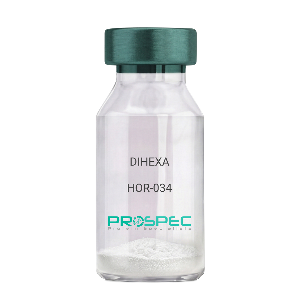
DIHEXADescription:
DIHEXA
Product # :
HOR-034Price :
Quantity :
Shipping Method :
Shipped at Room temp
More Info
- description
- formulation
- purity
- More Info
Description
DIHEXA Synthetic is a single, non-glycosylated polypeptide chain containing 3 amino acids, having a molecular mass of 504.28 Dalton and a Molecular formula of C27H44N4O5.
Formulation
The protein was lyophilized with no additives.
Purity
Greater than 97.0% as determined by analysis by RP-HPLC.
More Info
-
Physical Appearance
Sterile Filtered White lyophilized (freeze-dried) powder.
-
Stability
Lyophilized DIHEXA although stable at room temperature for 3 weeks, should be stored desiccated below -18°C. Upon reconstitution DIHEXA should be stored at 4°C between 2-7 days and for future use below -18°C. For long term storage it is recommended to add a carrier protein (0.1% HSA or BSA). Please prevent freeze-thaw cycles.
-
Solubility
It is recommended to reconstitute the lyophilized DIHEXA in sterile 18MΩ-cm H2O not less than 100 µg/ml, which can then be further diluted to other aqueous solutions.
-
Amino Acid Sequence
Hexanoyl-Tyr-Ile-Ahx-NH2.
-
Background
Dihexa, also known as N-hexanoic-Tyr-Ile-(6) aminohexanoic amide, is a potent and orally active small peptide that has been the focus of significant research due to its potential neurogenic and neuroprotective effects. This compound, derived from angiotensin IV, has been shown to possess a wide range of biological activities, including enhancing cognitive function, promoting neurogenesis, and potentially mitigating the effects of neurodegenerative diseases.
Dihexa's primary mechanism of action involves its interaction with hepatocyte growth factor (HGF) and its receptor, c-Met. By mimicking the effects of HGF, Dihexa can stimulate the c-Met receptor, leading to a cascade of events that promote neurogenesis and synaptic plasticity. Studies by Benoist et al. (2014) have demonstrated that Dihexa can enhance cognitive function in rats, suggesting potential applications in cognitive enhancement and the treatment of cognitive disorders.
In addition to its neurogenic effects, Dihexa has been shown to possess neuroprotective properties. Research by Kawas et al. (2017) found that Dihexa could protect neurons from apoptosis, suggesting potential applications in the treatment of neurodegenerative diseases such as Alzheimer's and Parkinson's disease.
Given its neurogenic and neuroprotective effects, Dihexa has been proposed as a potential therapeutic agent for a variety of conditions, including cognitive disorders, neurodegenerative diseases, and stroke. For instance, a study by Harding et al. (2018) found that Dihexa could improve outcomes in animal models of stroke, indicating its potential as a therapeutic agent in stroke recovery.
While research on Dihexa is promising, it is important to note that most studies have been conducted in animals or in vitro. More research is needed to fully understand the potential effects and applications of Dihexa in humans. However, the existing body of research suggests that Dihexa could be a promising tool in the treatment of cognitive disorders and neurodegenerative diseases.
ProSpec's products are furnished for LABORATORY RESEARCH USE ONLY. The product may not be used as drugs, agricultural or pesticidal products, food additives or household chemicals.
- View Data Sheet
Name :
CTSC MouseDescription:
Cathepsin-C Mouse Recombinant
DPPI, Cathepsin C, CTSC
Product # :
PRO-2769Price :
Quantity :
Shipping Method :
Shipped with Ice Packs
More Info
- description
- source
- formulation
- purity
- biological activity
- More Info
Description
CTSC Mouse Recombinant produced in HEK293 cells is a single, polypeptide chain containing 444 amino acids (25-462 a.a.) and having a molecular mass of 50.5kDa. CTSC is expressed with a 6 amino acid His tag at C-Terminus and purified by proprietary chromatographic techniques.
Source
HEK293 cells.
Formulation
CTSC protein solution (0.5mg/ml) containing Phosphate Buffered Saline (pH 7.4) and 10% glycerol.
Purity
Greater than 95% as determined by SDS-PAGE.
Biological Activity
> 50,000 pmol/min/ug and is defined as the amount of enzyme that hydrolyzes 1pmole of GlyArg-AMC / minute at pH 6.0 at 37C.
More Info
-
Introduction
CTSC has an important role in the activation of granule serine proteases in inflammatory cells.CTSC catalyses excision of dipeptides from the N-terminus of protein and peptide substrates. Once activated by CTSC, proteases are capable of degrading extracellular matrix components, which can result in tissue damage and chronic inflammation. CTSCcomprises of 4 subunits, each composed of the N-terminal proregion fragment, heavy chain and light chains. Defects in the CTSC protein have been demonstratedto result in Papillon-Lefevre disease which is an autosomal recessive disorder.
-
Synonyms
DPPI, Cathepsin C, CTSC
-
Physical Appearance
Sterile Filtered clear solution.
-
Stability
Store at 4°C if entire vial will be used within 2-4 weeks. Store, frozen at -20°C for longer periods of time.For long term storage it is recommended to add a carrier protein (0.1% HSA or BSA).Please avoid freeze thaw cycles.
-
Amino Acid Sequence
DTPANCTYPD LLGTWVFQVG PRSSRSDINC SVMEATEEKV VVHLKKLDTA YDELGNSGHF TLIYNQGFEI VLNDYKWFAF FKYEVRGHTA ISYCHETMTG WVHDVLGRNW ACFVGKKVES HIEKVNMNAA HLGGLQERYS ERLYTHNHNF VKAINTVQKS WTATAYKEYE KMSLRDLIRR SGHSQRIPRP KPAPMTDEIQ QQILNLPESW DWRNVQGVNY VSPVRNQESC GSCYSFASMG MLEARIRILT NNSQTPILSP QEVVSCSPYA QGCDGGFPYL IAGKYAQDFG VVEESCFPYT AKDSPCKPRE NCLRYYSSDY YYVGGFYGGC NEALMKLELV KHGPMAVAFE VHDDFLHYHS GIYHHTGLSD PFNPFELTNH AVLLVGYGRD PVTGIEYWII KNSWGSNWGE SGYFRIRRGT DECAIESIAV AAIPIPKL-HHHHHH
ProSpec's products are furnished for LABORATORY RESEARCH USE ONLY. The product may not be used as drugs, agricultural or pesticidal products, food additives or household chemicals.
- View Data Sheet
Name :
EREG Human, HEKDescription:
Epiregulin Human Recombinant, HEK
EPR, Epiregulin, Ep, ER, Proepiregulin, EREG.
Product # :
CYT-1206Price :
Quantity :
Shipping Method :
Shipped with Ice Packs
More Info
- description
- source
- formulation
- purity
- biological activity
- More Info
Description
EREG Human Recombinant produced in HEK293 cells is a single, glycosylated polypeptide chain (63-108a.a) containing 289 amino acids and having a molecular mass of 32.6 kDa.EREG is fused to a 239 amino acid hIgG-His-Tag at C-terminus & purified by proprietary chromatographic techniques.
Source
HEK293 cells.
Formulation
EREG protein (0.25mg/ml) contains 10% glycerol and Phosphate-Buffered Saline (pH 7.4).
Purity
Greater than 90.0% as determined by SDS-PAGE.
Biological Activity
Measured in a cell proliferation assay using Balb/3T3 mouse embryonic fibroblast cells. The ED50 range ≤ 1ug/ml.
More Info
-
Introduction
"Epiregulin is part of the EGF family. Epiregulin functions as a ligand of EGFR, as well as a ligand of most members of the ERBB (v-erb-b2 oncogene homolog) family of tyrosine-kinase receptors.Epiregulin is expressed mostly in the placenta and peripheral blood leukocytes and in specific carcinomas of the bladder, lung, kidney and colon. Epiregulin stimulates the proliferation of keratinocytes, hepatocytes, fibroblasts and vascular smooth muscle cells. Epiregulin inhibits the growth of several tumor-derived epithelial cell lines. Human Epiregulin is initially synthesized as a glycosylated 19.0 kDa transmembrane precursor protein, which is processed by proteolytic cleavage to produce a 6.0 kDa mature secreted sequence."
-
Synonyms
EPR, Epiregulin, Ep, ER, Proepiregulin, EREG.
-
Physical Appearance
Sterile filtered colorless solution.
-
Stability
Store at 4°C if entire vial will be used within 2-4 weeks.Store, frozen at -20°C for longer periods of time.For long term storage it is recommended to add a carrier protein (0.1% HSA or BSA).Avoid multiple freeze-thaw cycles.
-
Amino Acid Sequence
DGSMVSITKC SSDMNGYCLH GQCIYLVDMS QNYCRCEVGY TGVRCEHFFL LEPKSCDKTH TCPPCPAPEL LGGPSVFLFP PKPKDTLMIS RTPEVTCVVV DVSHEDPEVK FNWYVDGVEV HNAKTKPREE QYNSTYRVVS VLTVLHQDWL NGKEYKCKVS NKALPAPIEK TISKAKGQPR EPQVYTLPPS RDELTKNQVS LTCLVKGFYP SDIAVEWESN GQPENNYKTT PPVLDSDGSF FLYSKLTVDK SRWQQGNVFS CSVMHEALHN HYTQKSLSLS PGKHHHHHH
-
Background
What is the molecular weight/Mw of EREG Protein?
EREG Protein has a total Mw of 32.6kDa.
What is the source or expression system of EREG Protein?
HEK293 cells.
What is the Purity of EREG Protein?
EREG Protein is >90% pure as determined by SDS-PAGE.
What is the Biological Activity of EREG Protein?
Measured in a cell proliferation assay using Balb/3T3 mouse embryonic fibroblast cells. The ED50 range ≤ 1ug/ml.
What is the amino acid sequence of EREG Protein?
DGSMVSITKC SSDMNGYCLH GQCIYLVDMS QNYCRCEVGY TGVRCEHFFL LEPKSCDKTH TCPPCPAPEL LGGPSVFLFP PKPKDTLMIS RTPEVTCVVV DVSHEDPEVK FNWYVDGVEV HNAKTKPREE QYNSTYRVVS VLTVLHQDWL NGKEYKCKVS NKALPAPIEK TISKAKGQPR EPQVYTLPPS RDELTKNQVS LTCLVKGFYP SDIAVEWESN GQPENNYKTT PPVLDSDGSF FLYSKLTVDK SRWQQGNVFS CSVMHEALHN HYTQKSLSLS PGKHHHHHH
What applications can EREG Protein be used in?
EREG Protein can probably be used in western blot, ELISA and Lateral Flow.
What is the endotoxin level for EREG Protein?
The endotoxin level is minimal, EREG Protein was purified using conventional chromatography techniques.
ProSpec's products are furnished for LABORATORY RESEARCH USE ONLY. The product may not be used as drugs, agricultural or pesticidal products, food additives or household chemicals.
- View Data Sheet
Name :
CDH5 MouseDescription:
Cadherin 5 Mouse Recombinant
Cadherin-5, Vascular endothelial cadherin, VE-cadherin, CD144, Cdh5.
Product # :
PRO-2244Price :
Quantity :
Shipping Method :
Shipped with Ice Packs
More Info
- description
- source
- formulation
- purity
- More Info
Description
CDH5 produced in Sf9 Insect cells is a single, glycosylated polypeptide chain containing 583 amino acids (25-599a.a.) and having a molecular mass of 66.2kDa. (Molecular size on SDS-PAGE will appear at approximately 70-100kDa).CDH5 is expressed with an 8 amino acid His tag at C-Terminus and purified by proprietary chromatographic techniques.
Source
Sf9, Insect cells.
Formulation
CDH5 protein solution (0.5mg/ml) contains phosphate buffered saline (pH7.4) and 10% glycerol.
Purity
Greater than 90.0% as determined by SDS-PAGE.
More Info
-
Introduction
Cadherin-5, also known as CDH5 is a classical cadherin which belongs to the cadherin superfamily. The CDH5is located in a six-cadherin cluster in a region on the long arm of chromosome 16 which is involved in loss ofheterozygosity events in breast as well as prostate cancer. CDH5 is a calcium-dependent cell-celladhesion glycoprotein composed of five extracellular cadherin repeats, a transmembrane region and a highlyconserved cytoplasmic tail. Furthermore, CDH5 functions as a classic cadherin by passing on to cells the ability to adhere in ahomophilic manner, CDH5 plays a significant role in endothelial cell biology through control of the cohesion as well as organization of the intercellular junctions.
-
Synonyms
Cadherin-5, Vascular endothelial cadherin, VE-cadherin, CD144, Cdh5.
-
Physical Appearance
Sterile filtered colorless solution.
-
Stability
Store at 4°C if entire vial will be used within 2-4 weeks.Store, frozen at -20°C for longer periods of time. For long term storage it is recommended to add a carrier protein (0.1% HSA or BSA).Avoid multiple freeze-thaw cycles.
-
Amino Acid Sequence
GPNFPQIDTP NMLPAHHRQK RDWIWNQMHI DEEKNESLPH YVGKIKSNVN RQNAKYVLQG EFAGKIFGVD ANTGNVLAYE RLDREKVSEY FLTALIVDKN TNKNLEQPSS FTVKVHDIND NWPVFSHQVF NASVPEMSAI GTSVIRVTAV DADDPTVAGH ATVLYQIVKG NEYFSIDNSG LIFTKIKNLD REKQAEYKIV VETQDALGLR GESGTATVMI RLEDINDNFP VFTQSTYTFS VPEDIRVGKP LGFLTVVDPD EPQNRMTKYS IMQGEYRDTF TIETDPKRNE GIIKPTKSLD YEVIQQYTFY IEATDPTIRY EYLSSTSGKN KAMVTINVLD VDEPPVFQRH FYHFKLPENQ KKPLIGTVVA KDPDKAQRSI GYSIRKTSDR GQFFRITKQG NIYNEKELDR ETYAWYNLTV EANELDSRGN PVGKESIVQV YIEVLDENDN PPEFAQPYEP KVCENAAQGK LVVQISATDK DVVPVNPKFK FALKNEDSNF TLINNHDNTA NITVKYGQFN REHAKFHYLP VLISDNGVPS LTGTSTLTVG VCKCNEQGEF TFCEEMAAQA GVSIQLEHHH HHH.
ProSpec's products are furnished for LABORATORY RESEARCH USE ONLY. The product may not be used as drugs, agricultural or pesticidal products, food additives or household chemicals.
- View Data Sheet
Name :
COX5B HumanDescription:
Cytochrome C Oxidase Subunit Vb Human Recombinant
Cytochrome c oxidase subunit 5B, mitochondrial precursor, COXVB, COX5B, Mitochondrial, Cytochrome c oxidase polypeptide Vb.
Product # :
PRO-1513Price :
Quantity :
Shipping Method :
Shipped with Ice Packs
More Info
- description
- source
- formulation
- purity
- More Info
Description
COX5B Human Recombinant produced in E.Coli is a single, non-glycosylated polypeptide chain containing 121 amino acids (32-129a.a) and having a molecular mass of 13kDa. COX5B is fused to a 23 amino acid His-tag at N-terminus & purified by proprietary chromatographic techniques.
Source
Escherichia Coli.
Formulation
COX5B protein solution (0.5mg/ml) containing 20mM Tris-HCl buffer (pH8.0), 0.1M NaCl, 20% glycerol and 1mM DTT.
Purity
Greater than 95.0% as determined by SDS-PAGE.
More Info
-
Introduction
Cytochrome c oxidase subunit VB (COX5B) is the terminal enzyme of the mitochondrial respiratory chain. COX5B is a multi-subunit enzyme complex which couples the transfer of electrons from cytochrome c to molecular oxygen and contributes to a proton electrochemical gradient across the inner mitochondrial membrane. There are two isoforms of COX5:COX5a and COX5b. Transcription of COX5A (the aerobic isoform) is up-regulated as the rate of cellular respiration increases, when oxygen levels within the cell are high. However, when oxygen levels are low, COX5B (the hypoxic isoform) transcription increases and functions to maximize the turnover rate of the COX apoenzyme.
-
Synonyms
Cytochrome c oxidase subunit 5B, mitochondrial precursor, COXVB, COX5B, Mitochondrial, Cytochrome c oxidase polypeptide Vb.
-
Physical Appearance
Sterile filtered colorless solution.
-
Stability
Store at 4°C if entire vial will be used within 2-4 weeks. Store, frozen at -20°C for longer periods of time. For long term storage it is recommended to add a carrier protein (0.1% HSA or BSA).Avoid multiple freeze-thaw cycles.
-
Amino Acid Sequence
MGSSHHHHHH SSGLVPRGSH MGSASGGGVP TDEEQATGLE REIMLAAKKG LDPYNVLAPK GASGTREDPN LVPSISNKRI VGCICEEDNT SVVWFWLHKG EAQRCPRCGA HYKLVPQQLA H.
ProSpec's products are furnished for LABORATORY RESEARCH USE ONLY. The product may not be used as drugs, agricultural or pesticidal products, food additives or household chemicals.
- View Data Sheet
Name :
C1Q RatDescription:
Complement Component C1q Rat
Component C1q, Complement C1q, Complement Component C1q, C1q.
Product # :
PRO-2704Price :
Quantity :
Shipping Method :
Shipped with Ice Packs
More Info
- description
- source
- formulation
- purity
- More Info
Description
Rat Complement C1Q produced in Rat plasma having a molecular weight of 400kDa.
Source
Rat Plasma.
Formulation
C1Q solution contains 10mM HEPES and 300mM NaCl, pH 7.2.
Purity
Greater than 93.0% as determined by SDS-PAGE.
More Info
-
Introduction
C1q is the first component of the classical pathway of complement activation. C1q along with the enzymatically active components C1r and C1s forms the C1 complex. When C1 binds to immunoglobulins in the form of immune complexes, it leads to activation of C1r and C1s proteases and a further activates the classical pathway of complement. C1q is a glycoprotein that belongs to the collectin family, having a molecular weight of about 410-462 kDa. C1q is a hexamer composed of globular heads attached to collagen-like triple-helix tails. The globular heads of C1q exclusively bind to the CH2 domain of IgG molecules or the CH3 domain of IgM. Each heavy chain of the immunoglobulin molecule contains a single binding site for C1q. Given that C1q must bind to no less than two heavy chains in order to alter its conformation and activate C1r and C1s, its activation follows only after binding to immunoglobulins in the form of immune complexes bound to multivalent antigens. C1q’s main physiological role is in the clearance of immune complexes and apoptotic bodies from the organism. Interruption of this process may lead to development of autoimmunity. Individuals with genetic deficiencies of C1q or other components of the classical pathway are at risk to develop SLE. C1q specifically binds to apoptotic bodies of human keratinocytes, vascular endothelial cells and lymphocytes. Complement components C1q and bound C3 mediate the clearance of apoptotic bodies. Hence, C1q may advance the clearance of autoantigens, avoiding stimulation of the immune system. Nonetheless, an extended exposition of the immune system to the neoepitope exposed on C1q molecules bound to immune complexes or apoptotic bodies could ultimately lead to an autoimmune response against C1q itself and to an altered complement function. C1q deficiency may also lead to disruption of the negative selection of autoreactive B cells. C1q along with other specific recognition proteins bind to the highly conserved lupus antigens (dsDNA and nuclear proteins) and activate the complement system. Autoantibodies against C1q (anti-C1q) are found in a number of autoimmune and infectious diseases like glomerulonephritis (GN) and lupus erythematosus (SLE), these antibodies are significant in clinical practice due to their negative predictive value.
-
Synonyms
Component C1q, Complement C1q, Complement Component C1q, C1q.
-
Physical Appearance
Sterile filtered solution.
-
Stability
C1Q Rat is stable at 4°C if entire vial will be used within 2-4 weeks.Store, frozen below -20°C for longer periods of time.For long term storage it is recommended to add a carrier protein (0.1% HSA or BSA).Avoid multiple freeze-thaw cycles.
ProSpec's products are furnished for LABORATORY RESEARCH USE ONLY. The product may not be used as drugs, agricultural or pesticidal products, food additives or household chemicals.
- View Data Sheet
Name :
LIF HumanDescription:
Leukemia Inhibitory Factor Human Recombinant
CDF, HILDA, D-FACTOR, Differentiation- stimulating factor, Melanoma-derived LPL inhibitor, MLPLI, Emfilermin, Leukemia inhibitory factor, LIF, DIA.
Product # :
CYT-644Price :
Quantity :
Shipping Method :
Shipped at Room temp
More Info
- description
- source
- formulation
- purity
- biological activity
- More Info
Description
Leukemia Inhibitory Factor (LIF) Human Recombinant produced in E.Coli is a single, non-glycosylated, polypeptide chain containing 180 amino acids and having a molecular mass of 19.7kDa. The Leukemia Inhibitory Factor (LIF) is purified by proprietary chromatographic techniques.
Source
Escherichia Coli.
Formulation
Leukemia Inhibitory Factor (LIF) was lyophilized from a concentrated (1mg/ml) sterile solution containing 1xPBS pH 7.4.
Purity
Greater than 98.0% as determined by:
(a) Analysis by RP-HPLC.
(b) Analysis by SDS-PAGE.Biological Activity
The ED50 was determined by the M1 cell differentiation assay is < 0.01 ng/ml, corresponding to a specific activity of 100,000,000IU/mg.More Info
-
Introduction
Leukemia Inhibitory Factor also called LIF is a lymphoid factor that promotes long-term maintenance of embryonic stem cells by suppressing spontaneous differentiation. Leukemia Inhibitory Factor has several functions such as cholinergic neuron differentiation, control of stem cell pluripotency, bone & fat metabolism, mitogenesis of factor dependent cell lines & promotion of megakaryocyte production in vivo. Human and mouse LIF exhibit a 78% identity in its amino acid sequence.
-
Synonyms
CDF, HILDA, D-FACTOR, Differentiation- stimulating factor, Melanoma-derived LPL inhibitor, MLPLI, Emfilermin, Leukemia inhibitory factor, LIF, DIA.
-
Physical Appearance
Sterile Filtered White lyophilized (freeze-dried) powder.
-
Stability
Lyophilized Leukemia Inhibitory Factor (LIF) although stable at room temperature for 3 weeks, should be stored desiccated below -18°C. Upon reconstitution Leukemia Inhibitory Factor (LIF) should be stored at 4°C between 2-7 days and for future use below -18°C.For long term storage it is recommended to add a carrier protein (0.1% HSA or BSA).Please prevent freeze-thaw cycles.
-
Solubility
It is recommended to reconstitute the lyophilized Leukemia Inhibitory Factor (LIF) in sterile water not less than 100µg/ml, which can then be further diluted to other aqueous solutions.
-
Amino Acid Sequence
SPLPITPVNA TCAIRHPCHN NLMNQIRSQL AQLNGSANAL FILYYTAQGE PFPNNLDKLC GPNVTDFPPF HANGTEKAKL VELYRIVVYL GTSLGNITRD QKILNPSALS LHSKLNATAD ILRGLLSNVL CRLCSKYHVG HVDVTYGPDT SGKDVFQKKK LGCQLLGKYK QIIAVLAQAF.
-
Background
Leukemia Inhibitory Factor (LIF) Background
Leukemia Inhibitory Factor (LIF) is a cytokine within the interleukin-6 family. It is influential in the hypothalamus and involved in energy balance.
LIF stimulates ACTH secretion and affects the stress and immune response in the body. Administration of LIF has been shown to correct low plasma ACTH and repair hypothalamic-pituitary-adrenal (HPA) axis responses.
Function and Applications of LIF
LIF prevents the proliferation of myeloid leukemia cells by inducing their terminal differentiation. Beyond cancer, LIF influences bone metabolism, embryogenesis, and inflammation.
Moreover, it supports the self-renewal of stem cells in culture by activating Stat3, thus preventing spontaneous differentiation, and is studied for potential benefits in fertility treatments.
Structure and Interactions
LIF's structure consists of a four alpha-helix bundle similar to other hematopoietic cytokines. It interacts with components such as the ciliary neurotrophic factor (CNTF) through multimeric receptors, influencing both cytokine's effects on cells. This interaction is crucial for forming high-affinity binding sites that mediate their biological activities.
ProSpec's products are furnished for LABORATORY RESEARCH USE ONLY. The product may not be used as drugs, agricultural or pesticidal products, food additives or household chemicals.
- View Data Sheet
Name :
OSM Human, 209 a.aDescription:
Oncostatin M Human Recombinant (209 a.a.)
OSM, MGC20461, Oncostatin M.
Product # :
CYT-639Price :
Quantity :
Shipping Method :
Shipped at Room temp
More Info
- description
- source
- formulation
- purity
- biological activity
- More Info
Description
Oncostatin-M (209 a.a.) Human Recombinant produced in E.Coli is a single, non-glycosylated polypeptide chain containing 209 amino acids and having a molecular mass of 23.9kDa. The Oncostatin-M (209 a.a.) is purified by proprietary chromatographic techniques.
Source
Escherichia Coli.
Formulation
Oncostatin-M (209 a.a.) was lyophilized from a concentrated (1mg/ml) solution containing 1x PBS pH-7.4.
Purity
Greater than 97.0% as determined by(a) Analysis by RP-HPLC.
(b) Analysis by SDS-PAGE.Biological Activity
The ED50 as determined by the dose-dependant stimulation of Human TF-1 cells is < 2 ng/ml, corresponding to a Specific Activity of 500,000 IU/mg.More Info
-
Introduction
Oncostatin M is a member of a cytokine family that includes leukemia-inhibitory factor, granulocyte colony-stimulating factor, and interleukin 6. This gene encodes a growth regulator which inhibits the proliferation of a number of tumor cell lines. It regulates cytokine production, including IL-6, G-CSF and GM-CSF from endothelial cells.
-
Synonyms
OSM, MGC20461, Oncostatin M.
-
Physical Appearance
Sterile Filtered White lyophilized (freeze-dried) powder.
-
Stability
Lyophilized Oncostatin-M (209 a.a.) although stable at room temperature for 3 weeks, should be stored desiccated below -18°C. Upon reconstitution Oncostatin-M (209 a.a.) should be stored at 4°C between 2-7 days and for future use below -18°C.For long term storage it is recommended to add a carrier protein (0.1% HSA or BSA).Please prevent freeze-thaw cycles.
-
Solubility
It is recommended to reconstitute the lyophilized Oncostatin-M (209 a.a.) in sterile 18MΩ-cm H2O not less than 100µg/ml, which can then be further diluted to other aqueous solutions.
-
Amino Acid Sequence
AAIGSCSKEYRVLLGQLQKQTDLMQDTSRLLDPYIRIQGLDVPKLREHCRERPG
AFPSEETLRGLGRRGFLQTLNATLGCVLHRLADLEQRLPKAQDLERSGLNIEDL
EKLQMARPNILGLRNNIYCMAQLLDNSDTAEPTKAGRGASQPPTPTPASDAFQ
RKLEGCRFLHGYHRFMHSVGRVFKWGESPNRSRRHSPHQALRKGVRR.
ProSpec's products are furnished for LABORATORY RESEARCH USE ONLY. The product may not be used as drugs, agricultural or pesticidal products, food additives or household chemicals.
- View Data Sheet
Name :
VEGF Mouse, HisDescription:
Vascular Endothelial Growth Factor Mouse Recombinant, His Tag
Vascular endothelial growth factor A, VEGF-A, Vascular permeability factor, VPF, VEGF, Vegf120, Vegf164, Vegf188, Vegfa.
Product # :
CYT-680Price :
Quantity :
Shipping Method :
Shipped with Ice Packs
More Info
- description
- source
- formulation
- purity
- biological activity
- More Info
Description
VEGF Mouse Recombinant produced in E.Coli is a single, non-glycosylated, polypeptide chain containing 141 amino acids (205-324 a.a.) and having a total molecular mass of 16.3kDa. Mouse VEGF is fused to 20 amino acid His Tag at N-terminus and purified by proprietary chromatographic techniques.
Source
Escherichia Coli.
Formulation
The Mouse VEGF contains 20mM Tris-HCl buffer (pH8.0) and 10% glycerol.
Purity
Greater than 90.0% as determined by SDS-PAGE.
Biological Activity
Measured in a cell proliferation assay using NIH-3T3 mouse embryonic fibroblast. The ED50 for this effect is 0.5-1.5ng/ml.More Info
-
Introduction
Vascular endothelial growth factor is an important signaling protein involved in both vasculogenesis and angiogenesis. As its name implies, VEGF activity has been mostly studied on cells of the vascular endothelium, although it does have effects on a number of other cell types (e.g. stimulation monocyte/macrophage migration, neurons, cancer cells, kidney epithelial cells ).VEGF mediates increased vascular permeability, induces angiogenesis, vasculogenesis and endothelial cell growth, promotes cell migration, and inhibits apoptosis. In vitro, VEGF has been shown to stimulate endothelial cell mitogenesis and cell migration. VEGF is also a vasodilator and increases microvascular permeability and was originally referred to as vascular permeability factor.
Elevated levels of this protein is linked to POEMS syndrome, also known as Crow-Fukase syndrome. Mutations in this gene have been associated with proliferative and nonproliferative diabetic retinopathy. -
Synonyms
Vascular endothelial growth factor A, VEGF-A, Vascular permeability factor, VPF, VEGF, Vegf120, Vegf164, Vegf188, Vegfa.
-
Physical Appearance
Sterile Filtered colorless solution.
-
Stability
Store at 4°C if entire vial will be used within 2-4 weeks. Store, frozen at -20°C for longer periods of time. For long term storage it is recommended to add a carrier protein (0.1% HSA or BSA).Avoid multiple freeze-thaw cycles.
-
Amino Acid Sequence
MGSSHHHHHH SSGLVPRGSH MAPTTEGEQK SHEVIKFMDV YQRSYCRPIE TLVDIFQEYP DEIEYIFKPS CVPLMRCAGC CNDEALECVP TSESNITMQI MRIKPHQSQH IGEMSFLQHS RCECRPKKDR TKPEKCDKPR R.
ProSpec's products are furnished for LABORATORY RESEARCH USE ONLY. The product may not be used as drugs, agricultural or pesticidal products, food additives or household chemicals.
- View Data Sheet
Name :
Protein GDescription:
Protein G Recombinant
Product # :
PRO-402Price :
Quantity :
Shipping Method :
Shipped at Room temp
More Info
- description
- source
- formulation
- purity
- More Info
- sds-page
Description
The Protein G is a single, non-glycosylated protein contains 200 amino acids having a molecular mass of 21.8kDa. The Protein-G migrates on SDS-PAGE around 32kDa.
Source
Escherichia Coli.
Formulation
Lyophilized white powder containing no additives.
Purity
>96% as determined by SDS-PAGE and RP-HPLC.
sds-page
More Info
-
Physical Appearance
Sterile Filtered White lyophilized (freeze-dried) powder.
-
Stability
Lyophilized Recombinant Protein G although stable at room temperature for 3 weeks, should be stored desiccated below -18°C. Upon reconstitution Protein G should be stored at 4°C between 2-7 days and for future use below -18°C.Please prevent freeze-thaw cycles.
-
Solubility
Reconstitution with deionized water or PBS.
-
Amino Acid Sequence
LPKTDTYKLILNGKTLKGETTTEAVDAATAEKVFKQYANDNGVDGEWTYDDAT KTFTVTEKPEVIDASELTPAVTTYKLVINGKTLKGETTTEAVDAATAEKVFK QYANDNGVDGEWTYDDATKTFTVTEKPEVIDASELTPAVTTYKLVINGKTL KGETTTKAVDAETAEKAFKQYANDNGVDGVWTYDDATKTFTVTE.
ProSpec's products are furnished for LABORATORY RESEARCH USE ONLY. The product may not be used as drugs, agricultural or pesticidal products, food additives or household chemicals.
- View Data Sheet
Name :
Adiponectin Human (72-244)Description:
Adiponectin (72-244) Human Recombinant
Acrp30, AdipoQ, GBP-28, APM-1, ACDC.
Product # :
CYT-1231Price :
Quantity :
Shipping Method :
Shipped at Room temp
More Info
- description
- source
- formulation
- purity
- More Info
Description
The Adiponectin Human is created as a recombinant protein with a 4kda N-terminal fusion of His Tag. The Adiponectin His-Tagged Fusion Protein, produced in E. coli, is a 24kDa protein containing 173 amino acid residues of the Acrp30 Human, 72-244 amino acids.
Source
Escherichia Coli.
Formulation
Each mg was lyophilized with 1xPBS, 0.4% SDS and 4mM DTT.
Purity
Greater than 90% as determined by SDS-PAGE.
More Info
-
Synonyms
Acrp30, AdipoQ, GBP-28, APM-1, ACDC.
-
Physical Appearance
Filtered White lyophilized (freeze-dried) powder.
-
Stability
Store lyophilized Adiponectin at -20°C. Aliquot the product after reconstitution to avoid repeated freezing/thawing cycles. Reconstituted protein can be stored at 4°C for a limited period of time; it does not show any change after two weeks at 4°C.
-
Solubility
It is recommended to add deionized water to prepare a working stock solution of approximately 0.5mg/ml and let the lyophilized pellet dissolve completely. Product is not sterile! Please filter the product by an appropriate sterile filter before using it on cell culture.
-
Amino Acid Sequence
IGPKGDI GETGVPGAEG PRGFPGIQGR KGEPGEGAYV YRSAFSVGLE TYVTIPNMPI RFTKIFYNQQ NHYDGSTGKF HCNIPGLYYF AYHITVYMKD VKVSLFKKDK AMLFTYDQYQ ENNVDQASGS VLLHLEVGDQ VWLQVYGEGE RNGLYADNDN DSTFTGFLLY HDTN
-
Background
Adiponectin is a protein produced and secreted by adipose tissue. Adiponectin takes part in regulating glucose levels as well as fatty acid breakdown.
Adiponectin ‘s Functions:
Anti-Inflammatory Effects - Adiponectin has anti-inflammatory properties that helps mitigate chronic inflammation.
Regulation of Glucose and Lipid Metabolism - Adiponectin Enhances insulin sensitivity, helping in regulation of blood sugar levels and also promotes fatty acid oxidation, which helps reduce fat accumulation.
Cardiovascular Health - It may influence vascular health and is associated with a lower risk of cardiovascular diseases.
Levels and Health Implications:
Normal Levels - usually, higher levels of adiponectin are associated with a lower risk of metabolic syndrome, cardiovascular diseases and type 2 diabetes.
Low Levels - Reduced adiponectin levels are often linked with obesity, insulin resistance, and other metabolic disorders.
Factors Influencing on the Adiponectin Levels:
Weight - High body fat (especially visceral fat) can lower adiponectin levels.
Diet and Exercise - Regular physical activity and a healthy diet can increase adiponectin levels.
Genetics - Genetic factors might also be an influence on an individual adiponectin level.
Adiponectin is an important component in metabolic health, therefore continuing the research of its functions and regulation keeps advance our understanding of its role in diseases like diabetes and cardiovascular conditions.
What is the molecular weight / Mw of ADIPONECTIN Protein?
ADIPONECTIN Protein has a total Mw of 24kDa.What is the source or expression system of ADIPONECTIN Protein?
Escherichia Coli.
What is the Purity of ADIPONECTIN Protein?
ADIPONECTINProtein is >90% pure as determined by SDS-PAGE.
What is the Biological Activity of ADIPONECTIN Protein?
The biological functionality of ADIPONECTIN Protein will be determined in the future.
What is the amino acid sequence of ADIPONECTIN Protein?
IGPKGDI GETGVPGAEG PRGFPGIQGR KGEPGEGAYV YRSAFSVGLE TYVTIPNMPI RFTKIFYNQQ NHYDGSTGKF HCNIPGLYYF AYHITVYMKD VKVSLFKKDK AMLFTYDQYQ ENNVDQASGS VLLHLEVGDQ VWLQVYGEGE RNGLYADNDN DSTFTGFLLY HDTN.
What applications can ADIPONECTIN Protein be used in ?
ADIPONECTIN Protein can probably be used in western blot, ELISA and Lateral Flow.
What is the endotoxin level for ADIPONECTIN Protein?
The endotoxin level is minimal, ADIPONECTIN Protein was purified using conventional chromatography techniques.
ProSpec's products are furnished for LABORATORY RESEARCH USE ONLY. The product may not be used as drugs, agricultural or pesticidal products, food additives or household chemicals.
- View Data Sheet
Name :
IGJ HumanDescription:
Immunoglobulin J Human Recombinant
IGCJ, JCH, Immunoglobulin J chain, IGJ.
Product # :
PRO-1420Price :
Quantity :
Shipping Method :
Shipped with Ice Packs
More Info
- description
- source
- formulation
- purity
- More Info
Description
IGJ Human Recombinant produced in E. coli is a single polypeptide chain containing 160 amino acids (23-159) and having a molecular mass of 18kDa. IGJ is fused to a 23 amino acid His-tag at N-terminus & purified by proprietary chromatographic techniques.
Source
E.coli.
Formulation
The IGJ solution (0.5mg/ml) contains 20mM Tris-HCl buffer (pH 8.0), 0.15M NaCl, 10% glycerol and 1mM DTT.
Purity
Greater than 85% as determined by SDS-PAGE.
More Info
-
Introduction
Immunoglobulin J chain (IGJ) helps to link 2 monomer units of either IgM or IgA and also also helps to bind these immunoglobulins to secretory component. When it comes to IgM, the J chain-joined dimer is a nucleating unit for the IgM pentamer and in the case of IgA it induces larger polymers.
-
Synonyms
IGCJ, JCH, Immunoglobulin J chain, IGJ.
-
Physical Appearance
Sterile Filtered colorless solution.
-
Stability
Store at 4°C if entire vial will be used within 2-4 weeks. Store, frozen at -20°C for longer periods of time. For long term storage it is recommended to add a carrier protein (0.1% HSA or BSA).Avoid multiple freeze-thaw cycles.
-
Amino Acid Sequence
MGSSHHHHHH SSGLVPRGSH MGSQEDERIV LVDNKCKCAR ITSRIIRSSE DPNEDIVERN IRIIVPLNNR ENISDPTSPL RTRFVYHLSD LCKKCDPTEV ELDNQIVTAT QSNICDEDSA TETCYTYDRN KCYTAVVPLV YGGETKMVET ALTPDACYPD.
ProSpec's products are furnished for LABORATORY RESEARCH USE ONLY. The product may not be used as drugs, agricultural or pesticidal products, food additives or household chemicals.
- View Data Sheet
Name :
Protein ADescription:
Staphylococcal Protein A Recombinant
Immunoglobulin G-binding protein A, IgG-binding protein A, Staphylococcal protein A, SPA.
Product # :
PRO-356Price :
Quantity :
Shipping Method :
Shipped with Ice Packs
More Info
- description
- source
- formulation
- purity
- biological activity
- More Info
Description
Recombinant Staphylococcal Protein A produced in E.Coli is a non-glycosylated, Polypeptide chain having a molecular mass of 46.7 kDa.Recombinant Staphylococcal Protein A is purified by proprietary chromatographic techniques.
Source
Escherichia Coli.
Formulation
The protein solution contains no additives.
Purity
Greater than 95.0% as determined by RP-HPLC.
Biological Activity
Greater than 95.0% binding activity to human IgG.
More Info
-
Introduction
Protein A is a cell wall protein deriving from Staphylococcus aureus which exhibits unique binding properties for IgG from a variety of mammalian species and for some IgM and IgA as well. It binds with the Fc region of immunoglobulins through interaction with the heavy chain. It couples to a wide variety of reporter molecules including fluorescent dyes, enzyme markers, biotin, colloidal gold and radioactive iodine without affecting the antibody binding site. Recombinant Protein A was developed to increase the specificity of the molecule for IgG and is widely used both in research and bioprocessing. The recombinant protein A is produced by expressing a modified protein A gene in E.coli. A specific purification process with strict quality control was taken to get the recombinant protein A with the purity of more than 98% , no human IgG affinity step is used during validated fermentation and purification and devoid of bacterial contaminant found normally in native Protein A. (Free of Staphylococcus endotoxins and hemolysin).
-
Synonyms
Immunoglobulin G-binding protein A, IgG-binding protein A, Staphylococcal protein A, SPA.
-
Physical Appearance
Sterile Filtered clear colorless solution.
-
Stability
SPA should be stored at -20°C.
ProSpec's products are furnished for LABORATORY RESEARCH USE ONLY. The product may not be used as drugs, agricultural or pesticidal products, food additives or household chemicals.
- View Data Sheet
Name :
Protein A/GDescription:
Protein A/G Recombinant
Product # :
PRO-646Price :
Quantity :
Shipping Method :
Shipped at Room temp
More Info
- description
- source
- formulation
- purity
- More Info
Description
The recombinant Protein A/G consists of 5 IgG-binding regions of protein A and 2 of protein G, which corresponds to the Protein A and G domains that are included in the recombinant sequence. Cell wall binding region, cell membrane binding region and albumin binding region have been removed from the recombinant Protein A/G to ensure the maximum specific IgG binding. The Protein A portion is from Staphylococcus aureus segments E, D, A, B and C. The Protein G portion is from Streptococcus segments C1 and C3. The fusion protein has a predicted molecular mass of 47.7kDa and containing 429 amino acids.
Source
Escherichia coli.
Formulation
Lyophilized white Powder containing no additives.
Purity
>97% as determined by SDS-PAGE and RP-HPLC.
More Info
-
Introduction
Recombinant Protein A/G fusion protein joins IgG binding domains of both Protein A and Protein G.
Protein A/G includes four Fc binding domains from Protein A and two from Protein G, yielding a final mass of 50.4 kDa. The binding dependency to pH of Protein A/G lower than Protein A, but has the additive properties of Protein A and G together. Protein A/G binds to all subclasses of human IgG, making it helpful for purifying polyclonal or monoclonal IgG antibodies whose subclasses have not been identifieed. Protein A/G binds to IgA, IgE, IgM and IgD. Protein A/G binds to all subclasses of mouse IgG excluding mouse IgA, IgM or serum albumin. This permits Protein A/G to be used in purification and detection of mouse monoclonal IgG antibodies, with no interference from IgA, IgM and serum albumin. Mouse monoclonal antibodies normally have a stronger affinity to the chimeric Protein A/G than to either Protein A or Protein G. Protein A/G also has been used for purification of macaque IgG. -
Stability
After reconstitution, aliquot and store at -20°C. Avoid repeated freeze/thaw cycles.
-
Solubility
It is recommended to reconstitute the lyophilized Protein-A/G in sterile 18M-cm H2O not less than 0.1mg/ml, which can then be further diluted to other aqueous solutions.
-
Amino Acid Sequence
NAAQHDEAQQ NAFYQVLNMP NLNADQRNGF IQSLKDDPSQ SANVLGEAQK LNDSQAPKAD AQQNNFNKDQ QSAFYEILNM PNLNEAQRNG FIQSLKDDPS QSTNVLGEAK KLNESQAPKA DNNFNKEQQN AFYEILNMPN LNEEQRNGFI QSLKDDPSQS ANLLSEAKKL NESQAPKADN KFNKEQQNAF YEILHLPNLN EEQRNGFIQS LKDDPSQSAN LLAEAKKLND AQAPKADNKF NKEQQNAFYE ILHLPNLTEE QRNGFIQSLK DDPSVSKEIL AEAKKLNDAQ APKEEDSLEG SGSGTYKLIL NGKTLKGETT TEAVDAATAE KVFKQYANDN GVDGEWTYDD ATKTFTVTEK PEVIDASELT PAVTTYKLVI NGKTLKGETT TKAVDAETAE KAFKQYANDN GVDGVWTYDD ATKTFTVTE.
ProSpec's products are furnished for LABORATORY RESEARCH USE ONLY. The product may not be used as drugs, agricultural or pesticidal products, food additives or household chemicals.
- View Data Sheet
Name :
CTF2P MouseDescription:
Neuropoietin Mouse Recombinant
Cardiotrophin-2, CT-2, Neuropoietin, Np, Ctf2, Gm494.
Product # :
CYT-1128Price :
Quantity :
Shipping Method :
Shipped at Room temp
More Info
- description
- source
- formulation
- purity
- biological activity
- More Info
Description
Neuropoietin Mouse Recombinant produced in E.Coli is a non-glycosylated polypeptide chain containing 182 amino acids and having a molecular mass of approximately 19.7kDa.CTF2P is purified by proprietary chromatographic techniques.
Source
Escherichia Coli.
Formulation
Lyophilized from a 0.2μm filtered concentrated solution in 20mM Tris-HCl, pH 8.0, 0.5mM DTT and 500mM NaCl.
Purity
Greater than 98.0% as determined by:
(a) Analysis by RP-HPLC.
(b) Analysis by SDS-PAGE.Biological Activity
The ED50 as determined by a cell proliferation assay using human TF-1 cells is < 200 ng/ml, corresponding to a specific activity of > 5000 IU/mg.
More Info
-
Introduction
CTF2P, aka Neuropoietinis a part of the IL-6 family of cytokines. CTF2P is the outcome of a gene duplication event involving cardiotrophin-1 (CT-1) and it helps to define a subfamily within the IL-6 family that includes CT-1, CLC and CTNF. CTF2P Increases the platelet count associated with splenomegaly and takes part in neuronal precursor development and maturation.
-
Synonyms
Cardiotrophin-2, CT-2, Neuropoietin, Np, Ctf2, Gm494.
-
Physical Appearance
Sterile Filtered White lyophilized (freeze-dried) powder.
-
Stability
Lyophilized CTF2P although stable at room temperature for 3 weeks, should be stored desiccated below -18°C. Upon reconstitution Neuropoietin should be stored at 4°C between 2-7 days and for future use below -18°C. Please prevent freeze-thaw cycles.
-
Solubility
It is recommended to reconstitute the lyophilized Neuropoietin in sterile PBS not less than 100µg/ml, which can then be further diluted to other aqueous solutions.
-
Amino Acid Sequence
APISPSEPIG QAYSLALYMQ KNTSALLQTY LQHQGSPFSD PGFSAPELQL STLPSAAVSF KTWHAMEDAE RLSRAQGAFL ALTQHLQLVG DDQSYLNPGS PILLAQLGAA RLRAQGLLGN MAAIMTALGL PIPPEEDTLG FVPFGASAFE RKCRGYIVTR EYGHWTDRAV RDLALLKAKY SA.
-
Background
What is the molecular weight/Mw of CTF2P Protein?
CTF2P Protein has a total Mw of 19.7kDa.
What is the source or expression system of CTF2P Protein?
Escherichia Coli.
What is the Purity of CTF2P Protein?
CTF2P Protein is >98% pure as determined by SDS-PAGE.
What is the Biological Activity of CTF2P Protein?
The ED50 as determined by a cell proliferation assay using human TF-1 cells is < 200 ng/ml, corresponding to a specific activity of > 5000 IU/mg.
What is the amino acid sequence of CTF2P Protein?
APISPSEPIG QAYSLALYMQ KNTSALLQTY LQHQGSPFSD PGFSAPELQL STLPSAAVSF KTWHAMEDAE RLSRAQGAFL ALTQHLQLVG DDQSYLNPGS PILLAQLGAA RLRAQGLLGN MAAIMTALGL PIPPEEDTLG FVPFGASAFE RKCRGYIVTR EYGHWTDRAV RDLALLKAKY SA.
What applications can CTF2P Protein be used in?
CTF2P Protein can probably be used in western blot, ELISA and Lateral Flow.
What is the endotoxin level for CTF2P Protein?
The endotoxin level is minimal, CTF2P Protein was purified using conventional chromatography techniques.
ProSpec's products are furnished for LABORATORY RESEARCH USE ONLY. The product may not be used as drugs, agricultural or pesticidal products, food additives or household chemicals.
- View Data Sheet
Name :
HBsAg adwDescription:
Hepatitis B Surface Antigen, adw Recombinant
Product # :
HBS-872Price :
Quantity :
Shipping Method :
Shipped with Ice Packs
More Info
- description
- source
- formulation
- purity
- More Info
Description
HbsAg adw produced Pichia Pastoris, having a molecular weight of approximately 24.0 kDa as shown on SDS-PAGE.
Source
Pichia Pastoris.
Formulation
Sterile Filtered solution containing 20mM Phosphate Buffer, 154mM sodium chloride, pH 7.1.
Purity
Greater than 95.0% as determined by SDS-PAGE.
More Info
-
Introduction
HBsAg is the surface antigenof the Hepatitis-B-Virus (HBV). The capsidof a virus has different surface proteins from the rest of the virus. The antigen is a protein that binds specifically on one of these surface proteins. It is commonly referred to as the Australian Antigen.
-
Physical Appearance
Sterile Filtered pale solution.
-
Stability
HBsAg Should be stored at 4°C.DO NOT FREEZE.
ProSpec's products are furnished for LABORATORY RESEARCH USE ONLY. The product may not be used as drugs, agricultural or pesticidal products, food additives or household chemicals.
- View Data Sheet
Name :
Clusterin CanineDescription:
Clusterin Canine Recombinant
CLI, AAG4, KUB1, SGP2, SGP-2, SP-40, TRPM2, MGC24903, Glycoprotein 80, Gp80, CLU, Clusterin, Apolipoprotein J, Apo-J.
Product # :
CYT-549Price :
Quantity :
Shipping Method :
Shipped at Room temp
More Info
- description
- source
- formulation
- purity
- More Info
- sds-page
Description
Apolipoprotein-J canine Recombinant produced in E.Coli is a single, non-glycosylated, Polypeptide chain (Asn227~Glu445) and having a molecular mass of 30 kDa.
The protein is fused to His tag at N-Terminus.
The Apolipoprotein-J canine is purified by proprietary chromatographic techniques.Source
Escherichia Coli.
Formulation
Canine Clusterin was lyophilized from 20mM Tris, 150mM NaCl, pH8.0, 0.01% skl and 5%Trehalose.
Purity
Greater than 90% as determined by SDS PAGE.
sds-page
More Info
-
Introduction
Clusterin also named Apolipoprotein J (APO-J) is a 75-80 kD disulfide-linked heterodimeric protein containing about 30% of N-linked carbohydrate rich in sialic acid but truncated forms targeted to the nucleus have also been identified.
The precursor polypeptide chain is cleaved proteolytically to remove the 22-mer secretory signal peptide and subsequently between residues 227/228 to generate the a and b chains. These are assembled in anti-parallel to give a heterodimeric molecule in which the cysteine-rich centers are linked by five disulfide bridges and are flanked by two predicted coiled-coil a-helices and three predicted amphipathic a-helices.
Across a broad range of species clusterin shows a high degree of sequence homology ranging from 70% to 80%. It is nearly ubiquitously expressed in most mammalian tissues and can be found in plasma, milk, urine, cerebrospinal fluid and semen.
It is able to bind and form complexes with numerous partners such as immunoglobulins, lipids, heparin, bacteria, complement components, paraoxonase, beta amyloid, leptin and others. Clusterin has been ascribed a plethora of functions such as phagocyte recruitment, aggregation induction, complement attack prevention, apoptosis inhibition, membrane remodeling, lipid transport, hormone transport and/or scavenging, matrix metalloproteinase inhibition.
A genuine function of clusterin has not been defined. One tempting hypothesis says that clusterin is an extracellular chaperone protecting cells from stress induced insults caused by degraded and misfolded protein precipitates.
Clusterin is up- or down regulated on the mRNA or protein level in many pathological and clinically relevant situations including cancer, organ regeneration, infection, Alzheimer disease, retinitis pigmentosa, myocardial infarction, renal tubular damage, autoimmunity and others. -
Synonyms
CLI, AAG4, KUB1, SGP2, SGP-2, SP-40, TRPM2, MGC24903, Glycoprotein 80, Gp80, CLU, Clusterin, Apolipoprotein J, Apo-J.
-
Physical Appearance
Sterile Filtered White lyophilized (freeze-dried) powder.
-
Stability
Store lyophilized protein at -20°C. Aliquot the product after reconstitution to avoid repeated freezing/thawing cycles. Reconstituted protein can be stored at 4°C for a limited period of time; it does not show any change after two weeks at 4°C.
-
Solubility
Reconstitute in 20mM Tris and 150mM NaCl (pH8.0) to a concentration of 0.1-1.0 mg/mL and let the lyophilized pellet dissolve completely.
-
Amino Acid Sequence
NIIPFP RFQPLNFHDM FQPFFDMIHQ AQQAMDVNLH RIPYHFPIEF PEEDNRTVCK EIRHNSTGCL KMKDQCEKCQ EILSVDCSSN NPAQVQLRQE LSNSLQIAEK FTKLYDELLQ SYQEKMFNTS SLLKQLNEQF SWVSQLANLT QSEDPFYLQV TTVGSQTSDS NVPVGFTKVV VKLFDSDPIT VMIPEAVSRN NPKFMETVAE KALQEYRQKHREE.
-
Background
What is the molecular weight/Mw of CLUSTERIN Protein?
CLUSTERIN Protein has a total Mw of 30kDa.
What is the source or expression system of CLUSTERIN Protein?
Escherichia Coli.
What is the Purity of CLUSTERIN Protein?
CLUSTERIN Protein is >90% pure as determined by SDS-PAGE.
What is the Biological Activity of CLUSTERIN Protein?
The biological functionality of CLUSTERIN Protein will be determined in the future.
What is the amino acid sequence of CLUSTERIN Protein?
NIIPFP RFQPLNFHDM FQPFFDMIHQ AQQAMDVNLH RIPYHFPIEF PEEDNRTVCK EIRHNSTGCL KMKDQCEKCQ EILSVDCSSN NPAQVQLRQE LSNSLQIAEK FTKLYDELLQ SYQEKMFNTS SLLKQLNEQF SWVSQLANLT QSEDPFYLQV TTVGSQTSDS NVPVGFTKVV VKLFDSDPIT VMIPEAVSRN NPKFMETVAE KALQEYRQKHREE.
What applications can CLUSTERIN Protein be used in?
CLUSTERIN Protein can probably be used in western blot, ELISA and Lateral Flow.
What is the endotoxin level for CLUSTERIN Protein?
The endotoxin level is minimal, CLUSTERIN Protein was purified using conventional chromatography techniques
ProSpec's products are furnished for LABORATORY RESEARCH USE ONLY. The product may not be used as drugs, agricultural or pesticidal products, food additives or household chemicals.
- View Data Sheet
Name :
Noggin Human, HEKDescription:
Noggin Human Recombinant, HEK
Noggin, Symphalangism 1 (Proximal), Synostoses (Multiple) Syndrome 1, SYNS1A, SYNS1, SYM1.
Product # :
CYT-977Price :
Quantity :
Shipping Method :
Shipped with Ice Packs
More Info
- description
- source
- formulation
- purity
- More Info
Description
Noggin produced in HEK293 cells is a polypeptide chain containing 211 amino acids (28-232a.a.) and having a molecular mass of 23.8kDa. (Molecular size on SDS-PAGE will appear at approximately 28-40kDa).Noggin is expressed with a 6 amino acid His tag at C-Terminus and purified by proprietary chromatographic techniques.
Source
HEK293 Cells.
Formulation
Recombinant Human Noggin hek293 derived protein is provided as a solution (0.25mg/ml) containing 50mM MES (pH 6.5) and 30% glycerol.
Purity
Greater than 90.0% as determined by SDS-PAGE.
More Info
-
Introduction
The secreted polypeptide noggin, encoded by the NOG gene, binds and inactivates members of the transforming growth factor-beta (TGF-beta) superfamily signaling proteins, such as bone morphogenetic protein-4 (BMP4). By diffusing through extracellular matrices more efficiently than members of the TGF-beta superfamily, noggin may have a principal role in creating morphogenic gradients. Noggin appears to have pleiotropic effect, both early in development as well as in later stages. It was originally isolated from Xenopus based on its ability to restore normal dorsal-ventral body axis in embryos that had been artificially ventralized by UV treatment. The results of the mouse knockout of noggin suggest that it is involved in numerous developmental processes, such as neural tube fusion and joint formation. Recently, several dominant human NOG mutations in unrelated families with proximal symphalangism (SYM1) and multiple synostoses syndrome (SYNS1) were identified; both SYM1 and SYNS1 have multiple joint fusion as their principal feature, and map to the same region (17q22) as NOG. All NOG mutations altered evolutionarily conserved amino acid residues. The amino acid sequence of human noggin is highly homologous to that of Xenopus, rat and mouse.
-
Synonyms
Noggin, Symphalangism 1 (Proximal), Synostoses (Multiple) Syndrome 1, SYNS1A, SYNS1, SYM1.
-
Physical Appearance
Sterile filtered colorless solution.
-
Stability
Store at 4°C if entire vial will be used within 2-4 weeks.Store, frozen at -20°C for longer periods of time. For long term storage it is recommended to add a carrier protein (0.1% HSA or BSA).Avoid multiple freeze-thaw cycles.
-
Amino Acid Sequence
QHYLHIRPAP SDNLPLVDLI EHPDPIFDPK EKDLNETLLR SLLGGHYDPG FMATSPPEDR PGGGGGAAGG AEDLAELDQL LRQRPSGAMP SEIKGLEFSE GLAQGKKQRL SKKLRRKLQM WLWSQTFCPV LYAWNDLGSR FWPRYVKVGS CFSKRSCSVP EGMVCKPSKS VHLTVLRWRC QRRGGQRCGW IPIQYPIISE CKCSCHHHHH H.
ProSpec's products are furnished for LABORATORY RESEARCH USE ONLY. The product may not be used as drugs, agricultural or pesticidal products, food additives or household chemicals.