Search results
1000 results found for “visfatin”
Name
Description
Product #
Price
Quantity
Shipping Method
- View Data Sheet
Name :
VCAM1 HumanDescription:
Vascular Cell Adhesion Molecule 1 Human Recombinant
Vascular cell adhesion protein 1, V-CAM 1, INCAM-100, CD106, VCAM1, L1CAM, MGC99561, DKFZp779G2333.
Product # :
PRO-386Price :
Quantity :
Shipping Method :
Shipped with Ice Packs
More Info
- description
- source
- formulation
- purity
- More Info
- Citations
Description
VCAM1 Human Recombinant is expressed in E. coli containing 674 amino acids (a.a. 25-698) fused to an amino terminal hexahistidine tag, having a total MW of 78.8kDa.
Source
Escherichia Coli.
Formulation
VCAM1 is supplied in 10mM Tris-HCl (pH 8), 250mM NaCl, 0.5mM DTT, 1.5mM Cysteine, and 50% Glycerol.
Purity
Greater than 95% as determined by SDS-PAGE.
Citations
More Info
-
Introduction
VCAM1 is a member of the Ig superfamily. It is a cell surface sialoglycoprotein expressed by cytokine activated endothelium. VCAM-1 contains 6 or 7 immunoglobulin domains, and is expressed on both large and small vessels only after the endothelial cells are stimulated by cytokines. The protein has a number of functions including the regulation of leukocyte migration, leukocyte endothelial cell adhesion and signal transduction and may play a role in a number of inflammatory diseases (artherosclerosis and rheumatoid arthritis).
-
Synonyms
Vascular cell adhesion protein 1, V-CAM 1, INCAM-100, CD106, VCAM1, L1CAM, MGC99561, DKFZp779G2333.
-
Stability
Store at 4°C if entire vial will be used within 1-2 weeks. Store, frozen at -20°C for longer periods of time. Avoid multiple freeze-thaw cycles.
-
Applications
VCAM1 can be used directly as a positive control in Western blotting, ELISA, immunoprecipitation and other immunological experiments.
The biological activity of this product has not yet been tested.
ProSpec's products are furnished for LABORATORY RESEARCH USE ONLY. The product may not be used as drugs, agricultural or pesticidal products, food additives or household chemicals.
- View Data Sheet
Name :
RNF4 HumanDescription:
Ring Finger Protein 4 Human Recombinant
E3 ubiquitin-protein ligase RNF4, RING finger protein 4, Small nuclear ring finger protein, Protein SNURF, RNF4, SNURF, RES4-26, SLX5.
Product # :
PRO-1255Price :
Quantity :
Shipping Method :
Shipped with Ice Packs
More Info
- description
- source
- formulation
- purity
- More Info
- Citations
Description
RNF4 Human Recombinant produced in E.Coli is a single, non-glycosylated polypeptide chain containing 213 amino acids (1-190 a.a) and having a molecular mass of 23.7kDa.RNF4 is fused to a 23 amino acid His-tag at N-terminus & purified by proprietary chromatographic techniques.
Source
Escherichia Coli.
Formulation
RNF4 protein solution (0.5mg/ml) containing 20mM Tris-HCl buffer (pH 8.5), 0.2M NaCl, 30% glycerol and 1mM DTT.
Purity
Greater than 90.0% as determined by SDS-PAGE.
Citations
More Info
-
Introduction
E3 ubiquitin ligase RNF4 (RNF4) contains a RING finger motif and functions as a transcription regulator. RNF4 interacts with, and inhibits the activity of, TRPS1, a transcription suppressor of GATA-mediated transcription. Transcription repressor ZNF278/PATZ also interacts with the RNF4 protein thus reducing the enhancement of androgen receptor-dependent transcription mediated by RNF4. RNF4 is broadly expressed at low levels in many tissues and highly expressed in the testis.
-
Synonyms
E3 ubiquitin-protein ligase RNF4, RING finger protein 4, Small nuclear ring finger protein, Protein SNURF, RNF4, SNURF, RES4-26, SLX5.
-
Physical Appearance
Sterile Filtered colorless solution.
-
Stability
Store at 4°C if entire vial will be used within 2-4 weeks. Store, frozen at -20°C for longer periods of time. For long term storage it is recommended to add a carrier protein (0.1% HSA or BSA).Avoid multiple freeze-thaw cycles.
-
Amino Acid Sequence
MGSSHHHHHH SSGLVPRGSH MGSMSTRKRR GGAINSRQAQ KRTREATSTP EISLEAEPIE LVETAGDEIV DLTCESLEPV VVDLTHNDSV VIVDERRRPR RNARRLPQDH ADSCVVSSDD EELSRDRDVY VTTHTPRNAR DEGATGLRPS GTVSCPICMD GYSEIVQNGR LIVSTECGHV FCSQCLRDSL KNANTCPTCR KKINHKRYHP IYI.
ProSpec's products are furnished for LABORATORY RESEARCH USE ONLY. The product may not be used as drugs, agricultural or pesticidal products, food additives or household chemicals.
- View Data Sheet
Name :
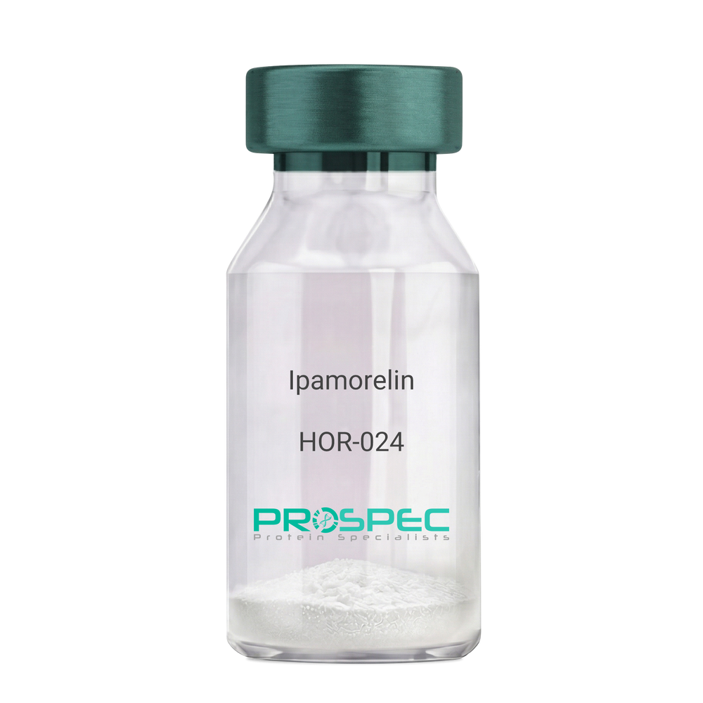
IpamorelinDescription:
Ipamorelin
Ipamorelin
Product # :
HOR-024Price :
Quantity :
Shipping Method :
Shipped at Room temp
More Info
- description
- formulation
- purity
- More Info
- Citations
Description
Ipamorelin Synthetic is a single, non-glycosylated polypeptide chain containing 4 amino acids, having a molecular mass of 711.85 Dalton and a Molecular formula of C38H49N9O5.
Formulation
The protein was lyophilized with no additives.
Purity
Greater than 97.0% as determined by analysis by RP-HPLC.
Citations
More Info
-
Introduction
Ipamorelin is a peptide selective agonist of the ghrelin/growth hormone secretagogue receptor and a growth hormone secretagogue. Ipamorelin is a pentapeptide that was derived from GHRP1. Ipamorelin significantly increases plasma growth hormone levels in both animals and humans. Like pralmorelin and GHRP-6, ipamorelin does not affect prolactin, FSH, LH or TSH levels. However, unlike GHRP2 and GHRP6, but as growth hormone-releasing hormone (GHRH), ipamorelin does not stimulate the secretion of adrenocorticotropic hormone (ACTH) or cortisol, and is highly selective for inducing the secretion only of GH.
-
Synonyms
Ipamorelin
-
Physical Appearance
Sterile Filtered White lyophilized (freeze-dried) powder.
-
Stability
Lyophilized Ipamorelin although stable at room temperature for 3 weeks, should be stored desiccated below -18°C. Upon reconstitution Ipamorelin should be stored at 4°C between 2-7 days and for future use below -18°C. For long term storage it is recommended to add a carrier protein (0.1% HSA or BSA). Please prevent freeze-thaw cycles.
-
Solubility
It is recommended to reconstitute the lyophilized Ipamorelin in sterile 18MΩ-cm H2O not less than 100 µg/ml, which can then be further diluted to other aqueous solutions.
-
Amino Acid Sequence
Aib-His-D-2-Nal-D-Phe-Lys-NH2.
ProSpec's products are furnished for LABORATORY RESEARCH USE ONLY. The product may not be used as drugs, agricultural or pesticidal products, food additives or household chemicals.
- View Data Sheet
Name :
Acrp30 AntibodyDescription:
Adiponectin, Mouse Anti Human
Acrp30, AdipoQ, GBP-28, APM-1, ACDC.
Product # :
ANT-232Price :
Quantity :
Shipping Method :
Shipped with Ice Packs
More Info
- formulation
- More Info
- Citations
Formulation
containing PBS, pH-7.4, & 0.1% Sodium Azide
Citations
More Info
-
Introduction
Human Adiponectin is a secreted protein expressed exclusively in differentiated adipocyte (adipokine). Adiponectin contains a modular structure comprising an N-terminal collagenous domain followed by a C-terminal globular domain. APM-1 plays a role in various physiological processes such as energy homeostasis and obesity. Plasma levels of adiponectin are reduced in obese humans, and decreased levels are associated with insulin resistance and hyperinsulinemia.
-
Synonyms
Acrp30, AdipoQ, GBP-28, APM-1, ACDC.
-
Physical Appearance
Sterile Filtered solution.
-
Immunogen
Anti-human adiponectin mAb, is derived from hybridization of mouse SP2/O myeloma cells with spleen cells from BALB/c mice immunized with Recombinant human adiponectin amino acids 15-244 purified from E. coli.
-
Ig Subclass
Mouse IgG1 heavy chain and k light chain.
-
Clone
P1G12AT.
-
Applications
Adiponectin antibody has been tested by ELISA and Western blot analysis to assure specificity and reactivity. Recommended dilution range for Western blot analysis is 1:250 ~ 1:1,000. The antibody has the specificity against globular domain of adiponectin. Recommended starting dilution is 1:500.
-
Type
Mouse Anti Human Monoclonal.
-
Storage Procedures
For periods up to 1 month store at 4°C, for longer periods of time, store at -20°C. Prevent freeze thaw cycles.
-
Purification Method
Adiponectin antibody was purified from mouse ascitic fluids by protein-G affinity chromatography.
ProSpec's products are furnished for LABORATORY RESEARCH USE ONLY. The product may not be used as drugs, agricultural or pesticidal products, food additives or household chemicals.
- View Data Sheet
Name :
LIN28 HumanDescription:
LIN28 Human Recombinant
CSDD1, FLJ12457, LIN-28, LIN28A, Protein lin-28 homolog A, ZCCHC1, Zinc finger CCHC domain-containing protein 1, Lin-28A, LIN28.
Product # :
PRO-743Price :
Quantity :
Shipping Method :
Shipped with Ice Packs
More Info
- description
- source
- formulation
- purity
- More Info
- Citations
Description
Recombinant Human LIN28 produced in E.Coli is a single, non-glycosylated polypeptide chain containing 191 amino acids (42-209) and having a molecular mass of 21.1 kDa.LIN28 is expressed with a 23 amino acid His tag fused at N-Terminus and purified by proprietary chromatographic techniques.
Source
Escherichia Coli.
Formulation
The LIN28 protein solution (0.5mg/ml) contains 20mM Tris-HCl, pH-8, 10% glycerol, 0.1mM PMSF and 0.1M NaCl.
Purity
Greater than 85.0% as determined by SDS-PAGE
Citations
More Info
-
Introduction
LIN28 plays an important role as a 'translational enhancer', leading specific mRNAs to polysomes and therefore increasing the competence of protein synthesis. LIN28 is a marker of undifferentiated human embryonic stem cells and it enhances the efficiency of the formation of induced pluripotent stem (iPS) cells from human fibroblasts. LIN28 binds to the let-7 pre-miRNA and blocks production of the mature let-7 microRNA in mouse embryonic stem cells. Overexpression of LIN28 is associated with human germ-cell tumors.
-
Synonyms
CSDD1, FLJ12457, LIN-28, LIN28A, Protein lin-28 homolog A, ZCCHC1, Zinc finger CCHC domain-containing protein 1, Lin-28A, LIN28.
-
Physical Appearance
Sterile filtered colorless solution.
-
Stability
Store at 4°C if entire vial will be used within 2-4 weeks. Store, frozen at -20°C for longer periods of time. For long term storage it is recommended to add a carrier protein (0.1% HSA or BSA). Avoid multiple freeze-thaw cycles.
-
Amino Acid Sequence
MGSSHHHHHH SSGLVPRGSHRSMGICKWFN VRMGFGFLSM TARAGVALDP PVDVFVHQSK LHMEGFRSLK EGEAVEFTFK KSAKGLESIR VTGPGGVFCI GSERRPKGKS MQKRRSKGDR CYNCGGLDHH AKECKLPPQP KKCHFCQSIS HMVASCPLKA QQGPSAQGKP TYFREEEEEI HSPTLLPEAQ N.
ProSpec's products are furnished for LABORATORY RESEARCH USE ONLY. The product may not be used as drugs, agricultural or pesticidal products, food additives or household chemicals.
- View Data Sheet
Name :
NFATC1 AntibodyDescription:
Nuclear factor of activated T-cells cytoplasmic 1, Mouse Anti Human
Nuclear factor of activated T-cells cytoplasmic 1, NFAT transcription complex cytosolic component, NF-ATc1, NFATc1, NF-ATc, NFATc, NFAT2, MGC138448.
Product # :
ANT-436Price :
Quantity :
Shipping Method :
Shipped with Ice Packs
More Info
- formulation
- More Info
- Citations
Formulation
1mg/ml containing PBS, pH-7.4, 0.02% Sodium Azide and 10% Glycerol.
Citations
More Info
-
Introduction
NFATC1 is a component of the nuclear factor of activated T cells DNA-binding transcription complex. This complex consists of at least 2 components: a preexisting cytosolic component that translocates to the nucleus upon T cell receptor (TCR) stimulation, and an inducible nuclear component. Proteins belonging to this family of transcription factors play an essential role in inducible gene transcription during immune response. NFATC1 is an inducible nuclear component. NFATC1 functions as a major molecular target for the immunosuppressive drugs such as cyclosporin A. Different isoforms of NFATC1 may regulate inducible expression of different cytokine genes.
-
Synonyms
Nuclear factor of activated T-cells cytoplasmic 1, NFAT transcription complex cytosolic component, NF-ATc1, NFATc1, NF-ATc, NFATc, NFAT2, MGC138448.
-
Physical Appearance
Sterile filtered colorless solution.
-
Immunogen
Anti-human NFATC1 mAb is derived from hybridization of mouse F0 myeloma cells with spleen cells from BALB/c mice immunized with recombinant human NFATC1 amino acids 428-716 purified from E. coli.
-
Ig Subclass
Mouse IgG2a heavy chain and κ light chain.
-
Clone
PAT1C3AT.
-
Applications
NFATC1 antibody has been tested by ELISA and Western blot analysis to assure specificity and reactivity. Since application varies, however, each investigation should be titrated by the reagent to obtain optimal results.
-
Type
Mouse Anti Human Monoclonal.
-
Storage Procedures
For periods up to 1 month store at 4°C, for longer periods of time, store at -20°C. Prevent freeze thaw cycles.
-
Purification Method
NFATC1 antibody was purified from mouse ascitic fluids by protein-G affinity chromatography.
ProSpec's products are furnished for LABORATORY RESEARCH USE ONLY. The product may not be used as drugs, agricultural or pesticidal products, food additives or household chemicals.
- View Data Sheet
Name :
AFP HumanDescription:
Alpha-Fetoprotein Human
Alpha-fetoprotein, Alpha-fetoglobulin, Alpha-1-fetoprotein, AFP, FETA, HPAFP.
Product # :
PRO-406Price :
Quantity :
Shipping Method :
Shipped with Ice Packs
More Info
- description
- source
- formulation
- purity
- More Info
- Citations
Description
Human alpha-fetoprotein purified from pooled human cord serum.
Source
Human cord serum.
Formulation
AFP protein filtered (0.2µm) solution in Tris buffered saline pH 7.5 and less than 0.1% NaN3.
Purity
Greater than 95%.
Citations
More Info
-
Introduction
AFP is normally synthesized in the liver, intestinal tract, and yolk sac of the fetus. Antibody to AFP has been shown to be useful in detecting hepatocellular carcinomas (HCC) and germ cell neoplasms, especially yolk sac tumors.
-
Synonyms
Alpha-fetoprotein, Alpha-fetoglobulin, Alpha-1-fetoprotein, AFP, FETA, HPAFP.
-
Physical Appearance
Sterile Filtered clear solution.
-
Stability
Human Alpha-Fetoprotein should be stored at 2-8°C.
ProSpec's products are furnished for LABORATORY RESEARCH USE ONLY. The product may not be used as drugs, agricultural or pesticidal products, food additives or household chemicals.
- View Data Sheet
Name :
SPOCK3 MouseDescription:
Sparc/Osteonectin 3 Mouse Recombinant
Testican-3, SPARC/osteonectin, CWCV, Kazal-like domains proteoglycan 3, Spock3.
Product # :
PRO-2328Price :
Quantity :
Shipping Method :
Shipped with Ice Packs
More Info
- description
- source
- formulation
- purity
- More Info
- Citations
Description
SPOCK3 Mouse Recombinant produced in Sf9 Baculovirus cells is a single, glycosylated polypeptide chain containing 423 amino acids (22-436 a.a.) and having a molecular mass of 47.9kDa (Migrates at 40-57kDa on SDS-PAGE under reducing conditions).SPOCK3 is expressed with an 8 amino acid His tag at C-Terminus and purified by proprietary chromatographic techniques.
Source
Sf9, Baculovirus cells.
Formulation
SPOCK3 protein solution (0.25mg/ml) containing Phosphate Buffered Saline (pH 7.4) and 10% glycerol.
Purity
Greater than 90% as determined by SDS-PAGE.
Citations
More Info
-
Introduction
Testican-3 (Spock3) is a nervous system-expressed heparan sulfate proteoglycan belonging to a subgroup of the BM-40/SPARC/osteonectin family, whose role of in brain development is unclear. Spock3 inhibits the processing of pro-matrix metalloproteinase 2(MMP-2) by MT1-MMP and MT3-MMP. Spock3 is mostly confined to the developmental stage of the brain.
-
Synonyms
Testican-3, SPARC/osteonectin, CWCV, Kazal-like domains proteoglycan 3, Spock3.
-
Physical Appearance
Sterile Filtered colorless solution.
-
Stability
Store at 4°C if entire vial will be used within 2-4 weeks. Store, frozen at -20°C for longer periods of time. For long term storage it is recommended to add a carrier protein (0.1% HSA or BSA).Avoid multiple freeze-thaw cycles.
-
Amino Acid Sequence
AAAAVAVAGG RSDGGNFLDE KQWLTTISQY DKEVGQWNKF RDEVEDDYFR TWNPGKPFDQ ALDPAKDPCL KTKCSRHKVC ITQDAQTALC ISHRRLTHSM KEVGGSHKQW RGLPSSTCKP CPIAYASPVC GSDGHSYSSQ CKLEYQACVL GKQISIKCEG RCPCPSDKSM NIGRNVKRAC
SDLEFREVAN RLRDWFKALH ESGSQNKKTK ALLRPERSRF DTSILPICKD SLGWMFNRLD TNYDLLLDQS ELGSIYLDKN EQCTKAFFNS CDTYKDSLIS NNEWCYCFQR QQDPPCHTEL SNIQKRQGIK KLLGQYIPLC DEDGYYKPTQ CHGSVGQCWC VDRYGNEVVG SRINGVADCA
IDFEISGDFA SGDFREWTDD EGEEDDIMND KDDIEDDDED EGDDDDDGDV HDGYILEHHH HHH
ProSpec's products are furnished for LABORATORY RESEARCH USE ONLY. The product may not be used as drugs, agricultural or pesticidal products, food additives or household chemicals.
- View Data Sheet
Name :
BMP3 HumanDescription:
Bone Morphogenetic protein-3 Human Recombinant
Bone Morphogenetic Protein 3, Osteogenin, Bone Morphogenetic Protein 3 (Osteogenic), Bone Morphogenetic Protein 3A, BMP-3A, BMP-3, Bone Morphogenetic Protein-3, BMP3A, BMP3.
Product # :
CYT-937Price :
Quantity :
Shipping Method :
Shipped at Room temp
More Info
- description
- source
- formulation
- purity
- biological activity
- More Info
- Citations
Description
BMP3 Human Recombinant produced in E.coli is a non-glycosylated disulfide linked homodimer containing 2 chains of 110 amino acids and having a molecular mass of 24.8kDa.The BMP-3 is purified by proprietary chromatographic techniques.
Source
Escherichia Coli.
Formulation
BMP-3 protein was lyophilized from a 0.2µm filtered concentrated solution in 30% Acetonitrile and 0.1% TFA.
Purity
Greater than 95.0% as determined by: (a) Analysis by HPLC. (b) Analysis by SDS-PAGE.
Biological Activity
The ED50 as determined by its ability to inhibit BMP-2-induced activity in murine MC3T3- E1 cells.Citations
More Info
-
Introduction
Bone Morphogenetic Protein 3 (BMP3) is one of the BMPs, some of which are members of the TGF-beta superfamily (BMP2-7). There are more than 13 BMPs, which are involved in inducing cartilage and bone formation, embryogenesis and morphogenesis of various tissues and organs. In addition, BMPs regulate the growth, differentiation, chemotaxis, and apoptosis of various cell types. Akin to most other TGF-beta family proteins, BMPs are extremely conserved across animal species. At the amino acid sequence level, mature human and rat BMP3 are 98% identical.
-
Synonyms
Bone Morphogenetic Protein 3, Osteogenin, Bone Morphogenetic Protein 3 (Osteogenic), Bone Morphogenetic Protein 3A, BMP-3A, BMP-3, Bone Morphogenetic Protein-3, BMP3A, BMP3.
-
Physical Appearance
Sterile Filtered White lyophilized (freeze-dried) powder.
-
Stability
Lyophilized BMP3 although stable at room temperature for 3 weeks, should be stored desiccated below -18°C. Upon reconstitution BMP-3 should be stored at 4°C between 2-7 days and for future use below -18°C. For long term storage it is recommended to add a carrier protein (0.1% HSA or BSA). Please prevent freeze-thaw cycles.
-
Solubility
It is recommended to reconstitute the lyophilized BMP3 in sterile 4mM HCl not less than 100µg/ml, which can then be further diluted to other aqueous solutions.
-
Amino Acid Sequence
QWIEPRNCAR RYLKVDFADI GWSEWIISPK SFDAYYCSGA CQFPMPKSLK PSNHATIQSI VRAVGVVPGI PEPCCVPEKM SSLSILFFDE NKNVVLKVYP NMTVESCACR.
-
Background
Bone Morphogenetic Protein-3 Human Recombinant: Unveiling the Potential of a Key Regulator in Tissue Regeneration
Abstract:
Bone Morphogenetic Protein-3 (BMP-3) human recombinant is a critical member of the bone morphogenetic protein family, known for its role in tissue development, repair, and regeneration. This research paper provides a comprehensive analysis of BMP-3, including its characteristics, signaling pathways, and potential therapeutic applications. Additionally, innovative methodologies for the production and optimization of BMP-3 human recombinant are proposed, shedding light on its future implications in the field of regenerative medicine.
Introduction:
Tissue regeneration is a complex biological process requiring precise molecular cues. BMP-3, a crucial member of the BMP family, plays a significant role in tissue development and regeneration. This paper explores the unique features of BMP-3 and presents novel approaches for its production and optimization, aiming to unlock its therapeutic potential in various regenerative contexts.
Characteristics and Signaling Pathways:
BMP-3 is a secreted protein that binds to cell surface receptors, initiating intracellular signaling cascades. It influences cell differentiation, proliferation, and extracellular matrix synthesis through both Smad-dependent and Smad-independent signaling pathways. BMP-3 signaling regulates critical processes involved in tissue regeneration, including chondrogenesis and osteogenesis.
Production of BMP-3 Human Recombinant:
Efficient production methodologies are essential for harnessing the therapeutic potential of BMP-3 human recombinant. Recombinant protein expression systems, such as Escherichia coli or mammalian cells, have been utilized to produce functional BMP-3. Optimization strategies, including codon optimization, signal peptide engineering, and protein folding optimization, have been employed to enhance the yield and activity of BMP-3 recombinant protein.
Potential Therapeutic Applications:
BMP-3 human recombinant holds significant promise in the field of regenerative medicine. It plays a crucial role in bone and cartilage regeneration, making it a potential candidate for the treatment of skeletal disorders and tissue injuries. Additionally, BMP-3 signaling influences tissue remodeling and wound healing, suggesting its broader therapeutic applications in other regenerative processes.
Conclusion:
BMP-3 human recombinant represents a key regulator in tissue regeneration, with immense potential in regenerative medicine. Optimizing production methodologies and further unraveling its signaling mechanisms will enhance its therapeutic applications. With its implications in bone and cartilage regeneration and its role in tissue remodeling, BMP-3 human recombinant emerges as a promising tool for promoting tissue repair and regeneration.
What is the molecular weight/Mw of BMP3 Protein?
BMP3 Protein has a total Mw of 24.8kDa.
What is the source or expression system of BMP3 Protein?
Escherichia Coli.
What is the Purity of BMP3 Protein?
BMP3 Protein is >95% pure as determined by SDS-PAGE.
What is the Biological Activity of BMP3 Protein?
The ED50 as determined by its ability to inhibit BMP-2-induced activity in murine MC3T3- E1 cells.
What is the amino acid sequence of BMP3 Protein?
QWIEPRNCAR RYLKVDFADI GWSEWIISPK SFDAYYCSGA CQFPMPKSLK PSNHATIQSI VRAVGVVPGI PEPCCVPEKM SSLSILFFDE NKNVVLKVYP NMTVESCACR.
What applications can BMP3 Protein be used in?
BMP3 Protein can probably be used in western blot, ELISA and Lateral Flow.
What is the endotoxin level for BMP3 Protein?
The endotoxin level is minimal, BMP3 Protein was purified using conventional chromatography techniques.
ProSpec's products are furnished for LABORATORY RESEARCH USE ONLY. The product may not be used as drugs, agricultural or pesticidal products, food additives or household chemicals.
- View Data Sheet
Name :
EREG Human, HEKDescription:
Epiregulin Human Recombinant, HEK
EPR, Epiregulin, Ep, ER, Proepiregulin, EREG.
Product # :
CYT-1206Price :
Quantity :
Shipping Method :
Shipped with Ice Packs
More Info
- description
- source
- formulation
- purity
- biological activity
- More Info
- Citations
Description
EREG Human Recombinant produced in HEK293 cells is a single, glycosylated polypeptide chain (63-108a.a) containing 289 amino acids and having a molecular mass of 32.6 kDa.EREG is fused to a 239 amino acid hIgG-His-Tag at C-terminus & purified by proprietary chromatographic techniques.
Source
HEK293 cells.
Formulation
EREG protein (0.25mg/ml) contains 10% glycerol and Phosphate-Buffered Saline (pH 7.4).
Purity
Greater than 90.0% as determined by SDS-PAGE.
Biological Activity
Measured in a cell proliferation assay using Balb/3T3 mouse embryonic fibroblast cells. The ED50 range ≤ 1ug/ml.
Citations
More Info
-
Introduction
"Epiregulin is part of the EGF family. Epiregulin functions as a ligand of EGFR, as well as a ligand of most members of the ERBB (v-erb-b2 oncogene homolog) family of tyrosine-kinase receptors.Epiregulin is expressed mostly in the placenta and peripheral blood leukocytes and in specific carcinomas of the bladder, lung, kidney and colon. Epiregulin stimulates the proliferation of keratinocytes, hepatocytes, fibroblasts and vascular smooth muscle cells. Epiregulin inhibits the growth of several tumor-derived epithelial cell lines. Human Epiregulin is initially synthesized as a glycosylated 19.0 kDa transmembrane precursor protein, which is processed by proteolytic cleavage to produce a 6.0 kDa mature secreted sequence."
-
Synonyms
EPR, Epiregulin, Ep, ER, Proepiregulin, EREG.
-
Physical Appearance
Sterile filtered colorless solution.
-
Stability
Store at 4°C if entire vial will be used within 2-4 weeks.Store, frozen at -20°C for longer periods of time.For long term storage it is recommended to add a carrier protein (0.1% HSA or BSA).Avoid multiple freeze-thaw cycles.
-
Amino Acid Sequence
DGSMVSITKC SSDMNGYCLH GQCIYLVDMS QNYCRCEVGY TGVRCEHFFL LEPKSCDKTH TCPPCPAPEL LGGPSVFLFP PKPKDTLMIS RTPEVTCVVV DVSHEDPEVK FNWYVDGVEV HNAKTKPREE QYNSTYRVVS VLTVLHQDWL NGKEYKCKVS NKALPAPIEK TISKAKGQPR EPQVYTLPPS RDELTKNQVS LTCLVKGFYP SDIAVEWESN GQPENNYKTT PPVLDSDGSF FLYSKLTVDK SRWQQGNVFS CSVMHEALHN HYTQKSLSLS PGKHHHHHH
-
Background
What is the molecular weight/Mw of EREG Protein?
EREG Protein has a total Mw of 32.6kDa.
What is the source or expression system of EREG Protein?
HEK293 cells.
What is the Purity of EREG Protein?
EREG Protein is >90% pure as determined by SDS-PAGE.
What is the Biological Activity of EREG Protein?
Measured in a cell proliferation assay using Balb/3T3 mouse embryonic fibroblast cells. The ED50 range ≤ 1ug/ml.
What is the amino acid sequence of EREG Protein?
DGSMVSITKC SSDMNGYCLH GQCIYLVDMS QNYCRCEVGY TGVRCEHFFL LEPKSCDKTH TCPPCPAPEL LGGPSVFLFP PKPKDTLMIS RTPEVTCVVV DVSHEDPEVK FNWYVDGVEV HNAKTKPREE QYNSTYRVVS VLTVLHQDWL NGKEYKCKVS NKALPAPIEK TISKAKGQPR EPQVYTLPPS RDELTKNQVS LTCLVKGFYP SDIAVEWESN GQPENNYKTT PPVLDSDGSF FLYSKLTVDK SRWQQGNVFS CSVMHEALHN HYTQKSLSLS PGKHHHHHH
What applications can EREG Protein be used in?
EREG Protein can probably be used in western blot, ELISA and Lateral Flow.
What is the endotoxin level for EREG Protein?
The endotoxin level is minimal, EREG Protein was purified using conventional chromatography techniques.
ProSpec's products are furnished for LABORATORY RESEARCH USE ONLY. The product may not be used as drugs, agricultural or pesticidal products, food additives or household chemicals.
- View Data Sheet
Name :
FGF2 (147), BovineDescription:
Fibroblast Growth Factor-basic (147 a.a.) Bovine Recombinant
HBGH-2, HBGF-2, Prostatropin, FGF-2, FGB-b.
Product # :
CYT-1130Price :
Quantity :
Shipping Method :
Shipped at Room temp
More Info
- description
- source
- formulation
- purity
- biological activity
- More Info
- Citations
Description
Fibroblast Growth Factor-basic (147 a.a.) Bovine Recombinant produced in E.Coli is a non-glycosylated polypeptide chain containing 147 amino acid and having a molecular mass of approximately 16.5kDa.FGF2 (147) is purified by proprietary chromatographic techniques.
Source
Escherichia Coli.
Formulation
Lyophilized from a 0.2μm filtered concentrated solution in PBS, pH 7.4.
Purity
Greater than 97.0% as determined by:
(a) Analysis by RP-HPLC.
(b) Analysis by SDS-PAGE.Biological Activity
The ED50 as determined by a cell proliferation assay using murine balb/c 3T3 cells is < 0.1 ng/ml, corresponding to a specific activity of > 1.0 ×10⁷IU/mg.
Citations
More Info
-
Introduction
FGF-basic is a member of the fibroblast growth factor (FGF) family. FGF family members bind heparin and possess broad mitogenic and angiogenic activities. This protein has been implicated in diverse biological processes, such as limb and nervous system development, wound healing, and tumor growth. The mRNA for this gene contains multiple polyadenylation sites, and is alternatively translated from AUG and non-AUG (CUG) initiation codons resulting in 5 different isoforms with distinct properties. The CUG-initiated isoforms are localized in the nucleus and are responsible for the intracrine effect, whereas, the AUG-initiated form is mostly cytosolic and is responsible for the paracrine and autocrine effects of this FGF.
The heparin-binding growth factors are angiogenic agents in vivo and are potent mitogens for a variety of cell types in vitro. there are differences in the tissue distribution and concentration of these 2 growth factors. -
Synonyms
HBGH-2, HBGF-2, Prostatropin, FGF-2, FGB-b.
-
Physical Appearance
Sterile Filtered White lyophilized (freeze-dried) powder.
-
Stability
Lyophilized FGF2 although stable at room temperature for 3 weeks, should be stored desiccated below -18°C. Upon reconstitution Fibroblast Growth Factor-basic (147 a.a.) should be stored at 4°C between 2-7 days and for future use below -18°C. Please prevent freeze-thaw cycles.
-
Solubility
It is recommended to reconstitute the lyophilized Fibroblast Growth Factor-basic (147 a.a.) in sterile PBS not less than 100µg/ml, which can then be further diluted to other aqueous solutions.
-
Amino Acid Sequence
MPALPEDGGS GAFPPGHFKD PKRLYCKNGG FFLRIHPDGR VDGVREKSDP HIKLQLQAEE RGVVSIKGVC ANRYLAMKED GRLLASKCVT DECFFFERLE SNNYNTYRSR KYSSWYVALK RTGQYKLGPK TGPGQKAILF LPMSAKS.
-
Background
What is the molecular weight/Mw of FGF2 (147), BOVINE Protein?
FGF2 (147), BOVINE Protein has a total Mw of 16.5kDa.
What is the source or expression system of FGF2 (147), BOVINE Protein?
Escherichia Coli.
What is the Purity of FGF2 (147), BOVINE Protein?
FGF2 (147), BOVINE Protein is >97% pure as determined by SDS-PAGE.
What is the Biological Activity of FGF2 (147), BOVINE Protein?
The ED50 as determined by a cell proliferation assay using murine balb/c 3T3 cells is < 0.1 ng/ml, corresponding to a specific activity of > 1.0 ×10⁷IU/mg.
What is the amino acid sequence of FGF2 (147), BOVINE Protein?
MPALPEDGGS GAFPPGHFKD PKRLYCKNGG FFLRIHPDGR VDGVREKSDP HIKLQLQAEE RGVVSIKGVC ANRYLAMKED GRLLASKCVT DECFFFERLE SNNYNTYRSR KYSSWYVALK RTGQYKLGPK TGPGQKAILF LPMSAKS.
What applications can FGF2 (147), BOVINE Protein be used in?
FGF2 (147), BOVINE Protein can probably be used in western blot, ELISA and Lateral Flow.
What is the endotoxin level for FGF2 (147), BOVINE Protein?
The endotoxin level is minimal, FGF2 (147), BOVINE Protein was purified using conventional chromatography techniques.
ProSpec's products are furnished for LABORATORY RESEARCH USE ONLY. The product may not be used as drugs, agricultural or pesticidal products, food additives or household chemicals.
- View Data Sheet
Name :
COX5B HumanDescription:
Cytochrome C Oxidase Subunit Vb Human Recombinant
Cytochrome c oxidase subunit 5B, mitochondrial precursor, COXVB, COX5B, Mitochondrial, Cytochrome c oxidase polypeptide Vb.
Product # :
PRO-1513Price :
Quantity :
Shipping Method :
Shipped with Ice Packs
More Info
- description
- source
- formulation
- purity
- More Info
- Citations
Description
COX5B Human Recombinant produced in E.Coli is a single, non-glycosylated polypeptide chain containing 121 amino acids (32-129a.a) and having a molecular mass of 13kDa. COX5B is fused to a 23 amino acid His-tag at N-terminus & purified by proprietary chromatographic techniques.
Source
Escherichia Coli.
Formulation
COX5B protein solution (0.5mg/ml) containing 20mM Tris-HCl buffer (pH8.0), 0.1M NaCl, 20% glycerol and 1mM DTT.
Purity
Greater than 95.0% as determined by SDS-PAGE.
Citations
More Info
-
Introduction
Cytochrome c oxidase subunit VB (COX5B) is the terminal enzyme of the mitochondrial respiratory chain. COX5B is a multi-subunit enzyme complex which couples the transfer of electrons from cytochrome c to molecular oxygen and contributes to a proton electrochemical gradient across the inner mitochondrial membrane. There are two isoforms of COX5:COX5a and COX5b. Transcription of COX5A (the aerobic isoform) is up-regulated as the rate of cellular respiration increases, when oxygen levels within the cell are high. However, when oxygen levels are low, COX5B (the hypoxic isoform) transcription increases and functions to maximize the turnover rate of the COX apoenzyme.
-
Synonyms
Cytochrome c oxidase subunit 5B, mitochondrial precursor, COXVB, COX5B, Mitochondrial, Cytochrome c oxidase polypeptide Vb.
-
Physical Appearance
Sterile filtered colorless solution.
-
Stability
Store at 4°C if entire vial will be used within 2-4 weeks. Store, frozen at -20°C for longer periods of time. For long term storage it is recommended to add a carrier protein (0.1% HSA or BSA).Avoid multiple freeze-thaw cycles.
-
Amino Acid Sequence
MGSSHHHHHH SSGLVPRGSH MGSASGGGVP TDEEQATGLE REIMLAAKKG LDPYNVLAPK GASGTREDPN LVPSISNKRI VGCICEEDNT SVVWFWLHKG EAQRCPRCGA HYKLVPQQLA H.
ProSpec's products are furnished for LABORATORY RESEARCH USE ONLY. The product may not be used as drugs, agricultural or pesticidal products, food additives or household chemicals.
- View Data Sheet
Name :
IGFBP4 Sf9, HumanDescription:
Insulin Like Growth Factor Binding Protein-4 Human Recombinant, Sf9
Insulin-like growth factor-binding protein 4, IBP-4, IGF-binding protein 4, IGFBP-4, IGFBP4, IBP4, BP-4, HT29-IGFBP.
Product # :
CYT-1114Price :
Quantity :
Shipping Method :
Shipped at Room temp
More Info
- description
- source
- formulation
- purity
- biological activity
- More Info
- Citations
Description
Insulin Like Growth Factor Binding Protein-4 Human Recombinant produced in Sf9 Insect cells is a single, glycosylated polypeptide chain containing 237 amino acids and having a molecular mass of 30kDa. The IGFBP4 is purified by proprietary chromatographic techniques.
Source
Sf9, Insect cells.
Formulation
Lyophilized from a 0.2μm filtered concentrated solution in 20mM Tris-HCl, pH 8.0 and 150mM NaCl.
Purity
Greater than 97.0% as determined by:
(a) Analysis by RP-HPLC.
(b) Analysis by SDS-PAGE.Biological Activity
The ED50 is determined by its ability to inhibit IGF-II induced proliferation of MCF-7 cells and is <than 0.1 μg/ml, corresponding to a specific activity of > 1.0 × 10⁴ IU/mg in the presence of 14 ng/ml of rHuIGF-II.
Citations
More Info
-
Introduction
Insulin-like growth factor-binding protein 4 (IGFBP-4) is a part of the insulin-like growth factor binding protein (IGFBP) family. IGFBP4 includes an IGFBP domain and a thyroglobulin type-I domain. IGFBP4 binds both insulin-like growth factors (IGFs) I and II. IGFBP-4 circulates in the plasma in both glycosylated and non-glycosylated forms. IGFBPs can either inhibit or enhance the biological activities of IGF, or act in an IGF independent manner. IGFBP-4 is consistently inhibits several cancer cells in vivo and in vitro, suggesting that it may function as an apoptotic factor. IGFBP4 is produced by all colon cancer cells. Binding of IGFBP-4 prolongs the half-life of the IGFs and changes their interaction with cell surface receptors.
-
Synonyms
Insulin-like growth factor-binding protein 4, IBP-4, IGF-binding protein 4, IGFBP-4, IGFBP4, IBP4, BP-4, HT29-IGFBP.
-
Physical Appearance
Sterile Filtered White lyophilized (freeze-dried) powder.
-
Stability
Lyophilized IGFBP4 although stable at room temperature for 3 weeks, should be stored desiccated below -18°C. Upon reconstitution Insulin Like Growth Factor Binding Protein-4 should be stored at 4°C between 2-7 days and for future use below -18°C. Please prevent freeze-thaw cycles.
-
Solubility
It is recommended to reconstitute the lyophilized Insulin Like Growth Factor Binding Protein-4 in sterile 18MΩ-cm H2O not less than 100µg/ml, which can then be further diluted to other aqueous solutions.
-
Amino Acid Sequence
DEAIHCPPCS EEKLARCRPP VGCEELVREP GCGCCATCAL GLGMPCGVYT PRCGSGLRCY PPRGVEKPLH TLMHGQGVCM ELAEIEAIQE SLQPSDKDEG DHPNNSFSPC SAHDRRCLQK HFAKIRDRST SGGKMKVNGA PREDARPVPQ GSCQSELHRA LERLAASQSR THEDLYIIPI PNCDRNGNFH PKQCHPALDG QRGKCWCVDR KTGVKLPGGL EPKGELDCHQ LADSFRE.
-
Background
What is the molecular weight/Mw of IGFBP4 SF9, HUMAN Protein?
IGFBP4 SF9, HUMAN Protein has a total Mw of 30kDa.
What is the source or expression system of IGFBP4 SF9, HUMAN Protein?
Sf9, Insect cells.
What is the Purity of IGFBP4 SF9, HUMAN Protein?
IGFBP4 SF9, HUMAN Protein is >97% pure as determined by SDS-PAGE.
What is the Biological Activity of IGFBP4 SF9, HUMAN Protein?
The ED50 is determined by its ability to inhibit IGF-II induced proliferation of MCF-7 cells and is 1.0 × 10⁴ IU/mg in the presence of 14 ng/ml of rHuIGF-II.
What is the amino acid sequence of IGFBP4 SF9, HUMAN Protein?
DEAIHCPPCS EEKLARCRPP VGCEELVREP GCGCCATCAL GLGMPCGVYT PRCGSGLRCY PPRGVEKPLH TLMHGQGVCM ELAEIEAIQE SLQPSDKDEG DHPNNSFSPC SAHDRRCLQK HFAKIRDRST SGGKMKVNGA PREDARPVPQ GSCQSELHRA LERLAASQSR THEDLYIIPI PNCDRNGNFH PKQCHPALDG QRGKCWCVDR KTGVKLPGGL EPKGELDCHQ LADSFRE.
What applications can IGFBP4 SF9, HUMAN Protein be used in?
IGFBP4 SF9, HUMAN Protein can probably be used in western blot, ELISA and Lateral Flow.
What is the endotoxin level for IGFBP4 SF9, HUMAN Protein?
The endotoxin level is minimal, IGFBP4 SF9, HUMAN Protein was purified using conventional chromatography techniques.
ProSpec's products are furnished for LABORATORY RESEARCH USE ONLY. The product may not be used as drugs, agricultural or pesticidal products, food additives or household chemicals.
- View Data Sheet
Name :
GM-CSF MonkeyDescription:
Granulocyte Macrophage-Colony Stimulating Factor Rhesus Macaque Recombinant
CSF-2, MGI-1GM, GM-CSF, Pluripoietin-alpha, MGC131935, MGC138897.
Product # :
CYT-720Price :
Quantity :
Shipping Method :
Shipped at Room temp
More Info
- description
- source
- formulation
- purity
- biological activity
- More Info
- Citations
Description
Granulocyte Macrophage Colony Stimulating Factor Monkey Recombinant produced in E.Coli is a single, non-glycosylated, polypeptide chain containing 127 amino acids and having a molecular mass of 14.4 kDa.GM-CSF is purified by proprietary chromatographic techniques.
Source
Escherichia Coli.
Formulation
GM-CSF was lyophilized from a concentrated (1mg/ml) solution containing 1x PBS pH 7.4.
Purity
Greater than 98.0% as determined by SDS-PAGE and RP-HPLC.
Biological Activity
The ED50 as determined by a cell proliferation assay using human TF-1 cells is less than 0.1ng/ml, corresponding to a specific activity of > 10,000,000 IU/mg.Citations
More Info
-
Introduction
GMCSF is a cytokine that controls the production, differentiation, and function of granulocytes and macrophages. The active form of the protein is found extracellularly as a homodimer. This gene has been localized to a cluster of related genes at chromosome region 5q31, which is known to be associated with interstitial deletions in the 5q- syndrome and acute myelogenous leukemia. Other genes in the cluster include those encoding interleukins 4, 5, and 13. GM-CSF stimulates the growth and differentiation of hematopoietic precursor cells from various lineages, including granulocytes, macrophages, eosinophils and erythrocytes.
-
Synonyms
CSF-2, MGI-1GM, GM-CSF, Pluripoietin-alpha, MGC131935, MGC138897.
-
Physical Appearance
Sterile Filtered White lyophilized (freeze-dried) powder.
-
Stability
Lyophilized GMCSF although stable at room temperature for 3 weeks, should be stored desiccated below -18°C. Upon reconstitution GMCSF should be stored at 4°C between 2-7 days and for future use below -18°C. For long term storage it is recommended to add a carrier protein (0.1% HSA or BSA). Please prevent freeze-thaw cycles.
-
Solubility
It is recommended to reconstitute the lyophilized GMCSF in sterile 18MΩ-cm H2O not less than 100µg/ml, which can then be further diluted to other aqueous solutions.
-
Amino Acid Sequence
APARSPSPGT QPWEHVNAIQ EARRLLNLSR DTAAEMNKTV EVVSEMFDLQ EPSCLQTRLE LYKQGLQGSL TKLKGPLTMM ASHYKQHCPP TPETSCATQI ITFQSFKENL KDFLLVIPFD CWEPVQE.
ProSpec's products are furnished for LABORATORY RESEARCH USE ONLY. The product may not be used as drugs, agricultural or pesticidal products, food additives or household chemicals.
- View Data Sheet
Name :
IFIH1 HumanDescription:
Interferon Induced With Helicase C Domain 1 Human Recombinant
Interferon-induced helicase C domain-containing protein 1, Clinically amyopathic dermatomyositis autoantigen 140 kDa, CADM-140 autoantigen, Helicase with 2 CARD domains, Helicard, Interferon-induced with helicase C domain protein 1, Melanoma differentiation-associated protein 5, MDA-5, Murabutide down-regulated protein, RIG-I-like receptor 2, RLR-2, RNA helicase-DEAD box protein 116, IFIH1, MDA5, RH116, Hlcd, IDDM19.
Product # :
PRO-1505Price :
Quantity :
Shipping Method :
Shipped with Ice Packs
More Info
- description
- source
- formulation
- purity
- More Info
- Citations
Description
IFIH1 Human Recombinant produced in SF9 is a glycosylated, polypeptide chain having a calculated molecular mass of 152,000 Dalton. IFIH1 is expressed with a -10xHis tag at N-terminus and purified by proprietary chromatographic techniques.
Source
Sf9 Insect Cells.
Formulation
IFIH1 is supplied in 20mM HEPES buffer pH-7.9, 550mM NaCl and 6M Urea.
Purity
Greater than 93.0% as determined by SDS-PAGE.
Citations
More Info
-
Introduction
IFIH1 is a DEAD box protein which is upregulated in response to treatment with beta-interferon and a protein kinase C-activating compound, mezerein. Irreversible reprogramming of melanomas can be attained by therapy with both these agents; treatment with either agent alone only achieves reversible differentiation. DEAD box proteins are implicated in several cellular processes involving alteration of RNA secondary structure such as translation initiation, nuclear and mitochondrial splicing, and ribosome and spliceosome assembly.
-
Synonyms
Interferon-induced helicase C domain-containing protein 1, Clinically amyopathic dermatomyositis autoantigen 140 kDa, CADM-140 autoantigen, Helicase with 2 CARD domains, Helicard, Interferon-induced with helicase C domain protein 1, Melanoma differentiation-associated protein 5, MDA-5, Murabutide down-regulated protein, RIG-I-like receptor 2, RLR-2, RNA helicase-DEAD box protein 116, IFIH1, MDA5, RH116, Hlcd, IDDM19.
-
Physical Appearance
Sterile Filtered clear solution.
-
Stability
Store at 4°C if entire vial will be used within 2-4 weeks. Store, frozen at -20°C for longer periods of time. Avoid multiple freeze-thaw cycles.
ProSpec's products are furnished for LABORATORY RESEARCH USE ONLY. The product may not be used as drugs, agricultural or pesticidal products, food additives or household chemicals.
- View Data Sheet
Name :
TGFB2 HumanDescription:
Transforming Growth Factor Beta 2 Human Recombinant
Transforming growth factor, beta 2, cetermin, Glioblastoma-derived T-cell suppressor factor, polyergin, G-TSF, TGF-beta2, TGF-beta-2, transforming growth factor beta-2, BSC-1 cell growth inhibitor, TGFB-2.
Product # :
CYT-441Price :
Quantity :
Shipping Method :
Shipped at Room temp
More Info
- description
- source
- formulation
- purity
- biological activity
- More Info
- Citations
Description
TGFB2 Human Recombinant produced in plants is a homodimeric polypeptide chain containing 2 x 118 amino acids and having a total molecular mass of 27.08kDa. The TGFB2 is fused to 6xHis Tag at N-terminus and purified by proprietary chromatographic techniques.
Source
Nicotiana benthamiana.
Formulation
Lyophilized from a concentrated (1mg/ml) solution containing 50mM Tris-HCl pH-7.4.
Purity
Greater than 97.0% as determined by SDS-PAGE.
Biological Activity
The biological activity of TGFB2 is measured in culture by its ability to inhibit the mink lung epithelial (Mv1Lu) cells proliferation. ED50 < 40ng/ml, corresponding to a specific activity of 25,000 units/mg.Citations
More Info
-
Introduction
TGFB2 is a 27.08 kDa protein having two identical 118 amino acid peptide chains linked by a single disulfide bond. TGFB2 is part of a family of five related cytokines that have an extensive variation of normal and neoplastic cells, indicating the importance of these homo-dimmer proteins as multi-functional regulators of cellular activity. The three mammalian isoforms of TGF-β (TGFb1, TGFb2 and TGFb3) signal through the same receptor and stimulate similar biological responses. They are involved in physiological processes as embryogenesis, tissue remodelling and wound healing.
-
Synonyms
Transforming growth factor, beta 2, cetermin, Glioblastoma-derived T-cell suppressor factor, polyergin, G-TSF, TGF-beta2, TGF-beta-2, transforming growth factor beta-2, BSC-1 cell growth inhibitor, TGFB-2.
-
Physical Appearance
Sterile Filtered White lyophilized (freeze-dried) powder.
-
Stability
Lyophilized TGFB2 although stable at room temperature for 3 weeks, should be stored desiccated below -18°C. Upon reconstitution TGFB2 Human should be stored at 4°C between 2-7 days and for future use below -18°C. For long term storage it is recommended to add a carrier protein (0.1% HSA or BSA). Please prevent freeze-thaw cycles.
-
Solubility
It is recommended to reconstitute the lyophilized TGFB2 in sterile 18M-cm H2O not less than 1µg/40µl, which can then be further diluted to other aqueous solutions.
-
Amino Acid Sequence
HHHHHHALDAAYCFRNVQDNCCLRPLYIDFKRDLGWKWIH
EPKGYNANFCAGACPYLWSSDTQHSRVLSLYNTINPEASAS
PCCVSQDLEPLTI LYYIGKTPKIEQLSNMIVKSCKCS.
ProSpec's products are furnished for LABORATORY RESEARCH USE ONLY. The product may not be used as drugs, agricultural or pesticidal products, food additives or household chemicals.
- View Data Sheet
Name :
CXCL14 Human, HisDescription:
BRAK Human Recombinant (CXCL14), His-Tag
C-X-C motif chemokine 14, Small-inducible cytokine B14, Chemokine BRAK, Bolekine, NJAC, KS1, Kec, BMAC, MIP-2g, SCYB14, CXCL14, BRAK, MGC10687.
Product # :
CHM-239Price :
Quantity :
Shipping Method :
Shipped at Room temp
More Info
- description
- source
- formulation
- purity
- More Info
- Citations
Description
CXCL14 Human Recombinant produced in E.Coli is a single, non-glycosylated, polypeptide chain containing 88 amino acids and having a molecular mass of 10.66 kDa. The Human BRAK contains a 10 a.a. fusion His tag at N-Terminus. The BRAK is purified by proprietary chromatographic techniques.
Source
Escherichia Coli.
Formulation
CXCL14 filtered (0.4µm) and lyophilized from a concentrated (0.5mg/ml) solution containing 20mM Tris buffer & 20mM NaCl pH-7.5.
Purity
Greater than 95.0% as determined by:
(a) Analysis by RP-HPLC.
(b) Analysis by SDS-PAGE.Citations
More Info
-
Introduction
CXCL14 is involved in immunoregulatory and inflammatory processes. BRAK protein is structurally related to the CXC (Cys-X-Cys) subfamily of cytokines. CXCL14 displays chemotactic activity for monocytes but not for lymphocytes, dendritic cells, neutrophils or macrophages. CXCL14 is involved in the homeostasis of monocyte-derived macrophages.
-
Synonyms
C-X-C motif chemokine 14, Small-inducible cytokine B14, Chemokine BRAK, Bolekine, NJAC, KS1, Kec, BMAC, MIP-2g, SCYB14, CXCL14, BRAK, MGC10687.
-
Physical Appearance
Filtered White lyophilized (freeze-dried) powder.
-
Stability
Lyophilized BRAK although stable at room temperature for 3 weeks, should be stored desiccated below -18°C. Upon reconstitution BRAK should be stored at 4°C between 2-7 days and for future use below -18°C.For long term storage it is recommended to add a carrier protein (0.1% HSA or BSA).Please prevent freeze-thaw cycles.
-
Solubility
It is recommended to reconstitute the lyophilized CXCL14 in sterile 18MΩ-cm H2O not less than 100µg/ml, which can then be further diluted to other aqueous solutions. Product is not sterile! Please filter the product by an appropriate sterile filter before using it in the cell culture.
-
Amino Acid Sequence
MKHHHHHHAS SKCKCSRKGP KIRYSDVKKL EMKPKYPHCE EKMVIITTKS VSRYRGQEHC LHPKLQSTKR FIKWYNAWNE KRRVYEE.
-
Background
What is the molecular weight/Mw of CXCL14 HUMAN, HIS Protein?
CXCL14 HUMAN, HIS Protein has a total Mw of 10.66kDa.
What is the source or expression system of CXCL14 HUMAN, HIS Protein?
Escherichia Coli.
What is the Purity of CXCL14 HUMAN, HIS Protein?
CXCL14 HUMAN, HIS Protein is >95% pure as determined by SDS-PAGE.
What is the Biological Activity of CXCL14 HUMAN, HIS Protein?
The biological functionality of CXCL14 HUMAN, HIS Protein will be determined in the future.
What is the amino acid sequence of CXCL14 HUMAN, HIS Protein?
MKHHHHHHAS SKCKCSRKGP KIRYSDVKKL EMKPKYPHCE EKMVIITTKS VSRYRGQEHC LHPKLQSTKR FIKWYNAWNE KRRVYEE.
What applications can CXCL14 HUMAN, HIS Protein be used in?
CXCL14 HUMAN, HIS Protein can probably be used in western blot, ELISA and Lateral Flow.
What is the endotoxin level for CXCL14 HUMAN, HIS Protein?
The endotoxin level is minimal, CXCL14 HUMAN, HIS Protein was purified using conventional chromatography techniques.
Who supplies recombinant CXCL14 HUMAN, HIS that is in stock and available to order?
ProSpec has the protein in stock and available for immediate delivery.
Where can I order bulk mg quantities of CXCL14 HUMAN, HIS?
ProSpec supplies bulk mg quantities at affordable prices that meets research and academic budgets.
ProSpec's products are furnished for LABORATORY RESEARCH USE ONLY. The product may not be used as drugs, agricultural or pesticidal products, food additives or household chemicals.
- View Data Sheet
Name :
TGFB1 Human, HEKDescription:
Transforming Growth Factor-Beta 1 Human Recombinant, HEK
Transforming growth factor beta-1, TGF-beta-1, CED, DPD1, TGFB, TGF-b 1, LAP, TGFB1.
Product # :
CYT-1260Price :
Quantity :
Shipping Method :
Shipped at Room temp
More Info
- description
- source
- formulation
- purity
- biological activity
- More Info
- Citations
Description
TGFB1 Human Recombinant produced in 293 cells is a glycosylated homodimer polypeptide chain containing 2 x 112 amino acids and having a total molecular mass of 25.0kDa. The TGFB1 is purified by proprietary chromatographic techniques.
Source
HEK 293 cells.
Formulation
Lyophilized from a sterile filtered solution containing TFA (0.1%).
Purity
Greater than 98.0% as determined by: (a) Analysis by HPLC. (b) Analysis by SDS-PAGE.
Biological Activity
The ED50 as determined by the dose-dependent inhibition of IL-4-induced proliferation of HT-2 cells is ≤ 0.05 ng/ml, corresponding to a specific activity of ≥ 2 x 10⁷ units/mg.
Citations
More Info
-
Synonyms
Transforming growth factor beta-1, TGF-beta-1, CED, DPD1, TGFB, TGF-b 1, LAP, TGFB1.
-
Physical Appearance
Sterile Filtered White lyophilized (freeze-dried) powder.
-
Stability
Lyophilized TGFB1 although stable at room temperature for 3 weeks, should be stored desiccated below -18°C. Upon reconstitution TGFB1 Human should be stored at 4°C between 2-7 days and for future use below -18°C. For long term storage it is recommended to add a carrier protein (0.1% HSA or BSA). Please prevent freeze-thaw cycles.
-
Solubility
It is recommended to reconstitute the lyophilized TGFB1 in sterile in 18MΩ-cm H2O at a concentration of 0.1 mg/ml, which can then be further diluted to other aqueous solutions.
-
Amino Acid Sequence
ALDTNYCFSS TEKNCCVRQL YIDFRKDLGW KWIHEPKGYH ANFCLGPCPY IWSLDTQYSK VLALYNQHNP GASAAPCCVP QALEPLPIVY YVGRKPKVEQ LSNMIVRSCK CS.
-
Background
Transforming growth factor betas (TGF Betas) mediate many cell-cell interactions that occur during embryonic development. 3 TGF Betas have been identified in mammals: TGF Beta 1, TGF Beta 2 and TGF Beta 3. each are synthesized as precursor proteins that are very similar in that each is cleaved to yield a 112 amino acid polypeptide that remains associated with the latent portion of the molecule.
ProSpec's products are furnished for LABORATORY RESEARCH USE ONLY. The product may not be used as drugs, agricultural or pesticidal products, food additives or household chemicals.
- View Data Sheet
Name :
CST5 HumanDescription:
Cystatin 5 Human Recombinant
Cystatin-D, CST5, Cystatin 5, Cystatin-5.
Product # :
PRO-2221Price :
Quantity :
Shipping Method :
Shipped with Ice Packs
More Info
- description
- source
- formulation
- purity
- biological activity
- More Info
- Citations
Description
CST5 Human Recombinant produced in Sf9 Baculovirus cells is a single, glycosylated polypeptide chain containing 123 amino acids (26-142 a.a) and having a molecular mass of 14.36kDa. CST5 is fused to a 6 amino acid His-tag at C-terminus & purified by proprietary chromatographic techniques.
Source
Sf9, Baculovirus cells.
Formulation
The CST5 solution (0.5mg/1ml) contains phosphate buffered saline (pH7.4) and 10% glycerol.
Purity
Greater than 90.0% as determined by SDS-PAGE.
Biological Activity
The IC50 value is < 2.0nM. The inhibitory function of Cystatin 5 on protease activity of papain was measured by a fluorometric assay using Z-FR-AMC at pH 7.5 at 25C.Citations
More Info
-
Introduction
Cystatin 5 (CST5) is a novel member of the cystatin superfamily, which encompasses proteins that contain multiple cystatin-like sequences. Some members are active cysteine protease inhibitors, while others have lost or possibly never had this inhibitory activity. The superfamily is comprised of 3 inhibitory families: the type 1 cystatins (stefins), type 2 cystatins and the kininogens. Type 2 cystatin proteins are a class of cysteine proteinase inhibitors found in various human fluids and secretions.
-
Synonyms
Cystatin-D, CST5, Cystatin 5, Cystatin-5.
-
Physical Appearance
Sterile Filtered clear solution.
-
Stability
Store at 4°C if entire vial will be used within 2-4 weeks. Store, frozen at -20°C for longer periods of time. For long term storage it is recommended to add a carrier protein (0.1% HSA or BSA).Avoid multiple freeze-thaw cycles.
-
Amino Acid Sequence
QSRTLAGGIH ATDLNDKSVQ RALDFAISEY NKVINKDEYY SRPLQVMAAY QQIVGGVNYY FNVKFGRTTC TKSQPNLDNC PFNDQPKLKE EEFCSFQINE VPWEDKISIL NYKCRKVHHH HHH.
ProSpec's products are furnished for LABORATORY RESEARCH USE ONLY. The product may not be used as drugs, agricultural or pesticidal products, food additives or household chemicals.
- View Data Sheet
Name :
MVD HumanDescription:
Mevalonate Decarboxylase Human Recombinant
Diphosphomevalonate decarboxylase, Mevalonate (diphospho)decarboxylase, MDDase, Mevalonate pyrophosphate decarboxylase, MVD, MPD, FP17780.
Product # :
ENZ-226Price :
Quantity :
Shipping Method :
Shipped with Ice Packs
More Info
- description
- source
- formulation
- purity
- More Info
- Citations
Description
MVD Human Recombinant produced in E.coli is a single, non-glycosylated polypeptide chain containing 420 amino acids (1-400) and having a molecular mass of 45.6kDa.MVD is fused to a 20 amino acid His-tag at N-terminus & purified by proprietary chromatographic techniques.
Source
E.coli.
Formulation
The MVD solution (0.5mg/ml) contains 20mM Tris-HCl buffer (pH8.0), 20% glycerol and 1mM DTT.
Purity
Greater than 85% as determined by SDS-PAGE.
Citations
More Info
-
Introduction
Diphosphomevalonate decarboxylase (MVD) catalyzes the conversion of mevalonate pyrophosphate into isopentenyl pyrophosphate in one of the early steps in cholesterol biosynthesis. MVD decarboxylates and dehydrates its substrate while hydrolyzing ATP. MVD is expressed in the heart, skeletal muscle, lung, liver, brain, pancreas, kidney and placenta.
-
Synonyms
Diphosphomevalonate decarboxylase, Mevalonate (diphospho)decarboxylase, MDDase, Mevalonate pyrophosphate decarboxylase, MVD, MPD, FP17780.
-
Physical Appearance
Sterile Filtered colorless solution.
-
Stability
Store at 4°C if entire vial will be used within 2-4 weeks. Store, frozen at -20°C for longer periods of time. For long term storage it is recommended to add a carrier protein (0.1% HSA or BSA).Avoid multiple freeze-thaw cycles.
-
Amino Acid Sequence
MGSSHHHHHH SSGLVPRGSH MASEKPLAAV TCTAPVNIAV IKYWGKRDEE LVLPINSSLS VTLHQDQLKT TTTAVISKDF TEDRIWLNGR EEDVGQPRLQ ACLREIRCLA RKRRNSRDGD PLPSSLSCKV HVASVNNFPT AAGLASSAAG YACLAYTLAR VYGVESDLSE VARRGSGSAC RSLYGGFVEW QMGEQADGKD SIARQVAPES HWPELRVLIL VVSAEKKLTG STVGMRASVE TSPLLRFRAE SVVPARMAEM ARCIRERDFP SFAQLTMKDS NQFHATCLDT FPPISYLNAI SWRIIHLVHR FNAHHGDTKV AYTFDAGPNA VIFTLDDTVA EFVAAVWHGF PPGSNGDTFL KGLQVRPAPL SAELQAALAM EPTPGGVKYI IVTQVGPGPQ ILDDPCAHLL GPDGLPKPAA.
ProSpec's products are furnished for LABORATORY RESEARCH USE ONLY. The product may not be used as drugs, agricultural or pesticidal products, food additives or household chemicals.
- View Data Sheet
Name :
SLC3A2 HumanDescription:
Solute Carrier Family 3 Member 2 Human Recombinant
Solute Carrier Family 3 Member 2, Lymphocyte Activation Antigen 4F2 Large Subunit, Solute Carrier Family 3 (Activators Of Dibasic And Neutral Amino Acid Transport), Member 2, Antigen Identified By Monoclonal Antibodies 4F2, TRA1.10, TROP4, And T43, Solute Carrier Family 3 (Amino Acid Transporter Heavy Chain), Member 2, 4F2 Cell-Surface Antigen Heavy Chain, Monoclonal Antibody 44D7, CD98 Heavy Chain, 4F2HC, MDU1, Antigen Defined By Monoclonal Antibody 4F2, Heavy Chain, Antigen Defined By Monoclonal Antibody 4F2, 4F2 Heavy Chain Antigen, 4F2 Heavy Chain, CD98 Antigen, Heavy Chain, CD98HC, 4T2HC, NACAE, CD98, 4F2, 4F2 cell-surface antigen heavy chain.
Product # :
PRO-2489Price :
Quantity :
Shipping Method :
Shipped with Ice Packs
More Info
- description
- source
- formulation
- purity
- More Info
- Citations
Description
SLC3A2 produced in Sf9 Baculovirus cells is a single, glycosylated polypeptide chain containing 434 amino acids (206-630a.a.) and having a molecular mass of 47.9kDa. (Molecular size on SDS-PAGE will appear at approximately 40-57kDa).SLC3A2 is expressed with a 6 amino acid His tag at C-Terminus and purified by proprietary chromatographic techniques.
Source
Sf9, Baculovirus cells.
Formulation
SLC3A2 protein solution (0.5mg/ml) contains Phosphate Buffered Saline (pH 7.4) and 10% glycerol.
Purity
Greater than 95.0% as determined by SDS-PAGE.
Citations
More Info
-
Introduction
SLC3A2, also known as Solute Carrier Family 3 Member 2, is a single-pass type II membrane protein which is part of the SLC3A transporter family. SLC3A2 is expressed ubiquitously in all tissues tested with the highest levels detected in kidney, placenta and testis and lowest level in thymus. SLC3A2 is essential for the function of light chain amino-acid transporters and also takes part in sodium-independent, high-affinity transport of large neutral amino acids such as phenylalanine, tyrosine, leucine, arginine and tryptophan. SLC3A2 participates in guiding and targeting of LAT1 and LAT2 to the plasma membrane.
-
Synonyms
Solute Carrier Family 3 Member 2, Lymphocyte Activation Antigen 4F2 Large Subunit, Solute Carrier Family 3 (Activators Of Dibasic And Neutral Amino Acid Transport), Member 2, Antigen Identified By Monoclonal Antibodies 4F2, TRA1.10, TROP4, And T43, Solute Carrier Family 3 (Amino Acid Transporter Heavy Chain), Member 2, 4F2 Cell-Surface Antigen Heavy Chain, Monoclonal Antibody 44D7, CD98 Heavy Chain, 4F2HC, MDU1, Antigen Defined By Monoclonal Antibody 4F2, Heavy Chain, Antigen Defined By Monoclonal Antibody 4F2, 4F2 Heavy Chain Antigen, 4F2 Heavy Chain, CD98 Antigen, Heavy Chain, CD98HC, 4T2HC, NACAE, CD98, 4F2, 4F2 cell-surface antigen heavy chain.
-
Physical Appearance
Sterile filtered colorless solution.
-
Stability
Store at 4°C if entire vial will be used within 2-4 weeks. Store, frozen at -20°C for longer periods of time. For long term storage it is recommended to add a carrier protein (0.1% HSA or BSA).Avoid multiple freeze-thaw cycles.
-
Amino Acid Sequence
ADPRAPRCRE LPAQKWWHTG ALYRIGDLQA FQGHGAGNLA GLKGRLDYLS SLKVKGLVLG PIHKNQKDDV AQTDLLQIDP NFGSKEDFDS LLQSAKKKSI RVILDLTPNY RGENSWFSTQ VDTVATKVKD ALEFWLQAGV DGFQVRDIEN LKDASSFLAE WQNITKGFSE DRLLIAGTNS SDLQQILSLL ESNKDLLLTS SYLSDSGSTG EHTKSLVTQY LNATGNRWCS WSLSQARLLT SFLPAQLLRL YQLMLFTLPG TPVFSYGDEI GLDAAALPGQ PMEAPVMLWD ESSFPDIPGA VSANMTVKGQ SEDPGSLLSL FRRLSDQRSK ERSLLHGDFH AFSAGPGLFS YIRHWDQNER FLVVLNFGDV GLSAGLQASD LPASASLPAK ADLLLSTQPG REEGSPLELE RLKLEPHEGL LLRFPYAAHH HHHH.
ProSpec's products are furnished for LABORATORY RESEARCH USE ONLY. The product may not be used as drugs, agricultural or pesticidal products, food additives or household chemicals.
- View Data Sheet
Name :
FGF22 HumanDescription:
Fibroblast Growth Factor-22 Human Recombinant
Fibroblast growth factor 22, FGF-22.
Product # :
CYT-428Price :
Quantity :
Shipping Method :
Shipped at Room temp
More Info
- description
- source
- formulation
- purity
- More Info
- Citations
Description
Fibroblast Growth Factor-22 Human Recombinant produced in E.Coli is a single, non-glycosylated polypeptide chain containing 149 amino acids and having a molecular mass of 17.3 kDa. The FGF-22 is purified by chromatographic techniques.
Source
Escherichia Coli.
Formulation
The sterile protein powder is lyophilized with no additives.
Purity
Greater than 97.0% as determined by(a) Analysis by RP-HPLC.
(b) Analysis by SDS-PAGE.Citations
More Info
-
Introduction
FGF22 is a member of the fibroblast growth factor (FGF) family. FGF family members possess broad mitogenic and cell survival activities and are involved in a variety of biological processes including embryonic development, cell growth, morphogenesis, tissue repair, tumor growth and invasion. The mouse homolog of this gene was found to be preferentially expressed in the inner root sheath of the hair follicle, which suggested a role in hair development.
-
Synonyms
Fibroblast growth factor 22, FGF-22.
-
Physical Appearance
Sterile Filtered white lyophilized powder.
-
Stability
Lyophilized Fibroblast Growth Factor 22 although stable at room temperature for 3 weeks, should be stored desiccated below -18°C. Upon reconstitution FGF-22 should be stored at 4°C between 2-7 days and for future use below -18°C.For long term storage it is recommended to add a carrier protein (0.1% HSA or BSA).Please prevent freeze-thaw cycles.
-
Solubility
It is recommended to reconstitute the lyophilized Fibroblast Growth Factor-22 sterile 18MΩ-cm H2O not less than 100µg/ml, which can then be further diluted to other aqueous solutions.
-
Amino Acid Sequence
MTPSASRGPR SYPHLEGDVR WRRLFSSTHF FLRVDPGGRV QGTRWRHGQD SILEIRSVHV GVVVIKAVSS GFYVAMNRRG RLYGSRLYTV DCRFRERIEE NGHNTYASQR WRRRGQPMFL ALDRRGGPRP GGRTRRYHLS AHFLPVLVS.
-
Background
What is the molecular weight/Mw of FGF22 HUMAN Protein?
FGF22 HUMAN Protein has a total Mw of 17.3kDa.
What is the source or expression system of FGF22 HUMAN Protein?
Escherichia Coli.
What is the Purity of FGF22 HUMAN Protein?
FGF22 HUMAN Protein is >97% pure as determined by SDS-PAGE.
What is the Biological Activity of FGF22 HUMAN Protein?
The biological functionality of FGF22 HUMAN Protein will be determined in the future.
What is the amino acid sequence of FGF22 HUMAN Protein?
MTPSASRGPR SYPHLEGDVR WRRLFSSTHF FLRVDPGGRV QGTRWRHGQD SILEIRSVHV GVVVIKAVSS GFYVAMNRRG RLYGSRLYTV DCRFRERIEE NGHNTYASQR WRRRGQPMFL ALDRRGGPRP GGRTRRYHLS AHFLPVLVS.
What applications can FGF22 HUMAN Protein be used in?
FGF22 HUMAN Protein can probably be used in western blot, ELISA and Lateral Flow.
What is the endotoxin level for FGF22 HUMAN Protein?
The endotoxin level is minimal, FGF22 HUMAN Protein was purified using conventional chromatography techniques.
ProSpec's products are furnished for LABORATORY RESEARCH USE ONLY. The product may not be used as drugs, agricultural or pesticidal products, food additives or household chemicals.
- View Data Sheet
Name :
CXCL12 Mouse betaDescription:
Stromal Cell Derived Factor-1 Beta Mouse Recombinant (CXCL12)
SDF-1, CXCL12, Pre-B cell growth-stimulating factor, PBSF, hIRH, chemokine (C-X-C motif) ligand 12, SDF1, SDF1B, TPAR1, SCYB12, SDF-1b, TLSF-b, 12-O-tetradecanoylphorbol 13-acetate repressed protein 1, Thymic lymphoma cell-stimulating factor, TLSF.
Product # :
CHM-326Price :
Quantity :
Shipping Method :
Shipped at Room temp
More Info
- description
- source
- formulation
- purity
- biological activity
- More Info
- Citations
Description
Stromal Cell-Derived Factor-1 beta Mouse Recombinant produced in E.Coli is a non-glycosylated, Polypeptide chain containing 72 amino acids and having a molecular mass of 8513 Dalton. The SDF-1b is purified by proprietary chromatographic techniques.
Source
Escherichia Coli.
Formulation
The CXCL12 protein was lyophilized from a concentrated (1mg/ml) sterile solution containing no additives.
Purity
Greater than 98.0% as determined by:
(a) Analysis by RP-HPLC.
(b) Analysis by SDS-PAGE.Biological Activity
The specific activity as determined by its ability to chemoattract human monocytes at 50-100ng/ml corresponding to a Specific Activity of 10,000-20,000IU/mg.Citations
More Info
-
Introduction
SDF-1 (stromal cell-derived factor-1) is small cytokine belonging to the chemokine family that is officially designated Chemokine (C-X-C motif) ligand 12 (CXCL12). It is produced in two forms, SDF-1?/CXCL12a and SDF-1?/CXCL12b, by alternate splicing of the same gene. Chemokines are characterized by the presence of four conserved cysteines, which form two disulfide bonds. The CXCL12 proteins belong to the group of CXC chemokines, whose initial pair of cysteines are separated by one intervening amino acid. CXCL12 is strongly chemotactic for lymphocytes and has been implicated as an important cell co-ordinator during development. During embryogenesis it directs the migration of hematopoietic cells from foetal liver to bone marrow. Mice which were knocked-out for CXCL12 gene were lethal before the birth or within just 1 hour of life. As another role, CXCL12a alters also the electrophysiology of neurons. CXCL12 was shown to be expressend in many tissues in mice (including brain, thymus, heart, lung, liver, kidney, spleen and bone marrow).
The receptor for this chemokine is CXCR4, which was previously called fusin. This CXCL12-CXCR4 interaction used to be considered exclusive (unlike for other chemokines and their receptors), but recently it was suggested that CXCL12 is also bound by CXCR7 receptor.
The gene for CXCL12 is located on human chromosome 10. In human and mouse both CXCL12 and CXCR4 show high identity of sequence: 99% and 90%, respectively. -
Synonyms
SDF-1, CXCL12, Pre-B cell growth-stimulating factor, PBSF, hIRH, chemokine (C-X-C motif) ligand 12, SDF1, SDF1B, TPAR1, SCYB12, SDF-1b, TLSF-b, 12-O-tetradecanoylphorbol 13-acetate repressed protein 1, Thymic lymphoma cell-stimulating factor, TLSF.
-
Physical Appearance
Sterile Filtered White lyophilized (freeze-dried) powder.
-
Stability
Lyophilized SDF-1b although stable at room temperature for 3 weeks, should be stored desiccated below -18°C. Upon reconstitution CXCL12 should be stored at 4°C between 2-7 days and for future use below -18°C.For long term storage it is recommended to add a carrier protein (0.1% HSA or BSA).Please prevent freeze-thaw cycles.
-
Solubility
It is recommended to reconstitute the lyophilized Stromal Cell-Derived Factor-1b in sterile 18MΩ-cm H2O not less than 100µg/ml, which can then be further diluted to other aqueous solutions.
-
Amino Acid Sequence
The sequence of the first five N-terminal amino acids was determined and was found to be Lys-Pro-Val-Ser-Leu.
-
Background
What is the molecular weight/Mw of CXCL12 MOUSE BETA Protein?
CXCL12 MOUSE BETA Protein has a total Mw of 8.5kDa.
What is the source or expression system of CXCL12 MOUSE BETA Protein?
Escherichia Coli.
What is the Purity of CXCL12 MOUSE BETA Protein?
CXCL12 MOUSE BETA Protein is > 98% pure as determined by SDS-PAGE.
What is the Biological Activity of CXCL12 MOUSE BETA Protein?
The specific activity as determined by its ability to chemoattract human monocytes at 50-100ng/ml corresponding to a Specific Activity of 10,000-20,000IU/mg.
What is the amino acid sequence of CXCL12 MOUSE BETA Protein?
The sequence of the first five N-terminal amino acids was determined and was found to be Lys-Pro-Val-Ser-Leu.
What applications can CXCL12 MOUSE BETA Protein be used in?
CXCL12 MOUSE BETA Protein can probably be used in western blot, ELISA and Lateral Flow.
What is the endotoxin level for CXCL12 MOUSE BETA Protein?
The endotoxin level is minimal, CXCL12 MOUSE BETA Protein was purified using conventional chromatography techniques.
Who supplies recombinant CXCL12 MOUSE BETA that is in stock and available to order?
ProSpec has the protein in stock and available for immediate delivery.
Where can I order bulk mg quantities of CXCL12 MOUSE BETA?
ProSpec supplies bulk mg quantities at affordable prices that meets research and academic budgets.
ProSpec's products are furnished for LABORATORY RESEARCH USE ONLY. The product may not be used as drugs, agricultural or pesticidal products, food additives or household chemicals.
- View Data Sheet
Name :
SNAPIN HumanDescription:
SNAP Associated Protein Human Recombinant
SNAPAP, SNARE-associated protein Snapin, Synaptosomal-associated protein 25-binding protein, SNAP-associated protein, SNAPIN, SNAP25BP.
Product # :
PRO-663Price :
Quantity :
Shipping Method :
Shipped with Ice Packs
More Info
- description
- source
- formulation
- purity
- More Info
- Citations
Description
SNAPIN Human Recombinant produced in E.Coli is a single, non-glycosylated polypeptide chain containing 156 amino acids (1-136) and having a molecular mass of 17 kDa. SNAPIN is fused to 20 amino acid His Tag at N-terminus and purified by standard chromatography techniques.
Source
Escherichia Coli.
Formulation
The protein solution contains 20mM Tris-HCl pH-8, 5mM DTT, 2mM EDTA, 0.2M NaCl, and 40% glycerol.
Purity
Greater than 90.0% as determined by SDS-PAGE.
Citations
More Info
-
Introduction
SNAPIN is involved in the neurotransmitter release process through its modulation of the sequential interactions between the SNAREs and synaptotagmin. SNAPIN is part of the SNARE complex of proteins that is needed for synaptic vesicle docking and fusion. SNAPAP is enriched in neurons and exclusively located on synaptic vesicle membrane protein. SNAPIN is also an important factor of the BLOC1 multisubunit protein complex. BLOC1 is required for normal biogenesis of specialized organelles of the endosomal-lysosomal system, such as melanosomes and platelet dense granules.
-
Synonyms
SNAPAP, SNARE-associated protein Snapin, Synaptosomal-associated protein 25-binding protein, SNAP-associated protein, SNAPIN, SNAP25BP.
-
Physical Appearance
Sterile Filtered colorless solution.
-
Stability
Store at 4°C if entire vial will be used within 2-4 weeks. Store, frozen at -20°C for longer periods of time. For long term storage it is recommended to add a carrier protein (0.1% HSA or BSA).Avoid multiple freeze-thaw cycles.
-
Amino Acid Sequence
MGSSHHHHHH SSGLVPRGSH MAGAGSAAVS GAGTPVAGPT GRDLFAEGLL EFLRPAVQQL DSHVHAVRES QVELREQIDN LATELCRINEDQKVALDLDP YVKKLLNARR RVVLVNNILQ NAQERLRRLN HSVAKETARR RAMLDSGIYP PGSPGK.
ProSpec's products are furnished for LABORATORY RESEARCH USE ONLY. The product may not be used as drugs, agricultural or pesticidal products, food additives or household chemicals.