Search results
860 results found for “thymus expressed chemokine (ccl25)”
Name
Description
Product #
Price
Quantity
Shipping Method
- View Data Sheet
Name :
Borrelia p41, Sf9Description:
Borrelia Burgdorferi p41 Recombinant, Sf9
Product # :
BOR-002Price :
Quantity :
Shipping Method :
Shipped with Ice Packs
More Info
- description
- source
- formulation
- purity
- More Info
- Citations
Description
Recombinant Borrelia Burgdorferi p41 produced in SF9 is a glycosylated, polypeptide chain having a calculated molecular mass of 36,578 Dalton. Borrelia p41 is expressed with a -6x His tag at N-terminus and purified by proprietary chromatographic techniques.
Source
Sf9 insect cells.
Formulation
Borrelia p41 (1.01mg/1ml) is supplied in 20mM HEPES buffer pH-7.5, 0.01mM EDTA and 0.02% SDS.
Purity
Greater than 80.0% as determined by SDS-PAGE.
Citations
More Info
-
Introduction
Borrelia belongs to a genus of bacteria of the spirochete phylum. Borrelia causes borreliosis, which is a zoonotic, vector-borne disease transmitted mainly by ticks and some by lice, depending on the species. Of the 36 known species of Borrelia, 12 are distinguished to cause Lyme disease or borreliosis and are transmitted by ticks. The main Borrelia species causing Lyme disease are Borrelia burgdorferi, Borrelia afzelii, and Borrelia garinii. The Borrelia genus members have a linear chromosome which is about 900 kbp in length as well as an excess of both linear and circular plasmids in the 5-220 kbp size range. The plasmids are atypical, as compared to most bacterial plasmids, since they contain many paralogous sequences, a large number of pseudogenes and, in some cases, essential genes. Moreover, a number of the plasmids have features suggesting that they are prophages.
-
Physical Appearance
Sterile Filtered clear solution.
-
Stability
Store at 4°C if entire vial will be used within 2-4 weeks. Store, frozen at -20°C for longer periods of time. Avoid multiple freeze-thaw cycles.
-
Applications
Western blot with Lyme positive plasma.
ProSpec's products are furnished for LABORATORY RESEARCH USE ONLY. The product may not be used as drugs, agricultural or pesticidal products, food additives or household chemicals.
- View Data Sheet
Name :
BD 3 HumanDescription:
Beta Defensin-3 Human Recombinant
HBD3, HBP3, DEFB3, HBD-3, HBP-3, DEFB103.
Product # :
CYT-461Price :
Quantity :
Shipping Method :
Shipped at Room temp
More Info
- description
- source
- formulation
- purity
- More Info
- Citations
Description
Beta Defensin-3 Human Recombinant produced in E.Coli is a single, non-glycosylated polypeptide chain containing 45 amino acids and having a molecular mass of 5161.2 Dalton. The BD-3 is purified by proprietary chromatographic techniques.
Source
Escherichia Coli.
Formulation
The HBD-3 was lyophilized without additives.
Purity
Greater than 95.0% as determined by(a) Analysis by RP-HPLC.
(b) Analysis by SDS-PAGE.Citations
More Info
-
Introduction
Defensins form a family of microbicidal and cytotoxic peptides made by neutrophils. Members of the defensin family are highly similar in protein sequence. This gene encodes defensin, beta 103A, which has broad spectrum antimicrobial activity and may play an important role in innate epithelial defense.
-
Synonyms
HBD3, HBP3, DEFB3, HBD-3, HBP-3, DEFB103.
-
Physical Appearance
Sterile Filtered lyophilized (freeze-dried) powder.
-
Stability
Lyophilized Beta Defensin-3 Recombinant although stable at room temperature for 3 weeks, should be stored desiccated below -18°C. Upon reconstitution BD-3 should be stored at 4°C between 2-7 days and for future use below -18°C.Please prevent freeze-thaw cycles.
-
Solubility
It is recommended to reconstitute the lyophilized Beta Defensin-3 in sterile 18MΩ-cm H2O not less than 100µg/ml, which can then be further diluted to other aqueous solutions.
-
Amino Acid Sequence
GIINTLQKYY CRVRGGRCAV LSCLPKEEQI GKCSTRGRKC CRRKK.
-
Background
Beta Defensin-3 Human Recombinant: Advancements in Antimicrobial Peptide Therapy
Abstract:
Beta Defensin-3 (hBD-3) human recombinant is a promising antimicrobial peptide with broad-spectrum activity against bacteria, viruses, and fungi. This research paper provides an overview of hBD-3, including its properties, mode of action, and potential applications. Additionally, novel methodologies for the production and optimization of hBD-3 human recombinant are discussed, highlighting its future implications in the field of infectious disease management.Introduction:
The rise of drug-resistant pathogens necessitates exploring alternative therapeutic approaches, such as antimicrobial peptides. Beta Defensin-3 (hBD-3) human recombinant has emerged as a potent candidate due to its broad-spectrum antimicrobial activity. This paper aims to examine the unique features of hBD-3 and propose innovative methodologies for its production and optimization.Properties and Mode of Action:
hBD-3 possesses a distinct structural composition consisting of 45 amino acids, including an N-terminal loop, three antiparallel β-strands, and a C-terminal α-helix. These structural elements contribute to its ability to disrupt microbial membranes and target selectivity. The mode of action involves electrostatic interactions with negatively charged microbial membranes, leading to membrane disruption and subsequent cell death. Furthermore, hBD-3 exhibits immunomodulatory functions by promoting chemotaxis, enhancing phagocytic activity, and modulating the release of pro-inflammatory cytokines.Production of hBD-3 Human Recombinant:
Various expression systems, such as bacterial, yeast, and mammalian cell-based platforms, have been explored for the efficient production of hBD-3 human recombinant. Each system offers distinct advantages and challenges, requiring careful selection to achieve high yields and desired protein quality. Optimization strategies, including codon optimization, fusion protein tags, and appropriate growth conditions, have been employed to enhance production efficiency. Purification techniques, such as chromatography and ultrafiltration, have been optimized to isolate high-quality hBD-3 recombinant.Applications and Future Perspectives:
hBD-3 human recombinant exhibits significant therapeutic potential against drug-resistant pathogens, making it a promising alternative to conventional antibiotics. It also demonstrates promise in wound healing and tissue regeneration by stimulating angiogenesis, extracellular matrix production, and keratinocyte migration. Moreover, the unique physicochemical properties of hBD-3 open avenues for its utilization in nanomedicine, enabling targeted therapy and improved drug delivery.Conclusion:
hBD-3 human recombinant represents a potent antimicrobial peptide with broad-spectrum activity against diverse pathogens. The optimization of production methodologies and further exploration of its mechanisms of action will contribute to its clinical utility. With its potential applications in infectious disease management, wound healing, and nanomedicine, hBD-3 human recombinant holds promise as a versatile therapeutic agent.What is the molecular weight/Mw of BD3 Protein?
BD3 Protein has a total Mw of 5.1kDa.
What is the source or expression system of BD3 Protein?
Escherichia Coli.
What is the Purity of BD3 Protein?
BD3 Protein is >95% pure as determined by SDS-PAGE.
What is the Biological Activity of BD3 Protein?
The biological functionality of BD3 Protein will be determined in the future.
What is the amino acid sequence of BD3 Protein?
GIINTLQKYY CRVRGGRCAV LSCLPKEEQI GKCSTRGRKC CRRKK.
What applications can BD3 Protein be used in?
BD3 Protein can probably be used in western blot, ELISA and Lateral Flow.
What is the endotoxin level for BD3 Protein?
The endotoxin level is minimal, BD3 Protein was purified using conventional chromatography techniques.
ProSpec's products are furnished for LABORATORY RESEARCH USE ONLY. The product may not be used as drugs, agricultural or pesticidal products, food additives or household chemicals.
- View Data Sheet
Name :
TNFRSF14 Human, Sf9Description:
HVEM-Fc Human Recombinant, Sf9
Tumor necrosis factor receptor superfamily member 14 isoform 1, TNFRSF14, ATAR, CD270, HVEA, HVEM, LIGHTR, TR2, HVEM-Fc, Sf9, Tumor necrosis factor receptor superfamily member 14, Herpes virus entry mediator A, Herpesvirus entry mediator A, HveA.
Product # :
CYT-981Price :
Quantity :
Shipping Method :
Shipped with Ice Packs
More Info
- description
- source
- formulation
- purity
- More Info
- Citations
Description
TNFRSF14 Human Recombinant produced in Sf9 Baculovirus cells is a single, glycosylated polypeptide chain containing 406 amino acids (39-202) and having a molecular mass of 44.6kDa (Molecular size on SDS-PAGE will appear at approximately 40-57kDa).TNFRSF14 is fused to a 239 amino acid IgG His-Tag at C-terminus and purified by proprietary chromatographic techniques.
Source
Sf9, Baculovirus cells.
Formulation
TNFRSF14 protein solution (0.25mg/ml) containing Phosphate Buffered Saline (pH 7.4) and 10% glycerol.
Purity
Greater than 90.0% as determined by analysis by SDS-PAGE.
Citations
More Info
-
Introduction
TNFRSF14, a member of the TNF receptor superfamily, is a type I transmembrane protein. TNFRSF14 is expressed in peripheral blood T cells, B cells, monocytes and in various tissues enriched in lymphoid cells. TNFRSF14 operates as a co-stimulatory factor for the activation of lymphoid cells and as a deterrent to infection by herpesvirus. Additionally, TNFRSF14 encourages the proliferation of T cells, and triggers apoptosis of various tumor cells.
-
Synonyms
Tumor necrosis factor receptor superfamily member 14 isoform 1, TNFRSF14, ATAR, CD270, HVEA, HVEM, LIGHTR, TR2, HVEM-Fc, Sf9, Tumor necrosis factor receptor superfamily member 14, Herpes virus entry mediator A, Herpesvirus entry mediator A, HveA.
-
Physical Appearance
Sterile Filtered clear solution.
-
Stability
Store at 4°C if entire vial will be used within 2-4 weeks. Store, frozen at -20°C for longer periods of time. For long term storage it is recommended to add a carrier protein (0.1% HSA or BSA).Avoid multiple freeze-thaw cycles.
-
Amino Acid Sequence
ADPLPSCKED EYPVGSECCP KCSPGYRVKE ACGELTGTVC EPCPPGTYIA HLNGLSKCLQ CQMCDPAMGL RASRNCSRTE NAVCGCSPGH FCIVQDGDHC AACRAYATSS PGQRVQKGGT ESQDTLCQNC PPGTFSPNGT LEECQHQTKC SWLVTKAGAG TSSSHWVLEP KSCDKTHTCP PCPAPELLGG PSVFLFPPKP KDTLMISRTP EVTCVVVDVS HEDPEVKFNW YVDGVEVHNA KTKPREEQYN STYRVVSVLT VLHQDWLNGK EYKCKVSNKA LPAPIEKTIS KAKGQPREPQ VYTLPPSRDE LTKNQVSLTC LVKGFYPSDI AVEWESNGQP ENNYKTTPPV LDSDGSFFLY SKLTVDKSRW QQGNVFSCSV MHEALHNHYT QKSLSLSPGK HHHHHH.
ProSpec's products are furnished for LABORATORY RESEARCH USE ONLY. The product may not be used as drugs, agricultural or pesticidal products, food additives or household chemicals.
- View Data Sheet
Name :
DIHEXADescription:
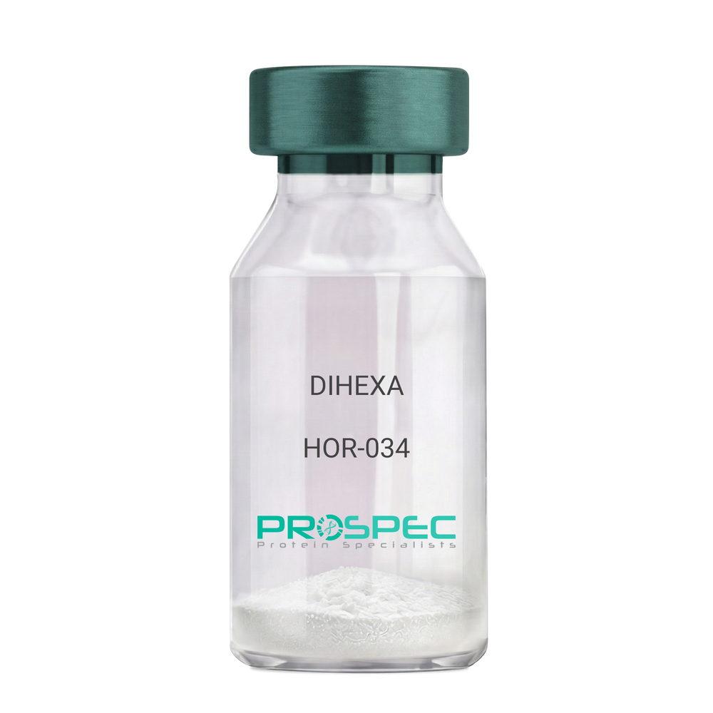
DIHEXA
Product # :
HOR-034Price :
Quantity :
Shipping Method :
Shipped at Room temp
More Info
- description
- formulation
- purity
- More Info
- Citations
Description
DIHEXA Synthetic is a single, non-glycosylated polypeptide chain containing 3 amino acids, having a molecular mass of 504.28 Dalton and a Molecular formula of C27H44N4O5.
Formulation
The protein was lyophilized with no additives.
Purity
Greater than 97.0% as determined by analysis by RP-HPLC.
Citations
More Info
-
Physical Appearance
Sterile Filtered White lyophilized (freeze-dried) powder.
-
Stability
Lyophilized DIHEXA although stable at room temperature for 3 weeks, should be stored desiccated below -18°C. Upon reconstitution DIHEXA should be stored at 4°C between 2-7 days and for future use below -18°C. For long term storage it is recommended to add a carrier protein (0.1% HSA or BSA). Please prevent freeze-thaw cycles.
-
Solubility
It is recommended to reconstitute the lyophilized DIHEXA in sterile 18MΩ-cm H2O not less than 100 µg/ml, which can then be further diluted to other aqueous solutions.
-
Amino Acid Sequence
Hexanoyl-Tyr-Ile-Ahx-NH2.
-
Background
Dihexa, also known as N-hexanoic-Tyr-Ile-(6) aminohexanoic amide, is a potent and orally active small peptide that has been the focus of significant research due to its potential neurogenic and neuroprotective effects. This compound, derived from angiotensin IV, has been shown to possess a wide range of biological activities, including enhancing cognitive function, promoting neurogenesis, and potentially mitigating the effects of neurodegenerative diseases.
Dihexa's primary mechanism of action involves its interaction with hepatocyte growth factor (HGF) and its receptor, c-Met. By mimicking the effects of HGF, Dihexa can stimulate the c-Met receptor, leading to a cascade of events that promote neurogenesis and synaptic plasticity. Studies by Benoist et al. (2014) have demonstrated that Dihexa can enhance cognitive function in rats, suggesting potential applications in cognitive enhancement and the treatment of cognitive disorders.
In addition to its neurogenic effects, Dihexa has been shown to possess neuroprotective properties. Research by Kawas et al. (2017) found that Dihexa could protect neurons from apoptosis, suggesting potential applications in the treatment of neurodegenerative diseases such as Alzheimer's and Parkinson's disease.
Given its neurogenic and neuroprotective effects, Dihexa has been proposed as a potential therapeutic agent for a variety of conditions, including cognitive disorders, neurodegenerative diseases, and stroke. For instance, a study by Harding et al. (2018) found that Dihexa could improve outcomes in animal models of stroke, indicating its potential as a therapeutic agent in stroke recovery.
While research on Dihexa is promising, it is important to note that most studies have been conducted in animals or in vitro. More research is needed to fully understand the potential effects and applications of Dihexa in humans. However, the existing body of research suggests that Dihexa could be a promising tool in the treatment of cognitive disorders and neurodegenerative diseases.
ProSpec's products are furnished for LABORATORY RESEARCH USE ONLY. The product may not be used as drugs, agricultural or pesticidal products, food additives or household chemicals.
- View Data Sheet
Name :
GPC3 HumanDescription:
Glypican-3 Human Recombinant
Glypican 3, Intestinal Protein OCI-5, Glypican Proteoglycan 3, GTR2-2, MXR7, Heparan Sulphate Proteoglycan, Secreted Glypican-3, Glypican-3, OCI-5, SGBS1, DGSX, SGBS, SDYS, OCI5, SGB, GPC3.
Product # :
PRO-2423Price :
Quantity :
Shipping Method :
Shipped with Ice Packs
More Info
- description
- source
- formulation
- purity
- More Info
- Citations
Description
GPC3 Human Recombinant produced in Sf9 Baculovirus cells is a single, glycosylated polypeptide chain containing 544 amino acids (25-559a.a.) and having a molecular mass of 61.8kDa (Molecular size on SDS-PAGE will appear at approximately 50-70kDa). GPC3 is expressed with a 6 amino acids His tag at C-Terminus and purified by proprietary chromatographic techniques.
Source
Sf9, Baculovirus cells.
Formulation
GPC3 protein solution (0.5mg/ml) contains Phosphate Buffered Saline (pH 7.4) and 10% glycerol.
Purity
Greater than 90.0% as determined by SDS-PAGE.
Citations
More Info
-
Introduction
Glypican-3 (GPC3) belongs to the glypican family, and is highly expressed in the lung, liver, and the kidney. In some tissues, GPC3 functionss as a tumor suppressor gene and an oncofetal protein. The Glypican-3 protein is currently considered as a tumor marker and potential target for immunotherapy. Glypican-3 binds to and inhibits the dipeptidyl peptidase activity of CD26, and it can also induce apoptosis in certain cell types. Deletion mutations in the GPC3 gene are linked with Simpson-Golabi-Behmel syndrome, aka Simpson dysmorphia syndrome.
-
Synonyms
Glypican 3, Intestinal Protein OCI-5, Glypican Proteoglycan 3, GTR2-2, MXR7, Heparan Sulphate Proteoglycan, Secreted Glypican-3, Glypican-3, OCI-5, SGBS1, DGSX, SGBS, SDYS, OCI5, SGB, GPC3.
-
Physical Appearance
Sterile filtered colorless solution.
-
Stability
Store at 4°C if entire vial will be used within 2-4 weeks. Store, frozen at -20°C for longer periods of time. For long term storage it is recommended to add a carrier protein (0.1% HSA or BSA).Avoid multiple freeze-thaw cycles.
-
Amino Acid Sequence
ADPQPPPPPP DATCHQVRSF FQRLQPGLKW VPETPVPGSD LQVCLPKGPT CCSRKMEEKY QLTARLNMEQ LLQSASMELK FLIIQNAAVF QEAFEIVVRH AKNYTNAMFK NNYPSLTPQA FEFVGEFFTD VSLYILGSDI NVDDMVNELF DSLFPVIYTQ LMNPGLPDSA LDINECLRGA RRDLKVFGNF PKLIMTQVSK SLQVTRIFLQ ALNLGIEVIN TTDHLKFSKD CGRMLTRMWY CSYCQGLMMV KPCGGYCNVV MQGCMAGVVE IDKYWREYIL SLEELVNGMY RIYDMENVLL GLFSTIHDSI QYVQKNAGKL TTTIGKLCAH SQQRQYRSAY YPEDLFIDKK VLKVAHVEHE ETLSSRRREL IQKLKSFISF YSALPGYICS HSPVAENDTL CWNGQELVER YSQKAARNGM KNQFNLHELK MKGPEPVVSQ IIDKLKHINQ LLRTMSMPKG RVLDKNLDEE GFESGDCGDD EDECIGGSGD GMIKVKNQLR FLAELAYDLD VDDAPGNSQQ ATPKDNEIST FHNLGNVHHH HHHH.
ProSpec's products are furnished for LABORATORY RESEARCH USE ONLY. The product may not be used as drugs, agricultural or pesticidal products, food additives or household chemicals.
- View Data Sheet
Name :
VEGF Human, BaculovirusDescription:
Vascular Endothelial Growth Factor Human Recombinant, Baculovirus
Vascular Endothelial Growth Factor A, VEGF, Vascular Permeability Factor, MVCD1, VPF, Vascular Endothelial Growth Factor, VEGF-A, Vascular endothelial growth factor A.
Product # :
CYT-849Price :
Quantity :
Shipping Method :
Shipped with Ice Packs
More Info
- description
- source
- formulation
- purity
- More Info
- Citations
Description
VEGF produced in Sf9 Baculovirus cells is a single, glycosylated polypeptide chain containing 171 amino acids (27-191 a.a.) and having a molecular mass of 19.9 kDa. VEGF is expressed with a 6 amino acid His tag at C-Terminus and purified by proprietary chromatographic techniques.
Source
Sf9, Baculovirus cells.
Formulation
VEGF protein solution (0.25mg/ml) contains Phosphate buffered saline (pH7.4), 30% glycerol, 1mM DTT and 0.1mM PMSF.
Purity
Greater than 85.0% as determined by SDS-PAGE.
Citations
More Info
-
Introduction
Vascular endothelial growth factor is an important signaling protein involved in both vasculogenesis and angiogenesis. As its name implies, VEGF activity has been mostly studied on cells of the vascular endothelium, although it does have effects on a number of other cell types (e.g. stimulation monocyte/ macrophagemigration, neurons, cancer cells, kidney epithelial cells ).VEGF mediates increased vascular permeability, induces angiogenesis, vasculogenesis and endothelial cell growth, promotes cell migration, and inhibits apoptosis. In vitro, VEGF has been shown to stimulate endothelial cell mitogenesisand cell migration. VEGF is also a vasodilator and increases microvascular permeability and was originally referred to as vascular permeability factor. Elevated levels of this protein are linked to POEMS syndrome, also known as Crow-Fukase syndrome. Mutations in this gene have been associated with proliferative and nonproliferative diabetic retinopathy.
-
Synonyms
Vascular Endothelial Growth Factor A, VEGF, Vascular Permeability Factor, MVCD1, VPF, Vascular Endothelial Growth Factor, VEGF-A, Vascular endothelial growth factor A.
-
Physical Appearance
Sterile filtered colorless solution.
-
Stability
Store at 4°C if entire vial will be used within 2-4 weeks. Store, frozen at -20°C for longer periods of time. For long term storage it is recommended to add a carrier protein (0.1% HSA or BSA). Avoid multiple freeze-thaw cycles.
-
Amino Acid Sequence
APMAEGGGQN HHEVVKFMDV YQRSYCHPIE TLVDIFQEYP DEIEYIFKPS CVPLMRCGGC CNDEGLECVP TEESNITMQI MRIKPHQGQH IGEMSFLQHN KCECRPKKDR ARQENPCGPC SERRKHLFVQ DPQTCKCSCK NTDSRCKARQ LELNERTCRC DKPRRHHHHH H.
ProSpec's products are furnished for LABORATORY RESEARCH USE ONLY. The product may not be used as drugs, agricultural or pesticidal products, food additives or household chemicals.
- View Data Sheet
Name :
SEMAXDescription:
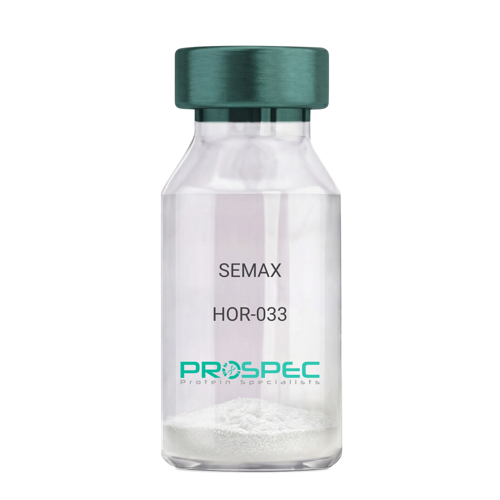
SEMAX
Product # :
HOR-033Price :
Quantity :
Shipping Method :
Shipped at Room temp
More Info
- description
- formulation
- purity
- More Info
- Citations
Description
SEMAX Synthetic is a single, non-glycosylated polypeptide chain containing 7 amino acids, having a molecular mass of 813.92 Dalton and a Molecular formula of C37H51N19O1S.
Formulation
The protein was lyophilized with no additives.
Purity
Greater than 97.0% as determined by analysis by RP-HPLC.
Citations
More Info
-
Physical Appearance
Sterile Filtered White lyophilized (freeze-dried) powder.
-
Stability
Lyophilized SEMAX although stable at room temperature for 3 weeks, should be stored desiccated below -18°C. Upon reconstitution SEMAX should be stored at 4°C between 2-7 days and for future use below -18°C. For long term storage it is recommended to add a carrier protein (0.1% HSA or BSA). Please prevent freeze-thaw cycles.
-
Solubility
It is recommended to reconstitute the lyophilized SEMAX in sterile 18MΩ-cm H2O not less than 100 µg/ml, which can then be further diluted to other aqueous solutions.
-
Amino Acid Sequence
H-Met-Glu-His-Phe-Pro-Gly-Pro-OH.
-
Background
Semax, also known as ACTH(4-10) Pro-Gly-Pro, is a synthetic peptide that has been the subject of extensive research due to its potential neuroprotective and nootropic effects. This heptapeptide, derived from the adrenocorticotropic hormone (ACTH), has been shown to possess a wide range of biological activities, including enhancing memory, learning, and neurogenesis.
Semax is unique in its ability to cross the blood-brain barrier and exert its effects directly on the central nervous system. It has been shown to stimulate the release of brain-derived neurotrophic factor (BDNF), a protein that plays a crucial role in the survival of neurons and the growth of new neurons and synapses. Studies by Dolotov et al. (2006) have demonstrated that Semax can enhance memory and learning in rats, suggesting potential applications in cognitive enhancement and the treatment of cognitive disorders.
In addition to its nootropic effects, Semax has been shown to possess neuroprotective properties. Research by Stavchansky et al. (2008) found that Semax could protect neurons from oxidative stress and apoptosis, suggesting potential applications in the treatment of neurodegenerative diseases such as Alzheimer's and Parkinson's disease.
Given its nootropic and neuroprotective effects, Semax has been proposed as a potential therapeutic agent for a variety of conditions, including cognitive disorders, stroke, and optic nerve disease. For instance, a study by Myasoedov et al. (2010) found that Semax could improve outcomes in patients with ischemic stroke, indicating its potential as a therapeutic agent in stroke recovery.
While research on Semax is promising, it is important to note that most studies have been conducted in animals or in vitro. More research is needed to fully understand the potential effects and applications of Semax in humans. However, the existing body of research suggests that Semax could be a promising tool in the treatment of cognitive disorders and neurodegenerative diseases.
What is the molecular weight/Mw of SEMAX Protein?
SEMAX Protein has a total Mw of 0.813kDa.
What is the Purity of SEMAX Protein?
SEMAX Protein is >97% pure as determined by SDS-PAGE.
What is the Biological Activity of SEMAX Protein?
The biological functionality of SEMAX Protein will be determined in the future.
What is the amino acid sequence of SEMAX Protein?
H-Met-Glu-His-Phe-Pro-Gly-Pro-OH.
What applications can SEMAX Protein be used in?
SEMAX Protein can probably be used in western blot, ELISA and Lateral Flow.
What is the endotoxin level for SEMAX Protein?
The endotoxin level is minimal, SEMAX Protein was purified using conventional chromatography techniques.
Who supplies SEMAX that is in stock and available to order?
ProSpec has the protein in stock and available for immediate delivery.
Where can I order bulk mg quantities of SEMAX?
ProSpec supplies bulk mg quantities at affordable prices that meets research and academic budgets.
ProSpec's products are furnished for LABORATORY RESEARCH USE ONLY. The product may not be used as drugs, agricultural or pesticidal products, food additives or household chemicals.
- View Data Sheet
Name :
SARS MERS, Sf9 ActiveDescription:
SARS MERS Spike Recombinant, Sf9 Active
Middle East respiratory syndrome coronavirus, Human betacoronavirus 2c EMC/2012, MERS-CoV, MERS, MERSCoV SP, Spike glycoprotein, S glycoprotein, S, Spike protein, E2, Peplomer protein
Product # :
SARS-061Price :
Quantity :
Shipping Method :
Shipped with Ice Packs
More Info
- description
- source
- formulation
- purity
- biological activity
- More Info
- Citations
Description
SARS MERS Recombinant produced in Sf9 Baculovirus cells is a single, glycosylated polypeptide chain containing 1285 amino acids (18-1296 aa) and having a molecular mass of 141.6kDa. SARS MERS is fused to a 6 amino acid His-tag at C-terminus & purified by proprietary chromatographic techniques.
Source
Sf9, Baculovirus cells.
Formulation
The SARS MERS solution (0.25mg/ml) contains 10% glycerol and Phosphate-Buffered Saline (pH 7.4).
Purity
Greater than 85.0% as determined by SDS-PAGE.
Biological Activity
Measured by its binding ability in a functional ELISA with Human DPPIV/CD26 (CAT# enz-1187).
Citations
More Info
-
Synonyms
Middle East respiratory syndrome coronavirus, Human betacoronavirus 2c EMC/2012, MERS-CoV, MERS, MERSCoV SP, Spike glycoprotein, S glycoprotein, S, Spike protein, E2, Peplomer protein
-
Physical Appearance
Sterile filtered colorless solution.
-
Stability
Store at 4°C if entire vial will be used within 2-4 weeks.Store, frozen at -20°C for longer periods of time.For long term storage it is recommended to add a carrier protein (0.1% HSA or BSA).Avoid multiple freeze-thaw cycles.
-
Amino Acid Sequence
YVDVGPDSVK SACIEVDIQQ TFFDKTWPRP IDVSKADGII YPQGRTYSNI TITYQGLFPY QGDHGDMYVY SAGHATGTTP QKLFVANYSQ DVKQFANGFV VRIGAAANST GTVIISPSTS ATIRKIYPAF MLGSSVGNFS DGKMGRFFNH TLVLLPDGCG TLLRAFYCIL EPRSGNHCPA GNSYTSFATY HTPATDCSDG NYNRNASLNS FKEYFNLRNC TFMYTYNITE DEILEWFGIT QTAQGVHLFS SRYVDLYGGN MFQFATLPVY DTIKYYSIIP HSIRSIQSDR KAWAAFYVYK LQPLTFLLDF SVDGYIRRAI DCGFNDLSQL HCSYESFDVE SGVYSVSSFE AKPSGSVVEQ AEGVECDFSP LLSGTPPQVY NFKRLVFTNC NYNLTKLLSL FSVNDFTCSQ ISPAAIASNC YSSLILDYFS YPLSMKSDLS VSSAGPISQF NYKQSFSNPT CLILATVPHN LTTITKPLKY SYINKCSRLL SDDRTEVPQL VNANQYSPCV SIVPSTVWED GDYYRKQLSP LEGGGWLVAS GSTVAMTEQL QMGFGITVQY GTDTNSVCPK LEFANDTKIA SQLGNCVEYS LYGVSGRGVF QNCTAVGVRQ QRFVYDAYQN LVGYYSDDGN YYCLRACVSV PVSVIYDKET KTHATLFGSV ACEHISSTMS QYSRSTRSML KRRDSTYGPL QTPVGCVLGL VNSSLFVEDC KLPLGQSLCA LPDTPSTLTP RSVRSVPGEM RLASIAFNHP IQVDQLNSSY FKLSIPTNFS FGVTQEYIQT TIQKVTVDCK QYVCNGFQKC EQLLREYGQF CSKINQALHG ANLRQDDSVR NLFASVKSSQ SSPIIPGFGG DFNLTLLEPV SISTGSRSAR SAIEDLLFDK VTIADPGYMQ GYDDCMQQGP ASARDLICAQ YVAGYKVLPP LMDVNMEAAY TSSLLGSIAG VGWTAGLSSF AAIPFAQSIF YRLNGVGITQ QVLSENQKLI ANKFNQALGA MQTGFTTTNE AFQKVQDAVN NNAQALSKLA SELSNTFGAI SASIGDIIQR LDVLEQDAQI DRLINGRLTT LNAFVAQQLV RSESAALSAQ LAKDKVNECV KAQSKRSGFC GQGTHIVSFV VNAPNGLYFM HVGYYPSNHI EVVSAYGLCD AANPTNCIAP VNGYFIKTNN TRIVDEWSYT GSSFYAPEPI TSLNTKYVAP QVTYQNISTN LPPPLLGNST GIDFQDELDE FFKNVSTSIP NFGSLTQINT TLLDLTYEML SLQQVVKALN ESYIDLKELG NYTYYNKWPH HHHHH.
-
Background
Severe acute respiratory syndrome coronavirus (SARS-CoV) and Middle East respiratory syndrome coronavirus (MERS-CoV) have caused significant outbreaks with severe respiratory illness in recent years. The spike proteins of these viruses play a crucial role in viral entry and host cell recognition. This research aims to explore the structure, function, and implications of SARS and MERS spike proteins in viral pathogenesis and the development of therapeutic interventions. Understanding the mechanisms of viral entry can provide valuable insights into the design of effective antiviral strategies.
Structure and Function of SARS and MERS Spike Proteins:
The spike proteins of SARS-CoV and MERS-CoV are trimeric glycoproteins located on the viral surface. They consist of S1 and S2 subunits, with the S1 subunit responsible for receptor binding and the S2 subunit involved in membrane fusion. The spike proteins contain key domains, including the receptor-binding domain (RBD) and the fusion peptide, which mediate the interactions with host cell receptors and membrane fusion during viral entry.
Interaction with Host Cell Receptors:
The SARS-CoV spike protein primarily interacts with the angiotensin-converting enzyme 2 (ACE2) receptor, which is expressed in various tissues, including the respiratory tract. The binding of the SARS-CoV spike protein to ACE2 is crucial for viral entry into host cells. Similarly, the MERS-CoV spike protein interacts with the dipeptidyl peptidase 4 (DPP4) receptor, which is predominantly expressed in the lungs and other tissues. These receptor interactions determine the host range and tissue tropism of the viruses.
Implications for Viral Pathogenesis:
The interaction between the spike proteins of SARS-CoV and MERS-CoV and their respective receptors initiates a cascade of events leading to viral entry and replication. The binding of the spike protein to the receptor triggers conformational changes that expose the fusion peptide, facilitating membrane fusion and viral entry into host cells. The specificity and affinity of the spike-receptor interaction play a critical role in determining viral tropism, disease severity, and transmission dynamics.
Therapeutic Strategies Targeting Spike Proteins:
The spike proteins of SARS-CoV and MERS-CoV present attractive targets for the development of antiviral interventions. Various approaches have been explored, including the development of neutralizing antibodies that specifically target the spike proteins and inhibit viral entry. Additionally, small molecule inhibitors and peptide-based therapeutics are being investigated to disrupt the spike-receptor interaction and block viral entry. Vaccine development efforts have also focused on generating immune responses against the spike proteins to prevent infection.
Challenges and Future Directions:
Although significant progress has been made in understanding the structure and function of SARS and MERS spike proteins, several challenges remain. The emergence of novel coronavirus variants necessitates continuous surveillance and adaptation of therapeutic strategies. Additionally, potential cross-reactivity and immunogenicity concerns need to be addressed to ensure the safety and efficacy of spike protein-based therapeutics.
Conclusion:
The study of SARS and MERS spike proteins provides valuable insights into the mechanisms of viral entry and pathogenesis. Understanding the interactions between spike proteins and host cell receptors is crucial for the development of effective antiviral strategies. Further research on spike protein structure, receptor binding, and fusion mechanisms is essential to combat current and future coronavirus outbreaks. By unraveling the complexities of viral entry, we can pave the way for the development of novel therapeutics to mitigate the impact of SARS-CoV, MERS-CoV, and related coronaviruses.
ProSpec's products are furnished for LABORATORY RESEARCH USE ONLY. The product may not be used as drugs, agricultural or pesticidal products, food additives or household chemicals.
- View Data Sheet
Name :
HIV-1 ProteaseDescription:
HIV-1 Protease Recombinant
Product # :
HIV-001Price :
Quantity :
Shipping Method :
Shipped with Ice Packs
More Info
- description
- source
- formulation
- purity
- More Info
- Citations
Description
HIV-1 protease is an active homodimer having a molecular mass of 21.6kDa (each monomer of 99 amino acids is 10.8kDa).
The HIV-1 has His-Tag at C-terminus and purified by proprietary chromatographic techniques.
Source
Escherichia Coli.
Formulation
The HIV-1 Protease filtered (0.4µm) solution is formulated in 20mM Tris, 20mM MES, 200mM NaCl, 1mM EDTA, 10% (v/v) glycerol and 0.05% 2-mercaptoethanol, pH 6.5.
Purity
Greater than 95.0% as determined by SDS-PAGE.
Citations
More Info
-
Introduction
HIV-1 protease is very significant in the life cycle of the HIV virus. It is expressed in the infected cells as a part of Gag-Pol polyprotein from which it is auto-catalytycaly released after formation of an immature viral particle. The enzyme subsequently cleaves the other parts of viral polyproteins resulting in the maturation of the virus. In HIV-infected patients the enzyme is subjected to intensive mutagenesis and mutants resistant to applied medicines are produced as a result of the selection pressure.
-
Physical Appearance
Sterile filtered colorless clear solution.
-
Stability
Store at 4°C if entire vial will be used within 2-4 weeks. Store, frozen at -20°C for longer periods of time.Please avoid freeze thaw cycles.
-
Amino Acid Sequence
PQITLWQRPL VTIKIGGQLK EALLDTGADD TVLEEMNLPG RWKPKMIGGI GGFIKVRQYDQILIEICGHK AIGTVLVGPT PVNIIGRNLL TQIGCTLNFHHHHHH.
-
Kinetic parameters
Km=15.1µM, Kcat = 30s-1, Kcat/Km= 1981 mM-1s-1 with peptide substrate KARVF (NO2)VRKA (F(NO2) ... p-nitrophenylalanine).
ProSpec's products are furnished for LABORATORY RESEARCH USE ONLY. The product may not be used as drugs, agricultural or pesticidal products, food additives or household chemicals.
- View Data Sheet
Name :
ECHS1 HumanDescription:
Enoyl CoA Hydratase, Short chain, 1, Mitochondrial Human Recombinant
Enoyl-CoA hydratase 1, SCEH.
Product # :
ENZ-556Price :
Quantity :
Shipping Method :
Shipped with Ice Packs
More Info
- description
- source
- formulation
- purity
- More Info
- Citations
Description
ECHS1 Human Recombinant produced in E.Coli is a single, non-glycosylated polypeptide chain containing 284 amino acids (28-290 a.a.) and having a molecular mass of 30.6kDa.ECHS1 is fused to a 21 amino acid His-tag at N-terminus & purified by proprietary chromatographic techniques.
Source
Escherichia Coli.
Formulation
The ECHS1 protein solution (1mg/ml) contains 20mM Tris-HCl buffer (pH-8), 1mM DTT,0.1M NaCl and 20% glycerol.
Purity
Greater than 95.0% as determined by SDS-PAGE.
Citations
More Info
-
Introduction
ECHS1 is part of the hydratase/isomerase superfamily. ECHS1 is localized in the mitochondrial matrix and Expressed in muscle, liver and fibroblasts, with low expression in kidney and spleen, ECHS1 exists as a homohexamer that takes part in the second phase of the mitochondrial fatty acid β-oxidation pathway.
-
Synonyms
Enoyl-CoA hydratase 1, SCEH.
-
Physical Appearance
Sterile filtered colorless solution.
-
Stability
Store at 4°C if entire vial will be used within 2-4 weeks. Store, frozen at -20°C for longer periods of time. For long term storage it is recommended to add a carrier protein (0.1% HSA or BSA).Avoid multiple freeze-thaw cycles.
-
Amino Acid Sequence
MGSSHHHHHH SSGLVPRGSH MASGANFEYI IAEKRGKNNT VGLIQLNRPK ALNALCDGLI DELNQALKIF EEDPAVGAIV LTGGDKAFAA GADIKEMQNL SFQDCYSSKF LKHWDHLTQV KKPVIAAVNG YAFGGGCELA MMCDIIYAGE KAQFAQPEIL IGTIPGAGGT QRLTRAVGKS LAMEMVLTGD RISAQDAKQA GLVSKICPVE TLVEEAIQCA EKIASNSKIV VAMAKESVNA AFEMTLTEGS KLEKKLFYST FATDDRKEGM TAFVEKRKAN FKDQ.
ProSpec's products are furnished for LABORATORY RESEARCH USE ONLY. The product may not be used as drugs, agricultural or pesticidal products, food additives or household chemicals.
- View Data Sheet
Name :
BCL2L10 HumanDescription:
BCL2 Like 10 Human Recombinant
Bcl-2-like protein 10, Bcl2-L-10, Anti-apoptotic protein NrH, Apoptosis regulator Bcl-B, BCL2L10, BCLB, Boo, Diva, BCL-B.
Product # :
PRO-1321Price :
Quantity :
Shipping Method :
Shipped with Ice Packs
More Info
- description
- source
- formulation
- purity
- More Info
- Citations
Description
BCL2L10 Human Recombinant produced in E.Coli is a single, non-glycosylated polypeptide chain containing 195 amino acids (1-172 a.a.) and having a molecular mass of 21.8kDa.BCL2L10 is fused to a 23 amino acid His-tag at N-terminus & purified by proprietary chromatographic techniques.
Source
E.coli.
Formulation
BCL2L10 protein solution (0.5mg/ml) containing 20mM Tris-HCl buffer (pH 8.0), 0.15M NaCl, 20% glycerol and 1mM DTT.
Purity
Greater than 90% as determined by SDS-PAGE.
Citations
More Info
-
Introduction
BCL2 Like 10 (BCL2L10) is a member of the BCL-2 protein family. BCL2 family members form hetero or homodimers and serve as anti or pro-apoptotic regulators which are involved in a wide variety of cellular activities. BCL2L10 includes the conserved BH4, BH1 and BH2 domains. BCL2L10 interacts with other members of BCL2 protein family including BCL2, BCL2L1/BCL-X(L), and BAX. BCL2L10 overexpression suppresses cell apoptosis possibly through the prevention of cytochrome C release from the mitochondria, and consequently activating caspase-3 activation.
-
Synonyms
Bcl-2-like protein 10, Bcl2-L-10, Anti-apoptotic protein NrH, Apoptosis regulator Bcl-B, BCL2L10, BCLB, Boo, Diva, BCL-B.
-
Physical Appearance
Sterile Filtered colorless solution.
-
Stability
Store at 4°C if entire vial will be used within 2-4 weeks. Store, frozen at -20°C for longer periods of time. For long term storage it is recommended to add a carrier protein (0.1% HSA or BSA).Avoid multiple freeze-thaw cycles.
-
Amino Acid Sequence
MGSSHHHHHH SSGLVPRGSH MGSMVDQLRE RTTMADPLRE RTELLLADYL GYCAREPGTP EPAPSTPEAA VLRSAAARLR QIHRSFFSAY LGYPGNRFEL VALMADSVLS DSPGPTWGRV VTLVTFAGTL LERGPLVTAR WKKWGFQPRL KEQEGDVARD CQRLVALLSS RLMGQHRAWL QAQGGWDGFC HFFRT.
ProSpec's products are furnished for LABORATORY RESEARCH USE ONLY. The product may not be used as drugs, agricultural or pesticidal products, food additives or household chemicals.
- View Data Sheet
Name :
SEPT3 HumanDescription:
Septin-3 Human Recombinant
Septin 3, SEP3, Neuronal-Specific Septin 3, Neuronal-Specific Septin-3, BK250D10.3, Neuronal-specific septin-3.
Product # :
PRO-2152Price :
Quantity :
Shipping Method :
Shipped with Ice Packs
More Info
- description
- source
- formulation
- purity
- More Info
- Citations
Description
SEPT3 Human Recombinant produced in E.Coli is a single, non-glycosylated polypeptide chain containing 381 amino acids (1-358 a.a) and having a molecular mass of 43.1kDa. SEPT3 is fused to a 23 amino acid His-tag at N-terminus & purified by proprietary chromatographic techniques.
Source
Escherichia Coli.
Formulation
SEPT3 protein solution (1mg/ml) containing Phosphate buffered saline (pH7.4), 10% glycerol and 1mM DTT.
Purity
Greater than 95% as determined by SDS-PAGE.
Citations
More Info
-
Introduction
Septin-3, also known as SEPT3 is a member of the septin family of GTPases. Members of this family are essential for cytokinesis. Furthermore, expression is up regulated by retinoic acid in a human teratocarcinoma cell line. The exact role of SEPT3 has not been determined yet. In addition, among its related pathways are Bacterial invasion of epithelial cells.
-
Synonyms
Septin 3, SEP3, Neuronal-Specific Septin 3, Neuronal-Specific Septin-3, BK250D10.3, Neuronal-specific septin-3.
-
Physical Appearance
Sterile filtered colorless solution.
-
Stability
Store at 4°C if entire vial will be used within 2-4 weeks. Store, frozen at -20°C for longer periods of time. For long term storage it is recommended to add a carrier protein (0.1% HSA or BSA).Avoid multiple freeze-thaw cycles.
-
Amino Acid Sequence
MGSSHHHHHH SSGLVPRGSH MGSMSKGLPE TRTDAAMSEL VPEPRPKPAV PMKPMSINSN LLGYIGIDTI IEQMRKKTMK TGFDFNIMVV GQSGLGKSTL VNTLFKSQVS RKASSWNREE KIPKTVEIKA IGHVIEEGGV KMKLTVIDTP GFGDQINNEN CWEPIEKYIN EQYEKFLKEE VNIARKKRIP DTRVHCCLYF ISPTGHSLRP LDLEFMKHLS KVVNIIPVIA KADTMTLEEK SEFKQRVRKE LEVNGIEFYP QKEFDEDLED KTENDKIRQE SMPFAVVGSD KEYQVNGKRV LGRKTPWGII EVENLNHCEF ALLRDFVIRT HLQDLKEVTH NIHYETYRAK RLNDNGGLPP GEGLLGTVLP PVPATPCPTA E.
ProSpec's products are furnished for LABORATORY RESEARCH USE ONLY. The product may not be used as drugs, agricultural or pesticidal products, food additives or household chemicals.
- View Data Sheet
Name :
ENTPD3 Human, sf9 BioactiveDescription:
Ectonucleoside Triphosphate Diphosphohydrolase 3 Human Recombinant, sf9 Bioactive
Ectonucleoside Triphosphate Diphosphohydrolase 3, Ecto-ATP Diphosphohydrolase 3, CD39 Antigen-Like 3, Ecto-ATPDase 3, Ecto-Apyrase 3, Ecto-ATPase 3, EC 3.6.1.5, NTPDase 3, CD39L3, HB6, NTPDase-3, EC 3.6.1, Ectonucleoside triphosphate diphosphohydrolase 3, NTPDase 3, CD39 antigen-like 3, Ecto-ATP diphosphohydrolase 3, Ecto-ATPDase 3, Ecto-ATPase 3, Ecto-apyrase 3, HB6.
Product # :
ENZ-1020Price :
Quantity :
Shipping Method :
Shipped with Ice Packs
More Info
- description
- source
- formulation
- purity
- biological activity
- More Info
- Citations
Description
ENTPD3 Human Recombinant produced in Sf9 Baculovirus cells is a single, glycosylated polypeptide chain containing 451 amino acids (44-485a.a.) and having a molecular mass of 50.7kDa (Molecular size on SDS-PAGE will appear at approximately 50-70kDa).ENTPD3 is expressed with a 6 amino acid His tag at C-Terminus and purified by proprietary chromatographic techniques.
Source
Sf9, Baculovirus cells.
Formulation
ENTPD3 protein solution (0.5mg/ml) contains Phosphate Buffered Saline (pH 7.4) and 10% glycerol.
Purity
Greater than 90.0% as determined by SDS-PAGE.
Biological Activity
Specific activity is > 250,000 pmol/min/ug, and is defined as the amount of enzyme that hydrolyze ATP per minute at pH 7.5 at 37C.Citations
More Info
-
Introduction
Ectonucleoside Triphosphate Diphosphohydrolase 3, also known as ENTPD3, which owns a threefold preference for the hydrolysis of ATP over ADP is similar to E-type nucleotidases (NTPases). ENTPD3 is a protein coding gene which contains four apyrase-conserved areas which is characteristic of NTPases.
-
Synonyms
Ectonucleoside Triphosphate Diphosphohydrolase 3, Ecto-ATP Diphosphohydrolase 3, CD39 Antigen-Like 3, Ecto-ATPDase 3, Ecto-Apyrase 3, Ecto-ATPase 3, EC 3.6.1.5, NTPDase 3, CD39L3, HB6, NTPDase-3, EC 3.6.1, Ectonucleoside triphosphate diphosphohydrolase 3, NTPDase 3, CD39 antigen-like 3, Ecto-ATP diphosphohydrolase 3, Ecto-ATPDase 3, Ecto-ATPase 3, Ecto-apyrase 3, HB6.
-
Physical Appearance
Sterile Filtered colorless solution.
-
Stability
Store at 4°C if entire vial will be used within 2-4 weeks.Store, frozen at -20°C for longer periods of time. For long term storage it is recommended to add a carrier protein (0.1% HSA or BSA).Avoid multiple freeze-thaw cycles.
-
Amino Acid Sequence
ADLQIHKQEV LPPGLKYGIV LDAGSSRTTV YVYQWPAEKE NNTGVVSQTF KCSVKGSGIS SYGNNPQDVP RAFEECMQKV KGQVPSHLHG STPIHLGATA GMRLLRLQNE TAANEVLESI QSYFKSQPFD FRGAQIISGQ EEGVYGWITA NYLMGNFLEK NLWHMWVHPH GVETTGALDL GGASTQISFV AGEKMDLNTS DIMQVSLYGY VYTLYTHSFQ CYGRNEAEKK FLAMLLQNSP TKNHLTNPCY PRDYSISFTM GHVFDSLCTV DQRPESYNPN DVITFEGTGD PSLCKEKVAS IFDFKACHDQ ETCSFDGVYQ PKIKGPFVAF AGFYYTASAL NLSGSFSLDT FNSSTWNFCS QNWSQLPLLL PKFDEVYARS YCFSANYIYH LFVNGYKFTE ETWPQIHFEK EVGNSSIAWS LGYMLSLTNQ IPAESPLIRL PIEPPHHHHHH.
ProSpec's products are furnished for LABORATORY RESEARCH USE ONLY. The product may not be used as drugs, agricultural or pesticidal products, food additives or household chemicals.
- View Data Sheet
Name :
BLNK HumanDescription:
B-Cell Linker Human Recombinant
B-cell linker protein, B-cell adapter containing a SH2 domain protein, B-cell adapter containing a Src homology 2 domain protein, Cytoplasmic adapter protein, Src homology 2 domain-containing leukocyte protein of 65 kDa, SLP-65, BLNK, BASH, SLP65, AGM4, LY57, BLNK-S, MGC111051.
Product # :
PRO-102Price :
Quantity :
Shipping Method :
Shipped with Ice Packs
More Info
- description
- source
- formulation
- purity
- More Info
- Citations
Description
BLNK Human Recombinant fused with a 20 amino acid His tag at N-terminus produced in E.Coli is a single, non-glycosylated, polypeptide chain containing 476 amino acids (1-456 a.a.) and having a molecular mass of 52.6kDa (Molecular weight on SDS-PAGE will appear higher). The BLNK is purified by proprietary chromatographic techniques.
Source
Escherichia Coli.
Formulation
The BLNK solution (1 mg/ml) contains 20mM Tris-HCl buffer (pH8.0), 20% glycerol, 0.1M NaCl, 1mM DTT and 0.1mM PMSF.
Purity
Greater than 90.0% as determined by SDS-PAGE.
Citations
More Info
-
Introduction
BLNK is a cytoplasmic linker or adaptor protein which has a significant role in B cell development. B-cell linker (BLNK) is essential for normal B-cell development. BLNK bridges B cell receptor-associated kinase activation with downstream signaling pathways, thus affecting different biological functions. BLNK associates with the effector proteins GRB2, Vav, NCK and PLC-g following activation of the B cell receptor. BLNK is phosphorylated by the Syk tyrosine kinase, which in turn permits activation of downstream effector proteins including GRB2 and PLC-g. Mutations in the BLNK gene cause hypoglobulinemia and absent B cells, a disease in which the pro- to pre-B-cell transition is developmentally blocked. Deficiency in the BLNK protein is seen in some cases of pre-B acute lymphoblastic leukemia.
-
Synonyms
B-cell linker protein, B-cell adapter containing a SH2 domain protein, B-cell adapter containing a Src homology 2 domain protein, Cytoplasmic adapter protein, Src homology 2 domain-containing leukocyte protein of 65 kDa, SLP-65, BLNK, BASH, SLP65, AGM4, LY57, BLNK-S, MGC111051.
-
Physical Appearance
Sterile Filtered colorless solution.
-
Stability
Store at 4°C if entire vial will be used within 2-4 weeks. Store, frozen at -20°C for longer periods of time. For long term storage it is recommended to add a carrier protein (0.1% HSA or BSA).Avoid multiple freeze-thaw cycles.
-
Amino Acid Sequence
MGSSHHHHHH SSGLVPRGSH MDKLNKITVP ASQKLRQLQK MVHDIKNNEG GIMNKIKKLK VKAPPSVPRR DYASESPADE EQQWSDDFDS DYENPDEHSD SEMYVMPAEE NADDSYEPPP VEQETRPVHP ALPFARGEYI DNRSSQRHSP PFSKTLPSKP SWPSEKARLT STLPALTALQ KPQVPPKPKG LLEDEADYVV PVEDNDENYI HPTESSSPPP EKAPMVNRST KPNSSTPASP PGTASGRNSG AWETKSPPPA APSPLPRAGK KPTTPLKTTP VASQQNASSV CEEKPIPAER HRGSSHRQEA VQSPVFPPAQ KQIHQKPIPL PRFTEGGNPT VDGPLPSFSS NSTISEQEAG VLCKPWYAGA CDRKSAEEAL HRSNKDGSFL IRKSSGHDSK QPYTLVVFFN KRVYNIPVRF IEATKQYALG RKKNGEEYFG SVAEIIRNHQ HSPLVLIDSQ NNTKDSTRLK YAVKVS.
ProSpec's products are furnished for LABORATORY RESEARCH USE ONLY. The product may not be used as drugs, agricultural or pesticidal products, food additives or household chemicals.
- View Data Sheet
Name :
GZMB Human, sf9Description:
Granzyme-B, Sf9 Human Recombinant
Granzyme B, T-Cell Serine Protease 1-3E, Cathepsin G-Like 1, Granzyme B (Granzyme 2, Cytotoxic T-Lymphocyte-Associated Serine Esterase 1, Cytotoxic T-Lymphocyte Proteinase 2, Cytotoxic Serine Protease B , Human Lymphocyte Protein, Fragmentin 2, EC 3.4.21.79, CTSGL1, CTLA1 , CSPB , CGL1, SECT , C11, HLP , Cytotoxic T-Lymphocyte-Associated Serine Esterase 1, Lymphocyte Protease, Fragmentin-2 , Granzyme 2 , Granzyme-2 , EC 3.4.21, CTLA-1 , CGL-1, CSP-B, CCPI , GRB.
Product # :
ENZ-1078Price :
Quantity :
Shipping Method :
Shipped with Ice Packs
More Info
- description
- source
- formulation
- purity
- biological activity
- More Info
- Citations
Description
GZMB produced in Sf9 Baculovirus cells is a single, glycosylated polypeptide chain containing 235 amino acids (19-247a.a.) and having a molecular mass of 26.5kDa. (Molecular size on SDS-PAGE will appear at approximately 28-40kDa). GZMB is expressed with an 6 amino acid His tag at C-Terminus and purified by proprietary chromatographic techniques.
Source
Sf9, Baculovirus cells.
Formulation
GZMB protein solution (0.5mg/ml) contains Phosphate Buffered Saline (pH 7.4) ,20% glycerol and 1mM DTT.
Purity
Greater than 95.0% as determined by SDS-PAGE.
Biological Activity
Specific activity is > 7,000 pmol/min/ug, and is defined as the amount of enzyme that cleave 1pmole of Boc-Ala-Ala-Asp-SBzl at 37C.
Citations
More Info
-
Introduction
Granzyme-B (GZMB) is essential for target cell lysis in cell-mediated immune responses. GZMB is related to an activation cascade of caspases responsible for apoptosis execution. GZMB cuts caspase-3, -7, -9 and 10 to activate enzymes mediating apoptosis.
-
Synonyms
Granzyme B, T-Cell Serine Protease 1-3E, Cathepsin G-Like 1, Granzyme B (Granzyme 2, Cytotoxic T-Lymphocyte-Associated Serine Esterase 1, Cytotoxic T-Lymphocyte Proteinase 2, Cytotoxic Serine Protease B , Human Lymphocyte Protein, Fragmentin 2, EC 3.4.21.79, CTSGL1, CTLA1 , CSPB , CGL1, SECT , C11, HLP , Cytotoxic T-Lymphocyte-Associated Serine Esterase 1, Lymphocyte Protease, Fragmentin-2 , Granzyme 2 , Granzyme-2 , EC 3.4.21, CTLA-1 , CGL-1, CSP-B, CCPI , GRB.
-
Physical Appearance
Sterile Filtered colorless solution.
-
Stability
Store at 4°C if entire vial will be used within 2-4 weeks. Store, frozen at -20°C for longer periods of time. For long term storage it is recommended to add a carrier protein (0.1% HSA or BSA). Avoid multiple freeze-thaw cycles.
-
Amino Acid Sequence
GEIIGGHEAK PHSRPYMAYL MIWDQKSLKR CGGFLIRDDF VLTAAHCWGS SINVTLGAHN IKEQEPTQQF IPVKRPIPHP AYNPKNFSND IMLLQLERKA KRTRAVQPLR LPSNKAQVKP GQTCSVAGWG QTAPLGKHSH TLQEVKMTVQ EDRKCESDLR HYYDSTIELC VGDPEIKKTS FKGDSGGPLV CNKVAQGIVS YGRNNGMPPR ACTKVSSFVH WIKKTMKRYH HHHHH
ProSpec's products are furnished for LABORATORY RESEARCH USE ONLY. The product may not be used as drugs, agricultural or pesticidal products, food additives or household chemicals.
- View Data Sheet
Name :
SARS Spike (306-515), Sf9Description:
Coronavirus Spike Receptor Binding Domain (306-515 a.a.)Recombinant, Sf9
Spike glycoprotein, S glycoprotein, Peplomer protein, E2 glycoprotein precursor, Severe acute repiratory Syndrome-related Coronavirus, SARS, SRAS-CoV, SARS-CoV1, E2.
Product # :
SARS-047Price :
Quantity :
Shipping Method :
Shipped with Ice Packs
More Info
- description
- source
- formulation
- purity
- biological activity
- More Info
- Citations
Description
SARS Spike produced in Sf9 Baculovirus cells is a single, glycosylated polypeptide chain containing 219 amino acids (306-515 aa) and having a molecular mass of 24.7kDa.SARS Spike is fused to a 6 amino acid His tag at C-terminus and purified by proprietary chromatographic techniques.
Source
Sf9, Baculovirus cells.
Formulation
The SARS Spike (306-515) solution (0.5mg/ml) contains Phosphate-Buffered Saline (pH 7.4) and 10% Glycerol.
Purity
Greater than 95.0% as determined by SDS-PAGE.
Biological Activity
Measured by its binding ability in a functional ELISA with Human ACE-2 (CAT# enz-1159).
Citations
More Info
-
Introduction
SARS Coronavirus is an enveloped virus containing 3 outer structural proteins, namely the membrane (M), envelope (E), and spike (S) proteins. Spike (S)-glycoprotein of the virus interacts with a cellular receptor and mediates membrane fusion to allow viral entry into susceptible target cells. Accordingly, S-protein takes part in virus infection cycle and is the primary target of neutralizing antibodies.
-
Synonyms
Spike glycoprotein, S glycoprotein, Peplomer protein, E2 glycoprotein precursor, Severe acute repiratory Syndrome-related Coronavirus, SARS, SRAS-CoV, SARS-CoV1, E2.
-
Physical Appearance
Sterile Filtered colorless solution.
-
Stability
Store at 4°C if entire vial will be used within 2-4 weeks. Store, frozen at -20°C for longer periods of time. For long term storage it is recommended to add a carrier protein (0.1% HSA or BSA). Avoid multiple freeze-thaw cycles.
-
Amino Acid Sequence
ADPRVVPSGD VVRFPNITNL CPFGEVFNAT KFPSVYAWER KKISNCVADY SVLYNSTFFS TFKCYGVSAT KLNDLCFSNV YADSFVVKGD DVRQIAPGQT GVIADYNYKL PDDFMGCVLA WNTRNIDATS TGNYNYKYRY LRHGKLRPFE RDISNVPFSP DGKPCTPPAL NCYWPLNDYG FYTTTGIGYQ PYRVVVLSFE LLNAPATVCG PKLHHHHHH
ProSpec's products are furnished for LABORATORY RESEARCH USE ONLY. The product may not be used as drugs, agricultural or pesticidal products, food additives or household chemicals.
- View Data Sheet
Name :
UGT8 HumanDescription:
UDP Glycosyltransferase 8 Human Recombinant
UDP Glycosyltransferase 8, CGT, UDP-Galactose Ceramide Galactosyltransferase, 2-Hydroxyacylsphingosine 1-Beta-Galactosyltransferase, Ceramide UDP-Galactosyltransferase, Cerebroside Synthase, UGT4, UDP-Galactose-Ceramide Galactosyltransferase, Uridine Diphosphate Glycosyltransferase 8, EC 2.4.1.45, EC 2.4.1.
Product # :
ENZ-754Price :
Quantity :
Shipping Method :
Shipped with Ice Packs
More Info
- description
- source
- formulation
- purity
- More Info
- Citations
Description
UGT8 Human Recombinant produced in E.Coli is a single, non-glycosylated polypeptide chain containing 544 amino acids (21-541 a.a) and having a molecular mass of 61.6kDa.UGT8 is fused to a 23 amino acid His-tag at N-terminus.
Source
Escherichia Coli.
Formulation
UGT8 protein solution (1mg/ml) containing 20mM Tris-HCl buffer (pH 8.0), 0.4M urea and 10% glycerol.
Purity
Greater than 85% as determined by SDS-PAGE.
Citations
More Info
-
Introduction
UDP Glycosyltransferase 8 (UGT8) is a member of the UDP-glycosyltransferase family. UGT8 catalyzes the transfer of galactose to ceramide, a crucial enzymatic step in the biosynthesis of galactocerebrosides, which are abundant sphingolipids of the myelin membrane of the central nervous system and peripheral nervous system.
-
Synonyms
UDP Glycosyltransferase 8, CGT, UDP-Galactose Ceramide Galactosyltransferase, 2-Hydroxyacylsphingosine 1-Beta-Galactosyltransferase, Ceramide UDP-Galactosyltransferase, Cerebroside Synthase, UGT4, UDP-Galactose-Ceramide Galactosyltransferase, Uridine Diphosphate Glycosyltransferase 8, EC 2.4.1.45, EC 2.4.1.
-
Physical Appearance
Sterile Filtered clear solution.
-
Stability
Store at 4°C if entire vial will be used within 2-4 weeks. Store, frozen at -20°C for longer periods of time.For long term storage it is recommended to add a carrier protein (0.1% HSA or BSA).Please avoid freeze thaw cycles.
-
Amino Acid Sequence
MGSSHHHHHH SSGLVPRGSH MGSAKIIIVP PIMFESHMYI FKTLASALHE RGHHTVFLLS EGRDIAPSNH YSLQRYPGIF NSTTSDAFLQ SKMRNIFSGR LTAIELFDIL DHYTKNCDLM VGNHALIQGL KKEKFDLLLV DPNDMCGFVI AHLLGVKYAV FSTGLWYPAE VGAPAPLAYV PEFNSLLTDR MNLLQRMKNT GVYLISRLGV SFLVLPKYER IMQKYNLLPE KSMYDLVHGS SLWMLCTDVA LEFPRPTLPN VVYVGGILTK PASPLPEDLQ RWVNGANEHG FVLVSFGAGV KYLSEDIANK LAGALGRLPQ KVIWRFSGPK PKNLGNNTKL IEWLPQNDLL GHSKIKAFLS HGGLNSIFET MYHGVPVVGI PLFGDHYDTM TRVQAKGMGI LLEWKTVTEK ELYEALVKVI NNPSYRQRAQ KLSEIHKDQP GHPVNRTIYW IDYIIRHNGA HHLRAAVHQI SFCQYFLLDI AFVLLLGAAL LYFLLSWVTK FIYRKIKSLW SRNKHSTVNG HYHNGILNGK YKRNGHIKHE KKVK.
ProSpec's products are furnished for LABORATORY RESEARCH USE ONLY. The product may not be used as drugs, agricultural or pesticidal products, food additives or household chemicals.
- View Data Sheet
Name :
ASFV P72Description:
African Swine Fever Virus P72 Recombinant
Product # :
ASFV-001Price :
Quantity :
Shipping Method :
Shipped with Ice Packs
More Info
- description
- source
- formulation
- purity
- More Info
- Citations
Description
Recombinant African Swine Fever Virus P72 (40-315 aa) produced in E.coli having a Mw of 33kDa.The African Swine Fever Virus P72 is fused to a 6xHis tag and purified by proprietary chromatographic technique.
Source
Escherichia Coli.
Formulation
African Swine Fever Virus P72 protein solution contains PBS and 25mM K2CO3.
Purity
Protein is >95% pure as determined by 10% PAGE (coomassie staining).
Citations
More Info
-
Introduction
African swine fever virus (ASFV) causes African swine fever (ASF), a disease of swine characterized by high morbidity and mortality. ASFV is a large DNA virus which belongs to Asfarviridae family. ASFV’s multilayered virion consists from up to 54 polypeptides while p72 is the major capsid component. Middle part of p72 protein contains most epitopes tested by monoclonal antibodies, while C-terminal peptide of P72 isused for the genotyping of ASFV isolates.
-
Physical Appearance
Sterile Filtered colorless solution.
-
Stability
Store at 4°C if entire vial will be used within 2-4 weeks.Store, frozen at -20°C for longer periods of time.For long term storage it is recommended to add a carrier protein (0.1% HSA or BSA).Avoid multiple freeze-thaw cycles.
-
Applications
ELISA, Western blot.
ProSpec's products are furnished for LABORATORY RESEARCH USE ONLY. The product may not be used as drugs, agricultural or pesticidal products, food additives or household chemicals.
- View Data Sheet
Name :
CASP3 Human, Sf9Description:
Caspase 3 Apoptosis-Related Cysteine Peptidase Human Recombinant, Sf9
CASP3, CPP32, CPP32B, SCA-1, CASP-3, Apopain, Cysteine protease CPP32, CPP-32, Protein Yama, SREBP cleavage activity 1.
Product # :
ENZ-1106Price :
Quantity :
Shipping Method :
Shipped with Ice Packs
More Info
- description
- source
- formulation
- purity
- biological activity
- More Info
- Citations
Description
CASP3 Human produced in Sf9 Baculovirus cells is a single, glycosylated polypeptide chain containing 256 amino acids (29-277 a.a.) and having a molecular mass of 29.4kDa (Migrates at 13.5-18kDa on SDS-PAGE under reducing conditions). CASP3 is expressed with a 6 amino acid His tag at C-Terminus and purified by proprietary chromatographic techniques.
Source
Sf9, Baculovirus cells.
Formulation
CASP3 protein solution (0.5mg/ml) containing 20mM HEPES buffer (pH 7.5), 0.1M NaCl, 1mM EDTA, 20% Glycerol and 1mM DTT.
Purity
Greater than 90.0% as determined by SDS-PAGE.
Biological Activity
Specific activity is greater than 5,000 pmol/min/ug. One unit will liberate 1 pmoles of Ac-DEVD-AFC to Ac-DEVD and AFC per minute at pH7.5 at 25C.
Citations
More Info
-
Introduction
Caspase 3 Apoptosis-Related Cysteine Peptidase (CASP3) belongs to the cysteine-aspartic acid protease (caspase) family. Sequential activation of caspases plays a key role in the execution-phase of cell apoptosis. Caspases exist as inactive proenzymes which undergo proteolytic processing at conserved aspartic residues to generate 2 subunits, large and small, that dimerize to create the active enzyme. CASP3 protein cleaves and activates caspases 6, 7 and 9, and the protein itself is processed by caspases 8, 9 and 10. CASP3 is the leading caspase involved in the cleavage of amyloid-beta 4A precursor protein, which is linked with neuronal death in Alzheimer's disease. In addition, CASP3 is involved in the cleavage of huntingtin. CASP3 also cleaves and activates sterol regulatory element binding proteins (SREBPs) between the basic helix-loop-helix leucine zipper domain and the membrane attachment domain. CASP3 initiates cell adhesion in sympathetic neurons through RET cleavage.
-
Synonyms
CASP3, CPP32, CPP32B, SCA-1, CASP-3, Apopain, Cysteine protease CPP32, CPP-32, Protein Yama, SREBP cleavage activity 1.
-
Physical Appearance
Sterile Filtered colorless solution.
-
Stability
Store at 4°C if entire vial will be used within 2-4 weeks. Store, frozen at -20°C for longer periods of time. For long term storage it is recommended to add a carrier protein (0.1% HSA or BSA).Avoid multiple freeze-thaw cycles.
-
Amino Acid Sequence
MSGISLDNSY KMDYPEMGLC IIINNKNFHK STGMTSRSGT DVDAANLRET FRNLKYEVRN
KNDLTREEIV ELMRDVSKED HSKRSSFVCV LLSHGEEGII FGTNGPVDLK KITNFFRGDR
CRSLTGKPKL FIIQACRGTE LDCGIETDSG VDDDMACHKI PVEADFLYAY STAPGYYSWR
NSKDGSWFIQ SLCAMLKQYA DKLEFMHILT RVNRKVATEF ESFSFDATFH AKKQIPCIVS MLTKELYFYH HHHHHH
ProSpec's products are furnished for LABORATORY RESEARCH USE ONLY. The product may not be used as drugs, agricultural or pesticidal products, food additives or household chemicals.
- View Data Sheet
Name :
SelankDescription:
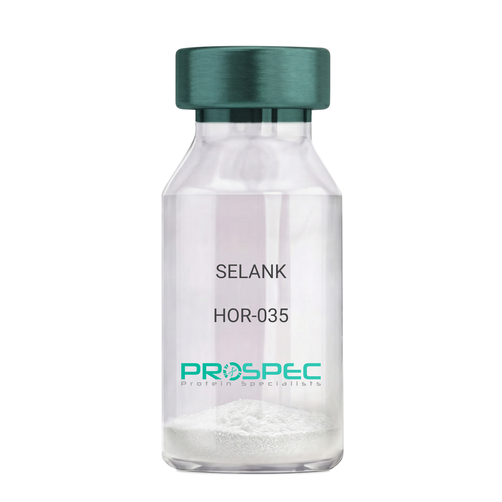
SELANK
Product # :
HOR-035Price :
Quantity :
Shipping Method :
Shipped at Room temp
More Info
- description
- formulation
- purity
- More Info
- Citations
Description
SELANK Synthetic is a single, non-glycosylated polypeptide chain containing 7 amino acids, having a molecular mass of 751.89 Dalton and a Molecular formula of C33H57N11O9.
Formulation
The protein was lyophilized with no additives.
Purity
Greater than 97.0% as determined by analysis by RP-HPLC.
Citations
More Info
-
Physical Appearance
Sterile Filtered White lyophilized (freeze-dried) powder.
-
Stability
Lyophilized SELANK although stable at room temperature for 3 weeks, should be stored desiccated below -18°C. Upon reconstitution SELANK should be stored at 4°C between 2-7 days and for future use below -18°C. For long term storage it is recommended to add a carrier protein (0.1% HSA or BSA). Please prevent freeze-thaw cycles.
-
Solubility
It is recommended to reconstitute the lyophilized SELANK in sterile 18MΩ-cm H2O not less than 100 µg/ml, which can then be further diluted to other aqueous solutions.
-
Amino Acid Sequence
H-Thr-Lys-Pro-Arg-Pro-Gly-Pro-OH.
-
Background
Selank, also known as TP-7, is a synthetic peptide that has garnered significant attention in the scientific community due to its potential anxiolytic, neuroprotective, and nootropic effects. This heptapeptide, derived from the naturally occurring human tetrapeptide Tuftsin, has been shown to possess a wide range of biological activities, including enhancing memory, reducing anxiety, and potentially mitigating the effects of neurodegenerative diseases.
Selank's primary mechanism of action involves its interaction with the GABAergic system, the body's main inhibitory neurotransmitter system. By modulating the activity of GABA receptors, Selank can exert a calming effect on the brain, reducing anxiety and enhancing cognitive function. Studies by Semenova et al. (2014) have demonstrated that Selank can enhance memory and learning in rats, suggesting potential applications in cognitive enhancement and the treatment of cognitive disorders.
In addition to its anxiolytic and nootropic effects, Selank has been shown to possess neuroprotective properties. Research by Ershov et al. (2016) found that Selank could protect neurons from oxidative stress, suggesting potential applications in the treatment of neurodegenerative diseases such as Alzheimer's and Parkinson's disease.
Given its anxiolytic, nootropic, and neuroprotective effects, Selank has been proposed as a potential therapeutic agent for a variety of conditions, including anxiety disorders, cognitive disorders, and neurodegenerative diseases. For instance, a study by Medvedev et al. (2017) found that Selank could improve symptoms in patients with generalized anxiety disorder, indicating its potential as a therapeutic agent in the treatment of anxiety disorders.
While research on Selank is promising, it is important to note that most studies have been conducted in animals or in vitro. More research is needed to fully understand the potential effects and applications of Selank in humans. However, the existing body of research suggests that Selank could be a promising tool in the treatment of anxiety disorders, cognitive disorders, and neurodegenerative diseases.
What is the molecular weight/Mw of SELANK Protein?
SELANK Protein has a total Mw of 0.751kDa.
What is the Purity of SELANK Protein?
SELANK Protein is >97% pure as determined by SDS-PAGE.
What is the Biological Activity of SELANK Protein?
The biological functionality of SELANK Protein will be determined in the future.
What is the amino acid sequence of SELANK Protein?
H-Thr-Lys-Pro-Arg-Pro-Gly-Pro-OH.
What applications can SELANK Protein be used in?
SELANK Protein can probably be used in western blot, ELISA and Lateral Flow.
What is the endotoxin level for SELANK Protein?
The endotoxin level is minimal, SELANK Protein was purified using conventional chromatography techniques.
Who supplies SELANK that is in stock and available to order?
ProSpec has the protein in stock and available for immediate delivery.
Where can I order bulk mg quantities of SELANK?
ProSpec supplies bulk mg quantities at affordable prices that meets research and academic budgets.
ProSpec's products are furnished for LABORATORY RESEARCH USE ONLY. The product may not be used as drugs, agricultural or pesticidal products, food additives or household chemicals.