Search results
1000 results found for “parvalbumin”
Name
Description
Product #
Price
Quantity
Shipping Method
- View Data Sheet
Name :
GREM1 HumanDescription:
GREM1 Human Recombinant
Gremlin-1 isoform 1, CKTSF1B1, DAND2, DRM, GREMLIN, IHG-2, PIG2, GREM1, Cell proliferation-inducing gene 2 protein, Cysteine knot superfamily 1, BMP antagonist 1, DAN domain family member 2, Down-regulated in Mos-transformed cells protein, Increased in high glucose protein 2.
Product # :
PRO-1359Price :
Quantity :
Shipping Method :
Shipped with Ice Packs
More Info
- description
- source
- formulation
- purity
- More Info
Description
GREM1 Human Recombinant produced in E.coli is a single, non-glycosylated polypeptide chain containing 183 amino acids (25-184) and having a molecular mass of 20.7 kDa. GREM1 is fused to a 23 amino acid His-tag at N-terminus & purified by proprietary chromatographic techniques.
Source
Escherichia Coli.
Formulation
The GREM1 solution (0.5mg/ml) contains 20mM Tris-HCl buffer (pH 8.0), 0.4M Urea and 10% glycerol.
Purity
Greater than 90.0% as determined by SDS-PAGE.
More Info
-
Introduction
GREM1 belongs to the BMP (bone morphogenic protein) antagonist family. Like BMPs, BMP antagonists comprise cystine knots and usually form homo- and heterodimers. The CAN (cerberus and dan) subfamily of BMP antagonists, to which GREM1 belongs, is characterized by a C-terminal cystine knot with an eight-membered ring. The antagonistic effect of the secreted glycosylated protein is because of its direct binding to BMP proteins. As an antagonist of BMP, GREM1 takes a place in regulating organogenesis, body patterning, and tissue differentiation. In mouse, GREM1 has been shown to convey the sonic hedgehog (SHH) signal from the polarizing region to the apical ectodermal ridge during limb bud outgrowth. Alternatively merged transcript variants encoding different isoforms have been found for this gene.
-
Synonyms
Gremlin-1 isoform 1, CKTSF1B1, DAND2, DRM, GREMLIN, IHG-2, PIG2, GREM1, Cell proliferation-inducing gene 2 protein, Cysteine knot superfamily 1, BMP antagonist 1, DAN domain family member 2, Down-regulated in Mos-transformed cells protein, Increased in high glucose protein 2.
-
Physical Appearance
Sterile Filtered colorless solution.
-
Stability
Store at 4°C if entire vial will be used within 2-4 weeks. Store, frozen at -20°C for longer periods of time. For long term storage it is recommended to add a carrier protein (0.1% HSA or BSA).Avoid multiple freeze-thaw cycles.
-
Amino Acid Sequence
MGSSHHHHHH SSGLVPRGSH MGSKKKGSQG AIPPPDKAQH NDSEQTQSPQ QPGSRNRGRG QGRGTAMPGE EVLESSQEAL HVTERKYLKR DWCKTQPLKQ TIHEEGCNSR TIINRFCYGQ CNSFYIPRHI RKEEGSFQSC SFCKPKKFTT MMVTLNCPEL QPPTKKKRVT RVKQCRCISI DLD.
ProSpec's products are furnished for LABORATORY RESEARCH USE ONLY. The product may not be used as drugs, agricultural or pesticidal products, food additives or household chemicals.
- View Data Sheet
Name :
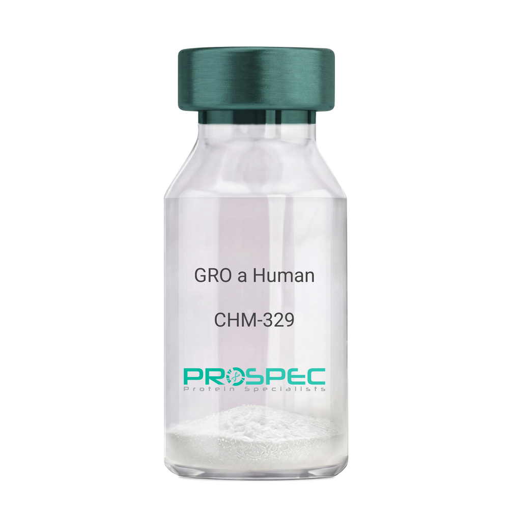
CXCL1 HumanDescription:
GRO-Alpha Human Recombinant (CXCL1)
Growth-regulated protein alpha, CXCL1, Melanoma growth stimulatory activity, MGSA, Neutrophil-activating protein 3, NAP-3, GRO-alpha (1-73), chemokine (C-X-C motif) ligand 1, GRO1, GROa, SCYB1, MGSA-a, MGSA alpha.
Product # :
CHM-329Price :
Quantity :
Shipping Method :
Shipped at Room temp
More Info
- description
- source
- formulation
- purity
- biological activity
- More Info
Description
GRO Alpha Human Recombinant produced in E.Coli is a single,non-glycosylated, polypeptide chain containing 73 amino acids and having a molecular mass of 7811 Dalton. The GRO-alpha is purified by proprietary chromatographic techniques.
Source
Escherichia Coli.
Formulation
Lyophilized from a 0.2µm filtered concentrated (1mg/ml) solution in 20mM PB, pH 7.4, 50mM NaCl.
Purity
Greater than 97.0% as determined by:
(a) Analysis by RP-HPLC.
(b) Analysis by SDS-PAGE.Biological Activity
Determined by its ability to chemoattract human peripheral blood neutrophils using a concentration range of 10.0-100.0 ng/ml.
More Info
-
Introduction
Chemokine (C-X-C motif) ligand 1 (CXCL1) is a small cytokine belonging to the CXC chemokine family that was previously called GRO1 oncogene, Neutrophil-activating protein 3 (NAP-3) and melanoma growth stimulating activity, alpha (MSGA-a). It is secreted by human melanoma cells, has mitogenic properties and is implicated in melanoma pathogenesis. CXCL1 is expressed by macrophages, neutrophilsand epithelial cells, and has neutrophil chemoattractant activity. CXCL1 plays a role in spinal cord development by inhibiting the migration of oligodendrocyte precursors and is involved in the processes of angiogenesis, inflammation, wound healing, and tumorigenesis. This chemokine elicits its effects by signaling through the chemokine receptor CXCR2. The gene for CXCL1 is located on human chromosome 4 amongst genes for other CXC chemokines.
-
Synonyms
Growth-regulated protein alpha, CXCL1, Melanoma growth stimulatory activity, MGSA, Neutrophil-activating protein 3, NAP-3, GRO-alpha (1-73), chemokine (C-X-C motif) ligand 1, GRO1, GROa, SCYB1, MGSA-a, MGSA alpha.
-
Physical Appearance
Sterile Filtered White lyophilized (freeze-dried) powder.
-
Stability
Lyophilized GRO-alpha although stable at room temperature for 3 weeks, should be stored desiccated below -18°C. Upon reconstitution CXCL1 should be stored at 4°C between 2-7 days and for future use below -18°C. For long term storage it is recommended to add a carrier protein (0.1% HSA or BSA).Please prevent freeze-thaw cycles.
-
Solubility
It is recommended to reconstitute the lyophilized CXCL1 in sterile 18MΩ-cm H2O not less than 100µg/ml, which can then be further diluted to other aqueous solutions.
-
Amino Acid Sequence
The sequence of the first five N-terminal amino acids was determined and was found to be Ala-Ser-Val-Ala-Thr.
-
Background
What is the molecular weight/Mw of CXCL1 HUMAN Protein?
CXCL1 HUMAN Protein has a total Mw of 7.81kDa.
What is the source or expression system of CXCL1 HUMAN Protein?
Escherichia Coli.
What is the Purity of CXCL1 HUMAN Protein?
CXCL1 HUMAN Protein is >97% pure as determined by SDS-PAGE.
What is the Biological Activity of CXCL1 HUMAN Protein?
Determined by its ability to chemoattract human peripheral blood neutrophils using a concentration range of 10.0-100.0 ng/ml.
What is the amino acid sequence of CXCL1 HUMAN Protein?
The sequence of the first five N-terminal amino acids was determined and was found to be Ala-Ser-Val-Ala-Thr.
What applications can CXCL1 HUMAN Protein be used in?
CXCL1 HUMAN Protein can probably be used in western blot, ELISA and Lateral Flow.
What is the endotoxin level for CXCL1 HUMAN Protein?
The endotoxin level is minimal, CXCL1 HUMAN Protein was purified using conventional chromatography techniques.
ProSpec's products are furnished for LABORATORY RESEARCH USE ONLY. The product may not be used as drugs, agricultural or pesticidal products, food additives or household chemicals.
- View Data Sheet
Name :
PSMD9 HumanDescription:
Proteasome 26S Subunit, Non-ATPase 9 Human Recombinant
p27, Rpn4, 26S proteasome non-ATPase regulatory subunit 9, 26S proteasome regulatory subunit p27, PSMD9.
Product # :
ENZ-666Price :
Quantity :
Shipping Method :
Shipped with Ice Packs
More Info
- description
- source
- formulation
- purity
- More Info
Description
PSMD9 Human Recombinant produced in E.coli is a single, non-glycosylated polypeptide chain containing 246 amino acids (1-223) and having a molecular mass of 27.1kDa. PSMD9 is fused to a 23 amino acid His-tag at N-terminus & purified by proprietary chromatographic techniques.
Source
Escherichia Coli.
Formulation
The PSMD9 solution (1mg/ml) contains 20mM Tris-HCl buffer (pH 8.0), 0.15M NaCl, 10% glycerol and 1mM DTT.
Purity
Greater than 90.0% as determined by SDS-PAGE.
More Info
-
Introduction
PSMD9 is a multicatalytic proteinase complex with a highly ordered structure composed of 2 complexes, a 20S core and a 19S regulator. PSMD9 acts as a chaperone through the assembly of the 26S proteasome, specifically of the base subcomplex of the PA700/19S regulatory complex (RC). During the base subcomplex compilation, PSMD9 is part of an intermediate PSMD9:PSMC6:PSMC3 module, also called modulator trimer complex. PSMD9 is released during the further base assembly process.
-
Synonyms
p27, Rpn4, 26S proteasome non-ATPase regulatory subunit 9, 26S proteasome regulatory subunit p27, PSMD9.
-
Physical Appearance
Sterile Filtered colorless solution.
-
Stability
Store at 4°C if entire vial will be used within 2-4 weeks.Store, frozen at -20°C for longer periods of time. For long term storage it is recommended to add a carrier protein (0.1% HSA or BSA).Avoid multiple freeze-thaw cycles.
-
Amino Acid Sequence
MGSSHHHHHH SSGLVPRGSH MGSMSDEEAR QSGGSSQAGV VTVSDVQELM RRKEEIEAQI KANYDVLESQ KGIGMNEPLV DCEGYPRSDV DLYQVRTARH NIICLQNDHK AVMKQVEEAL HQLHARDKEK QARDMAEAHK EAMSRKLGQS ESQGPPRAFA KVNSISPGSP ASIAGLQVDD EIVEFGSVNT QNFQSLHNIG SVVQHSEGKP LNVTVIRRGE KHQLRLVPTR WAGKGLLGCN IIPLQR.
ProSpec's products are furnished for LABORATORY RESEARCH USE ONLY. The product may not be used as drugs, agricultural or pesticidal products, food additives or household chemicals.
- View Data Sheet
Name :
MAP1LC3A HumanDescription:
Microtubule-Associated Protein 1 Light Chain 3 Alpha Human Recombinant
ATG8E, LC3, LC3A, MAP1ALC3, MAP1BLC3, MLP3A.
Product # :
PRO-495Price :
Quantity :
Shipping Method :
Shipped with Ice Packs
More Info
- description
- source
- formulation
- purity
- More Info
Description
MAP1LC3A Recombinant Human produced in E.Coli is a single, non-glycosylated polypeptide chain containing 128 amino acids (1-120 a.a.) and having a molecular mass of 15.1 kDa. The MAP1LC3A is fused to 8 amino acid His-Tag at C-terminus and purified by proprietary chromatographic techniques.
Source
Escherichia Coli.
Formulation
The MAP1LC3A solution (1mg/ml) contains 20mM Tris-HCl pH-8.0, 1mM DTT, 0.1M NaCl & 20% glycerol.
Purity
Greater than 95.0% as determined by SDS-PAGE.
More Info
-
Introduction
MAP1LC3A takes part in formation of autophagosomal vacuoles and is one of the light chain subunits that functions together with both MAP1A and/or MAP1B. MAP1LC3A has an important part in neuronal development and in maintaining the balance between neuronal plasticity and rigidity. MAP1LC3A is expressed as two alternatively spliced isoforms that are expressed in testis, brain, heart, liver and skeletal muscle, but are absent in thymus and peripheral blood leukocytes.
-
Synonyms
ATG8E, LC3, LC3A, MAP1ALC3, MAP1BLC3, MLP3A.
-
Physical Appearance
Sterile filtered colorless solution.
-
Stability
Store at 4°C if entire vial will be used within 2-4 weeks. Store, frozen at -20°C for longer periods of time. For long term storage it is recommended to add a carrier protein (0.1% HSA or BSA).Avoid multiple freeze-thaw cycles.
-
Amino Acid Sequence
MPSDRPFKQR RSFADRCKEV QQIRDQHPSK IPVIIERYKG EKQLPVLDKT KFLVPDHVNM SELVKIIRRR LQLNPTQAFF LLVNQHSMVS VSTPIADIYE QEKDEDGFLY MVYASQETFG LEHHHHHH.
ProSpec's products are furnished for LABORATORY RESEARCH USE ONLY. The product may not be used as drugs, agricultural or pesticidal products, food additives or household chemicals.
- View Data Sheet
Name :
Visfatin HumanDescription:
Visfatin Human Recombinant
PBEF, Pre-B cell colony-enhancing factor, Nicotinamide phosphoribosyltransferase NAmPRTase, Nampt, MGC117256, DKFZP666B131, 1110035O14Rik.
Product # :
CYT-318Price :
Quantity :
Shipping Method :
Shipped at Room temp
More Info
- description
- source
- formulation
- purity
- biological activity
- More Info
Description
Visfatin Human Recombinant produced in E.Coli is a single, non-glycosylated, polypeptide chain containing 466 amino acids. The total molecular mass is 52.6kDa (calculated). The Visfatin is purified by Flag-affinity chromatography.
Source
Escherichia Coli.
Formulation
Visfatin was lyophilized with no additives.
Purity
Greater than 90.0% as determined by SDS-PAGE.
Biological Activity
The activity is determined by its ability to induce IL-6, IL-1 beta and TNF alpha production from human PBMCs at 100ng/ml.More Info
-
Introduction
Excess adiposity is the most important risk in the development of type 2 diabetes mellitus (T2DM). Adipose tissue produces several proteins (adipocytokines) such as leptin, adiponectin, resistin, tumor necrosis factor-a, and IL-6, that modulate sensitivity and appear to play an important role in the pathogenesis, diabetes, dyslipidemia, inflammation, and atherosclerosis. Visfatin, also known as pre-B cell colony-enhancing factor (PBEF), is a cytokine that is highly expressed in visceral fat and was originally isolated as a secreted factor that synergizes with IL-7 and stem cell factors to promote the growth of B cell precursors. Visfatin homologs have been identified in carp, invertebrate mollusks, and bacteria, as well as in vertebrates, including humans and the mouse. It has been postulated to play a role in innate immunity.
Visfatin exerts mimetic effects that are dose-dependent and quantitatively similar to stimulating muscle and adipocyte glucose transport, and in inhibiting hepatocyte glucose production. Intravenous injection of recombinant visfatin in mice decreased plasma glucose in a dose-dependent fashion. In keeping with its mimetic effects, visfatin was as effective in reducing hyperglycemia in deficient diabetic mice. Visfatin was also found to be bound to and activate receptor, causing receptor phosphorylation and the activation of downstream signaling molecules. However, visfatin did not compete for binding to the receptor, indicating that the two proteins were recognized by different regions of the receptor. Thus, visfatin might play a role in glucose homeostasis and dysregulation in biosynthesis or signal transduction, and might contribute to the pathogenesis of diabetes. -
Synonyms
PBEF, Pre-B cell colony-enhancing factor, Nicotinamide phosphoribosyltransferase NAmPRTase, Nampt, MGC117256, DKFZP666B131, 1110035O14Rik.
-
Physical Appearance
Filtered White lyophilized (freeze-dried) powder.
-
Stability
Lyophilized Visfatin although stable at room temperature for 3 weeks, should be stored desiccated below -18°C. Upon reconstitution Visfatin should be stored at 4°C between 2-7 days and for future use below -18°C.For long term storage it is recommended to add a carrier protein (0.1% HSA or BSA).Please prevent freeze-thaw cycles.
-
Solubility
Centrifuge vial before opening. When reconstituting the product, gently pipet and wash down the sides of the vial to ensure full recovery of the protein into solution. It is recommended to reconstitute the lyophilized product with 20 mM HCl at a concentration of 0.1 mg/mL, which can be further diluted into other aqueous solutions. Wait several minutes for full reconstitution and solubility.
-
Amino Acid Sequence
MPPNTSKVYS YFECREKKTE NSKLRKVKYE ETVFYGLQYI LNKYLKGKVV TKEKIQEAKD VYKEHFQDDV FNEKGWNYIL EKYDGHLPIE IKAVPEGFVI PRGNVLFTVE NTDPECYWLT NWIETILVQS WYPITVATNS REQKKILAKY LLETSGNLDG LEYKLHDFGY RGVSSQETAG IGASAHLVNF KGTDTVAGLA LIKKYYGTKD PVPGYSVPAA EHSTITAWGK DHEKDAFEHI VTQFSSVPVS VVSDSYDIYN ACEKIWGEDL RHLIVSRSTQ APLIIRPDSG NPLDTVLKVL EILGKKFPVT ENSKGYKLLP PYLRVIQGDG VDINTLQEIV EGMKQKMWSI ENIAFGSGGG LLQKLTRDLL NCSFKCSYVV TNGLGINVFK DPVADPNKRS KKGRLSLHRT PAGNFVTLEE GKGDLEEYGQ DLLHTVFKNG KVTKSYSFDE IRKNAQLNIE LEAAHH.
-
Background
About Visfatin Human
Visfatin is a cytokine expressed in visceral fat that was originally isolated as a secreted
element that synergized with stem cell factors and IL-7. One of its main functions is to
enhance the development of B cell precursors.The cytokine is also known as the “Pre-B Cell Colony-Enhancing Factor (PBEF).” It has been
identified in vertebrates, including mice and humans, and it’s being studied due to its link
to inflammatory conditions, beta cell function, and cardiovascular disease.
What’s the Function of Visfatin Human Recombinant?Visfatin human recombinant is produced in E. Coli. It’s a single, non-glycosylated,
polypeptide chain that contains 466 amino acids, it’s purified by FLAG-affinity
chromatography, and it contains a total molecular mass of 52.6 kDa.
What Are the Main Applications of Visfatin Human Recombinant?The cytokine is being researched because of its involvement in glucose homeostasis,
dysregulation in biosynthesis and signal transduction, and the pathogenesis of diabetes.
Visfatin human recombinant is tailored exclusively for laboratory research, ensuring
experts can get further answers regarding the cytokine’s involvement in different
processes, including pathogenesis, diabetes, inflammation, dyslipidemia, and
atherosclerosis.Findings can also help during the identification of high-risk people for cardiovascular
disease and diabetes.
ProSpec's products are furnished for LABORATORY RESEARCH USE ONLY. The product may not be used as drugs, agricultural or pesticidal products, food additives or household chemicals.
- View Data Sheet
Name :
ZNHIT1 HumanDescription:
Zinc Finger HIT-Type Containing 1 Human Recombinant
Zinc Finger HIT-Type Containing 1, ZNHIT1, ZNFN4A1, Zinc Finger Protein, Subfamily 4A (HIT Domain Containing) Member 1, Zinc Finger HIT Domain Containing 1, Putative Cyclin G1 Interacting Protein, Cyclin-G1-Binding Protein 1, Zinc Finger Protein Subfamily 4A Member 1, P18 Hamlet, CG1I, H_DJ0747G18.14, p18Hamlet, Zinc Finger HIT Domain-Containing Protein 1, Zinc Finger HIT Type 1, CGBP1.
Product # :
PRO-1448Price :
Quantity :
Shipping Method :
Shipped with Ice Packs
More Info
- description
- source
- formulation
- purity
- More Info
Description
ZNHIT1 Human Recombinant produced in E.coli is a single, non-glycosylated polypeptide chain containing 177 amino acids (1-154) and having a molecular mass of 19.9kDa. ZNHIT1 is fused to a 23 amino acid His-tag at N-terminus.
Source
Escherichia Coli.
Formulation
The ZNHIT1 solution (1mg/ml) contains 20mM Tris-HCl buffer (pH 8.0), 10% glycerol and 0.4M Urea.
Purity
Greater than 95.0% as determined by SDS-PAGE.
More Info
-
Introduction
Zinc Finger HIT-Type Containing 1 (ZNHIT1) is a member of the ZNHIT1 family and contains 1 HIT-type zinc finger. The ZNHIT1 protein is induced by DNA damage and appears to have a role in p53-mediated apoptosis induction. ZNHIT1 interacts with MAPK11 and MAPK14 and is a component of the chromatin-remodeling SRCAP complex composed of at least SRCAP, DMAP1, RUVBL1, RUVBL2, ACTL6A, YEATS4, ACTR6 and ZNHIT1. ZNHIT1 binds to NR1D2 and discharges it of its inhibitory effect on the transcription of APOC3 without affecting its DNA-binding activity.
-
Synonyms
Zinc Finger HIT-Type Containing 1, ZNHIT1, ZNFN4A1, Zinc Finger Protein, Subfamily 4A (HIT Domain Containing) Member 1, Zinc Finger HIT Domain Containing 1, Putative Cyclin G1 Interacting Protein, Cyclin-G1-Binding Protein 1, Zinc Finger Protein Subfamily 4A Member 1, P18 Hamlet, CG1I, H_DJ0747G18.14, p18Hamlet, Zinc Finger HIT Domain-Containing Protein 1, Zinc Finger HIT Type 1, CGBP1.
-
Physical Appearance
Sterile Filtered colorless solution.
-
Stability
Store at 4°C if entire vial will be used within 2-4 weeks. Store, frozen at -20°C for longer periods of time. For long term storage it is recommended to add a carrier protein (0.1% HSA or BSA).Avoid multiple freeze-thaw cycles.
-
Amino Acid Sequence
MGSSHHHHHH SSGLVPRGSH MGSMVEKKTS VRSQDPGQRR VLDRAARQRR INRQLEALEN DNFQDDPHAG LPQLGKRLPQ FDDDADTGKK KKKTRGDHFK LRFRKNFQAL LEEQNLSVAE GPNYLTACAG PPSRPQRPFC AVCGFPSPYT CVSCGARYCT VRCLGTHQET RCLKWTV.
ProSpec's products are furnished for LABORATORY RESEARCH USE ONLY. The product may not be used as drugs, agricultural or pesticidal products, food additives or household chemicals.
- View Data Sheet
Name :
TIFA HumanDescription:
TRAF-Interacting Protein with Forkhead-Associated Domain Human Recombinant
TRAF-interacting protein with FHA domain-containing protein A, Putative MAPK-activating protein PM14, Putative NF-kappa-B-activating protein 20, TRAF2-binding protein, TIFA, T2BP, T6BP, TIFAA.
Product # :
PRO-1041Price :
Quantity :
Shipping Method :
Shipped with Ice Packs
More Info
- description
- source
- formulation
- purity
- More Info
Description
TIFA Human Recombinant produced in E.Coli is a single, non-glycosylated polypeptide chain containing 208 amino acids (1-184 a.a.) and having a molecular mass of 24kDa.TIFA is fused to a 24 amino acid His-tag at N-terminus & purified by proprietary chromatographic techniques.
Source
Escherichia Coli.
Formulation
TIFA protein solution (0.5mg/ml) containing 20mM Tris-HCl buffer (pH8.0), 20% glycerol and 1mM DTT.
Purity
Greater than 90.0% as determined by SDS-PAGE.
More Info
-
Introduction
TRAF-interacting protein with FHA domain-containing protein A (TIFA) is an adapter protein that mediates the IRAK1 and TRAF6 interaction following IL-1 stimulation, triggering the downstream activation of NF-kappa-B and AP-1 pathways. The TIFA protein stimulates the oligomerization and polyubiquitination of TRAF6, leading to the activation of TAK1 and IKK through a proteasome-independent mechanism.
-
Synonyms
TRAF-interacting protein with FHA domain-containing protein A, Putative MAPK-activating protein PM14, Putative NF-kappa-B-activating protein 20, TRAF2-binding protein, TIFA, T2BP, T6BP, TIFAA.
-
Physical Appearance
Sterile Filtered clear solution.
-
Stability
Store at 4°C if entire vial will be used within 2-4 weeks. Store, frozen at -20°C for longer periods of time. For long term storage it is recommended to add a carrier protein (0.1% HSA or BSA).Avoid multiple freeze-thaw cycles.
-
Amino Acid Sequence
MGSSHHHHHH SSGLVPRGSH MGSHMTSFED ADTEETVTCL QMTVYHPGQL QCGIFQSISF NREKLPSSEV VKFGRNSNIC HYTFQDKQVS RVQFSLQLFK KFNSSVLSFE IKNMSKKTNL IVDSRELGYL NKMDLPYRCM VRFGEYQFLM EKEDGESLEF FETQFILSPR SLLQENNWPP HRPIPEYGTY SLCSSQSSSP TEMDENES.
ProSpec's products are furnished for LABORATORY RESEARCH USE ONLY. The product may not be used as drugs, agricultural or pesticidal products, food additives or household chemicals.
- View Data Sheet
Name :
KLK3 ProteinDescription:
Kallikrein-3 Recombinant Human
Prostate-specific antigen, PSA, Gamma-seminoprotein, Seminin, Kallikrein-3, P-30 antigen, Semenogelase, KLK3, APS, hK3, KLK2A1.
Product # :
ENZ-1102Price :
Quantity :
Shipping Method :
Shipped at Room temp
More Info
- description
- source
- formulation
- purity
- More Info
Description
Kallikrein-3 Human Recombinant produced in E.Coli is a single, non- glycosylated polypeptide chain containing 237 amino acids and having a molecular mass of 26.1kDa.KLK3 is purified by proprietary chromatographic techniques.
Source
Escherichia Coli.
Formulation
Lyophilized from a 0.2μm filtered concentrated solution in 20mM Tris-HCl, pH 8.0, 150mM NaCl and 3% trehalose.
Purity
Greater than 95.0% as determined by:
(a) Analysis by RP-HPLC.
(b) Analysis by SDS-PAGE.More Info
-
Introduction
Kallikrein-3 (KLK3) is a part of the kallikrein-related peptidase family. Kallikreins are a subgroup of serine proteases having various physiological functions. Numerous kallikreins take part in carcinogenesis and some may be prospective cancer and other disease biomarkers. Kallikrein-3 is 1 of the 15 kallikrein subfamily members located in a cluster on chromosome 19 and is a protease present in seminal plasma. KLK3 hydrolyzes semenogelin-1 consequently leading to the liquefaction of the seminal coagulum. KLK3 acts normally in the liquefaction of seminal coagulum, probably by hydrolysis of the high molecular mass seminal vesicle protein. Serum level of the KLK3 protein, called PSA in the clinical setting, is beneficial in the diagnosis and monitoring of prostatic carcinoma.
-
Synonyms
Prostate-specific antigen, PSA, Gamma-seminoprotein, Seminin, Kallikrein-3, P-30 antigen, Semenogelase, KLK3, APS, hK3, KLK2A1.
-
Physical Appearance
Sterile Filtered White lyophilized (freeze-dried) powder.
-
Stability
Lyophilized KLK3 although stable at room temperature for 3 weeks, should be stored desiccated below -18°C. Upon reconstitution Kallikrein-3 should be stored at 4°C between 2-7 days and for future use below -18°C.Please prevent freeze-thaw cycles.
-
Solubility
It is recommended to reconstitute the lyophilized Kallikrein-3 in sterile 18MΩ-cm H2O not less than 100µg/ml, which can then be further diluted to other aqueous solutions.
-
Amino Acid Sequence
IVGGWECEKH SQPWQVLVAS RGRAVCGGVL VHPQWVLTAA HCIRNKSVIL LGRHSLFHPE DTGQVFQVSH SFPHPLYDMS LLKNRFLRPG DDSSHDLMLL RLSEPAELTDA VKVMDLPTQE PALGTTCYAS GWGSIEPEEF LTPKKLQCVD LHVISNDVCA QVHPQKVTKF MLCAGRWTGG KSTCSGDSGG PLVCNGVLQG ITSWGSEPCA LPERPSLYTK VVHYRKWIKD TIVANP.
ProSpec's products are furnished for LABORATORY RESEARCH USE ONLY. The product may not be used as drugs, agricultural or pesticidal products, food additives or household chemicals.
- View Data Sheet
Name :
AMTN HumanDescription:
Amelotin Human Recombinant
UNQ689, Amelotin, PRO1329.
Product # :
PRO-1301Price :
Quantity :
Shipping Method :
Shipped with Ice Packs
More Info
- description
- source
- formulation
- purity
- More Info
Description
AMTN Human Recombinant produced in E.Coli is a single, non-glycosylated polypeptide chain containing 216 amino acids (17-209 a.a.) and having a molecular mass of 22.2kDa.AMTN is fused to a 23 amino acid His-tag at N-terminus & purified by proprietary chromatographic techniques.
Source
Escherichia Coli.
Formulation
AMTN protein solution (0.5mg/ml) contains mM Tris-HCl buffer (pH 8.0), 0.2M NaCl, 20% glycerol and 2mM DTT.
Purity
Greater than 90.0% as determined by SDS-PAGE.
More Info
-
Introduction
AMTN is a member of the amelotin family. AMTN is a lately discovered secreted enamel protein. AMTN mainly expressed during the maturation stage of enamel formation. AMTN gathers in a basal lamina-like structure at the interface between ameloblasts and enamel mineral and it co-localizes with one more lately described enamel protein, odontogenic ameloblast- related protein (ODAM(.
-
Synonyms
UNQ689, Amelotin, PRO1329.
-
Physical Appearance
Sterile Filtered clear solution.
-
Stability
Store at 4°C if entire vial will be used within 2-4 weeks. Store, frozen at -20°C for longer periods of time. For long term storage it is recommended to add a carrier protein (0.1% HSA or BSA).Avoid multiple freeze-thaw cycles.
-
Amino Acid Sequence
MGSSHHHHHH SSGLVPRGSH MGSLPQLKPA LGLPPTKLAP DQGTLPNQQQ SNQVFPSLSL IPLTQMLTLG PDLHLLNPAA GMTPGTQTHP LTLGGLNVQQ QLHPHVLPIF VTQLGAQGTI LSSEELPQIF TSLIIHSLFP GGILPTSQAG ANPDVQDGSL PAGGAGVNPA TQGTPAGRLP TPSGTDDDFA VTTPAGIQRS THAIEEATTE SANGIQ.
ProSpec's products are furnished for LABORATORY RESEARCH USE ONLY. The product may not be used as drugs, agricultural or pesticidal products, food additives or household chemicals.
- View Data Sheet
Name :
HIV Type-O gp41 13kDaDescription:
HIV Type-O gp41 13kDa Recombinant
Product # :
HIV-007Price :
Quantity :
Shipping Method :
Shipped at Room temp
More Info
- description
- source
- formulation
- purity
- More Info
Description
Recombinant HIV-1 gp41 Type-O produced in E.coli is a non-glycosylated polypeptide chain having a molecular mass of 13kDa and fused to a His tag at N-terminus.
Source
Escherichia Coli.
Formulation
Lyophilized from 1mg/ml in 20mM Na-carbonate, pH 9.6.
Purity
Greater than 95.0% as determined by SDS-PAGE.
More Info
-
Introduction
Human immunodeficiency virus (HIV) is a retrovirusthat can lead to a condition in which the immune systembegins to fail, leading to opportunistic infections. HIV primarily infects vital cells in the humanimmune systemsuch as helper T cells(specifically CD4+ T cells), macrophagesand dendritic cells. HIV infection leads to low levels of CD4+ T cells through three main mechanisms: firstly, direct viral killing of infected cells; secondly, increased rates of apoptosisin infected cells; and thirdly, killing of infected CD4+ T cells by CD8 cytotoxic lymphocytesthat recognize infected cells. When CD4+ T cell numbers decline below a critical level, cell-mediated immunityis lost, and the body becomes progressively more susceptible to opportunistic infections. HIV was classified as a member of the genus Lentivirus, part of the family of Retroviridae. Lentiviruses have many common morphologies and biological properties. Many species are infected by lentiviruses, which are characteristically responsible for long-duration illnesses with a long incubation period. Lentiviruses are transmitted as single-stranded, positive-sense, enveloped RNA viruses. Upon entry of the target cell, the viral RNA genomeis converted to double-stranded DNAby a virally encoded reverse transcriptasethat is present in the virus particle. This viral DNA is then integrated into the cellular DNA by a virally encoded integraseso that the genome can be transcribed. Once the virus has infected the cell, two pathways are possible: either the virus becomes latentand the infected cell continues to function, or the virus becomes active and replicates, and a large number of virus particles are liberated that can then infect other cells.
-
Physical Appearance
Sterile Filtered White lyophilized (freeze-dried) powder.
-
Stability
HIV Type-O gp41 although stable at room temperature for 4 weeks, should be stored below -18°C. Please prevent freeze thaw cycles.
-
Solubility
It is recommended to reconstitute the lyophilized HIV Type-O gp41 in sterile 18M-cm H2O not less than 100µg/ml, which can then be further diluted to other aqueous solutions.
-
Amino Acid Sequence
MGHHHHHHGSVQTHTLLKGIVQQQDNLLRAIQAQQHLLRLSVWGIRQLRARLLALETLI QNQQLLNLWGAKGRLIAYTSVKWNTTWGGGGSIWGNLTWQEWDQQIDNVSSIIYEEIQ KAQDQQEQNEKKLLELDE.
ProSpec's products are furnished for LABORATORY RESEARCH USE ONLY. The product may not be used as drugs, agricultural or pesticidal products, food additives or household chemicals.
- View Data Sheet
Name :
FGF 2 Human (147 a.a.)Description:
Fibroblast Growth Factor Basic 147 a.a. Human Recombinant
Prostatropin, HBGH-2, HBGF-2, FGF-2, FGF-b.
Product # :
CYT-557Price :
Quantity :
Shipping Method :
Shipped at Room temp
More Info
- description
- source
- formulation
- purity
- biological activity
- More Info
Description
Fibroblast Growth Factor-2 Human Recombinant (FGF-2) produced in E.Coli is a single, non-glycosylated, polypeptide chain containing 147 amino acids and having a molecular mass of 16.5kDa. The FGF2 is purified by proprietary chromatographic techniques.
Source
Escherichia Coli.
Formulation
The bFGF was lyophilized from a sterile filtered solution containing 20mM Tris-HCl, pH 7.6 and 150mM NaCl.
Purity
Greater than 98.0% as determined by SDS-PAGE.
Biological Activity
The ED50, Calculated by the dose- dependent proliferation of mouse BALB/c 3T3 cells is < 0.05ng/ml corresponding to a specific activity of 2.0x10⁷ units/mg.
More Info
-
Introduction
FGF-basic is a member of the fibroblast growth factor (FGF) family. FGF family members possess broad mitogenic and cell survival activities, and are involved in a variety of biological processes, including embryonic development, cell growth, morphogenesis, tissue repair, tumor growth and invasion. This protein functions as a modifier of endothelial cell migration and proliferation, as well as an angiogenic factor. It acts as a mitogen for a variety of mesoderm- and neuroectoderm-derived cells in vitro, thus is thought to be involved in organogenesis. Three alternatively spliced variants encoding different isoforms have been described. The heparin-binding growth factors are angiogenic agents in vivo and are potent mitogens for a variety of cell types in vitro. There are differences in the tissue distribution and concentration of these 2 growth factors.
-
Synonyms
Prostatropin, HBGH-2, HBGF-2, FGF-2, FGF-b.
-
Physical Appearance
Sterile Filtered White lyophilized (freeze-dried) powder.
-
Stability
Lyophilized basic-FGF although stable at room temperature for 3 weeks, should be stored desiccated below -18°C. Upon reconstitution FGFb should be stored at 4°C between 2-7 days and for future use below -18°C.For long term storage it is recommended to add a carrier protein (0.1% HSA or BSA).Please prevent freeze-thaw cycles.
-
Solubility
It is recommended to reconstitute the lyophilized FGF-B in sterile 18MΩ-cm H2O not less than 100µg/ml, which can then be further diluted to other aqueous solutions.
-
Amino Acid Sequence
MPALPEDGGS GAFPPGHFKD PKRLYCKNGG FFLRIHPDGR VDGVREKSDP HIKLQLQAEE RGVVSIKGVC ANRYLAMKED GRLLASKCVT DECFFFERLE SNNYNTYRSR KYTSWYVALK RTGQYKLGSK TGPGQKAILF LPMSAKS.
-
Background
What is the molecular weight/Mw of FGF 2 Protein?
FGF 2 Protein has a total Mw of 16.5kDa.
What is the source or expression system of FGF 2 Protein?
Escherichia Coli.
What is the Purity of FGF 2 Protein?
FGF 2 Protein is >98% pure as determined by SDS-PAGE.
What is the Biological Activity of FGF 2 Protein?
The ED50, Calculated by the dose- dependent proliferation of mouse BALB/c 3T3 cells is < 0.05ng/ml corresponding to a specific activity of 2.0x10⁷ units/mg.
What is the amino acid sequence of FGF 2 Protein?
MPALPEDGGS GAFPPGHFKD PKRLYCKNGG FFLRIHPDGR VDGVREKSDP HIKLQLQAEE RGVVSIKGVC ANRYLAMKED GRLLASKCVT DECFFFERLE SNNYNTYRSR KYTSWYVALK RTGQYKLGSK TGPGQKAILF LPMSAKS.
What applications can FGF 2 Protein be used in?
FGF 2 Protein can probably be used in western blot, ELISA and Lateral Flow.
What is the endotoxin level for FGF 2 Protein?
The endotoxin level is minimal, FGF 2 Protein was purified using conventional chromatography techniques.
ProSpec's products are furnished for LABORATORY RESEARCH USE ONLY. The product may not be used as drugs, agricultural or pesticidal products, food additives or household chemicals.
- View Data Sheet
Name :
APP Human, HEKDescription:
Amyloid beta (A4) Precursor Protein Human Recombinant, HEK
ABPP, APPI, Alzheimer disease amyloid A4 protein homolog, Alzheimer disease amyloid protein, Amyloid precursor protein, Amyloid-beta precursor protein, Amyloid-beta A4 protein, Cerebral vascular amyloid peptide, CVAP, PreA4, Protease nexin-II, PN-II, A4, AD1, beta-amyloid peptide, beta-amyloid precursor protein, testicular tissue protein Li 2, AAA, ABETA, alpha-sApp, CTF gamma, PN2.
Product # :
PRO-2777Price :
Quantity :
Shipping Method :
Shipped with Ice Packs
More Info
- description
- source
- formulation
- purity
- biological activity
- More Info
Description
APP Human Recombinant is a single, glycosylated, polypeptide chain (18-701 a.a) containing a total of 690 amino acids, having a molecular mass of 78.2 kDa. APP is fused to a 6 amino acid His-tag at C-terminus and is purified by proprietary chromatographic techniques.
Source
HEK293 Cells.
Formulation
The APP solution (0.5mg/ml) contains 10% Glycerol and Phosphate-Buffered Saline (pH 7.4).
Purity
> 90% by SDS-PAGE.
Biological Activity
The inhibitory function of APP on activity of trypsin was measured by a fluorometric
assay using Mca-RPKPVE-Nval-WRK(Dnp)-NH2 at pH 7.5 at 37C. The IC50 ≤ 1 nM.
More Info
-
Synonyms
ABPP, APPI, Alzheimer disease amyloid A4 protein homolog, Alzheimer disease amyloid protein, Amyloid precursor protein, Amyloid-beta precursor protein, Amyloid-beta A4 protein, Cerebral vascular amyloid peptide, CVAP, PreA4, Protease nexin-II, PN-II, A4, AD1, beta-amyloid peptide, beta-amyloid precursor protein, testicular tissue protein Li 2, AAA, ABETA, alpha-sApp, CTF gamma, PN2.
-
Physical Appearance
Sterile Filtered colorless solution.
-
Stability
Store at 4°C if entire vial will be used within 2-4 weeks. Store, frozen at -20°C for longer periods of time. For long term storage it is recommended to add a carrier protein (0.1% HSA or BSA). Avoid multiple freeze-thaw cycles.
-
Amino Acid Sequence
LEVPTDGNAG LLAEPQIAMF CGRLNMHMNV QNGKWDSDPS GTKTCIDTKE GILQYCQEVY PELQITNVVE ANQPVTIQNW CKRGRKQCKT HPHFVIPYRC LVGEFVSDAL LVPDKCKFLH QERMDVCETH LHWHTVAKET CSEKSTNLHD YGMLLPCGID KFRGVEFVCC PLAEESDNVD SADAEEDDSD VWWGGADTDY ADGSEDKVVE VAEEEEVAEV EEEEADDDED DEDGDEVEEE AEEPYEEATE RTTSIATTTT TTTESVEEVV REVCSEQAET GPCRAMISRW YFDVTEGKCA PFFYGGCGGN RNNFDTEEYC MAVCGSAMSQ SLLKTTQEPL ARDPVKLPTT AASTPDAVDK YLETPGDENE HAHFQKAKER LEAKHRERMS QVMREWEEAE RQAKNLPKAD KKAVIQHFQE KVESLEQEAA NERQQLVETH MARVEAMLND RRRLALENYI TALQAVPPRP RHVFNMLKKY VRAEQKDRQH TLKHFEHVRM VDPKKAAQIR SQVMTHLRVI YERMNQSLSL LYNVPAVAEE IQDEVDELLQ KEQNYSDDVL ANMISEPRIS YGNDALMPSL TETKTTVELL PVNGEFSLDD LQPWHSFGAD SVPANTENEV EPVDARPAAD RGLTTRPGSG LTNIKTEEIS EVKMDAEFRH DSGYEVHHQK LVFFAEDVGS NKGA HHHHHH.
-
Background
Alzheimer's disease (AD) is a neurodegenerative disorder characterized by the accumulation of amyloid plaques and neurofibrillary tangles in the brain, leading to cognitive decline and memory loss. The amyloid beta (Aβ) peptide, derived from the amyloid beta (A4) precursor protein, has been identified as a key player in the pathogenesis of AD. This research aims to explore the significance of the amyloid beta precursor protein, its processing, and the implications it holds for understanding and treating Alzheimer's disease.
The amyloid beta precursor protein (APP) is a transmembrane protein widely expressed in various tissues, with higher concentrations found in the brain. APP undergoes sequential proteolytic processing by enzymes known as secretases, leading to the generation of Aβ peptides of different lengths. Of particular importance is the production of the Aβ42 peptide, which has a propensity to aggregate and form the characteristic amyloid plaques in AD.
Understanding the processing and metabolism of APP is crucial for unraveling the mechanisms underlying AD pathology. Mutations in the APP gene and dysregulation of its processing have been associated with familial forms of AD, highlighting the pivotal role of APP in disease development. Investigating the function of APP and its proteolytic fragments can provide valuable insights into the molecular events leading to AD and potentially lead to the identification of therapeutic targets.
This research will delve into the processing of the amyloid beta precursor protein, shedding light on the different cleavage pathways mediated by α-, β-, and γ-secretases. The paper will discuss the impact of these proteolytic events on the generation of Aβ peptides and how alterations in these pathways contribute to AD pathogenesis. Furthermore, it will explore the aggregation properties of Aβ peptides and their role in the formation of amyloid plaques, as well as their impact on neuronal function and viability.
The study will also examine the potential of APP and Aβ as biomarkers for AD diagnosis and progression monitoring. Investigating the levels of APP and Aβ peptides in biological fluids and utilizing imaging techniques to detect amyloid plaques could enhance early diagnosis and facilitate the development of novel therapeutic interventions.
By elucidating the molecular mechanisms involving APP and Aβ in AD, this research aims to contribute to the understanding of the disease pathogenesis and identify potential therapeutic targets. Additionally, it underscores the importance of ongoing research in this field to develop effective strategies for early diagnosis, disease modification, and improved patient outcomes.
ProSpec's products are furnished for LABORATORY RESEARCH USE ONLY. The product may not be used as drugs, agricultural or pesticidal products, food additives or household chemicals.
- View Data Sheet
Name :
Osteocrin Human, HEKDescription:
Osteocrin Human Recombinant, HEK
Osteocrin, Musclin, OSTN.
Product # :
PRO-2818Price :
Quantity :
Shipping Method :
Shipped with Ice Packs
More Info
- description
- source
- formulation
- purity
- biological activity
- More Info
Description
Osteocrin Human Recombinant is a single, glycosylated, polypeptide chain (28-133 a.a) containing a total of 112 amino acids and having a molecular mass of 12.5 kDa. Osteocrin is fused to a 6 a.a His-Tag at C-terminus and is purified by proprietary chromatographic techniques.
Source
HEK293 Cells.
Formulation
The Osteocrin solution (0.5mg/ml) contains 10% Glycerol and Phosphate-Buffered Saline (pH 7.4).
Purity
Greater than 95.0% as determined by SDS-PAGE.
Biological Activity
The ED50 range ≤ 20 ng/ml measured by its binding ability in a functional ELISA with Human NPRC.
More Info
-
Synonyms
Osteocrin, Musclin, OSTN.
-
Physical Appearance
Sterile Filtered colorless solution.
-
Stability
Store at 4°C if entire vial will be used within 2-4 weeks. Store, frozen at -20°C for longer periods of time. For long term storage it is recommended to add a carrier protein (0.1% HSA or BSA). Avoid multiple freeze-thaw cycles.
-
Amino Acid Sequence
VDVTTTEAFD SGVIDVQSTP TVREEKSATD LTAKLLLLDE LVSLENDVIE TKKKRSFSGF GSPLDRLSAG SVDHKGKQRK VVDHPKRRFG IPMDRIGRNR LSNSRGHHHH HH.
-
Background
Research has demonstrated that osteocrin influences bone formation by enhancing the anabolic effects of osteoblasts, the cells responsible for new bone formation. In addition to its skeletal roles, osteocrin has been shown to regulate cardiovascular functions by modulating blood pressure and cardiac hypertrophy. Furthermore, emerging evidence suggests that osteocrin may play a role in the central nervous system, impacting cognitive function and neuroprotection.
ProSpec's products are furnished for LABORATORY RESEARCH USE ONLY. The product may not be used as drugs, agricultural or pesticidal products, food additives or household chemicals.
- View Data Sheet
Name :
FCAR HumanDescription:
Fc Fragment Of IgA Receptor Human Recombinant
Fc Fragment Of IgA Receptor, Fc Fragment Of IgA, Receptor For, CD89, Immunoglobulin Alpha Fc Receptor, Fc Alpha Receptor, FCAR Variant 14, IgA Fc Receptor, CD89 Antigen, CTB-61M7.2, FcalphaRI, Immunoglobulin alpha Fc receptor, IgA Fc receptor.
Product # :
PRO-2471Price :
Quantity :
Shipping Method :
Shipped with Ice Packs
More Info
- description
- source
- formulation
- purity
- More Info
Description
FCAR produced in Sf9 Baculovirus cells is a single, glycosylated polypeptide chain containing 215 amino acids (22-227a.a.) and having a molecular mass of 24.5kDa. (Molecular size on SDS-PAGE will appear at approximately 28-40kDa).FCAR is expressed with a 9 amino acid His tag at C-Terminus and purified by proprietary chromatographic techniques.
Source
Sf9, Baculovirus cells.
Formulation
FCAR protein solution (0.25mg/ml) contains Phosphate Buffered Saline (pH 7.4) and 10% glycerol.
Purity
Greater than 85.0% as determined by SDS-PAGE.
More Info
-
Introduction
Fc Fragment Of IgA Receptor, also known as FCAR, is part of the multichain immune recognition receptor family which is the most abundant immunoglobulin in mucosal areas however it is only the second most common antibody isotype in serum. FCAR participates in both pro-and anti-inflammatory responses depending on the state of IgA bound. FCAR is also a vital Fc receptor for neutrophil killing of tumor cells. Once FCAR expressing neutrophils interact with IgA-opsonized tumor cells, the neutrophils not only show antibody-dependent cell-mediated cytotoxicity, but also release the cytokines TNF-α and IL-1β which cause increased neutrophil migration to the site.
-
Synonyms
Fc Fragment Of IgA Receptor, Fc Fragment Of IgA, Receptor For, CD89, Immunoglobulin Alpha Fc Receptor, Fc Alpha Receptor, FCAR Variant 14, IgA Fc Receptor, CD89 Antigen, CTB-61M7.2, FcalphaRI, Immunoglobulin alpha Fc receptor, IgA Fc receptor.
-
Physical Appearance
Sterile Filtered colorless solution.
-
Stability
Store at 4°C if entire vial will be used within 2-4 weeks. Store, frozen at -20°C for longer periods of time. For long term storage it is recommended to add a carrier protein (0.1% HSA or BSA).Avoid multiple freeze-thaw cycles.
-
Amino Acid Sequence
ADPQEGDFPM PFISAKSSPV IPLDGSVKIQ CQAIREAYLT QLMIIKNSTY REIGRRLKFW NETDPEFVID HMDANKAGRY QCQYRIGHYR FRYSDTLELV VTGLYGKPFL SADRGLVLMP GENISLTCSS AHIPFDRFSL AKEGELSLPQ HQSGEHPANF SLGPVDLNVS GIYRCYGWYN RSPYLWSFPS NALELVVTDS IHQDYTTQNH HHHHH.
ProSpec's products are furnished for LABORATORY RESEARCH USE ONLY. The product may not be used as drugs, agricultural or pesticidal products, food additives or household chemicals.
- View Data Sheet
Name :
VTCN1 Human, Sf9Description:
V-Set Domain Containing T Cell Activation Inhibitor 1 Human Recombinant, Sf9
V-set domain-containing T-cell activation inhibitor 1, B7 homolog 4, B7-H4, B7h.5, Immune costimulatory protein B7-H4, Protein B7S1, T-cell costimulatory molecule B7x, B7H4, VTCN1, B7S1, B7X, PRO1291, RP11-229A19.4.
Product # :
PRO-2439Price :
Quantity :
Shipping Method :
Shipped with Ice Packs
More Info
- description
- source
- formulation
- purity
- More Info
Description
VTCN1 Human Recombinant produced in Sf9 Baculovirus cells is a single, glycosylated polypeptide chain containing 244 amino acids (25-259a.a.) and having a molecular mass of 26.9kDa (Molecular size on SDS-PAGE will appear at approximately 28-40kDa). VTCN1 is expressed with a 9 amino acids His tag at C-Terminus and purified by proprietary chromatographic techniques.
Source
Sf9, Baculovirus cells.
Formulation
VTCN1 protein solution (0.25mg/ml) contains Phosphate Buffered Saline (pH 7.4) and 10% glycerol.
Purity
Greater than 90.0% as determined by SDS-PAGE.
More Info
-
Introduction
V-Set Domain Containing T Cell Activation Inhibitor 1, also known as VCTN1, is part of the B7 costimulatory protein family. Proteins in this family are located on the surface of antigen-presenting cells and interact with ligand bound to receptors on the surface of T cells. High level of the encoded protein has been associated with tumor progression. VCTN1 also takes part in promoting epithelial cell transformation.
-
Synonyms
V-set domain-containing T-cell activation inhibitor 1, B7 homolog 4, B7-H4, B7h.5, Immune costimulatory protein B7-H4, Protein B7S1, T-cell costimulatory molecule B7x, B7H4, VTCN1, B7S1, B7X, PRO1291, RP11-229A19.4.
-
Physical Appearance
Sterile Filtered colorless solution.
-
Stability
Store at 4°C if entire vial will be used within 2-4 weeks. Store, frozen at -20°C for longer periods of time. For long term storage it is recommended to add a carrier protein (0.1% HSA or BSA).Avoid multiple freeze-thaw cycles.
-
Amino Acid Sequence
ADPLIIGFGI SGRHSITVTT VASAGNIGED GILSCTFEPD IKLSDIVIQW LKEGVLGLVH EFKEGKDELS EQDEMFRGRT AVFADQVIVG NASLRLKNVQ LTDAGTYKCY IITSKGKGNA NLEYKTGAFS MPEVNVDYNA SSETLRCEAP RWFPQPTVVW ASQVDQGANF SEVSNTSFEL NSENVTMKVV SVLYNVTINN TYSCMIENDI AKATGDIKVT ESEIKRRSHL QLLNSKASHH HHHH.
ProSpec's products are furnished for LABORATORY RESEARCH USE ONLY. The product may not be used as drugs, agricultural or pesticidal products, food additives or household chemicals.
- View Data Sheet
Name :
Leptin Rat, PEGDescription:
Pegylated Rat Leptin Recombinant
OB Protein, Obesity Protein, OBS, Obesity factor.
Product # :
CYT-592Price :
Quantity :
Shipping Method :
Shipped at Room temp
More Info
- description
- source
- formulation
- purity
- biological activity
- More Info
Description
Mono-Pegylated Leptin Rat Recombinant produced in E.Coli is a single, non-glycosylated, polypeptide chain containing 146 amino acids and an additional Ala at N-terminus having a molecular mass of 35.6 kDa (with 20 kDa PEG) as determined by mass spectometry. However due to enlarged hydrodymanic volume it runs on the SDS-PAGE as 48 kDa protein and in gel-filtration on Superdex 200 as over 100 kDa protein. Its half-life in circulation after SC injection was over 20 hours. Rat Leptin was purified by proprietary chromatographic techniques according to Salomon et al (2006) Protein Expression and Purification 47, 128–136 and then pegylated.
Source
Escherichia Coli.
Formulation
The protein was lyophilized from a concentrated (0.65mg/ml) solution with 0.003mM NaHCO3.
Purity
Greater than 99.0% as determined by:
(a) Analysis by Gel-Filtration.
(b) Analysis by SDS-PAGE.Biological Activity
Pegylated Rat Leptin is capable of stimulatng proliferation of BAF/3 cells stably transfected with the long form of human leptin receptor. Its in vitro activity is only slightly lower than the non-pegylated antagonist but in vivo it has profound weight reducing effect (as compared to the non-pegylated leptin), resulting mainly from reduced food intake.More Info
-
Introduction
A 16-kDa peptide hormone secreted from white adipocytes and implicated in the regulation of food intake and energy balance. Leptin provides the key afferent signal from fat cells in the feedback system that controls body fat stores.
-
Synonyms
OB Protein, Obesity Protein, OBS, Obesity factor.
-
Physical Appearance
Sterile Filtered White lyophilized (freeze-dried) powder.
-
Stability
Lyophilized pegylated Rat Leptin Antagonist although stable at room temperature for several weeks, should be stored desiccated below -18°C. Upon reconstitution of pegylated Rat Leptin at > 0.1 mg/ml and up to 2 mg/ml and filter sterilization Rat leptin can be stored at 4C or even room temperature for several weeks making it suitable for long term infusion studies using osmotic pumps. At lower concentration addition of a carrier protein (0.1% HSA or BSA) is suggested. Please prevent freeze-thaw cycles.
-
Solubility
It is recommended to reconstitute the lyophilized pegylated Rat Leptin in sterile water or in sterile 0.4% NaHCO3 adjusted to pH-8.5, not less than 100µg/ml, which can then be further diluted to other aqueous solutions.
ProSpec's products are furnished for LABORATORY RESEARCH USE ONLY. The product may not be used as drugs, agricultural or pesticidal products, food additives or household chemicals.
- View Data Sheet
Name :
SIGLEC10 HumanDescription:
Sialic Acid Binding Ig Like Lectin 10 Human Recombinant
SIGLEC10, PRO940, SLG2, SIGLEC-10, Sialic acid-binding Ig-like lectin 10 isoform 3, Siglec-like protein 2.
Product # :
PRO-2610Price :
Quantity :
Shipping Method :
Shipped with Ice Packs
More Info
- description
- source
- formulation
- purity
- More Info
Description
SIGLEC10 Human produced in Sf9 Insect cells is a single, glycosylated polypeptide chain containing 678 amino acids (17-455 a.a.) and having a molecular mass of 75.6kDa. SIGLEC10 is expressed with a 239 hIgG-His-tag at C-Terminus and purified by proprietary chromatographic techniques.
Source
Sf9, Baculovirus cells.
Formulation
The SIGLEC10 solution (0.25mg/ml) contains Phosphate Buffered Saline (pH 7.4) and 10% glycerol.
Purity
Greater than 85.0% as determined by SDS-PAGE.
More Info
-
Introduction
Sialic Acid Binding Ig Like Lectin 10(SIGLEC10) is a part of the immunoglobulin superfamily that is expressed on eosinophils, B cells, monocytes and neutrophils. SIGLEC10 is an adhesion molecule that mediates sialic-acid dependent binding to cells.SIGLEC10 is a ligand for CD52, the target of the therapeutic monoclonal antibody Alemtuzumab. Also, it binds to Vascular adhesion protein 1 (VAP-1) and to the co-stimulatory molecule CD24.This binding is modulated by cis interactions of SIGLEC10 with sialated molecules on the same cell.
-
Synonyms
SIGLEC10, PRO940, SLG2, SIGLEC-10, Sialic acid-binding Ig-like lectin 10 isoform 3, Siglec-like protein 2.
-
Physical Appearance
Sterile Filtered colorless solution.
-
Stability
Store at 4°C if entire vial will be used within 2-4 weeks. Store, frozen at -20°C for longer periods of time. For long term storage it is recommended to add a carrier protein (0.1% HSA or BSA).Avoid multiple freeze-thaw cycles.
-
Amino Acid Sequence
MDGRFWIRVQ ESVMVPEGLC ISVPCSFSYP RQDWTGSTPA YGYWFKAVTE TTKGAPVATN
HQSREVEMST RGRFQLTGDP AKGNCSLVIR DAQMQDESQY FFRVERGSYV RYNFMNDGFF
LKVTALTQKP DVYIPETLEP GQPVTVICVF NWAFEECPPP SFSWTGAALS SQGTKPTTSH
FSVLSFTPRP QDHNTDLTCH VDFSRKGVSA QRTVRLRVAY APRDLVISIS RDNTPALEPQ
PQGNVPYLEA QKGQFLRLLC AADSQPPATL SWVLQNRVLS SSHPWGPRPL GLELPGVKAG
DSGRYTCRAE NRLGSQQRAL DLSVQYPPEN LRVMVSQANR TVLENLGNGT SLPVLEGQSL
CLVCVTHSSP PARLSWTQRG QVLSPSQPSD PGVLELPRVQ VEHEGEFTCH ARHPLGSQHV
SLSLSVHYKK GLISTAFSNL EPKSCDKTHT CPPCPAPELL GGPSVFLFPP KPKDTLMISR
TPEVTCVVVD VSHEDPEVKF NWYVDGVEVH NAKTKPREEQ YNSTYRVVSV LTVLHQDWLN
GKEYKCKVSN KALPAPIEKT ISKAKGQPRE PQVYTLPPSR DELTKNQVSL TCLVKGFYPS
DIAVEWESNG QPENNYKTTP PVLDSDGSFF LYSKLTVDKS RWQQGNVFSC SVMHEALHNH YTQKSLSLSP GKHHHHHH.
ProSpec's products are furnished for LABORATORY RESEARCH USE ONLY. The product may not be used as drugs, agricultural or pesticidal products, food additives or household chemicals.
- View Data Sheet
Name :
TACI Human, Sf9Description:
Tumor Necrosis Factor Receptor 13B Human Recombinant, Sf9
Tumor Necrosis Factor Receptor Superfamily Member 13B, Tumor Necrosis Factor Receptor Superfamily, Member 13B, Transmembrane Activator And CAML Interactor ,TACI , Tumor Necrosis Factor Receptor 13B, CD267 Antigen, TNFRSF14B, CD267, CVID2, IGAD2, CVID, RYZN.
Product # :
CYT-982Price :
Quantity :
Shipping Method :
Shipped with Ice Packs
More Info
- description
- source
- formulation
- purity
- More Info
Description
TACI produced in Sf9 Insect cells is a single, glycosylated polypeptide chain containing 407 amino acids (1-165a.a.) and having a molecular mass of 45.8kDa (Molecular size on SDS-PAGE will appear at approximately 25-50kDa). TACI is expressed with a 242 amino acid hIgG-His tag at C-Terminus and purified by proprietary chromatographic techniques.
Source
Sf9, Insect cells.
Formulation
TACI a protein solution (0.25mg/ml) contains Phosphate Buffered Saline (pH 7.4), 30% glycerol and 1mM DTT.
Purity
Greater than 90.0% as determined by SDS-PAGE.
More Info
-
Introduction
TNFRSF13B (TACI) is a transmembrane receptor protein found predominantly on the surface of B cells (a significant part of the immune system). TACI was at first discovered owing to its ability to interact with calcium-modulator and cyclophilin ligand (CAML). Later on, it was found that TACI plays a key role in humoral immunity by interacting with two members of the TNF family. Also, TACI controls T cell-independent B cell antibody responses, isotype switching, and B cell homeostasis.
-
Synonyms
Tumor Necrosis Factor Receptor Superfamily Member 13B, Tumor Necrosis Factor Receptor Superfamily, Member 13B, Transmembrane Activator And CAML Interactor ,TACI , Tumor Necrosis Factor Receptor 13B, CD267 Antigen, TNFRSF14B, CD267, CVID2, IGAD2, CVID, RYZN.
-
Physical Appearance
Sterile filtered colorless solution.
-
Stability
Store at 4°C if entire vial will be used within 2-4 weeks.Store, frozen at -20°C for longer periods of time. For long term storage it is recommended to add a carrier protein (0.1% HSA or BSA).Avoid multiple freeze-thaw cycles.
-
Amino Acid Sequence
ADLMSGLGRS RRGGRSRVDQ EERFPQGLWT GVAMRSCPEE QYWDPLLGTC MSCKTICNHQ SQRTCAAFCR SLSCRKEQGK FYDHLLRDCI SCASICGQHP KQCAYFCENK LRSPVNLPPE LRRQRSGEVE NNSDNSGRYQ GLEHRGSEAS PALPGLKLSA DQVALVYSLE PKSCDKTHTC PPCPAPELLG GPSVFLFPPK PKDTLMISRT PEVTCVVVDV SHEDPEVKFN WYVDGVEVHN AKTKPREEQY NSTYRVVSVL TVLHQDWLNG KEYKCKVSNK ALPAPIEKTI SKAKGQPREP QVYTLPPSRD ELTKNQVSLT CLVKGFYPSD IAVEWESNGQ PENNYKTTPP VLDSDGSFFL YSKLTVDKSR WQQGNVFSCS VMHEALHNHY TQKSLSLSPG KHHHHHH.
ProSpec's products are furnished for LABORATORY RESEARCH USE ONLY. The product may not be used as drugs, agricultural or pesticidal products, food additives or household chemicals.
- View Data Sheet
Name :
Resistin RatDescription:
Resistin Rat Recombinant
Cysteine-rich secreted protein FIZZ3, Adipose tissue-specific secretory factor, ADSF, C/EBP-epsilon-regulated myeloid-specific secreted cysteine-rich protein, Cysteine-rich secreted protein A12-alpha-like 2, RSTN, XCP1, RETN1, MGC126603, MGC126609.
Product # :
CYT-1129Price :
Quantity :
Shipping Method :
Shipped at Room temp
More Info
- description
- source
- formulation
- purity
- More Info
Description
Resistin Rat Recombinant produced in E.Coli is disulfide-linked homodimer consisting of 2x95 amino acid polypeptide chains and having a molecular mass of approximately 20.2kDa.Resistin is purified by proprietary chromatographic techniques.
Source
Escherichia Coli.
Formulation
Lyophilized from a 0.2μm filtered concentrated solution in PBS, pH7.4 and 0.02 % Tween-20.
Purity
Greater than 97.0% as determined by:
(a) Analysis by RP-HPLC.
(b) Analysis by SDS-PAGE.More Info
-
Introduction
Resistin, a product of the RSTN gene, is a peptide hormone belongs to the class of cysteine-rich secreted proteins (monomeric peptide contains 11 cysteine residues) referred to as the RELM family, and is also described as ADSF (Adipose Tissue-Specific Secretory Factor) or FIZZ3 (Found in Inflammatory Zone 3). Resistin may be an important link between obesity. Mouse resistin, specifically produced and secreted by adipocyte, acts on skeletal muscle myocytes, hepatocytes and adipocytes themselves so that it reduces their sensitivity. Steppan et al. have suggested that resistin suppressed the ability to stimulate glucose uptake. They have also suggested that resistin was present at elevated levels in blood of obese mice, and was down regulated by fasting and by antidiabetic drugs. Way et al., on the other hand, have found that resistin expression is severely suppressed in obesity.
Other studies have shown that mouse resistin increases during the differentiation of adipocytes, but it also seems to inhibit adipogenesis. In contrast, the human adipogenic differentiation is likely to be associated with a down regulation of resistin gene expression. -
Synonyms
Cysteine-rich secreted protein FIZZ3, Adipose tissue-specific secretory factor, ADSF, C/EBP-epsilon-regulated myeloid-specific secreted cysteine-rich protein, Cysteine-rich secreted protein A12-alpha-like 2, RSTN, XCP1, RETN1, MGC126603, MGC126609.
-
Physical Appearance
Sterile Filtered White lyophilized (freeze-dried) powder.
-
Stability
Lyophilized Resistin although stable at room temperature for 3 weeks, should be stored desiccated below -18°C. Upon reconstitution Resistin should be stored at 4°C between 2-7 days and for future use below -18°C. Please prevent freeze-thaw cycles.
-
Solubility
It is recommended to reconstitute the lyophilized Resistin in sterile PBS not less than 100µg/ml, which can then be further diluted to other aqueous solutions.
-
Amino Acid Sequence
MPSMSLCPMD EAISKKINQD FSSLLPAAMK NTVLHCWSVS SRGRLASCPE GTTVTSCSCG SGCGSWDVRE DTMCHCQCGS IDWTAARCCT LRVGS.
ProSpec's products are furnished for LABORATORY RESEARCH USE ONLY. The product may not be used as drugs, agricultural or pesticidal products, food additives or household chemicals.
- View Data Sheet
Name :
HBe VLP AntibodyDescription:
Anti Hepatitis B Virus e-Virus Like Particle Mouse
Product # :
ANT-771Price :
Quantity :
Shipping Method :
Shipped with Ice Packs
More Info
- description
- formulation
- More Info
Description
Monoclonal antibody Mab-HBe-VLP is used for gold conjugation to prepare competitive assay to test HBe antibody.
Formulation
Phosphate buffered saline pH 7.4.
More Info
-
Introduction
Hepatitis B virus is the main cause for human liver disease, chronic infection frequently causes liver cancer and cirrhosis. The e antigen is found in the circulation, it is found during the active HBV infection, positive result indicates the risk for contagiousness, and is also used as indicator for the effectiveness of HBV treatment. Positive anti-HBeAg specifies an active stage of acute HBV infection that is in its final stages, the risk for contagiousness is dramatically reduced. HBe VLP Antibody is a monoclonal antibody to the aggregated HBeparticle, used with HBe-VLP antigen to manufacture competitive HBe antibody rapid test or ELISA assay.
-
Physical Appearance
sterile filtered clear colorless solution.
-
Stability
Store at 4°C if entire vial will be used within 2-4 weeks.Store, frozen at -20°C for longer periods of time.For long term storage it is recommended to add a carrier protein (0.1% HSA or BSA).Avoid multiple freeze-thaw cycles.
-
Applications
ELISA, Lateral flow immunoassay.
-
Type
Mouse Anti Human Monoclonal.
-
Purification Method
Purified monoclonal IgG by protein A chromatography.
ProSpec's products are furnished for LABORATORY RESEARCH USE ONLY. The product may not be used as drugs, agricultural or pesticidal products, food additives or household chemicals.
- View Data Sheet
Name :
HEV MosaicDescription:
Hepatitis E Virus Mosaic Recombinant
Product # :
HEV-235Price :
Quantity :
Shipping Method :
Shipped with Ice Packs
More Info
- description
- formulation
- purity
- More Info
Description
The e.coli derived HEV Mosaic protein contains 12 HEV immunodominant regions from ORF2 and ORF3 having a molecular mass of 38.5 kDa.
Formulation
25mM Tris pH 8.0, 1mM EDTA, 0.5M urea and 50% glycerol.
Purity
HEV Mosaic protein is >95% pure as determined by 10% PAGE (coomassie staining).
More Info
-
Introduction
Hepatitis E virus (HEV), the major etiologic agent of enterically transmitted non-A, non-B hepatitis worldwide, is a spherical, non-enveloped, single stranded RNA virus that is approximately 32 to 34 nm in diameter. HEV belongs to a genus of HEV-like viruses (unassigned genus). HEV has a single-stranded polyadenylated RNA genome of approximately 8 kb. Based on its physicochemical properties it is presumed to be a calici-like virus.
-
Stability
HEV Mosaic protein although stable at 4°C for 1 week, should be stored below -18°C. Please prevent freeze thaw cycles.
-
Specificity
Immunoreactive with sera HEV-infected individuals.
-
Purification Method
GS-4B Sepharose-Affinity Purification.
ProSpec's products are furnished for LABORATORY RESEARCH USE ONLY. The product may not be used as drugs, agricultural or pesticidal products, food additives or household chemicals.
- View Data Sheet
Name :
T.pallidum p17Description:
Treponema pallidum p17 Recombinant
Product # :
TRP-241Price :
Quantity :
Shipping Method :
Shipped with Ice Packs
More Info
- description
- source
- formulation
- purity
- More Info
Description
The E.Coli derived recombinant protein contains the T. Pallidum p17 immunodominant regions. The protein contains beta- galactosidase (114 kDa) fused at the N- terminus.
Source
Escherichia Coli.
Formulation
8M urea, 20mM Tris-HCl pH-8 and10mM B-ME.
Purity
Treponema Pallidum protein protein is >90% pure as determined by 10% PAGE (coomassie staining).
More Info
-
Introduction
Treponema pallidum is a gram-negative spirochaete bacterium and is considered to be metabolically crippled. There are at least four known subspecies: T. pallidum pallidum, T. pallidum pertenue, T. pallidum carateum and T. pallidum endemicum. The helical structure of T. pallidum pallidum allows it to move in a corkscrew motion through viscous mediums such as mucus. Treponema pallidum sub sp. pallidum has one of the smallest bacterial genomes at 1.14 million base pairs (Mb) and has limited metabolic capabilities, reflecting its adaptation through genome reduction to the rich environment of mammalian tissue.
-
Stability
Treponema Pallidum protein although stable at 4°C for 1 week, should be stored below -18°C. Please prevent freeze thaw cycles.
-
Applications
Treponema Pallidum protein is suitable for ELISA and Western blots, excellent antigen for detection of Trp. Pallidum with minimal specificity problems.
-
Specificity
Immunoreactive with sera of Trp. Pallidum infected individuals.
-
Purification Method
Treponema Pallidum protein was purified by proprietary chromatographic technique.
ProSpec's products are furnished for LABORATORY RESEARCH USE ONLY. The product may not be used as drugs, agricultural or pesticidal products, food additives or household chemicals.
- View Data Sheet
Name :
HAV P2C-P3ADescription:
Hepatitis A Virus P2C-P3A Recombinant
Product # :
HAV-266Price :
Quantity :
Shipping Method :
Shipped with Ice Packs
More Info
- description
- source
- formulation
- purity
- More Info
Description
The E.Coli derived recombinant protein contains the P2C-P3A immunodominant regions, amino acids 1392-1521.
Source
Escherichia Coli.
Formulation
10mM CBB, pH9.6, 0.1% SDS and 50% glycerol.
Purity
HAV P2C-P3A protein is >90% pure as determined by 10% PAGE (coomassie staining).
More Info
-
Introduction
Forty-two antigenic domains were identified across the hepatitis A virus (HAV) polyprotein by using a set of 237 overlapping 20-mer synthetic peptides spanning the entire HAV polyprotein. Nineteen antigenic domains were found within the structural proteins, and 22 were found within the nonstructural proteins, with 1 domain spanning the junction of VP1 and P2A proteins. Five of these domains were considered immunodominant, as judged by both the breadth and the strength of their immunoreactivity. One domain is located within the VP2 protein at position 57-90 aa. A second domain, located at position 767-842 aa, contains the C-terminal part of the VP1 protein and the entire P2A protein. A third domain, located at position 1403-1456 aa, comprises the C-terminal part of the P2C protein and the N-terminal half of the P3A protein. The fourth domain, located at position 1500-1519 aa, includes almost the entire P3B, and the last domain, located at position 1719-1764 aa, contains the C-terminal region of the P3C protein and the N-terminal region of the P3D protein. Four of the five most immunoreactive domains are derived from small HAV proteins and/or encompass protein cleavage sites separating different HAV proteins.
-
Stability
HAV P2C-P3A protein although stable at 4°C for 1 week, should be stored below -18°C. Please prevent freeze thaw cycles.
-
Applications
HAV P2C-P3A antigen is suitable for ELISA and Western blots, excellent antigen for detection of HAV with minimal specificity problems.
-
Specificity
Immunoreactive with sera HAV-infected individuals.
-
Purification Method
HAV P2C-P3A protein was purified by proprietary chromatographic technique.
ProSpec's products are furnished for LABORATORY RESEARCH USE ONLY. The product may not be used as drugs, agricultural or pesticidal products, food additives or household chemicals.
- View Data Sheet
Name :
MMP 9 RabbitDescription:
Matrix Metalloproteinase-9 Rabbit Recombinant
Matrix metalloproteinase-9, MMP-9, 92 kDa type IV collagenase, 92 kDa gelatinase, Gelatinase B, GELB, MMP9, CLG4B.
Product # :
ENZ-121Price :
Quantity :
Shipping Method :
Shipped with Ice Packs
More Info
- description
- source
- formulation
- purity
- More Info
Description
MMP-9 Rabbit Recombinant is a full length secreted protein (688 amino acids - a.a. 20-707). The MMP-9 is expressed in insect cells and fused to a 30 aa C-terminal Myc-His tag, having a total MW of 79.94kDa. Purified MMP9 protein appears at 95kDa on SDS-PAGE gel due to protein modification.
Source
Baculovirus system, insect cells.
Formulation
The MMP-9 solution (0.3mg/ml) contains 50mM Tris, 150mM NaCl, 10% Glycerol, pH 7.5.
Purity
Greater than 85.0% as determined by SDS-PAGE.
More Info
-
Introduction
Matrix metalloproteinases are a family of zinc and calcium-dependent endopeptidases that break down extracellular matrix proteins. The MMP9 is secreted as a 92kDa zymogen. Cleavage of ProMMP-9 results in the active enzyme, having a molecular weight of approximately 82kDa. MMP9 is composed of the following domains: a gelatin-binding domain consisting of three fibronectin type II units, a catalytic domain containing the zinc-binding site, a proline-rich type V collagen-homologous domain and a hemopexin-like domain. MMP9 is produced by the several cell types: monocytes, macrophages, neutrophils, keratinocytes, fibroblasts, osteoclasts and endothelial cells. MMP9 is involved in inflammatory responses, tissue remodeling, wound healing, tumor growth and metastasis. MMP9 may also play an important part in local proteolysis of the extracellular matrix and in leukocyte migration, as well as in bone osteoclastic resorption. MMP9 cleaves type IV and type V collagens into large C-terminal three qu
-
Synonyms
Matrix metalloproteinase-9, MMP-9, 92 kDa type IV collagenase, 92 kDa gelatinase, Gelatinase B, GELB, MMP9, CLG4B.
-
Physical Appearance
Sterile Filtered clear solution.
-
Stability
Store at 4°C if entire vial will be used within 2-4 weeks. Store, frozen at -20°C for longer periods of time. For long term storage it is recommended to add a carrier protein (0.1% HSA or BSA).Avoid multiple freeze-thaw cycles.
-
Amino Acid Sequence
APRRRQPTLVVFPGELRTRLTDRQLAEEYLFRYGYTRVASMHGDSQSLRLPLLLLQK
HLSLPETGELDNATLEAMRAPRCGVPDVGKFQTFEGDLKWHHHNITYWIQNYSEDLP
RDVIDDAFARAFALWSAVTPLTFTRVYSRDADIVIQFGVAEHGDGYPFDGKDGLLAHA
FPPGPGIQGDAHFDDEELWSLGKGVVVPTYFGNADGAPCHFPFTFEGRSYTACTTD
GRSDGMAWCSTTADYDTDRRFGFCPSERLYTQDGNADGKPCEFPFIFQGRTYSACT
TDGRSDGHRWCATTASYDKDKLYGFCPTRADSTVVGGNSAGELCVFPFVFLGKEYS
SCTSEGRRDGRLWCATTSNFDSDKKWGFCPDKGYSLFLVAAHEFGHALGLDHSSVP
ERLMYPMYRYLEGSPLHEDDVRGIQHLYGPNPNPQPPATTTPEPQPTAPPTACPTWP
ATVRPSEHPTTSPTGAPSAGPTGPPTASPSAAPTASLDPAEDVCNVNVFDAIAEIGNK
LHVFKDGRYWRFSEGSGRRPQGPFLIADTWPALPAKLDSAFEEPLTKKLFFFSGRQV
WVYTGASVLGPRRLDKLGLGPEVPHVTGALPRAGGKVLLFGAQRFWRFDVKTQTVD
SRSGAPVDQMFPGVPLNTHDVFQYREKAYFCQDRFFWRVSTRNEVNLVDQVGYVS
FDILHCPEDENLYFQGLEEQKLISEEDLNSAVDHHHHHH.
ProSpec's products are furnished for LABORATORY RESEARCH USE ONLY. The product may not be used as drugs, agricultural or pesticidal products, food additives or household chemicals.