Skip to content

High-quality research proteins.

  • 1-800-805-6531

  • Mon - Sat 8.00-18.00, Sun - Closed

  • info@prospecbio.com

  • PO Box 6591 East Brunswick NJ 08816

  • Products
    • Cytokines
    • Growth factors
    • Chemokines
    • Cd antigens
    • Neurotrophins
    • Hormones
    • Enzymes
    • Viral antigens
    • Recombinant proteins
    • Natural proteins
    • Monoclonal antibodies
    • Polyclonal antibodies
    • Thermal Packaging
  • About
    • Company
    • Founder
    • Contribution
  • Customers
    • Eastern Asia
    • Eastern Europe
    • North America
    • South America
    • South East Asia
    • Southern Asia
    • Western Europe
  • Ordering
    • Ordering Method
    • Ordering - Credit Card
    • Ordering - PO
    • Shipping Fees
    • FAQs
  • Services
    • DNA Cloning
    • Protein Expression
    • Monoclonal Antibody
    • Fermentation
    • Technology License
  • Contact us
Log in

Country/region

  • USD $ | Albania
  • USD $ | Andorra
  • USD $ | Argentina
  • USD $ | Armenia
  • USD $ | Australia
  • USD $ | Austria
  • USD $ | Azerbaijan
  • USD $ | Belarus
  • USD $ | Belgium
  • USD $ | Bolivia
  • USD $ | Brazil
  • USD $ | Bulgaria
  • USD $ | Cambodia
  • USD $ | Canada
  • USD $ | Chile
  • USD $ | China
  • USD $ | Colombia
  • USD $ | Costa Rica
  • USD $ | Croatia
  • USD $ | Cyprus
  • USD $ | Czechia
  • USD $ | Denmark
  • USD $ | Estonia
  • USD $ | Finland
  • USD $ | France
  • USD $ | Georgia
  • USD $ | Germany
  • USD $ | Greece
  • USD $ | Hong Kong SAR
  • USD $ | Hungary
  • USD $ | Iceland
  • USD $ | India
  • USD $ | Ireland
  • USD $ | Israel
  • USD $ | Italy
  • USD $ | Japan
  • USD $ | Kazakhstan
  • USD $ | Latvia
  • USD $ | Liechtenstein
  • USD $ | Lithuania
  • USD $ | Madagascar
  • USD $ | Malta
  • USD $ | Mexico
  • USD $ | Moldova
  • USD $ | Monaco
  • USD $ | Netherlands
  • USD $ | New Zealand
  • USD $ | North Macedonia
  • USD $ | Norway
  • USD $ | Panama
  • USD $ | Paraguay
  • USD $ | Peru
  • USD $ | Philippines
  • USD $ | Poland
  • USD $ | Portugal
  • USD $ | Romania
  • USD $ | Singapore
  • USD $ | Slovakia
  • USD $ | Slovenia
  • USD $ | South Africa
  • USD $ | South Korea
  • USD $ | Spain
  • USD $ | Sri Lanka
  • USD $ | Sweden
  • USD $ | Switzerland
  • USD $ | Taiwan
  • USD $ | Thailand
  • USD $ | Türkiye
  • USD $ | Ukraine
  • USD $ | United Kingdom
  • USD $ | United States
  • USD $ | Uruguay
  • USD $ | Vatican City
  • USD $ | Venezuela
  • USD $ | Vietnam
  • Facebook
  • Instagram
logoProSpec - Recombinant Protein Manufacturer for academic and R&D industry
Become a distributor
Account Log in
Wishlist
Cart Cart(0)
  • Products
    +
    • Cytokines
    • Growth factors
    • Chemokines
    • Cd antigens
    • Neurotrophins
    • Hormones
    • Enzymes
    • Viral antigens
    • Recombinant proteins
    • Natural proteins
    • Monoclonal antibodies
    • Polyclonal antibodies
    • Thermal Packaging
  • About
    +
    • Company
    • Founder
    • Contribution
  • Customers
    +
    • Eastern Asia
    • Eastern Europe
    • North America
    • South America
    • South East Asia
    • Southern Asia
    • Western Europe
  • Ordering
    +
    • Ordering Method
    • Ordering - Credit Card
    • Ordering - PO
    • Shipping Fees
    • FAQs
  • Services
    +
    • DNA Cloning
    • Protein Expression
    • Monoclonal Antibody
    • Fermentation
    • Technology License
  • Contact us

Item added to your cart

View cart
View All
  • Cytokines
  • Growth factors
  • Chemokines
  • Cd antigens
  • Neurotrophins
  • Hormones
  • Enzymes
  • Viral antigens
  • Recombinant proteins
  • Natural proteins
  • Monoclonal antibodies
  • Polyclonal antibodies
  • Thermal Packaging
  • Tumor Necrosis Factor

    Tumor Necrosis Factor

    View All
  • 4-1BB

    4-1BB

    View All
  • Adiponectin

    Adiponectin

    View All
  • AIF1

    AIF1

    View All
  • Other Cytokines

    Other Cytokines

    View All
  • AITRL

    AITRL

    View All
  • Angiopoietin

    Angiopoietin

    View All
  • Apolipoprotein

    Apolipoprotein

    View All
  • B-Cell Activating Factor

    B-Cell Activating Factor

    View All
  • Beta Defensin

    Beta Defensin

    View All
  • Bone Morphogenetic Protein

    Bone Morphogenetic Protein

    View All
  • B type Natriuretic Peptide

    B type Natriuretic Peptide

    View All
  • BST

    BST

    View All
  • Betacellulin

    Betacellulin

    View All
  • Cardiotrophin

    Cardiotrophin

    View All
  • Activin

    Activin

    View All
  • Fibroblast Growth Factor

    Fibroblast Growth Factor

    View All
  • Other Growth Factors

    Other Growth Factors

    View All
  • CSF

    CSF

    View All
  • CTGF

    CTGF

    View All
  • IGFBP

    IGFBP

    View All
  • VEGF Protein

    VEGF Protein

    View All
  • EGF

    EGF

    View All
  • Epigen

    Epigen

    View All
  • Erythropoietin

    Erythropoietin

    View All
  • Insulin-Like Growth Factor

    Insulin-Like Growth Factor

    View All
  • Growth Hormone

    Growth Hormone

    View All
  • HDGF

    HDGF

    View All
  • Hepatocyte Growth Factor

    Hepatocyte Growth Factor

    View All
  • Insulin

    Insulin

    View All
  • BCA-1/ BLC (CXCL13)

    BCA-1/ BLC (CXCL13)

    View All
  • BRAK (CXCL14)

    BRAK (CXCL14)

    View All
  • C-10 (CCL6)

    C-10 (CCL6)

    View All
  • Other Chemokines

    Other Chemokines

    View All
  • MEC (CCL28)

    MEC (CCL28)

    View All
  • LD78-beta (CCL3L1)

    LD78-beta (CCL3L1)

    View All
  • CTACK (CCL27)

    CTACK (CCL27)

    View All
  • CXCL16

    CXCL16

    View All
  • CXCL17

    CXCL17

    View All
  • Platelet Factor-4 (CXCL4)

    Platelet Factor-4 (CXCL4)

    View All
  • ENA-78 (CXCL5)

    ENA-78 (CXCL5)

    View All
  • Eotaxin (CCL11,24,26)

    Eotaxin (CCL11,24,26)

    View All
  • Exodus-2 (CCL21)

    Exodus-2 (CCL21)

    View All
  • Fractalkine (CX3CL1)

    Fractalkine (CX3CL1)

    View All
  • CXCL6

    CXCL6

    View All
  • Other CD Antigens

    Other CD Antigens

    View All
  • CD14

    CD14

    View All
  • CD164

    CD164

    View All
  • CD1

    CD1

    View All
  • CD2

    CD2

    View All
  • CD200

    CD200

    View All
  • CD207

    CD207

    View All
  • CD226

    CD226

    View All
  • CD23

    CD23

    View All
  • CD244

    CD244

    View All
  • CD247

    CD247

    View All
  • CD27

    CD27

    View All
  • CD274

    CD274

    View All
  • CD300

    CD300

    View All
  • CD33

    CD33

    View All
  • Other Neurotrophins

    Other Neurotrophins

    View All
  • Beta-NGF

    Beta-NGF

    View All
  • BDNF

    BDNF

    View All
  • CDNF

    CDNF

    View All
  • CNTF

    CNTF

    View All
  • GDNF

    GDNF

    View All
  • Glia Maturation Factor

    Glia Maturation Factor

    View All
  • MANF

    MANF

    View All
  • Midkine

    Midkine

    View All
  • Neuroglobin

    Neuroglobin

    View All
  • Neurotrophic factors

    Neurotrophic factors

    View All
  • Neuregulin

    Neuregulin

    View All
  • Neuropilin

    Neuropilin

    View All
  • Pigment Epithelium-Derived Factor

    Pigment Epithelium-Derived Factor

    View All
  • Pleiotrophin

    Pleiotrophin

    View All
  • Peptide Hormones

    Peptide Hormones

    View All
  • Other Hormones

    Other Hormones

    View All
  • HCG

    HCG

    View All
  • Endothelin

    Endothelin

    View All
  • Exendin

    Exendin

    View All
  • FSH

    FSH

    View All
  • Glucagon

    Glucagon

    View All
  • GHRP

    GHRP

    View All
  • GLP

    GLP

    View All
  • Thyrostimulin

    Thyrostimulin

    View All
  • Inhibin A

    Inhibin A

    View All
  • LHRH

    LHRH

    View All
  • Melanotan

    Melanotan

    View All
  • Procalcitonin

    Procalcitonin

    View All
  • PTH

    PTH

    View All
  • Peroxiredoxin

    Peroxiredoxin

    View All
  • Peptidase

    Peptidase

    View All
  • Synthetase

    Synthetase

    View All
  • Transferase

    Transferase

    View All
  • Hydrolase

    Hydrolase

    View All
  • Dehydrogenase

    Dehydrogenase

    View All
  • ACE2

    ACE2

    View All
  • Esterase

    Esterase

    View All
  • Protein Kinases

    Protein Kinases

    View All
  • Other Enzymes

    Other Enzymes

    View All
  • Phosphatase

    Phosphatase

    View All
  • Deaminase

    Deaminase

    View All
  • Oxygenase

    Oxygenase

    View All
  • Adenylate Kinase

    Adenylate Kinase

    View All
  • Lyase

    Lyase

    View All
  • Chagas

    Chagas

    View All
  • SARS

    SARS

    View All
  • Influenza

    Influenza

    View All
  • ASFV

    ASFV

    View All
  • Astrovirus

    Astrovirus

    View All
  • Borrelia

    Borrelia

    View All
  • Helicobacter Pylori

    Helicobacter Pylori

    View All
  • Chikungunya

    Chikungunya

    View All
  • Chlamydia

    Chlamydia

    View All
  • Cytomegalo

    Cytomegalo

    View All
  • Dengue

    Dengue

    View All
  • EBV

    EBV

    View All
  • Ebola

    Ebola

    View All
  • Feline Leukemia Virus

    Feline Leukemia Virus

    View All
  • Hepatitis A

    Hepatitis A

    View All
  • Other

    Other

    View All
  • Actin

    Actin

    View All
  • ADAM

    ADAM

    View All
  • Complement Component

    Complement Component

    View All
  • Ag85

    Ag85

    View All
  • Eukaryotic Translation Initiation Factor

    Eukaryotic Translation Initiation Factor

    View All
  • Anterior Gradient Protein

    Anterior Gradient Protein

    View All
  • Heat Shock Protein

    Heat Shock Protein

    View All
  • Alpha-2-HS-Glycoprotein

    Alpha-2-HS-Glycoprotein

    View All
  • Hemoglobin

    Hemoglobin

    View All
  • Allergy

    Allergy

    View All
  • Anaplasma

    Anaplasma

    View All
  • Angiogenin

    Angiogenin

    View All
  • Ankyrin Repeat Domain

    Ankyrin Repeat Domain

    View All
  • Annexin

    Annexin

    View All
  • Other Natural Proteins

    Other Natural Proteins

    View All
  • Aprotinin

    Aprotinin

    View All
  • Transferrin

    Transferrin

    View All
  • Avidin

    Avidin

    View All
  • Anti Coagulation Factors

    Anti Coagulation Factors

    View All
  • Natural Albumin

    Natural Albumin

    View All
  • Natural Coagulation Factors

    Natural Coagulation Factors

    View All
  • Fibronectin

    Fibronectin

    View All
  • Thrombin

    Thrombin

    View All
  • Anti Human Enzyme

    Anti Human Enzyme

    View All
  • Other Monoclonal Antibodies

    Other Monoclonal Antibodies

    View All
  • Anti Human Cytokine

    Anti Human Cytokine

    View All
  • Anti Human Heat Shock Protein

    Anti Human Heat Shock Protein

    View All
  • Anti Mouse Lymphocyte

    Anti Mouse Lymphocyte

    View All
  • Anti Human Chemokine

    Anti Human Chemokine

    View All
  • Anti Human Lymphocyte

    Anti Human Lymphocyte

    View All
  • Anti Viral Monoclonal

    Anti Viral Monoclonal

    View All
  • Anti Mouse Cytokine

    Anti Mouse Cytokine

    View All
  • Anti-GST Monoclonal

    Anti-GST Monoclonal

    View All
  • Other Polyclonal Antibodies

    Other Polyclonal Antibodies

    View All
  • Anti Viral

    Anti Viral

    View All
  • Anti-GST Monoclonal

    Anti-GST Monoclonal

    View All
Back

Search results

1000 results found for “hdgf”

Name

Description

Product #

Price

Quantity

Shipping Method

  • View Data Sheet

    Name :

    HP-NAP

    Description:

    Neutrophil-activating protein A Helicobacter Pylori Recombinant

    DNA protection during starvation protein, Bacterioferritin, HP-NAP, Neutrophil-activating protein A, NAP A, dps, napA.

    Product # :

    PRO-2035

    Price :

    Quantity :

    Shipping Method :

    Room Temp Icon

    Shipped at Room temp

    Add To Cart

    More Info

    • description
    • source
    • formulation
    • purity
    • More Info

    Description

    HP-NAP Recombinant produced in E.Coli is a single, non-glycosylated, polypeptide chain (Met1-Ala144) containing 154 amino acids including a 10 aa His tag at N-terminus. The total calculated molecular mass is 18.2kDa.

    Source

    Escherichia Coli.

    Formulation

    HP-NAP was filtered (0.4 µm) and lyophilized in 20mM Tris buffer and 50mM NaCl, pH 7.5.

    Purity

    Greater than 90.0% as determined by SDS-PAGE.

    More Info

    • Introduction

      HP-NAP protects DNA from oxidative damage by sequestering intracellular Fe2+ ion and storing it in the form of Fe3+ oxyhydroxide mineral. One hydrogen peroxide oxidizes two Fe2+ ions, thus preventing hydroxyl radical production by the Fenton reaction. HP-NAP is necessary for the survival in the presence of oxidative stress. Dps is also a virulence factor which activates neutrophils, mast cells and monocytes. HP-NAP binds to neutrophil-glycosphingolipids and to sulfated carbohydrates on mucin. HP-NAP might play a part in the accumulation of neutrophils and monocytes at the site of infection. HP-NAP induces superoxide anion generation, adhesion and chemotaxis of neutrophils, via a pertussis toxin-sensitive pathway involving MAP kinases.

    • Synonyms

      DNA protection during starvation protein, Bacterioferritin, HP-NAP, Neutrophil-activating protein A, NAP A, dps, napA.

    • Physical Appearance

      Filtered White lyophilized (freeze-dried) powder.

    • Stability

      Store lyophilized protein at -20°C. Aliquot the product after reconstitution to avoid repeated freezing/thawing cycles. Reconstituted protein can be stored at 4°C for a limited period of time; it does not show any change after two weeks at 4°C.

    • Solubility

      It is recommended to add deionized water to prepare a working stock solution of approximately 0.5mg/ml and let the lyophilized pellet dissolve completely. HP-NAP is not sterile! Please filter the product by an appropriate sterile filter before using it in the cell culture.

    • Amino Acid Sequence

      MKHHHHHHASMKTFEILKHL QADAIVLFMK VHNFHWNVKG TDFFNVHKAT EEIYEEFADM FDDLAERIVQ LGHHPLVTLS EAIKLTRVKE ETKTSFHSKD IFKEILEDYK YLEKEFKELS NTAEKEGDKV TVTYADDQLA KLQKSIWMLQ AHLA.

    ProSpec's products are furnished for LABORATORY RESEARCH USE ONLY. The product may not be used as drugs, agricultural or pesticidal products, food additives or household chemicals.

    Hp Nap
  • View Data Sheet

    Name :

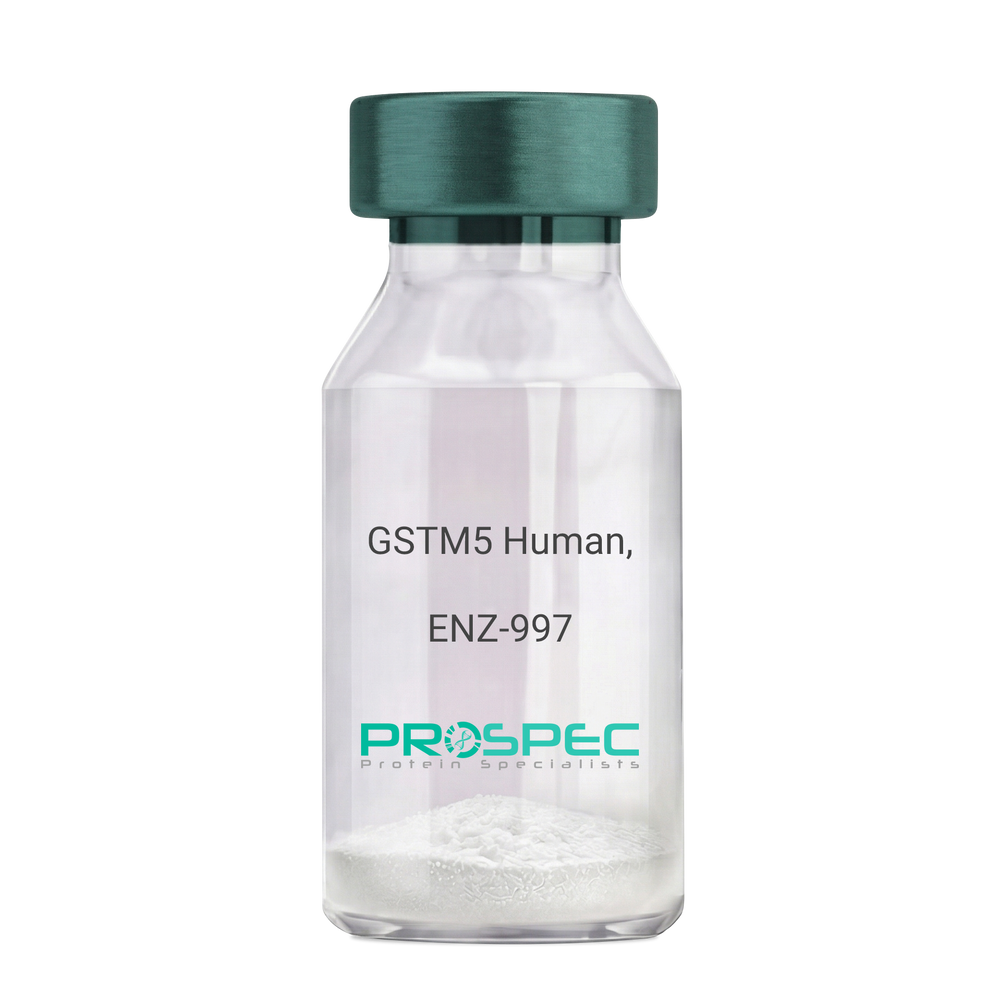
    GSTM5 Human, Active

    Description:

    Glutathione S-Transferase MU 5 Human Recombinant, Active

    Glutathione S-transferase mu 5, GSTM5-5, GST class-mu 5, GTM5, glutathione S-alkyltransferase M5, S-(hydroxyalkyl) glutathione lyase M5, EC 2.5.1.18.

    Product # :

    ENZ-997

    Price :

    Quantity :

    Shipping Method :

    Ice Icon

    Shipped with Ice Packs

    Add To Cart

    More Info

    • description
    • source
    • formulation
    • purity
    • biological activity
    • More Info

    Description

    GSTM5 Human Recombinant produced in E. coli is a single polypeptide chain containing 242 amino acids (1-218) and having a molecular mass of 28.2 kDa.GSTM5 is fused to a 24 amino acid His-tag at N-terminus & purified by proprietary chromatographic techniques.

    Source

    E.coli.

    Formulation

    The GSTM5 solution (1mg/ml) contains 20mM Tris-HCl buffer (pH 8.0), 0.1M NaCl, 1mM DTT and 10% glycerol.

    Purity

    Greater than 95% as determined by SDS-PAGE.

    Biological Activity

    Specific activity is > 90,000 pmol/min/ug, and is defined as the amount of enzyme that conjugate 1.0 u mole of 1-chloro-2,4- dinitrobenzene (CDNB) with reduced glutathione per minute at pH 6.5 at 25C.

    More Info

    • Introduction

      Glutathione S-transferase mu 5 (GSTM5) belongs to the glutathione s-transferase (GST) family of proteins. There are 8 families of GST proteins, specifically alpha, kappa, mu, omega, pi, sigma, theta and zeta, each of which is comprised of proteins which have various functions throughout the cell. GSTM5 belongs to the mu class of enzymes which function in the detoxification of electrophilic compounds, including carcinogens, therapeutic drugs, environmental toxins and products of oxidative stress, by conjugation with glutathione. GSTM5 has an imperative role in detoxification. GSTM5 conjugates reduced glutathione to a large number of exogenous and endogenous hydrophobic electrophiles.

    • Synonyms

      Glutathione S-transferase mu 5, GSTM5-5, GST class-mu 5, GTM5, glutathione S-alkyltransferase M5, S-(hydroxyalkyl) glutathione lyase M5, EC 2.5.1.18.

    • Physical Appearance

      Sterile Filtered colorless solution.

    • Stability

      Store at 4°C if entire vial will be used within 2-4 weeks. Store, frozen at -20°C for longer periods of time. For long term storage it is recommended to add a carrier protein (0.1% HSA or BSA).Avoid multiple freeze-thaw cycles.

    • Amino Acid Sequence

      MGSSHHHHHH SSGLVPRGSH MGSHMPMTLG YWDIRGLAHA IRLLLEYTDS SYVEKKYTLG DAPDYDRSQW LNEKFKLGLD FPNLPYLIDG AHKITQSNAI LRYIARKHNL CGETEEEKIR VDILENQVMD NHMELVRLCY DPDFEKLKPK YLEELPEKLK LYSEFLGKRP WFAGDKITFV DFLAYDVLDM KRIFEPKCLD AFLNLKDFIS RFEGLKKISA YMKSSQFLRG LLFGKSATWN SK.

    ProSpec's products are furnished for LABORATORY RESEARCH USE ONLY. The product may not be used as drugs, agricultural or pesticidal products, food additives or household chemicals.

    Gstm5 Human Active
  • View Data Sheet

    Name :

    CD40 Human, HEK

    Description:

    CD40 Human Recombinant, HEK

    CD40 Molecule, TNF Receptor Superfamily Member 5, TNFRSF5, Tumor Necrosis Factor Receptor Superfamily, Member 5, Bp50, B-Cell Surface Antigen CD40, CD40L Receptor, CDW40, B Cell Surface Antigen CD40, B Cell-Associated Molecule, CD40 Antigen (TNF Receptor Superfamily Member 5), CD40 Type II Isoform, Nerve Growth Factor Receptor-Related B-Lymphocyte Activation Molecule, p50, Tumor Necrosis Factor Receptor Superfamily Member 5, CDw40, CD40 Antigen

    Product # :

    PRO-2719

    Price :

    Quantity :

    Shipping Method :

    Ice Icon

    Shipped with Ice Packs

    Add To Cart

    More Info

    • description
    • source
    • formulation
    • purity
    • More Info

    Description

    CD40 Human Recombinant produced in HEK cells is a single, glycosylated, polypeptide chain containing 412 amino acids (21-193 a.a.) and having a molecular mass of 46.1kDa.CD40 is expressed with a 239 amino acid hIgG-His-Tag at C-Terminus and purified by proprietary chromatographic techniques.

    Source

    HEK 293.

    Formulation

    CD40 protein solution (1mg/ml) contains 10% glycerol and Phosphate-Buffered Saline (pH 7.4).

    Purity

    Greater than 95.0% as determined by SDS-PAGE.

    More Info

    • Introduction

      CD40 belongs to the TNF-receptor super family. CD40 has been found to be vital in mediating a wide range of immune and inflammatory responses including T cell-dependent immunoglobulin class switching, memory B cell development, and germinal center formation. AT-hook transcription factor AKNA is accounted to coordinately regulate the expression of CD40 and its ligand, which is significant for homotypic cell interactions. Adaptor protein TNFR2 interacts with CD40 and functions as a mediator of the signal transduction. The interaction of CD40 and its ligand is found to be essential for amyloid-beta-induced microglial activation, and therefore is considered to be an early event in Alzheimer disease pathogenesis.

    • Synonyms

      CD40 Molecule, TNF Receptor Superfamily Member 5, TNFRSF5, Tumor Necrosis Factor Receptor Superfamily, Member 5, Bp50, B-Cell Surface Antigen CD40, CD40L Receptor, CDW40, B Cell Surface Antigen CD40, B Cell-Associated Molecule, CD40 Antigen (TNF Receptor Superfamily Member 5), CD40 Type II Isoform, Nerve Growth Factor Receptor-Related B-Lymphocyte Activation Molecule, p50, Tumor Necrosis Factor Receptor Superfamily Member 5, CDw40, CD40 Antigen

    • Physical Appearance

      Sterile filtered colorless solution.

    • Stability

      Store at 4°C if entire vial will be used within 2-4 weeks.Store, frozen at -20°C for longer periods of time.For long term storage it is recommended to add a carrier protein (0.1% HSA or BSA).Avoid multiple freeze-thaw cycles.

    • Amino Acid Sequence

      EPPTACREKQ YLINSQCCSL CQPGQKLVSD CTEFTETECL PCGESEFLDT WNRETHCHQH KYCDPNLGLR VQQKGTSETD TICTCEEGWH CTSEACESCV LHRSCSPGFG VKQIATGVSD TICEPCPVGF FSNVSSAFEK CHPWTSCETK DLVVQQAGTN KTDVVCGPQD RLRLEPKSCD KTHTCPPCPA PELLGGPSVF LFPPKPKDTL MISRTPEVTC VVVDVSHEDP EVKFNWYVDG VEVHNAKTKP REEQYNSTYR VVSVLTVLHQ DWLNGKEYKC KVSNKALPAP IEKTISKAKG QPREPQVYTL PPSRDELTKN QVSLTCLVKG FYPSDIAVEW ESNGQPENNY KTTPPVLDSD GSFFLYSKLT VDKSRWQQGN VFSCSVMHEA LHNHYTQKSL SLSPGKHHHH HH

    ProSpec's products are furnished for LABORATORY RESEARCH USE ONLY. The product may not be used as drugs, agricultural or pesticidal products, food additives or household chemicals.

    Cd40 Protein
  • View Data Sheet

    Name :

    TFAM Human

    Description:

    Transcription Factor-A Recombinant Human

    Transcription factor A, mitochondrial, TCF6, TCF6L2, Mitochondrial transcription factor 1, Transcription factor 6-like 2, MtTF1, mtTFA, TCF6L1, TCF6L3.

    Product # :

    PRO-095

    Price :

    Quantity :

    Shipping Method :

    Ice Icon

    Shipped with Ice Packs

    Add To Cart

    More Info

    • description
    • source
    • formulation
    • purity
    • More Info

    Description

    TFAM produced in E.Coli is a single, non-glycosylated polypeptide chain containing 225 amino acids (43-246.a.a) and having a molecular mass of 26.6kDa. TFAM is fused to a 21 amino acid His-tag at N-terminus & purified by proprietary chromatographic techniques.

    Source

    Escherichia Coli.

    Formulation

    The TFAM protein solution (0.25mg/1ml) is formulated in 20 mM Tris-HCl buffer (pH8.0), 0.2M NaCl, 5mM DTT and 20% glycerol.

    Purity

    Greater than 90% as determined by SDS-PAGE.

    More Info

    • Introduction

      TFAM is a mitochondrial transcription factor which is a main activator of mitochondrial transcription and is also a participant in mitochondrial genome replication. TFAM is situated primarily in the nuclei of elongated spermatids and takes part in the regulation of gene expression of the haploid male genome. TFAM is linked to mitochondrial disorder in humans characterized by ocular myopathy, exercise intolerance and muscle wasting.

    • Synonyms

      Transcription factor A, mitochondrial, TCF6, TCF6L2, Mitochondrial transcription factor 1, Transcription factor 6-like 2, MtTF1, mtTFA, TCF6L1, TCF6L3.

    • Physical Appearance

      Sterile Filtered clear solution.

    • Stability

      Store at 4°C if entire vial will be used within 2-4 weeks. Store, frozen at -20°C for longer periods of time.Please avoid freeze thaw cycles.

    • Amino Acid Sequence

      MGSSHHHHHH SSGLVPRGSH MSSVLASCPK KPVSSYLRFS KEQLPIFKAQ NPDAKTTELI RRIAQRWREL PDSKKKIYQD AYRAEWQVYK EEISRFKEQL TPSQIMSLEK EIMDKHLKRK AMTKKKELTL LGKPKRPRSA YNVYVAERFQ EAKGDSPQEK LKTVKENWKN LSDSEKELYI QHAKEDETRY HNEMKSWEEQ MIEVGRKDLL RRTIKKQRKY GAEEC

    ProSpec's products are furnished for LABORATORY RESEARCH USE ONLY. The product may not be used as drugs, agricultural or pesticidal products, food additives or household chemicals.

    Tfam Human
  • View Data Sheet

    Name :

    GZMH Human

    Description:

    Granzyme-H Human Recombinant

    Granzyme H (Cathepsin G-Like 2, Protein H-CCPX), CTSGL2, Cytotoxic T-Lymphocyte Proteinase, CCP-X, CSP-C, Cytotoxic T-Lymphocyte-Associated Serine Esterase 1, Cytotoxin Serine Protease-C, Cytotoxic Serine Protease C, Cathepsin G-Like 2, EC 3.4.21.79, EC 3.4.21, Granzyme H, EC 3.4.21, CGL-2, CTLA1, CGL2, GZMH.

    Product # :

    ENZ-896

    Price :

    Quantity :

    Shipping Method :

    Ice Icon

    Shipped with Ice Packs

    Add To Cart

    More Info

    • description
    • source
    • formulation
    • purity
    • More Info

    Description

    GZMH Human Recombinant produced in E.Coli is a single, non-glycosylated polypeptide chain containing 248 amino acids (20-246 a.a) and having a molecular mass of 27.5kDa.GZMH is fused to a 21 amino acid His-tag at N-terminus & purified by proprietary chromatographic techniques.

    Source

    Escherichia Coli.

    Formulation

    GZMH protein solution (0.25mg/ml) containing 20mM Tris-HCl (pH 8.0) and 10% glycerol.

    Purity

    Greater than 85.0% as determined by SDS-PAGE.

    More Info

    • Introduction

      Granzyme H also known as GZMH belongs to the peptidase S1 family. GZMH is an essential part for HBV eradication. The HBx protein, which is required for the replication of HBV, is cleaved at Met(79) by GZMH. Furthermore, GZMH inhibitor can abolish GZNH- as well as lymphokine-activated killer cell-mediated HBx degradation and HBV clearance. A HBx-deficient HBV is resistant to GzmH- in addition to lymphokine-activated killer cell-mediated viral clearance.

    • Synonyms

      Granzyme H (Cathepsin G-Like 2, Protein H-CCPX), CTSGL2, Cytotoxic T-Lymphocyte Proteinase, CCP-X, CSP-C, Cytotoxic T-Lymphocyte-Associated Serine Esterase 1, Cytotoxin Serine Protease-C, Cytotoxic Serine Protease C, Cathepsin G-Like 2, EC 3.4.21.79, EC 3.4.21, Granzyme H, EC 3.4.21, CGL-2, CTLA1, CGL2, GZMH.

    • Physical Appearance

      Sterile Filtered clear solution.

    • Stability

      Store at 4°C if entire vial will be used within 2-4 weeks. Store, frozen at -20°C for longer periods of time. For long term storage it is recommended to add a carrier protein (0.1% HSA or BSA).Avoid multiple freeze-thaw cycles.

    • Amino Acid Sequence

      MGSSHHHHHH SSGLVPRGSH MEIIGGHEAK PHSRPYMAFV QFLQEKSRKR CGGILVRKDF VLTAAHCQGS SINVTLGAHN IKEQERTQQF IPVKRPIPHP AYNPKNFSND IMLLQLERKA KWTTAVRPLR LPSSKAQVKP GQLCSVAGWG YVSMSTLATT LQEVLLTVQK DCQCERLFHG NYSRATEICV GDPKKTQTGF KGDSGGPLVC KDVAQGILSY GNKKGTPPGV YIKVSHFLPW IKRTMKRL

    ProSpec's products are furnished for LABORATORY RESEARCH USE ONLY. The product may not be used as drugs, agricultural or pesticidal products, food additives or household chemicals.

    Gzmh Human
  • View Data Sheet

    Name :

    OPG Human

    Description:

    Osteoprotegerin Human Recombinant

    TNFRSF11B, OPG, OCIF, Osteoclastogenesis inhibitory factor, Osteoprotegerin, TR1, MGC29565.

    Product # :

    CYT-177

    Price :

    Quantity :

    Shipping Method :

    Room Temp Icon

    Shipped at Room temp

    Add To Cart

    More Info

    • description
    • source
    • formulation
    • purity
    • biological activity
    • More Info

    Description

    Recombinant Human Osteoprotegerin produced in E.coli cells is a single, non-glycosylated, polypeptide chain containing 174 amino acids and having a molecular mass of 20kDa. The OPG is purified by proprietary chromatographic techniques.

    Source

    Escherichia Coli.

    Formulation

    The OPG was lyophilized from a 0.2µm filtered concentrated (0.5mg/ml) solution in PBS, pH 7.4.

    Purity

    Greater than 95.0% as determined by:
    (a) Analysis by RP-HPLC.
    (b) Analysis by SDS-PAGE.

    Biological Activity

    The activity is determined by its ability to neutralize the stimulation of U937 cells treated with 10ng/ml of soluble RANKL corresponding to a specific activity of 100,000IU/mg.

    More Info

    • Introduction

      Osteoprotegerin acts as decoy receptor for rankl and thereby neutralizes its function in osteoclastogenesis. OPG inhibits the activation of osteoclasts and promotes osteoclast apoptosis in vitro. Bone homeostasis seems to depend on the local rankl/opg ratio. Osteoprotegerin may also play a role in preventing arterial calcification. May act as decoy receptor for trail and protect against apoptosis. Trail binding blocks the inhibition of osteoclastogenesis.

    • Synonyms

      TNFRSF11B, OPG, OCIF, Osteoclastogenesis inhibitory factor, Osteoprotegerin, TR1, MGC29565.

    • Physical Appearance

      Sterile Filtered White lyophilized (freeze-dried) powder.

    • Stability

      Lyophilized Osteoprotegerin although stable at room temperature for 3 weeks, should be stored desiccated below -18°C. Upon reconstitution OCIF should be stored at 4°C between 2-7 days and for future use below -18°C. For long term storage it is recommended to add a carrier protein (0.1% HSA or BSA).Please prevent freeze-thaw cycles.

    • Solubility

      It is recommended to reconstitute the lyophilized Osteoprotegerin in sterile 18MΩ-cm H2O not less than 100µg/ml, which can then be further diluted to other aqueous solutions.

    • Amino Acid Sequence

      METFPPKYLH YDEETSHQLL CDKCPPGTYL KQHCTAKWKT VCAPCPDHYY TDSWHTSDEC LYCSPVCKEL QYVKQECNRT HNRVCECKEG RYLEIEFCLK HRSCPPGFGV VQAGTPERNT VCKRCPDGFF SNETSSKAPC RKHTNCSVFG LLLTQKGNAT HDNICSGNSE STQK.

    ProSpec's products are furnished for LABORATORY RESEARCH USE ONLY. The product may not be used as drugs, agricultural or pesticidal products, food additives or household chemicals.

    Opg Human
  • View Data Sheet

    Name :

    HRSP12 Human

    Description:

    Heat-Responsive Protein 12 Human Recombinant

    Ribonuclease UK114, 14.5 kDa translational inhibitor protein, p14.5, Heat-responsive protein 12, UK114 antigen homolog, HRSP12, PSP.

    Product # :

    PRO-052

    Price :

    Quantity :

    Shipping Method :

    Ice Icon

    Shipped with Ice Packs

    Add To Cart

    More Info

    • description
    • source
    • formulation
    • purity
    • More Info

    Description

    HRSP12 Human Recombinant fused with a 20 amino acid His tag at N-terminus produced in E.Coli is a single, non-glycosylated, polypeptide chain containing 157 amino acids (1-137 a.a.) and having a molecular mass of 16.6kDa. The HRSP12 is purified by proprietary chromatographic techniques.

    Source

    Escherichia Coli.

    Formulation

    The HRSP12 solution (1 mg/ml) contains 20mM Tris-HCl buffer (pH8.0), 10% glycerol, 0.1M NaCl and 1mM DTT.

    Purity

    Greater than 95.0% as determined by SDS-PAGE.

    More Info

    • Introduction

      HRSP12 (UK114) is an endoribonuclease found mainly in the human adult kidney and liver, and which is responsible for inhibiting translation by cleaving mRNA. HRSP12 cleaves phosphodiester bonds only in single-stranded RNA. HRSP12 may be an important biomarker for heptatic carcinoma.

    • Synonyms

      Ribonuclease UK114, 14.5 kDa translational inhibitor protein, p14.5, Heat-responsive protein 12, UK114 antigen homolog, HRSP12, PSP.

    • Physical Appearance

      Sterile Filtered colorless solution.

    • Stability

      Store at 4°C if entire vial will be used within 2-4 weeks. Store, frozen at -20°C for longer periods of time. For long term storage it is recommended to add a carrier protein (0.1% HSA or BSA).Avoid multiple freeze-thaw cycles.

    • Amino Acid Sequence

      MGSSHHHHHH SSGLVPRGSH MSSLIRRVIS TAKAPGAIGP YSQAVLVDRT IYISGQIGMD PSSGQLVSGG VAEEAKQALK NMGEILKAAG CDFTNVVKTT VLLADINDFN TVNEIYKQYF KSNFPARAAY QVAALPKGSR IEIEAVAIQG PLTTASL.

    ProSpec's products are furnished for LABORATORY RESEARCH USE ONLY. The product may not be used as drugs, agricultural or pesticidal products, food additives or household chemicals.

    Hrsp12 Human
  • View Data Sheet

    Name :

    DLST Human

    Description:

    Dihydrolipoamide S-Succinyltransferase Human Recombinant

    dihydrolipoamide S-succinyltransferase (E2 component of 2-oxo-glutarate complex), Dihydrolipoamide succinyltransferase component of 2-oxoglutarate dehydrogenase complex, component E2, DLST, E2K, OGDC-E2, EC 2.3.1.61, EC 2.3.1.

    Product # :

    ENZ-083

    Price :

    Quantity :

    Shipping Method :

    Ice Icon

    Shipped with Ice Packs

    Add To Cart

    More Info

    • description
    • source
    • formulation
    • purity
    • More Info

    Description

    DLST is a Full-length cDNA coding for the mature form of the human OGDC-E2 protein having a molecular mass of 42,301 Dalton (pH 6.3).DLST protein is fused to a hexa-histidine purification tag.

    Source

    Sf9 insect cells.

    Formulation

    DLST (0.74mg/ml) is supplied in 20mM HEPES buffer pH-8.0, 200mM NaCl and 20% glycerol.

    Purity

    DLST purity was found to be greater than 75% as determined by SDS-PAGE.

    More Info

    • Introduction

      DLST catalyzes the general transformation of 2-oxoglutarate to succinyl-CoA and CO(2). DLST holds multiple copies of 3 enzymatic components: 2-oxoglutarate dehydrogenase (E1), dihydrolipoamide succinyltransferase (E2) and lipoamide dehydrogenase (E3).

    • Synonyms

      dihydrolipoamide S-succinyltransferase (E2 component of 2-oxo-glutarate complex), Dihydrolipoamide succinyltransferase component of 2-oxoglutarate dehydrogenase complex, component E2, DLST, E2K, OGDC-E2, EC 2.3.1.61, EC 2.3.1.

    • Stability

      Store DLST at 4°C if entire vial will be used within 2-4 weeks. Store, frozen at -20°C for longer periods of time. Avoid multiple freeze-thaw cycles.

    • Immunological Functions

      1. DLST binds IgG-type human auto-antibodies. 2. Standard ELISA test (checkerboard analysis of positive/negative sera panels); immunodot test with positive/negative sera panels.

    • coating concentration

      0.4-1.0 µg/ml (depending on the type of ELISA plate and coating buffer). Suitable for biotinylation and iodination.

    • Applications

      Western blot with anti-M2-Antigen autoantibody-positive patient sera or monoclonal
      anti-hexa-His-tag antibody.

    ProSpec's products are furnished for LABORATORY RESEARCH USE ONLY. The product may not be used as drugs, agricultural or pesticidal products, food additives or household chemicals.

    Dlst Human
  • View Data Sheet

    Name :

    DsbG E.Coli

    Description:

    Thiol Disulfide Interchange Protein E.Coli Recombinant DsbG

    Thiol:disulfide interchange protein dsbG, dsbG, ybdP, b0604, JW0597.

    Product # :

    HSP-024

    Price :

    Quantity :

    Shipping Method :

    Ice Icon

    Shipped with Ice Packs

    Add To Cart

    More Info

    • description
    • source
    • formulation
    • purity
    • More Info

    Description

    Recombinant DsbG produced in E.Coli is a single, non-glycosylated polypeptide chain containing 232 amino acids and having a molecular mass of 25.8 kDa. DsbG is purified by conventional chromatography techniques.

    Source

    Escherichia Coli.

    Formulation

    The DsbG protein solution contains 20mM Tris-HCl, pH-8, 2mM EDTA and 10% Glycerol.

    Purity

    Greater than 95.0% as determined by SDS-PAGE.

    More Info

    • Introduction

      Dsb proteins are in charge for the formation and rearrangement of disulfide bonds during the folding of secreted and membrane proteins in bacteria. DsbG has disulfide bond isomerase and chaperone activity. DsbG interacts with refolding intermediates of chemically denatured citrate synthase and prevents their aggregation in vitro. DsbG shares sequnce homology with DsbC. DsbG forms a stable periplasmic dimer and displays an equilibrium constant with glutathione comparable with DsbA and DsbC. DsbG is expressed at roughly 25% level of DsbC.

    • Synonyms

      Thiol:disulfide interchange protein dsbG, dsbG, ybdP, b0604, JW0597.

    • Physical Appearance

      Sterile filtered colorless solution.

    • Stability

      Store at 4°C if entire vial will be used within 2-4 weeks. Store, frozen at -20°C for longer periods of time. For long term storage it is recommended to add a carrier protein (0.1% HSA or BSA).Avoid multiple freeze-thaw cycles.

    • Amino Acid Sequence

      MEELPAPVKA IEKQGITIIK TFDAPGGMKG YLGKYQDMGV TIYLTPDGKH AISGYMYNEK GENLSNTLIE KEIYAPAGRE MWQRMEQSHW LLDGKKDAPV IVYVFADPFC PYCKQFWQQA RPWVDSGKVQ LRTLLVGVIK PESPATAAAI LASKDPAKTW QQYEASGGKL KLNVPANVST EQMKVLSDNE KLMDDLGANV TPAIYYMSKE NTLQQAVGLP DQKTLNIIMG NK.

    ProSpec's products are furnished for LABORATORY RESEARCH USE ONLY. The product may not be used as drugs, agricultural or pesticidal products, food additives or household chemicals.

    Dsbg Ecoli
  • View Data Sheet

    Name :

    CNTFR Human

    Description:

    Ciliary Neurotrophic Factor Receptor Human Recombinant

    Ciliary Neurotrophic Factor Receptor, CNTF Receptor Subunit Alpha, CNTFR-Alpha, Ciliary Neurotrophic Factor Receptor Subunit Alpha, Ciliary neurotrophic factor receptor subunit alpha, CNTF receptor subunit alpha, CNTFR-alpha.

    Product # :

    CYT-883

    Price :

    Quantity :

    Shipping Method :

    Ice Icon

    Shipped with Ice Packs

    Add To Cart

    More Info

    • description
    • source
    • formulation
    • purity
    • More Info
    • sds-page

    Description

    CNTFR Human Recombinant produced in E.Coli is a single, non-glycosylated polypeptide chain containing 341 amino acids (23-342 a.a) and having a molecular mass of 38.1kDa. CNTFR is fused to a 21 amino acid His-tag at N-terminus & purified by proprietary chromatographic techniques.

    Source

    Escherichia Coli.

    Formulation

    CNTFR protein solution (1mg/ml) containing 20mM Tris-HCl buffer (pH 8.0) and 10% glycerol.

    Purity

    Greater than 85.0% as determined by SDS-PAGE.

    sds-page

    CNTF-sds-page - Product image 1

    More Info

    • Introduction

      Ciliary Neurotrophic Factor Receptor, also known as CNTFR is a member of the type I cytokine receptor family. CNTFR binds to CNTF. The alpha subunit provides the receptor specificity. Sole nucleotide polymorphisms in CNTFR has been associated with variations in muscle strength, in addition to early onset of eating disorders.

    • Synonyms

      Ciliary Neurotrophic Factor Receptor, CNTF Receptor Subunit Alpha, CNTFR-Alpha, Ciliary Neurotrophic Factor Receptor Subunit Alpha, Ciliary neurotrophic factor receptor subunit alpha, CNTF receptor subunit alpha, CNTFR-alpha.

    • Physical Appearance

      Sterile Filtered colorless clear solution.

    • Stability

      Store at 4°C if entire vial will be used within 2-4 weeks. Store, frozen at -20°C for longer periods of time. For long term storage it is recommended to add a carrier protein (0.1% HSA or BSA).Avoid multiple freeze-thaw cycles.

    • Amino Acid Sequence

      MGSSHHHHHH SSGLVPRGSH MQRHSPQEAP HVQYERLGSD VTLPCGTANW DAAVTWRVNG TDLAPDLLNG SQLVLHGLEL GHSGLYACFH RDSWHLRHQV LLHVGLPPRE PVLSCRSNTY PKGFYCSWHL PTPTYIPNTF NVTVLHGSKI MVCEKDPALK NRCHIRYMHL FSTIKYKVSI SVSNALGHNA TAITFDEFTI VKPDPPENVV ARPVPSNPRR LEVTWQTPST WPDPESFPLK FFLRYRPLIL DQWQHVELSD GTAHTITDAY AGKEYIIQVA AKDNEIGTWS DWSVAAHATP WTEEPRHLTT EAQAAETTTS TTSSLAPPPT TKICDPGELG S.

    • Background

      Unveiling the Potential of Human Recombinant Ciliary Neurotrophic Factor Receptor: Insights and Applications

      Abstract:

      The Ciliary Neurotrophic Factor Receptor (CNTFR) holds a crucial role in mediating the effects of ciliary neurotrophic factor (CNTF) on neuronal survival and growth. This paper discusses the significance of Human Recombinant CNTFR, its production methods, and its potential applications in neurobiology and therapeutic interventions. The review sheds light on the pivotal role of CNTFR in neuroprotection and neuroregeneration research.

      Introduction:

      CNTFR, a transmembrane protein, plays a pivotal role in transmitting CNTF-mediated signals to the cell. The availability of Human Recombinant CNTFR allows researchers to investigate its role in neuronal function and develop targeted therapies for neurodegenerative disorders. CNTFR's involvement in modulating neuronal health and promoting regeneration makes it an essential component in neurobiology.

      Role in CNTF Signaling:

      CNTFR forms a receptor complex with other proteins, including gp130 and LIFRβ, to bind CNTF and initiate downstream signaling pathways. Activation of intracellular signaling cascades, such as JAK/STAT and MAPK, contributes to the neuroprotective and growth-promoting effects of CNTF.

      Production Methods:

      Human Recombinant CNTFR is produced through gene expression in suitable host cells, often employing bacterial or mammalian systems. Ensuring proper folding and post-translational modifications is essential to maintain its functionality and binding affinity for CNTF.

      Therapeutic Applications:

      The availability of Human Recombinant CNTFR offers potential therapeutic applications in neurodegenerative diseases like amyotrophic lateral sclerosis (ALS), multiple sclerosis, and retinal degeneration. Manipulating CNTFR-mediated signaling presents opportunities to enhance neuronal survival and regeneration, ultimately improving patient outcomes.

      Challenges and Future Directions:

      While promising, challenges include optimizing the interaction between CNTFR and CNTF, ensuring efficient delivery to target tissues, and understanding potential off-target effects. Ongoing research is essential to unravel the complete mechanisms of CNTFR-mediated signaling and its implications for therapy.

      Conclusion:

      Human Recombinant Ciliary Neurotrophic Factor Receptor serves as a crucial tool in advancing our understanding of neuroprotection and regeneration. Its potential to modulate CNTF-mediated effects opens avenues for innovative therapeutic strategies targeting neurodegenerative disorders, exemplifying the intersection of molecular biology and clinical application.

      What is the molecular weight/Mw of CNTF Protein?
      CNTF Protein has a total Mw of 38.1kDa.

      What is the source or expression system of CNTF Protein?
      Escherichia Coli.

      What is the Purity of CNTF Protein?
      CNTF Protein is >85% pure as determined by SDS-PAGE.

      What is the Biological Activity of CNTF Protein?
      The biological functionality of CNTF Protein will be determined in the future.

      What is the amino acid sequence of CNTF Protein?
      MGSSHHHHHH SSGLVPRGSH MQRHSPQEAP HVQYERLGSD VTLPCGTANW DAAVTWRVNG TDLAPDLLNG SQLVLHGLEL GHSGLYACFH RDSWHLRHQV LLHVGLPPRE PVLSCRSNTY PKGFYCSWHL PTPTYIPNTF NVTVLHGSKI MVCEKDPALK NRCHIRYMHL FSTIKYKVSI SVSNALGHNA TAITFDEFTI VKPDPPENVV ARPVPSNPRR LEVTWQTPST WPDPESFPLK FFLRYRPLIL DQWQHVELSD GTAHTITDAY AGKEYIIQVA AKDNEIGTWS DWSVAAHATP WTEEPRHLTT EAQAAETTTS TTSSLAPPPT TKICDPGELG S.

      What applications can CNTF Protein be used in?
      CNTF Protein can probably be used in western blot, ELISA and Lateral Flow.

      What is the endotoxin level for CNTF Protein?
      The endotoxin level is minimal, CNTF Protein was purified using conventional chromatography tech

    ProSpec's products are furnished for LABORATORY RESEARCH USE ONLY. The product may not be used as drugs, agricultural or pesticidal products, food additives or household chemicals.

    Cntfr Human
  • View Data Sheet

    Name :

    WIBG Human

    Description:

    within BCGN Homolog Human Recombinant

    PYM, Partner of Y14 and mago, MGC13064, WIBG, BCGN Homolog, Protein wibg homolog.

    Product # :

    PRO-864

    Price :

    Quantity :

    Shipping Method :

    Ice Icon

    Shipped with Ice Packs

    Add To Cart

    More Info

    • description
    • source
    • formulation
    • purity
    • More Info

    Description

    WIBG Recombinant Human produced in E.Coli is a single, non-glycosylated polypeptide chain containing 212 amino acids (1-204 a.a.) and having a molecular mass of 23.7 kDa. The WIBG is fused to 8 amino acid His-Tag at C-terminus and purified by proprietary chromatographic techniques.

    Source

    Escherichia Coli.

    Formulation

    WIBG Human solution containing 20mM Tris pH-8, 0.1M NaCl & 10% glycerol.

    Purity

    Greater than 85.0% as determined by SDS-PAGE.

    More Info

    • Introduction

      WIBG is a cooperateing partner of Mago-Y14. The Mago-Y14 heterodimer is a key protein of the EJC(exon junction complex) that is deposited on mRNAs as a consequence of splicing and influences postsplicing mRNA metabolism. WIBG is a cytoplasmic RNA-binding protein that is excluded from the nucleus by Crm1. WIBG relates directly with Mago-Y14 by means of its N-terminal domain.

    • Synonyms

      PYM, Partner of Y14 and mago, MGC13064, WIBG, BCGN Homolog, Protein wibg homolog.

    • Physical Appearance

      Sterile filtered colorless solution.

    • Stability

      Store at 4°C if entire vial will be used within 2-4 weeks. Store, frozen at -20°C for longer periods of time. For long term storage it is recommended to add a carrier protein (0.1% HSA or BSA).Avoid multiple freeze-thaw cycles.

    • Amino Acid Sequence

      MEAAGSPAAT ETGKYIASTQ RPDGTWRKQR RVKEGYVPQE EVPVYENKYV KFFKSKPELP PGLSPEATAP VTPSRPEGGE PGLSKTAKRN LKRKEKRRQQ QEKGEAEALS RTLDKVSLEE TAQLPSAPQG SRAAPTAASD QPDSAATTEK AKKIKNLKKK LRQVEELQQR IQAGEVSQPS KEQLEKLARR RALEEELEDL ELGLLEHHHH HH.

    ProSpec's products are furnished for LABORATORY RESEARCH USE ONLY. The product may not be used as drugs, agricultural or pesticidal products, food additives or household chemicals.

    Wibg Human
  • View Data Sheet

    Name :

    NANOGP8 Human

    Description:

    Nanog Homeobox Pseudogene 8 Human Recombinant

    Nanog Homeobox Pseudogene 8, NANOG, NANOGP1, PN8.

    Product # :

    PRO-1553

    Price :

    Quantity :

    Shipping Method :

    Ice Icon

    Shipped with Ice Packs

    Add To Cart

    More Info

    • description
    • source
    • formulation
    • purity
    • More Info

    Description

    NANOGP8 Human Recombinant produced in E.coli is a single, non-glycosylated polypeptide chain containing 305 amino acids (1-305) and having a molecular mass of 34.6kDa.NANOGP8 is purified by proprietary chromatographic techniques.

    Source

    Escherichia Coli.

    Formulation

    The NANOGP8 solution (1mg/ml) contains 20mM Tris-HCl buffer (pH 8.0), 0.4M Urea and 5% glycerol.

    Purity

    Greater than 85% as determined by SDS-PAGE.

    More Info

    • Introduction

      NANOGP8 belongs to the homeobox family of DNA binding transcription factors which is known to conserve pluripotency of embryonic stem cells. One change only separate NANOGP8 from NANOG – instead of 'Gln-253' in the inferred amino acid sequence in NANOG there is a ‘His-253’ in NANOGP8.

    • Synonyms

      Nanog Homeobox Pseudogene 8, NANOG, NANOGP1, PN8.

    • Physical Appearance

      Sterile Filtered clear solution.

    • Stability

      Store at 4°C if entire vial will be used within 2-4 weeks. Store, frozen at -20°C for longer periods of time. For long term storage it is recommended to add a carrier protein (0.1% HSA or BSA).Avoid multiple freeze-thaw cycles.

    • Amino Acid Sequence

      MSVDPACPQS LPCFEASDCK ESSPMPVICG PEENYPSLQM SSAEMPHTET VSPLPSSMDL LIQDSPDSST SPKGKQPTSA ENSVAKKEDK VPVKKQKTRT VFSSTQLCVL NDRFQRQKYL SLQQMQELSN ILNLSYKQVK TWFQNQRMKS KRWQKNNWPK NSNGVTQKAS APTYPSLYSS YHQGCLVNPT GNLPMWSNQT WNNSTWSNQT QNIQSWSNHS WNTQTWCTQS WNNQAWNSPF YNCGEESLQS CMHFQPNSPA SDLEAALEAA GEGLNVIQQT TRYFSTPQTM DLFLNYSMNM QPEDV

    ProSpec's products are furnished for LABORATORY RESEARCH USE ONLY. The product may not be used as drugs, agricultural or pesticidal products, food additives or household chemicals.

    Nanogp8 Human
  • View Data Sheet

    Name :

    HBcAg (1-149)

    Description:

    Hepatitis B Virus Core (1-149 a.a) Recombinant

    Product # :

    HBV-234

    Price :

    Quantity :

    Shipping Method :

    Ice Icon

    Shipped with Ice Packs

    Add To Cart

    More Info

    • description
    • source
    • formulation
    • purity
    • More Info

    Description

    HBcAg subtype adw2 produced in E.Coli, is a single, non-glycosylated, polypeptide chain containing 1-149 amino acids and having a molecular weight of approximately 20kDa. HBcAg adw2 1-149 protamine-like domain was truncated in order to enhance the differentiation of anti-HBe in ELISA test. HBcAg is fused to a his tag and purified by proprietary chromatographic techniques.

    Source

    Escherichia Coli.

    Formulation

    Tris base and 10mM K2CO3.

    Purity

    Greater than 90% as determined by SDS-PAGE.

    More Info

    • Introduction

      Hepatitis B is one of a few known non-retroviralviruses which employ reverse transcriptionas a part of its replication process. (HIV, a completely unrelated virus, also uses reverse transcription, but it is a retrovirus.) HBV invades the cell by binding to surface receptor and become internalized. The viral core particles then migrate to the hepatocyte nucleus and the partially double-stranded, relaxed circular genomes (RC-DNA) are repaired to form a covalently closed circular DNA (cccDNA), which is the template for viral genomic and sub-genomic RNAs by cellular RNA polymerase II. Of these, the pregenomic RNA(pgRNA is selectively packaged into progeny capsids and is then reverse-transcribed into new RC-DNA. The core can either bud into the endoplasmic reticulum to be enveloped or exported from the cell or recycled back into the genome for conversion to cccDNA.

    • Physical Appearance

      Sterile filtered colorless solution.

    • Stability

      Store at 4°C if entire vial will be used within 2-4 weeks. Store, frozen at -20°C for longer periods of time.For long term storage it is recommended to add a carrier protein (0.1% HSA or BSA). Avoid multiple freeze-thaw cycles.

    • Amino Acid Sequence

      MDIDPYKEFG ATVELLSFLP SDFFPSVRDL LDTASALYRE ALESPEHCSP HHTALRQAIL CWGELMTLAT WVGNNLEDPA SRDLVVNYVN TNVGLKIRQL LWFHISCLTF GRETVLEYLV SFGVWIRTPP AYRPPNAPIL

    ProSpec's products are furnished for LABORATORY RESEARCH USE ONLY. The product may not be used as drugs, agricultural or pesticidal products, food additives or household chemicals.

    Hbcag 1 149 Aa
  • View Data Sheet

    Name :

    GST Mouse Antibody

    Description:

    Glutathione-S-transferase, Antibody

    Product # :

    ANT-456

    Price :

    Quantity :

    Shipping Method :

    Ice Icon

    Shipped with Ice Packs

    Add To Cart

    More Info

    • formulation
    • More Info

    Formulation

    1mg/ml containing PBS, pH-7.4, & 0.1% Sodium Azide.

    More Info

    • Introduction

      GST family of enzymes comprises a long list of cytosolic, mitochondrial, and microsomal proteins that are 45-55 kDa (dimer form) size and are capable of multiple reactions with a multitude of substrates, both endogenous and xenobiotic. GST catalyses the conjugation of reduced glutathione meaning the sulfhydryl group, to electrophilic centers on a wide variety of substrates. This activity is useful in the detoxification of endogenous compounds such as peroxidised lipids, as well as the metabolism of xenobiotics. GST binds toxins and function as transport protein. Glutathione S-transferase is used to create the so-called ''GST gene fusion system''. The GST is used to purify and detect proteins of interest. In a GST gene fusion system, the GST sequence is incorporated into an expression vector alongside the gene sequence encoding the protein of interest. Induction of protein expression from the vector''s multiple cloning sites results in expression of a fusion protein - the protein of int

    • Physical Appearance

      Sterile filtered colorless solution.

    • Immunogen

      Anti GST mAb is derived from hybridization of mouse SP2/0 myeloma cells with spleen cells from BALB/c mice immunized with recombinant GST purified from E. coli.

    • Ig Subclass

      Mouse IgG2b heavy chain and κ light chain.

    • Clone

      P1E5AT.

    • Applications

      GST antibody has been tested ELISA and Western blot analysis to assure specificity and reactivity. Since application varies, however, each investigation should be titrated by the reagent to obtain optimal results. Recommended dilution range for Western blot analysis is 1:500 ~ 2,000. Recommended starting dilution is 1:1,000.

    • Type

      Mouse Antibody Monoclonal.

    • Storage Procedures

      For periods up to 1 month store at 4°C, for longer periods of time, store at -20°C. Prevent freeze thaw cycles.

    • Purification Method

      GST antibody was purified from mouse ascitic fluids by protein-G affinity chromatography.

    ProSpec's products are furnished for LABORATORY RESEARCH USE ONLY. The product may not be used as drugs, agricultural or pesticidal products, food additives or household chemicals.

    Glutathione S Transferase Antibody
  • View Data Sheet

    Name :

    GST Monoclonal antibody

    Description:

    Glutathione-S-Transferase, Mouse antibody

    Product # :

    ANT-219

    Price :

    Quantity :

    Shipping Method :

    Room Temp Icon

    Shipped at Room temp

    Add To Cart

    More Info

    • description
    • formulation
    • More Info

    Description

    Monoclonal antibodies are produced by immunizing mouse with GST protein.

    Formulation

    1×PBS & 50% glycerol.

    More Info

    • Introduction

      GST family of enzymes comprises a long list of cytosolic, mitochondrial, and microsomal proteins that are 45-55 kDa (dimer form) size and are capable of multiple reactions with a multitude of substrates, both endogenous and xenobiotic. GST catalyses the conjugation of reduced glutathione meaning the sulfhydryl group, to electrophilic centers on a wide variety of substrates. This activity is useful in the detoxification of endogenous compounds such as peroxidised lipids, as well as the metabolism of xenobiotics. GST binds toxins and function as transport protein. Glutathione S-transferase is used to create the so-called 'GST gene fusion system'. The GST is used to purify and detect proteins of interest. In a GST gene fusion system, the GST sequence is incorporated into an expression vector alongside the gene sequence encoding the protein of interest. Induction of protein expression from the vector's multiple cloning sites results in expression of a fusion protein - the protein of interest fused to the GST protein. This GST-fusion protein can then be purified from cells via its high affinity for glutathione. Fusion proteins offer an important biological assay for direct protein-to-protein interactions. The GST tag has the size of 220 amino acids, which, compared to other tags like the myc- or the FLAG-tag, is quite big. It is fused to the N-terminus of a protein. However, many commercially-available sources of GST-tagged plasmids include a thrombin domain for cleavage of the GST tag during protein purification. A GST-tag is often used to separate and purify proteins that contain the GST-fusion. GST-fusion proteins can be produced in Escherichia coli, as recombinant proteins.

    • Ig Subclass

      Mouse IgG2b.

    • Clone

      PGSTSHG.

    • Applications

      Western Blot, Immunoprecipitation.

    • Titer

      Western Blotting: 1μg/ml.

    • Type

      Mouse Antibody Monoclonal.

    ProSpec's products are furnished for LABORATORY RESEARCH USE ONLY. The product may not be used as drugs, agricultural or pesticidal products, food additives or household chemicals.

    Gst Igg2B Antibody
  • View Data Sheet

    Name :

    TGFB2 Antibody

    Description:

    Transforming Growth Factor-beta 2 Polyclonal Rabbit Anti Human Antibody

    Transforming growth factor, beta 2, cetermin, Glioblastoma-derived T-cell suppressor factor, polyergin, G-TSF, TGF-beta2, TGF-beta-2, transforming growth factor beta-2, BSC-1 cell growth inhibitor, TGFB-2.

    Product # :

    ANT-035

    Price :

    Quantity :

    Shipping Method :

    Ice Icon

    Shipped with Ice Packs

    Add To Cart

    More Info

    • formulation
    • purity
    • More Info

    Formulation

    Lyophilized from a sterile filtered (0.2µm) solution containing phosphate buffered saline.

    Purity

    Greater than 98%.

    More Info

    • Introduction

      TGFB2 is a 27.08 kDa protein having two identical 118 amino acid peptide chains linked by a single disulfide bond. TGFB2 is part of a family of five related cytokines that have an extensive variation of normal and neoplastic cells, indicating the importance of these homo-dimmer proteins as multi-functional regulators of cellular activity. The three mammalian isoforms of TGF-b (TGFb1, TGFb2 and TGFb3) signal through the same receptor and stimulate similar biological responses. They are involved in physiological processes as embryogenesis, tissue remodelling and wound healing.

    • Synonyms

      Transforming growth factor, beta 2, cetermin, Glioblastoma-derived T-cell suppressor factor, polyergin, G-TSF, TGF-beta2, TGF-beta-2, transforming growth factor beta-2, BSC-1 cell growth inhibitor, TGFB-2.

    • Physical Appearance

      Sterile Filtered White lyophilized (freeze-dried) powder.

    • Stability

      Store at -20°C. For long term storage freezes in working aliquots at -20°C. Repeated freezing and thawing is not recommended.

    • Solubility

      Add distilled water at 1:2 ratio and let the lyophilized pellet dissolve completely.

    • Immunogen

      IgG Anti Human TGFb-2 is developed in rabbit using recombinant Human TGFb-2 produced in plants.

    • Applications

      Western Blot: to detect human TGFb2 by WB analysis this antibody can be used in a dilution of 1/1,000.

    • Antigen Amino Acid Sequence

      ALDAAYCFRNVQDNCCLRPLYIDFKRDLGWKWIHEPKGYNANFCAGACPYLWSSDTQHSRVLSLYNTINPEASASPCCVSQDLEPLTILYYIGKTPKIEQLSNMIVKSCKCS

    • Type

      Polyclonal Rabbit Antibody.

    • Purification Method

      Purified IgG prepared by affinity chromatography on protein G.

    ProSpec's products are furnished for LABORATORY RESEARCH USE ONLY. The product may not be used as drugs, agricultural or pesticidal products, food additives or household chemicals.

    Tgfb2 Antibody
  • View Data Sheet

    Name :

    HADHB Human

    Description:

    2-Enoyl-Coenzyme A (CoA) Hydratase, Beta Human Recombinant

    Hydroxyacyl-CoA Dehydrogenase/3-Ketoacyl-CoA Thiolase/Enoyl-CoA Hydratase (Trifunctional Protein) Beta Subunit, Hydroxyacyl-Coenzyme A Dehydrogenase/3-Ketoacyl-Coenzyme A Thiolase/Enoyl-Coenzyme A Hydratase (Trifunctional Protein) Beta Subunit, TP-BETA, 3-Ketoacyl-Coenzyme A (CoA) Thiolase Of Mitochondrial Trifunctional Protein Beta Subunit, 2-Enoyl-Coenzyme A (CoA) Hydratase Beta Subunit, Trifunctional Enzyme Subunit Beta Mitochondrial, Mitochondrial Trifunctional Protein, Acetyl-CoA Acyltransferase, Beta-Ketothiolase, Beta Subunit, EC 2.3.1.16, EC 2.3.1, MSTP029, ECHB, MTPB.

    Product # :

    ENZ-845

    Price :

    Quantity :

    Shipping Method :

    Ice Icon

    Shipped with Ice Packs

    Add To Cart

    More Info

    • description
    • source
    • formulation
    • purity
    • More Info

    Description

    HADHB Human Recombinant produced in E.Coli is a single, non-glycosylated polypeptide chain containing 464 amino acids (34-474 a.a) and having a molecular mass of 49.9kDa. HADHB is fused to a 23 amino acid His-tag at N-terminus & purified by proprietary chromatographic techniques.

    Source

    Escherichia Coli.

    Formulation

    HADHB protein solution (0. 5mg/ml) containing 20mM Tris-HCl (pH8.0) and 10% glycerol.

    Purity

    Greater than 90.0% as determined by SDS-PAGE.

    More Info

    • Introduction

      2-Enoyl-Coenzyme A (CoA) Hydratase, Beta (HADHB) is the beta subunit of the mitochondrial trifunctional protein, that catalyzes the last 3 phases of mitochondrial beta-oxidation of long chain fatty acids. HADHB binds RNA and reduces the stability of various mRNAs. Mutations in HADHB cause trifunctional protein deficiency.

    • Synonyms

      Hydroxyacyl-CoA Dehydrogenase/3-Ketoacyl-CoA Thiolase/Enoyl-CoA Hydratase (Trifunctional Protein) Beta Subunit, Hydroxyacyl-Coenzyme A Dehydrogenase/3-Ketoacyl-Coenzyme A Thiolase/Enoyl-Coenzyme A Hydratase (Trifunctional Protein) Beta Subunit, TP-BETA, 3-Ketoacyl-Coenzyme A (CoA) Thiolase Of Mitochondrial Trifunctional Protein Beta Subunit, 2-Enoyl-Coenzyme A (CoA) Hydratase Beta Subunit, Trifunctional Enzyme Subunit Beta Mitochondrial, Mitochondrial Trifunctional Protein, Acetyl-CoA Acyltransferase, Beta-Ketothiolase, Beta Subunit, EC 2.3.1.16, EC 2.3.1, MSTP029, ECHB, MTPB.

    • Physical Appearance

      Sterile Filtered clear solution.

    • Stability

      Store at 4°C if entire vial will be used within 2-4 weeks. Store, frozen at -20°C for longer periods of time. For long term storage it is recommended to add a carrier protein (0.1% HSA or BSA).Avoid multiple freeze-thaw cycles.

    • Amino Acid Sequence

      MGSSHHHHHH SSGLVPRGSH MGSAAPAVQT KTKKTLAKPN IRNVVVVDGV RTPFLLSGTS YKDLMPHDLA RAALTGLLHR TSVPKEVVDY IIFGTVIQEV KTSNVAREAA LGAGFSDKTP AHTVTMACIS ANQAMTTGVG LIASGQCDVI VAGGVELMSD VPIRHSRKMR KLMLDLNKAK SMGQRLSLIS KFRFNFLAPE LPAVSEFSTS ETMGHSADRL AAAFAVSRLE QDEYALRSHS LAKKAQDEGL LSDVVPFKVP GKDTVTKDNG IRPSSLEQMA KLKPAFIKPY GTVTAANSSF LTDGASAMLI MAEEKALAMG YKPKAYLRDF MYVSQDPKDQ LLLGPTYATP KVLEKAGLTM NDIDAFEFHE AFSGQILANF KAMDSDWFAE NYMGRKTKVG LPPLEKFNNW GGSLSLGHPF GATGCRLVMA AANRLRKEGG QYGLVAACAA GGQGHAMIVE AYPK.

    ProSpec's products are furnished for LABORATORY RESEARCH USE ONLY. The product may not be used as drugs, agricultural or pesticidal products, food additives or household chemicals.

    Hadhb Human
  • View Data Sheet

    Name :

    MCFD2 Human

    Description:

    Multiple Coagulation Factor Deficiency 2 Human Recombinant

    SDNSF, LMAN1IP, Multiple coagulation factor deficiency protein 2, Neural stem cell-derived neuronal survival protein, MCFD2, F5F8D, DKFZp686G21263.

    Product # :

    PRO-769

    Price :

    Quantity :

    Shipping Method :

    Ice Icon

    Shipped with Ice Packs

    Add To Cart

    More Info

    • description
    • source
    • formulation
    • purity
    • More Info

    Description

    MCFD2 Human Recombinant produced in E.Coli is a single, non-glycosylated polypeptide chain containing 136 amino acids (27-146 a.a.) and having a molecular wieght of 15.1kDa. The MCFD2 is is fused to 16 a.a. T7-Tag at N-terminus and purified by proprietary chromatographic techniques.

    Source

    Escherichia Coli.

    Formulation

    The MCFD2 protein solution contains 20mM Tris-HCl, pH-7.5, 100mM NaCl and 10% glycerol.

    Purity

    Greater than 90.0% as determined by SDS-PAGE.

    More Info

    • Introduction

      The MCFD2-LMAN1 complex forms an explicit cargo receptor for the ER-to-Golgi transport of selected proteins. MCFD2 is involved in the secretion of coagulation factors. MCFD2 is expressed by neural stem/progenitor cells of the hippocampus, and localized to region where neurogenesis persists throughout life. MCFD2 prevents NSC cell death and maintains stem cell characteristics. MCFD2 forms a complex with LAMN1 that facilitates the transport of coagulation factors V and VIII from the endoplasmic reticulum to the Golgi apparatus through an endoplasmic reticulum Golgi intermediate compartment. Mutations in the MCFD2 cause Factor V and Factor VIII combined deficiency.

    • Synonyms

      SDNSF, LMAN1IP, Multiple coagulation factor deficiency protein 2, Neural stem cell-derived neuronal survival protein, MCFD2, F5F8D, DKFZp686G21263.

    • Physical Appearance

      Sterile filtered colorless solution.

    • Stability

      Store at 4°C if entire vial will be used within 2-4 weeks. Store, frozen at -20°C for longer periods of time. For long term storage it is recommended to add a carrier protein (0.1% HSA or BSA). Avoid multiple freeze-thaw cycles.

    • Amino Acid Sequence

      MASMTGGQQM GRGSHMEEPA ASFSQPGSMG LDKNTVHDQE HIMEHLEGVI NKPEAEMSPQ ELQLHYFKMH DYDGNNLLDG LELSTAITHV HKEEGSEQAP LMSEDELINI IDGVLRDDDK NNDGYIDYAE FAKSLQ.

    ProSpec's products are furnished for LABORATORY RESEARCH USE ONLY. The product may not be used as drugs, agricultural or pesticidal products, food additives or household chemicals.

    Mcfd2 Human
  • View Data Sheet

    Name :

    AZGP1 Human, HEK

    Description:

    Alpha-2-Glycoprotein 1 Zinc-Binding Human Recombinant, HEK

    Zn-alpha-2-glycoprotein, Zn-alpha-2-GP, AZGP1, ZAG, Zinc-alpha-2-glycoprotein, ZNGP1, ZA2G.

    Product # :

    PRO-403

    Price :

    Quantity :

    Shipping Method :

    Room Temp Icon

    Shipped at Room temp

    Add To Cart

    More Info

    • description
    • source
    • formulation
    • purity
    • biological activity
    • More Info

    Description

    ZA2G Human Recombinant produced in HEK cells is a single, glycosylated polypeptide chain containing a total of 290 amino acids encoding (13-290). ZA2G Human Recombinant is identical to Swiss-Prot-P25311 (AA 18-295, mature Zinc-Alpha-2-Glycoprotein). Twelve extra amino acids were fused with the N-terminus.

    Source

    293 Cell Line (Human Embryonic Kidney).

    Formulation

    Filtered (0.4 µm) and lyophilized in 0.5 mg/ml in 0.1M Tris-HCl pH 8.0 and 150mM NaCl.

    Purity

    Greater than 90.0% as determined by:
    (a) Analysis by RP-HPLC.
    (b) Analysis by SDS-PAGE.

    Biological Activity

    Differentiated human SGBS adipocytes were incubated for 18 h at two dose levels of rhZA2G - 5 and 20 µg/ml. Lipolysis was quantified by measuring glycerol release into the medium using a standard protocol. Isoproterenol (10 µM) and IBMX (100 µM) were used as positive controls. "Con" stands for the negative control. There was a 3-fold increase in glycerol release with both doses. The increase was statistically significant at 5 µg/ml dose of rhZA2G (p<0.01) as well as in positive controls.

    More Info

    • Introduction

      Zinc-alpha-2-glycoprotein (ZAG) is found in body fluids such as serum, sweat, and seminal and breast cyst fluids. It is identical in amino acid sequence to tumor-derived lipid mobilizing factor (LMF), a protein associated with the dramatic loss of adipose body stores in cancer cachexia, and has been shown to stimulate lipolysis by adipocytes in vivo and in vitro. A role for ZAG has been proposed in the regulation of body weight, and age-dependent changes in genetically influenced obesity, and also it regulates melanin production by normal and malignant melanocytes. It has also recently been classified as a novel adipokine in that it is produced by both white and brown fat adipocytes and may act in a local autocrine fashion in the reduction of adiposity in cachexia. Controlling ZAG/LMF's activity could be life-saving in the management of certain cancers and other cachexiainducing conditions, and its possible normal role in body fat store homeostasis is deserving of understanding in its own right. ZAG exhibits a class I major histocompatibility complex (MHC) fold but is a soluble protein rather than being anchored to plasma membranes and does not associate with alpha-2-microglobulin in humans. Like antigen-presenting MHC class I proteins, ZAG has an open apical groove, and X-ray crystallography of human derived ZAG revealed an unidentifiable electron density in a similar position to that occupied by antigenic peptides in classical MHC proteins and glycolipids in isoforms of CD1. This presumptive ligand is not a peptide, and the groove is too small to hold a glycolipid such as is presented by CD1 isoforms. By analogy with all other MHC class I-related proteins that have an open apical groove [some do not ], occupancy by a ligand is probably crucial to ZAG's biological function. Despite all of the structural and biochemical evidence that ZAG binds a ligand, none has so far been found by extraction from protein isolated from biological fluids. This difficulty could be because the ligand is labile, heterogeneous, or readily lost during purification procedures. Knowing more about how ZAG interacts with the compounds it has been found to bind, both natural and artificial, will inform searches for the elusive ligand(s) and its/their role in ZAG's signaling function.

    • Synonyms

      Zn-alpha-2-glycoprotein, Zn-alpha-2-GP, AZGP1, ZAG, Zinc-alpha-2-glycoprotein, ZNGP1, ZA2G.

    • Physical Appearance

      Filtered White lyophilized (freeze-dried) powder.

    • Stability

      Lyophilized ZA2G although stable at room temperature for 3 weeks, should be stored desiccated below -18°C. Upon reconstitution ZA2G should be stored at 4°C between 2-7 days and for future use below -18°C. For long term storage it is recommended to add a carrier protein (0.1% HSA or BSA). Please avoid freeze-thaw cycles.

    • Solubility

      Add deionized water to a working concentration approximately 0.5 mg/ml and let the lyophilized pellet dissolve completely.

    • Amino Acid Sequence

      ASWSHPQFEK GSQENQDGRY SLTYIYTGLS KHVEDVPAFQ ALGSLNDLQF FRYNSKDRKS QPMGLWRQVEGMEDWKQDSQ LQKAREDIFM ETLKDIVEYY NDSNGSHVLQ GRFGCEIENN RSSGAFWKYY YDGKDYIEFNKEIPAWVPFD PAAQITKQKW EAEPVYVQRA KAYLEEECPA TLRKYLKYSK NILDRQDPPS VVVTSHQAPG EKKKLKCLAYDFYPGKIDVH WTRAGEVQEP ELRGDVLHNG NGTYQSWVVV AVPPQDTAPY SCHVQHSSLA QPLVVPWEAS.

    ProSpec's products are furnished for LABORATORY RESEARCH USE ONLY. The product may not be used as drugs, agricultural or pesticidal products, food additives or household chemicals.

    Azgp1
  • View Data Sheet

    Name :

    EGFP Antibody

    Description:

    Enhanced Green Fluorescent Protein, Mouse Anti

    Green fluorescent protein, GFP.

    Product # :

    ANT-752

    Price :

    Quantity :

    Shipping Method :

    Ice Icon

    Shipped with Ice Packs

    Add To Cart

    More Info

    • formulation
    • More Info

    Formulation

    PBS, pH-7.4, 10% Glycerol and 0.02% Sodium Azide.

    More Info

    • Introduction

      GFP or Green Fluorescent Protein, is a protein derivedfroma jellyfish (Aequorea Victoria) that produces bioluminescence in the green area of the seen spectrum. GFP is a helpful and very commontool for manufacturing chimeric proteins, where it acts as a fluorescent protein tag. Green Fluorescent Protein is expressed in most known cell types and is used as a noninvasive fluorescent indicator in living cells and tissues.

    • Synonyms

      Green fluorescent protein, GFP.

    • Physical Appearance

      Sterile filtered colorless solution.

    • Immunogen

      Anti EGFP mAb 1-239 amino acid is purified from E. coli.

    • Ig Subclass

      Mouse IgG2a heavy chain and κ light chain.

    • Clone

      PAT1D9AT.

    • Applications

      EGFP antibody has been tested by ELISA, Western blot analysis to assure specificity and reactivity. Since application varies, however, each investigation should be titrated by the reagent to obtain optimal results.

    • Type

      Mouse Anti Monoclonal.

    • Storage Procedures

      For periods up to 1 month store at 4°C, for longer periods of time, store at -20°C. Prevent freeze thaw cycles.

    • Purification Method

      EGFP antibody was purified from mouse ascitic fluids by protein-G affinity chromatography.

    ProSpec's products are furnished for LABORATORY RESEARCH USE ONLY. The product may not be used as drugs, agricultural or pesticidal products, food additives or household chemicals.

    Egfp Antibody
  • View Data Sheet

    Name :

    GH1 Antibody

    Description:

    Growth Hormone-1, Mouse Anti Human

    GH1, GH, GHN, GH-N, hGH-N, Pituitary growth hormone, Growth hormone 1, Somatotropin.

    Product # :

    ANT-230

    Price :

    Quantity :

    Shipping Method :

    Ice Icon

    Shipped with Ice Packs

    Add To Cart

    More Info

    • formulation
    • More Info

    Formulation

    1mg/ml containing PBS, pH-7.4, & 0.1% sodium azide.

    More Info

    • Introduction

      GH is a member of the somatotropin/prolactin family of hormones which play an important role in growth control. The gene, along with four other related genes, is located at the growth hormone locus on chromosome 17 where they are interspersed in the same transcriptional orientation; an arrangement which is thought to have evolved by a series of gene duplications. The five genes share a remarkably high degree of sequence identity. Alternative splicing generates additional isoforms of each of the five growth hormones, leading to further diversity and potential for specialization. This particular family member is expressed in the pituitary but not in placental tissue as is the case for the other four genes in the growth hormone locus. Mutations in or deletions of the gene lead to growth hormone deficiency and short stature.

    • Synonyms

      GH1, GH, GHN, GH-N, hGH-N, Pituitary growth hormone, Growth hormone 1, Somatotropin.

    • Physical Appearance

      Sterile Filtered solution.

    • Immunogen

      GH Antibody is derived from hybridization of mouse SP2/O myeloma cells with spleen cells from BALB/c mice immunized with recombinant Growth hormone.

    • Ig Subclass

      Mouse IgG2b heavy chain and k light chain.

    • Clone

      PG3H5AT.

    • Applications

      The GH antibody has been tested by ELISA and by Western blot analysis to assure specificity and reactivity. Since application vary, however, each investigation should titrate the reagent to obtain optimal results. Recommended dilution range for Western blot analysis: 1:500 ~ 1:1000. Recommended starting dilution: 1:500.

    • Type

      Mouse Anti Human Monoclonal.

    • Storage Procedures

      For periods up to 1 month store at 4 C, for longer periods of time, store at -20 C. Prevent freeze thaw cycles.

    • Purification Method

      GH antibody was purified from mouse ascitic fluids by protein-G affinity chromatography.

    ProSpec's products are furnished for LABORATORY RESEARCH USE ONLY. The product may not be used as drugs, agricultural or pesticidal products, food additives or household chemicals.

    Growth Hormone Igg2B Antibody
  • View Data Sheet

    Name :

    IGFBP4 Sf9, Human

    Description:

    Insulin Like Growth Factor Binding Protein-4 Human Recombinant, Sf9

    Insulin-like growth factor-binding protein 4, IBP-4, IGF-binding protein 4, IGFBP-4, IGFBP4, IBP4, BP-4, HT29-IGFBP.

    Product # :

    CYT-1114

    Price :

    Quantity :

    Shipping Method :

    Room Temp Icon

    Shipped at Room temp

    Add To Cart

    More Info

    • description
    • source
    • formulation
    • purity
    • biological activity
    • More Info

    Description

    Insulin Like Growth Factor Binding Protein-4 Human Recombinant produced in Sf9 Insect cells is a single, glycosylated polypeptide chain containing 237 amino acids and having a molecular mass of 30kDa. The IGFBP4 is purified by proprietary chromatographic techniques.

    Source

    Sf9, Insect cells.

    Formulation

    Lyophilized from a 0.2μm filtered concentrated solution in 20mM Tris-HCl, pH 8.0 and 150mM NaCl.

    Purity

    Greater than 97.0% as determined by:
    (a) Analysis by RP-HPLC.
    (b) Analysis by SDS-PAGE.

    Biological Activity

    The ED50 is determined by its ability to inhibit IGF-II induced proliferation of MCF-7 cells and is <than 0.1 μg/ml, corresponding to a specific activity of > 1.0 × 10⁴ IU/mg in the presence of 14 ng/ml of rHuIGF-II.

    More Info

    • Introduction

      Insulin-like growth factor-binding protein 4 (IGFBP-4) is a part of the insulin-like growth factor binding protein (IGFBP) family. IGFBP4 includes an IGFBP domain and a thyroglobulin type-I domain. IGFBP4 binds both insulin-like growth factors (IGFs) I and II. IGFBP-4 circulates in the plasma in both glycosylated and non-glycosylated forms. IGFBPs can either inhibit or enhance the biological activities of IGF, or act in an IGF independent manner. IGFBP-4 is consistently inhibits several cancer cells in vivo and in vitro, suggesting that it may function as an apoptotic factor. IGFBP4 is produced by all colon cancer cells. Binding of IGFBP-4 prolongs the half-life of the IGFs and changes their interaction with cell surface receptors.

    • Synonyms

      Insulin-like growth factor-binding protein 4, IBP-4, IGF-binding protein 4, IGFBP-4, IGFBP4, IBP4, BP-4, HT29-IGFBP.

    • Physical Appearance

      Sterile Filtered White lyophilized (freeze-dried) powder.

    • Stability

      Lyophilized IGFBP4 although stable at room temperature for 3 weeks, should be stored desiccated below -18°C. Upon reconstitution Insulin Like Growth Factor Binding Protein-4 should be stored at 4°C between 2-7 days and for future use below -18°C. Please prevent freeze-thaw cycles.

    • Solubility

      It is recommended to reconstitute the lyophilized Insulin Like Growth Factor Binding Protein-4 in sterile 18MΩ-cm H2O not less than 100µg/ml, which can then be further diluted to other aqueous solutions.

    • Amino Acid Sequence

      DEAIHCPPCS EEKLARCRPP VGCEELVREP GCGCCATCAL GLGMPCGVYT PRCGSGLRCY PPRGVEKPLH TLMHGQGVCM ELAEIEAIQE SLQPSDKDEG DHPNNSFSPC SAHDRRCLQK HFAKIRDRST SGGKMKVNGA PREDARPVPQ GSCQSELHRA LERLAASQSR THEDLYIIPI PNCDRNGNFH PKQCHPALDG QRGKCWCVDR KTGVKLPGGL EPKGELDCHQ LADSFRE.

    ProSpec's products are furnished for LABORATORY RESEARCH USE ONLY. The product may not be used as drugs, agricultural or pesticidal products, food additives or household chemicals.

    Igfbp4 Protein
  • View Data Sheet

    Name :

    Flt3 Ligand Human, HEK

    Description:

    Flt3-Ligand Human Recombinant, HEK derived

    Fms-related tyrosine kinase 3 ligand, FLK2, STK1, CD135, Stem Cell Tyrosine Kinase 1, FLT3LG, Flt3.

    Product # :

    CYT-706

    Price :

    Quantity :

    Shipping Method :

    Room Temp Icon

    Shipped at Room temp

    Add To Cart

    More Info

    • description
    • source
    • formulation
    • purity
    • biological activity
    • More Info

    Description

    Flt3-Ligand Human Recombinant produced in HEK cells is a glycosylated monomer, having a molecular weight range of 24-30kDa due to glycosylation. The Flt3-Ligand is purified by proprietary chromatographic techniques.

    Source

    HEK293 (Human Embryonic Kidney cell line).

    Formulation

    Flt3-Ligand was lyophilized from a 0.2µm filtered solution containing 1xPBS.

    Purity

    Greater than 95.0% as determined by: (a) Analysis by RP-HPLC. (b) Analysis by SDS-PAGE.

    Biological Activity

    The activity was determined by the dose- dependent stimulation of the proliferation of the human acute myeloid leukemia cell line OCI-AML5. The ED50 is 0.56ng/ml.

    More Info

    • Introduction

      FLT3 ligand is a receptor for the fl cytokine has a tyrosine-protein kinase activity & a growth factor that regulates proliferation of early hematopoietic cells. Flt3-Ligand synergizes with other CSFs and interleukins to induce growth and differentiation.

    • Synonyms

      Fms-related tyrosine kinase 3 ligand, FLK2, STK1, CD135, Stem Cell Tyrosine Kinase 1, FLT3LG, Flt3.

    • Physical Appearance

      Sterile Filtered White lyophilized (freeze-dried) powder.

    • Stability

      Lyophilized Flt3-Ligand although stable at room temperature for 3 weeks, should be stored desiccated below -18°C. Upon reconstitution Flt3-Ligand should be stored at 4°C between 2-7 days and for future use below -18°C. For long term storage it is recommended to add a carrier protein (0.1% HSA or BSA). Please prevent freeze-thaw cycles.

    • Solubility

      It is recommended to reconstitute the lyophilized Flt3-Ligand in sterile 18MΩ-cm H2O not less than 100µg/ml, which can then be further diluted to other aqueous solutions.

    • Amino Acid Sequence

      TQDCSFQHSP ISSDFAVKIR ELSDYLLQDY PVTVASNLQD EELCGGLWRL VLAQRWMERL KTVAGSKMQG LLERVNTEIH FVTKCAFQPP PSCLRFVQTN ISRLLQETSE QLVALKPWIT RQNFSRCLEL QCQPDSSTLP PPWSPRPLEA TAPTAPQP.

    • Background

      What is the molecular weight/Mw of FLT3 LIGAND HUMAN, HEK Protein?
      FLT3 LIGAND HUMAN, HEK Protein has a total Mw of 27kDa.

      What is the source or expression system of FLT3 LIGAND HUMAN, HEK Protein?
      HEK293 (Human Embryonic Kidney cell line).

      What is the Purity of FLT3 LIGAND HUMAN, HEK Protein?
      FLT3 LIGAND HUMAN, HEK Protein is >95% pure as determined by SDS-PAGE.

      What is the Biological Activity of FLT3 LIGAND HUMAN, HEK Protein?
      The activity was determined by the dose- dependent stimulation of the proliferation of the human acute myeloid leukemia cell line OCI-AML5. The ED50 is 0.56ng/ml.

      What is the amino acid sequence of FLT3 LIGAND HUMAN, HEK Protein?
      TQDCSFQHSP ISSDFAVKIR ELSDYLLQDY PVTVASNLQD EELCGGLWRL VLAQRWMERL KTVAGSKMQG LLERVNTEIH FVTKCAFQPP PSCLRFVQTN ISRLLQETSE QLVALKPWIT RQNFSRCLEL QCQPDSSTLP PPWSPRPLEA TAPTAPQP.

      What applications can FLT3 LIGAND HUMAN, HEK Protein be used in?
      FLT3 LIGAND HUMAN, HEK Protein can probably be used in western blot, ELISA and Lateral Flow.

      What is the endotoxin level for FLT3 LIGAND HUMAN, HEK Protein?
      The endotoxin level is minimal, FLT3 LIGAND HUMAN, HEK Protein was purified using conventional chromatography techniques.

    ProSpec's products are furnished for LABORATORY RESEARCH USE ONLY. The product may not be used as drugs, agricultural or pesticidal products, food additives or household chemicals.

    Flt3 Ligand Human Hek
  • View Data Sheet

    Name :

    TNFR2 Human Fc

    Description:

    Tumor Necrosis Factor Receptor 2 Fusion Protein Human Recombinant

    Tumor necrosis factor receptor superfamily member 1B,Tumor necrosis factor receptor 2, TNF-R2, Tumor necrosis factor receptor type II, p75, p80 TNF-alpha receptor, CD120b antigen, Etanercept, TBPII, TNFBR, TNFR80, TNF-R75, p75TNFR, TNF-R-II.

    Product # :

    CYT-422

    Price :

    Quantity :

    Shipping Method :

    Room Temp Icon

    Shipped at Room temp

    Add To Cart

    More Info

    • description
    • source
    • formulation
    • purity
    • biological activity
    • More Info

    Description

    Recombinant Human Tumor Necrosis Factor Receptor 2 Fusion Protein produced in CHO is a dimeric, glycosylated, polypeptide chain consisting of the extracellular ligand-binding portion of the human 75 kilo Dalton (p75) tumor necrosis factor receptor 2 (TNFR2) linked to the Fc portion of human IgG1. The Fc component of TNFR2 contains the CH2 domain, the CH3 domain and hinge region, but not the CH1 domain of IgG1. It consists of 934 amino acids and has an apparent molecular weight of approximately 150 kilo Daltons.The TNFR2 is purified by standard chromatographic techniques.

    Source

    Chinese Hamster Ovarian Cells (CHO).

    Formulation

    Each mg contains 1.6mg mannitol, 0.4 mg sucrose and 48 µg tromethamine.

    Purity

    Greater than 98.0% as determined by:
    (a) Analysis by RP-HPLC.
    (c) Analysis by SDS-PAGE.

    Biological Activity

    Potency is determined by its ability to neutralize TNF-alpha mediated growth inhibition of A375 cells, corresponding to a Specific Activity of 17,000,000 IU/mg.

    More Info

    • Introduction

      TNFR binds specifically to tumor necrosis factor (TNF) and blocks its interaction with cell surface TNF receptors. TNF is a naturally occurring cytokine that is involved in normal inflammatory and immune responses. It plays an important role in the inflammatory processes of rheumatoid arthritis (RA), polyarticular-course juvenile rheumatoid arthritis (JRA), and ankylosing spondylitis and the resulting joint pathology. In addition, TNF plays a role in the inflammatory process of plaque psoriasis. Elevated levels of TNF are found in involved tissues and fluids of patients with RA, psoriatic arthritis, ankylosing spondylitis (AS), and plaque psoriasis. Two distinct receptors for TNF (TNFRs), a 55 kilodalton protein (p55) and a 75 kilodalton protein (p75), exist naturally as monomeric molecules on cell surfaces and in soluble forms. Biological activity of TNF is dependent upon binding to either cell surface TNFR. Recombinant Human TNFR is a dimeric soluble form of the p75 TNF receptor that can bind to two TNF molecules.
      It inhibits the activity of TNF in vitro and has been shown to affect several animal models of inflammation, including murine collagen-induced arthritis. TNFR inhibits binding of both TNF? and TNF? (lymphotoxin alpha [LT?]) to cell surface TNFRs, rendering TNF biologically inactive. Cells expressing transmembrane TNF that bind to TNFR are not lysed in vitro in the presence or absence of complement.
      TNFR can also modulate biological responses that are induced or regulated by TNF, including expression of adhesion molecules responsible for leukocyte migration (i.e., E-selectin and to a lesser extent intercellular adhesion molecule-1 [ICAM-1]), serum levels of cytokines (e.g., IL-6), and serum levels of matrix metalloproteinase-3 (MMP-3 or stromelysin).

    • Synonyms

      Tumor necrosis factor receptor superfamily member 1B,Tumor necrosis factor receptor 2, TNF-R2, Tumor necrosis factor receptor type II, p75, p80 TNF-alpha receptor, CD120b antigen, Etanercept, TBPII, TNFBR, TNFR80, TNF-R75, p75TNFR, TNF-R-II.

    • Physical Appearance

      Sterile Filtered White lyophilized (freeze-dried) powder.

    • Stability

      Lyophilized Tumor Necrosis Factor Receptor although stable at room temperature for 3 weeks, should be stored desiccated below -18°C. Upon reconstitution TNFR2 should be stored at 4°C between 2-7 days and for future use below -18°C.For long term storage it is recommended to add a carrier protein (0.1% HSA or BSA).Please prevent freeze-thaw cycles.

    • Solubility

      It is recommended to reconstitute the lyophilized TNFR2 in sterile 18M-cm H2O not less than 100µg/ml, which can then be further diluted to other aqueous solutions.

    ProSpec's products are furnished for LABORATORY RESEARCH USE ONLY. The product may not be used as drugs, agricultural or pesticidal products, food additives or household chemicals.

    Tnfr2 Human Fc
  • 1
  • …
  • 34
  • 35
  • 36
  • 37
  • 38
  • …
  • 42
Your browser does not support the video tag.

Products

  • Cytokines
  • Growth Factors
  • Chemokines
  • CD Antigens
  • Neurotrophins
  • Hormones
  • Enzymes
  • Viral Antigens
  • Recombinant Proteins
  • Natural Proteins
  • Monoclonal Antibodies
  • Polyclonal Antibodies
  • Thermal Packaging

About

  • Company
  • Founder
  • Contribution
  • Contact Us
  • My Account

Ordering

  • Ordering Method
  • Ordering - Credit Card
  • Ordering PO
  • Shipping Fees
  • FAQ's

Legal

  • Terms & Conditions
  • Privacy
  • Site Map
  • info@prospecbio.com
  • Facebook
  • Instagram
  • Linkedin Profile

© 2026 ProSpec-Tany TechnoGene Ltd. All Rights Reserved.

  • Choosing a selection results in a full page refresh.
  • Opens in a new window.