Search results
858 results found for “chromobox”
Name
Description
Product #
Price
Quantity
Shipping Method
- View Data Sheet
Name :
Axin1 AntibodyDescription:
Axin-1, Mouse Anti Human
Axin-1, Axis inhibition protein 1, hAxin, AXIN1, AXIN, MGC52315.
Product # :
ANT-021Price :
Quantity :
Shipping Method :
Shipped with Ice Packs
More Info
- formulation
- More Info
- Citations
Formulation
1mg/ml containing PBS, pH-7.4, 10% Glycerol and 0.02% Sodium Azide.
Citations
More Info
-
Introduction
AXIN1 is a cytoplasmic protein, which contains a regulation of G-protein signaling (RGS) domain and a dishevelled and axin (DIX) domain. Axin1 is has been characterized as a tumor suppressor. In addition, Axin1 has an essential role in the formation of the embryonic neural axis. It has also been reported that Axin is found in mitotic spindles and centrosomes. AXIN1 interacts with adenomatosis polyposis coli, catenin beta-1, glycogen synthase kinase 3 beta, protein phosphate 2, and itself. AXIN1 functions as a negative regulator of the WNT1 signaling pathway and is able to induce apoptosis. Axin can be identified in a mouse mutant strain, created by random integration of a transgene. Mutations in the AXIN1 gene are linked to hepatocellular carcinoma, hepatoblastomas, ovarian endometriod adenocarcinomas, and medullablastomas.
-
Synonyms
Axin-1, Axis inhibition protein 1, hAxin, AXIN1, AXIN, MGC52315.
-
Immunogen
Anti-human Axin1 mAb, is derived from hybridization of mouse F0 myeloma cells with spleen cells from BALB/c mice immunized with recombinant human Axin1 amino acids 546-752 purified from E. coli.
-
Ig Subclass
Mouse IgG2b heavy chain and κ light chain.
-
Clone
PAT1A4AT.
-
Applications
Axin1 antibody has been tested by ELISA, Western blot, Immunofluorescence, and Immunoprecipitation analyses to assure specificity and reactivity. Since applications vary, however, each investigation should be titrated by a reagent to obtain optimal results. Recommended dilution range for Western blot analysis and Immunofluorescence is 1:250 ~ 500.
Recommended starting dilution is 1:500. -
Type
Mouse Anti Human Monoclonal.
-
Storage Procedures
For periods up to 1 month store at 4°C, for longer periods of time, store at -20°C. Prevent freeze thaw cycles.
-
Purification Method
Axin1 antibody was purified from mouse ascitic fluids by protein-G affinity chromatography.
ProSpec's products are furnished for LABORATORY RESEARCH USE ONLY. The product may not be used as drugs, agricultural or pesticidal products, food additives or household chemicals.
- View Data Sheet
Name :
TGFB3 AntibodyDescription:
Transforming Growth Factor-beta 3 Polyclonal Rabbit Anti Human Antibody
Transforming Growth Factor-beta3, TGFB3, ARVD, FLJ16571, TGF-beta3.
Product # :
ANT-040Price :
Quantity :
Shipping Method :
Shipped with Ice Packs
More Info
- formulation
- More Info
- Citations
Formulation
lyophilized from 1mg/ml solution in 0.2µm sterile filtered solution in phosphate buffered saline.
Citations
More Info
-
Introduction
Transforming growth factor betas (TGF Betas) mediate many cell-cell interactions that occur during embryonic development. Three TGF Betas have been identified in mammals. TGF Beta 1, TGF Beta 2 and TGF Beta 3 are each synthesized as precursor proteins that are very similar in that each is cleaved to yield a 112 amino acid polypeptide that remains associated with the latent portion of the molecule.
-
Synonyms
Transforming Growth Factor-beta3, TGFB3, ARVD, FLJ16571, TGF-beta3.
-
Stability
Store at 4°C. For long term storage freezes in working aliquots at -20°C. Repeated freezing and thawing is not recommended.
-
Solubility
Reconstitute with H20. Mix gently, wash the sides of the vial and wait 30-60 seconds before use.
-
Immunogen
IgG Anti Human TGFb-3 is developed in rabbit using recombinant Human TGFb-3 produced in plants.
-
Applications
To detect Human TGFb-3 by WB analysis this IgG can be used in a dilution of 1/500-1/1000.
-
Antigen Amino Acid Sequence
ALDTNYCFRNLEENCCVRPLYIDFRQDLGWKWVHEPKGYYANFCSGPCPYLRSADTTHSTVLGLYNTLNPEASASPCCVPQDLEPLTILYYVGRTPKVEQLSNMVVKSCKCS
-
Type
Polyclonal Rabbit Antibody.
-
Purification Method
Purified IgG prepared by affinity chromatography on protein G.
ProSpec's products are furnished for LABORATORY RESEARCH USE ONLY. The product may not be used as drugs, agricultural or pesticidal products, food additives or household chemicals.
- View Data Sheet
Name :
IRF3 AntibodyDescription:
Interferon Regulatory Factor-3, Mouse Anti Human
IRF-3, IRF3, Interferon Regulatory Factor 3.
Product # :
ANT-365Price :
Quantity :
Shipping Method :
Shipped with Ice Packs
More Info
- formulation
- More Info
- Citations
Formulation
1mg/ml containing PBS, pH-7.4, & 0.1% Sodium Azide.
Citations
More Info
-
Introduction
Members of the Interferon regulatory factor (IRF) family regulate gene expression critical to immune response, hemopoiesis, and proliferation. IRF-3 is a member of the IRF family, and is distinct from other family members. Its transcriptional activity is regulated solely by posttranslational modifications. It plays a crucial role in activation of innate immunity and inflammation in response to viral infection. IRF-3 mediates interferon-stimulated response element (isre) promoter activation. Functions as a molecular switch for antiviral activity. Dsrna generated during the course of an viral infection leads to IRF3 phosphorylation on the c-terminal serine/threonine cluster. This induces a conformational change, leading to its dimerization, nuclear localization and association with creb binding protein (crebbp) to form dsrna-activated factor 1 (draf1), a complex which activates the transcription of genes under the control of isre. The complex binds to the ie and prdiii regions on the ifn
-
Synonyms
IRF-3, IRF3, Interferon Regulatory Factor 3.
-
Physical Appearance
Sterile Filtered clear solution.
-
Immunogen
Anti-human IRF3 mAb, is derived from hybridization of mouse SP2/0 myeloma cells with spleen cells from BALB/c mice immunized with Recombinant human IRF3 amino acids 108-166 purified from E. coli.
-
Ig Subclass
Mouse IgG1 heavy chain and κ light chain.
-
Clone
P3F10AT.
-
Applications
IRF3 antibody has been tested by ELISA and Western blot analysis to assure specificity and reactivity. Since application varies, however, each investigation should be titrated by the reagent to obtain optimal results. Recommended dilution range for Western blot analysis is 1:1,000 ~ 2,000. Recommended starting dilution is 1:1,000.
-
Type
Mouse Anti Human Monoclonal.
-
Storage Procedures
For periods up to 1 month store at 4°C, for longer periods of time, store at -20°C. Prevent freeze thaw cycles.
-
Purification Method
IRF3 antibody was purified from mouse ascitic fluids by protein-G affinity chromatography.
ProSpec's products are furnished for LABORATORY RESEARCH USE ONLY. The product may not be used as drugs, agricultural or pesticidal products, food additives or household chemicals.
- View Data Sheet
Name :
KRT19 AntibodyDescription:
Cytokeratin 19, Mouse Anti Human
Keratin type I cytoskeletal 19, Cytokeratin-19, CK-19, Keratin-19, K19, KRT19, CK19, K1CS, MGC15366.
Product # :
ANT-472Price :
Quantity :
Shipping Method :
Shipped with Ice Packs
More Info
- formulation
- More Info
- Citations
Formulation
1mg/ml containing PBS, pH-7.4, 10% Glycerol and 0.02% Sodium Azide.
Citations
More Info
-
Introduction
CTK-19 is a member of the keratin family. The keratins are intermediate filament proteins responsible for the structural integrity of epithelial cells and are subdivided into cytokeratins and hair keratins. The type I cytokeratins consist of acidic proteins which are arranged in pairs of heterotypic keratin chains. Unlike its related family members, this smallest known acidic cytokeratin is not paired with a basic cytokeratin in epithelial cells. It is specifically expressed in the periderm, the transiently superficial layer that envelopes the developing epidermis. The type I cytokeratins are clustered in a region of chromosome 17q12-q21.
-
Synonyms
Keratin type I cytoskeletal 19, Cytokeratin-19, CK-19, Keratin-19, K19, KRT19, CK19, K1CS, MGC15366.
-
Physical Appearance
Sterile filtered colorless solution.
-
Immunogen
Anti-human KRT19 mAb, clone PAT13D10A, is derived from hybridization of mouse F0 myeloma cells with spleen cells from BALB/c mice immunized with a recombinant human KRT19 protein purified from E. coli.
-
Ig Subclass
Mouse IgG2b heavy chain and Kappa light chain.
-
Clone
PAT13D10A.
-
Applications
The antibody has been tested by ELISA, Western blot analysis to assure specificity and reactivity. Since application varies, however, each investigation should be titrated by the reagent to obtain optimal results. Recommended starting dilution is 1:1000.
-
Type
Mouse Anti Human Monoclonal.
-
Storage Procedures
For periods up to 1 month store at 4°C, for longer periods of time, store at -20°C. Prevent freeze thaw cycles.
ProSpec's products are furnished for LABORATORY RESEARCH USE ONLY. The product may not be used as drugs, agricultural or pesticidal products, food additives or household chemicals.
- View Data Sheet
Name :
KRT8 AntibodyDescription:
Cytokeratin 8, Mouse Anti Human
Keratin type II cytoskeletal 8, Cytokeratin-8, CK-8, Keratin-8, K8, KRT8, CYK8, KO, CK8, K2C8, CARD2.
Product # :
ANT-470Price :
Quantity :
Shipping Method :
Shipped with Ice Packs
More Info
- formulation
- More Info
- Citations
Formulation
1mg/ml containing PBS, pH-7.4, 10% Glycerol and 0.02% Sodium Azide.
Citations
More Info
-
Introduction
Cytokeratin 8 (KRT8) belongs to the type II keratin family clustered on the long arm of chromosome 12. Type I and type II keratins heteropolymerize to form intermediate-sized filaments in the cytoplasm of epithelial cells. KRT8 typically dimerizes with keratin 18 to form an intermediate filament in simple single-layered epithelial cells. KRT8 has a role in retaining cellular structural integrity and also functions in signal transduction and cellular differentiation. KRT8 gene mutations cause cryptogenic cirrhosis.
-
Synonyms
Keratin type II cytoskeletal 8, Cytokeratin-8, CK-8, Keratin-8, K8, KRT8, CYK8, KO, CK8, K2C8, CARD2.
-
Physical Appearance
Sterile filtered colorless solution.
-
Immunogen
Anti-human KRT8 mAb, clone PAT3D11A, is derived from hybridization of mouse F0 myeloma cells with spleen cells from BALB/c mice immunized with a recombinant human KRT8 protein purified from E. coli.
-
Ig Subclass
Mouse IgG3 heavy chain and Kappa light chain.
-
Clone
PAT3D11A.
-
Applications
The antibody has been tested by ELISA, Western blot analysis to assure specificity and reactivity. Since application varies, however, each investigation should be titrated by the reagent to obtain optimal results. Recommended starting dilution is 1:1000.
-
Type
Mouse Anti Human Monoclonal.
-
Storage Procedures
For periods up to 1 month store at 4°C, for longer periods of time, store at -20°C. Prevent freeze thaw cycles.
ProSpec's products are furnished for LABORATORY RESEARCH USE ONLY. The product may not be used as drugs, agricultural or pesticidal products, food additives or household chemicals.
- View Data Sheet
Name :
CLEC4E AntibodyDescription:
C-type lectin domain family 4 member E, Mouse Anti Human
C-type lectin domain family 4 member E, C-type lectin superfamily member 9, Macrophage-inducible C-type lectin, CLEC4E, CLECSF9, MINCLE.
Product # :
ANT-012Price :
Quantity :
Shipping Method :
Shipped with Ice Packs
More Info
- formulation
- More Info
- Citations
Formulation
1mg/ml containing PBS, pH-7.4, & 0.1% Sodium Azide.
Citations
More Info
-
Introduction
CLEC4E (MINCLE or CLECSF9), belongs to a diverse family of proteins which were originally defined by their ability to recognize a wide range of ligands of carbohydrate structure. MINCLE is expressed in macrophages subjected to several types of stress. CLEC4E has an important role in response to trehalose-6,6''-dimycolate (TDM) and activated by a synthetic analogue, trehalose dibehenate (TDB). It was reported recently that MINCLE is connected with an immunoreceptor tyrosine-based activation motif–containing Fc receptor γ chain (FcRγ) and functions as an activating receptor for damaged self- and non–self-pathogenic fungi.
-
Synonyms
C-type lectin domain family 4 member E, C-type lectin superfamily member 9, Macrophage-inducible C-type lectin, CLEC4E, CLECSF9, MINCLE.
-
Physical Appearance
Sterile Filtered clear solution.
-
Immunogen
Anti-human CLEC4E mAb, is derived from hybridization of mouse F0 myeloma cells with spleen cells from BALB/c mice immunized with recombinant human CLEC4E amino acids 41-219 purified from E. coli.
-
Ig Subclass
Mouse IgG2b heavy chain and κ light chain.
-
Clone
PAT16E3AT.
-
Applications
CLEC4E antibody has been tested by ELISA, Western blot and Immunofluorescence analysis to assure specificity and reactivity. Since application varies, however, each investigation should be titrated by the reagent to obtain optimal results. Recommended dilution range for Western blot analysis and Immunofluorescence is 1:250 ~ 500.
Recommended starting dilution is 1:250. -
Type
Mouse Anti Human Monoclonal.
-
Storage Procedures
For periods up to 1 month store at 4°C, for longer periods of time, store at -20°C. Prevent freeze thaw cycles.
-
Purification Method
CLEC4E antibody was purified from mouse ascitic fluids by protein-G affinity chromatography.
ProSpec's products are furnished for LABORATORY RESEARCH USE ONLY. The product may not be used as drugs, agricultural or pesticidal products, food additives or household chemicals.
- View Data Sheet
Name :
VAMP2 AntibodyDescription:
Synaptobrevin-2, Mouse Anti Human
Vesicle-associated membrane protein 2, SYB2, VAMP-2, Synaptobrevin-2, VAMP2, FLJ11460.
Product # :
ANT-334Price :
Quantity :
Shipping Method :
Shipped with Ice Packs
More Info
- formulation
- More Info
- Citations
Formulation
1mg/ml containing PBS, pH-7.4, & 0.1% Sodium Azide.
Citations
More Info
-
Introduction
Synaptobrevin 2 which is an 18 kDa integral membrane protein localized to the cytoplasmic surface of synaptic vesicle, consists of a proline-rich N-terminal region, a highly conserved hydrophilic domain, followed by a transmembrane anchor and a C-terminal. Synaptobrevin 2 is predominantly expressed in Langerhans islets and glomerular cells. The N-terminal domain of the protein (residues 1-89) forms a specific SNARE complex with the target membrane-associated t- or Q-SNAREs syntaxin 1 and SNAP-25.
-
Synonyms
Vesicle-associated membrane protein 2, SYB2, VAMP-2, Synaptobrevin-2, VAMP2, FLJ11460.
-
Immunogen
Anti-human VAMP2 mAb is derived from hybridization of mouse SP2/O myeloma cells with spleen cells from BALB/c mice immunized with recombinant human VAMP2 amino acids 1-89 purified from E. coli.
-
Ig Subclass
Mouse IgG1 heavy chain and κ light chain.
-
Clone
P3E5AT.
-
Applications
VAMP2 antibody has been tested by ELISA and Western blot analysis to assure specificity and reactivity. Since application varies, however, each investigation should be titrated by the reagent to obtain optimal results. Recommended dilution range for Western blot analysis is 1:1,000 ~ 3,000. Recommended starting dilution is 1:2,000.
-
Type
Mouse Anti Human Monoclonal.
-
Storage Procedures
For periods up to 1 month store at 4°C, for longer periods of time, store at -20°C. Prevent freeze thaw cycles.
-
Purification Method
VAMP2 antibody was purified from mouse ascitic fluids by protein-G affinity chromatography.
ProSpec's products are furnished for LABORATORY RESEARCH USE ONLY. The product may not be used as drugs, agricultural or pesticidal products, food additives or household chemicals.
- View Data Sheet
Name :
COX5A AntibodyDescription:
Cytochrome C Oxidase Subunit Va, Mouse Anti Human
Cytochrome c oxidase subunit 5A, mitochondrial, Cytochrome c oxidase polypeptide Va, COX5A, VA, COX, COX-VA.
Product # :
ANT-605Price :
Quantity :
Shipping Method :
Shipped with Ice Packs
More Info
- formulation
- More Info
- Citations
Formulation
1mg/ml containing PBS, pH-7.4, 10% Glycerol and 0.02% Sodium Azide.
Citations
More Info
-
Introduction
Cytochrome C Oxidase Subunit Va (COX5A) is a member of the cytochrome c oxidase subunit 5A family. COX (Cytochrome C Oxidase), which is the terminal component of the mitochondrial respiratory chain, catalyzes the electron transfer from reduced Cytochrome C to oxygen. The COX component is a heteromeric complex comprised of three catalytic subunits encoded by mitochondrial genes and multiple structural subunits encoded by nuclear genes. The mitochondrially-encoded subunits serve in electron transfer, and the nuclear-encoded subunits act in the regulation and assembly of the complex. COX5A is the heme A-containing chain of Cytochrome C Oxidase,which is the terminal oxidase in mitochondrial electron transport.
-
Synonyms
Cytochrome c oxidase subunit 5A, mitochondrial, Cytochrome c oxidase polypeptide Va, COX5A, VA, COX, COX-VA.
-
Physical Appearance
Sterile filtered colorless solution.
-
Immunogen
Anti-human COX5A mAb, is derived from hybridization of mouse F0 myeloma cells with spleen cells from BALB/c mice immunized with a recombinant human COX5A protein 42-150 amino acids purified from E. coli.
-
Ig Subclass
Mouse IgG1 heavy chain and k light chain.
-
Clone
PAT7A6AT.
-
Applications
COX5A antibody has been tested by ELISA, Western blot analysis, ICC/IF and Flow cytometry to assure specificity and reactivity. Since application varies, however, each investigation should be titrated by the reagent to obtain optimal results.
-
Type
Mouse Anti Human Monoclonal.
-
Storage Procedures
For periods up to 1 month store at 4°C, for longer periods of time, store at -20°C. Prevent freeze thaw cycles.
-
Purification Method
COX5A antibody was purified from mouse ascitic fluids by protein-A affinity chromatography.
ProSpec's products are furnished for LABORATORY RESEARCH USE ONLY. The product may not be used as drugs, agricultural or pesticidal products, food additives or household chemicals.
- View Data Sheet
Name :
XPA AntibodyDescription:
Xeroderma Pigmentosum Complementation Group A, Mouse Anti Human
XP1, XPAC, DNA repair protein complementing XP-A cells.
Product # :
ANT-504Price :
Quantity :
Shipping Method :
Shipped with Ice Packs
More Info
- formulation
- More Info
- Citations
Formulation
1mg/ml containing PBS, pH-7.4, 10% Glycerol and 0.02% Sodium Azide.
Citations
More Info
-
Introduction
DNA repair protein complementing XP-A cells, (XPA), is a member of the XPA family. XPA protein takes a part in DNA excision repair. It Inductees repair by binding to damaged sites with different affinities depending on the photoproduct and the transcriptional state of the region. Defects in XPA is the reason of xeroderma pigmentosum complementation group A (XP-A), which is infrequent human autosomal recessive disease which characterized by solar sensitivity, high predisposition for developing cancers on areas exposed to sunlight, also may cause to neurological abnormalities.
-
Synonyms
XP1, XPAC, DNA repair protein complementing XP-A cells.
-
Physical Appearance
Sterile filtered colorless solution.
-
Immunogen
Anti-human XPA mAb, clone PAT71H3, is derived from hybridization of mouse F0 myeloma cells with spleen cells from BALB/c mice immunized with a recombinant human XPA protein 1-273 amino acids purified from E. coli.
-
Ig Subclass
Mouse IgG1 heavy chain and k light chain.
-
Clone
PAT71H3AT.
-
Applications
The antibody has been tested by ELISA, Western blot analysis, ICC/IF to assure specificity and reactivity. Since application varies, however, each investigation should be titrated by the reagent to obtain optimal results.
-
Type
Mouse Anti Human Monoclonal.
-
Storage Procedures
For periods up to 1 month store at 4°C, for longer periods of time, store at -20°C. Prevent freeze thaw cycles.
-
Purification Method
XPA antibody was purified from mouse ascitic fluids by protein-A affinity chromatography.
ProSpec's products are furnished for LABORATORY RESEARCH USE ONLY. The product may not be used as drugs, agricultural or pesticidal products, food additives or household chemicals.
- View Data Sheet
Name :
sCD40L Mouse, sf9Description:
soluble CD40 Ligand/TRAPMouse Recombinant, sf9
CD40 ligand, T-cell antigen Gp39, TNF-related activation protein, TRAP, Tumor necrosis factor ligand superfamily member 5, CD154, CD40lg, CD154, CD40-L, Cd40l, gp39, HIGM1, IGM, IMD3, Ly-62, Ly62, T-BAM, Tnfsf5.
Product # :
CYT-917Price :
Quantity :
Shipping Method :
Shipped with Ice Packs
More Info
- description
- source
- formulation
- purity
- More Info
- Citations
Description
sCD40 Mouse Recombinant produced in Sf9 Baculovirus cells is a single, glycosylated polypeptide chain containing 155 amino acids (112-260 a.a.) and having a molecular mass of 17.2kDa (Migrates at 18-28kDa on SDS-PAGE under reducing conditions).sCD40 is expressed with a 6 amino acid His tag at C-Terminus and purified by proprietary chromatographic techniques.
Source
Sf9, Baculovirus cells.
Formulation
sCD40 protein solution (0.25mg/ml) contains Phosphate Buffered Saline (pH 7.4) and 10% glycerol.
Purity
Greater than 95.0% as determined by SDS-PAGE.
Citations
More Info
-
Introduction
CD40L or CD154 is a membrane glycoprotein and differentiation antigen expressed on the surface of T-cells. The CD40 ligand stimulates B-cell proliferation and secretion of all immunoglobulin isotypes in the presence of cytokines . CD40 ligand has been shown to induce cytokine production and tumoricidal activity in peripheral blood monocytes. It also costimulates proliferation of activated T-cells and this is accompanied by the production of IFN-gamma , TNF-alpha , and IL2.
-
Synonyms
CD40 ligand, T-cell antigen Gp39, TNF-related activation protein, TRAP, Tumor necrosis factor ligand superfamily member 5, CD154, CD40lg, CD154, CD40-L, Cd40l, gp39, HIGM1, IGM, IMD3, Ly-62, Ly62, T-BAM, Tnfsf5.
-
Physical Appearance
Sterile Filtered colorless solution.
-
Stability
Store at 4°C if entire vial will be used within 2-4 weeks. Store, frozen at -20°C for longer periods of time. For long term storage it is recommended to add a carrier protein (0.1% HSA or BSA). Avoid multiple freeze-thaw cycles.
-
Amino Acid Sequence
MQRGDEDPQI AAHVVSEANS NAASVLQWAK KGYYTMKSNL VMLENGKQLT VKREGLYYVY TQVTFCSNRE PSSQRPFIVG LWLKPSSGSE RILLKAANTH SSSQLCEQQS VHLGGVFELQ AGASVFVNVT EASQVIHRVG FSSFGLLKLH HHHHH.
ProSpec's products are furnished for LABORATORY RESEARCH USE ONLY. The product may not be used as drugs, agricultural or pesticidal products, food additives or household chemicals.
- View Data Sheet
Name :
BRCC3 AntibodyDescription:
Lys-63-specific deubiquitinase BRCC36, Mouse Anti Human
Lys-63-specific deubiquitinase BRCC36, BRCA1-A complex subunit BRCC36, BRISC complex subunit BRCC36, BRCA1/BRCA2-containing complex subunit 3, BRCA1/BRCA2-containing complex subunit 36, BRCC3, BRCC36, C6.1A, CXorf53, C6.1A, RP11-143H17.2.
Product # :
ANT-448Price :
Quantity :
Shipping Method :
Shipped with Ice Packs
More Info
- formulation
- More Info
- Citations
Formulation
1mg/ml containing PBS, pH-7.4, & 0.1% Sodium Azide.
Citations
More Info
-
Introduction
BRCC3 is a subunit of the BRCA1-BRCA2-containing complex (BRCC), which is an E3 ubiquitin ligase. BRCC3 is also thought to be involved in the cellular response to ionizing radiation and progression through the G2/M checkpoint.
-
Synonyms
Lys-63-specific deubiquitinase BRCC36, BRCA1-A complex subunit BRCC36, BRISC complex subunit BRCC36, BRCA1/BRCA2-containing complex subunit 3, BRCA1/BRCA2-containing complex subunit 36, BRCC3, BRCC36, C6.1A, CXorf53, C6.1A, RP11-143H17.2.
-
Immunogen
Anti-human BRCC3 mAb, is derived from hybridization of mouse F0 myeloma cells with spleen cells from BALB/c mice immunized with recombinant human BRCC3 amino acids 1-316 purified from E. coli.
-
Ig Subclass
Mouse IgG1 heavy chain and κ light chain.
-
Clone
PAT3B1AT.
-
Applications
BRCC3 antibody has been tested by ELISA and Western blot analysis to assure specificity and reactivity. Since application varies, however, each investigation should be titrated by the reagent to obtain optimal results. Recommended dilution range for Western blot analysis is 1:1000 ~ 2000. Recommended starting dilution is 1:1000.
-
Type
Mouse Anti Human Monoclonal.
-
Storage Procedures
For periods up to 1 month store at 4°C, for longer periods of time, store at -20°C. Prevent freeze thaw cycles.
-
Purification Method
BRCC3 antibody was purified from mouse ascitic fluids by protein-G affinity chromatography.
ProSpec's products are furnished for LABORATORY RESEARCH USE ONLY. The product may not be used as drugs, agricultural or pesticidal products, food additives or household chemicals.
- View Data Sheet
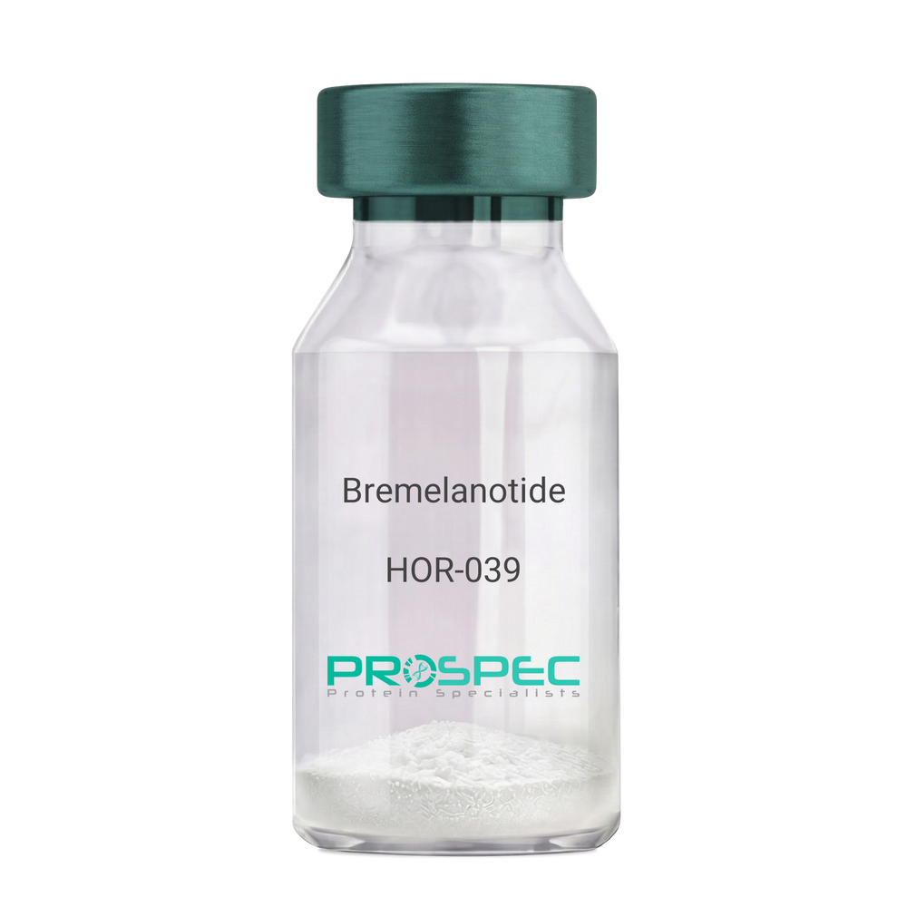
Name :
BremelanotideDescription:
Bremelanotide
PT-141, Rekynda, bremelanotide acetate.
Product # :
HOR-039Price :
Quantity :
Shipping Method :
Shipped at Room temp
More Info
- description
- formulation
- purity
- More Info
- Citations
Description
Bremelanotide Synthetic is a single, non-glycosylated polypeptide chain containing 6 amino acids, having a molecular mass of 1025.16 Dalton and a Molecular formula of C50H68N40O10.
Formulation
The protein was lyophilized with no additives.
Purity
Greater than 97.0% as determined by analysis by RP-HPLC.
Citations
More Info
-
Synonyms
PT-141, Rekynda, bremelanotide acetate.
-
Physical Appearance
Sterile Filtered White lyophilized (freeze-dried) powder.
-
Stability
Lyophilized Bremelanotide although stable at room temperature for 3 weeks, should be stored desiccated below -18°C. Upon reconstitution Bremelanotide should be stored at 4°C between 2-7 days and for future use below -18°C. For long term storage it is recommended to add a carrier protein (0.1% HSA or BSA). Please prevent freeze-thaw cycles.
-
Solubility
It is recommended to reconstitute the lyophilized Bremelanotide in sterile 18MΩ-cm H2O not less than 100 µg/ml, which can then be further diluted to other aqueous solutions.
-
Amino Acid Sequence
Ac-Nle-cyclo(-Asp-His-D-Phe-Arg-Trp-Lys)-OH.
-
Background
Bremelanotide (PT-141), a synthetic heptapeptide, is a novel therapeutic agent primarily utilized for the treatment of sexual dysfunction. This paper provides an exhaustive review of Bremelanotide, detailing its biochemical structure, mechanism of action, therapeutic applications, and future prospects in clinical medicine.
Bremelanotide (PT-141), derived from Melanotan II, is a synthetic peptide developed for its role in treating sexual dysfunction in both men and women (Clayton et al., 2016). This paper presents a comprehensive examination of Bremelanotide, its biological properties, and potential therapeutic uses.
Bremelanotide acts as a non-selective agonist of the melanocortin receptors, primarily MC3R and MC4R, present in the central nervous system. The peptide initiates its action in the hypothalamus, leading to downstream effects on sexual desire and arousal (King et al., 2007).
The US Food and Drug Administration approved Bremelanotide for the treatment of premenopausal women with hypoactive sexual desire disorder (HSDD) in 2019. In clinical trials, it demonstrated efficacy in enhancing sexual desire and reducing distress associated with HSDD (Kingsberg et al., 2019).
While the current focus of Bremelanotide is on treating sexual dysfunction, its future potential may extend to other areas due to its unique mechanism of action. Further research is needed to explore the full therapeutic potential of this intriguing peptide. In conclusion, Bremelanotide represents a significant advance in sexual dysfunction therapeutics, with potential implications extending beyond this realm.
ProSpec's products are furnished for LABORATORY RESEARCH USE ONLY. The product may not be used as drugs, agricultural or pesticidal products, food additives or household chemicals.
- View Data Sheet
Name :
FTCD HumanDescription:
Formiminotransferase Cyclodeaminase Human Recombinant
Formiminotransferase-cyclodeaminase, FTCD, LCHC1, LC-1.
Product # :
ENZ-302Price :
Quantity :
Shipping Method :
Shipped with Ice Packs
More Info
- description
- source
- formulation
- purity
- More Info
- Citations
Description
Formiminotransferase Cyclodeaminase Human Recombinant (also called liver cytosol type 1) produced in SF9, is a glycosylated, polypeptide chain having a molecular mass of 59,749 Dalton.The FTCD is expressed with a -6xHis tag and purified by proprietary chromatographic techniques.
Source
Sf9 insect cells.
Formulation
FTCD is supplied in 16mM HEPES buffer pH-7.6, 240mM sodium chloride, and 20% glycerol.
Purity
Greater than 95% as determined by SDS-PAGE.
Citations
More Info
-
Introduction
Formiminotransferase cyclodeaminase is an enzyme which catalyzes the conversion of formiminoglutamateand tetrahydrofolateinto formiminotetrahydrofolateand glutamate.
Formiminotransferase cyclodeaminase, a bifunctional enzyme of tetrahydrofolate synthesis, is the target antigen of anti-LC1 (liver cytosol antigen type 1) autoantibodies. Presence of LC1 autoantibodies is a marker for type 2 autoimmune hepatitis (for which anti-LKM 1/cytochrome P450 2D6 autoantibodies are a further marker).
Serum LC1 autoantibody concentrations appear to fluctuate in parallel with aminotransferase levels, a particularly intriguing observation that suggests a possible role of LC1 autoreactivity in the pathogenic mechanism leading to hepatocyte injury. -
Synonyms
Formiminotransferase-cyclodeaminase, FTCD, LCHC1, LC-1.
-
Stability
Store at 4°C if entire vial will be used within 2-4 weeks. Store, frozen at -20°Cfor longer periods of time. Avoid multiple freeze-thaw cycles.
-
Immunological Functions
1. Binds IgG-type human auto-antibodies.2. Standard ELISA test (checker-board analysis of positive/negative sera panels, immuno-dot test).
-
coating concentration
0.35-0.7 µg/ml (depending on the type of ELISA plate and coating buffer).Suitable for biotinylation and iodination.
ProSpec's products are furnished for LABORATORY RESEARCH USE ONLY. The product may not be used as drugs, agricultural or pesticidal products, food additives or household chemicals.
- View Data Sheet
Name :
RGS16 HumanDescription:
Regulator of G-Protein Signaling 16 Human Recombinant
A28-RGS14, A28-RGS14P, RGS-R, Regulator of G-protein signaling 16, RGS16, Retinally abundant regulator of G-protein signaling, Retinal-specific RGS, hRGS-r, RGSR.
Product # :
PRO-798Price :
Quantity :
Shipping Method :
Shipped with Ice Packs
More Info
- description
- source
- formulation
- purity
- More Info
- Citations
Description
RGS16 Human Recombinant produced in E.Coli is a single, non-glycosylated, polypeptide chain containing 222 amino acids (1-202 a.a.) and having a molecular mass of 24.9 kDa. The RGS16 is fused to a 20 amino acid His Tag at N-terminal and purified by proprietary chromatographic techniques.
Source
Escherichia Coli.
Formulation
The RGS16 solution (0.5mg/ml) contains Phosphate Buffered Saline (pH7.4), 0.1M NaCl and 20% glycerol.
Purity
Greater than 90% as determined by SDS-PAGE.
Citations
More Info
-
Introduction
RGS16 is part of the ''regulator of G protein signaling'' family. RGS16 inhibits signal transduction by increasing the GTPase activity of G protein alpha subunits thereby driving them into their inactive GDP-bound form. RGS16 Binds to G(i)-alpha and G(o)-alpha, but not to G(s)-alpha. RGS16 regulates the kinetics of signaling in the phototransduction cascade.
-
Synonyms
A28-RGS14, A28-RGS14P, RGS-R, Regulator of G-protein signaling 16, RGS16, Retinally abundant regulator of G-protein signaling, Retinal-specific RGS, hRGS-r, RGSR.
-
Physical Appearance
Sterile Filtered clear colorless solution.
-
Stability
Store at 4°C if entire vial will be used within 2-4 weeks. Store, frozen at -20°C for longer periods of time. For long term storage it is recommended to add a carrier protein (0.1% HSA or BSA). Avoid multiple freeze-thaw cycles.
-
Amino Acid Sequence
MGSSHHHHHH SSGLVPRGSH MCRTLAAFPT TCLERAKEFK TRLGIFLHKS ELGCDTGSTG KFEWGSKHSK ENRNFSEDVL GWRESFDLLL SSKNGVAAFH AFLKTEFSEE NLEFWLACEE FKKIRSATKL ASRAHQIFEE FICSEAPKEV NIDHETRELT RMNLQTATAT CFDAAQGKTR TLMEKDSYPR FLKSPAYRDL AAQASAASAT LSSCSLDEPS HT.
ProSpec's products are furnished for LABORATORY RESEARCH USE ONLY. The product may not be used as drugs, agricultural or pesticidal products, food additives or household chemicals.
- View Data Sheet
Name :
SIRPA HumanDescription:
Signal-Regulatory Protein Alpha Human Recombinant
Signal-Regulatory Protein Alpha, SHPS1, CD172 Antigen-Like Family Member A, Inhibitory Receptor SHPS-1, Macrophage Fusion Receptor, PTPNS1, SIRP, P84, BIT, MFR, Brain-Immunoglobulin-Like Molecule With Tyrosine-Based Activation Motifs, Brain Ig-Like Molecule With Tyrosine-Based Activation Motifs, Protein Tyrosine Phosphatase, Non-Receptor Type Substrate 1, Tyrosine-Protein Phosphatase Non-Receptor Type Substrate 1, Tyrosine Phosphatase SHP Substrate 1, Signal-Regulatory Protein Alpha-1 , ignal-Regulatory Protein Alpha-2, Signal-Regulatory Protein Alpha-3, SHP Substrate 1, CD172a Antigen, Myd-1 Antigen, MyD-1 Antigen, Sirp-Alpha-1, Sirp-Alpha-2, Sirp-Alpha-3, CD172A, SHPS-1, MYD-1, MYD1, Tyrosine-protein phosphatase non-receptor type substrate 1.
Product # :
PRO-2080Price :
Quantity :
Shipping Method :
Shipped with Ice Packs
More Info
- description
- source
- formulation
- purity
- More Info
- Citations
Description
SIRPA Human Recombinant produced in E.Coli is a single, non-glycosylated polypeptide chain containing 370 amino acids (27-373 a.a) and having a molecular mass of 40.4kDa. SIRPA is fused to a 23 amino acid His-tag at N-terminus & purified by proprietary chromatographic techniques.
Source
Escherichia Coli.
Formulation
SIRPA protein solution (0.5mg/ml) containing PBS buffer (pH 7.4), 10% glycerol and 1mM DTT.
Purity
Greater than 90.0% as determined by SDS-PAGE.
Citations
More Info
-
Introduction
Signal-Regulatory Protein Alpha, SIRPA belongs to the signal-regulatory-protein (SIRP) family, as well as theimmunoglobulin super family. The members of the SIRP family are receptor-type transmembrane glycoproteins which areinvolved in the negative regulation of receptor tyrosine kinase-coupled signaling processes. SIRPA can bephosphorylated by tyrosine kinases. The phospho-tyrosine residues of this PTP have been shown to recruit SH2domain containing tyrosine phosphatases (PTP), and perform as substrates of PTPs. SIRPA take part insignal transduction mediated by a variety of growth factor receptors. CD47 has been shown to be a ligand for SIRPA.
-
Synonyms
Signal-Regulatory Protein Alpha, SHPS1, CD172 Antigen-Like Family Member A, Inhibitory Receptor SHPS-1, Macrophage Fusion Receptor, PTPNS1, SIRP, P84, BIT, MFR, Brain-Immunoglobulin-Like Molecule With Tyrosine-Based Activation Motifs, Brain Ig-Like Molecule With Tyrosine-Based Activation Motifs, Protein Tyrosine Phosphatase, Non-Receptor Type Substrate 1, Tyrosine-Protein Phosphatase Non-Receptor Type Substrate 1, Tyrosine Phosphatase SHP Substrate 1, Signal-Regulatory Protein Alpha-1 , ignal-Regulatory Protein Alpha-2, Signal-Regulatory Protein Alpha-3, SHP Substrate 1, CD172a Antigen, Myd-1 Antigen, MyD-1 Antigen, Sirp-Alpha-1, Sirp-Alpha-2, Sirp-Alpha-3, CD172A, SHPS-1, MYD-1, MYD1, Tyrosine-protein phosphatase non-receptor type substrate 1.
-
Physical Appearance
Sterile Filtered colorless solution.
-
Stability
Store at 4°C if entire vial will be used within 2-4 weeks. Store, frozen at -20°C for longer periods of time. For long term storage it is recommended to add a carrier protein (0.1% HSA or BSA).Avoid multiple freeze-thaw cycles.
-
Amino Acid Sequence
MGSSHHHHHH SSGLVPRGSH MGSGVAGEEE LQVIQPDKSV LVAAGETATL RCTATSLIPV GPIQWFRGAG PGRELIYNQK EGHFPRVTTV SDLTKRNNMD FSIRIGNITP ADAGTYYCVK FRKGSPDDVE FKSGAGTELS VRAKPSAPVV SGPAARATPQ HTVSFTCESH GFSPRDITLK WFKNGNELSD FQTNVDPVGE SVSYSIHSTA KVVLTREDVH SQVICEVAHV TLQGDPLRGT ANLSETIRVP PTLEVTQQPV RAENQVNVTC QVRKFYPQRL QLTWLENGNV SRTETASTVT ENKDGTYNWM SWLLVNVSAH RDDVKLTCQV EHDGQPAVSK SHDLKVSAHP KEQGSNTAAE NTGSNERNIY.
ProSpec's products are furnished for LABORATORY RESEARCH USE ONLY. The product may not be used as drugs, agricultural or pesticidal products, food additives or household chemicals.
- View Data Sheet
Name :
TARDBP HumanDescription:
TAR DNA Binding Protein Human Recombinant
ALS10, TDP43, TAR DNA-binding protein 43, TDP-43, TARDBP.
Product # :
PRO-1410Price :
Quantity :
Shipping Method :
Shipped with Ice Packs
More Info
- description
- source
- formulation
- purity
- More Info
- Citations
Description
TARDBP Human Recombinant produced in E.Coli is a single, non-glycosylated polypeptide chain containing 296 amino acids (1-260 a.a) and having a molecular mass of 33.6kDa. TARDBP is fused to a 36 amino acid His-tag at N-terminus & purified by proprietary chromatographic techniques.
Source
E.coli.
Formulation
TARDBP protein solution (0.5mg/ml) containing 20mM Tris-HCl buffer (pH8.0), 10% glycerol and 0.1M NaCl.
Purity
Greater than 90% as determined by SDS-PAGE.
Citations
More Info
-
Introduction
TARDBP binds both DNA and RNA and have numerous roles in transcriptional repression, pre-mRNA splicing and translational regulation. TARDBP was initially identified as a transcriptional repressor that binds to chromosomally integrated TAR DNA and represses HIV-1 transcription. TARDBP has also been identified in individuals diagnosed with chronic traumatic ncephalopathy, a condition which frequently mimics ALS and that has been associated with athletes who have experienced multiple concussions and other types of head injury. TARDBP may also be involved in microRNA biogenesis, apoptosis and cell division.
-
Synonyms
ALS10, TDP43, TAR DNA-binding protein 43, TDP-43, TARDBP.
-
Physical Appearance
Sterile Filtered colorless solution.
-
Stability
Store at 4°C if entire vial will be used within 2-4 weeks. Store, frozen at -20°C for longer periods of time. For long term storage it is recommended to add a carrier protein (0.1% HSA or BSA).Avoid multiple freeze-thaw cycles.
-
Amino Acid Sequence
MRGSHHHHHH GMASMTGGQQ MGRDLYDDDD KDRWGSMSEY IRVTEDENDE PIEIPSEDDG TVLLSTVTAQ FPGACGLRYR NPVSQCMRGV RLVEGILHAP DAGWGNLVYV VNYPKDNKRK MDETDASSAV KVKRAVQKTS DLIVLGLPWK TTEQDLKEYF STFGEVLMVQ VKKDLKTGHS KGFGFVRFTE YETQVKVMSQ RHMIDGRWCD CKLPNSKQSQ DEPLRSRKVF VGRCTEDMTE DELREFFSQY GDVMDVFIPK PFRAFAFVTF ADDQIAQSLC GEDLIIKGIS VHISNA.
ProSpec's products are furnished for LABORATORY RESEARCH USE ONLY. The product may not be used as drugs, agricultural or pesticidal products, food additives or household chemicals.
- View Data Sheet
Name :
MG HumanDescription:
Menopausal Gonadotropin Human
Product # :
HOR-251Price :
Quantity :
Shipping Method :
Shipped at Room temp
More Info
- description
- source
- formulation
- biological activity
- More Info
- Citations
Description
Menopausal Gonadotropin Human is produced from a sterile preparation of placental glucoprotein urine of post-menopausal women.The MG is purified by proprietary chromatographic techniques.
Source
Urine of post-menopausal women.
Formulation
The Human MG was lyophilized from a concentrated (1mg/ml) solution with no additives.
Biological Activity
100 IU/mg of FSH and 100 IU/mg LH.Citations
More Info
-
Physical Appearance
Sterile Filtered White lyophilized (freeze-dried) powder.
-
Stability
Lyophilized Menopausal Gonadotropin Human although stable at room temperature for 3 weeks, should be stored desiccated below -18°C. Upon reconstitution MG should be stored at 4°C between 2-7 days and for future use below -18°C.For long term storage it is recommended to add a carrier protein (0.1% HSA or BSA).Please prevent freeze-thaw cycles.
-
Solubility
It is recommended to reconstitute the lyophilized MG in sterile 18MΩ-cm H2O not less than 100 µg/ml, which can then be further diluted to other aqueous solutions.
-
Contaminants
Free of: HbsAg, Hepatitis B surface antigen and antibodies to HIV, Hepatitis C and HIV.
ProSpec's products are furnished for LABORATORY RESEARCH USE ONLY. The product may not be used as drugs, agricultural or pesticidal products, food additives or household chemicals.
- View Data Sheet
Name :
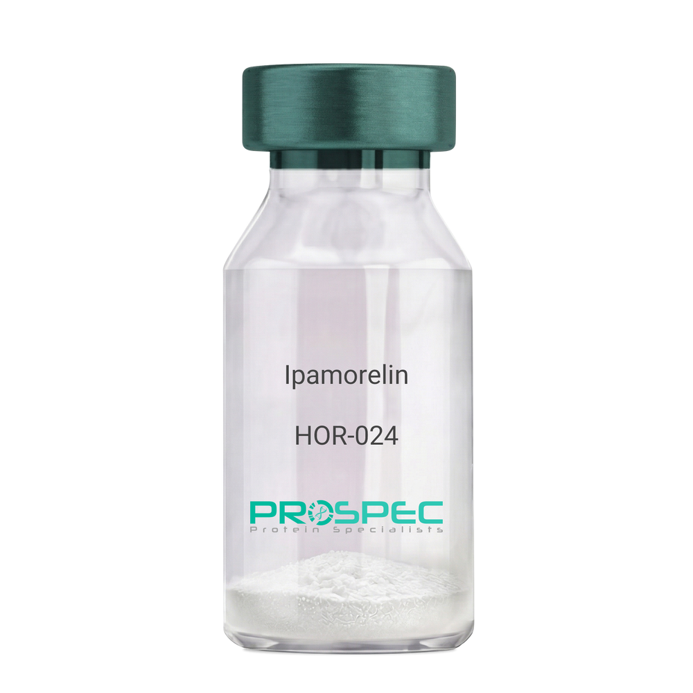
IpamorelinDescription:
Ipamorelin
Ipamorelin
Product # :
HOR-024Price :
Quantity :
Shipping Method :
Shipped at Room temp
More Info
- description
- formulation
- purity
- More Info
- Citations
Description
Ipamorelin Synthetic is a single, non-glycosylated polypeptide chain containing 4 amino acids, having a molecular mass of 711.85 Dalton and a Molecular formula of C38H49N9O5.
Formulation
The protein was lyophilized with no additives.
Purity
Greater than 97.0% as determined by analysis by RP-HPLC.
Citations
More Info
-
Introduction
Ipamorelin is a peptide selective agonist of the ghrelin/growth hormone secretagogue receptor and a growth hormone secretagogue. Ipamorelin is a pentapeptide that was derived from GHRP1. Ipamorelin significantly increases plasma growth hormone levels in both animals and humans. Like pralmorelin and GHRP-6, ipamorelin does not affect prolactin, FSH, LH or TSH levels. However, unlike GHRP2 and GHRP6, but as growth hormone-releasing hormone (GHRH), ipamorelin does not stimulate the secretion of adrenocorticotropic hormone (ACTH) or cortisol, and is highly selective for inducing the secretion only of GH.
-
Synonyms
Ipamorelin
-
Physical Appearance
Sterile Filtered White lyophilized (freeze-dried) powder.
-
Stability
Lyophilized Ipamorelin although stable at room temperature for 3 weeks, should be stored desiccated below -18°C. Upon reconstitution Ipamorelin should be stored at 4°C between 2-7 days and for future use below -18°C. For long term storage it is recommended to add a carrier protein (0.1% HSA or BSA). Please prevent freeze-thaw cycles.
-
Solubility
It is recommended to reconstitute the lyophilized Ipamorelin in sterile 18MΩ-cm H2O not less than 100 µg/ml, which can then be further diluted to other aqueous solutions.
-
Amino Acid Sequence
Aib-His-D-2-Nal-D-Phe-Lys-NH2.
ProSpec's products are furnished for LABORATORY RESEARCH USE ONLY. The product may not be used as drugs, agricultural or pesticidal products, food additives or household chemicals.