Skip to content

High-quality research proteins.

  • 1-800-805-6531

  • Mon - Sat 8.00-18.00, Sun - Closed

  • info@prospecbio.com

  • PO Box 6591 East Brunswick NJ 08816

  • Products
    • Cytokines
    • Growth factors
    • Chemokines
    • Cd antigens
    • Neurotrophins
    • Hormones
    • Enzymes
    • Viral antigens
    • Recombinant proteins
    • Natural proteins
    • Monoclonal antibodies
    • Polyclonal antibodies
    • Thermal Packaging
  • About
    • Company
    • Founder
    • Contribution
  • Customers
    • Eastern Asia
    • Eastern Europe
    • North America
    • South America
    • South East Asia
    • Southern Asia
    • Western Europe
  • Ordering
    • Ordering Method
    • Ordering - Credit Card
    • Ordering - PO
    • Shipping Fees
    • FAQs
  • Services
    • DNA Cloning
    • Protein Expression
    • Monoclonal Antibody
    • Fermentation
    • Technology License
  • Contact us
Log in

Country/region

  • USD $ | Albania
  • USD $ | Andorra
  • USD $ | Argentina
  • USD $ | Armenia
  • USD $ | Australia
  • USD $ | Austria
  • USD $ | Azerbaijan
  • USD $ | Belarus
  • USD $ | Belgium
  • USD $ | Bolivia
  • USD $ | Brazil
  • USD $ | Bulgaria
  • USD $ | Cambodia
  • USD $ | Canada
  • USD $ | Chile
  • USD $ | China
  • USD $ | Colombia
  • USD $ | Costa Rica
  • USD $ | Croatia
  • USD $ | Cyprus
  • USD $ | Czechia
  • USD $ | Denmark
  • USD $ | Estonia
  • USD $ | Finland
  • USD $ | France
  • USD $ | Georgia
  • USD $ | Germany
  • USD $ | Greece
  • USD $ | Hong Kong SAR
  • USD $ | Hungary
  • USD $ | Iceland
  • USD $ | India
  • USD $ | Ireland
  • USD $ | Israel
  • USD $ | Italy
  • USD $ | Japan
  • USD $ | Kazakhstan
  • USD $ | Latvia
  • USD $ | Liechtenstein
  • USD $ | Lithuania
  • USD $ | Madagascar
  • USD $ | Malta
  • USD $ | Mexico
  • USD $ | Moldova
  • USD $ | Monaco
  • USD $ | Netherlands
  • USD $ | New Zealand
  • USD $ | North Macedonia
  • USD $ | Norway
  • USD $ | Panama
  • USD $ | Paraguay
  • USD $ | Peru
  • USD $ | Philippines
  • USD $ | Poland
  • USD $ | Portugal
  • USD $ | Romania
  • USD $ | Singapore
  • USD $ | Slovakia
  • USD $ | Slovenia
  • USD $ | South Africa
  • USD $ | South Korea
  • USD $ | Spain
  • USD $ | Sri Lanka
  • USD $ | Sweden
  • USD $ | Switzerland
  • USD $ | Taiwan
  • USD $ | Thailand
  • USD $ | Türkiye
  • USD $ | Ukraine
  • USD $ | United Kingdom
  • USD $ | United States
  • USD $ | Uruguay
  • USD $ | Vatican City
  • USD $ | Venezuela
  • USD $ | Vietnam
  • Facebook
  • Instagram
logoProSpec - Recombinant Protein Manufacturer for academic and R&D industry
Become a distributor
Account Log in
Wishlist
Cart Cart(0)
  • Products
    +
    • Cytokines
    • Growth factors
    • Chemokines
    • Cd antigens
    • Neurotrophins
    • Hormones
    • Enzymes
    • Viral antigens
    • Recombinant proteins
    • Natural proteins
    • Monoclonal antibodies
    • Polyclonal antibodies
    • Thermal Packaging
  • About
    +
    • Company
    • Founder
    • Contribution
  • Customers
    +
    • Eastern Asia
    • Eastern Europe
    • North America
    • South America
    • South East Asia
    • Southern Asia
    • Western Europe
  • Ordering
    +
    • Ordering Method
    • Ordering - Credit Card
    • Ordering - PO
    • Shipping Fees
    • FAQs
  • Services
    +
    • DNA Cloning
    • Protein Expression
    • Monoclonal Antibody
    • Fermentation
    • Technology License
  • Contact us

Item added to your cart

View cart
View All
  • Cytokines
  • Growth factors
  • Chemokines
  • Cd antigens
  • Neurotrophins
  • Hormones
  • Enzymes
  • Viral antigens
  • Recombinant proteins
  • Natural proteins
  • Monoclonal antibodies
  • Polyclonal antibodies
  • Thermal Packaging
  • Tumor Necrosis Factor

    Tumor Necrosis Factor

    View All
  • 4-1BB

    4-1BB

    View All
  • Adiponectin

    Adiponectin

    View All
  • AIF1

    AIF1

    View All
  • Other Cytokines

    Other Cytokines

    View All
  • AITRL

    AITRL

    View All
  • Angiopoietin

    Angiopoietin

    View All
  • Apolipoprotein

    Apolipoprotein

    View All
  • B-Cell Activating Factor

    B-Cell Activating Factor

    View All
  • Beta Defensin

    Beta Defensin

    View All
  • Bone Morphogenetic Protein

    Bone Morphogenetic Protein

    View All
  • B type Natriuretic Peptide

    B type Natriuretic Peptide

    View All
  • BST

    BST

    View All
  • Betacellulin

    Betacellulin

    View All
  • Cardiotrophin

    Cardiotrophin

    View All
  • Activin

    Activin

    View All
  • Fibroblast Growth Factor

    Fibroblast Growth Factor

    View All
  • Other Growth Factors

    Other Growth Factors

    View All
  • CSF

    CSF

    View All
  • CTGF

    CTGF

    View All
  • IGFBP

    IGFBP

    View All
  • VEGF Protein

    VEGF Protein

    View All
  • EGF

    EGF

    View All
  • Epigen

    Epigen

    View All
  • Erythropoietin

    Erythropoietin

    View All
  • Insulin-Like Growth Factor

    Insulin-Like Growth Factor

    View All
  • Growth Hormone

    Growth Hormone

    View All
  • HDGF

    HDGF

    View All
  • Hepatocyte Growth Factor

    Hepatocyte Growth Factor

    View All
  • Insulin

    Insulin

    View All
  • BCA-1/ BLC (CXCL13)

    BCA-1/ BLC (CXCL13)

    View All
  • BRAK (CXCL14)

    BRAK (CXCL14)

    View All
  • C-10 (CCL6)

    C-10 (CCL6)

    View All
  • Other Chemokines

    Other Chemokines

    View All
  • MEC (CCL28)

    MEC (CCL28)

    View All
  • LD78-beta (CCL3L1)

    LD78-beta (CCL3L1)

    View All
  • CTACK (CCL27)

    CTACK (CCL27)

    View All
  • CXCL16

    CXCL16

    View All
  • CXCL17

    CXCL17

    View All
  • Platelet Factor-4 (CXCL4)

    Platelet Factor-4 (CXCL4)

    View All
  • ENA-78 (CXCL5)

    ENA-78 (CXCL5)

    View All
  • Eotaxin (CCL11,24,26)

    Eotaxin (CCL11,24,26)

    View All
  • Exodus-2 (CCL21)

    Exodus-2 (CCL21)

    View All
  • Fractalkine (CX3CL1)

    Fractalkine (CX3CL1)

    View All
  • CXCL6

    CXCL6

    View All
  • Other CD Antigens

    Other CD Antigens

    View All
  • CD14

    CD14

    View All
  • CD164

    CD164

    View All
  • CD1

    CD1

    View All
  • CD2

    CD2

    View All
  • CD200

    CD200

    View All
  • CD207

    CD207

    View All
  • CD226

    CD226

    View All
  • CD23

    CD23

    View All
  • CD244

    CD244

    View All
  • CD247

    CD247

    View All
  • CD27

    CD27

    View All
  • CD274

    CD274

    View All
  • CD300

    CD300

    View All
  • CD33

    CD33

    View All
  • Other Neurotrophins

    Other Neurotrophins

    View All
  • Beta-NGF

    Beta-NGF

    View All
  • BDNF

    BDNF

    View All
  • CDNF

    CDNF

    View All
  • CNTF

    CNTF

    View All
  • GDNF

    GDNF

    View All
  • Glia Maturation Factor

    Glia Maturation Factor

    View All
  • MANF

    MANF

    View All
  • Midkine

    Midkine

    View All
  • Neuroglobin

    Neuroglobin

    View All
  • Neurotrophic factors

    Neurotrophic factors

    View All
  • Neuregulin

    Neuregulin

    View All
  • Neuropilin

    Neuropilin

    View All
  • Pigment Epithelium-Derived Factor

    Pigment Epithelium-Derived Factor

    View All
  • Pleiotrophin

    Pleiotrophin

    View All
  • Peptide Hormones

    Peptide Hormones

    View All
  • Other Hormones

    Other Hormones

    View All
  • HCG

    HCG

    View All
  • Endothelin

    Endothelin

    View All
  • Exendin

    Exendin

    View All
  • FSH

    FSH

    View All
  • Glucagon

    Glucagon

    View All
  • GHRP

    GHRP

    View All
  • GLP

    GLP

    View All
  • Thyrostimulin

    Thyrostimulin

    View All
  • Inhibin A

    Inhibin A

    View All
  • LHRH

    LHRH

    View All
  • Melanotan

    Melanotan

    View All
  • Procalcitonin

    Procalcitonin

    View All
  • PTH

    PTH

    View All
  • Peroxiredoxin

    Peroxiredoxin

    View All
  • Peptidase

    Peptidase

    View All
  • Synthetase

    Synthetase

    View All
  • Transferase

    Transferase

    View All
  • Hydrolase

    Hydrolase

    View All
  • Dehydrogenase

    Dehydrogenase

    View All
  • ACE2

    ACE2

    View All
  • Esterase

    Esterase

    View All
  • Protein Kinases

    Protein Kinases

    View All
  • Other Enzymes

    Other Enzymes

    View All
  • Phosphatase

    Phosphatase

    View All
  • Deaminase

    Deaminase

    View All
  • Oxygenase

    Oxygenase

    View All
  • Adenylate Kinase

    Adenylate Kinase

    View All
  • Lyase

    Lyase

    View All
  • Chagas

    Chagas

    View All
  • SARS

    SARS

    View All
  • Influenza

    Influenza

    View All
  • ASFV

    ASFV

    View All
  • Astrovirus

    Astrovirus

    View All
  • Borrelia

    Borrelia

    View All
  • Helicobacter Pylori

    Helicobacter Pylori

    View All
  • Chikungunya

    Chikungunya

    View All
  • Chlamydia

    Chlamydia

    View All
  • Cytomegalo

    Cytomegalo

    View All
  • Dengue

    Dengue

    View All
  • EBV

    EBV

    View All
  • Ebola

    Ebola

    View All
  • Feline Leukemia Virus

    Feline Leukemia Virus

    View All
  • Hepatitis A

    Hepatitis A

    View All
  • Other

    Other

    View All
  • Actin

    Actin

    View All
  • ADAM

    ADAM

    View All
  • Complement Component

    Complement Component

    View All
  • Ag85

    Ag85

    View All
  • Eukaryotic Translation Initiation Factor

    Eukaryotic Translation Initiation Factor

    View All
  • Anterior Gradient Protein

    Anterior Gradient Protein

    View All
  • Heat Shock Protein

    Heat Shock Protein

    View All
  • Alpha-2-HS-Glycoprotein

    Alpha-2-HS-Glycoprotein

    View All
  • Hemoglobin

    Hemoglobin

    View All
  • Allergy

    Allergy

    View All
  • Anaplasma

    Anaplasma

    View All
  • Angiogenin

    Angiogenin

    View All
  • Ankyrin Repeat Domain

    Ankyrin Repeat Domain

    View All
  • Annexin

    Annexin

    View All
  • Other Natural Proteins

    Other Natural Proteins

    View All
  • Aprotinin

    Aprotinin

    View All
  • Transferrin

    Transferrin

    View All
  • Avidin

    Avidin

    View All
  • Anti Coagulation Factors

    Anti Coagulation Factors

    View All
  • Natural Albumin

    Natural Albumin

    View All
  • Natural Coagulation Factors

    Natural Coagulation Factors

    View All
  • Fibronectin

    Fibronectin

    View All
  • Thrombin

    Thrombin

    View All
  • Anti Human Enzyme

    Anti Human Enzyme

    View All
  • Other Monoclonal Antibodies

    Other Monoclonal Antibodies

    View All
  • Anti Human Cytokine

    Anti Human Cytokine

    View All
  • Anti Human Heat Shock Protein

    Anti Human Heat Shock Protein

    View All
  • Anti Mouse Lymphocyte

    Anti Mouse Lymphocyte

    View All
  • Anti Human Chemokine

    Anti Human Chemokine

    View All
  • Anti Human Lymphocyte

    Anti Human Lymphocyte

    View All
  • Anti Viral Monoclonal

    Anti Viral Monoclonal

    View All
  • Anti Mouse Cytokine

    Anti Mouse Cytokine

    View All
  • Anti-GST Monoclonal

    Anti-GST Monoclonal

    View All
  • Other Polyclonal Antibodies

    Other Polyclonal Antibodies

    View All
  • Anti Viral

    Anti Viral

    View All
  • Anti-GST Monoclonal

    Anti-GST Monoclonal

    View All
Back

Search results

1000 results found for “batf”

Name

Description

Product #

Price

Quantity

Shipping Method

  • View Data Sheet

    Name :

    PDCD5 Human

    Description:

    Programmed Cell Death-5 Human Recombinant

    Programmed cell death protein 5, TF-1 cell apoptosis-related protein 19, Protein TFAR19, PDCD5, TFAR19, MGC9294.

    Product # :

    PRO-624

    Price :

    Quantity :

    Shipping Method :

    Ice Icon

    Shipped with Ice Packs

    Add To Cart

    More Info

    • description
    • source
    • formulation
    • purity
    • More Info

    Description

    PDCD5 Recombinant produced in E.Coli is a single, non-glycosylated polypeptide chain containing 125 amino acids and having a molecular mass of 14 kDa.

    Source

    Escherichia Coli.

    Formulation

    PDCD5 protein solution contains 1x PBS pH-7.4.

    Purity

    Greater than 95.0% as determined by SDS-PAGE.

    More Info

    • Introduction

      PDCD5 is expressed in tumor cells during apoptosis independent of the apoptosis-inducing stimuli. Prior to apoptosis induction, PDCD5 is distributed in both the nucleus and cytoplasm. Once apoptosis is induced, the amount of PDCD5 increases and by relocation from the cytoplasm, it accumulates in the nucleus.PDCD5 protein has a stable helical core conformation consisting of a triple-helix bundle and two dissociated terminal regions. PDCD5 is an important novel protein that regulates both apoptotic and non-apoptotic programmed cell death. PDCD5 functions in the process of apoptosis.
      PDCD5 plays an important role in regulation of apoptotic processes in gastric cancer cells and gastric tumors.
      PDCD5 plays a role in the pathogenesis of rheumatoid arthritis.
      -27G/-11A SNP is associated with reduced PDCD5 promoter activity and increased susceptibility to chronic myelogenous leukemia.
      PDCD5 gene may be a target gene under the control of some important apoptosis-related transcriptional factors during the cell apoptosis.

    • Synonyms

      Programmed cell death protein 5, TF-1 cell apoptosis-related protein 19, Protein TFAR19, PDCD5, TFAR19, MGC9294.

    • Physical Appearance

      Sterile Filtered colorless solution.

    • Stability

      Store at 4°C if entire vial will be used within 2-4 weeks. Store, frozen at -20°C for longer periods of time. For long term storage it is recommended to add a carrier protein (0.1% HSA or BSA).Avoid multiple freeze-thaw cycles.

    • Amino Acid Sequence

      MADEELEALR RQRLAELQAK HGDPGDAAQQ EAKHRGAEMR NSILAQVLDQ SARARLSNLA LVKPEKTKAV ENYLIQMARY GQLSEKVSEQ GLIEILKKVS QQTEKTTTVK LNRRKVMDSD EDDDY.

    ProSpec's products are furnished for LABORATORY RESEARCH USE ONLY. The product may not be used as drugs, agricultural or pesticidal products, food additives or household chemicals.

    Pdcd5 Human
  • View Data Sheet

    Name :

    FASLG Human

    Description:

    FAS Ligand Human Recombinant

    Tumor necrosis factor ligand superfamily member 6, Apoptosis antigen ligand, APTL, CD95 ligand, CD95-L, Fas antigen ligand, Fas ligand, FasL, CD178, FASLG, APT1LG1, CD95L, TNFSF6, ALPS1B.

    Product # :

    CYT-031

    Price :

    Quantity :

    Shipping Method :

    Ice Icon

    Shipped with Ice Packs

    Add To Cart

    More Info

    • description
    • source
    • formulation
    • purity
    • More Info
    • sds-page

    Description

    FASLG Human Recombinant produced in E.Coli is a single, non-glycosylated polypeptide chain containing 173 amino acids (130-281 a.a.) and having a molecular mass of 19.6kDa.FASLG is fused to a 21 amino acid His-tag at N-terminus & purified by proprietary chromatographic techniques.

    Source

    Escherichia Coli.

    Formulation

    FASLG protein solution (1mg/ml) containing 20mM Tris-HCl buffer (pH 8.0), 0.4M urea and 10% glycerol.

    Purity

    Greater than 90% as determined by SDS-PAGE.

    sds-page

    FASL-sds-page - Product image 1

    More Info

    • Introduction

      The type II transmembrane protein FASLG is a member of the tumor necrosis factor (TNF) superfamily. A fas ligand/receptor interaction has a significant part in the regulation of the immune system and the advancement of cancer. FASLG is expressed on the activated T cell surface as a nondisulfidelinked homotrimer. FASLG binding to Fas/CD95/TNFRSF6 on a nearby cell prompts apoptosis in the Fas expressing cell. FASLG is released from the cell surface by metalloproteinases as a soluble molecule that stays trimeric and is able to bind with Fas, but its capability to activate apoptosis is radically reduced. In addition, FASLG binds to DcR3 - a soluble trap receptor with no signal transduction capabilities. Flawed Fas-mediated apoptosis causes oncogenesis in addition to drug resistance in existing tumors. Constitutive expression of FASLG in a variety of tumors enables their immune evasion. Both mouse and human FASLG are active on mouse and human cells.

    • Synonyms

      Tumor necrosis factor ligand superfamily member 6, Apoptosis antigen ligand, APTL, CD95 ligand, CD95-L, Fas antigen ligand, Fas ligand, FasL, CD178, FASLG, APT1LG1, CD95L, TNFSF6, ALPS1B.

    • Physical Appearance

      Sterile Filtered colorless solution.

    • Stability

      Store at 4°C if entire vial will be used within 2-4 weeks. Store, frozen at -20°C for longer periods of time. For long term storage it is recommended to add a carrier protein (0.1% HSA or BSA).Avoid multiple freeze-thaw cycles.

    • Amino Acid Sequence

      MGSSHHHHHH SSGLVPRGSH MQIGHPSPPP EKKELRKVAH LTGKSNSRSM PLEWEDTYGI VLLSGVKYKK GGLVINETGL YFVYSKVYFR GQSCNNLPLS HKVYMRNSKY PQDLVMMEGK MMSYCTTGQM WARSSYLGAV FNLTSADHLY VNVSELSLVN FEESQTFFGL YKL.

    • Background

      What is the molecular weight/Mw of FASL Protein?
      FASL Protein has a total Mw of 19.6kDa.

      What is the source or expression system of FASL Protein?
      Escherichia Coli.

      What is the Purity of FASL Protein?
      FASL Protein is >90% pure as determined by SDS-PAGE.

      What is the Biological Activity of FASL Protein?
      The biological functionality of FASL Protein will be determined in the future.

      What is the amino acid sequence of FASL Protein?
      MGSSHHHHHH SSGLVPRGSH MQIGHPSPPP EKKELRKVAH LTGKSNSRSM PLEWEDTYGI VLLSGVKYKK GGLVINETGL YFVYSKVYFR GQSCNNLPLS HKVYMRNSKY PQDLVMMEGK MMSYCTTGQM WARSSYLGAV FNLTSADHLY VNVSELSLVN FEESQTFFGL YKL.

      What applications can FASL Protein be used in?
      FASL Protein can probably be used in western blot, ELISA and Lateral Flow.

      What is the endotoxin level for FASL Protein?
      The endotoxin level is minimal, FASL Protein was purified using conventional chromatography techniques.

    ProSpec's products are furnished for LABORATORY RESEARCH USE ONLY. The product may not be used as drugs, agricultural or pesticidal products, food additives or household chemicals.

    Faslg Human
  • View Data Sheet

    Name :

    TCEA1 Human

    Description:

    Transcription Elongation Factor A (SII)-1 Human Recombinant

    Transcription elongation factor A (SII) 1, Transcription elongation factor TFIIS.o, TCEA, GTF2S, SII, TF2S.

    Product # :

    PRO-1098

    Price :

    Quantity :

    Shipping Method :

    Ice Icon

    Shipped with Ice Packs

    Add To Cart

    More Info

    • description
    • source
    • formulation
    • purity
    • More Info

    Description

    TCEA1 Human Recombinant produced in E. coli is a single polypeptide chain containing 325 amino acids (1-301) and having a molecular mass of 36.5kDa.TCEA1 is fused to a 24 amino acid His-tag at N-terminus & purified by proprietary chromatographic techniques.

    Source

    E.coli.

    Formulation

    The TCEA1 solution (1mg/1ml) contains 20mM Tris-HCl buffer (pH 7.5), 1mM DTT, 50mM NaCl and 20% glycerol.

    Purity

    Greater than 90% as determined by SDS-PAGE.

    More Info

    • Introduction

      TCEA1 is a member of the TFS-II family. TCEA1 binds to Pol II and operates to cleave the emerging transcript, thus unlocking the complex and permitting transcription to continue. TCEA1 is localized to the nucleus and holds three independently-folding domains that are essential for accurate binding to Pol II. The arresting sites in are able to trap a certain segment of elongating RNA polymerases that goes through, causing in a locked ternary complex. Cleavage of the emerging transcript by S-II enables the renewal of elongation from the new 3'-terminus.

    • Synonyms

      Transcription elongation factor A (SII) 1, Transcription elongation factor TFIIS.o, TCEA, GTF2S, SII, TF2S.

    • Physical Appearance

      Sterile Filtered colorless solution.

    • Stability

      Store at 4°C if entire vial will be used within 2-4 weeks. Store, frozen at -20°C for longer periods of time. For long term storage it is recommended to add a carrier protein (0.1% HSA or BSA).Avoid multiple freeze-thaw cycles.

    • Amino Acid Sequence

      MGSSHHHHHH SSGLVPRGSH MGSHMEDEVV RFAKKMDKMV QKKNAAGALD LLKELKNIPM TLELLQSTRI GMSVNAIRKQ STDEEVTSLA KSLIKSWKKL LDGPSTEKDL DEKKKEPAIT SQNSPEAREE STSSGNVSNR KDETNARDTY VSSFPRAPST SDSVRLKCRE MLAAALRTGD DYIAIGADEE ELGSQIEEAI YQEIRNTDMK YKNRVRSRIS NLKDAKNPNL RKNVLCGNIP PDLFARMTAE EMASDELKEM RKNLTKEAIR EHQMAKTGGT QTDLFTCGKC KKKNCTYTQV QTRSADEPMT TFVVCNECGN RWKFC

    ProSpec's products are furnished for LABORATORY RESEARCH USE ONLY. The product may not be used as drugs, agricultural or pesticidal products, food additives or household chemicals.

    Tcea1 Human
  • View Data Sheet

    Name :

    TIE1 Fc Human

    Description:

    TIE1 Fc Human Recombinant

    Tyrosine kinase with immunoglobulin-like and EGF-like domains 1, JTK14, TIE, TIE1.

    Product # :

    PKA-246

    Price :

    Quantity :

    Shipping Method :

    Room Temp Icon

    Shipped at Room temp

    Add To Cart

    More Info

    • description
    • source
    • formulation
    • purity
    • More Info

    Description

    Soluble TIE-1 Human Recombinant fused with the Fc part of human IgG1 produced in baculovirus is a homodimeric, glycosylated, polypeptide containing 749 amino acids and having a total molecular mass of 250 kDa. Human TIE-1/Fc monomer has a calculated molecular mass of approximately 105 kDa. As a result of glycosylation, the recombinant protein migrates as an approximately 125 kDa protein in SDS-PAGE under reducing conditions.The TIE1 Fc Chimera is purified by proprietary chromatographic techniques.

    Source

    Insect Cells.

    Formulation

    TIE-1 Fc Chimera was lyophilized from a concentrated (1mg/ml) sterile solution containing 20mM Tris, 0.5M NaCl, 10% Sucrose.

    Purity

    Greater than 90.0% as determined by:
    (A) Analysis by RP-HPLC.
    (B) Analysis by SDS-PAGE.

    More Info

    • Introduction

      TIE-1 (tyrosine kinase with Ig and EGF homology domains 1) and TIE-2/Tek comprise a receptor tyrosine kinase (RTK) subfamily with unique structural characteristics: two immunoglobulin-like domains flanking three epidermal growth factor (EGF)-like domains and followed by three fibronectin type III-like repeats in the extracellular region and a split tyrosine kinase domain in the cytoplasmic region. These receptors are expressed primarily on endothelial and hematopoietic progenitor cells and play critical roles in angiogenesis, vasculogenesis and hematopoiesis. Human TIE-1 cDNA encodes a 1124 amino acid (aa) residue precursor protein with an 18 residue putative signal peptide, a 727 residue extracellular domain and a 354 residue cytoplasmic domain. Whereas two ligands have been described for TIE-2 [angiopoietin-1 (Ang1) and angiopoietin-2 (Ang2)], so far no ligand was found for TIE-1.

    • Synonyms

      Tyrosine kinase with immunoglobulin-like and EGF-like domains 1, JTK14, TIE, TIE1.

    • Physical Appearance

      Sterile Filtered White lyophilized (freeze-dried) powder.

    • Stability

      Lyophilized sTIE-1 although stable at room temperature for 3 weeks, should be stored desiccated below -18°C. Upon reconstitution TIE-1 should be stored at 4°C between 2-7 days and for future use below -18°C.For long term storage it is recommended to add a carrier protein (0.1% HSA or BSA).Please prevent freeze-thaw cycles.

    • Solubility

      It is recommended to reconstitute the lyophilized TIE-1 Fc Chimera in sterile water not less than 100µg/ml, which can then be further diluted to other aqueous solutions.

    ProSpec's products are furnished for LABORATORY RESEARCH USE ONLY. The product may not be used as drugs, agricultural or pesticidal products, food additives or household chemicals.

    Tie 1 Human Fc
  • View Data Sheet

    Name :

    Flt3 Ligand Human

    Description:

    Flt3 Ligand Human Recombinant

    Fms-Related Tyrosine Kinase 3 Ligand, Flt3 Ligand, Flt-3 Ligand, SL Cytokine.

    Product # :

    CYT-331

    Price :

    Quantity :

    Shipping Method :

    Room Temp Icon

    Shipped at Room temp

    Add To Cart

    More Info

    • description
    • source
    • formulation
    • purity
    • biological activity
    • More Info
    • sds-page

    Description

    Flt3-Ligand Human Recombinant produced in E.Coli is a non-glycosylated, polypeptide chain containing 155 amino acids and having a molecular mass of approximately 17.6kDa. Flt3-Ligand is purified by proprietary chromatographic techniques.

    Source

    Escherichia Coli.

    Formulation

    The protein was lyophilized with no additives.

    Purity

    Greater than 97.0% as determined by:
    (a) Analysis by RP-HPLC.
    (b) Analysis by SDS-PAGE.

    Biological Activity

    The ED50 as calculated by the dose-dependent stimulation of the proliferation of human AML5 cells is less than 1.0 ng/ml, corresponding to a Specific Activity of 1.0×10⁶ IU/mg.

    sds-page

    FLT3 Ligand human sds-page - Product image 1

    More Info

    • Introduction

      FLT3 ligand is a receptor for the fl cytokine has a tyrosine-protein kinase activity & a growth factor that regulates proliferation of early hematopoietic cells. Flt3-Ligand synergizes with other CSFs and interleukins to induce growth and differentiation.

    • Synonyms

      Fms-Related Tyrosine Kinase 3 Ligand, Flt3 Ligand, Flt-3 Ligand, SL Cytokine.

    • Physical Appearance

      Sterile Filtered White lyophilized (freeze-dried) powder.

    • Stability

      Lyophilized Flt3-Ligand although stable at room temperature for 3 weeks, should be stored desiccated below -18°C. Upon reconstitution Flt3-Ligand should be stored at 4°C between 2-7 days and for future use below -18°C.For long term storage it is recommended to add a carrier protein (0.1% HSA or BSA).Please prevent freeze-thaw cycles.

    • Solubility

      It is recommended to reconstitute the lyophilized Flt3-L in sterile 18MΩ-cm H2O not less than 100µg/ml, which can then be further diluted to other aqueous solutions.

    • Amino Acid Sequence

      TQDCSFQHSP ISSDFAVKIR ELSDYLLQDY PVTVASNLQD EELCGGLWRL VLAQRWMERL KTVAGSKMQG LLERVNTEIH FVTKCAFQPP PSCLRFVQTN ISRLLQETSE QLVALKPWIT RQNFSRCLEL QCQPDSSTLP PPWSPRPLEA TAPTA.

    • Background


      What is the molecular weight/Mw of FLT3 LIGAND HUMAN Protein?
      FLT3 LIGAND HUMAN Protein has a total Mw of 17.6kDa.

      What is the source or expression system of FLT3 LIGAND HUMAN Protein?
      Escherichia Coli.

      What is the Purity of FLT3 LIGAND HUMAN Protein?
      FLT3 LIGAND HUMAN Protein is >97% pure as determined by SDS-PAGE.

      What is the Biological Activity of FLT3 LIGAND HUMAN Protein?
      The ED50 as calculated by the dose-dependent stimulation of the proliferation of human AML5 cells is less than 1.0 ng/ml, corresponding to a Specific Activity of 1.0×10⁶ IU/mg.

      What is the amino acid sequence of FLT3 LIGAND HUMAN Protein?
      TQDCSFQHSP ISSDFAVKIR ELSDYLLQDY PVTVASNLQD EELCGGLWRL VLAQRWMERL KTVAGSKMQG LLERVNTEIH FVTKCAFQPP PSCLRFVQTN ISRLLQETSE QLVALKPWIT RQNFSRCLEL QCQPDSSTLP PPWSPRPLEA TAPTA.

      What applications can FLT3 LIGAND HUMAN Protein be used in?
      FLT3 LIGAND HUMAN Protein can probably be used in western blot, ELISA and Lateral Flow.

      What is the endotoxin level for FLT3 LIGAND HUMAN Protein?
      The endotoxin level is minimal, FLT3 LIGAND HUMAN Protein was purified using conventional chromatography techniques.

    ProSpec's products are furnished for LABORATORY RESEARCH USE ONLY. The product may not be used as drugs, agricultural or pesticidal products, food additives or household chemicals.

    Flt3 Human
  • View Data Sheet

    Name :

    VEGI Human

    Description:

    Human Vascular Endothelial Growth Inhibitor Recombinant

    Tumor necrosis factor ligand superfamily member 15, TNFSF-15, TNFSF15, TNF ligand-related molecule 1, VEGI, TL-1, TL1, TL1A, VEGI192A, VEGI-192, MGC129934, MGC129935.

    Product # :

    CYT-517

    Price :

    Quantity :

    Shipping Method :

    Room Temp Icon

    Shipped at Room temp

    Add To Cart

    More Info

    • description
    • source
    • formulation
    • purity
    • biological activity
    • More Info

    Description

    TNFSF15 Human Recombinant produced in E.Coli is a single, non-glycosylated, polypeptide chain containing 180 amino acids and having a molecular mass of 20.5kDa. The TNFSF15 is purified by proprietary chromatographic techniques.

    Source

    Escherichia Coli.

    Formulation

    The TNFSF15 was lyophilized from a 0.2µm filtered concentrated solution in PBS, pH 7.4 with 0.02% Tween-20.

    Purity

    Greater than 95.0% as determined by:
    (a) Analysis by RP-HPLC.
    (b) Analysis by SDS-PAGE.

    Biological Activity

    The ED50 as determined by its ability to induce apoptosis using human TF-1 cells is less than 20ng/ml, corresponding to a specific activity of > 5.0×10⁴ IU/mg.

    More Info

    • Introduction

      TNFSF15 is a cytokine that belongs to the tumor necrosis factor (TNF) ligand family. This protein is abundantly expressed in endothelial cells, but is not expressed in either B or T cells. The expression of TNFSF15 is inducible by TNF and IL-1 alpha. This cytokine is a ligand for receptor TNFRSF25 and decoy receptor TNFRSF21/DR6. It can activate NF-kappaB and MAP kinases, and acts as an autocrine factor to induce apoptosis in endothelial cells. TNFSF15 is also found to inhibit endothelial cell proliferation, and thus may function as an angiogenesis inhibitor. An additional isoform encoded by an alternatively spliced transcript variant has been reported but the sequence of this transcript has not been determined.

    • Synonyms

      Tumor necrosis factor ligand superfamily member 15, TNFSF-15, TNFSF15, TNF ligand-related molecule 1, VEGI, TL-1, TL1, TL1A, VEGI192A, VEGI-192, MGC129934, MGC129935.

    • Physical Appearance

      Sterile Filtered White lyophilized (freeze-dried) powder.

    • Stability

      TNFSF15 although stable at room temperature for 3 weeks, should be stored desiccated below -18°C. Upon reconstitution VEGI should be stored at 4°C between 2-7 days and for future use below -18°C.For long term storage it is recommended to add a carrier protein (0.1% HSA or BSA).Please prevent freeze-thaw cycles.

    • Solubility

      It is recommended to reconstitute the lyophilized TNFSF15 in sterile 18MΩ-cm H2O not less than 100 µg/ml, which can then be further diluted to other aqueous solutions.

    • Amino Acid Sequence

      MQLTKGRLHFSHPLSHTKHISPFVTDAPLRADGDKPRAHL
      TVVRQTPTQHFKNQFPALHWEHELGLAFTKNRMNYTNKF
      LLIPESGDYFIYSQVTFRGMTSECSEIRQAGRPNKPDSIT
      VVITKVTDSYPEPTQLLMGTKSVCEVGSNWFQPIYLGAM
      FSLQEGDKLMVNVSDISLVDYTKEDKTFFGAFLL.

    ProSpec's products are furnished for LABORATORY RESEARCH USE ONLY. The product may not be used as drugs, agricultural or pesticidal products, food additives or household chemicals.

    Vegi Human
  • View Data Sheet

    Name :

    YIF1B Human

    Description:

    Yip1 Interacting Factor Homolog B Human Recombinant

    Yip1 Interacting Factor Homolog B (S. Cerevisiae), YIP1-Interacting Factor Homolog B, Protein YIF1B, FinGER8, YIF1B.

    Product # :

    PRO-1970

    Price :

    Quantity :

    Shipping Method :

    Ice Icon

    Shipped with Ice Packs

    Add To Cart

    More Info

    • description
    • source
    • formulation
    • purity
    • More Info

    Description

    YIF1B Human Recombinant produced in E. coli is a single polypeptide chain containing 179 amino acids (1-156) and having a molecular mass of 19.5 kDa.YIF1B is fused to a 23 amino acid His-tag at N-terminus & purified by proprietary chromatographic techniques.

    Source

    Escherichia Coli.

    Formulation

    The YIF1B solution (1mg/1ml) contains 20mM Tris-HCl buffer (pH 8.0), 10% glycerol and 0.4M Urea.

    Purity

    Greater than 90.0% as determined by SDS-PAGE.

    More Info

    • Introduction

      Yip1 Interacting Factor Homolog B (YIF1b) is a member of the YIF1 family.

    • Synonyms

      Yip1 Interacting Factor Homolog B (S. Cerevisiae), YIP1-Interacting Factor Homolog B, Protein YIF1B, FinGER8, YIF1B.

    • Physical Appearance

      Sterile Filtered colorless solution.

    • Stability

      Store at 4°C if entire vial will be used within 2-4 weeks. Store, frozen at -20°C for longer periods of time. For long term storage it is recommended to add a carrier protein (0.1% HSA or BSA).Avoid multiple freeze-thaw cycles.

    • Amino Acid Sequence

      MGSSHHHHHH SSGLVPRGSH MGSMHPAGLA AAAAGTPRLR KWPSKRRIPV SQPGMADPHQ LFDDTSSAQS RGYGAQRAPG GLSYPAASPT PHAAFLADPV SNMAMAYGSS LAAQGKELVD KNIDRFIPIT KLKYYFAVDT MYVGRKLGLL FFPYLHQDWE VQYQQDTPVA PRFDVNAPD.

    ProSpec's products are furnished for LABORATORY RESEARCH USE ONLY. The product may not be used as drugs, agricultural or pesticidal products, food additives or household chemicals.

    Yif1B Human
  • View Data Sheet

    Name :

    IGFBP4 Sf9, Human

    Description:

    Insulin Like Growth Factor Binding Protein-4 Human Recombinant, Sf9

    Insulin-like growth factor-binding protein 4, IBP-4, IGF-binding protein 4, IGFBP-4, IGFBP4, IBP4, BP-4, HT29-IGFBP.

    Product # :

    CYT-1114

    Price :

    Quantity :

    Shipping Method :

    Room Temp Icon

    Shipped at Room temp

    Add To Cart

    More Info

    • description
    • source
    • formulation
    • purity
    • biological activity
    • More Info

    Description

    Insulin Like Growth Factor Binding Protein-4 Human Recombinant produced in Sf9 Insect cells is a single, glycosylated polypeptide chain containing 237 amino acids and having a molecular mass of 30kDa. The IGFBP4 is purified by proprietary chromatographic techniques.

    Source

    Sf9, Insect cells.

    Formulation

    Lyophilized from a 0.2μm filtered concentrated solution in 20mM Tris-HCl, pH 8.0 and 150mM NaCl.

    Purity

    Greater than 97.0% as determined by:
    (a) Analysis by RP-HPLC.
    (b) Analysis by SDS-PAGE.

    Biological Activity

    The ED50 is determined by its ability to inhibit IGF-II induced proliferation of MCF-7 cells and is <than 0.1 μg/ml, corresponding to a specific activity of > 1.0 × 10⁴ IU/mg in the presence of 14 ng/ml of rHuIGF-II.

    More Info

    • Introduction

      Insulin-like growth factor-binding protein 4 (IGFBP-4) is a part of the insulin-like growth factor binding protein (IGFBP) family. IGFBP4 includes an IGFBP domain and a thyroglobulin type-I domain. IGFBP4 binds both insulin-like growth factors (IGFs) I and II. IGFBP-4 circulates in the plasma in both glycosylated and non-glycosylated forms. IGFBPs can either inhibit or enhance the biological activities of IGF, or act in an IGF independent manner. IGFBP-4 is consistently inhibits several cancer cells in vivo and in vitro, suggesting that it may function as an apoptotic factor. IGFBP4 is produced by all colon cancer cells. Binding of IGFBP-4 prolongs the half-life of the IGFs and changes their interaction with cell surface receptors.

    • Synonyms

      Insulin-like growth factor-binding protein 4, IBP-4, IGF-binding protein 4, IGFBP-4, IGFBP4, IBP4, BP-4, HT29-IGFBP.

    • Physical Appearance

      Sterile Filtered White lyophilized (freeze-dried) powder.

    • Stability

      Lyophilized IGFBP4 although stable at room temperature for 3 weeks, should be stored desiccated below -18°C. Upon reconstitution Insulin Like Growth Factor Binding Protein-4 should be stored at 4°C between 2-7 days and for future use below -18°C. Please prevent freeze-thaw cycles.

    • Solubility

      It is recommended to reconstitute the lyophilized Insulin Like Growth Factor Binding Protein-4 in sterile 18MΩ-cm H2O not less than 100µg/ml, which can then be further diluted to other aqueous solutions.

    • Amino Acid Sequence

      DEAIHCPPCS EEKLARCRPP VGCEELVREP GCGCCATCAL GLGMPCGVYT PRCGSGLRCY PPRGVEKPLH TLMHGQGVCM ELAEIEAIQE SLQPSDKDEG DHPNNSFSPC SAHDRRCLQK HFAKIRDRST SGGKMKVNGA PREDARPVPQ GSCQSELHRA LERLAASQSR THEDLYIIPI PNCDRNGNFH PKQCHPALDG QRGKCWCVDR KTGVKLPGGL EPKGELDCHQ LADSFRE.

    • Background

      What is the molecular weight/Mw of IGFBP4 SF9, HUMAN Protein?
      IGFBP4 SF9, HUMAN Protein has a total Mw of 30kDa.

      What is the source or expression system of IGFBP4 SF9, HUMAN Protein?
      Sf9, Insect cells.

      What is the Purity of IGFBP4 SF9, HUMAN Protein?
      IGFBP4 SF9, HUMAN Protein is >97% pure as determined by SDS-PAGE.

      What is the Biological Activity of IGFBP4 SF9, HUMAN Protein?
      The ED50 is determined by its ability to inhibit IGF-II induced proliferation of MCF-7 cells and is 1.0 × 10⁴ IU/mg in the presence of 14 ng/ml of rHuIGF-II.

      What is the amino acid sequence of IGFBP4 SF9, HUMAN Protein?
      DEAIHCPPCS EEKLARCRPP VGCEELVREP GCGCCATCAL GLGMPCGVYT PRCGSGLRCY PPRGVEKPLH TLMHGQGVCM ELAEIEAIQE SLQPSDKDEG DHPNNSFSPC SAHDRRCLQK HFAKIRDRST SGGKMKVNGA PREDARPVPQ GSCQSELHRA LERLAASQSR THEDLYIIPI PNCDRNGNFH PKQCHPALDG QRGKCWCVDR KTGVKLPGGL EPKGELDCHQ LADSFRE.
      What applications can IGFBP4 SF9, HUMAN Protein be used in?
      IGFBP4 SF9, HUMAN Protein can probably be used in western blot, ELISA and Lateral Flow.

      What is the endotoxin level for IGFBP4 SF9, HUMAN Protein?
      The endotoxin level is minimal, IGFBP4 SF9, HUMAN Protein was purified using conventional chromatography techniques.


    ProSpec's products are furnished for LABORATORY RESEARCH USE ONLY. The product may not be used as drugs, agricultural or pesticidal products, food additives or household chemicals.

    Igfbp4 Protein
  • View Data Sheet

    Name :

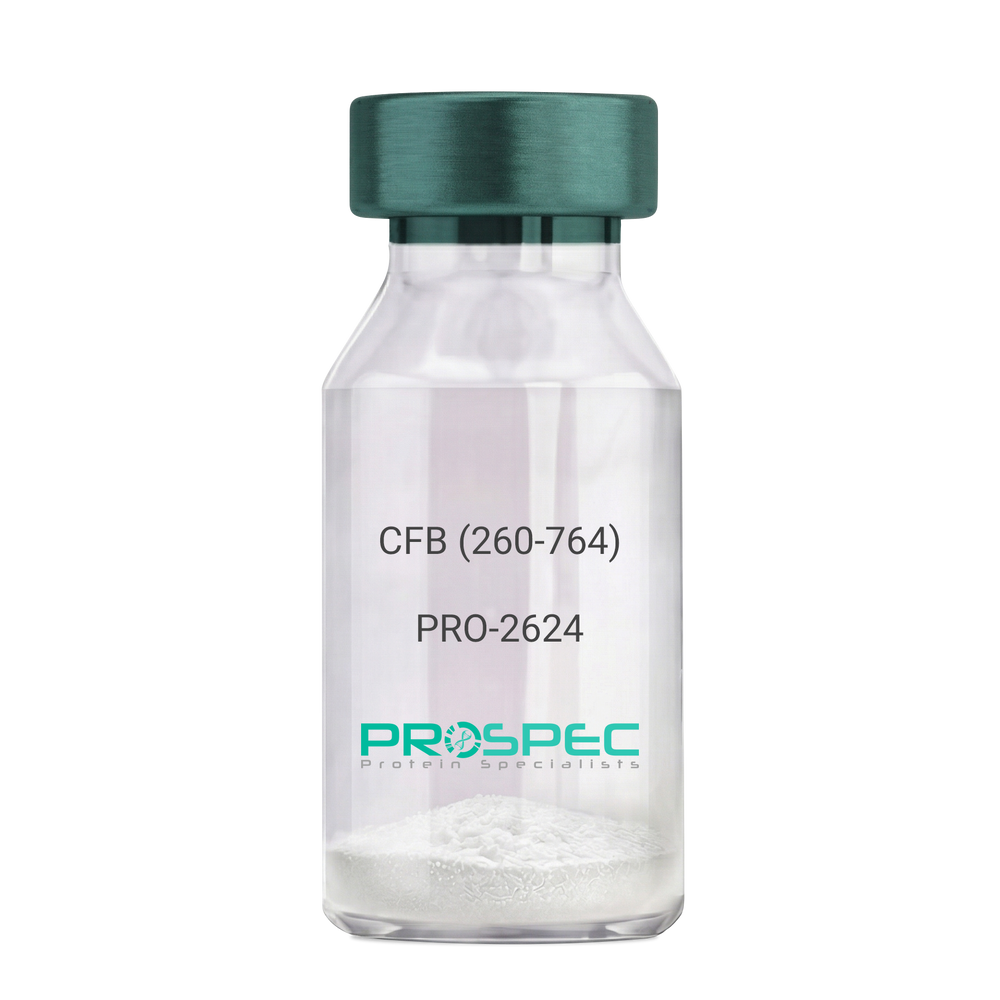
    CFB (260-764) Human, Sf9

    Description:

    Complement Factor B (260-764 a.a.) Human Recombinant, Sf9

    CFB, C3/C5 convertase, Glycine-rich beta glycoprotein, GBG, PBF2, Properdin factor B, BF, BFD, AHUS4, ARMD14, CFAB, CFBD, FB, FBI12, GBG, H2-Bf.

    Product # :

    PRO-2624

    Price :

    Quantity :

    Shipping Method :

    Ice Icon

    Shipped with Ice Packs

    Add To Cart

    More Info

    • description
    • source
    • formulation
    • purity
    • More Info

    Description

    CFB Human Recombinant produced in Sf9 Baculovirus cells is a single, glycosylated polypeptide chain containing 514 amino acids (260-764 a.a) and having a molecular mass of 58.1kDa.CFB is fused to an 9 amino acid His-tag at C-terminus & purified by proprietary chromatographic techniques.

    Source

    Sf9, Baculovirus cells.

    Formulation

    CFB protein solution (0.5mg/ml) containing Phosphate Buffered Saline (pH 7.4) and 20% glycerol.

    Purity

    Greater than 90.0% as determined by SDS-PAGE.

    More Info

    • Introduction

      Factor B is a protein, build from a single chain that circulates in the blood. Complement factor D cleaves and activates factor B, thus creating a catalytic subunit Bb and noncatalytic chain Ba. Subunit Bb is a serine protease that can bind to C3b and forms alternative pathway C3 convertase. Subunit Ba acts as an inhibitor for proliferation of lymphocytes, when Bb is part of the lymphocytes’ proliferation. The gene that codes for this protein is located on chromosome six.

    • Synonyms

      CFB, C3/C5 convertase, Glycine-rich beta glycoprotein, GBG, PBF2, Properdin factor B, BF, BFD, AHUS4, ARMD14, CFAB, CFBD, FB, FBI12, GBG, H2-Bf.

    • Physical Appearance

      Sterile Filtered colorless solution.

    • Stability

      Store at 4°C if entire vial will be used within 2-4 weeks. Store, frozen at -20°C for longer periods of time. For long term storage it is recommended to add a carrier protein (0.1% HSA or BSA).Avoid multiple freeze-thaw cycles.

    • Amino Acid Sequence

      ADPKIVLDPS GSMNIYLVLD GSDSIGASNF TGAKKCLVNL IEKVASYGVK PRYGLVTYAT
      YPKIWVKVSE ADSSNADWVT KQLNEINYED HKLKSGTNTK KALQAVYSMM SWPDDVPPEG
      WNRTRHVIIL MTDGLHNMGG DPITVIDEIR DLLYIGKDRK NPREDYLDVY VFGVGPLVNQ
      VNINALASKK DNEQHVFKVK DMENLEDVFY QMIDESQSLS LCGMVWEHRK GTDYHKQPWQ
      AKISVIRPSK GHESCMGAVV SEYFVLTAAH CFTVDDKEHS IKVSVGGEKR DLEIEVVLFH
      PNYNINGKKE AGIPEFYDYD VALIKLKNKL KYGQTIRPIC LPCTEGTTRA LRLPPTTTCQ
      QQKEELLPAQ DIKALFVSEE EKKLTRKEVY IKNGDKKGSC ERDAQYAPGY DKVKDISEVV
      TPRFLCTGGV SPYADPNTCR GDSGGPLIVH KRSRFIQVGV ISWGVVDVCK NQKRQKQVPA HARDFHINLF QVLPWLKEKL QDEDLGFLHH HHHH

    ProSpec's products are furnished for LABORATORY RESEARCH USE ONLY. The product may not be used as drugs, agricultural or pesticidal products, food additives or household chemicals.

    Complement Factor B
  • View Data Sheet

    Name :

    IGFBP6 (28-240) Human

    Description:

    Insulin Like Growth Factor Binding Protein-6 (28-240 a.a.) Human Recombinant

    Insulin-like growth factor-binding protein 6, IBP-6, IGF-binding protein 6, IGFBP-6, IGFBP6, IBP6.

    Product # :

    CYT-786

    Price :

    Quantity :

    Shipping Method :

    Ice Icon

    Shipped with Ice Packs

    Add To Cart

    More Info

    • description
    • source
    • formulation
    • purity
    • More Info

    Description

    IGFBP6 Human Recombinant produced in E. coli is a single polypeptide chain containing 236 amino acids (28-240) and having a molecular mass of 25.0kDa (Molecular size on SDS-PAGE will appear higher).IGFBP6 is fused to a 23 amino acid His-tag at N-terminus & purified by proprietary chromatographic techniques.

    Source

    E.coli.

    Formulation

    The IGFBP6 solution (0.5mg/ml) contains 20mM Tris-HCl buffer (pH 8.0), 0.15M NaCl, 20% glycerol and 1mM DTT.

    Purity

    Greater than 85% as determined by SDS-PAGE.

    More Info

    • Introduction

      IGFBP6 plays a role in lipoprotein assembly and dietary cholesterol absorption. in addition to its acyltransferase activity, it may act as a ligase. may provide cholesteryl esters for lipoprotein secretion from hepatocytes and intestinal mucosa.

    • Synonyms

      Insulin-like growth factor-binding protein 6, IBP-6, IGF-binding protein 6, IGFBP-6, IGFBP6, IBP6.

    • Physical Appearance

      Sterile Filtered colorless solution.

    • Stability

      Store at 4°C if entire vial will be used within 2-4 weeks. Store, frozen at -20°C for longer periods of time. For long term storage it is recommended to add a carrier protein (0.1% HSA or BSA).Avoid multiple freeze-thaw cycles.

    • Amino Acid Sequence

      MGSSHHHHHH SSGLVPRGSH MGSRCPGCGQ GVQAGCPGGC VEEEDGGSPA EGCAEAEGCL RREGQECGVY TPNCAPGLQC HPPKDDEAPL RALLLGRGRC LPARAPAVAE ENPKESKPQA GTARPQDVNR RDQQRNPGTS TTPSQPNSAG VQDTEMGPCR RHLDSVLQQL QTEVYRGAQT LYVPNCDHRG FYRKRQCRSS QGQRRGPCWC VDRMGKSLPG SPDGNGSSSC PTGSSG.

    • Background

      What is the molecular weight/Mw of IGFBP6 (28-240) HUMAN Protein?
      IGFBP6 (28-240) HUMAN Protein has a total Mw of 25kDa.

      What is the source or expression system of IGFBP6 (28-240) HUMAN Protein?
      Escherichia Coli.

      What is the Purity of IGFBP6 (28-240) HUMAN Protein?
      IGFBP6 (28-240) HUMAN Protein is >85% pure as determined by SDS-PAGE.

      What is the Biological Activity of IGFBP6 (28-240) HUMAN Protein?
      The biological functionality of IGFBP6 (28-240) HUMAN Protein will be determined in the future.

      What is the amino acid sequence of IGFBP6 (28-240) HUMAN Protein?
      MGSSHHHHHH SSGLVPRGSH MGSRCPGCGQ GVQAGCPGGC VEEEDGGSPA EGCAEAEGCL RREGQECGVY TPNCAPGLQC HPPKDDEAPL RALLLGRGRC LPARAPAVAE ENPKESKPQA GTARPQDVNR RDQQRNPGTS TTPSQPNSAG VQDTEMGPCR RHLDSVLQQL QTEVYRGAQT LYVPNCDHRG FYRKRQCRSS QGQRRGPCWC VDRMGKSLPG SPDGNGSSSC PTGSSG.

      What applications can IGFBP6 (28-240) HUMAN Protein be used in?
      IGFBP6 (28-240) HUMAN Protein can probably be used in western blot, ELISA and Lateral Flow.

      What is the endotoxin level for IGFBP6 (28-240) HUMAN Protein?
      The endotoxin level is minimal, IGFBP6 (28-240) HUMAN Protein was purified using conventional chromatography techniques.


    ProSpec's products are furnished for LABORATORY RESEARCH USE ONLY. The product may not be used as drugs, agricultural or pesticidal products, food additives or household chemicals.

    Igfbp6 28 240 Human
  • View Data Sheet

    Name :

    Tamm Horsfall

    Description:

    Recombinant Human Tamm Horsfall Glycoprotein

    Tamm-Horsfall urinary glycoprotein, THP, FJHN, HNFJ, THGP, MCKD2, ADMCKD2, UMOD, Uromodulin.


    Product # :

    ENZ-1206

    Price :

    Quantity :

    Shipping Method :

    Room Temp Icon

    Shipped at Room temp

    Add To Cart

    More Info

    • description
    • source
    • formulation
    • purity
    • More Info
    • SDS-PAGE

    Description

    Uromodulin Human Recombinant protein produced from HEK Cells, is a polypeptide chain containing 595 amino acids ( 25-613 a.a. ) and having a total Mw of 65 kDa.

    Source

    HEK293

    Formulation

    The UMOD protein was lyophilized from 0.4μm filtered solution containing 50mM NaCl, 0.02M TRIS, pH 7.5.

    Purity

    Greater than 90.0% as determined by SDS-PAGE analysis.

    SDS-PAGE

    Tamm Horsfall-SDS-PAGE - Product image 1

    More Info

    • Introduction

      Uromodulin (Tamm–Horsfall protein) is produced mainly by cells in the kidney’s thick ascending limb and is the most abundant protein in normal urine.
      Uromodulin takes part in salt and water regulation, helps prevent urinary tract infections and kidney stones, and influences inflammation and immune activity in the kidney.
      Reduced Uromodulin levels is associated with chronic kidney disease.
      UMOD mutations causes unproper protein folding and thus transported incorrectly, resulting in its accumulation inside kidney tubular cells that damages the tubules and cause kidney disease (ADTKD-UMOD), associated with high uric acid, gout, and progressive kidney failure.

    • Synonyms

      Tamm-Horsfall urinary glycoprotein, THP, FJHN, HNFJ, THGP, MCKD2, ADMCKD2, UMOD, Uromodulin.

    • Physical Appearance

      Filtered White lyophilized (freeze-dried) powder.

    • Stability

      Lyophilized UMOD although stable at room temperature for 3 weeks, should be stored at -18C. Upon reconstitution UMOD should be stored at 4C between 2-7 days and for future use below -18C.
      For long term storage it is recommended to add a carrier protein (0.1% HSA or BSA).
      Please prevent freeze-thaw cycles.

    • Solubility

      Add deionized water to prepare a working stock solution of approximately 0.5mg/ml and let the lyophilized pellet dissolve completely. Product is not sterile! Please filter the product by an appropriate sterile filter before using it in the cell culture.

    • Amino Acid Sequence

      DTSEARWCSE CHSNATCTED EAVTTCTCQE GFTGDGLTCV DLDECAIPGA HNCSANSSCV NTPGSFSCVC PEGFRLSPGL GCTDVDECAE PGLSHCHALA TCVNVVGSYL CVCPAGYRGD GWHCECSPGS CGPGLDCVPE GDALVCADPC QAHRTLDEYW RSTEYGEGYA CDTDLRGWYR FVGQGGARMA ETCVPVLRCN TAAPMWLNGT HPSSDEGIVS RKACAHWSGH CCLWDASVQV KACAGGYYVY NLTAPPECHL AYCTDPSSVE GTCEECSIDE DCKSNNGRWH CQCKQDFNIT DISLLEHRLE CGANDMKVSL GKCQLKSLGF DKVFMYLSDS RCSGFNDRDN RDWVSVVTPA RDGPCGTVLT RNETHATYSN TLYLADEIII RDLNIKINFA CSYPLDMKVS LKTALQPMVS ALNIRVGGTG MFTVRMALFQ TPSYTQPYQG SSVTLSTEAF LYVGTMLDGG DLSRFALLMT NCYATPSSNA TDPLKYFIIQ DRCPHTRDST IQVVENGESS QGRFSVQMFR FAGNYDLVYL HCEVYLCDTM NEKCKPTCSG TRFRSGSVID QSRVLNLGPI TRKGVQATVH HHHHH

    • Background

      Uromodulin is the most abundant protein in normal urine. Its secretion in urine follows proteolytic cleavage of the ectodomain of its glycosyl phosphatidylinosital-anchored counterpart that is situated on the luminal cell surface of the loop of Henle. Uromodulin plays a role as a constitutive inhibitor of calcium crystallization in renal fluids. Secretion of uromodulin in urine provides protection against urinary tract infections caused by uropathogenic bacteria. Defects in Uromodulin expression are associated with the autosomal dominant renal disorders medullary cystic kidney disease-2 (MCKD2) and familial juvenile hyperuricemic nephropathy (FJHN). These disorders are characterized by juvenile onset of hyperuricemia, gout, and progressive renal failure. While several transcript variants may exist for this gene, the full-length natures of only two have been described to date. UMOD is involved in regulating the circulating activity of cytokines as it binds to il-1, il-2 and tnf with high affinity.

      What is the molecular weight/Mw of UMOD Protein?
      UMOD Protein has a total Mw of 65kDa.

      What is the source or expression system of UMOD Protein?
      HEK293.

      What is the Purity of UMOD Protein?
      UMOD Protein is >90% pure as determined by SDS-PAGE.

      What is the Biological Activity of UMOD Protein?
      The biological functionality of UMOD Protein will be determined in the future.

      What is the amino acid sequence of UMOD Protein?
      UMOD Protein is composed from 595 amino acids.

      What applications can UMOD Protein be used in?
      UMOD Protein can probably be used in western blot, ELISA and Lateral Flow.

      What is the endotoxin level for UMOD Protein?
      The endotoxin level is minimal, UMOD Protein was purified using conventional chromatography techniques.

    ProSpec's products are furnished for LABORATORY RESEARCH USE ONLY. The product may not be used as drugs, agricultural or pesticidal products, food additives or household chemicals.

    Tamm Horsfall
  • View Data Sheet

    Name :

    HDGF2 Human

    Description:

    Hepatoma-Derived Growth Factor-2 Human Recombinant

    Hepatoma-derived growth factor-related protein 3, HRP-3, Hepatoma-derived growth factor 2, HDGF-2, HDGFRP3, HDGF2, CGI-142.

    Product # :

    CYT-132

    Price :

    Quantity :

    Shipping Method :

    Ice Icon

    Shipped with Ice Packs

    Add To Cart

    More Info

    • description
    • source
    • formulation
    • purity
    • More Info
    • SDS-PAGE

    Description

    HDGF2 Human Recombinant produced in E.Coli is a single, non-glycosylated polypeptide chain containing 227 amino acids (1-203 a.a.) and having a molecular mass of 25.1kDa (Molecular weight on SDS-PAGE will appear higher). HDGF2 is fused to a 24 amino acid His-tag at N-terminus & purified by proprietary chromatographic techniques.

    Source

    Escherichia Coli.

    Formulation

    HDGF2 protein solution (0.5mg/ml) containing 20mM Tris-HCl buffer (pH 8.0), 1mM DTT, 10% glycerol and 0.1M NaCl.

    Purity

    Greater than 90% as determined by SDS-PAGE.

    SDS-PAGE

    HDGF2 Human - Product image 1

    More Info

    • Introduction

      Hepatoma-derived growth factor, related protein 3 (HDGFRP3/HDGF2) is the original member of a family of polypeptides labeled HDGF related proteins (HRPs). The HDGF2 protein augments DNA synthesis and may play a part in cell proliferation. HDGF2 is expressed primarily in the testis and the brain, to an intermediate extent in the heart, and to a slight extent in the ovaries, kidneys, spleen, and liver in humans.

    • Synonyms

      Hepatoma-derived growth factor-related protein 3, HRP-3, Hepatoma-derived growth factor 2, HDGF-2, HDGFRP3, HDGF2, CGI-142.

    • Physical Appearance

      Sterile Filtered clear solution.

    • Stability

      Store at 4°C if entire vial will be used within 2-4 weeks. Store, frozen at -20°C for longer periods of time. For long term storage it is recommended to add a carrier protein (0.1% HSA or BSA).Avoid multiple freeze-thaw cycles.

    • Amino Acid Sequence

      MGSSHHHHHH SSGLVPRGSH MGSHMARPRP REYKAGDLVF AKMKGYPHWP ARIDELPEGA VKPPANKYPI FFFGTHETAF LGPKDLFPYK EYKDKFGKSN KRKGFNEGLW EIENNPGVKF TGYQAIQQQS SSETEGEGGN TADASSEEEG DRVEEDGKGK RKNEKAGSKR KKSYTSKKSS
      KQSRKSPGDE DDKDCKEEEN KSSSEGGDAG NDTRNTTSDL QKTSEGT.

    • Background

      What is the molecular weight/Mw of HDGF2 HUMAN Protein?
      HDGF2 HUMAN Protein has a total Mw of 25.1kDa.

      What is the source or expression system of HDGF2 HUMAN Protein?
      Escherichia Coli.

      What is the Purity of HDGF2 HUMAN Protein?
      HDGF2 HUMAN Protein is >90% pure as determined by SDS-PAGE.

      What is the Biological Activity of HDGF2 HUMAN Protein?
      The biological functionality of HDGF2 HUMAN Protein will be determined in the future.

      What is the amino acid sequence of HDGF2 HUMAN Protein?
      MGSSHHHHHH SSGLVPRGSH MGSHMARPRP REYKAGDLVF AKMKGYPHWP ARIDELPEGA VKPPANKYPI FFFGTHETAF LGPKDLFPYK EYKDKFGKSN KRKGFNEGLW EIENNPGVKF TGYQAIQQQS SSETEGEGGN TADASSEEEG DRVEEDGKGK RKNEKAGSKR KKSYTSKKSS
      KQSRKSPGDE DDKDCKEEEN KSSSEGGDAG NDTRNTTSDL QKTSEGT.

      What applications can HDGF2 HUMAN Protein be used in?
      HDGF2 HUMAN Protein can probably be used in western blot, ELISA and Lateral Flow.

      What is the endotoxin level for HDGF2 HUMAN Protein?
      The endotoxin level is minimal, HDGF2 HUMAN Protein was purified using conventional chromatography techniques.

    ProSpec's products are furnished for LABORATORY RESEARCH USE ONLY. The product may not be used as drugs, agricultural or pesticidal products, food additives or household chemicals.

    Hdgf2 Human
  • View Data Sheet

    Name :

    CD116 Human, sf9

    Description:

    GM-CSF Receptor Alpha Sf9 Human Recombinant

    Colony Stimulating Factor 2 Receptor Alpha Subunit, Colony Stimulating Factor 2 Receptor, Alpha, Low-Affinity (Granulocyte-Macrophage), Alpha-GM-CSF Receptor, GM-CSF-R-Alpha, CD116 Antigen, GMCSFR-Alpha, GMR-Alpha, CDw116, CSF2RY, CSF2R, Granulocyte-Macrophage Colony-Stimulating Factor Receptor Subunit Alpha, Granulocyte-Macrophage Colony-Stimulating Factor Receptor Alpha Chain, GM-CSF Receptor Alpha Subunit, AlphaGMR, CSF2RAX, CSF2RAY, CSF2RX,  GMCSFR, CD116, SMDP4, GMR.                  

    Product # :

    CYT-1044

    Price :

    Quantity :

    Shipping Method :

    Ice Icon

    Shipped with Ice Packs

    Add To Cart

    More Info

    • description
    • source
    • formulation
    • purity
    • biological activity
    • More Info

    Description

    CSF2RA produced in Sf9 Baculovirus cells is a single, glycosylated polypeptide chain containing 310 amino acids (20-320a.a.) and having a molecular mass of 35.9kDa. (Molecular size on SDS-PAGE will appear at approximately 40-57kDa). CSF2RA is expressed with a 9 amino acid His tag at C-Terminus and purified by proprietary chromatographic techniques.

    Source

    Sf9, Baculovirus cells.

    Formulation

    CSF2RA protein solution (0.5mg/ml) contains Phosphate Buffered Saline (pH 7.4) and 10% glycerol.

    Purity

    Greater than 90.0% as determined by SDS-PAGE.

    Biological Activity

    Measured by its ability to inhibit GM-CSF dependent proliferation of TF1 human erythroleukemic cells. The ED50 for this effect is less or equal to 10ug/ml in the presence of 0.5ng/ml GM-CSF.

    More Info

    • Introduction

      GM-CSF Receptor Alpha (CSF2RA) is the alpha subunit of the heterodimeric receptor for colony stimulating factor 2, a cytokine which controls the production, differentiation, and function of granulocytes and macrophages. CSFR2 is also a member of the cytokine family of receptors. In addition, this gene is found in the pseudoautosomal region (PAR) of the X and Y chromosomes. Multiple transcript variants encoding various isoforms have been found for this gene, while some of the isoforms being membrane-bound and others being soluble. Diseases associated with CSF2RA include surfactant metabolism dysfunction, pulmonary 4, and csf2ra-related pulmonary surfactant metabolism dysfunction.

    • Synonyms

      Colony Stimulating Factor 2 Receptor Alpha Subunit, Colony Stimulating Factor 2 Receptor, Alpha, Low-Affinity (Granulocyte-Macrophage), Alpha-GM-CSF Receptor, GM-CSF-R-Alpha, CD116 Antigen, GMCSFR-Alpha, GMR-Alpha, CDw116, CSF2RY, CSF2R, Granulocyte-Macrophage Colony-Stimulating Factor Receptor Subunit Alpha, Granulocyte-Macrophage Colony-Stimulating Factor Receptor Alpha Chain, GM-CSF Receptor Alpha Subunit, AlphaGMR, CSF2RAX, CSF2RAY, CSF2RX, GMCSFR, CD116, SMDP4, GMR.

    • Physical Appearance

      Sterile Filtered colorless solution.

    • Stability

      Store at 4°C if entire vial will be used within 2-4 weeks. Store, frozen at -20°C for longer periods of time. For long term storage it is recommended to add a carrier protein (0.1% HSA or BSA). Avoid multiple freeze-thaw cycles.

    • Amino Acid Sequence

      ADPLIPEKSD LRTVAPASSL NVRFDSRTMN LSWDCQENTT FSKCFLTDKK NRVVEPRLSN NECSCTFREI CLHEGVTFEV HVNTSQRGFQ QKLLYPNSGR EGTAAQNFSC FIYNADLMNC TWARGPTAPR DVQYFLYIRN SKRRREIRCP YYIQDSGTHV GCHLDNLSGL TSRNYFLVNG TSREIGIQFF DSLLDTKKIE RFNPPSNVTV RCNTTHCLVR WKQPRTYQKL SYLDFQYQLD VHRKNTQPGT ENLLINVSGD LENRYNFPSS EPRAKHSVKI RAADVRILNW SSWSEAIEFG SDDGHHHHHH

    • Background

      GM-CSF Receptor Alpha Human Recombinant: A Glimpse into Its Potential and Implications

      Abstract:

      Granulocyte-Macrophage Colony Stimulating Factor (GM-CSF) receptor alpha, a pivotal component in the GM-CSF signaling pathway, has been the focal point of numerous studies pertaining to hematopoiesis and immune responses. This paper provides an overview of the GM-CSF receptor alpha human recombinant, exploring its characteristics, production techniques, and potential therapeutic applications.

      Introduction

      GM-CSF, a cytokine responsible for the differentiation and proliferation of white blood cells, functions by binding to its receptor, GM-CSF receptor. The alpha subunit (GM-CSFRα) of this receptor plays a crucial role in ligand binding and is essential for initiating cellular responses. Modern biotechnological advancements have led to the successful production of its human recombinant form, offering new avenues in medical research.

      Recombinant GM-CSFRα:

      Production and Features Recombinant GM-CSFRα is synthesized using cutting-edge recombinant DNA technologies, predominantly in bacterial or mammalian expression systems. This human recombinant form retains its ability to bind to GM-CSF, maintaining its biological functionality and providing myriad research opportunities.

      Therapeutic and Clinical Prospects

      1. Autoimmune Diseases: GM-CSF is often overexpressed in various autoimmune disorders. By utilizing recombinant GM-CSFRα as a potential decoy receptor, it's feasible to mitigate the effects of excessive GM-CSF, offering a new therapeutic strategy.
      2. Hematopoietic Disorders: Given its integral role in white blood cell development, recombinant GM-CSFRα might hold promise in treatments or as a diagnostic tool for certain hematological conditions.
      3. Research Paradigm: Beyond therapeutic applications, the recombinant GM-CSFRα can serve as an invaluable research tool to elucidate the nuances of GM-CSF signaling, aiding in the understanding of immune response mechanisms.

      Conclusion:

      GM-CSF receptor alpha human recombinant stands at the forefront of exciting research and therapeutic potential. While its full capabilities are yet to be realized, current insights underscore its significance in the realms of immunology and medicine.

      What is the molecular weight/Mw of CD116 Protein?
      CD116 Protein has a total Mw of 35.9kDa.

      What is the source or expression system of CD116 Protein?
      Sf9, Baculovirus cells.
      What is the Purity of CD116 Protein?
      CD116 Protein is >90% pure as determined by SDS-PAGE.

      What is the Biological Activity of CD116 Protein?
      Measured by its ability to inhibit GM-CSF dependent proliferation of TF1 human erythroleukemic cells. The ED50 for this effect is less or equal to 10ug/ml in the presence of 0.5ng/ml GM-CSF.

      What is the amino acid sequence of CD116 Protein?
      CD116 Protein is composed from 310 amino acids.

      What applications can CD116 Protein be used in?
      CD116 Protein can probably be used in western blot, ELISA and Lateral Flow.

      What is the endotoxin level for CD116 Protein?
      The endotoxin level is minimal, CD116 Protein was purified using conventional chromatography techniques

    ProSpec's products are furnished for LABORATORY RESEARCH USE ONLY. The product may not be used as drugs, agricultural or pesticidal products, food additives or household chemicals.

    Gmcsf Receptor
  • View Data Sheet

    Name :

    OPG Fc Human

    Description:

    Osteoprotegerin Human Recombinant /Fc Chimera

    TNFRSF11B, OPG, OCIF, Osteoclastogenesis inhibitory factor, TR1, MGC29565.

    Product # :

    CYT-266

    Price :

    Quantity :

    Shipping Method :

    Room Temp Icon

    Shipped at Room temp

    Add To Cart

    More Info

    • description
    • source
    • formulation
    • purity
    • biological activity
    • More Info

    Description

    Recombinant OPG produced in yeast contains 2x412 amino acid residues, including 180 residues from mature OPG (a.a 22-201) and 232 residues from the Fc protein of human IgG1, and has a calculated molecular mass of 109.6kDa.

    Source

    Pichia Pastoris.

    Formulation

    OPG was lyophilized from a 0.2µm filtered concentrated solution in 20mM PB, pH 6.0, 150mM NaCl and 0.02 % Tween-80.

    Purity

    Greater than 90.0% as determined by:
    (a) Analysis by RP-HPLC.
    (b) Analysis by SDS-PAGE.

    Biological Activity

    The ED50 as determined by neutralizing the stimulation of U937 cells is less tha10ng/ml, corresponding to a specific activity of > 1.0 × 10⁵ IU/mg in the presence of 10ng/ml soluble Human RANKL (sRANKL).

    More Info

    • Introduction

      Osteoprotegerin acts as decoy receptor for rankl and thereby neutralizes its function in osteoclastogenesis. OPG inhibits the activation of osteoclasts and promotes osteoclast apoptosis in vitro. Bone homeostasis seems to depend on the local rankl/opg ratio. Osteoprotegerin may also play a role in preventing arterial calcification. May act as decoy receptor for trail and protect against apoptosis. Trail binding blocks the inhibition of osteoclastogenesis.

    • Synonyms

      TNFRSF11B, OPG, OCIF, Osteoclastogenesis inhibitory factor, TR1, MGC29565.

    • Physical Appearance

      Sterile Filtered White lyophilized (freeze-dried) powder.

    • Stability

      Lyophilized Osteoprotegerin although stable at room temperature for 3 weeks, should be stored desiccated below -18°C. Upon reconstitution OCIF should be stored at 4°C between 2-7 days and for future use below -18°C. For long term storage it is recommended to add a carrier protein (0.1% HSA or BSA).Please prevent freeze-thaw cycles.

    • Solubility

      It is recommended to reconstitute the lyophilized Osteoprotegerin in sterile 18MΩ-cm H2O not less than 100µg/ml, which can then be further diluted to other aqueous solutions.

    • Amino Acid Sequence

      OPG 22-201 ETFPPKYLHY DEETSHQLLC DKCPPGTYLK QHCTAKWKTV CAPCPDHYYT DSWHTSDECL YCSPVCKELQ YVKQECNRTH NRVCECKEGR YLEIEFCLKH RSCPPGFGVV QAGTPERNTV CKRCPDGFFS NETSSKAPCR KHTNCSVFGL LLTQKGNATH DNICSGNSES TQKCGIDVTL
      Fc232EPKSSDKTHT CPPCPAPEFE GAPSVFLFPP KPKDTLMISR TPEVTCVVVD VSHEDPEVKF NWYVDGVEVH NAKTKPREEQ YNSTYRVVSV LTVLHQDWLN GKEYKCKVSN KALPTPIEKTISKAKGQPRE PQVYTLPPSR DELTKNQVSL TCLVKGFYPS DIAVEWESNG QPENNYKTTP PVLDSDGSFF LYSKLTVDKS RWQQGNVFSC SVMHEALHNH YTQKSLSLSP GK

    ProSpec's products are furnished for LABORATORY RESEARCH USE ONLY. The product may not be used as drugs, agricultural or pesticidal products, food additives or household chemicals.

    Osteoprotegerin Human
  • View Data Sheet

    Name :

    BMPR1B Human

    Description:

    Bone Morphogenetic protein Receptor-1B Human Recombinant

    Bone Morphogenetic Protein Receptor, Type IB, BMP Type-1B Receptor, EC 2.7.11.30, BMPR-1B, ALK6, Bone Morphogenetic Protein Receptor Type-1B, Serine/Threonine Receptor Kinase, CDw293 Antigen, EC 2.7.11, CDw293, ALK-6, Bone morphogenetic protein receptor type-1B.

    Product # :

    CYT-897

    Price :

    Quantity :

    Shipping Method :

    Ice Icon

    Shipped with Ice Packs

    Add To Cart

    More Info

    • description
    • source
    • formulation
    • purity
    • More Info

    Description

    BMPR1B Human Recombinant produced in E.Coli is a single, non-glycosylated polypeptide chain containing 136 amino acids (14-126 a.a) and having a molecular mass of 15.1kDa. BMPR1B is fused to a 23 amino acid His-tag at N-terminus & purified by proprietary chromatographic techniques.

    Source

    Escherichia Coli.

    Formulation

    BMPR1B protein solution (0.5mg/ml) containing Phosphate buffered saline (pH7.4), 20% glycerol, 1mM DTT and 0.1mM PMSF.

    Purity

    Greater than 80.0% as determined by SDS-PAGE.

    More Info

    • Introduction

      Bone Morphogenetic protein Receptor-1B, also known as BMPR1B belongs to the TKL Ser/Thr protein kinase family. BMPR1Bis on ligand binding, forms a receptor complex consisting of two type II and two type I transmembrane serine/threonine kinases. Type II receptors phosphorylate as well as activate type I receptors which autophosphorylate, afterward bind and activate SMAD transcriptional regulators. BMPR1B is receptor for BMP7/OP-1 as well as GDF5.

    • Synonyms

      Bone Morphogenetic Protein Receptor, Type IB, BMP Type-1B Receptor, EC 2.7.11.30, BMPR-1B, ALK6, Bone Morphogenetic Protein Receptor Type-1B, Serine/Threonine Receptor Kinase, CDw293 Antigen, EC 2.7.11, CDw293, ALK-6, Bone morphogenetic protein receptor type-1B.

    • Physical Appearance

      Sterile Filtered colorless solution.

    • Stability

      Store at 4°C if entire vial will be used within 2-4 weeks. Store, frozen at -20°C for longer periods of time. For long term storage it is recommended to add a carrier protein (0.1% HSA or BSA).Avoid multiple freeze-thaw cycles.

    • Amino Acid Sequence

      MGSSHHHHHH SSGLVPRGSH MGSKKEDGES TAPTPRPKVL RCKCHHHCPE DSVNNICSTD GYCFTMIEED DSGLPVVTSG CLGLEGSDFQ CRDTPIPHQR RSIECCTERN ECNKDLHPTL PPLKNRDFVD GPIHHR.

    • Background

      Bone Morphogenetic Protein Receptor-1B Human Recombinant: A Key Regulator of Cellular Signaling in Development and Disease

      Abstract:

      Bone Morphogenetic Protein Receptor-1B (BMPR1B) human recombinant is a critical component of the bone morphogenetic protein (BMP) signaling pathway, governing cellular processes such as embryogenesis, tissue homeostasis, and disease progression. This research paper provides a comprehensive analysis of BMPR1B, including its characteristics, signaling mechanisms, and potential therapeutic applications. Additionally, innovative methodologies for the production and optimization of BMPR1B human recombinant are proposed, shedding light on its future implications in the field of regenerative medicine.

      Introduction:

      The precise regulation of cellular signaling pathways is essential for proper development and tissue maintenance. BMPR1B, a key receptor in the BMP pathway, plays a crucial role in various biological processes. This paper explores the unique features of BMPR1B and presents novel approaches for its production and optimization, aiming to unravel its therapeutic potential in a wide range of developmental and disease contexts.

      Characteristics and Signaling Mechanisms:

      BMPR1B belongs to the serine/threonine kinase receptor family and is predominantly expressed in embryonic tissues, skeletal structures, and reproductive organs. Upon binding to BMP ligands, BMPR1B initiates downstream signaling cascades, including Smad-dependent and Smad-independent pathways. These pathways regulate gene expression, cellular proliferation, differentiation, and apoptosis, ultimately influencing tissue development and homeostasis.

      Production of BMPR1B Human Recombinant:

      Efficient production methodologies are pivotal for harnessing the therapeutic potential of BMPR1B human recombinant. Mammalian expression systems, such as Chinese hamster ovary (CHO) cells, have been widely employed to ensure proper folding and post-translational modifications of the receptor. Optimization strategies, including codon optimization and vector engineering, have been utilized to enhance production efficiency. Purification techniques, such as affinity chromatography and size exclusion chromatography, have been optimized to obtain high-quality BMPR1B recombinant protein.

      Potential Therapeutic Applications:

      BMPR1B human recombinant holds tremendous promise in the field of regenerative medicine and disease therapy. Dysregulation of the BMP signaling pathway has been implicated in various developmental disorders, including skeletal abnormalities and congenital malformations. Modulating BMPR1B activity using BMPR1B human recombinant may offer a targeted approach for promoting tissue regeneration and repair in these conditions. Furthermore, BMPR1B signaling is involved in several diseases, such as cancer and cardiovascular disorders, highlighting its potential as a therapeutic target for intervention.

      Conclusion:

      BMPR1B human recombinant represents a vital regulator in cellular signaling, with significant implications in development and disease. Optimizing production methodologies and expanding our understanding of its signaling mechanisms will further enhance its therapeutic potential. With its involvement in various biological processes and disease contexts, BMPR1B human recombinant emerges as a promising tool for regenerative medicine and targeted therapeutics.

      What is the molecular weight/Mw of BMPR1B Protein?
      BMPR1B Protein has a total Mw of 15.1kDa.

      What is the source or expression system of BMPR1B Protein?
      Escherichia Coli.

      What is the Purity of BMPR1B Protein?
      BMPR1B Protein is >80% pure as determined by SDS-PAGE.

      What is the Biological Activity of BMPR1B Protein?
      The biological functionality of BMPR1B Protein will be determined in the future.

      What is the amino acid sequence of BMPR1B Protein?
      MGSSHHHHHH SSGLVPRGSH MGSKKEDGES TAPTPRPKVL RCKCHHHCPE DSVNNICSTD GYCFTMIEED DSGLPVVTSG CLGLEGSDFQ CRDTPIPHQR RSIECCTERN ECNKDLHPTL PPLKNRDFVD GPIHHR.

      What applications can BMPR1B Protein be used in?
      BMPR1B Protein can probably be used in western blot, ELISA and Lateral Flow.

      What is the endotoxin level for BMPR1B Protein?
      The endotoxin level is minimal, BMPR1B Protein was purified using conventional chromatography techniques.

    ProSpec's products are furnished for LABORATORY RESEARCH USE ONLY. The product may not be used as drugs, agricultural or pesticidal products, food additives or household chemicals.

    Bmpr1B Human
  • View Data Sheet

    Name :

    FGF 2 Rat

    Description:

    Fibroblast Growth Factor-basic Rat Recombinant

    HBGH-2, HBGF-2, Prostatropin, FGF-2, FGB-b, Fibroblast Growth Factor-basic, Basic fibroblast growth factor, bFGF, Heparin-binding growth factor 2.

    Product # :

    CYT-608

    Price :

    Quantity :

    Shipping Method :

    Room Temp Icon

    Shipped at Room temp

    Add To Cart

    More Info

    • description
    • source
    • formulation
    • purity
    • biological activity
    • More Info

    Description

    bFGF Rat Recombinant (FGF-2) produced in E.Coli is a single, non-glycosylated, polypeptide chain containing 146 amino acids and having a molecular mass of 16.4 kDa.The bFGF2 is purified by proprietary chromatographic techniques.

    Source

    Escherichia Coli.

    Formulation

    FGF-b was lyophilized from 1mg/ml solution after extensive dialysis against 10mM Na2PO4, pH 7.5 and 50mM NaCl.

    Purity

    Greater than 95.0% as determined by SDS-PAGE.

    Biological Activity

    The ED50 range as determined by the dose-dependant proliferation of NR6R-3T3 cells was found to be 0.903ng/ml, corresponding to a specific activity of 1.1x10⁷ units/mg.

    More Info

    • Introduction

      FGF-basic is a member of the fibroblast growth factor (FGF) family. FGF family members bind heparin and possess broad mitogenic and angiogenic activities. This protein has been implicated in diverse biological processes, such as limb and nervous system development, wound healing, and tumor growth. The mRNA for this gene contains multiple polyadenylation sites, and is alternatively translated from AUG and non-AUG (CUG) initiation codons resulting in five different isoforms with distinct properties. The CUG-initiated isoforms are localized in the nucleus and are responsible for the intracrine effect, whereas, the AUG-initiated form is mostly cytosolic and is responsible for the paracrine and autocrine effects of this FGF.
      The heparin-binding growth factors are angiogenic agents in vivo and are potent mitogens for a variety of cell types in vitro. there are differences in the tissue distribution and concentration of these 2 growth factors.

    • Synonyms

      HBGH-2, HBGF-2, Prostatropin, FGF-2, FGB-b, Fibroblast Growth Factor-basic, Basic fibroblast growth factor, bFGF, Heparin-binding growth factor 2.

    • Physical Appearance

      Sterile Filtered White lyophilized (freeze-dried) powder.

    • Stability

      Lyophilized Rat bFGF although stable at room temperature for 3 weeks, should be stored desiccated below -18°C. Upon reconstitution Rat FGF-2 should be stored at 4°C between 2-7 days and for future use below -18°C.For long term storage it is recommended to add a carrier protein (0.1% HSA or BSA).Please prevent freeze-thaw cycles.

    • Solubility

      It is recommended to reconstitute the lyophilized Rat bFGF in sterile 18MΩ-cm H2O not less than 100µg/ml, which can then be further diluted to other aqueous solutions.

    • Amino Acid Sequence

      MPALPEDGGGA FPPGHFKDPK RLYCKNGGFF LRIHPDGRVD GVREKSDPHV KLQLQAEERG VVSIKGVCAN RYLAMKEDGR LLASKCVTEE CFFFERLESN NYNTYRSRKY SSWYVALKRT GQYKLGSKTG PGQKAILFLP MSAKS.

    • Background

      What is the molecular weight/Mw of FGF 2 RAT Protein?
      FGF 2 RAT Protein has a total Mw of 16.4kDa.

      What is the source or expression system of FGF 2 RAT Protein?
      Escherichia Coli.

      What is the Purity of FGF 2 RAT Protein?
      FGF 2 RAT Protein is >95% pure as determined by SDS-PAGE.

      What is the Biological Activity of FGF 2 RAT Protein?
      The ED50 range as determined by the dose-dependant proliferation of NR6R-3T3 cells was found to be 0.903ng/ml, corresponding to a specific activity of 1.1x10⁷ units/mg.

      What is the amino acid sequence of FGF 2 RAT Protein?
      MPALPEDGGGA FPPGHFKDPK RLYCKNGGFF LRIHPDGRVD GVREKSDPHV KLQLQAEERG VVSIKGVCAN RYLAMKEDGR LLASKCVTEE CFFFERLESN NYNTYRSRKY SSWYVALKRT GQYKLGSKTG PGQKAILFLP MSAKS.

      What applications can FGF 2 RAT Protein be used in?
      FGF 2 RAT Protein can probably be used in western blot, ELISA and Lateral Flow.

      What is the endotoxin level for FGF 2 RAT Protein?
      The endotoxin level is minimal, FGF 2 RAT Protein was purified using conventional chromatography techniques.

    ProSpec's products are furnished for LABORATORY RESEARCH USE ONLY. The product may not be used as drugs, agricultural or pesticidal products, food additives or household chemicals.

    Bfgf Rat
  • View Data Sheet

    Name :

    FLT1 Human, HEK Active

    Description:

    Vascular Endothelial Growth Factor receptor-1 Human Recombinant, HEK Active

    FLT-1, FLT1, Tyrosine-protein kinase receptor FLT, Flt-1, Tyrosine-protein kinase FRT, Fms-like tyrosine kinase 1, VEGFR-1.

    Product # :

    PKA-134

    Price :

    Quantity :

    Shipping Method :

    Ice Icon

    Shipped with Ice Packs

    Add To Cart

    More Info

    • description
    • source
    • formulation
    • purity
    • biological activity
    • More Info

    Description

    FLT1 Human Recombinant is a single, glycosylated polypeptide chain containing 535 amino acids (27-328a.a) and having a molecular mass of 60.3kDa (calculated). FLT1 is fused to a 233 amino acid hIgG-Tag at C-terminus and is purified by proprietary chromatographic techniques.

    Source

    HEK293

    Formulation

    FLT1 protein solution (0.5mg/ml) contains 10% Glycerol and Phosphate-Buffered Saline (pH 7.4).

    Purity

    Greater than 90.0% as determined by SDS-PAGE.

    Biological Activity

    The ED50 range ≤ 60ng/ml and is measured by its ability to inhibit proliferation using HUVEC human umbilical vein endothelial cells in the
    presence of Human VEGF165. 

    More Info

    • Synonyms

      FLT-1, FLT1, Tyrosine-protein kinase receptor FLT, Flt-1, Tyrosine-protein kinase FRT, Fms-like tyrosine kinase 1, VEGFR-1.

    • Physical Appearance

      Filtered clear solution.

    • Stability

      Store at 4°C if entire vial will be used within 2-4 weeks.Store, frozen at -20°C for longer periods of time.Avoid multiple freeze-thaw cycles.

    • Amino Acid Sequence

      SKLKDPELSL KGTQHIMQAG QTLHLQCRGE AAHKWSLPEM VSKESERLSI TKSACGRNGK QFCSTLTLNT AQANHTGFYS CKYLAVPTSK KKETESAIYI FISDTGRPFV EMYSEIPEII HMTEGRELVI PCRVTSPNIT VTLKKFPLDT LIPDGKRIIW DSRKGFIISN ATYKEIGLLT CEATVNGHLY KTNYLTHRQT NTIIDVQIST PRPVKLLRGH TLVLNCTATT PLNTRVQMTW SYPDEKNKRA SVRRRIDQSN SHANIFYSVL TIDKMQNKDK GLYTCRVRSG PSFKSVNTSV HILEPKSCDK THTCPPCPAP ELLGGPSVFL FPPKPKDTLM ISRTPEVTCV VVDVSHEDPE VKFNWYVDGV EVHNAKTKPR EEQYNSTYRV VSVLTVLHQD WLNGKEYKCK VSNKALPAPI EKTISKAKGQ PREPQVYTLP PSRDELTKNQ VSLTCLVKGF YPSDIAVEWE SNGQPENNYK TTPPVLDSDG SFFLYSKLTV DKSRWQQGNV FSCSVMHEAL HNHYTQKSLS LSPGK.

    • Background

      VEGFR-1 (Vascular Endothelial Growth Factor Receptor-1), also known as Flt-1 (Fms-like tyrosine kinase 1), is a critical receptor involved in angiogenesis and vascular development. This research paper delves into the structure, function, and therapeutic implications of VEGFR-1, shedding light on its multifaceted role in various physiological and pathological processes.

      VEGFR-1 is a transmembrane receptor tyrosine kinase belonging to the VEGF receptor family. It is primarily expressed on endothelial cells and plays a pivotal role in mediating the cellular responses to VEGF ligands. Upon ligand binding, VEGFR-1 initiates intracellular signaling cascades that regulate endothelial cell proliferation, migration, and survival, ultimately contributing to the formation of new blood vessels.

      The structure of VEGFR-1 comprises distinct domains, including an extracellular ligand-binding domain, a transmembrane domain, and an intracellular tyrosine kinase domain. The extracellular domain facilitates the interaction between VEGF ligands and the receptor, while the intracellular domain transduces downstream signals by phosphorylating specific tyrosine residues.

      VEGFR-1 exhibits not only ligand-dependent but also ligand-independent functions. In addition to its role as a VEGF receptor, it can act as a decoy receptor, sequestering VEGF and modulating the bioavailability of VEGF ligands. This unique property allows VEGFR-1 to regulate VEGF signaling and influence angiogenic processes.

      The signaling pathways activated by VEGFR-1 are diverse and intricate, involving multiple downstream effectors, such as PI3K/AKT, MAPK/ERK, and STAT proteins. These pathways regulate endothelial cell behaviors, including proliferation, migration, and differentiation, which are crucial for angiogenesis. Perturbations in VEGFR-1 signaling have been implicated in various pathological conditions, including cancer, retinopathy, and inflammatory disorders.

      The therapeutic targeting of VEGFR-1 has gained considerable attention for its potential in managing angiogenesis-related diseases. Inhibitors specifically designed to block VEGFR-1 have been developed to suppress aberrant angiogenesis and impede tumor growth. Moreover, VEGFR-1-based therapies have been explored for ocular diseases like wet age-related macular degeneration (AMD) and diabetic retinopathy, aiming to alleviate pathological neovascularization.

      The availability of VEGFR-1 human recombinant proteins has facilitated in-depth research and the development of potential therapeutic interventions. Recombinant VEGFR-1 proteins serve as valuable tools for investigating VEGF-VEGFR-1 interactions, screening drug candidates, and elucidating the underlying molecular mechanisms of VEGFR-1-mediated signaling pathways.

    ProSpec's products are furnished for LABORATORY RESEARCH USE ONLY. The product may not be used as drugs, agricultural or pesticidal products, food additives or household chemicals.

    Flt1 Human Hek Active
  • View Data Sheet

    Name :

    CD274 Human, Sf9

    Description:

    CD274 Human Recombinant, Sf9

    CD274 molecule, CD274 antigen, programmed cell death 1 ligand 1, PD-L1, PDCD1L1, PDCD1LG1, PDCD1 ligand 1, B7 homolog 1, B7H1.

    Product # :

    PRO-2453

    Price :

    Quantity :

    Shipping Method :

    Ice Icon

    Shipped with Ice Packs

    Add To Cart

    More Info

    • description
    • source
    • formulation
    • purity
    • More Info

    Description

    CD274 Human Recombinant produced in Sf9 Baculovirus cells is a single, glycosylated polypeptide chain containing 462 amino acids (19-238a.a.) and having a molecular mass of 52.5kDa (Molecular size on SDS-PAGE will appear at approximately 50-70kDa). CD274 is expressed with a 239 amino acids hIgG-His tag at C-Terminus and purified by proprietary chromatographic techniques.

    Source

    Sf9, Baculovirus cells.

    Formulation

    CD274 protein solution (1mg/ml) contains Phosphate Buffered Saline (pH 7.4) and 10% glycerol.

    Purity

    Greater than 95% as determined by SDS-PAGE.

    More Info

    • Introduction

      CD274 takes part in the costimulatory signal, vital for T-cell proliferation and creation of IL10 and IFNG, in a PDCD1-independent and an IL2-dependent manner. CD274 collaboration with PDCD1 inhibits cytokine production and T-cell proliferation.

    • Synonyms

      CD274 molecule, CD274 antigen, programmed cell death 1 ligand 1, PD-L1, PDCD1L1, PDCD1LG1, PDCD1 ligand 1, B7 homolog 1, B7H1.

    • Physical Appearance

      Sterile Filtered colorless solution.

    • Stability

      Store at 4°C if entire vial will be used within 2-4 weeks. Store, frozen at -20°C for longer periods of time. For long term storage it is recommended to add a carrier protein (0.1% HSA or BSA).Avoid multiple freeze-thaw cycles.

    • Amino Acid Sequence

      ADLFTVTVPK DLYVVEYGSN MTIECKFPVE KQLDLAALIV YWEMEDKNII QFVHGEEDLK VQHSSYRQRA RLLKDQLSLG NAALQITDVK LQDAGVYRCM ISYGGADYKR ITVKVNAPYN KINQRILVVD PVTSEHELTC QAEGYPKAEV IWTSSDHQVL SGKTTTTNSK REEKLFNVTS TLRINTTTNE IFYCTFRRLD PEENHTAELV IPELPLAHPP NERLEPKSCD KTHTCPPCPA PELLGGPSVF LFPPKPKDTL MISRTPEVTC VVVDVSHEDP EVKFNWYVDG VEVHNAKTKP REEQYNSTYR VVSVLTVLHQ DWLNGKEYKC KVSNKALPAP IEKTISKAKG QPREPQVYTL PPSRDELTKN QVSLTCLVKG FYPSDIAVEW ESNGQPENNY KTTPPVLDSD GSFFLYSKLT VDKSRWQQGN VFSCSVMHEA LHNHYTQKSL SLSPGKHHHH HH.

    ProSpec's products are furnished for LABORATORY RESEARCH USE ONLY. The product may not be used as drugs, agricultural or pesticidal products, food additives or household chemicals.

    Cd274 Protein
  • View Data Sheet

    Name :

    KLRB1 Human, Sf9

    Description:

    Killer Cell Lectin-Like Receptor Subfamily B, Member 1 Human Recombinant, Sf9

    Killer Cell Lectin Like Receptor B1, Natural Killer Cell Surface Protein P1A, Killer Cell Lectin-Like Receptor Subfamily B, Member 1, C-Type Lectin Domain Family 5 Member B, HNKR-P1A, NKR-P1A, CLEC5B, NKRP1A, Killer Cell Lectin-Like Receptor Subfamily B Member 1, CD161 Antigen, NKR-P1, CD161, NKR.

    Product # :

    PRO-2488

    Price :

    Quantity :

    Shipping Method :

    Ice Icon

    Shipped with Ice Packs

    Add To Cart

    More Info

    • description
    • source
    • formulation
    • purity
    • More Info

    Description

    KLRB1 produced in Sf9 Baculovirus cells is a single, glycosylated polypeptide chain containing 401 amino acids (67-225a.a.) and having a molecular mass of 45.7kDa. KLRB1 is expressed with a 239 amino acid hIgG-His-tag at C-Terminus and purified by proprietary chromatographic techniques.

    Source

    Sf9, Baculovirus cells.

    Formulation

    KLRB1 protein solution (0.25mg/ml) contains Phosphate Buffered Saline (pH 7.4) and 10% glycerol.

    Purity

    Greater than 90% as determined by SDS-PAGE.

    More Info

    • Introduction

      KLRB1 has an inhibitory role on natural killer (NK) cells cytotoxicity which are lymphocytes who facilitate cytotoxicity and secrete cytokines subsequent to immune stimulation. Some genes of the C-type lectin superfamily, like the rodent NKRP1 family of glycoproteins, are expressed by NK cells and take part in NK cell function regulation. KLRB1 holds an extracellular domain with a few characteristic motifs of C-type lectins, a transmembrane domain, and a cytoplasmic domain. Due to its external C terminus KLRB1 is considered to be a type II membrane protein.

    • Synonyms

      Killer Cell Lectin Like Receptor B1, Natural Killer Cell Surface Protein P1A, Killer Cell Lectin-Like Receptor Subfamily B, Member 1, C-Type Lectin Domain Family 5 Member B, HNKR-P1A, NKR-P1A, CLEC5B, NKRP1A, Killer Cell Lectin-Like Receptor Subfamily B Member 1, CD161 Antigen, NKR-P1, CD161, NKR.

    • Physical Appearance

      Sterile Filtered colorless solution.

    • Stability

      Store at 4°C if entire vial will be used within 2-4 weeks. Store, frozen at -20°C for longer periods of time. For long term storage it is recommended to add a carrier protein (0.1% HSA or BSA).Avoid multiple freeze-thaw cycles.

    • Amino Acid Sequence

      ADPQKSSIEK CSVDIQQSRN KTTERPGLLN CPIYWQQLRE KCLLFSHTVN PWNNSLADCS TKESSLLLIR DKDELIHTQN LIRDKAILFW IGLNFSLSEK NWKWINGSFL NSNDLEIRGD AKENSCISIS QTSVYSEYCS TEIRWICQKE LTPVRNKVYP DSLEPKSCDK THTCPPCPAP ELLGGPSVFL FPPKPKDTLM ISRTPEVTCV VVDVSHEDPE VKFNWYVDGV EVHNAKTKPR EEQYNSTYRV VSVLTVLHQD WLNGKEYKCK VSNKALPAPI EKTISKAKGQ PREPQVYTLP PSRDELTKNQ VSLTCLVKGF YPSDIAVEWE SNGQPENNYK TTPPVLDSDG SFFLYSKLTV DKSRWQQGNV FSCSVMHEAL HNHYTQKSLS LSPGKHHHHH H.

    ProSpec's products are furnished for LABORATORY RESEARCH USE ONLY. The product may not be used as drugs, agricultural or pesticidal products, food additives or household chemicals.

    Klrb1 Protein
  • View Data Sheet

    Name :

    GKN1 Human

    Description:

    Gastrokine 1 Human Recombinant

    BRICHOS domain containing 1, 18 kDa antrum mucosa protein, gastrokine 1, Protein CA11, AMP-18, BRICD1, foveolin, FOV.

    Product # :

    PRO-1206

    Price :

    Quantity :

    Shipping Method :

    Ice Icon

    Shipped with Ice Packs

    Add To Cart

    More Info

    • description
    • source
    • formulation
    • purity
    • More Info

    Description

    GKN1 Human Recombinant produced in E. coli is a single polypeptide chain containing 223 amino acids (1-199) and having a molecular mass of 24.5 kDa.GKN1 is fused to a 24 amino acid His-tag at N-terminus & purified by proprietary chromatographic techniques.

    Source

    E.coli.

    Formulation

    The GKN1 solution (0.25mg/1ml) contains 20mM Tris-HCl buffer (pH 8.0), 0.1M NaCl, 1mM DTT and 10% glycerol.

    Purity

    Greater than 85% as determined by SDS-PAGE.

    More Info

    • Introduction

      Gastrokine-1 (GKN1) is a member of the gastrokine family. GKN1 has mitogenic activity and is involved in preserving the integrity of the gastric mucosal epithelium. GKN1 is down-regulated in human gastric cancer tissue in comparison to normal gastric mucosa. GKN1 is expressed in the stomach; however no expression is detected in cancer tissue or gastric cancer cell lines.

    • Synonyms

      BRICHOS domain containing 1, 18 kDa antrum mucosa protein, gastrokine 1, Protein CA11, AMP-18, BRICD1, foveolin, FOV.

    • Physical Appearance

      Sterile Filtered colorless solution.

    • Stability

      Store at 4°C if entire vial will be used within 2-4 weeks. Store, frozen at -20°C for longer periods of time. For long term storage it is recommended to add a carrier protein (0.1% HSA or BSA).Avoid multiple freeze-thaw cycles.

    • Amino Acid Sequence

      MGSSHHHHHH SSGLVPRGSH MGSH MLAYSS VHCFREDKMK FTIVFAGLLG VFLAPALANY NINVNDDNNN AGSGQQSVSV NNEHNVANVD NNNGWDSWNS IWDYGNGFAA TRLFQKKTCI VHKMNKEVMP SIQSLDALVK EKKLQGKGPG GPPPKGLMYS VNPNKVDDLS KFGKNIANMC RGIPTYMAEE MQEASLFFYS GTCYTTSVLW IVDISFCGDT VEN.

    ProSpec's products are furnished for LABORATORY RESEARCH USE ONLY. The product may not be used as drugs, agricultural or pesticidal products, food additives or household chemicals.

    Gkn1 Human
  • View Data Sheet

    Name :

    HSPBAP1 Human

    Description:

    HSPB Associated Protein 1 Human Recombinant

    HSPB (heat shock 27kDa) associated protein 1, Protein associated with small stress protein 1, PASS1, FLJ22623, FLJ39386.

    Product # :

    HSP-045

    Price :

    Quantity :

    Shipping Method :

    Ice Icon

    Shipped with Ice Packs

    Add To Cart

    More Info

    • description
    • source
    • formulation
    • purity
    • More Info

    Description

    HSPBAP1 Human Recombinant fused with a 20 amino acid His tag at N-terminus produced in E.Coli is a single, non-glycosylated, polypeptide chain containing 508 amino acids (1-488a.a.) and having a molecular mass of 57.3kDa. The HSPBAP1 is purified by proprietary chromatographic techniques.

    Source

    Escherichia Coli.

    Formulation

    The HSPBAP1 solution (0.25mg/1ml) contains phosphate-buffered saline (pH 7.4).

    Purity

    Greater than 80.0% as determined by SDS-PAGE.

    More Info

    • Introduction

      HSPBAP1 holds a jumonji C (JMJC) domain. HSPBAP1 protein has a part in regulating stress response in the living cell. HSPBAP1 is known to interact with CRYAB and HSPB1.

    • Synonyms

      HSPB (heat shock 27kDa) associated protein 1, Protein associated with small stress protein 1, PASS1, FLJ22623, FLJ39386.

    • Physical Appearance

      Sterile Filtered colorless solution.

    • Stability

      HSPBAP1 should be stored below -18°C. For long term storage it is recommended to add a carrier protein (0.1% HSA or BSA). Please prevent freeze-thaw cycles.

    • Amino Acid Sequence

      MGSSHHHHHH SSGLVPRGSH MAAGSEATTP VIVAAGAGGE EGEHVKPFKP EKAKEIIMSL QQPAIFCNMV FDWPARHWNA KYLSQVLHGK QIRFRMGMKS MSTVPQFETT CNYVEATLEE FLTWNCDQSS ISGPFRDYDH SKFWAYADYK YFVSLFEDKT DLFQDVKWSD FGFPGRNGQE
      STLWIGSLGA HTPCHLDSYG CNLVFQVQGR KRWHLFPPED TPFLYPTRIP YEESSVFSKI NVVNPDLKRF PQFRKAQRHA VTLSPGQVLF VPRHWWHYVE SIDPVTVSIN SWIELEEDHL ARVEEAITRM LVCALKTAEN PQNTRAWLNP TEVEETSHAV NCCYLNAAVS AFFDRCRTSE VVEIQALRTD GEHMKKEELN VCNHMEVGQT GSQNLTTGTD KPEAASPFGP DLVPVAQRSE EPPSERGGIF GSDGKDFVDK DGEHFGKLHC AKRQQIMSNS ENAIEEQIAS NTTTTPQTFI STDDLLDCLV NPQVTRIVAQ LLIQGRSL

    ProSpec's products are furnished for LABORATORY RESEARCH USE ONLY. The product may not be used as drugs, agricultural or pesticidal products, food additives or household chemicals.

    Hspbap1 Human
  • View Data Sheet

    Name :

    Exodus-2 Mouse, Sf9

    Description:

    Exodus-2 (CCL21) Mouse Recombinant, Sf9

    Ccl21a, C-C motif chemokine 21a, 6Ckine, Beta-chemokine exodus-2, Small-inducible cytokine A21a,Thymus-derived chemotactic agent 4, TCA4, Ccl21a, Scya21, Scya21a, 6CKBAC2, 6Ckine, ALP,AW987545, CKb9, plt, Scya21b, SLC.

    Product # :

    CHM-043

    Price :

    Quantity :

    Shipping Method :

    Ice Icon

    Shipped with Ice Packs

    Add To Cart

    More Info

    • description
    • source
    • formulation
    • purity
    • More Info

    Description

    Exodus-2 Mouse Recombinant produced in Sf9 Baculovirus cells is a single, glycosylated polypeptide chain containing 119 amino acids (24-133aa) and having a molecular mass of 13.1kDa.Exodus-2 is fused to a 9 amino acid His tag at C-terminus and purified by proprietary chromatographic techniques.

    Source

    Sf9, Baculovirus cells.

    Formulation

    The Exodus-2 solution (0.5mg/ml) contains Phosphate-Buffered Saline (pH 7.4) and 10% glycerol.

    Purity

    Greater than 90.0% as determined by SDS-PAGE.

    More Info

    • Introduction

      Exodus-2 protein or CCL21 is a cytokine, part of the CC chemokines subgroup. Because of its 6 conserved residues of the cysteine amino acid (instead of the 4 residues we usually see in chemokines), the protein is also named 6Ckine and SLC (secondary lymphoid-tissue chemokine). The gene that codes for this protein is located on chromosome 9 in humans. The Exodus-2 is binding to the CCR7 receptor, which is a chemokine receptor located to the cell’s surface.

    • Synonyms

      Ccl21a, C-C motif chemokine 21a, 6Ckine, Beta-chemokine exodus-2, Small-inducible cytokine A21a,Thymus-derived chemotactic agent 4, TCA4, Ccl21a, Scya21, Scya21a, 6CKBAC2, 6Ckine, ALP,AW987545, CKb9, plt, Scya21b, SLC.

    • Physical Appearance

      Sterile Filtered colorless solution.

    • Stability

      Store at 4°C if entire vial will be used within 2-4 weeks. Store, frozen at -20°C for longer periods of time. For long term storage it is recommended to add a carrier protein (0.1% HSA or BSA).Avoid multiple freeze-thaw cycles.

    • Amino Acid Sequence

      ADPSDGGGQD CCLKYSQKKI PYSIVRGYRK QEPSLGCPIP AILFSPRKHS KPELCANPEE GWVQNLMRRL DQPPAPGKQS PGCRKNRGTS KSGKKGKGSK GCKRTEQTQP SRGHHHHHH

    ProSpec's products are furnished for LABORATORY RESEARCH USE ONLY. The product may not be used as drugs, agricultural or pesticidal products, food additives or household chemicals.

    Exodus 2 Mouse
  • View Data Sheet

    Name :

    FGF 2 Human

    Description:

    Fibroblast Growth Factor-Basic Human Recombinant

    Prostatropin, FGF-basic, fgf2, Basic FGF, HBGF-2, FGF-2, FGF-b.

    Product # :

    CYT-218

    Price :

    Quantity :

    Shipping Method :

    Room Temp Icon

    Shipped at Room temp

    Add To Cart

    More Info

    • description
    • source
    • formulation
    • purity
    • biological activity
    • More Info

    Description

    Fibroblast Growth Factor-2 Human Recombinant (FGF-2) produced in E.Coli is a single, non-glycosylated, polypeptide chain containing 154 amino acids and having a molecular mass of 17.2kDa.The FGF-b is purified by proprietary chromatographic techniques.

    Source

    Escherichia Coli.

    Formulation

    The protein was lyophilized from a concentrated (1mg/ml) solution in 20mM Tris-HCl, pH7.4 and 1M NaCl.

    Purity

    Greater than 98.0% as determined by Analysis by SDS-PAGE.

    Biological Activity

    The ED50, calculated by the dose-dependant proliferation of murine balb/c 3T3 cells is <0.1ng/ml, corresponding to a specific activity of graeter than 1.0x10⁷ Units/mg.

    More Info

    • Introduction

      Basic fibroblast growth factor is a member of the fibroblast growth factor (FGF) family. FGF family members possess broad mitogenic and cell survival activities, and are involved in a variety of biological processes, including embryonic development, cell growth, morphogenesis, tissue repair, tumor growth and invasion. This protein functions as a modifier of endothelial cell migration and proliferation, as well as an angiogenic factor. It acts as a mitogen for a variety of mesoderm- and neuroectoderm-derived cells in vitro, thus is thought to be involved in organogenesis. Three alternatively spliced variants encoding different isoforms have been described. The HPR -binding growth factors are angiogenic agents in vivo and are potent mitogens for a variety of cell types in vitro. There are differences in the tissue distribution and concentration of these 2 growth factors.

    • Synonyms

      Prostatropin, FGF-basic, fgf2, Basic FGF, HBGF-2, FGF-2, FGF-b.

    • Physical Appearance

      Sterile Filtered White lyophilized (freeze-dried) powder.

    • Stability

      Lyophilized Fibroblast Growth Factor-2 although stable at room temperature for 3 weeks, should be stored desiccated below -18°C. Upon reconstitution FGF-b should be stored at 4°C between 2-7 days and for future use below -18°C.For long term storage it is recommended to add a carrier protein (0.1% HSA or BSA).Please prevent freeze-thaw cycles.

    • Solubility

      It is recommended to reconstitute the lyophilized Fibroblast Growth Factor Basic in sterile 18MΩ-cm H2O not less than 100µg/ml, which can then be further diluted to other aqueous solutions.

    • Amino Acid Sequence

      AAGSITTLPA LPEDGGSGAF PPGHFKDPKR LYCKNGGFFL RIHPDGRVDG VREKSDPHIK LQLQAEERGV VSIKGVCANR YLAMKEDGRL LASKCVTDEC FFFERLESNN YNTYRSRKYT SWYVALKRTG QYKLGSKTGP GQKAILFLPM SAKS.

    • Background

      FGF 2 HUMAN: Insights into Fibroblast Growth Factor-2

      Basic Fibroblast Growth Factor or FGF 2 HUMAN is a protein with crucial roles in cell growth, tissue repair, and embryonic development. This is part of the larger fibroblast growth factor family and is vital for various biological processes, including the modulation of cell survival activities.

      Production and Properties

      Produced in E. coli, FGF 2 is a non-glycosylated polypeptide chain possessing 154 amino acids with a molecular weight of about 17.2 kDa. It is purified through advanced chromatographic techniques, ensuring high purity and activity for laboratory use.

      Physical Characteristics and Preparation

      The physical form of FGF 2 HUMAN is a sterile, white lyophilized powder. For experimental use, it is reconstituted with sterile water to at least 100µg/ml. This reconstitution is crucial for maintaining the integrity and effectiveness of the protein in various research applications.

      Storage and Handling

      To maintain stability, lyophilized FGF 2 should be stored at -18°C and used within three weeks if kept at room temperature. Once reconstituted, it should be kept at 4°C and used within 2-7 days or stored at -18°C for longer-term storage.

      Proper handling and avoiding repeated freeze-thaw cycles are essential to preserve the protein's functionality.

      Purity and Biological Activity

      FGF 2 is characterized by a purity greater than 98%, verified by SDS-PAGE analysis. Its biological activity is primarily defined by its efficacy in promoting the proliferation of specific cell lines, with an effective dose (ED50) typically below 0.1 ng/ml.

      Research Applications and Impact

      In the research context, FGF 2 is used extensively to study its effects on cell migration, proliferation, and angiogenesis. Moreover, its role in disease models, particularly in cancer and tissue repair studies, makes it a valuable resource for developing new therapeutic approaches.

      Usage Guidelines

      FGF 2 HUMAN is strictly for laboratory research use and is not suitable for drug development, food production, or cosmetic applications. Researchers are advised to comply with safety and handling guidelines to ensure that experiments are conducted under optimal conditions.

      The Broad Impact on Development and Disease

      FGF-2 is known for its multifunctional role across numerous biological processes such as tissue repair, embryonic development, angiogenesis, and even tumorigenesis.

      This growth factor, existing in various synonymous forms such as Basic FGF, FGF-b, and HBGF-2, is essential in cellular processes that underpin both health and disease.

      Furthermore, FGF-2's ability to bind to cellular receptors triggers a cascade of signaling pathways, including PI3K/Akt, MAPK/ERK, and PLCγ, which in turn influence cell growth, migration, and survival.

      These pathways are pivotal in mediating the factor's diverse effects on cell behavior, contributing to its critical roles in wound healing, angiogenesis, and tissue remodeling.

      What is the molecular weight/Mw of FGF 2 Protein?
      FGF 2 Protein has a total Mw of 17.2kDa.

      What is the source or expression system of FGF 2 Protein?
      Escherichia Coli.

      What is the Purity of FGF 2 Protein?
      FGF 2 Protein is >98% pure as determined by SDS-PAGE.

      What is the Biological Activity of FGF 2 Protein?
      The ED50, calculated by the dose-dependant proliferation of murine balb/c 3T3 cells is <0.1ng/ml, corresponding to a specific activity of graeter than 1.0x10⁷ Units/mg.

      What is the amino acid sequence of FGF 2 Protein?
      AAGSITTLPA LPEDGGSGAF PPGHFKDPKR LYCKNGGFFL RIHPDGRVDG VREKSDPHIK LQLQAEERGV VSIKGVCANR YLAMKEDGRL LASKCVTDEC FFFERLESNN YNTYRSRKYT SWYVALKRTG QYKLGSKTGP GQKAILFLPM SAKS.

      What applications can FGF 2 Protein be used in?
      FGF 2 Protein can probably be used in western blot, ELISA and Lateral Flow.

      What is the endotoxin level for FGF 2 Protein?
      The endotoxin level is minimal, FGF 2 Protein was purified using conventional chromatography techniques.

    ProSpec's products are furnished for LABORATORY RESEARCH USE ONLY. The product may not be used as drugs, agricultural or pesticidal products, food additives or household chemicals.

    Fgf 2 Human
  • View Data Sheet

    Name :

    BD 3 Antibody

    Description:

    Beta Defensin-3, Mouse Anti Human

    HBD3, HBP3, DEFB3, HBD-3, HBP-3, DEFB103.

    Product # :

    ANT-050

    Price :

    Quantity :

    Shipping Method :

    Ice Icon

    Shipped with Ice Packs

    Add To Cart

    More Info

    • formulation
    • More Info

    Formulation

    1mg/ml in PBS (after reconstitution).

    More Info

    • Introduction

      Defensins form a family of microbicidal and cytotoxic peptides made by neutrophils. Members of the defensin family are highly similar in protein sequence. This gene encodes defensin, beta 103A, which has broad spectrum antimicrobial activity and may play an important role in innate epithelial defense.

    • Synonyms

      HBD3, HBP3, DEFB3, HBD-3, HBP-3, DEFB103.

    • Solubility

      Reconstitute with H20. Mix gently, wash the sides of the vial and wait 30-60 seconds before use.

    • Immunogen

      r.Human beta-defensin-3

    • Ig Subclass

      mouse IgG2a

    • Clone

      hb-def3

    • Applications

      Direct ELISA, Immuneprecipitation, blotting , Immunohistochemistry.

    • Titer

      By direct ELISA, 1:7,000 dilution will yield 0.4 O.D using alkaline phosphatase conjugated rabbit anti-mouse Ig (Jackson Laboratories).

    • Shipping Conditions

      Antibody is shipped lyophilized at ambient temperature.

    • Type

      Mouse Anti Human Monoclonal.

    • Storage Procedures

      In lyophilized form, for long periods, store at 4°C in a dry environment. After reconstitution, if not intended for use within a month, aliquot and store at -20°C.

    • Purification Method

      Ion exchange

    ProSpec's products are furnished for LABORATORY RESEARCH USE ONLY. The product may not be used as drugs, agricultural or pesticidal products, food additives or household chemicals.

    Bd 3 Antibody
  • 1
  • …
  • 34
  • 35
  • 36
  • 37
  • 38
  • …
  • 42
Your browser does not support the video tag.

Products

  • Cytokines
  • Growth Factors
  • Chemokines
  • CD Antigens
  • Neurotrophins
  • Hormones
  • Enzymes
  • Viral Antigens
  • Recombinant Proteins
  • Natural Proteins
  • Monoclonal Antibodies
  • Polyclonal Antibodies
  • Thermal Packaging

About

  • Company
  • Founder
  • Contribution
  • Contact Us
  • My Account

Ordering

  • Ordering Method
  • Ordering - Credit Card
  • Ordering PO
  • Shipping Fees
  • FAQ's

Legal

  • Terms & Conditions
  • Privacy
  • Site Map
  • info@prospecbio.com
  • Facebook
  • Instagram
  • Linkedin Profile

© 2026 ProSpec-Tany TechnoGene Ltd. All Rights Reserved.

  • Choosing a selection results in a full page refresh.
  • Opens in a new window.