Skip to content

High-quality research proteins.

  • 1-800-805-6531

  • Mon - Sat 8.00-18.00, Sun - Closed

  • info@prospecbio.com

  • PO Box 6591 East Brunswick NJ 08816

  • Products
    • Cytokines
    • Growth factors
    • Chemokines
    • Cd antigens
    • Neurotrophins
    • Hormones
    • Enzymes
    • Viral antigens
    • Recombinant proteins
    • Natural proteins
    • Monoclonal antibodies
    • Polyclonal antibodies
    • Thermal Packaging
  • About
    • Company
    • Founder
    • Contribution
  • Customers
    • Eastern Asia
    • Eastern Europe
    • North America
    • South America
    • South East Asia
    • Southern Asia
    • Western Europe
  • Ordering
    • Ordering Method
    • Ordering - Credit Card
    • Ordering - PO
    • Shipping Fees
    • FAQs
  • Services
    • DNA Cloning
    • Protein Expression
    • Monoclonal Antibody
    • Fermentation
    • Technology License
  • Contact us
Log in

Country/region

  • USD $ | Albania
  • USD $ | Andorra
  • USD $ | Argentina
  • USD $ | Armenia
  • USD $ | Australia
  • USD $ | Austria
  • USD $ | Azerbaijan
  • USD $ | Belarus
  • USD $ | Belgium
  • USD $ | Bolivia
  • USD $ | Brazil
  • USD $ | Bulgaria
  • USD $ | Cambodia
  • USD $ | Canada
  • USD $ | Chile
  • USD $ | China
  • USD $ | Colombia
  • USD $ | Costa Rica
  • USD $ | Croatia
  • USD $ | Cyprus
  • USD $ | Czechia
  • USD $ | Denmark
  • USD $ | Estonia
  • USD $ | Finland
  • USD $ | France
  • USD $ | Georgia
  • USD $ | Germany
  • USD $ | Greece
  • USD $ | Hong Kong SAR
  • USD $ | Hungary
  • USD $ | Iceland
  • USD $ | India
  • USD $ | Ireland
  • USD $ | Israel
  • USD $ | Italy
  • USD $ | Japan
  • USD $ | Kazakhstan
  • USD $ | Latvia
  • USD $ | Liechtenstein
  • USD $ | Lithuania
  • USD $ | Madagascar
  • USD $ | Malta
  • USD $ | Mexico
  • USD $ | Moldova
  • USD $ | Monaco
  • USD $ | Netherlands
  • USD $ | New Zealand
  • USD $ | North Macedonia
  • USD $ | Norway
  • USD $ | Panama
  • USD $ | Paraguay
  • USD $ | Peru
  • USD $ | Philippines
  • USD $ | Poland
  • USD $ | Portugal
  • USD $ | Romania
  • USD $ | Singapore
  • USD $ | Slovakia
  • USD $ | Slovenia
  • USD $ | South Africa
  • USD $ | South Korea
  • USD $ | Spain
  • USD $ | Sri Lanka
  • USD $ | Sweden
  • USD $ | Switzerland
  • USD $ | Taiwan
  • USD $ | Thailand
  • USD $ | Türkiye
  • USD $ | Ukraine
  • USD $ | United Kingdom
  • USD $ | United States
  • USD $ | Uruguay
  • USD $ | Vatican City
  • USD $ | Venezuela
  • USD $ | Vietnam
  • Facebook
  • Instagram
logoProSpec - Recombinant Protein Manufacturer for academic and R&D industry
Become a distributor
Account Log in
Wishlist
Cart Cart(0)
  • Products
    +
    • Cytokines
    • Growth factors
    • Chemokines
    • Cd antigens
    • Neurotrophins
    • Hormones
    • Enzymes
    • Viral antigens
    • Recombinant proteins
    • Natural proteins
    • Monoclonal antibodies
    • Polyclonal antibodies
    • Thermal Packaging
  • About
    +
    • Company
    • Founder
    • Contribution
  • Customers
    +
    • Eastern Asia
    • Eastern Europe
    • North America
    • South America
    • South East Asia
    • Southern Asia
    • Western Europe
  • Ordering
    +
    • Ordering Method
    • Ordering - Credit Card
    • Ordering - PO
    • Shipping Fees
    • FAQs
  • Services
    +
    • DNA Cloning
    • Protein Expression
    • Monoclonal Antibody
    • Fermentation
    • Technology License
  • Contact us

Item added to your cart

View cart
View All
  • Cytokines
  • Growth factors
  • Chemokines
  • Cd antigens
  • Neurotrophins
  • Hormones
  • Enzymes
  • Viral antigens
  • Recombinant proteins
  • Natural proteins
  • Monoclonal antibodies
  • Polyclonal antibodies
  • Thermal Packaging
  • Tumor Necrosis Factor

    Tumor Necrosis Factor

    View All
  • 4-1BB

    4-1BB

    View All
  • Adiponectin

    Adiponectin

    View All
  • AIF1

    AIF1

    View All
  • Other Cytokines

    Other Cytokines

    View All
  • AITRL

    AITRL

    View All
  • Angiopoietin

    Angiopoietin

    View All
  • Apolipoprotein

    Apolipoprotein

    View All
  • B-Cell Activating Factor

    B-Cell Activating Factor

    View All
  • Beta Defensin

    Beta Defensin

    View All
  • Bone Morphogenetic Protein

    Bone Morphogenetic Protein

    View All
  • B type Natriuretic Peptide

    B type Natriuretic Peptide

    View All
  • BST

    BST

    View All
  • Betacellulin

    Betacellulin

    View All
  • Cardiotrophin

    Cardiotrophin

    View All
  • Activin

    Activin

    View All
  • Fibroblast Growth Factor

    Fibroblast Growth Factor

    View All
  • Other Growth Factors

    Other Growth Factors

    View All
  • CSF

    CSF

    View All
  • CTGF

    CTGF

    View All
  • IGFBP

    IGFBP

    View All
  • VEGF Protein

    VEGF Protein

    View All
  • EGF

    EGF

    View All
  • Epigen

    Epigen

    View All
  • Erythropoietin

    Erythropoietin

    View All
  • Insulin-Like Growth Factor

    Insulin-Like Growth Factor

    View All
  • Growth Hormone

    Growth Hormone

    View All
  • HDGF

    HDGF

    View All
  • Hepatocyte Growth Factor

    Hepatocyte Growth Factor

    View All
  • Insulin

    Insulin

    View All
  • BCA-1/ BLC (CXCL13)

    BCA-1/ BLC (CXCL13)

    View All
  • BRAK (CXCL14)

    BRAK (CXCL14)

    View All
  • C-10 (CCL6)

    C-10 (CCL6)

    View All
  • Other Chemokines

    Other Chemokines

    View All
  • MEC (CCL28)

    MEC (CCL28)

    View All
  • LD78-beta (CCL3L1)

    LD78-beta (CCL3L1)

    View All
  • CTACK (CCL27)

    CTACK (CCL27)

    View All
  • CXCL16

    CXCL16

    View All
  • CXCL17

    CXCL17

    View All
  • Platelet Factor-4 (CXCL4)

    Platelet Factor-4 (CXCL4)

    View All
  • ENA-78 (CXCL5)

    ENA-78 (CXCL5)

    View All
  • Eotaxin (CCL11,24,26)

    Eotaxin (CCL11,24,26)

    View All
  • Exodus-2 (CCL21)

    Exodus-2 (CCL21)

    View All
  • Fractalkine (CX3CL1)

    Fractalkine (CX3CL1)

    View All
  • CXCL6

    CXCL6

    View All
  • Other CD Antigens

    Other CD Antigens

    View All
  • CD14

    CD14

    View All
  • CD164

    CD164

    View All
  • CD1

    CD1

    View All
  • CD2

    CD2

    View All
  • CD200

    CD200

    View All
  • CD207

    CD207

    View All
  • CD226

    CD226

    View All
  • CD23

    CD23

    View All
  • CD244

    CD244

    View All
  • CD247

    CD247

    View All
  • CD27

    CD27

    View All
  • CD274

    CD274

    View All
  • CD300

    CD300

    View All
  • CD33

    CD33

    View All
  • Other Neurotrophins

    Other Neurotrophins

    View All
  • Beta-NGF

    Beta-NGF

    View All
  • BDNF

    BDNF

    View All
  • CDNF

    CDNF

    View All
  • CNTF

    CNTF

    View All
  • GDNF

    GDNF

    View All
  • Glia Maturation Factor

    Glia Maturation Factor

    View All
  • MANF

    MANF

    View All
  • Midkine

    Midkine

    View All
  • Neuroglobin

    Neuroglobin

    View All
  • Neurotrophic factors

    Neurotrophic factors

    View All
  • Neuregulin

    Neuregulin

    View All
  • Neuropilin

    Neuropilin

    View All
  • Pigment Epithelium-Derived Factor

    Pigment Epithelium-Derived Factor

    View All
  • Pleiotrophin

    Pleiotrophin

    View All
  • Peptide Hormones

    Peptide Hormones

    View All
  • Other Hormones

    Other Hormones

    View All
  • HCG

    HCG

    View All
  • Endothelin

    Endothelin

    View All
  • Exendin

    Exendin

    View All
  • FSH

    FSH

    View All
  • Glucagon

    Glucagon

    View All
  • GHRP

    GHRP

    View All
  • GLP

    GLP

    View All
  • Thyrostimulin

    Thyrostimulin

    View All
  • Inhibin A

    Inhibin A

    View All
  • LHRH

    LHRH

    View All
  • Melanotan

    Melanotan

    View All
  • Procalcitonin

    Procalcitonin

    View All
  • PTH

    PTH

    View All
  • Peroxiredoxin

    Peroxiredoxin

    View All
  • Peptidase

    Peptidase

    View All
  • Synthetase

    Synthetase

    View All
  • Transferase

    Transferase

    View All
  • Hydrolase

    Hydrolase

    View All
  • Dehydrogenase

    Dehydrogenase

    View All
  • ACE2

    ACE2

    View All
  • Esterase

    Esterase

    View All
  • Protein Kinases

    Protein Kinases

    View All
  • Other Enzymes

    Other Enzymes

    View All
  • Phosphatase

    Phosphatase

    View All
  • Deaminase

    Deaminase

    View All
  • Oxygenase

    Oxygenase

    View All
  • Adenylate Kinase

    Adenylate Kinase

    View All
  • Lyase

    Lyase

    View All
  • Chagas

    Chagas

    View All
  • SARS

    SARS

    View All
  • Influenza

    Influenza

    View All
  • ASFV

    ASFV

    View All
  • Astrovirus

    Astrovirus

    View All
  • Borrelia

    Borrelia

    View All
  • Helicobacter Pylori

    Helicobacter Pylori

    View All
  • Chikungunya

    Chikungunya

    View All
  • Chlamydia

    Chlamydia

    View All
  • Cytomegalo

    Cytomegalo

    View All
  • Dengue

    Dengue

    View All
  • EBV

    EBV

    View All
  • Ebola

    Ebola

    View All
  • Feline Leukemia Virus

    Feline Leukemia Virus

    View All
  • Hepatitis A

    Hepatitis A

    View All
  • Other

    Other

    View All
  • Actin

    Actin

    View All
  • ADAM

    ADAM

    View All
  • Complement Component

    Complement Component

    View All
  • Ag85

    Ag85

    View All
  • Eukaryotic Translation Initiation Factor

    Eukaryotic Translation Initiation Factor

    View All
  • Anterior Gradient Protein

    Anterior Gradient Protein

    View All
  • Heat Shock Protein

    Heat Shock Protein

    View All
  • Alpha-2-HS-Glycoprotein

    Alpha-2-HS-Glycoprotein

    View All
  • Hemoglobin

    Hemoglobin

    View All
  • Allergy

    Allergy

    View All
  • Anaplasma

    Anaplasma

    View All
  • Angiogenin

    Angiogenin

    View All
  • Ankyrin Repeat Domain

    Ankyrin Repeat Domain

    View All
  • Annexin

    Annexin

    View All
  • Other Natural Proteins

    Other Natural Proteins

    View All
  • Aprotinin

    Aprotinin

    View All
  • Transferrin

    Transferrin

    View All
  • Avidin

    Avidin

    View All
  • Anti Coagulation Factors

    Anti Coagulation Factors

    View All
  • Natural Albumin

    Natural Albumin

    View All
  • Natural Coagulation Factors

    Natural Coagulation Factors

    View All
  • Fibronectin

    Fibronectin

    View All
  • Thrombin

    Thrombin

    View All
  • Anti Human Enzyme

    Anti Human Enzyme

    View All
  • Other Monoclonal Antibodies

    Other Monoclonal Antibodies

    View All
  • Anti Human Cytokine

    Anti Human Cytokine

    View All
  • Anti Human Heat Shock Protein

    Anti Human Heat Shock Protein

    View All
  • Anti Mouse Lymphocyte

    Anti Mouse Lymphocyte

    View All
  • Anti Human Chemokine

    Anti Human Chemokine

    View All
  • Anti Human Lymphocyte

    Anti Human Lymphocyte

    View All
  • Anti Viral Monoclonal

    Anti Viral Monoclonal

    View All
  • Anti Mouse Cytokine

    Anti Mouse Cytokine

    View All
  • Anti-GST Monoclonal

    Anti-GST Monoclonal

    View All
  • Other Polyclonal Antibodies

    Other Polyclonal Antibodies

    View All
  • Anti Viral

    Anti Viral

    View All
  • Anti-GST Monoclonal

    Anti-GST Monoclonal

    View All
Back

Search results

1000 results found for “Osteoprotegerin”

Name

Description

Product #

Price

Quantity

Shipping Method

  • View Data Sheet

    Name :

    APOD Human, HEK

    Description:

    Apolipoprotein-D Human Recombinant, HEK

    Apolipoprotein D, Apo-D, ApoD.

    Product # :

    CYT-768

    Price :

    Quantity :

    Shipping Method :

    Room Temp Icon

    Shipped at Room temp

    Add To Cart

    More Info

    • description
    • source
    • formulation
    • purity
    • More Info

    Description

    Apolipoprotein-D Human Recombinant produced in HEK cells is a single, glycosylated, polypeptide chain (aa 21-189) containing a total of 175 amino acids, having a molecular mass of 20.1kDa (calculated) and fused to a 6 aa His tag at C-Terminus.The Human APOD is purified by proprietary chromatographic techniques.

    Source

    HEK 293.

    Formulation

    Filtered (0.4µm) and lyophilized from 0.5mg/ml in 0.05M phosphate buffer and 0.075M NaCl, pH 7.4.

    Purity

    Greater than 80.0% as determined by SDS-PAGE.

    More Info

    • Introduction

      Apolipoprotein-D is mainly associated with high density lipoproteins in human plasma. Apolipoprotein-D is an atypical apolipoprotein and, based on its primary structure, Apolipoprotein-D is a member of the lipocalin family. Lipocalins adopt a beta-barrel tertiary structure and transport small hydrophobic ligands. Apolipoprotein-D binds cholesterol, progesterone, pregnenolone, bilirubin and arachidonic acid.
      Apolipoprotein-D is expressed in numerous tissues having high levels of expression in spleen, testes and brain. Apolipoprotein-D is present at high concentrations in the cyst fluid of women with gross cystic disease of the breast, a condition associated with increased risk of breast cancer. Apolipoprotein-D accumulates in regenerating peripheral nerves and in the cerebrospinal fluid of patients with neurodegenerative conditions, such as Alzheimer's disease. Apolipoprotein-D participates in maintenance and repair within the central and peripheral nervous systems. Apolipoprotein-D is a multi-ligand, multi-functional transporter and transports a ligand from 1 cell to another within an organ, scavenge a ligand within an organ for transport to the blood or could transport a ligand from the circulation to specific cells within a tissue.

    • Synonyms

      Apolipoprotein D, Apo-D, ApoD.

    • Physical Appearance

      Filtered White lyophilized (freeze-dried) powder.

    • Stability

      Store lyophilized protein at -20°C. Aliquot the product after reconstitution to avoid repeated freezing/thawing cycles. Reconstituted protein can be stored at 4°C for a limited period of time.

    • Solubility

      It is recommended to add deionized water to a working concentration of 0.5mg/ml and let the lyophilized pellet dissolve completely. Product is not sterile! Please filter the product by an appropriate sterile filter before using it in the cell culture.

    • Amino Acid Sequence

      QAFHLGKCPN PPVQENFDVN KYLGRWYEIE KIPTTFENGR CIQANYSLME NGKIKVLNQE LRADGTVNQI EGEATPVNLT EPAKLEVKFS WFMPSAPYWI LATDYENYAL VYSCTCIIQL FHVDFAWILA RNPNLPPETV DSLKNILTSN NIDVKKMTVT DQVNCPKLSH HHHHH.

    • Background

      What is the molecular weight/Mw of APO D Protein?
      APO D Protein has a total Mw of 20.1kDa.

      What is the source or expression system of APO D Protein?
      HEK 293.

      What is the Purity of APO D Protein?
      APO D Protein is >80% pure as determined by SDS-PAGE.

      What is the Biological Activity of APO D Protein?
      The biological functionality of APO D Protein will be determined in the future.

      What is the amino acid sequence of APO D Protein?
      QAFHLGKCPN PPVQENFDVN KYLGRWYEIE KIPTTFENGR CIQANYSLME NGKIKVLNQE LRADGTVNQI EGEATPVNLT EPAKLEVKFS WFMPSAPYWI LATDYENYAL VYSCTCIIQL FHVDFAWILA RNPNLPPETV DSLKNILTSN NIDVKKMTVT DQVNCPKLSH HHHHH.

      What applications can APO D Protein be used in?
      APO D Protein can probably be used in western blot, ELISA and Lateral Flow.

      What is the endotoxin level for APO D Protein?
      The endotoxin level is minimal, APO D Protein was purified using conventional chromatography techniques.

    ProSpec's products are furnished for LABORATORY RESEARCH USE ONLY. The product may not be used as drugs, agricultural or pesticidal products, food additives or household chemicals.

    Apod Human Hek
  • View Data Sheet

    Name :

    Follistatin Human

    Description:

    Follistatin Human Recombinant

    FST, FS

    Product # :

    CYT-232

    Price :

    Quantity :

    Shipping Method :

    Room Temp Icon

    Shipped at Room temp

    Add To Cart

    More Info

    • description
    • source
    • formulation
    • purity
    • biological activity
    • More Info

    Description

    Follistatin Human Recombinant produced in E.Coli is a single, non-glycosylated polypeptide chain containing 288 amino acids and having a total molecular mass of 31.5kDa.The FST is purified by proprietary chromatographic techniques.

    Source

    Escherichia Coli.

    Formulation

    Lyophilized from a concentrated (1mg/ml) solution containing no additives.

    Purity

    Greater than 95.0% as determined by:
    (a) Analysis by RP-HPLC.
    (b) Analysis by SDS-PAGE.

    Biological Activity

    The activity is determined by the ability to neutralize ACTV inhibitory effect of mouse MPC-11 cells. The expected ED50 is 100-400ng/ml, corresponding to a Specific Activity of 2,500-10,000units/mg in the presence of 7.5ng/ml ACTV A.

    More Info

    • Introduction

      Follistatin is a single-chain gonadal protein that specifically inhibits follicle-stimulating hormone release. The single FST gene encodes two isoforms, FST317 and FST344 containing 317 and 344 amino acids respectively, resulting from alternative splicing of the precursor mRNA. In a study in which 37 candidate genes were tested for linkage and association with polycystic ovary syndrome (PCOS) or hyperandrogenemia in 150 families, evidence was found for linkage between PCOS and follistatin. Follistatin functions as an ACTV antagonist. specific inhibitor of the biosynthesis and secretion of pituitary follicle stimulating hormone (fsh).

    • Synonyms

      FST, FS

    • Physical Appearance

      Sterile Filtered White lyophilized (freeze-dried) powder.

    • Stability

      Lyophilized Follistatin although stable at room temperature for 3 weeks, should be stored desiccated below -18°C. Upon reconstitution FST should be stored at 4°C between 2-7 days and for future use below -18°C.For long term storage it is recommended to add a carrier protein (0.1% HSA or BSA).Please prevent freeze-thaw cycles.

    • Solubility

      It is recommended to reconstitute the lyophilized Follistatin in sterile 18MΩ-cm H2O not less than 100µg/ml, which can then be further diluted to other aqueous solutions.

    • Amino Acid Sequence

      The sequence of the first five N-terminal amino acids was determined and was found to be Gly-Asn-Cys-Trp-Leu.

    • Background

      What is the molecular weight/Mw of FOLLISTATIN HUMAN Protein?
      FOLLISTATIN HUMAN Protein has a total Mw of 31.5kDa.

      What is the source or expression system of FOLLISTATIN HUMAN Protein?
      Escherichia Coli.

      What is the Purity of FOLLISTATIN HUMAN Protein?
      FOLLISTATIN HUMAN Protein is >95% pure as determined by SDS-PAGE.

      What is the Biological Activity of FOLLISTATIN HUMAN Protein?
      The activity is determined by the ability to neutralize ACTV inhibitory effect of mouse MPC-11 cells. The expected ED50 is 100-400ng/ml, corresponding to a Specific Activity of 2,500-10,000units/mg in the presence of 7.5ng/ml ACTV A.

      What is the amino acid sequence of FOLLISTATIN HUMAN Protein?
      The sequence of the first five N-terminal amino acids was determined and was found to be Gly-Asn-Cys-Trp-Leu.

      What applications can FOLLISTATIN HUMAN Protein be used in?
      FOLLISTATIN HUMAN Protein can probably be used in western blot, ELISA and Lateral Flow.

      What is the endotoxin level for FOLLISTATIN HUMAN Protein?
      The endotoxin level is minimal, FOLLISTATIN HUMAN Protein was purified using conventional chromatography techniques.

    ProSpec's products are furnished for LABORATORY RESEARCH USE ONLY. The product may not be used as drugs, agricultural or pesticidal products, food additives or household chemicals.

    Follistatin Human
  • View Data Sheet

    Name :

    FGF 21 Mouse

    Description:

    Fibroblast Growth Factor-21 Mouse Recombinant

    Fibroblast growth factor 21, FGF-21.

    Product # :

    CYT-339

    Price :

    Quantity :

    Shipping Method :

    Room Temp Icon

    Shipped at Room temp

    Add To Cart

    More Info

    • description
    • source
    • formulation
    • purity
    • More Info

    Description

    Fibroblast Growth Factor -21 Mouse Recombinant produced in E.Coli is a single, non-glycosylated, polypeptide chain containing 183 amino acids including N-terminal Methionin and having a molecular mass of 20.1 kDa. The FGF-21 is purified by proprietary chromatographic techniques.

    Source

    Escherichia Coli.

    Formulation

    Filtered (0.4µm) and lyophilized from 0.5mg/ml in 20mM TRIS, 20mM NaCl, pH 7.5.

    Purity

    Greater than 95.0% as determined by SDS-PAGE.

    More Info

    • Introduction

      The FGFs are a family of more than 20 small (~17–26 kDa) secreted peptides. The initial characterization of these proteins focused on their ability to stimulate fibroblast proliferation. This mitogenic activity was mediated through FGF receptors (FGFRs) 1, 2, or 3. A fourth closely related tyrosine kinase receptor (FGFR4) was able to bind the FGFs but did not lead to a mitogenic response.
      FGFs modulate cellular activity via at least 5 distinct subfamilies of high-affinity FGF receptors (FGFRs): FGFR-1, -2, -3, and -4, all with intrinsic tyrosine kinase activity and, except for FGFR-4, multiple splice isoforms, and FGFR-5, which lacks an intracellular kinase domain. There is growing evidence that FGFRs can be important for regulation of glucose and lipid homeostasis. The overexpression of a dominant negative form of FGFR-1 in ? cells leads to diabetes in mice, which thus implies that proper FGF signaling is required for normal ? cell function and glycemia maintenance. FGFR-2 appears to be a key molecule during pancreatic development. Moreover, FGFR-4 has been implicated in cholesterol metabolism and bile acid synthesis.
      FGF-19, has been shown to cause resistance to diet-induced obesity desensitization and to improve glucose, and lipid profiles in diabetic rodents. Since these effects, at least in part, are mediated through the observed changes in metabolic rates, FGF-19 can be considered as a regulator of energy expenditure.
      FGF-21 is preferentially expressed in liver, but an exact knowledge of FGF-21 bioactivity and its mode of action have been lacking to date. FGF-21 is a potent activator of glucose uptake on adipocytes, protects animals from diet-induced obesity when overexpressed in transgenic mice, and lowers blood glucose and triglyceride levels when therapeutically administered to diabetic rodents.

    • Synonyms

      Fibroblast growth factor 21, FGF-21.

    • Physical Appearance

      Filtered white lyophilized powder.

    • Stability

      Lyophilized FGF-21 Mouse Recombinant although stable at room temperature for 3 weeks, should be stored desiccated below -18°C. Upon reconstitution Fibroblast Growth Factor 21 should be stored at 4°C between 2-7 days and for future use below -18°C.For long term storage it is recommended to add a carrier protein (0.1% HSA or BSA).Please prevent freeze-thaw cycles.

    • Solubility

      It is recommended to add deionized water to prepare a working stock solution of approximately 0.5mg/ml and let the lyophilized pellet dissolve completely. Product is not sterile! Please filter the product by an appropriate sterile filter before using it in the cell culture. Add DTT (0.2mM) and NaCl (0.1-0.15M) before freezing to prevent potential aggregation.

    • Amino Acid Sequence

      MAY PIPDSSPLLQ FGGQVRQRYL YTDDDQDTEA HLEIREDGTV VGAAHRSPES LLELKALKPG VIQILGVKAS RFLCQQPDGA LYGSPHFDPE ACSFRELLLE DGYNVYQSEA HGLPLRLPQK DSPNQDATSW GPVRFLPMPG LLHEPQDQAG FLPPEPPDVG SSDPLSMVEP LQGRSPSYAS.

    • Background

      What is the molecular weight/Mw of FGF21 MOUSE Protein?
      FGF21 MOUSE Protein has a total Mw of 20.1kDa.

      What is the source or expression system of FGF21 MOUSE Protein?
      Escherichia Coli.

      What is the Purity of FGF21 MOUSE Protein?
      FGF21 MOUSE Protein is >95% pure as determined by SDS-PAGE.

      What is the Biological Activity of FGF21 MOUSE Protein?
      The biological functionality of FGF21 MOUSE Protein will be determined in the future.

      What is the amino acid sequence of FGF21 MOUSE Protein?
      MAY PIPDSSPLLQ FGGQVRQRYL YTDDDQDTEA HLEIREDGTV VGAAHRSPES LLELKALKPG VIQILGVKAS RFLCQQPDGA LYGSPHFDPE ACSFRELLLE DGYNVYQSEA HGLPLRLPQK DSPNQDATSW GPVRFLPMPG LLHEPQDQAG FLPPEPPDVG SSDPLSMVEP LQGRSPSYAS.

      What applications can FGF21 MOUSE Protein be used in?
      FGF21 MOUSE Protein can probably be used in western blot, ELISA and Lateral Flow.

      What is the endotoxin level for FGF21 MOUSE Protein?
      The endotoxin level is minimal, FGF21 MOUSE Protein was purified using conventional chromatography techniques.

    ProSpec's products are furnished for LABORATORY RESEARCH USE ONLY. The product may not be used as drugs, agricultural or pesticidal products, food additives or household chemicals.

    Fgf21 Mouse
  • View Data Sheet

    Name :

    sRAGE Mouse

    Description:

    Advanced Glycosylation End Product-Specific Receptor Mouse Recombinant

    Advanced glycosylation end product-specific receptor, Receptor for advanced glycosylation end products, AGER, SRAGE, RAGE, MGC22357.

    Product # :

    PRO-2781

    Price :

    Quantity :

    Shipping Method :

    Room Temp Icon

    Shipped at Room temp

    Add To Cart

    More Info

    • description
    • source
    • formulation
    • purity
    • More Info

    Description

    sRAGE Mouse Recombinant produced in HEK cells is a single, glycosylated, polypeptide chain containing 317 amino acids (Gly23–Asp333) and having a molecular mass of 34.0kDa. sRAGE Mouse is fused to a 6 a.a his tag at C-Terminus and is purified by proprietary chromatographic techniques.

    Source

    HEK 293.

    Formulation

    The filtered (0.4µm) concentrated protein solution was lyophilized with PBS, PH 7.4.

    Purity

    Greater than 95% as determined by SDS-PAGE.

    More Info

    • Synonyms

      Advanced glycosylation end product-specific receptor, Receptor for advanced glycosylation end products, AGER, SRAGE, RAGE, MGC22357.

    • Physical Appearance

      Sterile Filtered White lyophilized (freeze-dried) powder.

    • Stability

      Store lyophilized protein at -20°C. Aliquot the product after reconstitution to avoid repeated freezing/thawing cycles. Reconstituted protein can be stored at 4°C for a limited period of time.

    • Solubility

      It is recommended to add deionized water to prepare a working stock solution of approximately 0.5mg/ml and let the lyophilized pellet dissolve completely.

    • Amino Acid Sequence

      GQNITARIGE PLVLSCKGAP KKPPQQLEWK LNTGRTEAWK VLSPQGGPWD SVARILPNGS LLLPATGIVD EGTFRCRATN RRGKEVKSNY RVRVYQIPGK PEIVDPASEL TASVPNKVGT CVSEGSYPAG TLSWHLDGKL LIPDGKETLV KEETRRHPET GLFTLRSELT VIPTQGGTHP TFSCSFSLGL PRRRPLNTAP IQLRVREPGP PEGIQLLVEP EGGIVAPGGT VTLTCAISAQ PPPQVHWIKD GAPLPLAPSP VLLLPEVGHE DEGTYSCVAT HPSHGPQESP PVSIRVTETG DEGPAEGEGL DHHHHHH.

    • Background

      The soluble receptor for advanced glycation end products, sRAGE, is a multifunctional protein known for its involvement in diverse physiological processes, including inflammation, aging, and chronic diseases. Research using mouse models has been instrumental in unraveling the complexities of sRAGE biology and its implications for health and disease. This study aims to provide a comprehensive exploration of sRAGE in mouse physiology, shedding light on its various functions and potential applications in understanding aging and disease mechanisms.

      The primary objective of this research is to elucidate the impact of sRAGE in mouse models on aging processes. In vivo experiments utilizing genetically modified mice with altered sRAGE expression will be conducted to investigate how sRAGE influences the aging process, including effects on tissue homeostasis, oxidative stress, and longevity. Understanding these mechanisms is fundamental for deciphering the role of sRAGE in age-related diseases.

      The second objective is to assess the clinical relevance of sRAGE in mouse models of chronic diseases. Mouse models of diseases such as diabetes, Alzheimer's disease, and cancer will be employed to explore how sRAGE modulation affects disease progression, inflammation, and tissue damage. These investigations may provide valuable insights into potential therapeutic strategies targeting sRAGE in various chronic diseases.

      The third objective is to explore the broader implications of sRAGE in mouse physiology, including its effects on immunity, tissue repair, and metabolic regulation. Research will investigate its roles in immune cell function, wound healing, and glucose homeostasis. Understanding the multifaceted properties of sRAGE in mouse models may open new avenues for therapeutic interventions in various health and disease contexts.

      By delving into the diverse functions of sRAGE in mouse physiology, this research aims to expand our knowledge of its physiological roles and clinical applications. The findings may contribute to the development of targeted interventions for age-related diseases and chronic conditions influenced by sRAGE.

    ProSpec's products are furnished for LABORATORY RESEARCH USE ONLY. The product may not be used as drugs, agricultural or pesticidal products, food additives or household chemicals.

    Srage Mouse
  • View Data Sheet

    Name :

    NAP 2 Rat

    Description:

    Neutrophil Activating Protein-2 Rat Recombinant (CXCL7)

    Platelet basic protein, PBP, Small inducible cytokine B7, CXCL7, Leukocyte-derived growth factor, LDGF, Macrophage-derived growth factor, MDGF, pro-platelet basic protein (chemokine (C-X-C motif) ligand 7), TC1, TC2, TGB, TGB1, B-TG1, CTAP3, NAP-2, SCYB7, THBGB, LA-PF4, THBGB1, Beta-TG, CTAPIII, CTAP-III.

    Product # :

    CHM-269

    Price :

    Quantity :

    Shipping Method :

    Room Temp Icon

    Shipped at Room temp

    Add To Cart

    More Info

    • description
    • source
    • formulation
    • purity
    • biological activity
    • More Info

    Description

    NAP-2 Rat Recombinant produced in E.coli is a single, non-glycosylated polypeptide chain containing 62 amino acids and having a molecular mass of 6.8kDa.The NAP 2 is purified by proprietary chromatographic techniques.

    Source

    Escherichia Coli.

    Formulation

    NAP-2 protein was lyophilized from a 0.2µm filtered concentrated solution in 1×PBS, pH 7.4.

    Purity

    Greater than 97.0% as determined by:
    (a) Analysis by RP-HPLC.
    (b) Analysis by SDS-PAGE.

    Biological Activity

    Measured by its ability to chemoattract BaF3 mouse pro-B cells transfected with human CXCR2. The ED50 for this effect is less than 10ng/ml, corresponding to a specific activity of 100,000units/mg.

    More Info

    • Introduction

      Chemokine (C-X-C motif) ligand (CXCL7) is a small cytokine belonging to the CXC chemokine family. It is a protein that is released in large amounts from platelets following their activation. It stimulates various processes including mitogenesis, synthesis of extracellular matrix, glucose metabolism and synthesis of plasminogen activator.

    • Synonyms

      Platelet basic protein, PBP, Small inducible cytokine B7, CXCL7, Leukocyte-derived growth factor, LDGF, Macrophage-derived growth factor, MDGF, pro-platelet basic protein (chemokine (C-X-C motif) ligand 7), TC1, TC2, TGB, TGB1, B-TG1, CTAP3, NAP-2, SCYB7, THBGB, LA-PF4, THBGB1, Beta-TG, CTAPIII, CTAP-III.

    • Physical Appearance

      Sterile Filtered White lyophilized (freeze-dried) powder.

    • Stability

      Lyophilized NAP-2 although stable at room temperature for 3 weeks, should be stored desiccated below -18°C. Upon reconstitution CXCL7 should be stored at 4°C between 2-7 days and for future use below -18°C. For long term storage it is recommended to add a carrier protein (0.1% HSA or BSA). Please prevent freeze-thaw cycles.

    • Solubility

      It is recommended to reconstitute the lyophilized NAP-2 in sterile 18M-cm H2O not less than 100µg/ml, which can then be further diluted to other aqueous solutions.

    • Amino Acid Sequence

      IELRCRCTNT LSGIPLNSIS RVNVFRPGAH CDNVEVIATL KNGKEVCLDP TAPMIKKIVK KI.

    ProSpec's products are furnished for LABORATORY RESEARCH USE ONLY. The product may not be used as drugs, agricultural or pesticidal products, food additives or household chemicals.

    Nap 2 Rat
  • View Data Sheet

    Name :

    IFNG Canine, His

    Description:

    Interferon-gamma Canine Recombinant, His Tag

    Immune Interferon, type II interferon, T cell interferon, MAF, IFNG, IFG, IFI, IFN-gamma. 

    Product # :

    CYT-1022

    Price :

    Quantity :

    Shipping Method :

    Ice Icon

    Shipped with Ice Packs

    Add To Cart

    More Info

    • description
    • source
    • formulation
    • purity
    • More Info

    Description

    IFNG Canine Recombinant produced in E.Coli is a single, non-glycosylated polypeptide chain containing 166 amino acids (24-166 a.a) and having a molecular mass of 19.3kDa. IFNG is fused to a 23 amino acid His-tag at N-terminus & purified by proprietary chromatographic techniques.

    Source

    Escherichia Coli.

    Formulation

    IFNG protein solution (0.5mg/ml) contains 20mM MES (pH6.0), 20% glycerol, 0.1M NaCl and 1mM EDTA.

    Purity

    Greater than 95.0% as determined by SDS-PAGE.

    More Info

    • Introduction

      IFN-gamma produced by lymphocytes activated by specific antigens or mitogens.
      IFN-gamma, in addition to having antiviral activity, has important immunoregulatory functions, it is a potent activator of macrophages, and has antiproliferative effects on transformed cells and it can potentiate the antiviral and antitumor effects of the type I interferons.

    • Synonyms

      Immune Interferon, type II interferon, T cell interferon, MAF, IFNG, IFG, IFI, IFN-gamma.

    • Physical Appearance

      Sterile Filtered colorless solution.

    • Stability

      Store at 4°C if entire vial will be used within 2-4 weeks. Store, frozen at -20°C for longer periods of time. For long term storage it is recommended to add a carrier protein (0.1% HSA or BSA).Avoid multiple freeze-thaw cycles.

    • Amino Acid Sequence

      MGSSHHHHHH SSGLVPRGSH MGSQAMFFKE IENLKEYFNA SNPDVSDGGS LFVDILKKWR EESDKTIIQS QIVSFYLKLF DNFKDNQIIQ RSMDTIKEDM LGKFLNSSTS KREDFLKLIQ IPVNDLQVQR KAINELIKVM NDLSPRSNLR KRKRSQNLFR GRRASK.

    • Background

      What is the molecular weight/Mw of IFNG CANINE, HIS Protein?
      IFNG CANINE, HIS Protein has a total Mw of 19.3kDa.

      What is the source or expression system of IFNG CANINE, HIS Protein?
      Escherichia Coli.

      What is the Purity of IFNG CANINE, HIS Protein?
      IFNG CANINE, HIS Protein is >95% pure as determined by SDS-PAGE.

      What is the Biological Activity of IFNG CANINE, HIS Protein?
      The biological functionality of IFNG CANINE, HIS Protein will be determined in the future.

      What is the amino acid sequence of IFNG CANINE, HIS Protein?
      MGSSHHHHHH SSGLVPRGSH MGSQAMFFKE IENLKEYFNA SNPDVSDGGS LFVDILKKWR EESDKTIIQS QIVSFYLKLF DNFKDNQIIQ RSMDTIKEDM LGKFLNSSTS KREDFLKLIQ IPVNDLQVQR KAINELIKVM NDLSPRSNLR KRKRSQNLFR GRRASK.

      What applications can IFNG CANINE, HIS Protein be used in?
      IFNG CANINE, HIS Protein can probably be used in western blot, ELISA and Lateral Flow.

      What is the endotoxin level for IFNG CANINE, HIS Protein?
      The endotoxin level is minimal, IFNG CANINE, HIS Protein was purified using conventional chromatography techniques.


    ProSpec's products are furnished for LABORATORY RESEARCH USE ONLY. The product may not be used as drugs, agricultural or pesticidal products, food additives or household chemicals.

    Canine Ifng
  • View Data Sheet

    Name :

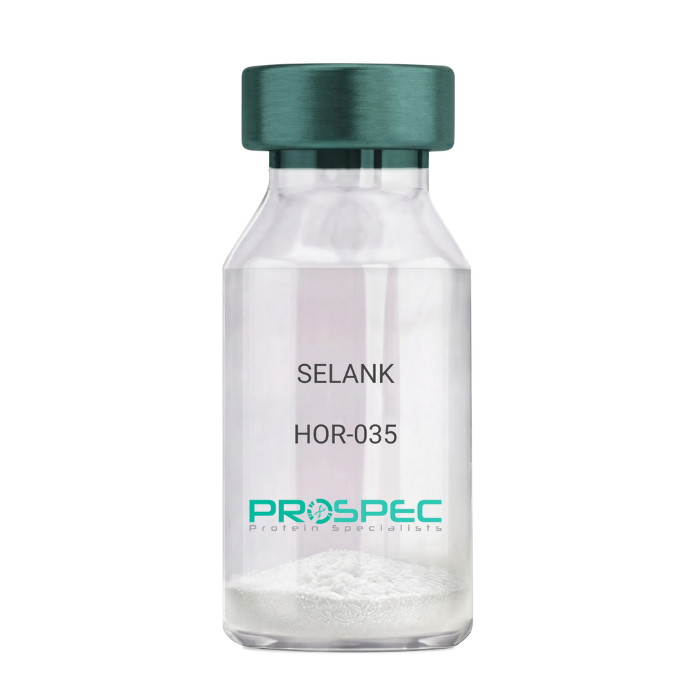
    SELANK

    Description:

    SELANK

    Product # :

    HOR-035

    Price :

    Quantity :

    Shipping Method :

    Room Temp Icon

    Shipped at Room temp

    Add To Cart

    More Info

    • description
    • formulation
    • purity
    • More Info

    Description

    SELANK Synthetic is a single, non-glycosylated polypeptide chain containing 7 amino acids, having a molecular mass of 751.89 Dalton and a Molecular formula of C33H57N11O9.

    Formulation

    The protein was lyophilized with no additives.

    Purity

    Greater than 97.0% as determined by analysis by RP-HPLC.

    More Info

    • Physical Appearance

      Sterile Filtered White lyophilized (freeze-dried) powder.

    • Stability

      Lyophilized SELANK although stable at room temperature for 3 weeks, should be stored desiccated below -18°C. Upon reconstitution SELANK should be stored at 4°C between 2-7 days and for future use below -18°C. For long term storage it is recommended to add a carrier protein (0.1% HSA or BSA). Please prevent freeze-thaw cycles.

    • Solubility

      It is recommended to reconstitute the lyophilized SELANK in sterile 18MΩ-cm H2O not less than 100 µg/ml, which can then be further diluted to other aqueous solutions.

    • Amino Acid Sequence

      H-Thr-Lys-Pro-Arg-Pro-Gly-Pro-OH.

    • Background

      Selank, also known as TP-7, is a synthetic peptide that has garnered significant attention in the scientific community due to its potential anxiolytic, neuroprotective, and nootropic effects. This heptapeptide, derived from the naturally occurring human tetrapeptide Tuftsin, has been shown to possess a wide range of biological activities, including enhancing memory, reducing anxiety, and potentially mitigating the effects of neurodegenerative diseases.

      Selank's primary mechanism of action involves its interaction with the GABAergic system, the body's main inhibitory neurotransmitter system. By modulating the activity of GABA receptors, Selank can exert a calming effect on the brain, reducing anxiety and enhancing cognitive function. Studies by Semenova et al. (2014) have demonstrated that Selank can enhance memory and learning in rats, suggesting potential applications in cognitive enhancement and the treatment of cognitive disorders.

      In addition to its anxiolytic and nootropic effects, Selank has been shown to possess neuroprotective properties. Research by Ershov et al. (2016) found that Selank could protect neurons from oxidative stress, suggesting potential applications in the treatment of neurodegenerative diseases such as Alzheimer's and Parkinson's disease.

      Given its anxiolytic, nootropic, and neuroprotective effects, Selank has been proposed as a potential therapeutic agent for a variety of conditions, including anxiety disorders, cognitive disorders, and neurodegenerative diseases. For instance, a study by Medvedev et al. (2017) found that Selank could improve symptoms in patients with generalized anxiety disorder, indicating its potential as a therapeutic agent in the treatment of anxiety disorders.

      While research on Selank is promising, it is important to note that most studies have been conducted in animals or in vitro. More research is needed to fully understand the potential effects and applications of Selank in humans. However, the existing body of research suggests that Selank could be a promising tool in the treatment of anxiety disorders, cognitive disorders, and neurodegenerative diseases.

    ProSpec's products are furnished for LABORATORY RESEARCH USE ONLY. The product may not be used as drugs, agricultural or pesticidal products, food additives or household chemicals.

    Selank
  • View Data Sheet

    Name :

    SERPING1 Human, Native

    Description:

    Serpin Peptidase Inhibitor, Clade G Member 1 Human

    C1IN, C1INH, C1NH, HAE1, HAE2 , Plasma protease C1 inhibitor, C1 esterase inhibitor, C1-inhibiting factor, Serpin G1, Name, SERPING1.

    Product # :

    PRO-2711

    Price :

    Quantity :

    Shipping Method :

    Ice Icon

    Shipped with Ice Packs

    Add To Cart

    More Info

    • description
    • source
    • formulation
    • purity
    • More Info

    Description

    Human Serpin Peptidase Inhibitor, Clade G Member 1 produced in Human plasma having a molecular mass of 110 kDa.

    Source

    Human Plasma.

    Formulation

    10mM sodium phosphate and 145mM NaCl, pH 7.3.

    Purity

    Greater than 95.0% as determined by SDS-PAGE.

    More Info

    • Introduction

      Plasma protease C1 inhibitor (SERPING1) is a part of the serpin superfamily of serine protease inhibitors. SERPING1 plays an important role in regulating activation of both the complement and contact systems. SERPING1 is also activating C1 by coupling with the active catalytic site at the light chains of C1r and C1s. SERPING1 insufficiency results in hereditary angioedema, which is characterized by recurrent episodes of localized angioedema of the skin, gastrointestinal mucosa or upper respiratory mucosa.

    • Synonyms

      C1IN, C1INH, C1NH, HAE1, HAE2 , Plasma protease C1 inhibitor, C1 esterase inhibitor, C1-inhibiting factor, Serpin G1, Name, SERPING1.

    • Physical Appearance

      Sterile Filtered solution.

    • Stability

      Native SERPING1 Human is stable at 4°C if entire vial will be used within 2-4 weeks.Store, frozen below -20°C for longer periods of time.For long term storage it is recommended to add a carrier protein (0.1% HSA or BSA).Avoid multiple freeze-thaw cycles.

    • Human Virus Test

      Plasma from each donor has been tested and found negative for antibody to HIV-1,HIV-2, HCV, HTLV-I &II, STS and HBSAG.

    ProSpec's products are furnished for LABORATORY RESEARCH USE ONLY. The product may not be used as drugs, agricultural or pesticidal products, food additives or household chemicals.

    C1 Esterase Inhibitor
  • View Data Sheet

    Name :

    CXCL6 Human

    Description:

    Granulocyte Chemotactic Protein 2 (CXCL6) Human Recombinant

    C-X-C motif chemokine 6, Chemokine alpha 3, CKA-3, Granulocyte chemotactic protein 2, GCP-2, Small-inducible cytokine B6.

    Product # :

    CHM-025

    Price :

    Quantity :

    Shipping Method :

    Room Temp Icon

    Shipped at Room temp

    Add To Cart

    More Info

    • description
    • source
    • formulation
    • purity
    • biological activity
    • More Info

    Description

    GCP-2 Human Recombinant produced in E.Coli is a single, non-glycosylated, polypeptide chain containing 72 amino acids and having a molecular mass of 7.9kDa.

    Source

    Escherichia Coli.

    Formulation

    GCP-2 protein was lyophilized from a 0.2 µm filtered concentrated solution in PBS, pH 7.4.

    Purity

    Greater than 98.0% as determined by:
    (a) Analysis by RP-HPLC.
    (b) Analysis by SDS-PAGE.

    Biological Activity

    Fully biologically active when compared to standard. The biological activity determined by a chemotaxis bioassay using human neutrophils is in a concentration range of 10-50 ng/ml corresponding to a specific activity of 20,000-100,000 IU/mg.

    More Info

    • Introduction

      Granulocyte Chemotactic Protein 2 (CXCL6), also known as GCP-2, is a Chemotactic for neutrophil granulocytes. GCP-2 signals through binding and activation of its receptors (CXCR1 and CXCR2). GCP-2 has strong antibacterial activity against Gram-positive and Gram-negative bacteria In addition to its chemotactic and angiogenic property.

    • Synonyms

      C-X-C motif chemokine 6, Chemokine alpha 3, CKA-3, Granulocyte chemotactic protein 2, GCP-2, Small-inducible cytokine B6.

    • Physical Appearance

      Sterile Filtered White lyophilized (freeze-dried) powder.

    • Stability

      Lyophilized GCP-2 although stable at room temperature for 3 weeks, should be stored desiccated below -18°C. Upon reconstitution GCP-2 should be stored at 4°C between 2-7 days and for future use below -18°C. For long term storage it is recommended to add a carrier protein (0.1% HSA or BSA). Please prevent freeze-thaw cycles.

    • Solubility

      It is recommended to reconstitute the lyophilized GCP-2 in sterile 18M-cm H2O not less than 100µg/ml, which can then be further diluted to other aqueous solutions.

    • Amino Acid Sequence

      VLTELRCTCL RVTLRVNPKT IGKLQVFPAG PQCSKVEVVA SLKNGKQVCL DPEAPFLKKV IQKILDSGNK KN.

    • Background

      What is the molecular weight/Mw of CXCL6 HUMAN Protein?
      CXCL6 HUMAN Protein has a total Mw of 7.9kDa.

      What is the source or expression system of CXCL6 HUMAN Protein?
      Escherichia Coli.

      What is the Purity of CXCL6 HUMAN Protein?
      CXCL6 HUMAN Protein is >98% pure as determined by SDS-PAGE.

      What is the Biological Activity of CXCL6 HUMAN Protein?
      Fully biologically active when compared to standard. The biological activity determined by a chemotaxis bioassay using human neutrophils is in a concentration range of 10-50 ng/ml corresponding to a specific activity of 20,000-100,000 IU/mg.

      What is the amino acid sequence of CXCL6 HUMAN Protein?
      VLTELRCTCL RVTLRVNPKT IGKLQVFPAG PQCSKVEVVA SLKNGKQVCL DPEAPFLKKV IQKILDSGNK KN.

      What applications can CXCL6 HUMAN Protein be used in?
      CXCL6 HUMAN Protein can probably be used in western blot, ELISA and Lateral Flow.

      What is the endotoxin level for CXCL6 HUMAN Protein?
      The endotoxin level is minimal, CXCL6 HUMAN Protein was purified using conventional chromatography techniques.


    ProSpec's products are furnished for LABORATORY RESEARCH USE ONLY. The product may not be used as drugs, agricultural or pesticidal products, food additives or household chemicals.

    Gcp 2 Human
  • View Data Sheet

    Name :

    Thymosin a1 human

    Description:

    Thymosin a1 Acetate

    Product # :

    HOR-243

    Price :

    Quantity :

    Shipping Method :

    Room Temp Icon

    Shipped at Room temp

    Add To Cart

    More Info

    • description
    • formulation
    • purity
    • More Info

    Description

    Thymosin a1 acetate, also known as thymalfasin has immunoregulatory properties enhancing immune functions. Thymosin a1 has a molecular formula of C129H215N33O55 a.a. sequence of Ac-Ser-Asp-Ala-Ala-Val-Asp-Thr-Ser-Ser-Glu-Ile-Thr-Thr-Lys-Asp-Leu-Lys-Glu-Lys-Lys-Glu-Val-Val-Glu-Glu-Ala-Glu-Asn-OH and having a Mw of 3108.32 Dalton.

    Formulation

    The protein (1mg/ml) was lyophilized with no additives.

    Purity

    Greater than 99.0% as determined by:
    (a) Analysis by RP-HPLC.
    (b) Analysis by SDS-PAGE.

    More Info

    • Introduction

      Thymalfasin is a synthetic analogue of thymosin-alpha-1, a 28-amino acid protein derived from the precursor protein prothymosin-alpha. Exhibiting a variety of immunoregulating properties, thymosin-alpha-1 induces differentiation of murine T-cell precursors and human thymocytes and the terminal differentiation of functionally immature cord blood lymphocytes and induces production of IL-2, high affinity IL-2 receptors, and B-cell growth factors by peripheral blood mononuclear cells. T-helper and cytotoxic/suppressor T-cell populations are targets of thymosin activity. Thymosin-alpha-1 has been shown to increase the efficiency of antigen presentation by macrophages and to be an endogenous modulator of alpha-thrombin activity.

    • Physical Appearance

      Sterile Filtered White lyophilized (freeze-dried) powder.

    • Stability

      Lyophilized Thymosin a1 although stable at room temperature for 3 weeks, should be stored desiccated below -18°C. Upon reconstitution Thymalfasin should be stored at 4°C between 2-7 days and for future use below -18°C.For long term storage it is recommended to add a carrier protein (0.1% HSA or BSA).Please prevent freeze-thaw cycles.

    • Solubility

      It is recommended to reconstitute the lyophilized Thymosin a1 in sterile 18MΩ-cm H2O not less than 100 µg/ml, which can then be further diluted to other aqueous solutions.

    • Background

      What is the molecular weight/Mw of THYMOSIN-A1 HUMAN Protein?
      THYMOSIN-A1 HUMAN Protein has a total Mw of 3.1kDa.

      What is the Purity of THYMOSIN-A1 HUMAN Protein?
      THYMOSIN-A1 HUMAN Protein is >99% pure as determined by SDS-PAGE.

      What is the Biological Activity of THYMOSIN-A1 HUMAN Protein?
      The biological functionality of THYMOSIN-A1 HUMAN Protein will be determined in the future.

      What applications can THYMOSIN-A1 HUMAN Protein be used in?
      THYMOSIN-A1 HUMAN Protein can probably be used in western blot, ELISA and Lateral Flow.

      What is the endotoxin level for THYMOSIN-A1 HUMAN Protein?
      The endotoxin level is minimal, THYMOSIN-A1 HUMAN Protein was purified using conventional chromatography techniques.

    ProSpec's products are furnished for LABORATORY RESEARCH USE ONLY. The product may not be used as drugs, agricultural or pesticidal products, food additives or household chemicals.

    Thymosin Alpha 1
  • View Data Sheet

    Name :

    MOG Antibody

    Description:

    myelin oligodendrocyte glycoprotein, Mouse Antibody

    MGC26137, MOG, MYELIN-OLIGODENDROCYTE GLYCOPROTEIN.

    Product # :

    ANT-199

    Price :

    Quantity :

    Shipping Method :

    Room Temp Icon

    Shipped at Room temp

    Add To Cart

    More Info

    • formulation
    • More Info

    Formulation

    1mg/ml in PBS (after reconstitution).

    More Info

    • Introduction

      MOG is a membrane protein expressed on the oligodendrocyte cell surface and the outermost surface of myelin sheaths. Due to this localization, it is a primary target antigen involved in immune-mediated demyelination. MOG is involved in completion and maintenance of the myelin sheath and in cell-cell communication. Alternatively spliced transcript variants encoding different isoforms have been identified Minor component of the myelin sheath is involved in completion and/or maintenance of the myelin sheath and in cell- cell communication.

    • Synonyms

      MGC26137, MOG, MYELIN-OLIGODENDROCYTE GLYCOPROTEIN.

    • Solubility

      Reconstitute with H20. Mix gently, wash the sides of the vial and wait 30-60 seconds before use.

    • Immunogen

      synthetic pMOG 35-55.

    • Ig Subclass

      mouse IgG1.

    • Clone

      NYRMOG.

    • Applications

      Direct ELISA, Western Blot, Immuneprecipitation, immunohistochemistry.

    • Titer

      By direct ELISA, 1:10,000 dilution will yield 0.5 O.D using alkaline phosphatase conjugated rabbit anti-mouse Ig (Jackson Laboratories).

    • Shipping Conditions

      Antibody is shipped lyophilized at ambient temperature.

    • Type

      Mouse Antibody Monoclonal.

    • Storage Procedures

      In lyophilized form, for long periods, store at 4oC in a dry environment. After reconstitution, if not intended for use within a month, aliquot and store at -20oC.

    • Purification Method

      Ion exchange column.

    ProSpec's products are furnished for LABORATORY RESEARCH USE ONLY. The product may not be used as drugs, agricultural or pesticidal products, food additives or household chemicals.

    Mog Antibody
  • View Data Sheet

    Name :

    CRP Human Recombinant

    Description:

    c-Reactive Protein Human Recombinant

    C-reactive protein, CRP, PTX1, MGC88244, MGC149895.

    Product # :

    PRO-335

    Price :

    Quantity :

    Shipping Method :

    Ice Icon

    Shipped with Ice Packs

    Add To Cart

    More Info

    • description
    • source
    • formulation
    • purity
    • More Info
    • sds-page

    Description

    Recombinant Human CRP produced in E.Coli is a non-glycosylated polypeptide chain having a total molecular mass of 115 kDa that corresponds to the pentamer structure of 23 kDa monomer determined by amino acid sequence. The CRP is purified by proprietary chromatographic techniques.

    Source

    Escherichia Coli.

    Formulation

    The sterile protein solution contains 20mM Tris (pH 7.5), 2mM CaCl2, 0.14M NaCl and 0.05% NaN3.

    Purity

    Greater than 95.0% as determined by Analysis by SDS-PAGE.

    sds-page

    CRP Human Recombinant SDS-PAGE - Product image 1

    More Info

    • Introduction

      CRP is an acute phase protein that correlates with inflammatory disease and is synthesized by hepatocytes during the acute phase response by certain cytokines (IL-1 and TNF Alpha and Beta). CRP levels increase dramatically (up to 1,000 fold) and serve as a useful marker of inflammation in such conditions as bacterial infection, rheumatoid arthritis, viral infections, transplantation rejection, meningitis, myocardial infarction, septicemia, osteomyelitis and others. CRP is also highly correlated to Serum Amyloid A levels.

    • Synonyms

      C-reactive protein, CRP, PTX1, MGC88244, MGC149895.

    • Physical Appearance

      Sterile Filtered clear solution.

    • Stability

      CRP should be stored at all times at 4°C.

    ProSpec's products are furnished for LABORATORY RESEARCH USE ONLY. The product may not be used as drugs, agricultural or pesticidal products, food additives or household chemicals.

    CRP Human Recombinant
  • View Data Sheet

    Name :

    FLRT3 Human, HEK

    Description:

    Fibronectin Leucine Rich Transmembrane Protein 3 Human Recombinant, HEK

    Fibronectin Leucine Rich Transmembrane Protein 3, Fibronectin-Like Domain, Containing Leucine-Rich Transmembrane Protein 3, HH21, Leucine-Rich Repeat, Transmembrane Protein FLRT3, KIAA1469.

    Product # :

    PRO-2805

    Price :

    Quantity :

    Shipping Method :

    Ice Icon

    Shipped with Ice Packs

    Add To Cart

    More Info

    • description
    • source
    • formulation
    • purity
    • biological activity
    • More Info

    Description

    FLRT3 Human Recombinant is a single, glycosylated, polypeptide chain (29-528 a.a) containing a total of 506 amino acids and having a molecular mass of 57.3 kDa. FLRT3 is fused to a 6 a.a his-Tag at C-terminus and is purified by proprietary chromatographic techniques.

    Source

    HEK293 Cells.

    Formulation

    FLRT3 protein solution (0.25mg/ml) contains Phosphate Buffered Saline (pH 7.4) and 10% glycerol.

    Purity

    Greater than 95.0% as determined by SDS-PAGE.

    Biological Activity

    >40%.  Measured by the ability of the immobilized protein to support the adhesion of Neuro-2a neuroblast cells. When cells are added to human FLRT3 coated plates 5 ug/ml.

    More Info

    • Synonyms

      Fibronectin Leucine Rich Transmembrane Protein 3, Fibronectin-Like Domain, Containing Leucine-Rich Transmembrane Protein 3, HH21, Leucine-Rich Repeat, Transmembrane Protein FLRT3, KIAA1469.

    • Physical Appearance

      Sterile filtered colorless solution.

    • Stability

      Store at 4°C if entire vial will be used within 2-4 weeks.Store, frozen at -20°C for longer periods of time. For long term storage it is recommended to add a carrier protein (0.1% HSA or BSA).Avoid multiple freeze-thaw cycles.

    • Amino Acid Sequence

      KSCPSVCRCD AGFIYCNDRF LTSIPTGIPE DATTLYLQNN QINNAGIPSD LKNLLKVERI YLYHNSLDEF PTNLPKYVKE LHLQENNIRT ITYDSLSKIP YLEELHLDDN SVSAVSIEEG AFRDSNYLRL LFLSRNHLST IPWGLPRTIE ELRLDDNRIS TISSPSLQGL TSLKRLVLDG NLLNNHGLGD KVFFNLVNLT ELSLVRNSLT AAPVNLPGTN LRKLYLQDNH INRVPPNAFS
      YLRQLYRLDM SNNNLSNLPQ GIFDDLDNIT QLILRNNPWY CGCKMKWVRD WLQSLPVKVN VRGLMCQAPE KVRGMAIKDL NAELFDCKDS GIVSTIQITT AIPNTVYPAQ GQWPAPVTKQ PDIKNPKLTK DHQTTGSPSR KTITITVKSV TSDTIHISWK LALPMTALRL SWLKLGHSPA FGSITETIVT GERSEYLVTA LEPDSPYKVC MVPMETSNLY LFDETPVCIE TETAPLRMYN
      PTTTLNREQE KEPYKNPNLP HHHHHH.

    • Background

      Fibronectin leucine-rich transmembrane protein 3, commonly known as FLRT3, stands as a molecular architect in the intricate landscape of neural development. Its roles, initially discovered in the embryonic nervous system, have expanded to encompass various physiological and pathological processes in both the brain and beyond. This research endeavors to unravel the enigma of FLRT3 protein, exploring its structural intricacies, physiological functions, and its far-reaching implications in neurobiology, embryogenesis, and disease. By delving into FLRT3's multifaceted roles, scientists aim to decipher the underlying mechanisms that govern its diverse functions and explore potential therapeutic avenues in the realms of neuroscience and beyond.

      Structural Complexities of FLRT3:

      FLRT3 belongs to the FLRT family, characterized by extracellular leucine-rich repeats (LRRs) and a transmembrane domain. These structural motifs enable FLRT3 to participate in a myriad of interactions, including binding with cell adhesion molecules and guidance cues. Understanding the three-dimensional architecture of FLRT3 is fundamental for unraveling its molecular partnerships, biological activities, and its contributions to cell adhesion and signaling.

      Physiological Functions in Neural Development:

      In the developing nervous system, FLRT3 acts as a guidance molecule, steering growing axons and dendrites to their precise destinations. Through interactions with other cell surface receptors and ligands, FLRT3 modulates axon pathfinding, synapse formation, and neuronal migration. Its presence in growth cones and developing neural circuits underscores its significance in sculpting the intricate neural networks essential for proper brain function.

      Beyond Neural Development:

      Beyond its canonical roles in neurodevelopment, FLRT3 has emerged as a versatile player in various physiological processes. It participates in tissue morphogenesis, modulates cell adhesion, and influences immune responses. Recent studies have also implicated FLRT3 in cancer progression, highlighting its involvement in pathological conditions and making it a potential target for therapeutic interventions in cancer therapy.

      FLRT3 as a Therapeutic Target:

      The diverse roles of FLRT3 in neural development and diseases position it as an attractive target for therapeutic interventions. Modulating FLRT3 interactions offers novel avenues for neurological disorder treatments, including neurodevelopmental disorders and neurodegenerative diseases. Moreover, understanding FLRT3's involvement in cancer biology opens doors for innovative cancer therapies, making it a promising target for precision medicine approaches.

      FLRT3, with its intricate structural features and diverse functional roles, stands as a linchpin in the realms of neuroscience, embryogenesis, and disease. Its multifaceted contributions to neural development, tissue morphogenesis, and disease pathogenesis underscore its significance in both health and pathology. As researchers continue to unravel FLRT3’s complexities, they not only deepen our understanding of fundamental biological processes but also pave the way for groundbreaking discoveries in neuroscience and therapeutic interventions, ultimately shaping the future landscape of medicine and scientific inquiry.

    ProSpec's products are furnished for LABORATORY RESEARCH USE ONLY. The product may not be used as drugs, agricultural or pesticidal products, food additives or household chemicals.

    Flrt3 Protein
  • View Data Sheet

    Name :

    TINAGL1 Human, Sf9

    Description:

    Tubulointerstitial Nephritis Antigen Like 1 Human Recombinant, Sf9

    Tubulointerstitial nephritis antigen-like isoform 1, TINAGL1, Tubulointerstitial nephritis antigen-like, Glucocorticoid-inducible protein 5, Oxidized LDL-responsive gene 2 protein, OLRG-2, Tubulointerstitial nephritis antigen-related protein, TIN Ag-related protein, TIN-Ag-RP, GIS5, LCN7, OLRG2, TINAGL, PP6614, PSEC0088, UNQ204/PRO230.

    Product # :

    PRO-2422

    Price :

    Quantity :

    Shipping Method :

    Ice Icon

    Shipped with Ice Packs

    Add To Cart

    More Info

    • description
    • source
    • formulation
    • purity
    • More Info

    Description

    TINAGL1 Human Recombinant produced in Sf9 Baculovirus cells is a single, glycosylated polypeptide chain containing 455 amino acids (22-467a.a.) and having a molecular mass of 51.2kDa (Molecular size on SDS-PAGE will appear at approximately 50-70kDa). TINAGL1 is expressed with a 6 amino acid His tag at C-Terminus and purified by proprietary chromatographic techniques.

    Source

    Sf9, Baculovirus cells.

    Formulation

    TINAGL1 protein solution (0.25mg/ml) contains Phosphate Buffered Saline (pH 7.4), 30% glycerol and 1mM DTT.

    Purity

    Greater than 90.0% as determined by SDS-PAGE.

    More Info

    • Introduction

      Tubulointerstitial Nephritis Antigen Like 1 also known as TINAGL1 is similar in sequence to tubulointerstitial nephritis antigen, a secreted glycoprotein which is identified by antibodies in several types of immune-related tubulointerstitial nephritis. Three transcript variants encoding different isoforms are known for TINAGL1.

    • Synonyms

      Tubulointerstitial nephritis antigen-like isoform 1, TINAGL1, Tubulointerstitial nephritis antigen-like, Glucocorticoid-inducible protein 5, Oxidized LDL-responsive gene 2 protein, OLRG-2, Tubulointerstitial nephritis antigen-related protein, TIN Ag-related protein, TIN-Ag-RP, GIS5, LCN7, OLRG2, TINAGL, PP6614, PSEC0088, UNQ204/PRO230.

    • Physical Appearance

      Sterile filtered colorless solution.

    • Stability

      Store at 4°C if entire vial will be used within 2-4 weeks. Store, frozen at -20°C for longer periods of time. For long term storage it is recommended to add a carrier protein (0.1% HSA or BSA).Avoid multiple freeze-thaw cycles.

    • Amino Acid Sequence

      ADLAQQGRGR RELAPGLHLR GIRDAGGRYC QEQDLCCRGR ADDCALPYLG AICYCDLFCN RTVSDCCPDF WDFCLGVPPP FPPIQGCMHG GRIYPVLGTY WDNCNRCTCQ ENRQWQCDQE PCLVDPDMIK AINQGNYGWQ AGNHSAFWGM TLDEGIRYRL GTIRPSSSVM NMHEIYTVLN PGEVLPTAFE ASEKWPNLIH EPLDQGNCAG SWAFSTAAVA SDRVSIHSLG HMTPVLSPQN LLSCDTHQQQ GCRGGRLDGA WWFLRRRGVV SDHCYPFSGR ERDEAGPAPP CMMHSRAMGR GKRQATAHCP NSYVNNNDIY QVTPVYRLGS NDKEIMKELM ENGPVQALME VHEDFFLYKG GIYSHTPVSL GRPERYRRHG THSVKITGWG EETLPDGRTL KYWTAANSWG PAWGERGHFR IVRGVNECDI ESFVLGVWGR VGMEDMGHHH HHHHH.

    ProSpec's products are furnished for LABORATORY RESEARCH USE ONLY. The product may not be used as drugs, agricultural or pesticidal products, food additives or household chemicals.

    Tinagl1 Human Sf9
  • View Data Sheet

    Name :

    TNFR2 Human Fc

    Description:

    Tumor Necrosis Factor Receptor 2 Fusion Protein Human Recombinant

    Tumor necrosis factor receptor superfamily member 1B,Tumor necrosis factor receptor 2, TNF-R2, Tumor necrosis factor receptor type II, p75, p80 TNF-alpha receptor, CD120b antigen, Etanercept, TBPII, TNFBR, TNFR80, TNF-R75, p75TNFR, TNF-R-II.

    Product # :

    CYT-422

    Price :

    Quantity :

    Shipping Method :

    Room Temp Icon

    Shipped at Room temp

    Add To Cart

    More Info

    • description
    • source
    • formulation
    • purity
    • biological activity
    • More Info

    Description

    Recombinant Human Tumor Necrosis Factor Receptor 2 Fusion Protein produced in CHO is a dimeric, glycosylated, polypeptide chain consisting of the extracellular ligand-binding portion of the human 75 kilo Dalton (p75) tumor necrosis factor receptor 2 (TNFR2) linked to the Fc portion of human IgG1. The Fc component of TNFR2 contains the CH2 domain, the CH3 domain and hinge region, but not the CH1 domain of IgG1. It consists of 934 amino acids and has an apparent molecular weight of approximately 150 kilo Daltons.The TNFR2 is purified by standard chromatographic techniques.

    Source

    Chinese Hamster Ovarian Cells (CHO).

    Formulation

    Each mg contains 1.6mg mannitol, 0.4 mg sucrose and 48 µg tromethamine.

    Purity

    Greater than 98.0% as determined by:
    (a) Analysis by RP-HPLC.
    (c) Analysis by SDS-PAGE.

    Biological Activity

    Potency is determined by its ability to neutralize TNF-alpha mediated growth inhibition of A375 cells, corresponding to a Specific Activity of 17,000,000 IU/mg.

    More Info

    • Introduction

      TNFR binds specifically to tumor necrosis factor (TNF) and blocks its interaction with cell surface TNF receptors. TNF is a naturally occurring cytokine that is involved in normal inflammatory and immune responses. It plays an important role in the inflammatory processes of rheumatoid arthritis (RA), polyarticular-course juvenile rheumatoid arthritis (JRA), and ankylosing spondylitis and the resulting joint pathology. In addition, TNF plays a role in the inflammatory process of plaque psoriasis. Elevated levels of TNF are found in involved tissues and fluids of patients with RA, psoriatic arthritis, ankylosing spondylitis (AS), and plaque psoriasis. Two distinct receptors for TNF (TNFRs), a 55 kilodalton protein (p55) and a 75 kilodalton protein (p75), exist naturally as monomeric molecules on cell surfaces and in soluble forms. Biological activity of TNF is dependent upon binding to either cell surface TNFR. Recombinant Human TNFR is a dimeric soluble form of the p75 TNF receptor that can bind to two TNF molecules.
      It inhibits the activity of TNF in vitro and has been shown to affect several animal models of inflammation, including murine collagen-induced arthritis. TNFR inhibits binding of both TNF? and TNF? (lymphotoxin alpha [LT?]) to cell surface TNFRs, rendering TNF biologically inactive. Cells expressing transmembrane TNF that bind to TNFR are not lysed in vitro in the presence or absence of complement.
      TNFR can also modulate biological responses that are induced or regulated by TNF, including expression of adhesion molecules responsible for leukocyte migration (i.e., E-selectin and to a lesser extent intercellular adhesion molecule-1 [ICAM-1]), serum levels of cytokines (e.g., IL-6), and serum levels of matrix metalloproteinase-3 (MMP-3 or stromelysin).

    • Synonyms

      Tumor necrosis factor receptor superfamily member 1B,Tumor necrosis factor receptor 2, TNF-R2, Tumor necrosis factor receptor type II, p75, p80 TNF-alpha receptor, CD120b antigen, Etanercept, TBPII, TNFBR, TNFR80, TNF-R75, p75TNFR, TNF-R-II.

    • Physical Appearance

      Sterile Filtered White lyophilized (freeze-dried) powder.

    • Stability

      Lyophilized Tumor Necrosis Factor Receptor although stable at room temperature for 3 weeks, should be stored desiccated below -18°C. Upon reconstitution TNFR2 should be stored at 4°C between 2-7 days and for future use below -18°C.For long term storage it is recommended to add a carrier protein (0.1% HSA or BSA).Please prevent freeze-thaw cycles.

    • Solubility

      It is recommended to reconstitute the lyophilized TNFR2 in sterile 18M-cm H2O not less than 100µg/ml, which can then be further diluted to other aqueous solutions.

    ProSpec's products are furnished for LABORATORY RESEARCH USE ONLY. The product may not be used as drugs, agricultural or pesticidal products, food additives or household chemicals.

    Tnfr2 Human Fc
  • View Data Sheet

    Name :

    Omentin Human, His

    Description:

    Omentin Human Recombinant, His Tag

    Intelectin-1, HL1, LFR, HL-1, INTL, ITLN, hIntL.

    Product # :

    CYT-551

    Price :

    Quantity :

    Shipping Method :

    Room Temp Icon

    Shipped at Room temp

    Add To Cart

    More Info

    • description
    • source
    • formulation
    • purity
    • More Info

    Description

    Omentin Human Recombinant is produced in E.Coli by recombinant DNA technology is a single, polypeptide chain containing 294 amino acids and having a molecular mass of 32.7 kDa. Recombinant Human Omentin contains His tag fused at N-Terminus.Intelectin is purified by proprietary chromatographic techniques.

    Source

    Escherichia Coli.

    Formulation

    Each mg of lyophilized powder contains 20mM Tris & 50mM NaCl pH-8.0.

    Purity

    Greater than 95.0% as determined by(a) Analysis by RP-HPLC.
    (b) Analysis by SDS-PAGE.

    More Info

    • Introduction

      Omentin is a recently recognized gene highly localized to the mental tissue (visceral adipose tissue). Omentin is present in the stromal vascular cells in the adipose tissue rather than in the adipocytes. Omentin is predominantly expressed in the visceral adipose tissue than the subcutaneous tissue, with the omentin mRNA being 150 times higher in the visceral adipose tissue. Omentin has also been detected in human blood using western blot analysis, and seems to increase insulin-stimulated glucose uptake in 3T3-L1 adipocytes in mice. Omentin seems to increase Akt phosphorylation irrespective of insulin presence. Its role in glucose metabolism and obesity remains to be described; an insulin-sensitizing action is possible.Differences in Omentin expression has been noted in adipose tissue from normals and patients with inflammatory bowel disease although its significance is unknown.

    • Synonyms

      Intelectin-1, HL1, LFR, HL-1, INTL, ITLN, hIntL.

    • Physical Appearance

      Sterile Filtered White lyophilized (freeze-dried) powder.

    • Stability

      Lyophilized Intelectin His Tag although stable at room temperature for 3 weeks, should be stored desiccated below -18°C. Upon reconstitution Intelectin should be stored at 4°C between 2-7 days and for future use below -18°C.For long term storage it is recommended to add a carrier protein (0.1% HSA or BSA).Please prevent freeze-thaw cycles.

    • Solubility

      It is recommended to reconstitute the lyophilized Omentin His Tag in sterile 18MΩ-cm H2O not less than 100µg/ml, which can then be further diluted to other aqueous solutions.

    • Amino Acid Sequence

      MRGSHHHHHH GMASTDEANT YFKEWTCSSS PSLPRSCKEI KDECPSAFDG LYFLRTENGV IYQTFCDMTS GGGGWTLVAS VHENDMRGKC TVGDRWSSQQ GSKAVYPEGD GNWANYNTFG SAEAATSDDY KNPGYYDIQA KDLGIWHVPN KSPMQHWRNS SLLRYRTDTG FLQTLGHNLF GIYQKYPVKY GEGKCWTDNG PVIPVVYDFG DAQKTASYYS PYGQREFTAG FVQFRVFNNE RAANALCAGM RVTGCNTEHH CIGGGGYFPE ASPQQCGDFS GFDWSGYGTH VGYS.

    • Applications

      Elisa
      Western blot.

    ProSpec's products are furnished for LABORATORY RESEARCH USE ONLY. The product may not be used as drugs, agricultural or pesticidal products, food additives or household chemicals.

    Omentin Human His
  • View Data Sheet

    Name :

    C1QTNF8 Human

    Description:

    Complement C1q Tumor Necrosis Factor-Related Protein 8 Human Recombinant

    CTRP8, UNQ5829, C1q/TNF-related protein 8, Complement C1q tumor necrosis factor-related protein 8.

    Product # :

    PRO-134

    Price :

    Quantity :

    Shipping Method :

    Ice Icon

    Shipped with Ice Packs

    Add To Cart

    More Info

    • description
    • source
    • formulation
    • purity
    • More Info

    Description

    C1QTNF8 Human Recombinant produced in E.Coli is a single, non-glycosylated, polypeptide chain containing 255 amino acids including a 10 a.a N-terminal His tag. The total molecular mass is 28.4kDa (calculated).

    Source

    Escherichia Coli.

    Formulation

    C1QTNF8 filtered (0.4 µm) solution in 0.03M Acetate buffer pH-4.0.

    Purity

    Greater than 95.0% as determined by SDS-PAGE.

    More Info

    • Synonyms

      CTRP8, UNQ5829, C1q/TNF-related protein 8, Complement C1q tumor necrosis factor-related protein 8.

    • Physical Appearance

      Filtered colorless solution.

    • Stability

      Store at 4°C if entire vial will be used within 2-4 weeks. Store, frozen at -20°C for longer periods of time. For long term storage it is recommended to add a carrier protein (0.1% HSA or BSA).Avoid multiple freeze-thaw cycles.

    • Amino Acid Sequence

      MKHHHHHHAS WPGLPRRPCV HCCRPAWPPG PYARVSDRDL WRGDLWRGLP RVRPTIDIEI LKGEKGEAGV RGRAGRSGKE GPPGARGLQG RRGQKGQVGP PGAACRRAYA AFSVGRRAYA AFSVGRREGL HSSDHFQAVP FDTELVNLDG AFDLAAGRFL CTVPGVYFLS LNVHTWNYKE TYLHIMLNRR PAAVLYAQPS ERSVMQAQSL MLLLAAGDAV WVRMFQRDRD NAIYGEHGDL YITFSGHLVK PAAEL.

    ProSpec's products are furnished for LABORATORY RESEARCH USE ONLY. The product may not be used as drugs, agricultural or pesticidal products, food additives or household chemicals.

    C1Qtnf8 Human
  • View Data Sheet

    Name :

    Fibronectin Rat

    Description:

    Fibronectin Rat

    Product # :

    PRO-131

    Price :

    Quantity :

    Shipping Method :

    Room Temp Icon

    Shipped at Room temp

    Add To Cart

    More Info

    • description
    • source
    • formulation
    • purity
    • More Info

    Description

    Plasma fibronectin level is elevated in severe coronary artery disease. Increased plasma fibronectin levels are related with venous thromboembolism (VTE) particularly in males, and extend the probable association between biomarkers and risk factors for arterial atherothrombosis and VTE. Fibronectin plays a role in several cellular processes, including tissue repair, embryogenesis, blood clotting, and cell migration/adhesion. Fibronectin consists in two main forms: 1) as an insoluble glycoprotein dimer that serves as a linker in the etracellular matrix and 2) as a soluble disulphide linked dimer found in the plasma. The plasma form is produced by hepatocytes, and the ECM form is synthesized by fibroblasts, chondrocytes, endothelial cells, macrophages, as well as certain epithelial cells. Fibronectin alos takes part as a general cell adhesion molecule by anchoring cells to collagen or proteoglycan substrates. Fibronectin organizes cellular interaction with the ECM by binding to different components of the extracellular matrix and to membrane-bound Fibronectin receptors on cell surfaces. Molecular Weight 220kDa.

    Source

    Rat Plasma.

    Formulation

    The Rat Fibronectin was lyophilized from a concentrated 1mg/ml solution containing 20mM Tris Cl pH-7.5.

    Purity

    Greater than 95.0% as determined by SDS-PAGE.

    More Info

    • Physical Appearance

      Sterile Filtered White lyophilized (freeze-dried) powder.

    • Stability

      Store the lyophilized fibronectin at 4°C. Upon reconstitution fibronectin should be stored at 4°C for 2 weeks and for future use below -18°C.Please prevent freeze-thaw cycles.

    • Solubility

      It is recommended to reconstitute the lyophilized Rat Fibronectin in sterile 18M-cm H2O not less than 100µg/ml, which can then be further diluted to other aqueous solutions.

    ProSpec's products are furnished for LABORATORY RESEARCH USE ONLY. The product may not be used as drugs, agricultural or pesticidal products, food additives or household chemicals.

    Fibronectin Rat
  • View Data Sheet

    Name :

    Clusterin Human, His

    Description:

    Apolipoprotein-J Human Recombinant, His Tag

    CLI, AAG4, APOJ, KUB1, SGP2, SGP-2, SP-40, TRPM2, TRPM-2, MGC24903, Clusterin, ging-associated gene 4 protein, Apolipoprotein J,Complement cytolysis inhibitor, Complement-associated protein SP-40,40, Ku70-binding protein 1, NA1/NA2, Testosterone-repressed prostate message 2, CLU.

    Product # :

    CYT-814

    Price :

    Quantity :

    Shipping Method :

    Ice Icon

    Shipped with Ice Packs

    Add To Cart

    More Info

    • description
    • source
    • formulation
    • purity
    • More Info
    • sds-page

    Description

    Clusterin Human Recombinant produced in E.Coli is a single, non-glycosylated polypeptide chain containing 463 amino acids (23-449 a.a.) and having a molecular mass of 54.1kDa. Clusterin is fused to a 36 amino acid His-tag at N-terminus & purified by proprietary chromatographic techniques.

    Source

    Escherichia Coli.

    Formulation

    Clusterin protein solution (0.5mg/ml) contains 20mM Tris-HCl buffer (pH 8.0), 0.15M NaCl, 10% glycerol and 1mM DTT.

    Purity

    Greater than 85% as determined by SDS-PAGE.

    sds-page

    Clusterin-sds-page - Product image 1

    More Info

    • Introduction

      Clusterin also named Apolipoprotein J (APO-J) is a 75-80 kD disulfide-linked heterodimeric protein containing about 30% of N-linked carbohydrate rich in sialic acid but truncated forms targeted to the nucleus have also been identified.
      The precursor polypeptide chain is cleaved proteolytically to remove the 22-mer secretory signal peptide and subsequently between residues 227/228 to generate the a and b chains. These are assembled in anti-parallel to give a heterodimeric molecule in which the cysteine-rich centers are linked by five disulfide bridges and are flanked by two predicted coiled-coil a-helices and three predicted amphipathic a-helices.
      Across a broad range of species clusterin shows a high degree of sequence homology ranging from 70% to 80%. It is nearly ubiquitously expressed in most mammalian tissues and can be found in plasma, milk, urine, cerebrospinal fluid and semen.
      It is able to bind and form complexes with numerous partners such as immunoglobulins, lipids, heparin, bacteria, complement components, paraoxonase, beta amyloid, leptin and others. Clusterin has been ascribed a plethora of functions such as phagocyte recruitment, aggregation induction, complement attack prevention, apoptosis inhibition, membrane remodeling, lipid transport, hormone transport and/or scavenging, matrix metalloproteinase inhibition.
      A genuine function of clusterin has not been defined. One tempting hypothesis says that clusterin is an extracellular chaperone protecting cells from stress induced insults caused by degraded and misfolded protein precipitates.
      Clusterin is up- or down regulated on the mRNA or protein level in many pathological and clinically relevant situations including cancer, organ regeneration, infection, Alzheimer disease, retinitis pigmentosa, myocardial infarction, renal tubular damage, autoimmunity and others.

    • Synonyms

      CLI, AAG4, APOJ, KUB1, SGP2, SGP-2, SP-40, TRPM2, TRPM-2, MGC24903, Clusterin, ging-associated gene 4 protein, Apolipoprotein J,Complement cytolysis inhibitor, Complement-associated protein SP-40,40, Ku70-binding protein 1, NA1/NA2, Testosterone-repressed prostate message 2, CLU.

    • Physical Appearance

      Sterile filtered colorless solution.

    • Stability

      Store at 4°C if entire vial will be used within 2-4 weeks. Store, frozen at -20°C for longer periods of time. For long term storage it is recommended to add a carrier protein (0.1% HSA or BSA).Avoid multiple freeze-thaw cycles.

    • Amino Acid Sequence

      MRGSHHHHHH GMASMTGGQQ MGRDLYDDDD KDRWGSDQTV SDNELQEMSN QGSKYVNKEI QNAVNGVKQI KTLIEKTNEE RKTLLSNLEE AKKKKEDALN ETRESETKLK ELPGVCNETM MALWEECKPC LKQTCMKFYA RVCRSGSGLV GRQLEEFLNQ SSPFYFWMNG DRIDSLLEND RQQTHMLDVM QDHFSRASSI IDELFQDRFF TREPQDTYHY LPFSLPHRRP HFFFPKSRIV RSLMPFSPYE PLNFHAMFQP FLEMIHEAQQ AMDIHFHSPA FQHPPTEFIR EGDDDRTVCR EIRHNSTGCL RMKDQCDKCR EILSVDCSTN NPSQAKLRRE LDESLQVAER LTRKYNELLK SYQWKMLNTS SLLEQLNEQF NWVSRLANLT QGEDQYYLRV TTVASHTSDS DVPSGVTEVV VKLFDSDPIT VTVPVEVSRK NPKFMETVAE KALQEYRKKH REE.

    • Background

      What is the molecular weight/Mw of CLUSTERIN Protein?
      CLUSTERIN Protein has a total Mw of 54.1kDa.

      What is the source or expression system of CLUSTERIN Protein?
      Escherichia Coli.

      What is the Purity of CLUSTERIN Protein?
      CLUSTERIN Protein is >85% pure as determined by SDS-PAGE.

      What is the Biological Activity of CLUSTERIN Protein?
      The biological functionality of CLUSTERIN Protein will be determined in the future.

      What is the amino acid sequence of CLUSTERIN Protein?
      MRGSHHHHHH GMASMTGGQQ MGRDLYDDDD KDRWGSDQTV SDNELQEMSN QGSKYVNKEI QNAVNGVKQI KTLIEKTNEE RKTLLSNLEE AKKKKEDALN ETRESETKLK ELPGVCNETM MALWEECKPC LKQTCMKFYA RVCRSGSGLV GRQLEEFLNQ SSPFYFWMNG DRIDSLLEND RQQTHMLDVM QDHFSRASSI IDELFQDRFF TREPQDTYHY LPFSLPHRRP HFFFPKSRIV RSLMPFSPYE PLNFHAMFQP FLEMIHEAQQ AMDIHFHSPA FQHPPTEFIR EGDDDRTVCR EIRHNSTGCL RMKDQCDKCR EILSVDCSTN NPSQAKLRRE LDESLQVAER LTRKYNELLK SYQWKMLNTS SLLEQLNEQF NWVSRLANLT QGEDQYYLRV TTVASHTSDS DVPSGVTEVV VKLFDSDPIT VTVPVEVSRK NPKFMETVAE KALQEYRKKH REE.

      What applications can CLUSTERIN Protein be used in?
      CLUSTERIN Protein can probably be used in western blot, ELISA and Lateral Flow.

      What is the endotoxin level for CLUSTERIN Protein?
      The endotoxin level is minimal, CLUSTERIN Protein was purified using conventional chromatography techniques.

    ProSpec's products are furnished for LABORATORY RESEARCH USE ONLY. The product may not be used as drugs, agricultural or pesticidal products, food additives or household chemicals.

    Clusterin Human His
  • View Data Sheet

    Name :

    BCDIN3D Human

    Description:

    BCDIN3D Human Recombinant

    Pre-miRNA 5'-monophosphate methyltransferase, BCDIN3 domain-containing protein, BCDIN3D.

    Product # :

    PRO-1262

    Price :

    Quantity :

    Shipping Method :

    Ice Icon

    Shipped with Ice Packs

    Add To Cart

    More Info

    • description
    • source
    • formulation
    • purity
    • More Info

    Description

    BCDIN3D Human Recombinant produced in E.Coli is a single, non-glycosylated polypeptide chain containing 315 amino acids (1-292 a.a) and having a molecular mass of 35kDa.BCDIN3D is fused to a 23 amino acid His-tag at N-terminus & purified by proprietary chromatographic techniques.

    Source

    Escherichia Coli.

    Formulation

    BCDIN3D protein solution (0.25mg/ml) containing 20mM Tris-HCl buffer (pH 8.0), 0.2M NaCl, 40% glycerol, 1mM DTT and 2mM EDTA.

    Purity

    Greater than 85.0% as determined by SDS-PAGE.

    More Info

    • Introduction

      BCDIN3D is a member of the methyltransferase superfamily and contains 1 Bin3-type SAM domain. BCDIN3D acts in the catalysis of the transfer of a methyl group to an acceptor molecule. BCDIN3D is an O-methyltransferase which specifically dimethylates the 5' monophosphate of pre-miRNAs, serving as a negative regulator of miRNA processing. BCDIN3D mediates the methylation of pre-miR-145, as well as other pre-miRNAs.

    • Synonyms

      Pre-miRNA 5'-monophosphate methyltransferase, BCDIN3 domain-containing protein, BCDIN3D.

    • Physical Appearance

      Sterile Filtered colorless solution.

    • Stability

      Store at 4°C if entire vial will be used within 2-4 weeks. Store, frozen at -20°C for longer periods of time. For long term storage it is recommended to add a carrier protein (0.1% HSA or BSA).Avoid multiple freeze-thaw cycles.

    • Amino Acid Sequence

      MGSSHHHHHH SSGLVPRGSH MGSMAVPTEL DGGSVKETAA EEESRVLAPG AAPFGNFPHY SRFHPPEQRL RLLPPELLRQ LFPESPENGP ILGLDVGCNS GDLSVALYKH FLSLPDGETC SDASREFRLL CCDIDPVLVK RAEKECPFPD ALTFITLDFM NQRTRKVLLS SFLSQFGRSV FDIGFCMSIT MWIHLNHGDH GLWEFLAHLS SLCHYLLVEP QPWKCYRAAA RRLRKLGLHD FDHFHSLAIR GDMPNQIVQI LTQDHGMELI CCFGNTSWDR SLLLFRAKQT IETHPIPESL IEKGKEKNRL SFQKQ.

    ProSpec's products are furnished for LABORATORY RESEARCH USE ONLY. The product may not be used as drugs, agricultural or pesticidal products, food additives or household chemicals.

    Bcdin3D Human
  • View Data Sheet

    Name :

    IL18BP Human, Sf9

    Description:

    Interleukin-18 Binding Protein Human Recombinant, Sf9

    Interleukin 18 Binding Protein, MC51L-53L-54L Homolog Gene Product, Tadekinig-Alfa, IL-18BP, Interleukin-18-Binding Protein, IL18BPa, Interleukin-18-binding protein, IL-18BP, Tadekinig-alfa.

    Product # :

    CYT-878

    Price :

    Quantity :

    Shipping Method :

    Ice Icon

    Shipped with Ice Packs

    Add To Cart

    More Info

    • description
    • source
    • formulation
    • purity
    • More Info

    Description

    IL18BP Human Recombinant produced in Sf9 Baculovirus cells is a single, non-glycosylated polypeptide chain containing 406 amino acids (31-194a.a) and having a molecular mass of 44.9kDa. (Molecular size on SDS-PAGE will appear at approximately 40-57kDa). IL18BP is fused to a 239 amino acid hIgG-His-tag at C-terminus & purified by proprietary chromatographic techniques.

    Source

    Sf9, Baculovirus cells.

    Formulation

    IL18BP protein solution (0.5mg/ml) containing Phosphate Buffered Saline (pH 7.4) and 10% glycerol.

    Purity

    Greater than 90.0% as determined by SDS-PAGE.

    More Info

    • Introduction

      Interleukin-18 Binding Protein (IL18BP) serves as an inhibitor of the proinflammatory cytokine, IL18. IL18BP binds IL18, inhibits the binding of IL18 to its receptor, and consequently inhibits IL18-induced IFN-gamma production, resulting in reduced T-helper type 1 immune responses. The IL18BP protein is constitutively expressed and secreted in mononuclear cells. Elevated levels of IL18BP protein are detected in the intestinal tissues of patients with Crohn's disease.

    • Synonyms

      Interleukin 18 Binding Protein, MC51L-53L-54L Homolog Gene Product, Tadekinig-Alfa, IL-18BP, Interleukin-18-Binding Protein, IL18BPa, Interleukin-18-binding protein, IL-18BP, Tadekinig-alfa.

    • Physical Appearance

      Sterile filtered colorless solution.

    • Stability

      Store at 4°C if entire vial will be used within 2-4 weeks. Store, frozen at -20°C for longer periods of time. For long term storage it is recommended to add a carrier protein (0.1% HSA or BSA).Avoid multiple freeze-thaw cycles.

    • Amino Acid Sequence

      ADPTPVSQTT TAATASVRST KDPCPSQPPV FPAAKQCPAL EVTWPEVEVP LNGTLSLSCV ACSRFPNFSI LYWLGNGSFI EHLPGRLWEG STSRERGSTG TQLCKALVLE QLTPALHSTN FSCVLVDPEQ VVQRHVVLAQ LWAGLRATLP PTQEALPSSH SSPQQQGLEP KSCDKTHTCP PCPAPELLGG PSVFLFPPKP KDTLMISRTP EVTCVVVDVS HEDPEVKFNW YVDGVEVHNA KTKPREEQYN STYRVVSVLT VLHQDWLNGK EYKCKVSNKA LPAPIEKTIS KAKGQPREPQ VYTLPPSRDE LTKNQVSLTC LVKGFYPSDI AVEWESNGQP ENNYKTTPPV LDSDGSFFLY SKLTVDKSRW QQGNVFSCSV MHEALHNHYT QKSLSLSPGK HHHHHH.

    ProSpec's products are furnished for LABORATORY RESEARCH USE ONLY. The product may not be used as drugs, agricultural or pesticidal products, food additives or household chemicals.

    Il18Bp Human Sf9
  • View Data Sheet

    Name :

    PTGR2 Human

    Description:

    Prostaglandin Reductase 2 Human Recombinant

    Prostaglandin reductase 2, PRG-2, 15-oxoprostaglandin 13-reductase, Zinc-binding alcohol dehydrogenase domain-containing protein 1, PTGR2, ZADH1, PGR2.

    Product # :

    ENZ-601

    Price :

    Quantity :

    Shipping Method :

    Ice Icon

    Shipped with Ice Packs

    Add To Cart

    More Info

    • description
    • source
    • formulation
    • purity
    • More Info

    Description

    PTGR2 Human Recombinant produced in E.coli is a single, non-glycosylated polypeptide chain containing 375 amino acids (1-351) and having a molecular mass of 41.1kDa.PTGR2 is fused to a 24 amino acid His-tag at N-terminus & purified by proprietary chromatographic techniques.

    Source

    Escherichia Coli.

    Formulation

    The PTGR2 solution (1mg/ml) contains 20mM Tris-HCl buffer, pH8.0, 10% glycerol, 1mM DTT and 50mM NaCl.

    Purity

    Greater than 90.0% as determined by SDS-PAGE.

    More Info

    • Introduction

      Prostaglandin Reductase 2 (PTGR2) is a member of the medium-chain dehydrogenase/reductase superfamily. PTGR2 is an enzyme involved in the metabolism of prostaglandins. PTGR2 catalyzes an NADPH-dependent reduction of the conjugated alpha, beta-unsaturated double bond of 15-keto-PGE(2), which is a fundamental step in terminal inactivation of prostaglandins and suppression of PPARgamma-mediated adipocyte differentiation. PTGR2 may also be involved in controlling activation of the peroxisome proliferator-activated receptor.

    • Synonyms

      Prostaglandin reductase 2, PRG-2, 15-oxoprostaglandin 13-reductase, Zinc-binding alcohol dehydrogenase domain-containing protein 1, PTGR2, ZADH1, PGR2.

    • Physical Appearance

      Sterile filtered colorless solution.

    • Stability

      Store at 4°C if entire vial will be used within 2-4 weeks. Store, frozen at -20°C for longer periods of time. For long term storage it is recommended to add a carrier protein (0.1% HSA or BSA).Avoid multiple freeze-thaw cycles.

    • Amino Acid Sequence

      MGSSHHHHHH SSGLVPRGSH MGSHMIVQRV VLNSRPGKNG NPVAENFRME EVYLPDNINE GQVQVRTLYL SVDPYMRCRM NEDTGTDYIT PWQLSQVVDG GGIGIIEESK HTNLTKGDFV TSFYWPWQTK VILDGNSLEK VDPQLVDGHL SYFLGAIGMP GLTSLIGIQE KGHITAGSNK
      TMVVSGAAGA CGSVAGQIGH FLGCSRVVGI CGTHEKCILL TSELGFDAAI NYKKDNVAEQ LRESCPAGVD VYFDNVGGNI SDTVISQMNE NSHIILCGQI SQYNKDVPYP PPLSPAIEAI QKERNITRER FLVLNYKDKF EPGILQLSQW FKEGKLKIKE TVINGLENMG AAFQSMMTGG
      NIGKQIVCIS EEISL.

    ProSpec's products are furnished for LABORATORY RESEARCH USE ONLY. The product may not be used as drugs, agricultural or pesticidal products, food additives or household chemicals.

    Ptgr2 Human
  • View Data Sheet

    Name :

    CXCL17 Rat

    Description:

    VEGF Co-regulated Chemokine 1 Rat Recombinant

    Protein Cxcl17, Cxcl17, RGD1304717, C-X-C motif chemokine 17, VEGF co-regulated chemokine 1, Vcc1, VCC-1.

    Product # :

    CHM-281

    Price :

    Quantity :

    Shipping Method :

    Room Temp Icon

    Shipped at Room temp

    Add To Cart

    More Info

    • description
    • source
    • formulation
    • purity
    • biological activity
    • More Info

    Description

    CXCL17 Rat Recombinant produced in E.coli is a single, non-glycosylated polypeptide chain containing 97 amino acids and having a molecular mass of 11.5kDa.The CXCL17 is purified by proprietary chromatographic techniques.

    Source

    Escherichia Coli.

    Formulation

    CXCL17 protein was lyophilized from a 0.2µm filtered concentrated solution in 20mM PB, 300mM NaCl, pH 7.4.

    Purity

    Greater than 95.0% as determined by:
    (a) Analysis by RP-HPLC.
    (b) Analysis by SDS-PAGE.

    Biological Activity

    Measured by its ability to induce VEGF expression in mouse endothelial cells. The ED50 for this effect is typically 1-5 µg/ml.

    More Info

    • Introduction

      Dendritic cell and monocyte chemokinelike protein (DMC/CXCL17/VEGF-correlated chemokine 1/VCC1), is a secreted molecule with a size and predicted 3-dimensional folding pattern similar to that of chemokines CXCL8/IL8 and CXCL14/BRAK. CXCL17 is constitutively generated by airway and intestinal epithelium. CXCL17 induces the chemotaxis of quiescent, but not LPS-activated peripheral blood monocytes and dendritic cells, and it also binds these cells specifically. The expression of CXCL17 is increased in endothelial cells when they are induced to form tubes in vitro. CXCL17, CXCL1/GRO and CXCL8/IL8 which have roles in angiogenesis, show significantly correlated expression with that of VEGF in primary lung, breast and esophageal tumors. Therefore, CXCL17 is suggested to have a role in tumor angiogenesis. The mature Rat CXCL17 shares 82%, 71% amino acid sequence identity with mouse, human CXCL17, respectively.

    • Synonyms

      Protein Cxcl17, Cxcl17, RGD1304717, C-X-C motif chemokine 17, VEGF co-regulated chemokine 1, Vcc1, VCC-1.

    • Physical Appearance

      Sterile Filtered White lyophilized (freeze-dried) powder.

    • Stability

      Lyophilized CXCL17 although stable at room temperature for 3 weeks, should be stored desiccated below -18°C. Upon reconstitution CXCL17 should be stored at 4°C between 2-7 days and for future use below -18°C. For long term storage it is recommended to add a carrier protein (0.1% HSA or BSA). Please prevent freeze-thaw cycles.

    • Solubility

      It is recommended to reconstitute the lyophilized CXCL17 in sterile 18M-cm H2O not less than 100µg/ml, which can then be further diluted to other aqueous solutions.

    • Amino Acid Sequence

      SPNQEVARHH GDQHQAPRRW LWEGGQECDC KDWSLRVSKR KTTAVLEPPR KQCPCDHVKG SEKKNRRQKH HRKSQRPSRT CQQFLKRCQL ASFTLPL.

    • Background

      What is the molecular weight/Mw of CXCL17 RAT Protein?
      CXCL17 RAT Protein has a total Mw of 11.5kDa.

      What is the source or expression system of CXCL17 RAT Protein?
      Escherichia Coli.

      What is the Purity of CXCL17 RAT Protein?
      CXCL17 RAT Protein is >95% pure as determined by SDS-PAGE.

      What is the Biological Activity of CXCL17 RAT Protein?
      Measured by its ability to induce VEGF expression in mouse endothelial cells. The ED50 for this effect is typically 1-5 µg/ml.

      What is the amino acid sequence of CXCL17 RAT Protein?
      SPNQEVARHH GDQHQAPRRW LWEGGQECDC KDWSLRVSKR KTTAVLEPPR KQCPCDHVKG SEKKNRRQKH HRKSQRPSRT CQQFLKRCQL ASFTLPL.

      What applications can CXCL17 RAT Protein be used in?
      CXCL17 RAT Protein can probably be used in western blot, ELISA and Lateral Flow.

      What is the endotoxin level for CXCL17 RAT Protein?
      The endotoxin level is minimal, CXCL17 RAT Protein was purified using conventional chromatography techniques.


    ProSpec's products are furnished for LABORATORY RESEARCH USE ONLY. The product may not be used as drugs, agricultural or pesticidal products, food additives or household chemicals.

    Cxcl17 Rat
  • View Data Sheet

    Name :

    CRYGS Human

    Description:

    Crystallin, Gamma S Human Recombinant

    Crystallin gamma S, Gamma-crystallin S, CRYG8, crystallin, gamma 8, Beta-crystallin S.

    Product # :

    PRO-963

    Price :

    Quantity :

    Shipping Method :

    Ice Icon

    Shipped with Ice Packs

    Add To Cart

    More Info

    • description
    • source
    • formulation
    • purity
    • More Info

    Description

    CRYGS Human Recombinant produced in E.coli is a single, non-glycosylated polypeptide chain containing 202 amino acids (1-178) and having a molecular mass of 23.6 kDa.The CRYGS is fused to a 24 amino acid His-Tag at N-terminus and purified by proprietary chromatographic techniques.

    Source

    E.coli.

    Formulation

    The CRYGS solution (1mg/ml) contains 20mM Tris-HCl buffer (pH 8.0), 1mM DTT, 0.1M NaCl and 10% glycerol.

    Purity

    Greater than 95% as determined by SDS-PAGE.

    More Info

    • Introduction

      Mammalian crystallins which are water soluble structural proteins located in the vertebrate eye are classified in three forms, labeled alpha, beta and gamma. Crystallins, the primary components of the lens, raise the refractive index of the eye all through the accommodation by creating high-molecular weight aggregates that maintain transparency. CRYGS is a monomer that does not aggregate. CRYGS encodes the most substantial gamma-crystallin in adult eye lens tissue. Gamma-crystallins has a part in cataract formation due to aging or mutations in specific genes,

    • Synonyms

      Crystallin gamma S, Gamma-crystallin S, CRYG8, crystallin, gamma 8, Beta-crystallin S.

    • Physical Appearance

      Sterile Filtered colorless solution.

    • Stability

      Store at 4°C if entire vial will be used within 2-4 weeks. Store, frozen at -20°C for longer periods of time. For long term storage it is recommended to add a carrier protein (0.1% HSA or BSA).Avoid multiple freeze-thaw cycles.

    • Amino Acid Sequence

      MGSSHHHHHH SSGLVPRGSH MGSHMSKTGT KITFYEDKNF QGRRYDCDCD CADFHTYLSR CNSIKVEGGT WAVYERPNFA GYMYILPQGE YPEYQRWMGL NDRLSSCRAV HLPSGGQYKI QIFEKGDFSG QMYETTEDCP SIMEQFHMRE IHSCKVLEGV WIFYELPNYR GRQYLLDKKE YRKPIDWGAA SPAVQSFRRI VE

    ProSpec's products are furnished for LABORATORY RESEARCH USE ONLY. The product may not be used as drugs, agricultural or pesticidal products, food additives or household chemicals.

    Crygs Human
  • 1
  • …
  • 34
  • 35
  • 36
  • 37
  • 38
  • …
  • 42
Your browser does not support the video tag.

Products

  • Cytokines
  • Growth Factors
  • Chemokines
  • CD Antigens
  • Neurotrophins
  • Hormones
  • Enzymes
  • Viral Antigens
  • Recombinant Proteins
  • Natural Proteins
  • Monoclonal Antibodies
  • Polyclonal Antibodies
  • Thermal Packaging

About

  • Company
  • Founder
  • Contribution
  • Contact Us
  • My Account

Ordering

  • Ordering Method
  • Ordering - Credit Card
  • Ordering PO
  • Shipping Fees
  • FAQ's

Legal

  • Terms & Conditions
  • Privacy
  • Site Map
  • info@prospecbio.com
  • Facebook
  • Instagram
  • Linkedin Profile

© 2026 ProSpec-Tany TechnoGene Ltd. All Rights Reserved.

  • Choosing a selection results in a full page refresh.
  • Opens in a new window.