Skip to content

High-quality research proteins.

  • 1-800-805-6531

  • Mon - Sat 8.00-18.00, Sun - Closed

  • info@prospecbio.com

  • PO Box 6591 East Brunswick NJ 08816

  • Products
    • Cytokines
    • Growth factors
    • Chemokines
    • Cd antigens
    • Neurotrophins
    • Hormones
    • Enzymes
    • Viral antigens
    • Recombinant proteins
    • Natural proteins
    • Monoclonal antibodies
    • Polyclonal antibodies
    • Thermal Packaging
  • About
    • Company
    • Founder
    • Contribution
  • Customers
    • Eastern Asia
    • Eastern Europe
    • North America
    • South America
    • South East Asia
    • Southern Asia
    • Western Europe
  • Ordering
    • Ordering Method
    • Ordering - Credit Card
    • Ordering - PO
    • Shipping Fees
    • FAQs
  • Services
    • DNA Cloning
    • Protein Expression
    • Monoclonal Antibody
    • Fermentation
    • Technology License
  • Contact us
Log in

Country/region

  • USD $ | Albania
  • USD $ | Andorra
  • USD $ | Argentina
  • USD $ | Armenia
  • USD $ | Australia
  • USD $ | Austria
  • USD $ | Azerbaijan
  • USD $ | Belarus
  • USD $ | Belgium
  • USD $ | Bolivia
  • USD $ | Brazil
  • USD $ | Bulgaria
  • USD $ | Cambodia
  • USD $ | Canada
  • USD $ | Chile
  • USD $ | China
  • USD $ | Colombia
  • USD $ | Costa Rica
  • USD $ | Croatia
  • USD $ | Cyprus
  • USD $ | Czechia
  • USD $ | Denmark
  • USD $ | Estonia
  • USD $ | Finland
  • USD $ | France
  • USD $ | Georgia
  • USD $ | Germany
  • USD $ | Greece
  • USD $ | Hong Kong SAR
  • USD $ | Hungary
  • USD $ | Iceland
  • USD $ | India
  • USD $ | Ireland
  • USD $ | Israel
  • USD $ | Italy
  • USD $ | Japan
  • USD $ | Kazakhstan
  • USD $ | Latvia
  • USD $ | Liechtenstein
  • USD $ | Lithuania
  • USD $ | Madagascar
  • USD $ | Malta
  • USD $ | Mexico
  • USD $ | Moldova
  • USD $ | Monaco
  • USD $ | Netherlands
  • USD $ | New Zealand
  • USD $ | North Macedonia
  • USD $ | Norway
  • USD $ | Panama
  • USD $ | Paraguay
  • USD $ | Peru
  • USD $ | Philippines
  • USD $ | Poland
  • USD $ | Portugal
  • USD $ | Romania
  • USD $ | Singapore
  • USD $ | Slovakia
  • USD $ | Slovenia
  • USD $ | South Africa
  • USD $ | South Korea
  • USD $ | Spain
  • USD $ | Sri Lanka
  • USD $ | Sweden
  • USD $ | Switzerland
  • USD $ | Taiwan
  • USD $ | Thailand
  • USD $ | Türkiye
  • USD $ | Ukraine
  • USD $ | United Kingdom
  • USD $ | United States
  • USD $ | Uruguay
  • USD $ | Vatican City
  • USD $ | Venezuela
  • USD $ | Vietnam
  • Facebook
  • Instagram
logoProSpec - Recombinant Protein Manufacturer for academic and R&D industry
Become a distributor
Account Log in
Wishlist
Cart Cart(0)
  • Products
    +
    • Cytokines
    • Growth factors
    • Chemokines
    • Cd antigens
    • Neurotrophins
    • Hormones
    • Enzymes
    • Viral antigens
    • Recombinant proteins
    • Natural proteins
    • Monoclonal antibodies
    • Polyclonal antibodies
    • Thermal Packaging
  • About
    +
    • Company
    • Founder
    • Contribution
  • Customers
    +
    • Eastern Asia
    • Eastern Europe
    • North America
    • South America
    • South East Asia
    • Southern Asia
    • Western Europe
  • Ordering
    +
    • Ordering Method
    • Ordering - Credit Card
    • Ordering - PO
    • Shipping Fees
    • FAQs
  • Services
    +
    • DNA Cloning
    • Protein Expression
    • Monoclonal Antibody
    • Fermentation
    • Technology License
  • Contact us

Item added to your cart

View cart
View All
  • Cytokines
  • Growth factors
  • Chemokines
  • Cd antigens
  • Neurotrophins
  • Hormones
  • Enzymes
  • Viral antigens
  • Recombinant proteins
  • Natural proteins
  • Monoclonal antibodies
  • Polyclonal antibodies
  • Thermal Packaging
  • Tumor Necrosis Factor

    Tumor Necrosis Factor

    View All
  • 4-1BB

    4-1BB

    View All
  • Adiponectin

    Adiponectin

    View All
  • AIF1

    AIF1

    View All
  • Other Cytokines

    Other Cytokines

    View All
  • AITRL

    AITRL

    View All
  • Angiopoietin

    Angiopoietin

    View All
  • Apolipoprotein

    Apolipoprotein

    View All
  • B-Cell Activating Factor

    B-Cell Activating Factor

    View All
  • Beta Defensin

    Beta Defensin

    View All
  • Bone Morphogenetic Protein

    Bone Morphogenetic Protein

    View All
  • B type Natriuretic Peptide

    B type Natriuretic Peptide

    View All
  • BST

    BST

    View All
  • Betacellulin

    Betacellulin

    View All
  • Cardiotrophin

    Cardiotrophin

    View All
  • Activin

    Activin

    View All
  • Fibroblast Growth Factor

    Fibroblast Growth Factor

    View All
  • Other Growth Factors

    Other Growth Factors

    View All
  • CSF

    CSF

    View All
  • CTGF

    CTGF

    View All
  • IGFBP

    IGFBP

    View All
  • VEGF Protein

    VEGF Protein

    View All
  • EGF

    EGF

    View All
  • Epigen

    Epigen

    View All
  • Erythropoietin

    Erythropoietin

    View All
  • Insulin-Like Growth Factor

    Insulin-Like Growth Factor

    View All
  • Growth Hormone

    Growth Hormone

    View All
  • HDGF

    HDGF

    View All
  • Hepatocyte Growth Factor

    Hepatocyte Growth Factor

    View All
  • Insulin

    Insulin

    View All
  • BCA-1/ BLC (CXCL13)

    BCA-1/ BLC (CXCL13)

    View All
  • BRAK (CXCL14)

    BRAK (CXCL14)

    View All
  • C-10 (CCL6)

    C-10 (CCL6)

    View All
  • Other Chemokines

    Other Chemokines

    View All
  • MEC (CCL28)

    MEC (CCL28)

    View All
  • LD78-beta (CCL3L1)

    LD78-beta (CCL3L1)

    View All
  • CTACK (CCL27)

    CTACK (CCL27)

    View All
  • CXCL16

    CXCL16

    View All
  • CXCL17

    CXCL17

    View All
  • Platelet Factor-4 (CXCL4)

    Platelet Factor-4 (CXCL4)

    View All
  • ENA-78 (CXCL5)

    ENA-78 (CXCL5)

    View All
  • Eotaxin (CCL11,24,26)

    Eotaxin (CCL11,24,26)

    View All
  • Exodus-2 (CCL21)

    Exodus-2 (CCL21)

    View All
  • Fractalkine (CX3CL1)

    Fractalkine (CX3CL1)

    View All
  • CXCL6

    CXCL6

    View All
  • Other CD Antigens

    Other CD Antigens

    View All
  • CD14

    CD14

    View All
  • CD164

    CD164

    View All
  • CD1

    CD1

    View All
  • CD2

    CD2

    View All
  • CD200

    CD200

    View All
  • CD207

    CD207

    View All
  • CD226

    CD226

    View All
  • CD23

    CD23

    View All
  • CD244

    CD244

    View All
  • CD247

    CD247

    View All
  • CD27

    CD27

    View All
  • CD274

    CD274

    View All
  • CD300

    CD300

    View All
  • CD33

    CD33

    View All
  • Other Neurotrophins

    Other Neurotrophins

    View All
  • Beta-NGF

    Beta-NGF

    View All
  • BDNF

    BDNF

    View All
  • CDNF

    CDNF

    View All
  • CNTF

    CNTF

    View All
  • GDNF

    GDNF

    View All
  • Glia Maturation Factor

    Glia Maturation Factor

    View All
  • MANF

    MANF

    View All
  • Midkine

    Midkine

    View All
  • Neuroglobin

    Neuroglobin

    View All
  • Neurotrophic factors

    Neurotrophic factors

    View All
  • Neuregulin

    Neuregulin

    View All
  • Neuropilin

    Neuropilin

    View All
  • Pigment Epithelium-Derived Factor

    Pigment Epithelium-Derived Factor

    View All
  • Pleiotrophin

    Pleiotrophin

    View All
  • Peptide Hormones

    Peptide Hormones

    View All
  • Other Hormones

    Other Hormones

    View All
  • HCG

    HCG

    View All
  • Endothelin

    Endothelin

    View All
  • Exendin

    Exendin

    View All
  • FSH

    FSH

    View All
  • Glucagon

    Glucagon

    View All
  • GHRP

    GHRP

    View All
  • GLP

    GLP

    View All
  • Thyrostimulin

    Thyrostimulin

    View All
  • Inhibin A

    Inhibin A

    View All
  • LHRH

    LHRH

    View All
  • Melanotan

    Melanotan

    View All
  • Procalcitonin

    Procalcitonin

    View All
  • PTH

    PTH

    View All
  • Peroxiredoxin

    Peroxiredoxin

    View All
  • Peptidase

    Peptidase

    View All
  • Synthetase

    Synthetase

    View All
  • Transferase

    Transferase

    View All
  • Hydrolase

    Hydrolase

    View All
  • Dehydrogenase

    Dehydrogenase

    View All
  • ACE2

    ACE2

    View All
  • Esterase

    Esterase

    View All
  • Protein Kinases

    Protein Kinases

    View All
  • Other Enzymes

    Other Enzymes

    View All
  • Phosphatase

    Phosphatase

    View All
  • Deaminase

    Deaminase

    View All
  • Oxygenase

    Oxygenase

    View All
  • Adenylate Kinase

    Adenylate Kinase

    View All
  • Lyase

    Lyase

    View All
  • Chagas

    Chagas

    View All
  • SARS

    SARS

    View All
  • Influenza

    Influenza

    View All
  • ASFV

    ASFV

    View All
  • Astrovirus

    Astrovirus

    View All
  • Borrelia

    Borrelia

    View All
  • Helicobacter Pylori

    Helicobacter Pylori

    View All
  • Chikungunya

    Chikungunya

    View All
  • Chlamydia

    Chlamydia

    View All
  • Cytomegalo

    Cytomegalo

    View All
  • Dengue

    Dengue

    View All
  • EBV

    EBV

    View All
  • Ebola

    Ebola

    View All
  • Feline Leukemia Virus

    Feline Leukemia Virus

    View All
  • Hepatitis A

    Hepatitis A

    View All
  • Other

    Other

    View All
  • Actin

    Actin

    View All
  • ADAM

    ADAM

    View All
  • Complement Component

    Complement Component

    View All
  • Ag85

    Ag85

    View All
  • Eukaryotic Translation Initiation Factor

    Eukaryotic Translation Initiation Factor

    View All
  • Anterior Gradient Protein

    Anterior Gradient Protein

    View All
  • Heat Shock Protein

    Heat Shock Protein

    View All
  • Alpha-2-HS-Glycoprotein

    Alpha-2-HS-Glycoprotein

    View All
  • Hemoglobin

    Hemoglobin

    View All
  • Allergy

    Allergy

    View All
  • Anaplasma

    Anaplasma

    View All
  • Angiogenin

    Angiogenin

    View All
  • Ankyrin Repeat Domain

    Ankyrin Repeat Domain

    View All
  • Annexin

    Annexin

    View All
  • Other Natural Proteins

    Other Natural Proteins

    View All
  • Aprotinin

    Aprotinin

    View All
  • Transferrin

    Transferrin

    View All
  • Avidin

    Avidin

    View All
  • Anti Coagulation Factors

    Anti Coagulation Factors

    View All
  • Natural Albumin

    Natural Albumin

    View All
  • Natural Coagulation Factors

    Natural Coagulation Factors

    View All
  • Fibronectin

    Fibronectin

    View All
  • Thrombin

    Thrombin

    View All
  • Anti Human Enzyme

    Anti Human Enzyme

    View All
  • Other Monoclonal Antibodies

    Other Monoclonal Antibodies

    View All
  • Anti Human Cytokine

    Anti Human Cytokine

    View All
  • Anti Human Heat Shock Protein

    Anti Human Heat Shock Protein

    View All
  • Anti Mouse Lymphocyte

    Anti Mouse Lymphocyte

    View All
  • Anti Human Chemokine

    Anti Human Chemokine

    View All
  • Anti Human Lymphocyte

    Anti Human Lymphocyte

    View All
  • Anti Viral Monoclonal

    Anti Viral Monoclonal

    View All
  • Anti Mouse Cytokine

    Anti Mouse Cytokine

    View All
  • Anti-GST Monoclonal

    Anti-GST Monoclonal

    View All
  • Other Polyclonal Antibodies

    Other Polyclonal Antibodies

    View All
  • Anti Viral

    Anti Viral

    View All
  • Anti-GST Monoclonal

    Anti-GST Monoclonal

    View All
Back

Search results

1000 results found for “EGF Receptor”

Name

Description

Product #

Price

Quantity

Shipping Method

  • View Data Sheet

    Name :

    G CSF Human, HEK

    Description:

    Granulocyte-Colony Stimulating Factor Human Recombinant, HEK

    CSF-3, MGI-1G, GM-CSF beta, Pluripoietin, Filgrastim, Lenograstim, G-CSF, MGC45931, GCSF.

    Product # :

    CYT-088

    Price :

    Quantity :

    Shipping Method :

    Room Temp Icon

    Shipped at Room temp

    Add To Cart

    More Info

    • description
    • source
    • formulation
    • purity
    • biological activity
    • More Info

    Description

    G-CSF Human Recombinant produced in HEK cells is a glycosylated monomer, having a molecular weight range of 21-25kDa due to glycosylation.The G-CSF is purified by proprietary chromatographic techniques.

    Source

    HEK.

    Formulation

    The G-CSF was lyophilized from 1mg/ml in 1xPBS.

    Purity

    Greater than 95% as obsereved by SDS-PAGE.

    Biological Activity

    The specific activity was determined by the dose-dependent stimulation of the proliferation of murine M-NFS-60 cells (Mouse Myeloid Leukemia indicator cell line), the ED50 is <0.01ng/ml.

    More Info

    • Introduction

      GCSF is a cytokine that controls the production, differentiation, and function of granulocytes. The active protein is found extracellularly. Three transcript variants encoding three different isoforms have been found for the GCSF gene. Granulocyte/macrophage colony-stimulating factors are cytokines that act in hematopoiesis by controlling the production, differentiation, and function of 2 related white cell populations of the blood, the granulocytes and the monocytes-macrophages. This csf induces granulocytes.

    • Synonyms

      CSF-3, MGI-1G, GM-CSF beta, Pluripoietin, Filgrastim, Lenograstim, G-CSF, MGC45931, GCSF.

    • Physical Appearance

      Sterile Filtered White lyophilized (freeze-dried) powder.

    • Stability

      Lyophilized G-CSF although stable at room temperature for 3 weeks, should be stored desiccated below -18°C. Upon reconstitution G-CSF should be stored at 4°C between 2-7 days and for future use below -18°C.For long term storage it is recommended to add a carrier protein (0.1% HSA or BSA).Please prevent freeze-thaw cycles.

    • Solubility

      It is recommended to reconstitute the lyophilized G-CSF in sterile 1xPBS containing 0.1% endotoxin-free recombinant HSA.

    • Background

      What is the molecular weight/Mw of G CSF HUMAN, HEK Protein?
      G CSF HUMAN, HEK Protein has a total Mw of 23kDa.

      What is the source or expression system of G CSF HUMAN, HEK Protein?
      HEK.

      What is the Purity of G CSF HUMAN, HEK Protein?
      G CSF HUMAN, HEK Protein is >95% pure as determined by SDS-PAGE.

      What is the Biological Activity of G CSF HUMAN, HEK Protein?
      The specific activity was determined by the dose-dependent stimulation of the proliferation of murine M-NFS-60 cells (Mouse Myeloid Leukemia indicator cell line), the ED50 is <0.01ng/ml.

      What applications can G CSF HUMAN, HEK Protein be used in?
      G CSF HUMAN, HEK Protein can probably be used in western blot, ELISA and Lateral Flow.

      What is the endotoxin level for G CSF HUMAN, HEK Protein?
      The endotoxin level is minimal, G CSF HUMAN, HEK Protein was purified using conventional chromatography techniques.


    ProSpec's products are furnished for LABORATORY RESEARCH USE ONLY. The product may not be used as drugs, agricultural or pesticidal products, food additives or household chemicals.

    G Csf Human Hek
  • View Data Sheet

    Name :

    KGF 2 Mouse

    Description:

    Keratinocyte Growth Factor-2 Mouse Recombinant

    FGFA, FGF10, FGF-10, KGF-2, Fibroblast growth factor 10.

    Product # :

    CYT-126

    Price :

    Quantity :

    Shipping Method :

    Room Temp Icon

    Shipped at Room temp

    Add To Cart

    More Info

    • description
    • source
    • formulation
    • purity
    • biological activity
    • More Info

    Description

    KGF 2 Mouse Recombinant produced in E.Coli is a single, non-glycosylated polypeptide chain containing 173 amino acids and having a molecular mass of 19.5kDa. The KGF 2 is purified by proprietary chromatographic techniques.

    Source

    Escherichia Coli.

    Formulation

    Lyophilized from a 0.2µm filtered concentrated solution in 1×PBS, pH 7.4 containing 5% trehalose.

    Purity

    Greater than 97.0% as determined by:
    (a) Analysis by RP-HPLC.
    (b) Analysis by SDS-PAGE.

    Biological Activity

    Fully biologically active when compared to standard. The ED50 as determined by the dose-dependent stimulation of thymidine uptake by BaF3 cells expressing FGF receptors is <0.5ng/ml.

    More Info

    • Introduction

      KGF-2 is a member of the fibroblast growth factor (FGF) family. FGF family members possess broad mitogenic and cell survival activities, and are involved in a variety of biological processes, including embryonic development, cell growth, morphogenesis, tissue repair, tumor growth and invasion. FGF-10 exhibits mitogenic activity for keratinizing epidermal cells, but essentially no activity for fibroblasts, which is similar to the biological activity of FGF7. Studies of the mouse homolog of suggested that this gene is required for embryonic epidermal morphogenesis including brain development, lung morphogenesis, and initiation of lim bud formation. This gene is also implicated to be a primary factor in the process of wound healing.

    • Synonyms

      FGFA, FGF10, FGF-10, KGF-2, Fibroblast growth factor 10.

    • Physical Appearance

      Sterile Filtered White lyophilized (freeze-dried) powder.

    • Stability

      Lyophilized KGF 2 although stable at room temperature for 3 weeks, should be stored desiccated below -18°C. Upon reconstitution KGF 2 should be stored at 4°C between 2-7 days and for future use below -18°C. For long term storage it is recommended to add a carrier protein (0.1% HSA or BSA).Please prevent freeze-thaw cycles.

    • Solubility

      It is recommended to reconstitute the lyophilized KGF 2 Mouse Recombinant in sterile 18M-cm H2O not less than 100µg/ml, which can then be further diluted to other aqueous solutions.

    • Amino Acid Sequence

      QALGQDMVSQ EATNCSSSSS SFSSPSSAGR HVRSYNHLQG DVRWRRLFSF TKYFLTIEKN GKVSGTKNED CPYSVLEITS VEIGVVAVKA INSNYYLAMN KKGKLYGSKE FNNDCKLKER IEENGYNTYA SFNWQHNGRQ MYVALNGKGA PRRGQKTRRK NTSAHFLPMT IQT

    ProSpec's products are furnished for LABORATORY RESEARCH USE ONLY. The product may not be used as drugs, agricultural or pesticidal products, food additives or household chemicals.

    Kgf 2 Mouse
  • View Data Sheet

    Name :

    IGF1 N15 Human

    Description:

    Insulin Like Growth Factor-1 N15 Labeled Human Recombinant

    Somatomedin C, IGF-I, IGFI, IGF1, IGF-IA, Mechano growth factor, MGF.

    Product # :

    CYT-128

    Price :

    Quantity :

    Shipping Method :

    Room Temp Icon

    Shipped at Room temp

    Add To Cart

    More Info

    • description
    • source
    • formulation
    • purity
    • biological activity
    • More Info
    • sds-page

    Description

    IGF1 N15 Human Recombinant produced in E.coli is a single, non-glycosylated polypeptide chain containing 70 amino acids and having a molecular mass of 7.74kDa. The N15 is stable isotope labeled. The IGF1 N15 is purified by proprietary chromatographic techniques.

    Source

    Escherichia Coli.

    Formulation

    IGF1 N15 protein was lyophilized from a 0.2µm filtered concentrated solution in PBS, pH 7.2.

    Purity

    Greater than 97.0% as determined by:
    (a) Analysis by RP-HPLC.
    (b) Analysis by SDS-PAGE.

    Biological Activity

    The ED50 as determined by a cell proliferation assay using serum free human MCF-7 cells is less than 2ng/ml, corresponding to a specific activity of > 5.0 × 10⁵ IU/mg.

    sds-page

    IGF-1 15N Human sds-page - Product image 1

    More Info

    • Introduction

      The somatomedins, or insulin-like growth factors (IGFs), comprise a family of peptides that play important roles in mammalian growth and development. IGF1 mediates many of the growth-promoting effects of growth hormone (GH; MIM 139250). Early studies showed that growth hormone did not directly stimulate the incorporation of sulfate into cartilage, but rather acted through a serum factor, termed 'sulfation factor,' which later became known as 'somatomedin' (Daughaday et al., 1972). Three main somatomedins have been characterized: somatomedin C (IGF1), somatomedin A (IGF2; MIM 147470), and somatomedin B (MIM 193190) (Rotwein, 1986; Rosenfeld, 2003).

    • Synonyms

      Somatomedin C, IGF-I, IGFI, IGF1, IGF-IA, Mechano growth factor, MGF.

    • Physical Appearance

      Sterile Filtered White lyophilized (freeze-dried) powder.

    • Stability

      Lyophilized IGF1 N15 although stable at room temperature for 3 weeks, should be stored desiccated below -18°C. Upon reconstitution IGF1 N15 should be stored at 4°C between 2-7 days and for future use below -18°C. For long term storage it is recommended to add a carrier protein (0.1% HSA or BSA). Please prevent freeze-thaw cycles.

    • Solubility

      It is recommended to reconstitute the lyophilized IGF1 N15 in sterile 18M-cm H2O not less than 100µg/ml, which can then be further diluted to other aqueous solutions.

    • Amino Acid Sequence

      GPETLCGAEL VDALQFVCGD RGFYFNKPTG YGSSSRRAPQ TGIVDECCFR SCDLRRLEMY CAPLKPAKSA.

    ProSpec's products are furnished for LABORATORY RESEARCH USE ONLY. The product may not be used as drugs, agricultural or pesticidal products, food additives or household chemicals.

    Igf1 N15 Human
  • View Data Sheet

    Name :

    NKp46 Antibody

    Description:

    Natural Cytotoxicity Receptor NKp46, Mouse Anti Human

    Natural cytotoxicity triggering receptor 1, Natural killer cell p46-related protein, hNKp46, NK-p46, NKp46, NK cell-activating receptor, Lymphocyte antigen 94 homolog, CD335 antigen, NCR1, LY94, NCRNKp46, CD335.

    Product # :

    ANT-303

    Price :

    Quantity :

    Shipping Method :

    Ice Icon

    Shipped with Ice Packs

    Add To Cart

    More Info

    • formulation
    • More Info

    Formulation

    1mg/ml containing PBS, pH-7.4, & 0.1% Sodium Azide.

    More Info

    • Introduction

      A natural cytotoxicity receptor (NCR) NKp46 has been shown to represent a novel NK cell-specific molecule involved in human NK cell activation. The natural cytotoxicity receptors (NCRs) are a recently characterized family of Ig-like activation receptors that appear to be major triggering receptors in tumor cell recognition. The three known NCRs include NKp46 and NKp30, which are expressed on circulating NKcells, and NKp44, which is expressed only on activating NK cells. NKp46 has been implicated in NK cell-mediated lysis of several autologous tumor cells, pathogen-infected cell lines and mononuclear phagocytes infected with an intracellular bacterium. The Lysis of tumor cells by NK-cells involves recognition by NKp46 of heparan sulfate moieties of membrane heparan sulfate proteoglycans. Furthermore, NKp46 is a surface receptor involved in NK-cell cell death by apoptosis. NKp46 has two extracellular Ig-like domains followed by a ~40 residue stalk region, a type I transmembrane domain, and a short cytoplasmic tail. The extracellular Ig-like domain of NKp46 (22-255aa) is purified by FPLC gel-filtration chromatography, after refolding of the isolated inclusion bodies in a redox buffer. In addition, engagement of the antigen with the monoclonal antibody stimulates intracellular calcium levels and the synthesis of cytokines. CD59 is an NKp46 coreceptor (by physical association) together they activate cytotoxicity of human NK-cells, their engagement results in tyrosine phosphorylation of CD3-zeta chains associated with NKp46. Reduced cell surface expression of NKp46 and other NK-cell receptors is linked to the impaired NK-cell cytolytic function in viremic HIV-1 infection.

    • Synonyms

      Natural cytotoxicity triggering receptor 1, Natural killer cell p46-related protein, hNKp46, NK-p46, NKp46, NK cell-activating receptor, Lymphocyte antigen 94 homolog, CD335 antigen, NCR1, LY94, NCRNKp46, CD335.

    • Physical Appearance

      Sterile Filtered colorless solution.

    • Immunogen

      Anti-human NKp46 mAb, is derived from hybridization of mouse SP2/0 myeloma cells with spleen cells from BALB/c mice immunized with recombinant human NKp46 amino acids 22-255 purified from E. coli.

    • Ig Subclass

      Mouse IgG1 heavy chain and κ light chain.

    • Clone

      Pn1D9AT.

    • Applications

      NKp46 antibody has been tested by immunofluorescent staining with flow cytometric analysis and by Western blot analysis to assure specificity and reactivity. Since application varies, however, each investigation should be titrated by the reagent to obtain optimal results.

    • Type

      Mouse Anti Human Monoclonal.

    • Storage Procedures

      For periods up to 1 month store at 4°C, for longer periods of time, store at -20°C. Prevent freeze thaw cycles.

    • Purification Method

      NKp46 antibody was purified from mouse ascitic fluids by protein-G affinity chromatography.

    ProSpec's products are furnished for LABORATORY RESEARCH USE ONLY. The product may not be used as drugs, agricultural or pesticidal products, food additives or household chemicals.

    Nkp46 Antibody
  • View Data Sheet

    Name :

    Myostatin Human, HEK

    Description:

    Myostatin Human Recombinant, HEK

    GDF-8, MSTN, Growth Differentiation Factor 8, MSTN Muscle Hypertrophy.

    Product # :

    CYT-833

    Price :

    Quantity :

    Shipping Method :

    Ice Icon

    Shipped with Ice Packs

    Add To Cart

    More Info

    • description
    • source
    • formulation
    • purity
    • More Info

    Description

    Myostatin Human Recombinant produced in HEK cells is a single, glycosylated, polypeptide chain (Asn24-Ser375) containing a total of 360 amino acids, having a calculated molecular mass of 41.1kDa. Myostatin is fused to a 2 aa N-terminal linker and a 6 aa His tag at N-Terminus.

    Source

    HEK 293.

    Formulation

    Myostatin solution at a concentration of 0.25mg/ml in phosphate buffered saline (PBS) pH 8.0 and 20% (w/v) glycerol.

    Purity

    Greater than 95.0% as determined by SDS-PAGE.

    More Info

    • Introduction

      GDF8 is a member of the bone morphogenetic protein (BMP) family and the TGF-beta superfamily. This group of proteins is characterized by a polybasic proteolytic processing site which is cleaved to produce a mature protein containing seven conserved cysteine residues. The members of this family are regulators of cell growth and differentiation in both embryonic and adult tissues. This gene is thought to encode a secreted protein which negatively regulates skeletal muscle growth.

    • Synonyms

      GDF-8, MSTN, Growth Differentiation Factor 8, MSTN Muscle Hypertrophy.

    • Physical Appearance

      Filtered colorless solution.

    • Stability

      Store at 4°C if entire vial will be used within 2-4 weeks. Store, frozen at -20°C for longer periods of time. For long term storage it is recommended to add a carrier protein (0.1% HSA or BSA).Avoid multiple freeze-thaw cycles.

    • Amino Acid Sequence

      HHHHHHASNE NSEQKENVEK EGLCNACTWR QNTKSSRIEA IKIQILSKLR LETAPNISKD VIRQLLPKAP PLRELIDQYD VQRDDSSDGS LEDDDYHATT ETIITMPTES DFLMQVDGKP KCCFFKFSSK IQYNKVVKAQ LWIYLRPVET PTTVFVQILR LIKPMKDGTR YTGIRSLKLD MNPGTGIWQS IDVKTVLQNW LKQPESNLGI EIKALDENGH DLAVTFPGPG EDGLNPFLEV KVTDTPKRSR RDFGLDCDEH STESRCCRYP LTVDFEAFGW DWIIAPKRYK ANYCSGECEF VFLQKYPHTH LVHQANPRGS AGPCCTPTKM SPINMLYFNG KEQIIYGKIP AMVVDRCGCS.

    ProSpec's products are furnished for LABORATORY RESEARCH USE ONLY. The product may not be used as drugs, agricultural or pesticidal products, food additives or household chemicals.

    Myostatin Human Hek
  • View Data Sheet

    Name :

    AGR3 Human

    Description:

    Anterior Gradient Protein 3 Homolog Human Recombinant

    Anterior gradient protein 3 homolog, AG-3, AG3, hAG-3, Breast cancer membrane protein 11, AGR3, BCMP11, UNQ642/PRO1272, HAG3, PDIA18.

    Product # :

    PRO-242

    Price :

    Quantity :

    Shipping Method :

    Ice Icon

    Shipped with Ice Packs

    Add To Cart

    More Info

    • description
    • source
    • formulation
    • purity
    • More Info

    Description

    AGR3 produced in E.Coli is a single, non-glycosylated polypeptide chain containing 169 amino acids (22-166 a.a) and having a molecular mass of 19.5kDa.AGR3 is fused to a 20 amino acid His-tag at N-terminus & purified by proprietary chromatographic techniques.

    Source

    Escherichia Coli.

    Formulation

    AGR3 protein solution (1mg/ml) containing 20mM Tris-HCl buffer (pH8.0), 20% glycerol, 0.1M NaCl and 1mM DTT.

    Purity

    Greater than 90.0% as determined by SDS-PAGE.

    More Info

    • Introduction

      Anterior gradient protein 3 homolog precursor (AGR3) is a secreted cytoplasmic protein that is involved in metastasis induction and p53 tumor suppressor inhibition. AGR3 may function as molecular marker and potential therapeutic target for hormone-responsive breast tumors.

    • Synonyms

      Anterior gradient protein 3 homolog, AG-3, AG3, hAG-3, Breast cancer membrane protein 11, AGR3, BCMP11, UNQ642/PRO1272, HAG3, PDIA18.

    • Physical Appearance

      Sterile Filtered colorless solution.

    • Stability

      Store at 4°C if entire vial will be used within 2-4 weeks. Store, frozen at -20°C for longer periods of time. For long term storage it is recommended to add a carrier protein (0.1% HSA or BSA). Avoid multiple freeze-thaw cycles.

    • Amino Acid Sequence

      MGSSHHHHHH SSGLVPRGSH MGSMIAIKKE KRPPQTLSRG WGDDITWVQT YEEGLFYAQK SKKPLMVIHH LEDCQYSQAL KKVFAQNEEI QEMAQNKFIM LNLMHETTDK NLSPDGQYVP RIMFVDPSLT VRADIAGRYS NRLYTYEPRD LPLLIENMKK ALRLIQSEL.

    ProSpec's products are furnished for LABORATORY RESEARCH USE ONLY. The product may not be used as drugs, agricultural or pesticidal products, food additives or household chemicals.

    Agr3 Human
  • View Data Sheet

    Name :

    ERP27 Human

    Description:

    Endoplasmic Reticulum Protein 27 Human Recombinant

    Endoplasmic reticulum resident protein 27, ER protein 27, ERp27, ERP27, PDIA8, C12orf46, FLJ32115.

    Product # :

    PRO-046

    Price :

    Quantity :

    Shipping Method :

    Ice Icon

    Shipped with Ice Packs

    Add To Cart

    More Info

    • description
    • source
    • formulation
    • purity
    • More Info

    Description

    ERP27 Human Recombinant fused with a 21 amino acid His tag at N-terminus produced in E.Coli is a single, non-glycosylated, polypeptide chain containing 269 amino acids (26-273 a.a.) and having a molecular mass of 30kDa. The ERP27 is purified by proprietary chromatographic techniques.

    Source

    Escherichia Coli.

    Formulation

    The ERP27 solution (1mg/ml) contains 20mM Tris buffer (pH 8.0), 10% glycerol and 1mM DTT.

    Purity

    Greater than 90.0% as determined by SDS-PAGE.

    More Info

    • Introduction

      Endoplasmic reticulum protein 27kDa (ERP27) is a widely expressed protein which localizes to the ER and may act as a protease, protein disulfide isomerase, thiol-disulfide oxidase or phospholipase. ERP27 doesn’t contain a CXXC active site motif indicating that it is a catalytically redox-inactive member of the protein disulfide isomerase family.

    • Synonyms

      Endoplasmic reticulum resident protein 27, ER protein 27, ERp27, ERP27, PDIA8, C12orf46, FLJ32115.

    • Physical Appearance

      Sterile Filtered colorless solution.

    • Stability

      Store at 4°C if entire vial will be used within 2-4 weeks. Store, frozen at -20°C for longer periods of time. For long term storage it is recommended to add a carrier protein (0.1% HSA or BSA).Avoid multiple freeze-thaw cycles.

    • Amino Acid Sequence

      MGSSHHHHHH SSGLVPRGSH MEVEKSSDGP GAAQEPTWLT DVPAAMEFIA ATEVAVIGFF QDLEIPAVPI LHSMVQKFPG VSFGISTDSE VLTHYNITGN TICLFRLVDN EQLNLEDEDI ESIDATKLSR FIEINSLHMV TEYNPVTVIG LFNSVIQIHL LLIMNKASPE YEENMHRYQK AAKLFQGKIL FILVDSGMKE NGKVISFFKL KESQLPALAI YQTLDDEWDT LPTAEVSVEH VQNFCDGFLS GKLLKENRES EGKTPKVEL.

    ProSpec's products are furnished for LABORATORY RESEARCH USE ONLY. The product may not be used as drugs, agricultural or pesticidal products, food additives or household chemicals.

    Erp27 Human
  • View Data Sheet

    Name :

    ZMAT3 Human

    Description:

    Zinc Finger, Matrin-Type 3 Human Recombinant

    Zinc Finger Matrin-Type 3, Zinc Finger Protein WIG1, P53-Activated Gene 608 Protein, P53 Target Zinc Finger Protein, WIG1.

    Product # :

    PRO-1635

    Price :

    Quantity :

    Shipping Method :

    Ice Icon

    Shipped with Ice Packs

    Add To Cart

    More Info

    • description
    • source
    • formulation
    • purity
    • More Info

    Description

    ZMAT3 Human Recombinant produced in E.coli is a single, non-glycosylated polypeptide chain containing 312 amino acids (1-289) and having a molecular mass of 34.4kDa.ZMAT3 is fused to a 23 amino acid His-tag at N-terminus & purified by proprietary chromatographic techniques.

    Source

    Escherichia Coli.

    Formulation

    The ZMAT3 solution contains 20mM Tris-HCl buffer (pH 8.0), 0.2M NaCl, 2mM DTT and 50% glycerol.

    Purity

    Greater than 85% as determined by SDS-PAGE.

    More Info

    • Introduction

      ZMAT3 is a protein with a nuclear localization signal and 3 zinc finger domains. The mRNA and the protein of this gene are upregulated by wildtype p53. When overexpressed, ZMAT3 can produce tumor cell growth, indicating that ZMAT3 takes part in the p53-dependent growth regulatory pathway. Alternative splicing of ZMAT3 produce two transcript variants encoding two isoforms differing in only one amino acid.

    • Synonyms

      Zinc Finger Matrin-Type 3, Zinc Finger Protein WIG1, P53-Activated Gene 608 Protein, P53 Target Zinc Finger Protein, WIG1.

    • Physical Appearance

      Sterile filtered colorless solution.

    • Stability

      Store at 4°C if entire vial will be used within 2-4 weeks. Store, frozen at -20°C for longer periods of time. For long term storage it is recommended to add a carrier protein (0.1% HSA or BSA).Avoid multiple freeze-thaw cycles.

    • Amino Acid Sequence

      MGSSHHHHHH SSGLVPRGSH MGSMILLQHA VLPPPKQPSP SPPMSVATRS TGTLQLPPQK PFGQEASLPL AGEEELSKGG EQDCALEELC KPLYCKLCNV TLNSAQQAQA HYQGKNHGKK LRNYYAANSC PPPARMSNVV EPAATPVVPV PPQMGSFKPG GRVILATEND YCKLCDASFS SPAVAQAHYQ GKNHAKRLRL AEAQSNSFSE SSELGQRRAR KEGNEFKMMP NRRNMYTVQN NSAGPYFNPR SRQRIPRDLA MCVTPSGQFY CSMCNVGAGE EMEFRQHLES KQHKSKVSEQ RYRNEMENLG YV

    ProSpec's products are furnished for LABORATORY RESEARCH USE ONLY. The product may not be used as drugs, agricultural or pesticidal products, food additives or household chemicals.

    Zmat3 Human
  • View Data Sheet

    Name :

    LGALS4 Mouse

    Description:

    Galectin-4 Mouse Recombinant

    gal-4 , Galectin-4, Lactose-binding lectin 4, lectin galactoside-binding soluble 4.

    Product # :

    CYT-187

    Price :

    Quantity :

    Shipping Method :

    Ice Icon

    Shipped with Ice Packs

    Add To Cart

    More Info

    • description
    • source
    • formulation
    • purity
    • biological activity
    • More Info
    • SDS-PAGE

    Description

    LGALS4 Mouse Recombinant produced in E.Coli is a single, non-glycosylated polypeptide chain containing 349 amino acids (1-326a.a) and having a molecular mass of 38.8kDa.LGALS4 is fused to a 23 amino acid His-tag at N-terminus & purified by proprietary chromatographic techniques.

    Source

    Escherichia Coli.

    Formulation

    LGALS4 protein solution (0.5mg/ml) containing 20mM Tris-HCl buffer (pH 8.0), 0.1M NaCl, 10% glycerol and 1mM DTT.

    Purity

    Greater than 90.0% as determined by SDS-PAGE.

    Biological Activity

    The ED50 was measured by its ability to agglutinate human red blood cells and was found to be <5 ug/ml.

    SDS-PAGE

    LGALS4 Mouse-SDS-PAGE - Product image 1

    More Info

    • Introduction

      Galectin-4 is a member of the subfamily of galectins composed of two carbohydrate recognition domains having similar peptide chains. The galectins are a family of beta-galactoside-binding proteins having a role in modulating cell-cell and cell-matrix interactions, which inhibits chronic inflammations, GVHD, and allergic responses. LGALS4 expression is limited to small intestine, colon, and rectum, and it is underexpressed in colorectal cancer. LGALS4 binds as an endogenous ligand to glycosphingolipids having 3-O-sulfated Gal residues and bind as well to cholesterol-3-sulfate. LGALS4 takes part in cell adhesion. LGALS4 plays a role in crosslinking the lateral cell membranes of the surface-lining epithelial cells, thus supporting epithelial integrity against mechanical stress exerted by the bowel lume. LGALS4 is in charge of intestinal inflammation via selective regulation of peripheral and mucosal T-cell cell cycle, in addition to cell death by apoptosis of T-cells by a pathway independent of the activation of caspases. LGALS4 blockade decreases TNF-alpha inhibitor induced T-cell death. LGALS4 decreases pro-inflammatory cytokine secretion including IL-6 & IL-17.

    • Synonyms

      gal-4 , Galectin-4, Lactose-binding lectin 4, lectin galactoside-binding soluble 4.

    • Physical Appearance

      Sterile filtered colorless solution.

    • Stability

      Store at 4°C if entire vial will be used within 2-4 weeks. Store, frozen at -20°C for longer periods of time. For long term storage it is recommended to add a carrier protein (0.1% HSA or BSA).Avoid multiple freeze-thaw cycles.

    • Amino Acid Sequence

      MGSSHHHHHH SSGLVPRGSH MGSMAYVPAP GYQPTYNPTL PYKRPIPGGL SVGMSVYIQG MAKENMRRFH VNFAVGQDDG ADVAFHFNPR FDGWDKVVFN TMQSGQWGKE EKKKSMPFQK GKHFELVFMV MPEHYKVVVN GNSFYEYGHR LPVQMVTHLQ VDGDLELQSI NFLGGQPAAA PYPGAMTIPA YPAGSPGYNP PQMNTLPVMT GPPVFNPRVP YVGALQGGLT VRRTIIIKGY VLPTARNFVI NFKVGSSGDI ALHLNPRIGD SVVRNSFMNG SWGAEERKVA YNPFGPGQFF DLSIRCGMDR FKVFANGQHL FDFSHRFQAF QMVDTLEING DITLSYVQI.

    • Background

      What is the molecular weight/Mw of LGALS4 MOUSE Protein?
      LGALS4 MOUSE Protein has a total Mw of 38.8kDa.

      What is the source or expression system of LGALS4 MOUSE Protein?
      Escherichia Coli.

      What is the Purity of LGALS4 MOUSE Protein?
      LGALS4 MOUSE Protein is >90% pure as determined by SDS-PAGE.

      What is the Biological Activity of LGALS4 MOUSE Protein?
      The ED50 was measured by its ability to agglutinate human red blood cells and was found to be <5 ug/ml.

      What is the amino acid sequence of LGALS4 MOUSE Protein?
      MGSSHHHHHH SSGLVPRGSH MGSMAYVPAP GYQPTYNPTL PYKRPIPGGL SVGMSVYIQG MAKENMRRFH VNFAVGQDDG ADVAFHFNPR FDGWDKVVFN TMQSGQWGKE EKKKSMPFQK GKHFELVFMV MPEHYKVVVN GNSFYEYGHR LPVQMVTHLQ VDGDLELQSI NFLGGQPAAA PYPGAMTIPA YPAGSPGYNP PQMNTLPVMT GPPVFNPRVP YVGALQGGLT VRRTIIIKGY VLPTARNFVI NFKVGSSGDI ALHLNPRIGD SVVRNSFMNG SWGAEERKVA YNPFGPGQFF DLSIRCGMDR FKVFANGQHL FDFSHRFQAF QMVDTLEING DITLSYVQI.

      What applications can LGALS4 MOUSE Protein be used in?
      LGALS4 MOUSE Protein can probably be used in western blot, ELISA and Lateral Flow.

      What is the endotoxin level for LGALS4 MOUSE Protein?
      The endotoxin level is minimal, LGALS4 MOUSE Protein was purified using conventional chromatography techniques.


    ProSpec's products are furnished for LABORATORY RESEARCH USE ONLY. The product may not be used as drugs, agricultural or pesticidal products, food additives or household chemicals.

    Lgals4 Mouse
  • View Data Sheet

    Name :

    LGALS3 Mouse, Active

    Description:

    Galectin-3 Mouse Recombinant, BioActive

    Lectin galactose binding soluble 3, Lectin, galactose binding, soluble 3, CBP35, GAL3, GALBP, GALIG, LGALS2, MAC2.

    Product # :

    CYT-1151

    Price :

    Quantity :

    Shipping Method :

    Ice Icon

    Shipped with Ice Packs

    Add To Cart

    More Info

    • description
    • source
    • formulation
    • purity
    • biological activity
    • More Info
    • sds-page

    Description

    LGALS3 Mouse Recombinant produced in E.Coli is a single, non-glycosylated polypeptide chain containing 287 amino acids ( 1-264 a.a) and having a molecular mass of 29.8kDa.LGALS3 is fused to a 23 amino acid His-tag at N-terminus & purified by proprietary chromatographic techniques.

    Source

    E.coli.

    Formulation

    LGALS3 protein (0.5mg/ml) contains 20mM Tris-HCl buffer (pH 8.0), 0.15M NaCl, 50% glycerol,1mM DTT and 2mM EDTA.

    Purity

    Greater than 95% as determined by SDS-PAGE.

    Biological Activity

    Measured by its ability to agglutinate human red blood cells. The ED50 for this effect is ≥ 25ug/ml. 

    sds-page

    LGALS3-sds-page - Product image 1

    More Info

    • Introduction

      Galectin 3, or LGALS3, is a protein which belongs to the animal lectins family, that binds betagalactoside residues selectively. LGALS3 is originated and leaves cells through ectocytosis. The protein is capable of inhibition apoptosis and the development of cancer. Galectin 3 is found in epithelial tissues in organisms, it can be located in dendritic cells, Kupffer cells, macrophages etc. LGALS3 levels elevated when inflammation is generating, cell proliferation, trans-activation by viral proteins and cell differentiation.

    • Synonyms

      Lectin galactose binding soluble 3, Lectin, galactose binding, soluble 3, CBP35, GAL3, GALBP, GALIG, LGALS2, MAC2.

    • Physical Appearance

      Sterile Filtered colorless solution.

    • Stability

      Store at 4°C if entire vial will be used within 2-4 weeks. Store, frozen at -20°C for longer periods of time. For long term storage it is recommended to add a carrier protein (0.1% HSA or BSA).Avoid multiple freeze-thaw cycles.

    • Amino Acid Sequence

      MGSSHHHHHH SSGLVPRGSH MGSMADSFSL NDALAGSGNP NPQGYPGAWG NQPGAGGYPG AAYPGAYPGQ APPGAYPGQA PPGAYPGQAP PSAYPGPTAP GAYPGPTAPG AYPGSTAPGA FPGQPGAPGA YPSAPGGYPA AGPYGVPAGP LTVPYDLPLP GGVMPRMLIT IMGTVKPNAN RIVLDFRRGN DVAFHFNPRF NENNRRVIVC NTKQDNNWGK EERQSAFPFE SGKPFKIQVL VEADHFKVAV NDAHLLQYNH RMKNLREISQ LGISGDITLT SANHAMI.

    • Background

      What is the molecular weight/Mw of LGALS3 MOUSE Protein?
      LGALS3 MOUSE Protein has a total Mw of 29.8kDa.

      What is the source or expression system of LGALS3 MOUSE Protein?
      Escherichia Coli.

      What is the Purity of LGALS3 MOUSE Protein?
      LGALS3 MOUSE Protein is >95% pure as determined by SDS-PAGE.

      What is the Biological Activity of LGALS3 MOUSE Protein?
      Measured by its ability to agglutinate human red blood cells. The ED50 for this effect is ≥ 25ug/ml.

      What is the amino acid sequence of LGALS3 MOUSE Protein?
      MGSSHHHHHH SSGLVPRGSH MGSMADSFSL NDALAGSGNP NPQGYPGAWG NQPGAGGYPG AAYPGAYPGQ APPGAYPGQA PPGAYPGQAP PSAYPGPTAP GAYPGPTAPG AYPGSTAPGA FPGQPGAPGA YPSAPGGYPA AGPYGVPAGP LTVPYDLPLP GGVMPRMLIT IMGTVKPNAN RIVLDFRRGN DVAFHFNPRF NENNRRVIVC NTKQDNNWGK EERQSAFPFE SGKPFKIQVL VEADHFKVAV NDAHLLQYNH RMKNLREISQ LGISGDITLT SANHAMI.

      What applications can LGALS3 MOUSE Protein be used in?
      LGALS3 MOUSE Protein can probably be used in western blot, ELISA and Lateral Flow.

    ProSpec's products are furnished for LABORATORY RESEARCH USE ONLY. The product may not be used as drugs, agricultural or pesticidal products, food additives or household chemicals.

    Galectin 3 Mouse
  • View Data Sheet

    Name :

    VEGF Mouse (121 a.a.), Yeast

    Description:

    Vascular Endothelial Growth Factor (121 a.a.) Mouse Recombinant, Yeast

    Vascular endothelial growth factor A, VEGF-A, Vascular permeability factor, VPF, VEGF, MGC70609.

    Product # :

    CYT-1125

    Price :

    Quantity :

    Shipping Method :

    Room Temp Icon

    Shipped at Room temp

    Add To Cart

    More Info

    • description
    • source
    • formulation
    • purity
    • biological activity
    • More Info

    Description

    Vascular Endothelial Growth Factor (121 a.a.) Mouse Recombinant produced in yeast is a disulfide-linked homodimer consisting of 2x121 amino acid polypeptide chains, having a molecular mass of approximately 20.7kDa each.VEGF (121 a.a.) is purified by proprietary chromatographic techniques.

    Source

    Saccharomyces cerevisiae

    Formulation

    Lyophilized from a 0.2μm filtered concentrated solution in PBS, pH 7.4.

    Purity

    Greater than 95.0% as determined by:
    (a) Analysis by RP-HPLC.
    (b) Analysis by SDS-PAGE.

    Biological Activity

    The activity was measured in a cell proliferation assay using HUVEC human umbilical vein endothelial cells and was found to be 1-4 ng/ml.

    More Info

    • Introduction

      Vascular endothelial growth factor is an important signaling protein involved in both angiogenesis and vasculogenesis.VEGF activity has been mostly studied on cells of the vascular endothelium, although it does have effects on a number of other cell types (e.g. stimulation monocyte/macrophage migration, neurons, cancer cells, kidney epithelial cells ).VEGF mediates increased vascular permeability, induces angiogenesis, vasculogenesis and endothelial cell growth, promotes cell migration, and inhibits apoptosis. In vitro, VEGF has been shown to stimulate endothelial cell mitogenesis and cell migration. VEGF is also a vasodilator and increases microvascular permeability and was originally referred to as vascular permeability factor.
      Elevated levels of VEGF is linked to POEMS syndrome, also known as Crow-Fukase syndrome. Mutations in VEGF have been associated with proliferative and nonproliferative diabetic retinopathy.

    • Synonyms

      Vascular endothelial growth factor A, VEGF-A, Vascular permeability factor, VPF, VEGF, MGC70609.

    • Physical Appearance

      Sterile Filtered White lyophilized (freeze-dried) powder.

    • Stability

      Lyophilized VEGF (121 a.a.) although stable at room temperature for 3 weeks, should be stored desiccated below -18°C. Upon reconstitution Vascular Endothelial Growth Factor (121 a.a.) should be stored at 4°C between 2-7 days and for future use below -18°C.Please prevent freeze-thaw cycles.

    • Solubility

      It is recommended to reconstitute the lyophilized Vascular Endothelial Growth Factor (121 a.a.) in sterile 18MΩ-cm H2O not less than 100µg/ml, which can then be further diluted to other aqueous solutions.

    • Amino Acid Sequence

      MAPTTEGEQK SHEVIKFMDV YQRSYCRPIE TLVDIFQEYP DEIEYIFKPS CVPLMRCAGC CNDEALECVP TSESNITMQI MRIKPHQSQH IGEMSFLQHS RCECRPKKDR TKPEKCDKPR R.

    ProSpec's products are furnished for LABORATORY RESEARCH USE ONLY. The product may not be used as drugs, agricultural or pesticidal products, food additives or household chemicals.

    Vegf Murine
  • View Data Sheet

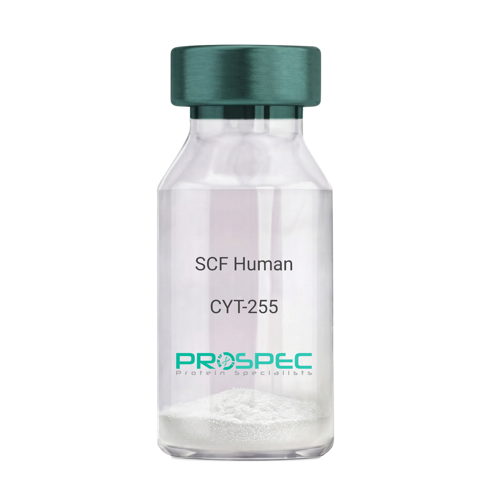
    Name :

    SCF Human

    Description:

    Stem Cell Factor Human Recombinant

    Kit ligand Precursor, C-kit ligand, SCF, Mast cell growth factor, MGF, SF, KL-1, Kitl, DKFZp686F2250.

    Product # :

    CYT-255

    Price :

    Quantity :

    Shipping Method :

    Room Temp Icon

    Shipped at Room temp

    Add To Cart

    More Info

    • description
    • source
    • formulation
    • purity
    • biological activity
    • More Info

    Description

    Stem Cell Factor Human Recombinant produced in E.Coli is a single, non-glycosylated polypeptide chain containing 165 amino acids and having a molecular mass of 18409 Dalton. The SCF is purified by proprietary chromatographic techniques.

    Source

    Escherichia Coli.

    Formulation

    Lyophilized from a concentrated (1mg/ml) solution in water containing 0.02% NaHCO3.

    Purity

    Greater than 98.0% as determined by:
    (a) Analysis by SEC-HPLC.
    (b) Analysis by SDS-PAGE.

    Biological Activity

    The ED50 as determined by the dose-dependant stimulation of Human TF-1 cells is < 2 ng/ml, corresponding to a Specific Activity of 500,000 IU/mg.

    More Info

    • Introduction

      Stem cell factor / KIT ligand (SCF) is a cytokine which binds CD117(c-Kit). SCF is also known as "steel factor" or "c-kit ligand". SCF exists in two forms, cell surface bound SCF and soluble (or free) SCF. Soluble SCF is produced by the cleavage of surface bound SCF by metalloproteases. SCF is a growth factor important for the survival, proliferation, and differentiation of hematopoietic stem cells and other hematopoietic progenitor cells. One of its roles is to change the BFU-E (burst-forming unit-erythroid) cells, which are the earliest erythrocyte precursors in the erythrocytic series, into the CFU-E (colony-forming unit-erythroid).

    • Synonyms

      Kit ligand Precursor, C-kit ligand, SCF, Mast cell growth factor, MGF, SF, KL-1, Kitl, DKFZp686F2250.

    • Physical Appearance

      Sterile Filtered White lyophilized (freeze-dried) powder.

    • Stability

      Lyophilized KIT ligand although stable at room temperature for 3 weeks, should be stored desiccated below -18°C. Upon reconstitution SCF should be stored at 4°C between 2-7 days and for future use below -18°C.For long term storage it is recommended to add a carrier protein (0.1% HSA or BSA).Please prevent freeze-thaw cycles.

    • Solubility

      It is recommended to reconstitute the lyophilized Stem Cell Factor in sterile 18MΩ-cm H2O not less than 100µg/ml, which can then be further diluted to other aqueous solutions.

    • Amino Acid Sequence

      The sequence of the first five N-terminal amino acids was determined and was found to be Met-Glu-Gly-Ile-Cys.

    • Protein content

      Protein quantitation was carried out by two independent methods1. UV spectroscopy at 280 nm using the absorbency value of 0.52 as the extinction coefficient for a 0.1% (1mg/ml) solution. This value is calculated by the PC GENE computer analysis program of protein sequences (IntelliGenetics). 2. Analysis by RP-HPLC, using a standard solution of Stem Cell Factor as a Reference Standard.

    ProSpec's products are furnished for LABORATORY RESEARCH USE ONLY. The product may not be used as drugs, agricultural or pesticidal products, food additives or household chemicals.

    Scf Human
  • View Data Sheet

    Name :

    ESM1 Human

    Description:

    Endothelial Cell-Specific Molecule 1 Human Recombinant

    Endothelial cell-specific molecule 1, ESM-1, ESM1, endocan.

    Product # :

    PRO-1328

    Price :

    Quantity :

    Shipping Method :

    Ice Icon

    Shipped with Ice Packs

    Add To Cart

    More Info

    • description
    • source
    • formulation
    • purity
    • More Info

    Description

    ESM1 Human Recombinant produced in E.Coli is a single, non-glycosylated polypeptide chain containing 188 amino acids (20-184 a.a.) and having a molecular mass of 20.5kDa.ESM1 is fused to a 23 amino acid His-tag at N-terminus & purified by proprietary chromatographic techniques.

    Source

    Escherichia Coli.

    Formulation

    ESM1 protein solution (1mg/ml) contains 20mM Tris-HCl buffer (pH 8.0), 2M Urea and 10% glycerol.

    Purity

    Greater than 90.0% as determined by SDS-PAGE.

    More Info

    • Introduction

      Endothelial cell-specific molecule 1 (ESM1) is a proteoglycan secreted by endothelial cells (mostly in the human lung and kidney tissues) and its mRNA expression is regulated by inflammatory cytokines. ESM1 has potent implications in lung endothelial cell-leukocyte interactions. In addition, ESM1 expression is detected in various epithelia and in adipocytes. ESM1 is involved in angiogenesis and it also promotes angiogenic sprouting. ESM1 expression is upregulated by TNF alpha, IL1 beta, or lipopolysaccharide and downregulated by IFN gamma. Genetically engineered cells overexpressing ESM1 induce tumor formation, implying that ESM1 might be involved in the pathophysiology of tumor growth in vivo.

    • Synonyms

      Endothelial cell-specific molecule 1, ESM-1, ESM1, endocan.

    • Physical Appearance

      Sterile Filtered colorless solution.

    • Stability

      Store at 4°C if entire vial will be used within 2-4 weeks. Store, frozen at -20°C for longer periods of time. For long term storage it is recommended to add a carrier protein (0.1% HSA or BSA).Avoid multiple freeze-thaw cycles.

    • Amino Acid Sequence

      MGSSHHHHHH SSGLVPRGSH MGSWSNNYAV DCPQHCDSSE CKSSPRCKRT VLDDCGCCRV CAAGRGETCY RTVSGMDGMK CGPGLRCQPS NGEDPFGEEF GICKDCPYGT FGMDCRETCN CQSGICDRGT GKCLKFPFFQ YSVTKSSNRF VSLTEHDMAS GDGNIVREEV VKENAAGSPV MRKWLNPR.

    ProSpec's products are furnished for LABORATORY RESEARCH USE ONLY. The product may not be used as drugs, agricultural or pesticidal products, food additives or household chemicals.

    Esm1 Human
  • View Data Sheet

    Name :

    CoV-2-S1 (319-541), Biotin

    Description:

    Coronavirus 2019 Spike Glycoprotein-S1 Receptor Binding Domain (319-541 a.a), Biotinylated Recombinant

    Product # :

    SARS-030

    Price :

    Quantity :

    Shipping Method :

    Room Temp Icon

    Shipped at Room temp

    Add To Cart

    More Info

    • description
    • source
    • formulation
    • purity
    • More Info

    Description

    The HEK293 derived Biotinylated recombinant protein contains the Coronavirus 2019 Spike Glycoprotein S1 Receptor Binding Domain [ RBD ], Wuhan-Hu-1 strain, amino acids 319-541 fused to His tag & AVI tag at C-terminal and having a molecular mass of 28.7kDa.

    Source

    HEK293 Cells.

    Formulation

    CoV-2 S1 RBD protein is lyophilized from 1x PBS pH-7.4 + 10% trehalose.

    Purity

    Protein is >90% pure as determined SDS-PAGE.

    More Info

    • Introduction

      A human infecting coronavirus (viral pneumonia) called 2019 novel coronavirus, 2019-nCoV was found in the fish market at the city of Wuhan, Hubei province of China on December 2019.

      The 2019-nCoV shares an 87% identity to the 2 bat-derived severe acute respiratory syndrome 2018 SARS-CoV-2 located in Zhoushan of eastern China. 2019-nCoV has an analogous receptor-BD-structure to that of 2018 SARS-CoV, even though there is a.a. diversity so thus the 2019-nCoV might bind to ACE2 receptor protein (angiotensin-converting enzyme 2) in humans.

      While bats are possibly the host of 2019-nCoV, researchers suspect that animal from the ocean sold at the seafood market was an intermediate host. RSCU analysis proposes that the 2019-nCoV is a recombinant within the viral spike glycoprotein between the bat coronavirus and an unknown coronavirus.

    • Physical Appearance

      Lyophilized freezed dried powder.

    • Stability

      Lyophilized Cov-2 S1 Glycoprotein RBD although stable at room temperature for 3 weeks, should be stored desiccated below -18°C. Upon reconstitution CoV2 Spike protein should be stored at 4°C between 2-7 days and for future use below -18°C.For long term storage it is recommended to add a carrier protein (0.1% HSA or BSA).Please prevent freeze-thaw cycles.

    • Solubility

      It is recommended to reconstitute the lyophilized CoV-2 S1 protein in sterile 18M-cm H2O at aconcentration of 0.2mg/ml and not less than 0.1mg/ml, which can then be further diluted to other aqueous solutions.

    • Purification Method

      Purified by Metal-Afinity chromatographic technique.

    ProSpec's products are furnished for LABORATORY RESEARCH USE ONLY. The product may not be used as drugs, agricultural or pesticidal products, food additives or household chemicals.

    product_image.jpg
  • View Data Sheet

    Name :

    LASP1 Human

    Description:

    LIM and SH3 Protein 1 Human Recombinant

    LIM and SH3 protein 1, MLN50, Lasp-1, Metastatic lymph node gene 50 protein.

    Product # :

    PRO-1031

    Price :

    Quantity :

    Shipping Method :

    Ice Icon

    Shipped with Ice Packs

    Add To Cart

    More Info

    • description
    • source
    • formulation
    • purity
    • More Info

    Description

    LASP1 Human Recombinant produced in E.coli is a single, non-glycosylated polypeptide chain containing 285 amino acids (1-261) and having a molecular mass of 32.3kDa.LASP1 is fused to a 24 amino acid His-tag at N-terminus & purified by proprietary chromatographic techniques.

    Source

    E.coli.

    Formulation

    The LASP1 solution (0.25mg/ml) contains 20mM Tris-HCl buffer (pH 8.0), 1mM DTT and 20% glycerol.

    Purity

    Greater than 80% as determined by SDS-PAGE.

    More Info

    • Introduction

      LASP1 has a vital part in the regulation of dynamic actin-based, cytoskeletal activities. Agonist-dependent changes in LASP1 phosphorylation can additionally assist in regulation of actin-associated ion transport activities in the parietal cell and in several other F-actin-rich secretory epithelial cell types.

    • Synonyms

      LIM and SH3 protein 1, MLN50, Lasp-1, Metastatic lymph node gene 50 protein.

    • Physical Appearance

      Sterile Filtered colorless solution.

    • Stability

      Store at 4°C if entire vial will be used within 2-4 weeks. Store, frozen at -20°C for longer periods of time. For long term storage it is recommended to add a carrier protein (0.1% HSA or BSA).Avoid multiple freeze-thaw cycles.

    • Amino Acid Sequence

      MGSSHHHHHH SSGLVPRGSH MGSHMNPNCA RCGKIVYPTE KVNCLDKFWH KACFHCETCK MTLNMKNYKG YEKKPYCNAH YPKQSFTMVA DTPENLRLKQ QSELQSQVRY KEEFEKNKGK GFSVVADTPE LQRIKKTQDQ ISNIKYHEEF EKSRMGPSGG EGMEPERRDS QDGSSYRRPL EQQQPHHIPT SAPVYQQPQQ QPVAQSYGGY KEPAAPVSIQ RSAPGGGGKR YRAVYDYSAA DEDEVSFQDG DTIVNVQQID DGWMYGTVER TGDTGMLPAN YVEAI

    ProSpec's products are furnished for LABORATORY RESEARCH USE ONLY. The product may not be used as drugs, agricultural or pesticidal products, food additives or household chemicals.

    Lasp1 Human
  • View Data Sheet

    Name :

    TGFB1 Mouse, CHO

    Description:

    Transforming Growth Factor-Beta 1 Mouse Recombinant, CHO

    Transforming growth factor beta-1, TGF-beta-1, Tgfb, Tgfb-1, TGFbeta1.

    Product # :

    CYT-1264

    Price :

    Quantity :

    Shipping Method :

    Room Temp Icon

    Shipped at Room temp

    Add To Cart

    More Info

    • description
    • source
    • formulation
    • purity
    • biological activity
    • More Info

    Description

    Transforming Growth Factor-Beta 1 Mouse Recombinant produced in CHO is a homodimer, polypeptide chain containing 2 x 112 amino acids and having a total molecular mass of 25.6kDa.
    TGFB1 Mouse Recombinant is purified by proprietary chromatographic techniques.

    Source

    CHO Cells.

    Formulation

    The protein was lyophilized with 0.1% (v/v) TFA and 35% (v/v) Acetonitrile.

    Purity

    Greater than 97.0% as determined by SDS-PAGE and SEC-HPLC analyses.

    Biological Activity

    The biological activity was determined by TGFB1 ability to inhibit the mouse IL-4-dependent proliferation of mouse HT-2 cells. The expected ED50 for this effect is <0.05ng/ml, corresponding to a specific activity of ≥ 2.0 × 10⁷ units/mg.

    More Info

    • Synonyms

      Transforming growth factor beta-1, TGF-beta-1, Tgfb, Tgfb-1, TGFbeta1.

    • Physical Appearance

      Sterile Filtered White lyophilized (freeze-dried) powder.

    • Stability

      Lyophilized TGFB1 although stable at room temperature for 3 weeks, should be stored desiccated below -18°C. Upon reconstitution Transforming Growth Factor-Beta 1 should be stored at 4°C between 2-7 days and for future use below -18°C.
      For long term storage it is recommended to add a carrier protein (0.1% HSA or BSA).
      Please prevent freeze-thaw cycles.

    • Solubility

      It is recommended to reconstitute the lyophilized Transforming Growth Factor-Beta 1 in sterile 4mM HCl not less than 100µg/ml, which can then be further diluted to other aqueous solutions.

    • Amino Acid Sequence

      MGSSHHHHHH SSGLVPRGSH MGSALDTNYC FSSTEKNCCV RQLYIDFRKD LGWKWIHEPK GYHANFCLGP CPYIWSLDTQ YSKVLALYNQ HNPGASASPC CVPQALEPLP IVYYVGRKPK VEQLSNMIVR SCKCS.

    • Background

      Mouse TGF-β1 as an inducer of EMT [epithelial–mesenchymal transition ] therefore used in in fibrosis, wound healing, cancer invasion, and metastasis. Mouse TGF-β1 decreases E-cadherin expression and increases N-cadherin, vimentin and fibronectin.
      TGF-β1 is produced by T regulatory cells (Tregs), Macrophages and monocytes, Platelets, Fibroblasts, Epithelial cells, Endothelial cells, Smooth muscle cells, Tumor cells, Activated immune cells
      What is the source or expression system of Mouse TGFB1 Protein?
      CHO Cells

      What is the Purity of Mouse TGFB1 Protein?
      Mouse TGFB1 Protein is >97% pure as determined by SDS-PAGE and SEC-HPLC analyses.

      What is the molecular weight of Mouse TGFB1 Protein?
      Mouse TGFB1 Protein having a total Mw of 25.6kDa.

      What is the Biological Activity of Mouse TGFB1 Protein?
      The biological functionality of Mouse TGFB1 Protein is determined by mouse HT-2 cells.

      What is the endotoxin level for Mouse TGFB1 Protein?
      The endotoxin level is minimal, Mouse TGFB1 Protein was purified using conventional chromatography techniques.

      What is the amino acid sequence of Mouse TGFB1 Protein?
      ALDTNYCFSS TEKNCCVRQL YIDFRKDLGW KWIHEPKGYH ANFCLGPCPY IWSLDTQYSK VLALYNQHNP GASASPCCVP QALEPLPIVY YVGRKPKVEQ LSNMIVRSCK CS.

      Is TGFB1 a homodimer / homodimeric protein?
      Yes, TGFB1 is homo dimer consisting of 2 identical chains.

    ProSpec's products are furnished for LABORATORY RESEARCH USE ONLY. The product may not be used as drugs, agricultural or pesticidal products, food additives or household chemicals.

    tgfb1 mouse cho
  • View Data Sheet

    Name :

    MIF Human

    Description:

    Macrophage Migration Inhibitory Factor Human Recombinant

    Phenylpyruvate tautomerase, Glycosylation-inhibiting factor, GIF, MMIF, MIF.

    Product # :

    CYT-575

    Price :

    Quantity :

    Shipping Method :

    Ice Icon

    Shipped with Ice Packs

    Add To Cart

    More Info

    • description
    • source
    • formulation
    • purity
    • More Info

    Description

    Macrophage Inducing Factor Human Recombinant produced in E. coli is a single, non-glycosylated, polypeptide chain containing 115 amino acids (1-115aa) and having a molecular mass of 12kDa. MIF human Recombinant is purified by proprietary chromatographic techniques.

    Source

    Escherichia Coli.

    Formulation

    1mg/ml solution containing 50mM Tris-HCl pH-8, 0.5mM DTT & 10% glycerol.

    Purity

    Greater than 98.0% as determined by SDS-PAGE.

    More Info

    • Introduction

      The cytokine Macrophage migration inhibitory factor (MIF) has been identified to be secreted by the pituitary gland and the monocyte/macrophage and to play an important role in endotoxic shock. MIF has the unique property of being released from macrophages and T cells in response to physiological concentrations of glucocorticoids. The secretion of MIF is tightly regulated and decreases at high, anti-inflammatory steroid concentration.

    • Synonyms

      Phenylpyruvate tautomerase, Glycosylation-inhibiting factor, GIF, MMIF, MIF.

    • Physical Appearance

      Sterile Filtered clear solution.

    • Stability

      Store at 4°C if entire vial will be used within 2-4 weeks. Store, frozen at -20°C for longer periods of time. For long term storage it is recommended to add a carrier protein (0.1% HSA or BSA).Avoid multiple freeze-thaw cycles.

    • Amino Acid Sequence

      MPMFIVNTNV PRASVPDGFL SELTQQLAQA TGKPPQYIAV HVVPDQLMAF GGSSEPCALC SLHSIGKIGG AQNRSYSKLL CGLLAERLRI SPDRVYINYY DMNAANVGWN NSTFA.

    ProSpec's products are furnished for LABORATORY RESEARCH USE ONLY. The product may not be used as drugs, agricultural or pesticidal products, food additives or household chemicals.

    Mif Human
  • View Data Sheet

    Name :

    ING2 Human

    Description:

    Inhibitor of Growth Family, Member 2 Human Recombinant

    Inhibitor Of Growth Family Member 2, ING1L, Inhibitor Of Growth 1-Like Protein, P33ING2, ING1Lp, P32, Inhibitor Of Growth Family Member 1-Like, Inhibitor Of Growth Protein 2, ING2.

    Product # :

    PRO-1739

    Price :

    Quantity :

    Shipping Method :

    Ice Icon

    Shipped with Ice Packs

    Add To Cart

    More Info

    • description
    • source
    • formulation
    • purity
    • More Info

    Description

    ING2 Human Recombinant produced in E.Coli is a single, non-glycosylated polypeptide chain containing 303 amino acids (1-280 a.a) and having a molecular mass of 35.2kDa.ING2 is fused to a 23 amino acid His-tag at N-terminus & purified by proprietary chromatographic techniques.

    Source

    Escherichia Coli.

    Formulation

    ING2 protein solution (1mg/ml) containing 20mM Tris-HCl buffer (pH 8.0), 0.4M UREA and 10% glycerol.

    Purity

    Greater than 85.0% as determined by SDS-PAGE.

    More Info

    • Introduction

      Inhibitor of Growth Family, Member 2 (ING2) belongs to the inhibitor of growth (ING) family. ING family members associate with and modulate the activity of histone acetyltransferase (HAT) and histone deacetylase (HDAC) complexes and serve in DNA repair and apoptosis. ING2 appears to be involved in p53/TP53 activation and p53/TP53-dependent apoptotic pathways, most likely by enhancing acetylation of p53/TP53. ING2 is a component of an mSin3A-like corepressor complex, which is probably involved in deacetylation of nucleosomal histones. ING2 activity is modulated by binding to phosphoinositides (PtdInsPs).

    • Synonyms

      Inhibitor Of Growth Family Member 2, ING1L, Inhibitor Of Growth 1-Like Protein, P33ING2, ING1Lp, P32, Inhibitor Of Growth Family Member 1-Like, Inhibitor Of Growth Protein 2, ING2.

    • Physical Appearance

      Sterile Filtered clear solution.

    • Stability

      Store at 4°C if entire vial will be used within 2-4 weeks. Store, frozen at -20°C for longer periods of time. For long term storage it is recommended to add a carrier protein (0.1% HSA or BSA).Avoid multiple freeze-thaw cycles.

    • Amino Acid Sequence

      MGSSHHHHHH SSGLVPRGSH MGSMLGQQQQ QLYSSAALLT GERSRLLTCY VQDYLECVES LPHDMQRNVS VLRELDNKYQ ETLKEIDDVY EKYKKEDDLN QKKRLQQLLQ RALINSQELG DEKIQIVTQM LELVENRARQ MELHSQCFQD PAESERASDK AKMDSSQPER SSRRPRRQRT SESRDLCHMA NGIEDCDDQP PKEKKSKSAK KKKRSKAKQE REASPVEFAI DPNEPTYCLC NQVSYGEMIG CDNEQCPIEW FHFSCVSLTY KPKGKWYCPK CRGDNEKTMD KSTEKTKKDR RSR.

    ProSpec's products are furnished for LABORATORY RESEARCH USE ONLY. The product may not be used as drugs, agricultural or pesticidal products, food additives or household chemicals.

    Ing2 Human
  • View Data Sheet

    Name :

    SLAMF6 Human

    Description:

    SLAMF6 Human Recombinant

    CD352, KALI, KALIb, Ly108, NTB-A, NTBA, Activating NK receptor, SLAM family member 6.

    Product # :

    PRO-1286

    Price :

    Quantity :

    Shipping Method :

    Ice Icon

    Shipped with Ice Packs

    Add To Cart

    More Info

    • description
    • source
    • formulation
    • purity
    • More Info

    Description

    SLAMF6 Human Recombinant produced in E.Coli is a single, non-glycosylated polypeptide chain containing 228 amino acids (22-226 a.a.) and having a molecular mass of 25.5kDa. SLAMF6 is fused to a 23 amino acid His-tag at N-terminus & purified by proprietary chromatographic techniques.

    Source

    Escherichia Coli.

    Formulation

    SLAMF6 protein solution (1mg/ml) contains 20mM Tris-HCl buffer, (pH 8.0), 0.4M Urea and 10% glycerol.

    Purity

    Greater than 85.0% as determined by SDS-PAGE.

    More Info

    • Introduction

      SLAM family member 6 (SLAMF6) is a member of the SLAM family of immune cell receptors. SLAMF6 is a unique receptor on T cells which, when triggered potentiates T cell expansion in a CD28-independent manner. SLAMF6 has a high expression on NK-, T-, and B cells. SLAMF6 exhibits homotypic interactions and can connect with adaptor molecules such as SAP to alter immune cell function.

    • Synonyms

      CD352, KALI, KALIb, Ly108, NTB-A, NTBA, Activating NK receptor, SLAM family member 6.

    • Physical Appearance

      Sterile Filtered clear solution.

    • Stability

      Store at 4°C if entire vial will be used within 2-4 weeks. Store, frozen at -20°C for longer periods of time. For long term storage it is recommended to add a carrier protein (0.1% HSA or BSA).Avoid multiple freeze-thaw cycles.

    • Amino Acid Sequence

      MGSSHHHHHH SSGLVPRGSH MGSQSSLTPL MVNGILGESV TLPLEFPAGE KVNFITWLFN ETSLAFIVPH ETKSPEIHVT NPKQGKRLNF TQSYSLQLSN LKMEDTGSYR AQISTKTSAK LSSYTLRILR QLRNIQVTNH SQLFQNMTCE LHLTCSVEDA DDNVSFRWEA LGNTLSSQPN LTVSWDPRIS SEQDYTCIAE NAVSNLSFSV SAQKLCEDVK IQYTDTKM.

    ProSpec's products are furnished for LABORATORY RESEARCH USE ONLY. The product may not be used as drugs, agricultural or pesticidal products, food additives or household chemicals.

    Slamf6 Human
  • View Data Sheet

    Name :

    EXOSC10 Human

    Description:

    Exosome Component 10 Human Recombinant

    Exosome component 10, Polymyositis/scleroderma autoantigen 2, Autoantigen PM/Scl 2, Polymyositis/scleroderma autoantigen 100 kDa, PM/Scl-100, P100 polymyositis-scleroderma overlap syndrome-associated autoantigen, EXOSC10, PMSCL, PMSCL2, p2, p3, p4, RRP6, Rrp6p, PM-Scl.

    Product # :

    PRO-444

    Price :

    Quantity :

    Shipping Method :

    Ice Icon

    Shipped with Ice Packs

    Add To Cart

    More Info

    • description
    • source
    • formulation
    • purity
    • More Info

    Description

    EXOSC10 Human Recombinant produced in SF9 is a glycosylated, polypeptide chain having a molecular mass of 102 kDa. EXOSC10 is expressed with a -6xHis tag and purified by proprietary chromatographic techniques.

    Source

    Sf9 insect cells.

    Formulation

    EXOSC10 is supplied in 20mM HEPES, pH-7.5, 100mM NaCl, 0.01mM EDTA, 0.02% SDS.

    Purity

    Greater than 93% as determined by SDS-PAGE.

    More Info

    • Introduction

      EXOSC10 is a nucleolar protein and represents one crucial component of the so-called "PM/Scl complex" or "nuclear exosome", which is an RNA-processing complex consisting of a number of exoribonucleases (not to be confused with certain multivesicular bodies which are shed by cultured cells into the culture fluid and are also called exosomes).
      Autoantibodies against EXOSC10 occur in patients suffering from polymyositis, scleroderma or polymyositis/scleroderma overlap syndromes. While antibodies to other components of the exosome complex (PM/Scl 75, several Rrp proteins) can be found in some sera, practically all patients with autoantibodies against the PM/Scl complex are positive for EXOSC10 antibodies, making this specificity an important marker. The presence of EXOSC10 autoantibodies is a comparatively "good" prognostic sign in polymyositis/scleroderma overlap syndrome unlike the poor prognosis seen when other myositis and systemic sclerosis specific autoantibodies are present.

    • Synonyms

      Exosome component 10, Polymyositis/scleroderma autoantigen 2, Autoantigen PM/Scl 2, Polymyositis/scleroderma autoantigen 100 kDa, PM/Scl-100, P100 polymyositis-scleroderma overlap syndrome-associated autoantigen, EXOSC10, PMSCL, PMSCL2, p2, p3, p4, RRP6, Rrp6p, PM-Scl.

    • Physical Appearance

      Sterile Filtered clear solution.

    • Stability

      Store at 4°C if entire vial will be used within 2-4 weeks. Store, frozen at -20°C for longer periods of time.Avoid multiple freeze-thaw cycles.

    • Immunological Functions

      1. Binds IgG-type human auto-antibodies.2. Standard ELISA test (checker-board analysis of positive/negative sera panels).

    • coating concentration

      0.4-0.8 µg/ml (depending on the type of ELISA plate and coating buffer).Suitable for biotinylation and iodination.

    • Applications

      Western-Blot with monoclonal anti-hexa-His-tag antibody & polymyositis patient sera.

    ProSpec's products are furnished for LABORATORY RESEARCH USE ONLY. The product may not be used as drugs, agricultural or pesticidal products, food additives or household chemicals.

    Exosc10 Human
  • View Data Sheet

    Name :

    MIF Human, GST

    Description:

    Macrophage Migration Inhibitor Factor Human Recombinant, GST tag

    Macrophage Migration Inhibitory Factor (Glycosylation-Inhibiting Factor), Phenylpyruvate Tautomerase, L-Dopachrome Tautomerase, L-Dopachrome Isomerase, GLIF, MMIF, GIF, Macrophage Migration Inhibitory Factor, Glycosylation-Inhibiting Factor, EC 5.3.3.12, EC 5.3.2.1, Macrophage migration inhibitory factor.

    Product # :

    CYT-401

    Price :

    Quantity :

    Shipping Method :

    Ice Icon

    Shipped with Ice Packs

    Add To Cart

    More Info

    • description
    • source
    • formulation
    • purity
    • More Info

    Description

    MIF Human Recombinant produced in E.Coli is a single, non-glycosylated polypeptide chain containing 345 amino acids (1-115 a.a) and having a molecular mass of 39.2kDa. MIF is fused to a 230 amino acid GST-tag at N-terminus & purified by proprietary chromatographic techniques.

    Source

    Escherichia Coli.

    Formulation

    MIF protein solution (1mg/ml) containing Phosphate Buffered Saline (pH7.4) and 10% glycerol.

    Purity

    Greater than 90.0% as determined by SDS-PAGE.

    More Info

    • Introduction

      The cytokine Macrophage migration inhibitory factor (MIF) has been identified to be secreted by the pituitary gland and the monocyte/macrophage and to play an important role in endotoxic shock. MIF has the unique property of being released from macrophages and T cells in response to physiological concentrations of glucocorticoids. The secretion of MIF is tightly regulated and decreases at high, anti-inflammatory steroid concentration.

    • Synonyms

      Macrophage Migration Inhibitory Factor (Glycosylation-Inhibiting Factor), Phenylpyruvate Tautomerase, L-Dopachrome Tautomerase, L-Dopachrome Isomerase, GLIF, MMIF, GIF, Macrophage Migration Inhibitory Factor, Glycosylation-Inhibiting Factor, EC 5.3.3.12, EC 5.3.2.1, Macrophage migration inhibitory factor.

    • Physical Appearance

      Sterile Filtered clear solution.

    • Stability

      Store at 4°C if entire vial will be used within 2-4 weeks. Store, frozen at -20°C for longer periods of time. For long term storage it is recommended to add a carrier protein (0.1% HSA or BSA).Avoid multiple freeze-thaw cycles.

    • Amino Acid Sequence

      MSPILGYWKI KGLVQPTRLL LEYLEEKYEE HLYERDEGDK WRNKKFELGL EFPNLPYYID GDVKLTQSMA IIRYIADKHN MLGGCPKERA EISMLEGAVL DIRYGVSRIA YSKDFETLKV DFLSKLPEML KMFEDRLCHK TYLNGDHVTH PDFMLYDALD VVLYMDPMCL DAFPKLVCFK KRIEAIPQID KYLKSSKYIA WPLQGWQATF GGGDHPPKSD LVPRGSPEFA MPMFIVNTNV PRASVPDGFL SELTQQLAQA TGKPPQYIAV HVVPDQLMAF GGSSEPCALC SLHSIGKIGG AQNRSYSKLL CGLLAERLRI SPDRVYINYY DMNAANVGWN NSTFA.

    ProSpec's products are furnished for LABORATORY RESEARCH USE ONLY. The product may not be used as drugs, agricultural or pesticidal products, food additives or household chemicals.

    Mif Human Gst
  • View Data Sheet

    Name :

    IL36RN Mouse

    Description:

    Interleukin-36 Receptor Antagonist Mouse Recombinant

    Product # :

    CYT-154

    Price :

    Quantity :

    Shipping Method :

    Room Temp Icon

    Shipped at Room temp

    Add To Cart

    More Info

    • description
    • source
    • formulation
    • purity
    • biological activity
    • More Info

    Description

    IL36RN Mouse Recombinant produced in E.Coli is a single, non-glycosylated, polypeptide chain containing 154 amino acids and having a molecular mass of 17.0kDa.The IL36RN is purified by proprietary chromatographic techniques.

    Source

    Escherichia Coli.

    Formulation

    Lyophilized from a 0.2µm filtered concentrated solution in PBS, pH 7.4.

    Purity

    Greater than 95.0% as determined by SDS-PAGE.

    Biological Activity

    Fully biologically active when compared to standard. Measured by its ability to inhibit IL-36a, IL-36b or IL-36g induced IL-6 secretion by NIH-3T3 mouse embryonic fibroblast cells. The ED50 for this effect is typically 0.8-4ug/ml (corresponding to a specific activity of 250-125units/mg) in the presence of 15ng/ml of recombinant mouse IL-36b.

    More Info

    • Introduction

      IL36RA belongs to the IL-1 family of proteins. IL36RA is produced as a 156 aa protein which holds no signal sequence, no prosegment and no potential N-linked glycosylation site but, IL36RA is secreted as a 17 kDa monomer. Cells known to express IL36Ra/IL1F5 include monocytes, B cells, keratinocytes, dendritic cells/Langerhans cells and gastric fundus Parietal and Chief cells.

    • Physical Appearance

      Sterile Filtered White lyophilized (freeze-dried) powder.

    • Stability

      Lyophilized IL 36RN although stable at room temperature for 3 weeks, should be stored desiccated below -18°C. Upon reconstitution IL 36RN should be stored at 4°C between 2-7 days and for future use below -18°C.Please prevent freeze-thaw cycles.

    • Solubility

      It is recommended to reconstitute the lyophilized IL36RN in sterile 18M-cm H2O not less than 100µg/ml, which can then be further diluted to other aqueous solutions.

    • Amino Acid Sequence

      VLSGALCFRM KDSALKVLYL HNNQLLAGGL HAEKVIKGEE ISVVPNRALD ASLSPVILGV QGGSQCLSCG TEKGPILKLE PVNIMELYLG AKESKSFTFY RRDMGLTSSF ESAAYPGWFL CTSPEADQPV RLTQIPEDPA WDAPITDFYF QQCD

    ProSpec's products are furnished for LABORATORY RESEARCH USE ONLY. The product may not be used as drugs, agricultural or pesticidal products, food additives or household chemicals.

    Il36Ra Mouse
  • View Data Sheet

    Name :

    GH Antagonist Ovine

    Description:

    Growth Hormone Antagonist Ovine Recombinant

    GH1, GH, GHN, GH-N, hGH-N, Pituitary growth hormone, Growth hormone 1, Somatotropin.

    Product # :

    CYT-215

    Price :

    Quantity :

    Shipping Method :

    Room Temp Icon

    Shipped at Room temp

    Add To Cart

    More Info

    • description
    • source
    • formulation
    • purity
    • biological activity
    • More Info

    Description

    Somatotropin Ovine Antagonist Recombinant G119R produced in E.Coli is a single, non-glycosylated polypeptide chain containing 191 amino acids and having a molecular mass of 22 kDa. The Somatotropin Ovine Antagonist Recombinant is purified by proprietary chromatographic techniques.

    Source

    Escherichia Coli.

    Formulation

    The protein was lyophilized from a concentrated (1mg/ml) solution with 0.0045M NaHCO3, pH 9.

    Purity

    Greater than 98.0% as determined by:
    (a) Analysis by RP-HPLC
    (b) Analysis by Gel Filtration. (c) Analysis by SDS-PAGE.

    Biological Activity

    GH G119R acts as antagonist using an in vitro bioassay in PDF-P1 3B9 cells stably transfected with rabbit GH receptors. It is capable of forming a 1:1 complex with the recombinant ovine growth hormone receptor extracellular domain (ECD) and binds to this ECD with affinity similar the the wild type oGH.

    More Info

    • Introduction

      GH is a member of the somatotropin/prolactin family of hormones which play an important role in growth control. The gene, along with four other related genes, is located at the growth hormone locus on chromosome 17 where they are interspersed in the same transcriptional orientation; an arrangement which is thought to have evolved by a series of gene duplications. The five genes share a remarkably high degree of sequence identity. Alternative splicing generates additional isoforms of each of the five growth hormones, leading to further diversity and potential for specialization. This particular family member is expressed in the pituitary but not in placental tissue as is the case for the other four genes in the growth hormone locus. Mutations in or deletions of the gene lead to growth hormone deficiency and short stature.

    • Synonyms

      GH1, GH, GHN, GH-N, hGH-N, Pituitary growth hormone, Growth hormone 1, Somatotropin.

    • Physical Appearance

      Sterile Filtered White lyophilized (freeze-dried) powder.

    • Stability

      Lyophilized GH G119R although stable at room temperature for at least two weeks, should be stored desiccated below -18°C. Upon reconstitution and filter sterilization GH can be stored at 4°C, pH 9 for up to 4 weeks. For long term storage and more diluted solutions it is recommended to add a carrier protein (0.1% HSA or BSA). Please prevent freeze-thaw cycles.

    • Solubility

      It is recommended to reconstitute the lyophilized GH G119R in 0.4% NaHCO3 or water adjusted to pH 9, not less than 100µg/ml, which can then be further diluted to other aqueous solutions, preferably in a presence of a carrier protein such as BSA or similar.

    • Amino Acid Sequence

      The sequence of the first five N-terminal amino acids was determined and was found to be Ala-Thr-Phe-Pro-Ala.

    • Protein content

      Protein quantitation was carried out by UV spectroscopy at 280 nm using the absorbency value of 0.63 as the extinction coefficient for a 0.1% (1mg/ml) solution. This value is calculated by the DNAman computer analysis program of protein sequences.

    ProSpec's products are furnished for LABORATORY RESEARCH USE ONLY. The product may not be used as drugs, agricultural or pesticidal products, food additives or household chemicals.

    Gh Antagonist Ovine
  • View Data Sheet

    Name :

    IL 1RA Antibody

    Description:

    Interleukin-1 Receptor Antagonist, Mouse Anti-Human

    IRAP, IL1F3, IL1RA, IL-1ra3, ICIL-1RA, IL1RN, IL1 inhibitor, IL-1ra, MGC10430.

    Product # :

    ANT-238

    Price :

    Quantity :

    Shipping Method :

    Room Temp Icon

    Shipped at Room temp

    Add To Cart

    More Info

    • formulation
    • More Info

    Formulation

    1mg/ml in PBS (after reconstitution)

    More Info

    • Introduction

      Interleukin-1 ra is a member of the interleukin 1 cytokine family. This protein inhibits the activities of interleukin 1, alpha (IL1A) and interleukin 1, beta (IL1B), and modulates a variety of interleukin 1 related immune and inflammatory responses. This gene and five other closely related cytokine genes form a gene cluster spanning approximately 400 kb on chromosome 2. A polymorphism of this gene is reported to be associated with increased risk of osteoporotic fractures and gastric cancer. Four alternatively spliced transcript variants encoding distinct isoforms have been reported.

    • Synonyms

      IRAP, IL1F3, IL1RA, IL-1ra3, ICIL-1RA, IL1RN, IL1 inhibitor, IL-1ra, MGC10430.

    • Solubility

      Reconstitute with sterile H20. Mix gently, wash the sides of the vial and wait 30-60 seconds before use.

    • Immunogen

      r.Human IL-1RA.

    • Ig Subclass

      Mouse IgG2b.

    • Clone

      NYR-hIL1RA.

    • Applications

      Direct ELISA, Western Blot, Immuneprecipitation.

    • Titer

      By direct ELISA, 1:10,000 dilution will yield 0.8 O.D using alkaline phosphatase conjugated rabbit anti-mouse Ig (Jackson Laboratories).

    • Shipping Conditions

      Antibody is shipped lyophilized at ambient temperature.

    • Type

      Mouse Anti Human Monoclonal.

    • Storage Procedures

      In lyophilized form, for long periods, store at 4oC in a dry environment. After reconstitution, if not intended for use within a month, aliquot and store at -20oC.

    • Purification Method

      Ion exchange.

    ProSpec's products are furnished for LABORATORY RESEARCH USE ONLY. The product may not be used as drugs, agricultural or pesticidal products, food additives or household chemicals.

    Il 1Ra Antibody
  • 1
  • …
  • 34
  • 35
  • 36
  • 37
  • 38
  • …
  • 42
Your browser does not support the video tag.

Products

  • Cytokines
  • Growth Factors
  • Chemokines
  • CD Antigens
  • Neurotrophins
  • Hormones
  • Enzymes
  • Viral Antigens
  • Recombinant Proteins
  • Natural Proteins
  • Monoclonal Antibodies
  • Polyclonal Antibodies
  • Thermal Packaging

About

  • Company
  • Founder
  • Contribution
  • Contact Us
  • My Account

Ordering

  • Ordering Method
  • Ordering - Credit Card
  • Ordering PO
  • Shipping Fees
  • FAQ's

Legal

  • Terms & Conditions
  • Privacy
  • Site Map
  • info@prospecbio.com
  • Facebook
  • Instagram
  • Linkedin Profile

© 2026 ProSpec-Tany TechnoGene Ltd. All Rights Reserved.

  • Choosing a selection results in a full page refresh.
  • Opens in a new window.