Search results
1000 results found for “stem cell”
Name
Description
Product #
Price
Quantity
Shipping Method
- View Data Sheet
Name :
SNCG HumanDescription:
Gamma-Synuclein Human Recombinant
Gamma-synuclein, Persyn, Breast cancer-specific gene 1 protein, Synoretin, SR, SNCG, BCSG1, PERSYN, PRSN, g-Synuclein.
Product # :
PRO-395Price :
Quantity :
Shipping Method :
Shipped with Ice Packs
More Info
- description
- source
- formulation
- purity
- More Info
- Citations
Description
g-Synuclein Human Recombinant produced in E.Coli is a single,non-glycosylated polypeptide chain containing 127 amino acids and having a molecular mass of 13,300 Dalton. The protein coding region of g-synuclein was amplified by RT-PCR and cloned into an E.coli expression vector. g-synuclein was overexpressed in E. coli and purified to apparent homogeneity by taking advantage of the thermosolubility of the protein and by using conventional column chromatography techniques.
Source
Escherichia Coli.
Formulation
The protein (1mg/ml) contains 20mM Tris-HCl buffer (pH 7.5) and 0.1M NaCl.
Purity
Greater than 90.0% as determined by SDS-PAGE.
Citations
More Info
-
Introduction
g-synuclein(Originally known as a breast cancer specific gene product, BCSG1) is an acidic neuronal protein of 127 amino acids. Gamma-Synuclein is a member of the Synuclein protein family, which is believed to be involved in the pathogenesis of neurodegenerative diseases. High levels of Gamma-Synuclein have been found in advanced breast carcinomas suggesting a correlation between overexpression of SNCG and breast tumor development. Synuclein-Gamma is found mostly in the peripheral nervous system (in primary sensory neurons, sympathetic neurons, and motor neurons) and retina. SNCG is also identified in the brain, ovarian tumors, and in the olfactory epithelium. SNCG expression in breast tumors is a marker for tumor progression. A modification in the expression of gamma-synuclein has been detected in the retina of Alzheimer's patients.
-
Synonyms
Gamma-synuclein, Persyn, Breast cancer-specific gene 1 protein, Synoretin, SR, SNCG, BCSG1, PERSYN, PRSN, g-Synuclein.
-
Physical Appearance
Sterile filtered colorless solution.
-
Stability
Store at 4°C if entire vial will be used within 2-4 weeks. Store, frozen at -20°C for longer periods of time. For long term storage it is recommended to add a carrier protein (0.1% HSA or BSA). Avoid multiple freeze-thaw cycles.
-
Amino Acid Sequence
MDVFKKGFSI AKEGVVGAVE KTKQGVTEAA EKTKEGVMYV GAKTKENVVQSVTSVAEKTK EQANAVSEAV VSSVNTVATK TVEEAENIAV TSGVVRKEDL RPSAPQQEGV ASKEKEEVAE EAQSGGD.
ProSpec's products are furnished for LABORATORY RESEARCH USE ONLY. The product may not be used as drugs, agricultural or pesticidal products, food additives or household chemicals.
- View Data Sheet
Name :
CDKN3 HumanDescription:
Cyclin-Dependent Kinase Inhibitor 3 Human Recombinant
Cyclin-dependent kinase inhibitor 3, CDK2-associated dual-specificity phosphatase, Cyclin-dependent kinase interactor 1, Cyclin-dependent kinase-interacting protein 2, Kinase-associated phosphatase, KAP, CDI1, CIP2, KAP1, FLJ25787, MGC70625, CDKN3.
Product # :
PKA-336Price :
Quantity :
Shipping Method :
Shipped with Ice Packs
More Info
- description
- source
- formulation
- purity
- More Info
- Citations
Description
CDKN3 Human Recombinant fused with a 20 amino acid His tag at N-terminus produced in E.Coli is a single, non-glycosylated, polypeptide chain containing 232 amino acids (1-212 a.a.) and having a molecular mass of 25.9kDa. The CDKN3 is purified by proprietary chromatographic techniques.
Source
Escherichia Coli.
Formulation
The CDKN3 solution (0.5 mg/ml) contains 20mM Tris-HCl buffer (pH 8.0), 1mM DTT, 40% glycerol and 0.1M NaCl.
Purity
Greater than 90.0% as determined by SDS-PAGE.
Citations
More Info
-
Introduction
Cyclin-dependent kinase inhibitor 3 (CDKN3) is a member of the dual specificity protein phosphatase family. CDKN3 was identified as a cyclin-dependent kinase inhibitor, and was shown to interact with, and dephosphorylate CDK2 kinase, consequently prevent the activation of CDK2 kinase. In addition CDKN3 is important in cell cycle regulation. The CDKN3 gene was reported to be deleted, mutated, or overexpressed in several kinds of cancers.
-
Synonyms
Cyclin-dependent kinase inhibitor 3, CDK2-associated dual-specificity phosphatase, Cyclin-dependent kinase interactor 1, Cyclin-dependent kinase-interacting protein 2, Kinase-associated phosphatase, KAP, CDI1, CIP2, KAP1, FLJ25787, MGC70625, CDKN3.
-
Physical Appearance
Sterile Filtered colorless solution.
-
Stability
Store at 4°C if entire vial will be used within 2-4 weeks. Store, frozen at -20°C for longer periods of time. For long term storage it is recommended to add a carrier protein (0.1% HSA or BSA).Avoid multiple freeze-thaw cycles.
-
Amino Acid Sequence
MGSSHHHHHH SSGLVPRGSH MKPPSSIQTS EFDSSDEEPI EDEQTPIHIS WLSLSRVNCS QFLGLCALPG CKFKDVRRNV QKDTEELKSC GIQDIFVFCT RGELSKYRVP NLLDLYQQCG IITHHHPIAD GGTPDIASCC EIMEELTTCL KNYRKTLIHC YGGLGRSCLV AACLLLYLSD TISPEQAIDS LRDLRGSGAI QTIKQYNYLH EFRDKLAAHL SSRDSQSRSV SR.
ProSpec's products are furnished for LABORATORY RESEARCH USE ONLY. The product may not be used as drugs, agricultural or pesticidal products, food additives or household chemicals.
- View Data Sheet
Name :
CSTA Human, HisDescription:
Cystatin A Human Recombinant, His tag
Cystatin-A, Cystatin-AS, Stefin-A, CSTA, STF1, STFA.
Product # :
PRO-504Price :
Quantity :
Shipping Method :
Shipped with Ice Packs
More Info
- description
- source
- formulation
- More Info
- Citations
Description
CSTA produced in E.Coli is a single, non-glycosylated polypeptide chain containing 118 amino acids (1-98 a.a.) and having a molecular mass of 13.1kDa.CSTA is fused to a 20 amino acid His-tag at N-terminus & purified by proprietary chromatographic techniques.
Source
Escherichia Coli.
Formulation
Cystatin-A (1mg/ml) in 20mM Tris-HCl buffer (pH8.0), 1mM DTT and 10% glycerol.
Citations
More Info
-
Introduction
Cystatin-A is the intracellular inhibitor of cysteine proteinase cathepsin B (CatB) and belongs to family 1 of the cystatin superfamily of inhibitor proteins. CSTA is a stefin that functions as a cysteine protease inhibitor, forming tight complexes with papain and the cathepsins B, H, and L. It has been proposed that extracellular CatB together with other classes of proteinases are all subjected to a cascade-like manner of activation. CatB and its inhibitor Cystatin-A are involved in degradation of extracellular matrix proteins during tissue remodeling. CSTA is one of the precursor proteins of cornified cell envelope in keratinocytes and plays a role in epidermal development and maintenance. Cystatin-A has been reported to be found in high concentrations in epithelial cells, polymorphonuclear leukocytes and lymphoid tissue. Stefins may be used as prognostic and diagnostic tools for cancer.
-
Synonyms
Cystatin-A, Cystatin-AS, Stefin-A, CSTA, STF1, STFA.
-
Physical Appearance
Sterile Filtered clear solution.
-
Stability
Store at 4°C if entire vial will be used within 2-4 weeks. Store, frozen at -20°C for longer periods of time. For long term storage it is recommended to add a carrier protein (0.1% HSA or BSA).Avoid multiple freeze-thaw cycles.
-
Amino Acid Sequence
MGSSHHHHHH SSGLVPRGSH MIPGGLSEAK PATPEIQEIV DKVKPQLEEK TNETYGKLEA VQYKTQVVAG TNYYIKVRAG DNKYMHLKVF KSLPGQNEDL VLTGYQVDKN KDDELTGF.
ProSpec's products are furnished for LABORATORY RESEARCH USE ONLY. The product may not be used as drugs, agricultural or pesticidal products, food additives or household chemicals.
- View Data Sheet
Name :
UBE2T HumanDescription:
Ubiquitin-Conjugating Enzyme E2T Human Recombinant
EC 6.3.2.19, HSPC150, PIG50, Ubiquitin-conjugating enzyme E2 T, Ubiquitin-protein ligase T, Ubiquitin carrier protein T, Cell proliferation-inducing gene 50 protein, UBE2T.
Product # :
ENZ-506Price :
Quantity :
Shipping Method :
Shipped with Ice Packs
More Info
- description
- source
- formulation
- purity
- More Info
- Citations
Description
UBE2T Human Recombinant produced in E.Coli is a single, non-glycosylated, polypeptide chain containing 205 amino acids (1-197 a.a.) and having a molecular mass of 23.6 kDa. The UBE2T is fused to an 8 amino acid His Tag at C-Terminus and purified by proprietary chromatographic techniques.
Source
Escherichia Coli.
Formulation
The UBE2T solution contains 20mM Tris-HCl pH-8, 1mM DTT, and 10% glycerol.
Purity
Greater than 95% as determined by SDS-PAGE.
Citations
More Info
-
Introduction
UBE2T is part of the E2 ubiquitin-conjugating enzyme family that participates in the protein degradation pathway. UBE2T catalyzes the ATP-dependent attachment of ubiquitin to target proteins, thus tagging them for subsequent destruction by the proteasome. UBE2T is an important factor of the Faconi anemia pathway of DNA damage repair and, upon self-inactivation, may negatively regulate the Faconi pathway.
-
Synonyms
EC 6.3.2.19, HSPC150, PIG50, Ubiquitin-conjugating enzyme E2 T, Ubiquitin-protein ligase T, Ubiquitin carrier protein T, Cell proliferation-inducing gene 50 protein, UBE2T.
-
Physical Appearance
Sterile Filtered clear colorless solution.
-
Stability
Store at 4°C if entire vial will be used within 2-4 weeks. Store, frozen at -20°C for longer periods of time. For long term storage it is recommended to add a carrier protein (0.1% HSA or BSA).Avoid multiple freeze-thaw cycles.
-
Amino Acid Sequence
MQRASRLKRE LHMLATEPPP GITCWQDKDQ MDDLRAQILG GANTPYEKGV FKLEVIIPER YPFEPPQIRF LTPIYHPNID SAGRICLDVL KLPPKGAWRP SLNIATVLTS IQLLMSEPNP DDPLMADISS EFKYNKPAFL KNARQWTEKH ARQKQKADEE EMLDNLPEAG DSRVHNSTQK RKASQLVGIE KKFHPDVLEH HHHHH.
ProSpec's products are furnished for LABORATORY RESEARCH USE ONLY. The product may not be used as drugs, agricultural or pesticidal products, food additives or household chemicals.
- View Data Sheet
Name :
NME1 AntibodyDescription:
Non-Metastatic Cells 1, Mouse Anti Human
Nucleoside diphosphate kinase A, NDP kinase A, NDK A, Tumor metastatic process-associated protein, Metastasis inhibition factor nm23, nm23-H1, Granzyme A-activated DNase, GAAD, NME1, NDPKA, NM23, NB, AWD, NBS, NDPK-A.
Product # :
ANT-691Price :
Quantity :
Shipping Method :
Shipped with Ice Packs
More Info
- formulation
- More Info
- Citations
Formulation
1mg/ml containing PBS, pH-7.4, 10% Glycerol and 0.02% Sodium Azide.
Citations
More Info
-
Introduction
NDK (Nucleoside diphosphate kinase) exists as a hexamer composed of 'A' (encoded by NME1) and 'B' (encoded by NME2) isoforms. NME1 is involved in cell proliferation, differentiation and development, signal transduction, G protein-coupled receptor endocytosis, and gene expression. It also has tumor metastasis-suppressive capacity. NME1 has a key role in the synthesis of nucleoside triphosphates other than ATP. NME1 is essential for neural development including neural patterning and cell future determination.
The NME1 gene is expressed in various tumor types where its levels have been alternatively linked to reduced or increased metastatic potential.
Decrease in NME1 expression is notably connected to aggressive behavior in melanoma, breast, colon, and gastric carcinomas. In contrast, elevated levels of NME1 gene expression are noted in the advanced stage of thyroid carcinomas.
Somatic mutations of the NME1 gene are found in neuroblastoma. Increased NME1 in neuroblastoma is linked to features of the disease that are associated with aggressive tumors. -
Synonyms
Nucleoside diphosphate kinase A, NDP kinase A, NDK A, Tumor metastatic process-associated protein, Metastasis inhibition factor nm23, nm23-H1, Granzyme A-activated DNase, GAAD, NME1, NDPKA, NM23, NB, AWD, NBS, NDPK-A.
-
Physical Appearance
Sterile filtered colorless solution.
-
Immunogen
Anti-human NME1 mAb, is derived from hybridization of mouse F0 myeloma cells with spleen cells from BALB/c mice immunized with recombinant human NME1 amino acids 1-152 purified from E. coli.
-
Ig Subclass
Mouse IgG2b heavy chain and k light chain.
-
Clone
PAT5F4AT.
-
Applications
NME1 antibody has been tested by ELISA, Western blot analysis, Flow cytometry and ICC/IF to assure specificity and reactivity. Since application varies, however, each investigation should be titrated by the reagent to obtain optimal results.
-
Type
Mouse Anti Human Monoclonal.
-
Storage Procedures
For periods up to 1 month store at 4°C, for longer periods of time, store at -20°C. Prevent freeze thaw cycles.
-
Purification Method
NME1 antibody was purified from mouse ascitic fluids by protein-A affinity chromatography.
ProSpec's products are furnished for LABORATORY RESEARCH USE ONLY. The product may not be used as drugs, agricultural or pesticidal products, food additives or household chemicals.
- View Data Sheet
Name :
NKp46 AntibodyDescription:
Natural Cytotoxicity Receptor NKp46, Mouse Anti Human
Natural cytotoxicity triggering receptor 1, Natural killer cell p46-related protein, hNKp46, NK-p46, NKp46, NK cell-activating receptor, Lymphocyte antigen 94 homolog, CD335 antigen, NCR1, LY94, NCRNKp46, CD335.
Product # :
ANT-303Price :
Quantity :
Shipping Method :
Shipped with Ice Packs
More Info
- formulation
- More Info
- Citations
Formulation
1mg/ml containing PBS, pH-7.4, & 0.1% Sodium Azide.
Citations
More Info
-
Introduction
A natural cytotoxicity receptor (NCR) NKp46 has been shown to represent a novel NK cell-specific molecule involved in human NK cell activation. The natural cytotoxicity receptors (NCRs) are a recently characterized family of Ig-like activation receptors that appear to be major triggering receptors in tumor cell recognition. The three known NCRs include NKp46 and NKp30, which are expressed on circulating NKcells, and NKp44, which is expressed only on activating NK cells. NKp46 has been implicated in NK cell-mediated lysis of several autologous tumor cells, pathogen-infected cell lines and mononuclear phagocytes infected with an intracellular bacterium. The Lysis of tumor cells by NK-cells involves recognition by NKp46 of heparan sulfate moieties of membrane heparan sulfate proteoglycans. Furthermore, NKp46 is a surface receptor involved in NK-cell cell death by apoptosis. NKp46 has two extracellular Ig-like domains followed by a ~40 residue stalk region, a type I transmembrane domain, and a short cytoplasmic tail. The extracellular Ig-like domain of NKp46 (22-255aa) is purified by FPLC gel-filtration chromatography, after refolding of the isolated inclusion bodies in a redox buffer. In addition, engagement of the antigen with the monoclonal antibody stimulates intracellular calcium levels and the synthesis of cytokines. CD59 is an NKp46 coreceptor (by physical association) together they activate cytotoxicity of human NK-cells, their engagement results in tyrosine phosphorylation of CD3-zeta chains associated with NKp46. Reduced cell surface expression of NKp46 and other NK-cell receptors is linked to the impaired NK-cell cytolytic function in viremic HIV-1 infection.
-
Synonyms
Natural cytotoxicity triggering receptor 1, Natural killer cell p46-related protein, hNKp46, NK-p46, NKp46, NK cell-activating receptor, Lymphocyte antigen 94 homolog, CD335 antigen, NCR1, LY94, NCRNKp46, CD335.
-
Physical Appearance
Sterile Filtered colorless solution.
-
Immunogen
Anti-human NKp46 mAb, is derived from hybridization of mouse SP2/0 myeloma cells with spleen cells from BALB/c mice immunized with recombinant human NKp46 amino acids 22-255 purified from E. coli.
-
Ig Subclass
Mouse IgG1 heavy chain and κ light chain.
-
Clone
Pn1D9AT.
-
Applications
NKp46 antibody has been tested by immunofluorescent staining with flow cytometric analysis and by Western blot analysis to assure specificity and reactivity. Since application varies, however, each investigation should be titrated by the reagent to obtain optimal results.
-
Type
Mouse Anti Human Monoclonal.
-
Storage Procedures
For periods up to 1 month store at 4°C, for longer periods of time, store at -20°C. Prevent freeze thaw cycles.
-
Purification Method
NKp46 antibody was purified from mouse ascitic fluids by protein-G affinity chromatography.
ProSpec's products are furnished for LABORATORY RESEARCH USE ONLY. The product may not be used as drugs, agricultural or pesticidal products, food additives or household chemicals.
- View Data Sheet
Name :
ENTPD3 Human, sf9Description:
Ectonucleoside Triphosphate Diphosphohydrolase 3 Human Recombinant, sf9
Ectonucleoside Triphosphate Diphosphohydrolase 3, CD39L3, Ecto-ATP Diphosphohydrolase 3, CD39 Antigen-Like 3, Ecto-ATPDase 3, Ecto-Apyrase 3, Ecto-ATPase 3, EC 3.6.1.5, NTPDase 3, HB6, NTPDase-3.
Product # :
ENZ-958Price :
Quantity :
Shipping Method :
Shipped with Ice Packs
More Info
- description
- source
- formulation
- purity
- More Info
- Citations
Description
ENTPD3 Human Recombinant produced in in Sf9 Baculovirus cells is a single, non-glycosylated polypeptide chain containing 451 amino acids (44-485a.a) and having a molecular mass of 50.7kDa (Migrates at 50-70kDa on SDS-PAGE under reducing conditions). ENTPD3 is fused to a 6 amino acids His-tag at C-Terminus and purified by proprietary chromatographic techniques.
Source
Sf9, Baculovirus cells.
Formulation
ENTPD3 protein solution (0.5mg/ml) containing Phosphate Buffered Saline (pH 7.4) and 10% glycerol.
Purity
Greater than 90.0% as determined by SDS-PAGE.
Citations
More Info
-
Introduction
Ectonucleoside Triphosphate Diphosphohydrolase 3, also known as ENTPD3, which owns a threefold preference for the hydrolysis of ATP over ADP is similar to E-type nucleotidases (NTPases). ENTPD3 is a protein coding gene which contains four apyrase-conserved areas which is characteristic of NTPases.
-
Synonyms
Ectonucleoside Triphosphate Diphosphohydrolase 3, CD39L3, Ecto-ATP Diphosphohydrolase 3, CD39 Antigen-Like 3, Ecto-ATPDase 3, Ecto-Apyrase 3, Ecto-ATPase 3, EC 3.6.1.5, NTPDase 3, HB6, NTPDase-3.
-
Physical Appearance
Sterile Filtered colorless solution.
-
Stability
Store at 4°C if entire vial will be used within 2-4 weeks. Store, frozen at -20°C for longer periods of time. For long term storage it is recommended to add a carrier protein (0.1% HSA or BSA).Avoid multiple freeze-thaw cycles.
-
Amino Acid Sequence
ADLQIHKQEV LPPGLKYGIV LDAGSSRTTV YVYQWPAEKE NNTGVVSQTF KCSVKGSGIS SYGNNPQDVP RAFEECMQKV KGQVPSHLHG STPIHLGATA GMRLLRLQNE TAANEVLESI QSYFKSQPFD FRGAQIISGQ EEGVYGWITA NYLMGNFLEK NLWHMWVHPH GVETTGALDL GGASTQISFV AGEKMDLNTS DIMQVSLYGY VYTLYTHSFQ CYGRNEAEKK FLAMLLQNSP TKNHLTNPCY PRDYSISFTM GHVFDSLCTV DQRPESYNPN DVITFEGTGD PSLCKEKVAS IFDFKACHDQ ETCSFDGVYQ PKIKGPFVAF AGFYYTASAL NLSGSFSLDT FNSSTWNFCS QNWSQLPLLL PKFDEVYARS YCFSANYIYH LFVNGYKFTE ETWPQIHFEK EVGNSSIAWS LGYMLSLTNQ IPAESPLIRL PIEPPHHHHHH.
ProSpec's products are furnished for LABORATORY RESEARCH USE ONLY. The product may not be used as drugs, agricultural or pesticidal products, food additives or household chemicals.
- View Data Sheet
Name :
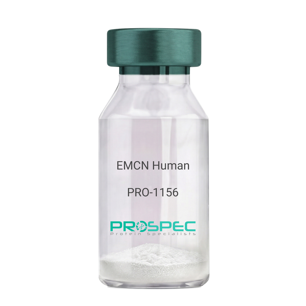
EMCN HumanDescription:
Endomucin Human Recombinant
Endomucin, Endomucin-2, Gastric cancer antigen Ga34, Mucin-14, MUC-14, EMCN, EMCN2, MUC14.
Product # :
PRO-1156Price :
Quantity :
Shipping Method :
Shipped with Ice Packs
More Info
- description
- source
- formulation
- purity
- More Info
- Citations
Description
EMCN Human Recombinant produced in E.Coli is a single, non-glycosylated polypeptide chain containing 197 amino acids (19-190 a.a) and having a molecular mass of 20.5kDa.EMCN is fused to a 25 amino acid His-tag at N-terminus & purified by proprietary chromatographic techniques.
Source
Escherichia Coli.
Formulation
EMCN protein solution (0.25mg/ml) containing 20mM Tris-HCl buffer, pH8.0, 10% glycerol, 1mM DTT and 100mM NaCl.
Purity
Greater than 90% as determined by SDS-PAGE.
Citations
More Info
-
Introduction
Endothelial sialomucin (Endomucin) interferes with the assembly of focal adhesion complexes and inhibits interaction between cells and the extracellular matrix. Endomucin is expressed in the heart, kidney and lung.
-
Synonyms
Endomucin, Endomucin-2, Gastric cancer antigen Ga34, Mucin-14, MUC-14, EMCN, EMCN2, MUC14.
-
Physical Appearance
Sterile filtered colorless solution.
-
Stability
Store at 4°C if entire vial will be used within 2-4 weeks. Store, frozen at -20°C for longer periods of time. For long term storage it is recommended to add a carrier protein (0.1% HSA or BSA).Avoid multiple freeze-thaw cycles.
-
Amino Acid Sequence
MGSSHHHHHH SSGLVPRGSH MGSHMNSTGV LEAANNSLVV TTTKPSITTP NTESLQKNVV TPTTGTTPKG TITNELLKMS LMSTATFLTS KDEGLKATTT DVRKNDSIIS NVTVTSVTLP NAVSTLQSSK PKTETQSSIK TTEIPGSVLQ PDASPSKTGT LTSIPVTIPE NTSQSQVIGT EGGKNASTSA TSRSYSS.
ProSpec's products are furnished for LABORATORY RESEARCH USE ONLY. The product may not be used as drugs, agricultural or pesticidal products, food additives or household chemicals.
- View Data Sheet
Name :
BMP3 HumanDescription:
Bone Morphogenetic protein-3 Human Recombinant
Bone Morphogenetic Protein 3, Osteogenin, Bone Morphogenetic Protein 3 (Osteogenic), Bone Morphogenetic Protein 3A, BMP-3A, BMP-3, Bone Morphogenetic Protein-3, BMP3A, BMP3.
Product # :
CYT-937Price :
Quantity :
Shipping Method :
Shipped at Room temp
More Info
- description
- source
- formulation
- purity
- biological activity
- More Info
- Citations
Description
BMP3 Human Recombinant produced in E.coli is a non-glycosylated disulfide linked homodimer containing 2 chains of 110 amino acids and having a molecular mass of 24.8kDa.The BMP-3 is purified by proprietary chromatographic techniques.
Source
Escherichia Coli.
Formulation
BMP-3 protein was lyophilized from a 0.2µm filtered concentrated solution in 30% Acetonitrile and 0.1% TFA.
Purity
Greater than 95.0% as determined by: (a) Analysis by HPLC. (b) Analysis by SDS-PAGE.
Biological Activity
The ED50 as determined by its ability to inhibit BMP-2-induced activity in murine MC3T3- E1 cells.Citations
More Info
-
Introduction
Bone Morphogenetic Protein 3 (BMP3) is one of the BMPs, some of which are members of the TGF-beta superfamily (BMP2-7). There are more than 13 BMPs, which are involved in inducing cartilage and bone formation, embryogenesis and morphogenesis of various tissues and organs. In addition, BMPs regulate the growth, differentiation, chemotaxis, and apoptosis of various cell types. Akin to most other TGF-beta family proteins, BMPs are extremely conserved across animal species. At the amino acid sequence level, mature human and rat BMP3 are 98% identical.
-
Synonyms
Bone Morphogenetic Protein 3, Osteogenin, Bone Morphogenetic Protein 3 (Osteogenic), Bone Morphogenetic Protein 3A, BMP-3A, BMP-3, Bone Morphogenetic Protein-3, BMP3A, BMP3.
-
Physical Appearance
Sterile Filtered White lyophilized (freeze-dried) powder.
-
Stability
Lyophilized BMP3 although stable at room temperature for 3 weeks, should be stored desiccated below -18°C. Upon reconstitution BMP-3 should be stored at 4°C between 2-7 days and for future use below -18°C. For long term storage it is recommended to add a carrier protein (0.1% HSA or BSA). Please prevent freeze-thaw cycles.
-
Solubility
It is recommended to reconstitute the lyophilized BMP3 in sterile 4mM HCl not less than 100µg/ml, which can then be further diluted to other aqueous solutions.
-
Amino Acid Sequence
QWIEPRNCAR RYLKVDFADI GWSEWIISPK SFDAYYCSGA CQFPMPKSLK PSNHATIQSI VRAVGVVPGI PEPCCVPEKM SSLSILFFDE NKNVVLKVYP NMTVESCACR.
-
Background
Bone Morphogenetic Protein-3 Human Recombinant: Unveiling the Potential of a Key Regulator in Tissue Regeneration
Abstract:
Bone Morphogenetic Protein-3 (BMP-3) human recombinant is a critical member of the bone morphogenetic protein family, known for its role in tissue development, repair, and regeneration. This research paper provides a comprehensive analysis of BMP-3, including its characteristics, signaling pathways, and potential therapeutic applications. Additionally, innovative methodologies for the production and optimization of BMP-3 human recombinant are proposed, shedding light on its future implications in the field of regenerative medicine.
Introduction:
Tissue regeneration is a complex biological process requiring precise molecular cues. BMP-3, a crucial member of the BMP family, plays a significant role in tissue development and regeneration. This paper explores the unique features of BMP-3 and presents novel approaches for its production and optimization, aiming to unlock its therapeutic potential in various regenerative contexts.
Characteristics and Signaling Pathways:
BMP-3 is a secreted protein that binds to cell surface receptors, initiating intracellular signaling cascades. It influences cell differentiation, proliferation, and extracellular matrix synthesis through both Smad-dependent and Smad-independent signaling pathways. BMP-3 signaling regulates critical processes involved in tissue regeneration, including chondrogenesis and osteogenesis.
Production of BMP-3 Human Recombinant:
Efficient production methodologies are essential for harnessing the therapeutic potential of BMP-3 human recombinant. Recombinant protein expression systems, such as Escherichia coli or mammalian cells, have been utilized to produce functional BMP-3. Optimization strategies, including codon optimization, signal peptide engineering, and protein folding optimization, have been employed to enhance the yield and activity of BMP-3 recombinant protein.
Potential Therapeutic Applications:
BMP-3 human recombinant holds significant promise in the field of regenerative medicine. It plays a crucial role in bone and cartilage regeneration, making it a potential candidate for the treatment of skeletal disorders and tissue injuries. Additionally, BMP-3 signaling influences tissue remodeling and wound healing, suggesting its broader therapeutic applications in other regenerative processes.
Conclusion:
BMP-3 human recombinant represents a key regulator in tissue regeneration, with immense potential in regenerative medicine. Optimizing production methodologies and further unraveling its signaling mechanisms will enhance its therapeutic applications. With its implications in bone and cartilage regeneration and its role in tissue remodeling, BMP-3 human recombinant emerges as a promising tool for promoting tissue repair and regeneration.
What is the molecular weight/Mw of BMP3 Protein?
BMP3 Protein has a total Mw of 24.8kDa.
What is the source or expression system of BMP3 Protein?
Escherichia Coli.
What is the Purity of BMP3 Protein?
BMP3 Protein is >95% pure as determined by SDS-PAGE.
What is the Biological Activity of BMP3 Protein?
The ED50 as determined by its ability to inhibit BMP-2-induced activity in murine MC3T3- E1 cells.
What is the amino acid sequence of BMP3 Protein?
QWIEPRNCAR RYLKVDFADI GWSEWIISPK SFDAYYCSGA CQFPMPKSLK PSNHATIQSI VRAVGVVPGI PEPCCVPEKM SSLSILFFDE NKNVVLKVYP NMTVESCACR.
What applications can BMP3 Protein be used in?
BMP3 Protein can probably be used in western blot, ELISA and Lateral Flow.
What is the endotoxin level for BMP3 Protein?
The endotoxin level is minimal, BMP3 Protein was purified using conventional chromatography techniques.
ProSpec's products are furnished for LABORATORY RESEARCH USE ONLY. The product may not be used as drugs, agricultural or pesticidal products, food additives or household chemicals.
- View Data Sheet
Name :
JAM3 HumanDescription:
Junctional Adhesion Molecule 3 Human Recombinant
Junctional Adhesion Molecule 3, Junctional Adhesion Molecule C, JAM-C, JAM-2, JAM-3.
Product # :
PRO-1223Price :
Quantity :
Shipping Method :
Shipped with Ice Packs
More Info
- description
- source
- formulation
- purity
- More Info
- Citations
Description
JAM3 Human Recombinant produced in E. coli is a single polypeptide chain containing 234 amino acids (32-241) and having a molecular mass of 26.0 kDa.JAM3 is fused to a 24 amino acid His-tag at N-terminus & purified by proprietary chromatographic techniques.
Source
E.coli.
Formulation
The JAM3 solution contains 20mM Tris-HCl buffer (pH 8.0), 0.2M NaCl, 2mM EDTA, 5mM DTT and 50% glycerol.
Purity
Greater than 90% as determined by SDS-PAGE.
Citations
More Info
-
Introduction
JAM3 belongs to the junctional adhesion molecule protein family and functions as a receptor for another member of this family. JAM3 plays a part in cell-cell adhesion. The soluble form of JAM3 is a mediator of angiogenesis. JAM3 is a counter-receptor for ITGAM, mediating leukocyte-platelet interactions and is involved in the regulation of transepithelial migration of polymorphonuclear neutrophils (PMN). A mutation in an intron of the JAM3 gene is linked with hemorrhagic destruction of the brain, subependymal calcification, and congenital cataracts.
-
Synonyms
Junctional Adhesion Molecule 3, Junctional Adhesion Molecule C, JAM-C, JAM-2, JAM-3.
-
Physical Appearance
Sterile Filtered colorless solution.
-
Stability
Store at 4°C if entire vial will be used within 2-4 weeks. Store, frozen at -20°C for longer periods of time. For long term storage it is recommended to add a carrier protein (0.1% HSA or BSA).Avoid multiple freeze-thaw cycles.
-
Amino Acid Sequence
MGSSHHHHHH SSGLVPRGSH MGSMVNLKSS NRTPVVQEFE SVELSCIITD SQTSDPRIEW KKIQDEQTTY VFFDNKIQGD LAGRAEILGK TSLKIWNVTR RDSALYRCEV VARNDRKEID EIVIELTVQV KPVTPVCRVP KAVPVGKMAT LHCQESEGHP RPHYSWYRND VPLPTDSRAN PRFRNSSFHL NSETGTLVFT AVHKDDSGQY YCIASNDAGS ARCEEQEMEV YDLN
ProSpec's products are furnished for LABORATORY RESEARCH USE ONLY. The product may not be used as drugs, agricultural or pesticidal products, food additives or household chemicals.
- View Data Sheet
Name :
NECTIN3 HumanDescription:
Nectin Cell Adhesion Molecule 3 Human Recombinant
Nectin-3, CDw113, Nectin cell adhesion molecule 3, Poliovirus receptor-related protein 3, CD113, PVRL3, PRR3, NECTIN-3, PVRR3, PPR3.
Product # :
PRO-2504Price :
Quantity :
Shipping Method :
Shipped with Ice Packs
More Info
- description
- source
- formulation
- purity
- More Info
- Citations
Description
NECTIN3 produced in Sf9 Insect cells is a single, glycosylated polypeptide chain containing 355 amino acids (58-404 a.a.) and having a molecular mass of 39.1kDa (Molecular size on SDS-PAGE will appear at approximately 40-57kDa).NECTIN3 is expressed with an 8 amino acid His tag at C-Terminus and purified by proprietary chromatographic techniques.
Source
Sf9, Insect cells.
Formulation
NECTIN3 protein solution (0.25mg/ml) contains Phosphate Buffered Saline (pH 7.4), 20% glycerol and 1mM DTT.
Purity
Greater than 90.0% as determined by SDS-PAGE.
Citations
More Info
-
Introduction
NECTIN3, also known as Nectin Cell Adhesion Molecule 3, is part of the nectin family. NECTIN3 is intended to initiate cell-cell adhesion to stimulate cell attachment and to allow subsequent formation of JAM-and cadherin-based intercellular junctions. NECTIN3 induces endocytosis-mediated down-regulation of PVR from the cell surface, which results in reduction of cell movement & proliferation. Following Nectin-3 activity adds strength to the junction through trans-interaction with various molecules.
-
Synonyms
Nectin-3, CDw113, Nectin cell adhesion molecule 3, Poliovirus receptor-related protein 3, CD113, PVRL3, PRR3, NECTIN-3, PVRR3, PPR3.
-
Physical Appearance
Sterile filtered colorless solution.
-
Stability
Store at 4°C if entire vial will be used within 2-4 weeks. Store, frozen at -20°C for longer periods of time. For long term storage it is recommended to add a carrier protein (0.1% HSA or BSA).Avoid multiple freeze-thaw cycles.
-
Amino Acid Sequence
GPIIVEPHVT AVWGKNVSLK CLIEVNETIT QISWEKIHGK SSQTVAVHHP QYGFSVQGEY QGRVLFKNYS LNDATITLHN IGFSDSGKYI CKAVTFPLGN AQSSTTVTVL VEPTVSLIKG PDSLIDGGNE TVAAICIAAT GKPVAHIDWE GDLGEMESTT TSFPNETATI ISQYKLFPTR FARGRRITCV VKHPALEKDI RYSFILDIQY APEVSVTGYD GNWFVGRKGV NLKCNADANP PPFKSVWSRL DGQWPDGLLA SDNTLHFVHP LTFNYSGVYI CKVTNSLGQR SDQKVIYISD PPTTTTLQPT IQWHPSTADI EDLATEPKKL PFPLSTLATI KDDTIATLEH HHHHH.
ProSpec's products are furnished for LABORATORY RESEARCH USE ONLY. The product may not be used as drugs, agricultural or pesticidal products, food additives or household chemicals.
- View Data Sheet
Name :
NEDD8 HumanDescription:
Neural Precursor Cell Expressed Developmentally Down-Regulated 8 Human Recombinant
Nedd-8, FLJ43224, MGC104393, MGC125896, MGC125897, NEDD8, Ubiquitin-like protein Nedd8, Neddylin, Neural precursor cell expressed developmentally down-regulated protein 8.
Product # :
ENZ-396Price :
Quantity :
Shipping Method :
Shipped with Ice Packs
More Info
- description
- source
- formulation
- purity
- More Info
- Citations
Description
NEDD8 Human Recombinant fused with 37 amino acid His tag at N-terminus produced in E.Coli is a single, non-glycosylated, polypeptide chain containing 113 amino acids (1-76 a.a.) and having a molecular mass of 12.8 kDa.The NEDD8 is purified by proprietary chromatographic techniques.
Source
Escherichia Coli.
Formulation
The NEDD8 solution contains 20mM Tris pH 8.0, 50mM NaCl, 0.5mM DTT & 10% glycerol.
Purity
Greater than 95.0% as determined by SDS-PAGE.
Citations
More Info
-
Introduction
NEDD8 is part of the ubiquitin family. Human NEDD8 shares 60% amino acid sequence homology to ubiquitin. The NEDD8 system is essential for the regulation of protein degradation pathways involved in cell cycle progression, morphogenesis and tumorigenesis. NEDD8 is involved in cell cycle control and embryogenesis. Covalent attachment to its substrates requires prior activation by the E-1 complex UbE1c- appbp1 and linkage to the E-2 enzyme UbE2m. Attachment of NEDD8 to cullins activates their associated E-3 ubiquitin ligase activity, and thus promotes polyubiquitination and proteasomal degradation of cyclins and other regulatory proteins.
-
Synonyms
Nedd-8, FLJ43224, MGC104393, MGC125896, MGC125897, NEDD8, Ubiquitin-like protein Nedd8, Neddylin, Neural precursor cell expressed developmentally down-regulated protein 8.
-
Physical Appearance
Sterile Filtered colorless solution.
-
Stability
Store at 4°C if entire vial will be used within 2-4 weeks. Store, frozen at -20°C for longer periods of time. For long term storage it is recommended to add a carrier protein (0.1% HSA or BSA).Avoid multiple freeze-thaw cycles.
-
Amino Acid Sequence
MHHHHHHMKI EEGKLVIWIN GDKGYNGLAE VGKKFEKDTG IKVTVEHPDK LEEKFPQVAA TGDGPDIIFW AHDRFGGYAQ SGLLAEITPD KAFQDKLYPF TWDAVRYNGK LIAYPIAVEA LSLIYNKDLL PNPPKTWEEI PALDKELKAK GKSALMFNLQ EPYFTWPLIA ADGGYAFKYE NGKYDIKDVG VDNAGAKAGL TFLVDLIKNK HMNADTDYSI AEAAFNKGET AMTINGPWAW SNIDTSKVNY GVTVLPTFKG QPSKPFVGVL SAGINAASPN KELAKEFLEN YLLTDEGLEA
VNKDKPLGAV ALKSYEEELA KDPRIAATME NAQKGEIMPN IPQMSAFWYA VRTAVINAAS GRQTVDEALK DAQTNSSSNN NNNNNNNNLG IEGRGSHMAA AEAANCIMEV SCGQAESSEK PNAEDMTSKD YYFDSYAHFG IHEEMLKDEV RTLTYRNSMF HNRHLFKDKV VLDVGSGTGILCMFAAKAGA RKVIGIECSS ISDYAVKIVK ANKLDHVVTI IKGKVEEVEL PVEKVDIIIS EWMGYCLFYE SMLNTVLHAR DKWLAPDGLI FPDRATLYVT AIEDRQYKDY KIHWWENVYG
FDMSCIKDVA IKEPLVDVVD PKQLVTNACL IKEVDIYTVK VEDLTFTSPF CLQVKRNDYVHALVAYFNIE FTRCHKRTGF STSPESPYTH WKQTVFYMED YLTVKTGEEI FGTIGMRPNA KNNRDLDFTI DLDFKGQLCE LSCSTDYRMR.
ProSpec's products are furnished for LABORATORY RESEARCH USE ONLY. The product may not be used as drugs, agricultural or pesticidal products, food additives or household chemicals.
- View Data Sheet
Name :
AHCY Human, Sf9Description:
Adenosylhomocysteinase Human Recombinant, Sf9
EC 3.3.1.1, SAHH, AdoHcyase, S-adenosyl-L-homocysteine hydrolase, AHCY, Adenosylhomocysteinase.
Product # :
ENZ-1034Price :
Quantity :
Shipping Method :
Shipped with Ice Packs
More Info
- description
- source
- formulation
- purity
- More Info
- Citations
Description
AHCY Human Recombinant produced in Sf9 Baculovirus cells is a single, glycosylated polypeptide chain containing 441 amino acids (1-432 a.a.) and having a molecular mass of 48.8kDa (Migrates at 40-57kDa on SDS-PAGE under reducing conditions). AHCY is fused to a 6 amino acids His-Tag at C-terminus and purified by proprietary chromatographic techniques.
Source
Sf9, Baculovirus cells.
Formulation
AHCY protein solution (0.25mg/ml) contains Phosphate Buffered Saline (pH 7.4) and 10% glycerol.
Purity
Greater than 90% as determined by SDS-PAGE.
Citations
More Info
-
Introduction
AHCY is an enzyme that catalyzes the reversible hydrolysis of S-adenosylhomocysteine (AdoHcy) to adenosine (Ado) and L-homocysteine (Hcy). AHCY controls the intracellular S-adenosylhomocysteine (SAH) concentration that is crucial for transmethylation reactions. AHCY deficiency causes hypermethioninemia.
-
Synonyms
EC 3.3.1.1, SAHH, AdoHcyase, S-adenosyl-L-homocysteine hydrolase, AHCY, Adenosylhomocysteinase.
-
Physical Appearance
Sterile Filtered colorless solution.
-
Stability
Store at 4°C if entire vial will be used within 2-4 weeks. Store, frozen at -20°C for longer periods of time. For long term storage it is recommended to add a carrier protein (0.1% HSA or BSA).Avoid multiple freeze-thaw cycles.
-
Amino Acid Sequence
ADLMSDKLPY KVADIGLAAW GRKALDIAEN EMPGLMRMRE RYSASKPLKG ARIAGCLHMT VETAVLIETL VTLGAEVQWS SCNIFSTQDH AAAAIAKAGI PVYAWKGETD EEYLWCIEQT LYFKDGPLNM ILDDGGDLTN LIHTKYPQLL PGIRGISEET TTGVHNLYKM MANGILKVPA INVNDSVTKS KFDNLYGCRE SLIDGIKRAT DVMIAGKVAV VAGYGDVGKG CAQALRGFGA RVIITEIDPI NALQAAMEGY EVTTMDEACQ EGNIFVTTTG CIDIILGRHF EQMKDDAIVC NIGHFDVEID VKWLNENAVE KVNIKPQVDR YRLKNGRRII LLAEGRLVNL GCAMGHPSFV MSNSFTNQVM AQIELWTHPD KYPVGVHFLP KKLDEAVAEA HLGKLNVKLT KLTEKQAQYL GMSCDGPFKP DHYRYHHHHH H.
ProSpec's products are furnished for LABORATORY RESEARCH USE ONLY. The product may not be used as drugs, agricultural or pesticidal products, food additives or household chemicals.
- View Data Sheet
Name :
CHMP1A HumanDescription:
Chromatin Modifying Protein 1A Human Recombinant
Charged multivesicular body protein 1a, Chromatin-modifying protein 1a, Vacuolar protein sorting-associated protein 46-1, Vps46-1, hVps46-1, CHMP1A, CHMP1, KIAA0047, PCOLN3, PRSM1, PCH8, VPS46A.
Product # :
PRO-1308Price :
Quantity :
Shipping Method :
Shipped with Ice Packs
More Info
- description
- source
- formulation
- purity
- More Info
- Citations
Description
CHMP1A Human Recombinant produced in E.Coli is a single, non-glycosylated polypeptide chain containing 219 amino acids (1-196 a.a.) and having a molecular mass of 24.1kDa (Molecular size on SDS-PAGE will appear higher).CHMP1A is fused to a 23 amino acid His-tag at N-terminus & purified by proprietary chromatographic techniques.
Source
Escherichia Coli.
Formulation
CHMP1A protein solution (0.5mg/ml) containing 20mM Tris-HCl buffer (pH 8.0), 0.1M NaCl, 20% glycerol and 1mM DTT.
Purity
Greater than 85.0% as determined by SDS-PAGE.
Citations
More Info
-
Introduction
Charged multivesicular body protein 1a (CHMP1A) is a member of the vacuolar sorting protein family and functions as chromatin-modifying protein. These complexes are essential for sorting endosomal articles into multivesicular bodies (MVBs), as well as vital for the formation of these bodies. The MVBs pathway mediates distribution of transmembrane proteins into the lumen of the lysosome for degradation. CHMP1 interacts with VPS4B and localizes to early endosomes.
-
Synonyms
Charged multivesicular body protein 1a, Chromatin-modifying protein 1a, Vacuolar protein sorting-associated protein 46-1, Vps46-1, hVps46-1, CHMP1A, CHMP1, KIAA0047, PCOLN3, PRSM1, PCH8, VPS46A.
-
Physical Appearance
Sterile Filtered colorless solution.
-
Stability
Store at 4°C if entire vial will be used within 2-4 weeks. Store, frozen at -20°C for longer periods of time. For long term storage it is recommended to add a carrier protein (0.1% HSA or BSA).Avoid multiple freeze-thaw cycles.
-
Amino Acid Sequence
MGSSHHHHHH SSGLVPRGSH MGSMDDTLFQ LKFTAKQLEK LAKKAEKDSK AEQAKVKKAL LQKNVECARV YAENAIRKKN EGVNWLRMAS RVDAVASKVQ TAVTMKGVTK NMAQVTKALD KALSTMDLQK VSSVMDRFEQ QVQNLDVHTS VMEDSMSSAT TLTTPQEQVD SLIMQIAEEN GLEVLDQLSQ LPEGASAVGE SSVRSQEDQL SRRLAALRN.
ProSpec's products are furnished for LABORATORY RESEARCH USE ONLY. The product may not be used as drugs, agricultural or pesticidal products, food additives or household chemicals.
- View Data Sheet
Name :
PTMS HumanDescription:
Prothymosin Human Recombinant
ParaT, parathymosin.
Product # :
PRO-1735Price :
Quantity :
Shipping Method :
Shipped with Ice Packs
More Info
- description
- source
- formulation
- purity
- More Info
- Citations
Description
PTMS Human Recombinant produced in E.Coli is a single, non-glycosylated polypeptide chain containing 125 amino acids (1-102aa) and having a molecular mass of 13.9kDa.PTMS is fused to a 23 amino acid His-tag at N-terminus & purified by proprietary chromatographic techniques.
Source
Escherichia Coli.
Formulation
PTMS protein solution (0.5mg/ml) containing 20mM Tris-HCl buffer (pH 8.0), 0.15M NaCl, 20% glycerol and 1mM DTT.
Purity
Greater than 95.0% as determined by SDS-PAGE.
Citations
More Info
-
Introduction
Parathymosin, also known as PTMS is a member of the pro/parathymosin family. PTMS may mediate immune function by blocking the effect of prothymosin alpha which confers resistance to particular opportunistic infections. Among the diseases associated with PTMS are hepatitis b, and alzheimer's disease.
-
Synonyms
ParaT, parathymosin.
-
Physical Appearance
Sterile Filtered clear solution.
-
Stability
Store at 4°C if entire vial will be used within 2-4 weeks. Store, frozen at -20°C for longer periods of time. For long term storage it is recommended to add a carrier protein (0.1% HSA or BSA).Avoid multiple freeze-thaw cycles.
-
Amino Acid Sequence
MGSSHHHHHH SSGLVPRGSH MGSMSEKSVE AAAELSAKDL KEKKEKVEEK ASRKERKKEV VEEEENGAEE EEEETAEDGE EEDEGEEEDE EEEEEDDEGP ALKRAAEEED EADPKRQKTE NGASA
ProSpec's products are furnished for LABORATORY RESEARCH USE ONLY. The product may not be used as drugs, agricultural or pesticidal products, food additives or household chemicals.
- View Data Sheet
Name :
MAD2L1 HumanDescription:
MAD2 Mitotic Arrest Deficient-Like 1 Human Recombinant
Mitotic spindle assembly checkpoint protein MAD2A, HsMAD2, Mitotic arrest deficient 2-like protein 1, MAD2-like protein 1, MAD2L1, MAD2.
Product # :
PRO-934Price :
Quantity :
Shipping Method :
Shipped with Ice Packs
More Info
- description
- source
- formulation
- purity
- More Info
- Citations
Description
MAD2L1 Human Recombinant produced in E.Coli is a single, non-glycosylated polypeptide chain containing 225 amino acids (1-205 a.a.) and having a molecular mass of 25.7kDa.MAD2L1 is fused to a 20 amino acid His-tag at N-terminus & purified by proprietary chromatographic techniques.
Source
Escherichia Coli.
Formulation
MAD2L1 protein solution (0.5mg/ml) containing 20mM Tris-HCl buffer (pH8.0), 20% glycerol, 0.1M NaCl and 1mM DTT.
Purity
Greater than 95.0% as determined by SDS-PAGE.
Citations
More Info
-
Introduction
MAD2L1-binding protein (MAD2L1) is a component of the mitotic spindle assembly checkpoint that prevents the onset of anaphase until all chromosomes are properly aligned at the metaphase plate. MAD2L1 is vital for the execution of the mitotic checkpoint which monitors the process of kinetochore-spindle attachment and inhibits the activity of the anaphase promoting complex by sequestering CDC20 until all chromosomes are aligned at the metaphase plate.
-
Synonyms
Mitotic spindle assembly checkpoint protein MAD2A, HsMAD2, Mitotic arrest deficient 2-like protein 1, MAD2-like protein 1, MAD2L1, MAD2.
-
Physical Appearance
Sterile Filtered colorless solution.
-
Stability
Store at 4°C if entire vial will be used within 2-4 weeks. Store, frozen at -20°C for longer periods of time. For long term storage it is recommended to add a carrier protein (0.1% HSA or BSA).Avoid multiple freeze-thaw cycles.
-
Amino Acid Sequence
MGSSHHHHHH SSGLVPRGSH MALQLSREQG ITLRGSAEIV AEFFSFGINS ILYQRGIYPS ETFTRVQKYG LTLLVTTDLE LIKYLNNVVE QLKDWLYKCS VQKLVVVISN IESGEVLERW QFDIECDKTA KDDSAPREKS QKAIQDEIRS VIRQITATVT FLPLLEVSCS FDLLIYTDKD LVVPEKWEES GPQFITNSEE VRLRSFTTTI HKVNSMVAYK IPVND.
ProSpec's products are furnished for LABORATORY RESEARCH USE ONLY. The product may not be used as drugs, agricultural or pesticidal products, food additives or household chemicals.
- View Data Sheet
Name :
EPO MouseDescription:
Erythropoietin Mouse Recombinant
Erythropoietin, erythropoietin isoform 1 precursor, Epo.
Product # :
CYT-1171Price :
Quantity :
Shipping Method :
Shipped with Ice Packs
More Info
- description
- source
- formulation
- purity
- biological activity
- More Info
- sds-page
- Citations
Description
EPO Mouse Recombinant produced in Baculovirus is a single glycosylated polypeptide chain containing 176 amino acids (27-192 aa) and having a molecular mass of 19.8kDa.EPO is fused to a 9 amino acid His tag at C-terminus and purified by proprietary chromatographic techniques.
Source
Sf9, Baculovirus cells.
Formulation
EPO Mouse protein (0.25mg/ml) contains 10% glycerol and Phosphate-Buffered Saline (pH 7.4).
Purity
Greater than 95.0% as determined by SDS-PAGE.
Biological Activity
Measured in a cell proliferation assay using TF-1 human erythroleukemic cells. The ED50 range ≤ 2ng/ml.
sds-page
Citations
More Info
-
Introduction
Erythropoietin or EPO is a hormone (glycoprotein), part of the type I cytokine group of proteins. EPO is found mainly in the kidney tissue, produced from fibroblast-like cortical interstitial cells near the proximal tubules. EPO is also present in the blood, where it acts as red cell production regulator, by the promotion of differentiation of erythroid and thereby starts hemoglobin synthesis. Furthermore, EPO has neuroprotective activity towards brain injuries & anti-apoptotic activity in different tissues.
-
Synonyms
Erythropoietin, erythropoietin isoform 1 precursor, Epo.
-
Physical Appearance
Sterile Filtered colorless solution.
-
Stability
Store at 4°C if entire vial will be used within 2-4 weeks. Store, frozen at -20°C for longer periods of time. For long term storage it is recommended to add a carrier protein (0.1% HSA or BSA).Avoid multiple freeze-thaw cycles.
-
Amino Acid Sequence
ADPMAPPRLI CDSRVLERYI LEAKEAENVT MGCAEGPRLS ENITVPDTKV NFYAWKRMEV EEQAIEVWQG LSLLSEAILQ AQALLANSSQ PPETLQLHID KAISGLRSLT SLLRVLGAQK ELMSPPDTTP PAPLRTLTVD TFCKLFRVYA NFLRGKLKLY TGEVCRRGDR HHHHHH.
-
Background
What is the molecular weight/Mw of EPO Protein?
EPO Protein has a total Mw of 19.8kDa.
What is the source or expression system of EPO Protein?
Sf9, Baculovirus cells.
What is the Purity of EPO Protein?
EPO Protein is >95% pure as determined by SDS-PAGE.
What is the Biological Activity of EPO Protein?
Measured in a cell proliferation assay using TF-1 human erythroleukemic cells. The ED50 range ≤ 2ng/ml.
What is the amino acid sequence of EPO Protein?
ADPMAPPRLI CDSRVLERYI LEAKEAENVT MGCAEGPRLS ENITVPDTKV NFYAWKRMEV EEQAIEVWQG LSLLSEAILQ AQALLANSSQ PPETLQLHID KAISGLRSLT SLLRVLGAQK ELMSPPDTTP PAPLRTLTVD TFCKLFRVYA NFLRGKLKLY TGEVCRRGDR HHHHHH.
What applications can EPO Protein be used in?
EPO Protein can probably be used in western blot, ELISA and Lateral Flow.
What is the endotoxin level for EPO Protein?
The endotoxin level is minimal, EPO Protein was purified using conventional chromatography techniques.
ProSpec's products are furnished for LABORATORY RESEARCH USE ONLY. The product may not be used as drugs, agricultural or pesticidal products, food additives or household chemicals.
- View Data Sheet
Name :
SCGB1A1 HumanDescription:
Uteroglobin Human Recombinant
Uteroglobin, Clara cell phospholipid-binding protein, CCPBP, Clara cells 10 kDa secretory protein, CC10, Secretoglobin family 1A member 1, Urinary protein 1, UP-1, UP1, Urine protein 1, SCGB1A1, CCSP, UGB, CC16.
Product # :
CYT-743Price :
Quantity :
Shipping Method :
Shipped at Room temp
More Info
- description
- source
- formulation
- purity
- biological activity
- More Info
- Citations
Description
Uteroglobin Human Recombinant produced in E.coli is a single, non-glycosylated polypeptide chain containing 70 amino acids and having a molecular mass of 15.8kDa.The SCGB1A1 is purified by proprietary chromatographic techniques.
Source
Escherichia Coli.
Formulation
The Uteroglobin protein was lyophilized from a 0.2µm filtered concentrated solution in PBS, pH 7.4.
Purity
Greater than 98.0% as determined by:
(a) Analysis by RP-HPLC.
(b) Analysis by SDS-PAGE.Biological Activity
The ED50 as determined by the ability of the immobilized protein to support the adhesion of the A549 human lung carcinoma cells is less than 5.0µg/ml, corresponding to a specific activity of > 200 IU/mg.Citations
More Info
-
Introduction
Uteroglobin (SCGB1A1) which belongs to the Secretoglobin (SCGBs) superfamily, is a multifunctional protein that exerts anti-inflammatory and anti-tumorigenic effects by binding small hydrophobic molecules such as phospholipids and prostaglandins. Uteroglobin is involved in numerous functions including anti-inflammation, inhibition of phospholipase A2 and the sequestering of hydrophobic ligands. SCGB1A1 is expressed by Clara cells, the non-ciliated, non-mucous secretory cells predominant in lung bronchioles, and by other epithelia which communicate with the external environment. On top of sequestering pro-inflammatory mediators and carcinogens, Uteroglobin is implicated in the inhibition of cell migration and invasion, platelet aggregation, and T cell differentiation. SCGB1A1 gene defects are associated with a susceptibility to asthma.
-
Synonyms
Uteroglobin, Clara cell phospholipid-binding protein, CCPBP, Clara cells 10 kDa secretory protein, CC10, Secretoglobin family 1A member 1, Urinary protein 1, UP-1, UP1, Urine protein 1, SCGB1A1, CCSP, UGB, CC16.
-
Physical Appearance
Sterile Filtered White lyophilized (freeze-dried) powder.
-
Stability
Lyophilized Uteroglobin although stable at room temperature for 3 weeks, should be stored desiccated below -18°C. Upon reconstitution SCGB1A1 should be stored at 4°C between 2-7 days and for future use below -18°C. For long term storage it is recommended to add a carrier protein (0.1% HSA or BSA). Please prevent freeze-thaw cycles.
-
Solubility
It is recommended to reconstitute the lyophilized SCGB1A1 in sterile 18M-cm H2O not less than 100µg/ml, which can then be further diluted to other aqueous solutions.
-
Amino Acid Sequence
EICPSFQRVI ETLLMDTPSS YEAAMELFSP DQDMREAGAQ LKKLVDTLPQ KPRESIIKLM EKIAQSSLCN.
ProSpec's products are furnished for LABORATORY RESEARCH USE ONLY. The product may not be used as drugs, agricultural or pesticidal products, food additives or household chemicals.
- View Data Sheet
Name :
SCGB1A1 RatDescription:
Uteroglobin Rat Recombinant
Uteroglobin, Clara cell 17 kDa protein, Clara cell phospholipid-binding protein, CCPBP, Clara cells 10 kDa secretory protein, CC10, PCB-binding protein, Secretoglobin family 1A member 1, Scgb1a1, Ugb, Utg, UG, CC16, CCSP, PCB-BP.
Product # :
CYT-820Price :
Quantity :
Shipping Method :
Shipped at Room temp
More Info
- description
- source
- formulation
- purity
- biological activity
- More Info
- Citations
Description
SCGB1A1 Rat Recombinant produced in E.Coli is a homodimeric non-glycosylated polypeptide chains consisting of two 77 amino acids and having a molecular mass of 17.0kDa.
Source
Escherichia Coli.
Formulation
SCGB1A1 protein was lyophilized from a 0.2 µm filtered concentrated solution in PBS, pH 7.4.
Purity
Greater than 97.0% as determined by:
(a) Analysis by RP-HPLC.
(b) Analysis by SDS-PAGE.Biological Activity
Fully biologically active when compared to standard. The ED50 as determined by the ability of the immobilized protein to support the adhesion of the A549 human lung carcinoma cells is less than 5.0 ?g/ml, corresponding to a specific activity of >200IU/mg.Citations
More Info
-
Introduction
Uteroglobin (SCGB1A1) which belongs to the Secretoglobin (SCGBs) superfamily, is a multifunctional protein that exerts anti-inflammatory and anti-tumorigenic effects by binding small hydrophobic molecules such as phospholipids and prostaglandins. Uteroglobin is involved in numerous functions including anti-inflammation, inhibition of phospholipase A2 and the sequestering of hydrophobic ligands. SCGB1A1 is expressed by Clara cells, the non-ciliated, non-mucous secretory cells predominant in lung bronchioles, and by other epithelia which communicate with the external environment. On top of sequestering pro-inflammatory mediators and carcinogens, Uteroglobin is implicated in the inhibition of cell migration and invasion, platelet aggregation, and T cell differentiation. SCGB1A1 gene defects are associated with a susceptibility to asthma.
-
Synonyms
Uteroglobin, Clara cell 17 kDa protein, Clara cell phospholipid-binding protein, CCPBP, Clara cells 10 kDa secretory protein, CC10, PCB-binding protein, Secretoglobin family 1A member 1, Scgb1a1, Ugb, Utg, UG, CC16, CCSP, PCB-BP.
-
Physical Appearance
Sterile Filtered White lyophilized (freeze-dried) powder.
-
Stability
Lyophilized SCGB1A1 although stable at room temperature for 3 weeks, should be stored desiccated below -18°C. Upon reconstitution SCGB1A1 should be stored at 4°C between 2-7 days and for future use below -18°C. For long term storage it is recommended to add a carrier protein (0.1% HSA or BSA). Please prevent freeze-thaw cycles.
-
Solubility
It is recommended to reconstitute the lyophilized SCGB1A1 in sterile 18MΩ-cm H2O not less than 100µg/ml, which can then be further diluted to other aqueous solutions.
-
Amino Acid Sequence
SSDICPGFLQ VLEALLLGSE SNYEAALKPF NPASDLQNAG TQLKRLVDTL PQETRINIVK LTEKILTSPL CEQDLRV
ProSpec's products are furnished for LABORATORY RESEARCH USE ONLY. The product may not be used as drugs, agricultural or pesticidal products, food additives or household chemicals.
- View Data Sheet
Name :
MME Human, ActiveDescription:
Membrane Metalloendopeptidase Human Recombinant, Active
Membrane Metalloendopeptidase, Common Acute Lymphocytic Leukemia Antigen, Neutral Endopeptidase 24.11, Skin Fibroblast Elastase, Neutral Endopeptidase, Atriopeptidase, Enkephalinase, EC 3.4.24.11, Neprilysin, CALLA, NEP, SFE,Membrane Metallo-Endopeptidase (Neutral Endopeptidase, Enkephalinase, CALLA, CD10), Membrane Metallo-Endopeptidase Variant 1, Membrane Metallo-Endopeptidase Variant 2, Neprilysin-390, Neprilysin-411, CD10 Antigen, EC 3.4.24, CMT2T, SCA43, CD10, EPN, MME.
Product # :
ENZ-1116Price :
Quantity :
Shipping Method :
Shipped with Ice Packs
More Info
- description
- source
- formulation
- purity
- biological activity
- More Info
- Citations
Description
MME Human produced in Sf9 Baculovirus cells is a single, glycosylated polypeptide chain containing 708 amino acids (52-750 aa) and having a molecular mass of 80.9kDa.MME is fused to a 6 amino acid His tag at C-terminus and purified by proprietary chromatographic techniques.
Source
Sf9, Baculovirus cells.
Formulation
The MME solution (1mg/ml) contains 10% Glycerol, 20 mM Tris-HCl buffer (pH 8.0), 0.1mM PMSF and 100mM NaCl.
Purity
Greater than 95.0% as determined by SDS-PAGE.
Biological Activity
Specific activity > 5,000 pmol/min/ug. One unit will convert 1.0 pmole of Mca-SEVNLDAEFRK(Dnp)RR-NH2 to MCA- Pro-Leu-OH per minute, at pH 8.8 at 25C˚.
Citations
More Info
-
Introduction
Neutral endopeptidase (NEP) is an enzyme located in the cell membrane (bound to it) that is able to dissolve biologically active proteins and is expressed on the surface of lymphoid progenitors, human podocytes, syncytiotrophoblastic cells, and many other epithelial cells including polymorphonuclear leukocytes.
-
Synonyms
Membrane Metalloendopeptidase, Common Acute Lymphocytic Leukemia Antigen, Neutral Endopeptidase 24.11, Skin Fibroblast Elastase, Neutral Endopeptidase, Atriopeptidase, Enkephalinase, EC 3.4.24.11, Neprilysin, CALLA, NEP, SFE,Membrane Metallo-Endopeptidase (Neutral Endopeptidase, Enkephalinase, CALLA, CD10), Membrane Metallo-Endopeptidase Variant 1, Membrane Metallo-Endopeptidase Variant 2, Neprilysin-390, Neprilysin-411, CD10 Antigen, EC 3.4.24, CMT2T, SCA43, CD10, EPN, MME.
-
Physical Appearance
Sterile Filtered colorless solution.
-
Stability
Store at 4°C if entire vial will be used within 2-4 weeks. Store, frozen at -20°C for longer periods of time. For long term storage it is recommended to add a carrier protein (0.1% HSA or BSA).Avoid multiple freeze-thaw cycles.
-
Amino Acid Sequence
ADPYDDGICK SSDCIKSAAR LIQNMDATTE PCTDFFKYAC GGWLKRNVIP ETSSRYGNFD ILRDELEVVL KDVLQEPKTE DIVAVQKAKA LYRSCINESA IDSRGGEPLL KLLPDIYGWP VATENWEQKY GASWTAEKAI AQLNSKYGKK VLINLFVGTD DKNSVNHVIH IDQPRLGLPS RDYYECTGIY KEACTAYVDF MISVARLIRQ EERLPIDENQ LALEMNKVME LEKEIANATA KPEDRNDPML LYNKMTLAQI QNNFSLEING KPFSWLNFTN EIMSTVNISIT NEEDVVVYAP EYLTKLKPI LTKYSARDLQ NLMSWRFIMD LVSSLSRTYK ESRNAFRKAL YGTTSETATW RRCANYVNGN MENAVGRLYV EAAFAGESKH VVEDLIAQIR EVFIQTLDDL TWMDAETKKR AEEKALAIKE RIGYPDDIVS NDNKLNNEYL ELNYKEDEYF ENIIQNLKFS QSKQLKKLRE KVDKDEWISG AAVVNAFYSS GRNQIVFPAG ILQPPFFSAQ QSNSLNYGGI GMVIGHEITH GFDDNGRNFN KDGDLVDWWT QQSASNFKEQ SQCMVYQYGN FSWDLAGGQH LNGINTLGEN IADNGGLGQA YRAYQNYIKK NGEEKLLPGL DLNHKQLFFL NFAQVWCGTY RPEYAVNSIK TDVHSPGNFR IIGTLQNSAE FSEAFHCRKN SYMNPEKKCR VWHHHHHH
ProSpec's products are furnished for LABORATORY RESEARCH USE ONLY. The product may not be used as drugs, agricultural or pesticidal products, food additives or household chemicals.
- View Data Sheet
Name :
MBL2 Human, Sf9Description:
Mannose-Binding Lectin 2 Human Recombinant, Sf9
Mannose Binding Lectin 2, Mannose-Binding Lectin (Protein C) 2, Soluble (Opsonic Defect), Collectin-1, COLEC1, MBP-C, MBP1, MBL, Mannose-Binding Lectin 2, Soluble (Opsonic Defect), Mannose-Binding Lectin (Protein C) 2, Soluble, Mannose-Binding Protein C, Mannan-Binding Protein, Mannose-Binding Lectin, Mannan-Binding Lectin, HSMBPC, MBL2D, MBPD, MBP.
Product # :
PRO-2526Price :
Quantity :
Shipping Method :
Shipped with Ice Packs
More Info
- description
- source
- formulation
- purity
- More Info
- Citations
Description
MBL2 produced in Sf9 Baculovirus cells is a single, glycosylated polypeptide chain containing 237 amino acids (21-248a.a.) and having a molecular mass of 25.1kDa. (Molecular size on SDS-PAGE will appear at approximately 28-40kDa). MBL2 is expressed with a 6 amino acid His tag at C-Terminus and purified by proprietary chromatographic techniques.
Source
Sf9, Baculovirus cells.
Formulation
MBL2 protein solution (1mg/ml) contains Phosphate Buffered Saline (pH 7.4) and 10% glycerol.
Purity
Greater than 90.0% as determined by SDS-PAGE.
Citations
More Info
-
Introduction
Mannose-binding protein C (MBL2), belongs to the collectin family of patternrecognition molecules and is an important component in the innate immune system. MBL2 is a secreted glycoprotein which recognizes mannose and N-acetylglucosamine on various microorganisms, and is capable of activating the classical complement pathway. Lacking MBL2 has been associated with susceptibility to autoimmune and infectious diseases.
-
Synonyms
Mannose Binding Lectin 2, Mannose-Binding Lectin (Protein C) 2, Soluble (Opsonic Defect), Collectin-1, COLEC1, MBP-C, MBP1, MBL, Mannose-Binding Lectin 2, Soluble (Opsonic Defect), Mannose-Binding Lectin (Protein C) 2, Soluble, Mannose-Binding Protein C, Mannan-Binding Protein, Mannose-Binding Lectin, Mannan-Binding Lectin, HSMBPC, MBL2D, MBPD, MBP.
-
Physical Appearance
Sterile Filtered colorless solution.
-
Stability
Store at 4°C if entire vial will be used within 2-4 weeks. Store, frozen at -20°C for longer periods of time. For long term storage it is recommended to add a carrier protein (0.1% HSA or BSA). Avoid multiple freeze-thaw cycles.
-
Amino Acid Sequence
ADPETVTCED AQKTCPAVIA CSSPGINGFP GKDGRDGTKG EKGEPGQGLR GLQGPPGKLG PPGNPGPSGS PGPKGQKGDP GKSPDGDSSL AASERKALQT EMARIKKWLT FSLGKQVGNK FFLTNGEIMT FEKVKALCVK FQASVATPRN AAENGAIQNL IKEEAFLGIT DEKTEGQFVD LTGNRLTYTN WNEGEPNNAG SDEDCVLLLK NGQWNDVPCS TSHLAVCEFP IHHHHHH
ProSpec's products are furnished for LABORATORY RESEARCH USE ONLY. The product may not be used as drugs, agricultural or pesticidal products, food additives or household chemicals.
- View Data Sheet
Name :
TSG HumanDescription:
Twisted Gastrulation Protein Human Recombinant
Twisted Gastrulation BMP Signaling Modulator 1, TSG, Twisted Gastrulation Homolog 1 (Drosophila), Twisted Gastrulation Protein Homolog 1, Twisted Gastrulation Homolog 1, TWSG1.
Product # :
CYT-873Price :
Quantity :
Shipping Method :
Shipped at Room temp
More Info
- description
- source
- formulation
- purity
- biological activity
- More Info
- Citations
Description
TWSG1 Human Recombinant (26-223) produced in CHO is a single, glycosylated, polypeptide chain containing 198 amino acids and having a molecular mass ranging from 35-43kDa on SDS-PAGE due to glycosylation.The TWSG1 is purified by proprietary chromatographic techniques.
Source
CHO.
Formulation
Lyophilized from a 0.2µm filtered solution in PBS.
Purity
Greater than 95.0% as determined by SDS-PAGE.
Biological Activity
The ED50, as measured by its ability to inhibit alkaline phosphatase production induced by rHuBMP-6 in mouse ATDC5 cells, is less than 16µg/ml.Citations
More Info
-
Introduction
Twisted gastrulation Protein (TSG) is a secreted, cysteine-rich protein which has a role in dorsal/ventral patterning in Drosophila and Xenopus by regulating BMP signaling. TSG functions as an agonist for BMP signaling by controlling the inhibitory actions of the BMP antagonist, Chordin/Sog, and the cleavage properties of the metalloprotease, xolloid/tolloid. TSG N-terminal domain binds BMP protein directly and displays BMP antagonist activity.
-
Synonyms
Twisted Gastrulation BMP Signaling Modulator 1, TSG, Twisted Gastrulation Homolog 1 (Drosophila), Twisted Gastrulation Protein Homolog 1, Twisted Gastrulation Homolog 1, TWSG1.
-
Physical Appearance
Sterile Filtered White lyophilized (freeze-dried) powder.
-
Stability
Lyophilized TSG although stable at room temperature for 3 weeks, should be stored desiccated below -18°C. Upon reconstitution TSG should be stored at 4°C between 2-7 days and for future use below -18°C.For long term storage it is recommended to add a carrier protein (0.1% HSA or BSA).Please prevent freeze-thaw cycles.
-
Solubility
It is recommended to reconstitute the lyophilized TSG in sterile 18M-cm H2O not less than 100µg/ml, which can then be further diluted to other aqueous solutions.
-
Amino Acid Sequence
CNKALCASDV SKCLIQELCQ CRPGEGNCSC CKECMLCLGA LWDECCDCVG MCNPRNYSDT PPTSKSTVEE LHEPIPSLFR ALTEGDTQLN WNIVSFPVAE ELSHHENLVS FLETVNQPHH QNVSVPSNNV HAPYSSDKEH MCTVVYFDDC MSIHQCKISC ESMGASKYRW FHNACCECIG PECIDYGSKT VKCMN CMF.
ProSpec's products are furnished for LABORATORY RESEARCH USE ONLY. The product may not be used as drugs, agricultural or pesticidal products, food additives or household chemicals.
- View Data Sheet
Name :
ANAPC13 HumanDescription:
Anaphase Promoting Complex Subunit 13 Human Recombinant
Anaphase-promoting complex subunit 13, APC13, Cylosome subunit 13, ANAPC13, SWM1.
Product # :
PRO-1037Price :
Quantity :
Shipping Method :
Shipped with Ice Packs
More Info
- description
- source
- formulation
- purity
- More Info
- Citations
Description
ANAPC13 Human Recombinant produced in E.Coli is a single, non-glycosylated, polypeptide chain containing 89 amino acids (1-74) and having a molecular mass of 10kDa (Molecular weight on SDS-PAGE will appear higher).ANAPC13 protein is fused to a 15 amino acid T7-tag at N-terminus and is purified by standard chromatography.
Source
Escherichia Coli.
Formulation
The ANAPC13 solution (1mg/ml) contains 20mM Tris-HCl buffer (pH8.0), 20% glycerol, 1mM DTT and 0.1M NaCl.
Purity
Greater than 90.0% as determined by SDS-PAGE.
Citations
More Info
-
Introduction
Anaphase-promoting complex subunit 13 (ANAPC13) is a component of the anaphase promoting complex, which is a large ubiquitin-protein ligase that controls cell cycle progression by regulating the degradation of cell cycle regulators such as B-type cyclins. The ANAPC13 protein is evolutionarily conserved and is essential for the integrity and ubiquitin ligase activity of the anaphase promoting complex.
-
Synonyms
Anaphase-promoting complex subunit 13, APC13, Cylosome subunit 13, ANAPC13, SWM1.
-
Physical Appearance
Sterile Filtered clear solution.
-
Stability
Store at 4°C if entire vial will be used within 2-4 weeks. Store, frozen at -20°C for longer periods of time. For long term storage it is recommended to add a carrier protein (0.1% HSA or BSA).Avoid multiple freeze-thaw cycles.
-
Amino Acid Sequence
MASMTGGQQM GRGSHMDSEV QRDGRILDLI DDAWREDKLP YEDVAIPLNE LPEPEQDNGG TTESVKEQEM KWTDLALQYL HENVPPIGN.
ProSpec's products are furnished for LABORATORY RESEARCH USE ONLY. The product may not be used as drugs, agricultural or pesticidal products, food additives or household chemicals.
- View Data Sheet
Name :
GPNMB Human, Sf9Description:
Glycoprotein Nmb Human Recombinant, Sf9
Transmembrane glycoprotein NMB, Transmembrane glycoprotein HGFIN, GPNMB, HGFIN, NMB, Glycoprotein (transmembrane) nmb.
Product # :
PRO-2394Price :
Quantity :
Shipping Method :
Shipped with Ice Packs
More Info
- description
- source
- formulation
- purity
- More Info
- Citations
Description
GPNMB Human Recombinant produced in Sf9 Baculovirus cells is a single, glycosylated polypeptide chain containing 462 amino acids (22-474a.a.) and having a molecular mass of 51.8kDa. GPNMB is expressed with a 6 amino acid His tag at C-Terminus and purified by proprietary chromatographic techniques.
Source
Sf9, Baculovirus cells.
Formulation
GPNMB protein solution (0.5mg/ml) contains Phosphate Buffered Saline (pH 7.4) and 10% glycerol.
Purity
Greater than 90.0% as determined by SDS-PAGE.
Citations
More Info
-
Introduction
Glycoprotein Nmb (GPNMB) is a member of the PMEL/NMB family. GPNMB is a type I transmembrane glycoprotein which exhibits homology to the pMEL17 precursor, a melanocyte-specific protein. GPNMB is expressed in the lowly metastatic human melanoma cell lines and xenografts but has no expression in the highly metastatic cell lines. GPNMB might be involved in growth delay and reduction of metastatic potential. GPNMB is up-regulated in a number of cancer cells, including in glioblastoma multiforme. GPNMB is expressed in many melanoma cells, as well as in tissue macrophages, including liver Kuppfer cells and lung alveolar macrophages, in podocytes and in some cells of the ciliary body of the eye (at protein level). GPNMB is hardly detectable in the healthy brain.
-
Synonyms
Transmembrane glycoprotein NMB, Transmembrane glycoprotein HGFIN, GPNMB, HGFIN, NMB, Glycoprotein (transmembrane) nmb.
-
Physical Appearance
Sterile Filtered colorless solution.
-
Stability
Store at 4°C if entire vial will be used within 2-4 weeks. Store, frozen at -20°C for longer periods of time. For long term storage it is recommended to add a carrier protein (0.1% HSA or BSA).Avoid multiple freeze-thaw cycles.
-
Amino Acid Sequence
ADPAKRFHDV LGNERPSAYM REHNQLNGWS SDENDWNEKL YPVWKRGDMR WKNSWKGGRV QAVLTSDSPA LVGSNITFAV NLIFPRCQKE DANGNIVYEK NCRNEAGLSA DPYVYNWTAW SEDSDGENGT GQSHHNVFPD GKPFPHHPGW RRWNFIYVFH TLGQYFQKLG RCSVRVSVNT ANVTLGPQLM EVTVYRRHGR AYVPIAQVKD VYVVTDQIPV FVTMFQKNDR NSSDETFLKD LPIMFDVLIH DPSHFLNYST INYKWSFGDN TGLFVSTNHT VNHTYVLNGT FSLNLTVKAA APGPCPPPPP PPRPSKPTPS LGPAGDNPLE LSRIPDENCQ INRYGHFQAT ITIVEGILEV NIIQMTDVLM PVPWPESSLI DFVVTCQGSI PTEVCTIISD PTCEITQNTV CSPVDVDEMC LLTVRRTFNG SGTYCVNLTL GDDTSLALTS TLISVPHHHH HH.
ProSpec's products are furnished for LABORATORY RESEARCH USE ONLY. The product may not be used as drugs, agricultural or pesticidal products, food additives or household chemicals.