Skip to content

High-quality research proteins.

  • 1-800-805-6531

  • Mon - Sat 8.00-18.00, Sun - Closed

  • info@prospecbio.com

  • PO Box 6591 East Brunswick NJ 08816

  • Products
    • Cytokines
    • Growth factors
    • Chemokines
    • Cd antigens
    • Neurotrophins
    • Hormones
    • Enzymes
    • Viral antigens
    • Recombinant proteins
    • Natural proteins
    • Monoclonal antibodies
    • Polyclonal antibodies
    • Thermal Packaging
  • About
    • Company
    • Founder
    • Contribution
  • Customers
    • Eastern Asia
    • Eastern Europe
    • North America
    • South America
    • South East Asia
    • Southern Asia
    • Western Europe
  • Ordering
    • Ordering Method
    • Ordering - Credit Card
    • Ordering - PO
    • Shipping Fees
    • FAQs
  • Services
    • DNA Cloning
    • Protein Expression
    • Monoclonal Antibody
    • Fermentation
    • Technology License
  • Contact us
Log in

Country/region

  • USD $ | Albania
  • USD $ | Andorra
  • USD $ | Argentina
  • USD $ | Armenia
  • USD $ | Australia
  • USD $ | Austria
  • USD $ | Azerbaijan
  • USD $ | Belarus
  • USD $ | Belgium
  • USD $ | Bolivia
  • USD $ | Brazil
  • USD $ | Bulgaria
  • USD $ | Cambodia
  • USD $ | Canada
  • USD $ | Chile
  • USD $ | China
  • USD $ | Colombia
  • USD $ | Costa Rica
  • USD $ | Croatia
  • USD $ | Cyprus
  • USD $ | Czechia
  • USD $ | Denmark
  • USD $ | Estonia
  • USD $ | Finland
  • USD $ | France
  • USD $ | Georgia
  • USD $ | Germany
  • USD $ | Greece
  • USD $ | Hong Kong SAR
  • USD $ | Hungary
  • USD $ | Iceland
  • USD $ | India
  • USD $ | Ireland
  • USD $ | Israel
  • USD $ | Italy
  • USD $ | Japan
  • USD $ | Kazakhstan
  • USD $ | Latvia
  • USD $ | Liechtenstein
  • USD $ | Lithuania
  • USD $ | Madagascar
  • USD $ | Malta
  • USD $ | Mexico
  • USD $ | Moldova
  • USD $ | Monaco
  • USD $ | Netherlands
  • USD $ | New Zealand
  • USD $ | North Macedonia
  • USD $ | Norway
  • USD $ | Panama
  • USD $ | Paraguay
  • USD $ | Peru
  • USD $ | Philippines
  • USD $ | Poland
  • USD $ | Portugal
  • USD $ | Romania
  • USD $ | Singapore
  • USD $ | Slovakia
  • USD $ | Slovenia
  • USD $ | South Africa
  • USD $ | South Korea
  • USD $ | Spain
  • USD $ | Sri Lanka
  • USD $ | Sweden
  • USD $ | Switzerland
  • USD $ | Taiwan
  • USD $ | Thailand
  • USD $ | Türkiye
  • USD $ | Ukraine
  • USD $ | United Kingdom
  • USD $ | United States
  • USD $ | Uruguay
  • USD $ | Vatican City
  • USD $ | Venezuela
  • USD $ | Vietnam
  • Facebook
  • Instagram
logoProSpec - Recombinant Protein Manufacturer for academic and R&D industry
Become a distributor
Account Log in
Wishlist
Cart Cart(0)
  • Products
    +
    • Cytokines
    • Growth factors
    • Chemokines
    • Cd antigens
    • Neurotrophins
    • Hormones
    • Enzymes
    • Viral antigens
    • Recombinant proteins
    • Natural proteins
    • Monoclonal antibodies
    • Polyclonal antibodies
    • Thermal Packaging
  • About
    +
    • Company
    • Founder
    • Contribution
  • Customers
    +
    • Eastern Asia
    • Eastern Europe
    • North America
    • South America
    • South East Asia
    • Southern Asia
    • Western Europe
  • Ordering
    +
    • Ordering Method
    • Ordering - Credit Card
    • Ordering - PO
    • Shipping Fees
    • FAQs
  • Services
    +
    • DNA Cloning
    • Protein Expression
    • Monoclonal Antibody
    • Fermentation
    • Technology License
  • Contact us

Item added to your cart

View cart
View All
  • Cytokines
  • Growth factors
  • Chemokines
  • Cd antigens
  • Neurotrophins
  • Hormones
  • Enzymes
  • Viral antigens
  • Recombinant proteins
  • Natural proteins
  • Monoclonal antibodies
  • Polyclonal antibodies
  • Thermal Packaging
  • Tumor Necrosis Factor

    Tumor Necrosis Factor

    View All
  • 4-1BB

    4-1BB

    View All
  • Adiponectin

    Adiponectin

    View All
  • AIF1

    AIF1

    View All
  • Other Cytokines

    Other Cytokines

    View All
  • AITRL

    AITRL

    View All
  • Angiopoietin

    Angiopoietin

    View All
  • Apolipoprotein

    Apolipoprotein

    View All
  • B-Cell Activating Factor

    B-Cell Activating Factor

    View All
  • Beta Defensin

    Beta Defensin

    View All
  • Bone Morphogenetic Protein

    Bone Morphogenetic Protein

    View All
  • B type Natriuretic Peptide

    B type Natriuretic Peptide

    View All
  • BST

    BST

    View All
  • Betacellulin

    Betacellulin

    View All
  • Cardiotrophin

    Cardiotrophin

    View All
  • Activin

    Activin

    View All
  • Fibroblast Growth Factor

    Fibroblast Growth Factor

    View All
  • Other Growth Factors

    Other Growth Factors

    View All
  • CSF

    CSF

    View All
  • CTGF

    CTGF

    View All
  • IGFBP

    IGFBP

    View All
  • VEGF Protein

    VEGF Protein

    View All
  • EGF

    EGF

    View All
  • Epigen

    Epigen

    View All
  • Erythropoietin

    Erythropoietin

    View All
  • Insulin-Like Growth Factor

    Insulin-Like Growth Factor

    View All
  • Growth Hormone

    Growth Hormone

    View All
  • HDGF

    HDGF

    View All
  • Hepatocyte Growth Factor

    Hepatocyte Growth Factor

    View All
  • Insulin

    Insulin

    View All
  • BCA-1/ BLC (CXCL13)

    BCA-1/ BLC (CXCL13)

    View All
  • BRAK (CXCL14)

    BRAK (CXCL14)

    View All
  • C-10 (CCL6)

    C-10 (CCL6)

    View All
  • Other Chemokines

    Other Chemokines

    View All
  • MEC (CCL28)

    MEC (CCL28)

    View All
  • LD78-beta (CCL3L1)

    LD78-beta (CCL3L1)

    View All
  • CTACK (CCL27)

    CTACK (CCL27)

    View All
  • CXCL16

    CXCL16

    View All
  • CXCL17

    CXCL17

    View All
  • Platelet Factor-4 (CXCL4)

    Platelet Factor-4 (CXCL4)

    View All
  • ENA-78 (CXCL5)

    ENA-78 (CXCL5)

    View All
  • Eotaxin (CCL11,24,26)

    Eotaxin (CCL11,24,26)

    View All
  • Exodus-2 (CCL21)

    Exodus-2 (CCL21)

    View All
  • Fractalkine (CX3CL1)

    Fractalkine (CX3CL1)

    View All
  • CXCL6

    CXCL6

    View All
  • Other CD Antigens

    Other CD Antigens

    View All
  • CD14

    CD14

    View All
  • CD164

    CD164

    View All
  • CD1

    CD1

    View All
  • CD2

    CD2

    View All
  • CD200

    CD200

    View All
  • CD207

    CD207

    View All
  • CD226

    CD226

    View All
  • CD23

    CD23

    View All
  • CD244

    CD244

    View All
  • CD247

    CD247

    View All
  • CD27

    CD27

    View All
  • CD274

    CD274

    View All
  • CD300

    CD300

    View All
  • CD33

    CD33

    View All
  • Other Neurotrophins

    Other Neurotrophins

    View All
  • Beta-NGF

    Beta-NGF

    View All
  • BDNF

    BDNF

    View All
  • CDNF

    CDNF

    View All
  • CNTF

    CNTF

    View All
  • GDNF

    GDNF

    View All
  • Glia Maturation Factor

    Glia Maturation Factor

    View All
  • MANF

    MANF

    View All
  • Midkine

    Midkine

    View All
  • Neuroglobin

    Neuroglobin

    View All
  • Neurotrophic factors

    Neurotrophic factors

    View All
  • Neuregulin

    Neuregulin

    View All
  • Neuropilin

    Neuropilin

    View All
  • Pigment Epithelium-Derived Factor

    Pigment Epithelium-Derived Factor

    View All
  • Pleiotrophin

    Pleiotrophin

    View All
  • Peptide Hormones

    Peptide Hormones

    View All
  • Other Hormones

    Other Hormones

    View All
  • HCG

    HCG

    View All
  • Endothelin

    Endothelin

    View All
  • Exendin

    Exendin

    View All
  • FSH

    FSH

    View All
  • Glucagon

    Glucagon

    View All
  • GHRP

    GHRP

    View All
  • GLP

    GLP

    View All
  • Thyrostimulin

    Thyrostimulin

    View All
  • Inhibin A

    Inhibin A

    View All
  • LHRH

    LHRH

    View All
  • Melanotan

    Melanotan

    View All
  • Procalcitonin

    Procalcitonin

    View All
  • PTH

    PTH

    View All
  • Peroxiredoxin

    Peroxiredoxin

    View All
  • Peptidase

    Peptidase

    View All
  • Synthetase

    Synthetase

    View All
  • Transferase

    Transferase

    View All
  • Hydrolase

    Hydrolase

    View All
  • Dehydrogenase

    Dehydrogenase

    View All
  • ACE2

    ACE2

    View All
  • Esterase

    Esterase

    View All
  • Protein Kinases

    Protein Kinases

    View All
  • Other Enzymes

    Other Enzymes

    View All
  • Phosphatase

    Phosphatase

    View All
  • Deaminase

    Deaminase

    View All
  • Oxygenase

    Oxygenase

    View All
  • Adenylate Kinase

    Adenylate Kinase

    View All
  • Lyase

    Lyase

    View All
  • Chagas

    Chagas

    View All
  • SARS

    SARS

    View All
  • Influenza

    Influenza

    View All
  • ASFV

    ASFV

    View All
  • Astrovirus

    Astrovirus

    View All
  • Borrelia

    Borrelia

    View All
  • Helicobacter Pylori

    Helicobacter Pylori

    View All
  • Chikungunya

    Chikungunya

    View All
  • Chlamydia

    Chlamydia

    View All
  • Cytomegalo

    Cytomegalo

    View All
  • Dengue

    Dengue

    View All
  • EBV

    EBV

    View All
  • Ebola

    Ebola

    View All
  • Feline Leukemia Virus

    Feline Leukemia Virus

    View All
  • Hepatitis A

    Hepatitis A

    View All
  • Other

    Other

    View All
  • Actin

    Actin

    View All
  • ADAM

    ADAM

    View All
  • Complement Component

    Complement Component

    View All
  • Ag85

    Ag85

    View All
  • Eukaryotic Translation Initiation Factor

    Eukaryotic Translation Initiation Factor

    View All
  • Anterior Gradient Protein

    Anterior Gradient Protein

    View All
  • Heat Shock Protein

    Heat Shock Protein

    View All
  • Alpha-2-HS-Glycoprotein

    Alpha-2-HS-Glycoprotein

    View All
  • Hemoglobin

    Hemoglobin

    View All
  • Allergy

    Allergy

    View All
  • Anaplasma

    Anaplasma

    View All
  • Angiogenin

    Angiogenin

    View All
  • Ankyrin Repeat Domain

    Ankyrin Repeat Domain

    View All
  • Annexin

    Annexin

    View All
  • Other Natural Proteins

    Other Natural Proteins

    View All
  • Aprotinin

    Aprotinin

    View All
  • Transferrin

    Transferrin

    View All
  • Avidin

    Avidin

    View All
  • Anti Coagulation Factors

    Anti Coagulation Factors

    View All
  • Natural Albumin

    Natural Albumin

    View All
  • Natural Coagulation Factors

    Natural Coagulation Factors

    View All
  • Fibronectin

    Fibronectin

    View All
  • Thrombin

    Thrombin

    View All
  • Anti Human Enzyme

    Anti Human Enzyme

    View All
  • Other Monoclonal Antibodies

    Other Monoclonal Antibodies

    View All
  • Anti Human Cytokine

    Anti Human Cytokine

    View All
  • Anti Human Heat Shock Protein

    Anti Human Heat Shock Protein

    View All
  • Anti Mouse Lymphocyte

    Anti Mouse Lymphocyte

    View All
  • Anti Human Chemokine

    Anti Human Chemokine

    View All
  • Anti Human Lymphocyte

    Anti Human Lymphocyte

    View All
  • Anti Viral Monoclonal

    Anti Viral Monoclonal

    View All
  • Anti Mouse Cytokine

    Anti Mouse Cytokine

    View All
  • Anti-GST Monoclonal

    Anti-GST Monoclonal

    View All
  • Other Polyclonal Antibodies

    Other Polyclonal Antibodies

    View All
  • Anti Viral

    Anti Viral

    View All
  • Anti-GST Monoclonal

    Anti-GST Monoclonal

    View All
Back

Search results

1000 results found for “procalcitonin”

Name

Description

Product #

Price

Quantity

Shipping Method

  • View Data Sheet

    Name :

    STC 2 Human

    Description:

    Stanniocalcin-2 Human Recombinant

    Stanniocalcin-2, STC, STC2, STCRP, STC-2, Stanniocalcin-related protein, STC-related protein.

    Product # :

    HOR-296

    Price :

    Quantity :

    Shipping Method :

    Room Temp Icon

    Shipped at Room temp

    Add To Cart

    More Info

    • description
    • source
    • formulation
    • purity
    • More Info
    • Citations

    Description

    Stanniocalcin-2 Human Recombinant produced in HEK 293 cell line is a single, glycosylated, polypeptide chain containing 289 amino acids and having a total molecular mass of 31.9kDa (calculated). The Stanniocalcin contains four extra residues which were used as a spacer and 8 residues form the C-Terminal Flag- tag.Stanniocalcin is purified by proprietary chromatographic techniques.The amino acid sequence of the recombinant human Stanniocalcin-2 is 100% homologous to the amino acid sequence AA 25-302 of the human mature Human Stanniocalcin-2.

    Source

    HEK 293 cell line (Human embryonic kidney).

    Formulation

    Filtered (0.4µm) and lyophilized in 0.5mg/ml in 20mM Tris buffer, 20mM NaCl, pH 7.5.

    Purity

    Greater than 90.0% as determined by SDS-PAGE.

    Citations

    More Info

    • Introduction

      STC2 is a homodimeric glycoprotein that is expressed in a wide variety of tissues and may have autocrine or paracrine functions. The encoded protein has 10 of its 15 cysteine residues conserved among stanniocalcin family members and is phosphorylated by casein kinase 2 exclusively on its serine residues. Its C-terminus contains a cluster of histidine residues which may interact with metal ions. The protein may play a role in the regulation of renal and intestinal calcium and phosphate transport, cell metabolism, or cellular calcium/phosphate homeostasis. Constitutive over expression of human stanniocalcin 2 in mice resulted in pre- and postnatal growth restriction, reduced bone and skeletal muscle growth, and organomegaly. Expression of this gene is induced by estrogen and altered in some breast cancers.

    • Synonyms

      Stanniocalcin-2, STC, STC2, STCRP, STC-2, Stanniocalcin-related protein, STC-related protein.

    • Physical Appearance

      White lyophilized (freeze-dried) powder.

    • Stability

      Lyophilized STC-2 although stable at room temperature for 3 weeks, should be stored desiccated below -18°C. Upon reconstitution Stanniocalcin should be stored at 4°C between 2-7 days and for future use below -18°C.For long term storage it is recommended to add a carrier protein (0.1% HSA or BSA).Please prevent freeze-thaw cycles.

    • Solubility

      Add deionized water to a working concentration approximately 0.5 mg/ml and let the lyophilized pellet dissolve completely. Product is not sterile! Please filter the product by appropriate sterile filter before using it in the cell culture.

    • Amino Acid Sequence

      TDATNPPEGP QDRSSQQKGR LSLQNTAEIQ HCLVNAGDVG CGVFECFENN SCEIRGLHGI CMTFLHNAGKFDAQGKSFIK DALKCKAHAL RHRFGCISRK CPAIREMVSQ LQRECYLKHD LCAAAQENTR VIVEMIHFKDLLLHEPYVDL VNLLLTCGEE VKEAITHSVQ VQCEQNWGSL CSILSFCTSA IQKPPTAPPE RQPQVDRTKLSRAHHGEAGH HLPEPSSRET GRGAKGERGS KSHPNAHARG RVGGLGAQGP SGSSEWEDEQ SEYSDIRRAAADYKDDDDK.

    ProSpec's products are furnished for LABORATORY RESEARCH USE ONLY. The product may not be used as drugs, agricultural or pesticidal products, food additives or household chemicals.

    Stc2 Human
  • View Data Sheet

    Name :

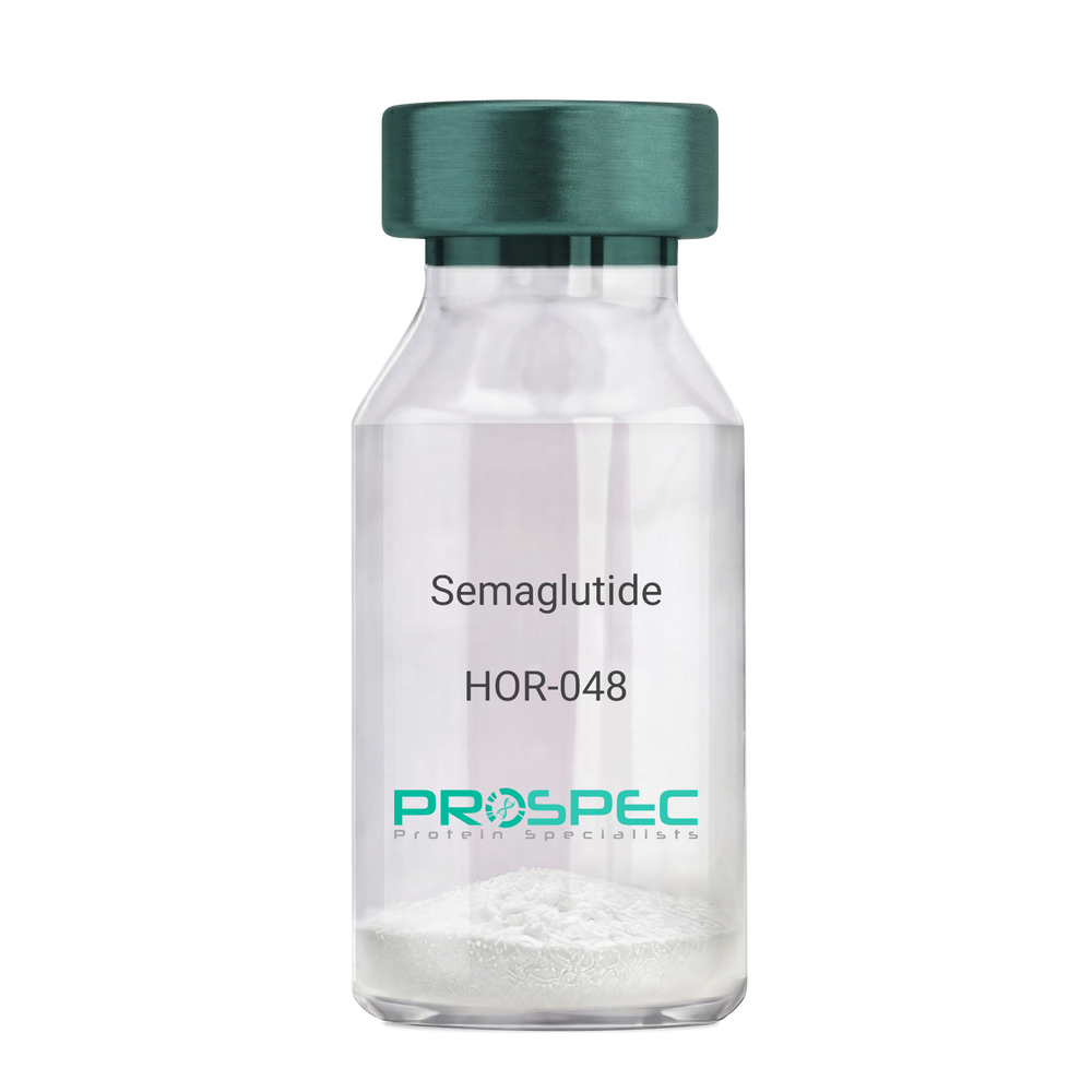
    Semaglutide

    Description:

    Semaglutide

    Product # :

    HOR-048

    Price :

    Quantity :

    Shipping Method :

    Room Temp Icon

    Shipped at Room temp

    Add To Cart

    More Info

    • description
    • formulation
    • purity
    • More Info
    • HPLC, MS
    • Citations

    Description

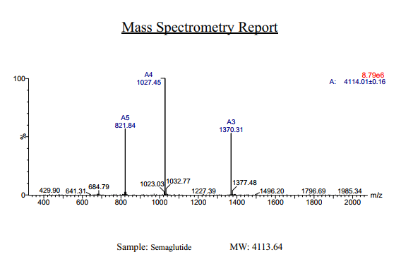
    Semaglutide Synthetic is a single, non-glycosylated polypeptide chain containing 31 amino acids, having a molecular mass of 4113 Dalton and a Molecular formula of C187H291N45O59.

    Formulation

    The protein was lyophilized with no additives.

    Purity

    Greater than 97.0% as determined by analysis by RP-HPLC.

    HPLC, MS

    Semaglutide hplc - Product image 1
    semaglutide mass spec - Product image 2

    Citations

    More Info

    • Physical Appearance

      Sterile Filtered White lyophilized (freeze-dried) powder.

    • Stability

      Lyophilized Semaglutide although stable at room temperature for 3 weeks, should be stored desiccated below -18°C. Upon reconstitution Semaglutide should be stored at 4°C between 2-7 days and for future use below -18°C. For long term storage it is recommended to add a carrier protein (0.1% HSA or BSA). Please prevent freeze-thaw cycles.

    • Solubility

      It is recommended to reconstitute the lyophilized Semaglutide in sterile 18MΩ-cm H2O not less than 100 µg/ml, which can then be further diluted to other aqueous solutions.

    • Amino Acid Sequence

      H-His-Aib-Glu-Gly-Thr-Phe-Thr-Ser-Asp-Val-Ser-Ser-Tyr-Leu-Glu-Gly-Gln-Ala-Ala-Lys(AEEAc-AEEAc-γ-Glu-17-carboxyheptadecanoyl)-Glu-Phe-Ile-Ala-Trp-Leu-Val-Arg-Gly-Arg-Gly-OH.

    • Background

      Semaglutide, a glucagon-like peptide-1 receptor agonist (GLP-1 RA), has emerged as a breakthrough in the field of diabetes management and metabolic disorders. It is recognized for its potent glucose-lowering effects, weight management properties, and cardiovascular benefits. Beyond diabetes, semaglutide is under investigation for its potential applications in obesity treatment and other related conditions. This research aims to comprehensively explore semaglutide, shedding light on its multifaceted mechanisms of action and its broader therapeutic implications.

      The primary objective of this research is to elucidate the mechanisms underlying the glucose-lowering and metabolic effects of semaglutide. In vitro and in vivo experiments will be conducted to investigate how semaglutide interacts with GLP-1 receptors, influences insulin secretion, and modulates glucose homeostasis. Understanding these mechanisms is crucial for harnessing the full therapeutic potential of semaglutide.

      The second objective is to assess the clinical relevance of semaglutide in diabetes management. Clinical trials involving individuals with type 2 diabetes will be conducted to evaluate the efficacy, safety, and long-term outcomes of semaglutide treatment. These investigations may provide valuable insights into its use as a monotherapy or adjunct therapy in diabetes care.

      The third objective is to explore the potential applications of semaglutide beyond diabetes. Research will investigate its role in obesity treatment, cardiovascular risk reduction, and non-alcoholic fatty liver disease (NAFLD) management. Understanding the multifaceted properties of semaglutide may open new avenues for therapeutic interventions in various metabolic and cardiovascular conditions.

      By delving into the diverse functions of semaglutide, this research aims to expand our understanding of its therapeutic potential and clinical applications. The findings may contribute to improved treatment strategies for individuals affected by diabetes, obesity, and related metabolic disorders.

    ProSpec's products are furnished for LABORATORY RESEARCH USE ONLY. The product may not be used as drugs, agricultural or pesticidal products, food additives or household chemicals.

    Semaglutide
  • View Data Sheet

    Name :

    IL 2 Antibody

    Description:

    Interleukin-2, Mouse Anti-Human

    T-cell growth factor (TCGF), Interleukin-2, Lymphokine, IL-2.

    Product # :

    ANT-102

    Price :

    Quantity :

    Shipping Method :

    Room Temp Icon

    Shipped at Room temp

    Add To Cart

    More Info

    • formulation
    • More Info
    • Citations

    Formulation

    1mg/ml in PBS (after reconstitution).

    Citations

    More Info

    • Introduction

      IL2 is a secreted cytokine that is important for the proliferation of T and B lymphocytes. The receptor of this cytokine is a heterotrimeric protein complex whose gamma chain is also shared by interleukin 4 (IL4) and interleukin 7 (IL7). The expression of this gene in mature thymocytes is monoallelic, which represents an unusual regulatory mode for controlling the precise expression of a single gene. The targeted disruption of a similar gene in mice leads to ulcerative colitis-like disease, which suggests an essential role of this gene in the immune response to antigenic stimuli.

    • Synonyms

      T-cell growth factor (TCGF), Interleukin-2, Lymphokine, IL-2.

    • Solubility

      Reconstitute with sterile H20. Mix gently, wash the sides of the vial and wait 30-60 seconds before use.

    • Immunogen

      r.Human IL-2.

    • Ig Subclass

      Mouse IgG2a.

    • Clone

      YNRhIL2.

    • Applications

      Direct ELISA, Western Blot, Immuneprecipitation, immunohistochemistry, blocking T cell poroliferation.

    • Cross reactivity

      A certain degree of functional cross reactivity with murine IL-2 was reported by some investigators.

    • Titer

      By direct ELISA: 1:10,000 dilution will yield 0.4 O.D using alkaline phosphatase conjugated rabbit anti-mouse Ig (Jackson Laboratories). 1:50 dilution will block 70-80% of CTLL proliferative response to 50 units of r.human IL-2.

    • Shipping Conditions

      Antibody is shipped lyophilized at ambient temperature.

    • Type

      Mouse Anti Human Monoclonal.

    • Storage Procedures

      In lyophilized form, for long periods, store at 4°C in a dry environment. After reconstitution, if not intended for use within a month, aliquot and store at -20°C.

    • Purification Method

      Ion Exchange.

    ProSpec's products are furnished for LABORATORY RESEARCH USE ONLY. The product may not be used as drugs, agricultural or pesticidal products, food additives or household chemicals.

    Interleukin 2 Human Antibody
  • View Data Sheet

    Name :

    IL 2r Antibody, FITC

    Description:

    Mouse Anti Human Interleukin-2 receptor FITC

    CD25, IL2R, TCGFR, IL-2RA, sIL-2RA, TAC antigen, sIL-2R, IDDM10, p55 TypeMouse Anti Human Monoclonal.

    Product # :

    ANT-072

    Price :

    Quantity :

    Shipping Method :

    Room Temp Icon

    Shipped at Room temp

    Add To Cart

    More Info

    • formulation
    • More Info
    • Citations

    Formulation

    1mg/ml in PBS (after reconstitution).

    Citations

    More Info

    • Introduction

      IL2-Ra is one of the three constituent subunits of the IL2 receptor. IL-2Ra is released into the serum after increased cellular expression such as increased activation of B and T cells. Clinical manifestations of IL2-Ra elevation include autoimmune conditions and some leukemias and lymphomas.

    • Synonyms

      CD25, IL2R, TCGFR, IL-2RA, sIL-2RA, TAC antigen, sIL-2R, IDDM10, p55 TypeMouse Anti Human Monoclonal.

    • Solubility

      Reconstitute with sterile H20. Mix gently, wash the sides of the vial and wait 30-60 seconds before use.

    • Immunogen

      Con A-activated human T cells.

    • Ig Subclass

      Mouse IgG2a.

    • Clone

      YNRhIL2R.

    • Titer

      This antibody will stain 70% of PHA-activated human T cells (by FACS). 10µl will stain 106 IL-2R positive cells for FACS analysis or sorting.

    • Shipping Conditions

      Antibody is shipped lyophilized at ambient temperature.

    • Storage Procedures

      In lyophilized form, for long periods, store at 4°C in a dry environment. After reconstitution, if not intended for use within a month, aliquot and store at -20°C.

    • Purification Method

      Ion Exchange.

    ProSpec's products are furnished for LABORATORY RESEARCH USE ONLY. The product may not be used as drugs, agricultural or pesticidal products, food additives or household chemicals.

    Il 2R Antibody Fitc
  • View Data Sheet

    Name :

    IL 17 A/F Mouse

    Description:

    Interleukin-17 A/F Heterodimer Mouse Recombinant

    IL17A/F, IL17 A/F, IL-17A/F, IL-17 A/F, IL17AF, IL-17 AF, Interleukin-17 A/F, Interleukin-17 AF.

    Product # :

    CYT-640

    Price :

    Quantity :

    Shipping Method :

    Room Temp Icon

    Shipped at Room temp

    Add To Cart

    More Info

    • description
    • source
    • formulation
    • purity
    • More Info
    • Citations

    Description

    Interleukin-17 A/F Mouse Recombinant produced in E.Coli is a heterodimeric, non-glycosylated polypeptide comprised of IL17A monomeric subunit & and IL17F monomeric subunit containing a total of 266 amino acids and having a total molecular mass of 29.8kDa. The IL-17 A/F is purified by proprietary chromatographic techniques.

    Source

    Escherichia Coli.

    Formulation

    Lyophilized from a concentrated (1mg/ml) solution containing no additives.

    Purity

    Greater than 97.0% as determined by SDS-PAGE.

    Citations

    More Info

    • Introduction

      Human IL-17A/F is a 40kDa glycoprotein which is secreted as a disulfide-linked heterodimer. IL-17A/F consists of two proteins of the IL-17 family, IL-17A and IL17F. Proteins of the 6 homodimeric IL17 family show a cysteine knot motif that contains two disulfide-bonds. Human IL17A is produced as a 155 a.a precursor that includes a 23 amino acids signal sequence and a 132 amino acid chain that includes an N-linked glycosylation site. Human IL17F is produced as a 153 amino acid precursor with a 20 amino acid signal sequence and a 133 amino acid region. Similar to IL17A, IL17F also has an N-linked glycosylation site. Both proteins (IL17A & IL17F) share 50% amino acid sequence identity. Human IL17A & IL17F show approximately 60% homology in their amino acid sequence to mouse IL-17A and IL-17F. Interleukin-17A/F and IL17A, IL17F homodimers are manufactured by activted CD4+ T cells, called Th17. IL-23 causes Th17 lymphocytes to manufacture IL-17A/F. IL17RA and IL17RC form a heterodimer for the binding of IL17A and IL17F. IL-17A/F binds IL-17RA. Interleukin-17A/F induces chemokine production and airway neutrophilia with intermediate potency between IL17A (most potent) and IL17F (least potent).

    • Synonyms

      IL17A/F, IL17 A/F, IL-17A/F, IL-17 A/F, IL17AF, IL-17 AF, Interleukin-17 A/F, Interleukin-17 AF.

    • Physical Appearance

      Sterile Filtered White lyophilized (freeze-dried) powder.

    • Stability

      Lyophilized Mouse IL17 A/F although stable at room temperature for 3 weeks, should be stored desiccated below -18°C. Upon reconstitution Mouse IL17 A/F should be stored at 4°C between 2-7 days and for future use below -18°C.For long term storage it is recommended to add a carrier protein (0.1% HSA or BSA).Please prevent freeze-thaw cycles.

    • Solubility

      It is recommended to reconstitute the lyophilized Mouse IL17 A/F in sterile 18MΩ-cm H2O not less than 100µg/ml, which can then be further diluted to other aqueous solutions.

    • Amino Acid Sequence

      RKNPKAGVPALQKAGNCPPLEDNTVRVDIRIFNQNQGISVPREFQNRSSSP
      WDYNITRDPHRFPSEIAEAQCRHSGCINAQGQEDSTMNSVAIQQEILVLRR
      EPQGCSNSFRLEKMLLKVGCTCVKPIVHQAAAAIIPQSSACPNTEAKDFLQ
      NVKVNLKVFNSLGAKVSSRRPSDYLNRSTSPWTLHRNEDPDRYPSVIWE
      AQCRHQRCVNAEGKLDHHMNSVLIQQEILVLKREPESCPFTFRVEKMLV
      GVGCTCVASIVRQAA.

    ProSpec's products are furnished for LABORATORY RESEARCH USE ONLY. The product may not be used as drugs, agricultural or pesticidal products, food additives or household chemicals.

    Il 17 A F Mouse
  • View Data Sheet

    Name :

    IL 17A/F Rat

    Description:

    Interleukin-17A/F Heterodimer Rat Recombinant

    IL17A/F, IL17 A/F, IL-17A/F, IL-17 A/F, IL17AF, IL-17 AF, Interleukin-17 A/F, Interleukin-17 AF.

    Product # :

    CYT-677

    Price :

    Quantity :

    Shipping Method :

    Room Temp Icon

    Shipped at Room temp

    Add To Cart

    More Info

    • description
    • source
    • formulation
    • purity
    • biological activity
    • More Info
    • Citations

    Description

    IL-17A/F Rat Recombinant produced in E.Coli is a heterodimeric, non-glycosylated polypeptide chain containing 1 monomeric subunit of each IL-17A & IL-17F. The dimer contains 269 amino acids and having a total molecular mass of 30.7 kDa. The IL-17A/F Rat is purified by proprietary chromatographic techniques.

    Source

    Escherichia Coli.

    Formulation

    Lyophilized from a concentrated (1mg/ml) solution containing no additives.

    Purity

    Greater than 98.0% as determined by HPLC and SDS-PAGE.

    Biological Activity

    The ED50 as determined by its ability to induce of IL-6 production from NIH-3T3 cells, is 15.3-23 ng/ml.

    Citations

    More Info

    • Introduction

      Rat IL-17A/F is a glycoprotein which is secreted as a disulfide-linked heterodimer. IL-17A/F consists of two proteins of the IL-17 family, IL-17A and IL17F. Proteins of the 6 homodimeric IL17 family show a cysteine knot motif that contains two disulfide-bonds. Rat IL17A is produced as a 155 a.a precursor that includes a 23 amino acids signal sequence and a 132 amino acid chain that includes an N-linked glycosylation site. Rat IL17F is produced as a 153 amino acid precursor with a 20 amino acid signal sequence and a 133 amino acid region. Similar to IL17A, IL17F also has an N-linked glycosylation site. Both proteins (IL17A & IL17F) share 50% amino acid sequence identity. Human IL17A & IL17F show approximately 60% homology in their amino acid sequence to mouse IL-17A and IL-17F. Interleukin-17A/F and IL17A, IL17F homodimers are manufactured by activted CD4+ T cells, called Th17. IL-23 causes Th17 lymphocytes to manufacture IL-17A/F. IL17RA and IL17RC form a heterodimer for the binding of IL17A and IL17F. IL-17A/F binds IL-17RA. Interleukin-17A/F induces chemokine production and airway neutrophilia with intermediate potency between IL17A (most potent) and IL17F (least potent).

    • Synonyms

      IL17A/F, IL17 A/F, IL-17A/F, IL-17 A/F, IL17AF, IL-17 AF, Interleukin-17 A/F, Interleukin-17 AF.

    • Physical Appearance

      Sterile Filtered White lyophilized (freeze-dried) powder.

    • Stability

      Lyophilized Rat IL-17A/F although stable at room temperature for 3 weeks, should be stored desiccated below -18°C. Upon reconstitution Rat IL-17A/F should be stored at 4°C between 2-7 days and for future use below -18°C.For long term storage it is recommended to add a carrier protein (0.1% HSA or BSA).Please prevent freeze-thaw cycles.

    • Solubility

      It is recommended to reconstitute the lyophilized Rat IL-17A/F in sterile water not more than 1mg/ml, which can then be further diluted to other aqueous solutions.

    • Amino Acid Sequence

      IL-17A: MAVLIPQSSV CPNAEANNFL QNVKVNLKVL NSLSSKASSR RPSDYLNRST SPWTLSRNED PDRYPSVIWE AQCRHQRCVN AEGKLDHHMN SVLIQQEILV LKREPEKCPF TFRVEKMLVG VGCTCVSSIV RHAS.
      IL-17F: MARRNPKVGL SALQKAGNCP PLEDNSVRVD IRIFNQNQGI SVPRDFQNRS SSPWDYNITR DPDRFPSEIA EAQCRHSGCI NAQGQEDGSM NSVPIQQEIL VLRREPQGCS NSFRLEKMLI KVGCTCVTPI VHHAA.

    ProSpec's products are furnished for LABORATORY RESEARCH USE ONLY. The product may not be used as drugs, agricultural or pesticidal products, food additives or household chemicals.

    Il 17 A F Rat
  • View Data Sheet

    Name :

    Activin-A Mouse

    Description:

    Activin-A Mouse Recombinant

    Inhba, Inhibin beta A, FSH releasing protein.

    Product # :

    CYT-146

    Price :

    Quantity :

    Shipping Method :

    Room Temp Icon

    Shipped at Room temp

    Add To Cart

    More Info

    • description
    • source
    • formulation
    • purity
    • biological activity
    • More Info
    • Citations

    Description

    Active form Activin-A Murine Recombinant produced in e.coli is a homodimeric, non-glycosylated, polypeptide chain containing 2 x 117 amino acids and having a molecular weight of 26.2kDa.The Active form Activin-A is purified by standard chromatographic techniques.

    Source

    E.Coli.

    Formulation

    Mouse Activin-A lyophilized from a concentrated 1mg/ml protein solution containing 0.1% TFA.

    Purity

    Greater than 95% as obsereved by SDS-PAGE.

    Biological Activity

    Biological activity is assessed by the ability to induce cytoxicity of MPC-11 cells and was found to be 8.8ng/ml corresponding to a specific activity of 1.1 x 10⁵ units/mg.

    Citations

    More Info

    • Introduction

      Activins are homodimers or heterodimers of the different β subunit isoforms, part of the TGFβ family. Mature Activin A has two 116 amino acids residues βA subunits (βA-βA). Activin displays an extensive variety of biological activities, including mesoderm induction, neural cell differentiation, bone remodelling, haematopoiesis, and reproductive physiology. Activins takes part in the production and regulation of hormones such as FSH, LH, GnRH and ACTH. Cells that are identified to express Activin A include fibroblasts, endothelial cells, hepatocytes, vascular smooth muscle cells, macrophages, keratinocytes, osteoclasts, bone marrow monocytes, prostatic epithelium, neurons, chondrocytes, osteoblasts, Leydig cells, Sertoli cells, and ovarian granulosa cells.

    • Synonyms

      Inhba, Inhibin beta A, FSH releasing protein.

    • Physical Appearance

      Lyophilized freeze dried powder.

    • Stability

      Lyophilized Activin-A although stable at room temperature for 3 weeks, should be stored desiccated below -18°C. Upon reconstitution Activin-A should be stored at 4°C between 2-7 days and for future use below -18°C.For long term storage it is recommended to add a carrier protein (0.1% HSA or BSA).Please prevent freeze-thaw cycles.

    • Solubility

      Murine INHBA protein should be reconstituted in distilled pyrogen free water to a concentration of 100ug /ml which can then be further diluted to other aqueous solutions.

    • Amino Acid Sequence

      MGLECDGKVN ICCKKQFFVS FKDIGWNDWI IAPSGYHANY CEGECPSHIA GTSGSSLSFH STVINHYRMR GHSPFANLKS CCVPTKLRPM SMLYYDDGQN IIKKDIQNMI VEECGCS.

    • Background

      What is the molecular weight / Mw of Activin A Protein?
      Activin A Protein has a total Mw of 26.2 kDa.

      What is the source or expression system of Activin A Protein?
      Ecoli

      What is the Purity of Activin A Protein?
      Activin A Protein is >95% pure as determined by SDS-PAGE.

      What is the Biological Activity of Activin A Protein?
      Biological activity is assessed by the ability to induce cytoxicity of MPC-11 cells and was found to be 8.8ng/ml corresponding to a specific activity of 110,000 units/mg.

      What is the endotoxin level for Activin A Protein?
      The endotoxin level is minimal, ACTIVIN A Protein was purified using conventional chromatography techniques.

      What is the amino acid sequence of ACTIVIN A Protein?
      MGLECDGKVN ICCKKQFFVS FKDIGWNDWI IAPSGYHANY CEGECPSHIA GTSGSSLSFH STVINHYRMR GHSPFANLKS CCVPTKLRPM SMLYYDDGQN IIKKDIQNMI VEECGCS.

      What applications can ACTIVIN A Protein be used in?
      ACTIVIN A Protein can probably be used in western blot, ELISA and Lateral Flow.

    ProSpec's products are furnished for LABORATORY RESEARCH USE ONLY. The product may not be used as drugs, agricultural or pesticidal products, food additives or household chemicals.

    Inhba Mouse
  • View Data Sheet

    Name :

    MSR1 Human, sf9

    Description:

    Macrophage Scavenger Receptor 1, sf9 Human Recombinant

    Macrophage Scavenger Receptor 1, Macrophage Acetylated LDL Receptor I And II, Scavenger Receptor Class A Member 1, SCARA1, Macrophage Scavenger Receptor Type III, CD204 Antigen, CD204, PhSR1, PhSR2, SR-A, SRA.

    Product # :

    PRO-2318

    Price :

    Quantity :

    Shipping Method :

    Ice Icon

    Shipped with Ice Packs

    Add To Cart

    More Info

    • description
    • source
    • formulation
    • purity
    • More Info
    • Citations

    Description

    MSR1 produced in Sf9 Baculovirus cells is a single, glycosylated polypeptide chain containing 384 amino acids (77-451 a.a.) and having a molecular mass of 42.4kDa (Migrates at 40-57kDa on SDS-PAGE under reducing conditions). MSR1 is expressed with a 6 amino acid His tag at C-Terminus and purified by proprietary chromatographic techniques.

    Source

    Sf9, Baculovirus cells.

    Formulation

    MSR1 protein solution (0.5mg/ml) contains Phosphate Buffered Saline (pH 7.4) and 10% glycerol.

    Purity

    Greater than 95.0% as determined by SDS-PAGE.

    Citations

    More Info

    • Introduction

      Macrophage Scavenger Receptor 1, also known as MSR1 is a member of the class A macrophage scavenger receptors, which comprises three different types 1, 2 and 3 generated by alternative splicing. Furthermore, these receptors or isoforms are macrophage-specific trimeric integral membrane glycoproteins and have been implicated in many macrophage-associated physiological as well as pathological processes which include atherosclerosis, Alzheimer's disease, and host defense.

    • Synonyms

      Macrophage Scavenger Receptor 1, Macrophage Acetylated LDL Receptor I And II, Scavenger Receptor Class A Member 1, SCARA1, Macrophage Scavenger Receptor Type III, CD204 Antigen, CD204, PhSR1, PhSR2, SR-A, SRA.

    • Physical Appearance

      Sterile Filtered colorless solution.

    • Stability

      Store at 4°C if entire vial will be used within 2-4 weeks. Store, frozen at -20°C for longer periods of time. For long term storage it is recommended to add a carrier protein (0.1% HSA or BSA). Avoid multiple freeze-thaw cycles.

    • Amino Acid Sequence

      ADPKWETKNC SVSSTNANDI TQSLTGKGND SEEEMRFQEV FMEHMSNMEK RIQHILDMEA NLMDTEHFQN FSMTTDQRFN DILLQLSTLF SSVQGHGNAI DEISKSLISL NTTLLDLQLN IENLNGKIQE NTFKQQEEIS KLEERVYNVS AEIMAMKEEQ VHLEQEIKGE VKVLNNITND LRLKDWEHSQ TLRNITLIQG PPGPPGEKGD RGPTGESGPR GFPGPIGPPG LKGDRGAIGF PGSRGLPGYA GRPGNSGPKG QKGEKGSGNT LTPFTKVRLV GGSGPHEGRV EILHSGQWGT ICDDRWEVRV GQVVCRSLGY PGVQAVHKAA HFGQGTGPIW LNEVFCFGRE SSIEECKIRQ WGTRACSHSE DAGVTCTLHH HHHH.

    ProSpec's products are furnished for LABORATORY RESEARCH USE ONLY. The product may not be used as drugs, agricultural or pesticidal products, food additives or household chemicals.

    Msr1 Human Sf9
  • View Data Sheet

    Name :

    SDCBP2 Human

    Description:

    Syndecan Binding Protein 2 Human Recombinant

    SITAC, SITAC18, ST-2, Syntenin-2, Syndecan-binding protein 2.

    Product # :

    PRO-1297

    Price :

    Quantity :

    Shipping Method :

    Ice Icon

    Shipped with Ice Packs

    Add To Cart

    More Info

    • description
    • source
    • formulation
    • purity
    • More Info
    • Citations

    Description

    SDCBP2 Human Recombinant produced in E.Coli is a single, non-glycosylated polypeptide chain containing 315 amino acids (1-292 a.a.) and having a molecular mass of 34.0kDa. SDCBP2 is fused to a 23 amino acid His-tag at N-terminus & purified by proprietary chromatographic techniques.

    Source

    Escherichia Coli.

    Formulation

    SDCBP2 protein solution (0.5mg/ml) contains 20mM Tris-HCl buffer (pH 8.0), 0.15M NaCl, 10% glycerol and 1mM DTT.

    Purity

    Greater than 95.0% as determined by SDS-PAGE.

    Citations

    More Info

    • Introduction

      SDCBP2 has two class II PDZ domains. PDZ domains assist protein-protein interactions by attaching to the cytoplasmic C-terminus of transmembrane proteins, and PDZ-containing proteins mediate cell signaling and the organization of protein complexes. SDCBP2 attaches to phosphatidylinositol 4, 5-bisphosphate (PIP2) and take part in nuclear PIP2 arrangement and cell division.

    • Synonyms

      SITAC, SITAC18, ST-2, Syntenin-2, Syndecan-binding protein 2.

    • Physical Appearance

      Sterile Filtered clear solution.

    • Stability

      Store at 4°C if entire vial will be used within 2-4 weeks. Store, frozen at -20°C for longer periods of time. For long term storage it is recommended to add a carrier protein (0.1% HSA or BSA).Avoid multiple freeze-thaw cycles.

    • Amino Acid Sequence

      MGSSHHHHHH SSGLVPRGSH MGSMSSLYPS LEDLKVDQAI QAQVRASPKM PALPVQATAI SPPPVLYPNL AELENYMGLS LSSQEVQESL LQIPEGDSTA VSGPGPGQMV APVTGYSLGV RRAEIKPGVR EIHLCKDERG KTGLRLRKVD QGLFVQLVQA NTPASLVGLR FGDQLLQIDG RDCAGWSSHK AHQVVKKASG DKIVVVVRDR PFQRTVTMHK DSMGHVGFVI KKGKIVSLVK GSSAARNGLL TNHYVCEVDG QNVIGLKDKK IMEILATAGN VVTLTIIPSV IYEHMVKKLP PVLLHHTMDH SIPDA.

    ProSpec's products are furnished for LABORATORY RESEARCH USE ONLY. The product may not be used as drugs, agricultural or pesticidal products, food additives or household chemicals.

    Sdcbp2 Human
  • View Data Sheet

    Name :

    Apo D Human

    Description:

    Apolipoprotein-D Human Recombinant

    Apolipoprotein D, Apo-D, ApoD.

    Product # :

    CYT-547

    Price :

    Quantity :

    Shipping Method :

    Room Temp Icon

    Shipped at Room temp

    Add To Cart

    More Info

    • description
    • source
    • formulation
    • purity
    • More Info
    • Citations

    Description

    Apolipoprotein-D Human Recombinant His Tag fusion protein at C-terminus (7 highlighted a.a.) produced in E.Coli is a single, non-glycosylated, Polypeptide chain containing 174 amino acids and having a molecular mass of 19.82kDa. The protein a.a sequence corresponds to the UniProtKB/Swiss-Prot entry P05090.The Following gene modifications were made:Trp99His, Cys116Ser, Ile118Ser, Leu120Ser amino acids exchanges were introduced at the surface of Apolipoprotein-D to enhance the protein’s solubility and another three Leu23Pro, Pro133Val, Asn134Ala amino acids exchanges which facilitate its genetic manipulation. The Apolipoprotein-D is purified by proprietary chromatographic techniques.

    Source

    Escherichia Coli.

    Formulation

    Filtered (0.4µm) and lyophilized from 1mg/ml in 4mM KH2PO4, 16mM Na2HPO4 and 115mM NaCl pH 7.5.

    Purity

    Greater than 90% as determined by SDS-PAGE.

    Citations

    More Info

    • Introduction

      Apolipoprotein-D is mainly associated with high density lipoproteins in human plasma. Apolipoprotein-D is an atypical apolipoprotein and, based on its primary structure, Apolipoprotein-D is a member of the lipocalin family. Lipocalins adopt a beta-barrel tertiary structure and transport small hydrophobic ligands. Apolipoprotein-D binds cholesterol, progesterone, pregnenolone, bilirubin and arachidonic acid.
      Apolipoprotein-D is expressed in numerous tissues having high levels of expression in spleen, testes and brain. Apolipoprotein-D is present at high concentrations in the cyst fluid of women with gross cystic disease of the breast, a condition associated with increased risk of breast cancer. Apolipoprotein-D accumulates in regenerating peripheral nerves and in the cerebrospinal fluid of patients with neurodegenerative conditions, such as Alzheimer's disease. Apolipoprotein-D participates in maintenance and repair within the central and peripheral nervous systems. Apolipoprotein-D is a multi-ligand, multi-functional transporter and transports a ligand from 1 cell to another within an organ, scavenge a ligand within an organ for transport to the blood or could transport a ligand from the circulation to specific cells within a tissue.

    • Synonyms

      Apolipoprotein D, Apo-D, ApoD.

    • Physical Appearance

      Filtered White lyophilized (freeze-dried) powder.

    • Stability

      Store lyophilized protein at -20°C. Aliquot the product after reconstitution to avoid repeated freezing/thawing cycles. Reconstituted protein can be stored at 4°C for a limited period of time; it does not show any change after two weeks at 4°C.

    • Solubility

      It is recommended to add deionized H2O to a working volume of 0.5mg/ml and let the lyophilized pellet dissolve completely. Product is not sterile! Please filter this product by an appropriate sterile filter before using it in the cell culture.

    • Amino Acid Sequence

      FHLGKCPNPP VQENFDVNKY PGRWYEIEKI PTTFENGRCI QANYSLMENG KIKVLNQELR ADGTVNQIEG EATPVNLTEP AKLEVKFSWF MPSAPYHILA TDYENYALVY SCTSISQSFH VDFAWILARN VALPPETVDS LKNILTSNNI DVKKMTVTDQ VNCPKLSAHHHHHH.

    • Background

      Apolipoprotein-D Human Recombinant: Illuminating the Role of a Multifaceted Lipid-Binding Protein

      Abstract:


      Apolipoprotein-D (ApoD), a multifunctional lipid-binding protein, has emerged as a fascinating player in lipid metabolism and neuroprotection. This research paper aims to provide an insightful overview of ApoD human recombinant, exploring its physiological functions, production methods, and potential therapeutic applications. By unraveling the complexities of ApoD, we gain valuable insights into its role in lipid homeostasis and its potential as a therapeutic target for neurodegenerative diseases. This article presents a concise yet comprehensive analysis of ApoD, humanizing its significance in the context of human health.

      Introduction:


      Understanding the intricate mechanisms underlying lipid metabolism and neuroprotection is crucial for the development of novel therapeutic strategies. ApoD, a versatile protein expressed in various tissues, offers unique insights into these areas. This paper delves into the multifaceted nature of ApoD, shedding light on its significance in lipid homeostasis and neuronal health.

      Structure and Function of Apolipoprotein-D:


      ApoD exhibits a complex molecular structure, comprising distinct domains that facilitate its binding to lipids and other biomolecules. It engages in diverse functions, including lipid transport, antioxidant defense, and modulation of neuroinflammatory responses. The versatility of ApoD underscores its pivotal role in maintaining cellular and tissue integrity.

      Regulation of Apolipoprotein-D Expression:


      The expression of ApoD is subject to intricate regulatory mechanisms influenced by hormonal and environmental cues. Understanding the factors governing ApoD expression provides valuable insights into its physiological roles and potential therapeutic applications.

      Apolipoprotein-D and Neurodegenerative Diseases:


      Growing evidence implicates ApoD in neuroprotection, particularly in the context of neurodegenerative diseases. ApoD exhibits neuroprotective properties by modulating oxidative stress, lipid peroxidation, and inflammatory responses, making it an intriguing target for therapeutic interventions.

      Production of Apolipoprotein-D Human Recombinant:


      Advanced biotechnological approaches, including recombinant DNA technology and protein expression systems, enable the production of ApoD human recombinant. These methods facilitate large-scale production, purification, and characterization of ApoD, paving the way for potential therapeutic applications.

      Therapeutic Potential of Apolipoprotein-D Human Recombinant:


      Targeting ApoD holds promise for the development of therapeutics aimed at neurodegenerative diseases. Modulating ApoD expression or function may provide neuroprotection, enhance neuronal survival, and mitigate the progression of neurodegenerative disorders.

      Conclusion:


      Apolipoprotein-D human recombinant represents a captivating area of research, bridging the fields of lipid metabolism and neurodegeneration. Understanding the intricate interplay between ApoD, lipid homeostasis, and neuroprotection is crucial for unraveling its full therapeutic potential. Continued investigation into the functions and mechanisms of ApoD will likely lead to novel therapeutic strategies for neurodegenerative diseases.

      What is the molecular weight/Mw of APO D Protein?
      APO D Protein has a total Mw of 19.82kDa.

      What is the source or expression system of APO D Protein?
      Escherichia Coli.

      What is the Purity of APO D Protein?
      APO D Protein is >90% pure as determined by SDS-PAGE.

      What is the Biological Activity of APO D Protein?
      The biological functionality of APO D Protein will be determined in the future.

      What is the amino acid sequence of APO D Protein?
      FHLGKCPNPP VQENFDVNKY PGRWYEIEKI PTTFENGRCI QANYSLMENG KIKVLNQELR ADGTVNQIEG EATPVNLTEP AKLEVKFSWF MPSAPYHILA TDYENYALVY SCTSISQSFH VDFAWILARN VALPPETVDS LKNILTSNNI DVKKMTVTDQ VNCPKLSAHHHHHH.

      What applications can APO D Protein be used in?
      APO D Protein can probably be used in western blot, ELISA and Lateral Flow.

      What is the endotoxin level for APO D Protein?
      The endotoxin level is minimal, APO D Protein was purified using conventional chromatography techniques.

    ProSpec's products are furnished for LABORATORY RESEARCH USE ONLY. The product may not be used as drugs, agricultural or pesticidal products, food additives or household chemicals.

    Apo D Human
  • View Data Sheet

    Name :

    NPPA Human

    Description:

    Natriuretic Peptide A Human Recombinant

    Natriuretic peptides A, CDD-ANF, Cardiodilatin, CDD, Cardiodilatin-related peptide, CDP, N-terminal proatrial natriuretic peptide, ANP, PND. 

    Product # :

    CYT-1028

    Price :

    Quantity :

    Shipping Method :

    Room Temp Icon

    Shipped at Room temp

    Add To Cart

    More Info

    • description
    • source
    • formulation
    • purity
    • More Info
    • Citations

    Description

    NPPA Human Recombinant produced in E.Coli is a single, non-glycosylated, polypeptide chain (a.a 26-123) containing 106 amino acids including an 8 a.a N-terminal His tag. The total molecular mass is 11.7kDa (calculated).

    Source

    Escherichia Coli.

    Formulation

    NPPA filtered (0.4 µm) and lyophilized from 0.5mg/ml in 20 mM Tris buffer, 50 mM NaCl and 5% w/v trehalosa, pH 7.5.

    Purity

    Greater than 95.0% as determined by SDS-PAGE.

    Citations

    More Info

    • Introduction

      Natriuretic Peptide A (NPPA) is a part of the natriuretic peptide family and is involved in cardiovascular homeostasis through regulation of natriuresis, diuresis, and vasodilation. NPPA is synthesized as a great precursor which releases a peptide from the N-terminus with similarity to vasoactive peptide, cardiodilatin, and another peptide from the C-terminus with natriuretic-diuretic activity. In female pregnancy, NPPA is promoting trophoblast invasion and spiral artery remodeling in uterus.

    • Synonyms

      Natriuretic peptides A, CDD-ANF, Cardiodilatin, CDD, Cardiodilatin-related peptide, CDP, N-terminal proatrial natriuretic peptide, ANP, PND.

    • Physical Appearance

      Filtered White lyophilized (freeze-dried) powder.

    • Stability

      Store lyophilized protein at -20°C. Aliquot the product after reconstitution to avoid repeated freezing/thawing cycles. Reconstituted protein can be stored at 4°C for a limited period of time; it does not show any change after two weeks at 4°C.

    • Solubility

      It is recommended to add deionized water to prepare a working stock solution of approximately 0.5mg/ml and let the lyophilized pellet dissolve completely. NPPA is not sterile! Please filter the product by an appropriate sterile filter before using it in the cell culture.

    • Amino Acid Sequence

      MKHHHHHHNP MYNAVSNADL MDFKNLLDHL EEKMPLEDEV VPPQVLSEPN EEAGAALSPL PEVPPWTGEV SPAQRDGGAL GRGPWDSSDR SALLKSKLRA LLTAPR.

    ProSpec's products are furnished for LABORATORY RESEARCH USE ONLY. The product may not be used as drugs, agricultural or pesticidal products, food additives or household chemicals.

    Nppa Human
  • View Data Sheet

    Name :

    SRGN Human, HEK

    Description:

    Serglycin Human Recombinant, HEK

    Serglycin, PRG, PRG1, PPG, Proteoglycan 1 secretory granule, Hematopoetic proteoglycan core protein, Platelet proteoglycan core protein, proteoglycan protein core for mast cell secretory granule.

    Product # :

    PRO-2046

    Price :

    Quantity :

    Shipping Method :

    Ice Icon

    Shipped with Ice Packs

    Add To Cart

    More Info

    • description
    • source
    • formulation
    • purity
    • More Info
    • Citations

    Description

    Serglycin Human Recombinant produced in HEK cells is a single, glycosylated, polypeptide chain (Tyr28-Leu158) containing a total of 137 amino acids, having a calculated molecular mass of 15.5kDa and fused to a 6 aa His tag at C-Terminus.

    Source

    HEK 293.

    Formulation

    SRGN filtered (0.4µm) solution in phosphate buffered saline and 20% (w/v) glycerol.

    Purity

    Greater than 95.0% as determined by SDS-PAGE.

    Citations

    More Info

    • Introduction

      SRGN is identified as a hematopoietic cell granule proteoglycan. Proteoglycans stored in the secretory granules of various hematopoietic cells also hold a protease-resistant peptide core, and is vital for neutralizing hydrolytic enzymes. SRGN is related to the macromolecular complex of granzymes and perforin that acts as a intermediary of granule-mediated apoptosis.

    • Synonyms

      Serglycin, PRG, PRG1, PPG, Proteoglycan 1 secretory granule, Hematopoetic proteoglycan core protein, Platelet proteoglycan core protein, proteoglycan protein core for mast cell secretory granule.

    • Physical Appearance

      Filtered colorless solution.

    • Stability

      Store at 4°C if entire vial will be used within 2-4 weeks. Store, frozen at -20°C for longer periods of time. For long term storage it is recommended to add a carrier protein (0.1% HSA or BSA).Avoid multiple freeze-thaw cycles.

    • Amino Acid Sequence

      YPTRRARYQW VRCNPDSNSA NCLEEKGPMF ELLPGESNKI PRLRTDLFPK TRIQDLNRIF PLSEDYSGSG FGSGSGSGSG SGSGFLTEME QDYQLVDESD AFHDNLRSLD RNLPSDSQDL GQHGLEEDFM L HHHHHH.

    ProSpec's products are furnished for LABORATORY RESEARCH USE ONLY. The product may not be used as drugs, agricultural or pesticidal products, food additives or household chemicals.

    Srgn Human Hek
  • View Data Sheet

    Name :

    STMN2 Human

    Description:

    Stathmin Like-2 Human Recombinant

    SCG10, STMB2, SCGN10, AI159727, STMN-2, Stathmin-2, Superior cervical ganglion-10 protein, Protein SCG10, STMN2, SGC10, stathmin-like 2.

    Product # :

    PRO-752

    Price :

    Quantity :

    Shipping Method :

    Ice Icon

    Shipped with Ice Packs

    Add To Cart

    More Info

    • description
    • source
    • formulation
    • purity
    • More Info
    • Citations

    Description

    Recombinant Human Stathmin Like-2 produced in E.Coli is a single, non-glycosylated polypeptide chain containing 142 amino acids (39-179 a.a) and having a molecular mass of 16.4 kDa. STMN2 is purified by conventional chromatography techniques.

    Source

    Escherichia Coli.

    Formulation

    The STMN2 protein solution contains 50mM MES, pH-6.0, 0.1mM PMSF, 1 mM EDTA and 10% Glycerol.

    Purity

    Greater than 85.0% as determined by SDS-PAGE.

    Citations

    More Info

    • Introduction

      STMN2 is a neuronal growth-associated protein which shares a majority of its amino acid sequence with the phosphoprotein stathmin. STMN2 is involved in neuronal differentiation and in modulating membrane interaction with the cytoskeleton through neurite outgrowth. STMN2 is necessary for maintaining the anchorage-independent growth state of beta-catenin/TCF-activated hepatoma cells. SCG10 is a novel indicator of osteogenesis and osteoblast that takes part in the regulation of the adipocyte/osteoblast balance. STMN2 plays a role as an effector downstream of Rnd1 to regulate axon extensions by modulating microtubule organization.

    • Synonyms

      SCG10, STMB2, SCGN10, AI159727, STMN-2, Stathmin-2, Superior cervical ganglion-10 protein, Protein SCG10, STMN2, SGC10, stathmin-like 2.

    • Physical Appearance

      Sterile filtered colorless solution.

    • Stability

      Store at 4°C if entire vial will be used within 2-4 weeks. Store, frozen at -20°C for longer periods of time. For long term storage it is recommended to add a carrier protein (0.1% HSA or BSA).Avoid multiple freeze-thaw cycles.

    • Amino Acid Sequence

      MDMEVKQINK RASGQAFELI LKPPSPISEA PRTLASPKKK DLSLEEIQKK LEAAEERRKS QEAQVLKQLA EKREHEREVL QKALEENNNF SKMAEEKLIL KMEQIKENRE ANLAAIIERL QEKERHAAEV RRNKELQVEL SG.

    ProSpec's products are furnished for LABORATORY RESEARCH USE ONLY. The product may not be used as drugs, agricultural or pesticidal products, food additives or household chemicals.

    Stmn2 Human
  • View Data Sheet

    Name :

    FAS Human, Sf9

    Description:

    sFas Receptor Human Recombinant, Sf9

    Tumor necrosis factor receptor superfamily member 6, Apo-1 antigen, Apoptosis-mediating surface antigen FAS, FASLG receptor, CD95, FAS, APT1, FAS1, APO-1, FASTM, ALPS1A, TNFRSF6.

    Product # :

    CYT-1153

    Price :

    Quantity :

    Shipping Method :

    Ice Icon

    Shipped with Ice Packs

    Add To Cart

    More Info

    • description
    • source
    • formulation
    • purity
    • More Info
    • Citations

    Description

    FAS Human Recombinant produced in Baculovirus is a single glycosylated polypeptide chain containing 156 amino acids (26-173 aa) and having a molecular mass of 17.7KDa.FAS is fused to a 8 amino acid His-Tag at C-terminus and purified by proprietary chromatographic techniques.

    Source

    Sf9, Baculovirus cells.

    Formulation

    FAS protein (0.5mg/ml) contains 10% glycerol and Phosphate-Buffered Saline (pH 7.4).

    Purity

    Greater than 95.0% as determined by SDS-PAGE.

    Citations

    More Info

    • Introduction

      FAS or tumor necrosis factor receptor superfamily member 6 or TNFRSF6, is part of the death receptor family, which is part of the TNF receptor protein family. TNFRSF6 has a crucial part in regulating viral infections. FAS protein can be found in almost all cell types, when its cognate ligand (FasL) can be found in activated T cells, NK cells & dendritic cells. The regulating protein of FasL & TRAIL on HCMV-infected dendritic enhances direct destruction of activated T lymphocytes. furthermore, subvertneutrophil function in HCMV retinitis can occur when FasL is activated in HCMV infected retinal pigment epithelial cells.

    • Synonyms

      Tumor necrosis factor receptor superfamily member 6, Apo-1 antigen, Apoptosis-mediating surface antigen FAS, FASLG receptor, CD95, FAS, APT1, FAS1, APO-1, FASTM, ALPS1A, TNFRSF6.

    • Physical Appearance

      Sterile Filtered colorless solution.

    • Stability

      Store at 4°C if entire vial will be used within 2-4 weeks. Store, frozen at -20°C for longer periods of time. For long term storage it is recommended to add a carrier protein (0.1% HSA or BSA).Avoid multiple freeze-thaw cycles.

    • Amino Acid Sequence

      QVTDINSKGL ELRKTVTTVE TQNLEGLHHD GQFCHKPCPP GERKARDCTV NGDEPDCVPC QEGKEYTDKA HFSSKCRRCR LCDEGHGLEV EINCTRTQNT KCRCKPNFFC NSTVCEHCDP CTKCEHGIIK ECTLTSNTKC KEEGSRSNLE HHHHHH

    • Background

      What is the molecular weight/Mw of FAS Protein?
      FAS Protein has a total Mw of 17.7kDa.

      What is the source or expression system of FAS Protein?
      Sf9, Baculovirus cells

      What is the Purity of FAS Protein?
      FAS Protein is >95% pure as determined by SDS-PAGE.

      What is the Biological Activity of FAS Protein?
      The biological functionality of FAS Protein will be determined in the future.

      What is the amino acid sequence of FAS Protein?
      QVTDINSKGL ELRKTVTTVE TQNLEGLHHD GQFCHKPCPP GERKARDCTV NGDEPDCVPC QEGKEYTDKA HFSSKCRRCR LCDEGHGLEV EINCTRTQNT KCRCKPNFFC NSTVCEHCDP CTKCEHGIIK ECTLTSNTKC KEEGSRSNLE HHHHHH

      What applications can FAS Protein be used in?
      FAS Protein can probably be used in western blot, ELISA and Lateral Flow.

      What is the endotoxin level for FAS Protein?
      The endotoxin level is minimal, FAS Protein was purified using conventional chromatography techniques.

    ProSpec's products are furnished for LABORATORY RESEARCH USE ONLY. The product may not be used as drugs, agricultural or pesticidal products, food additives or household chemicals.

    Fas Tnfrsf6
  • View Data Sheet

    Name :

    C1QTNF3 Human

    Description:

    Complement C1q Tumor Necrosis Factor-Related Protein 3 Human Recombinant

    Complement C1q tumor necrosis factor-related protein 3, Secretory protein CORS26, C1QTNF3, CTRP3, Cors, Corcs, CORS26, FLJ37576, Cartducin.

    Product # :

    PRO-653

    Price :

    Quantity :

    Shipping Method :

    Room Temp Icon

    Shipped at Room temp

    Add To Cart

    More Info

    • description
    • source
    • formulation
    • purity
    • More Info
    • Citations

    Description

    C1QTNF3 Human Recombinant produced in E.Coli is a single, non-glycosylated, Polypeptide chain containing 234 amino acids and having a molecular mass of 25.4 kDa. The protein contains an extra 10 aa His tag at N-terminus. The C1QTNF3 amino acid sequence is identical to UniProtKB/Swiss-Prot entry Q9BXJ4 amino acids 23–246. The C1QTNF3 is purified by proprietary chromatographic techniques.

    Source

    Escherichia Coli.

    Formulation

    Human C1QTNF3 was filtered (0.4µm) and lyophilized in 0.5 mg/ml in 0.05M Acetate buffer pH4.

    Purity

    The purity of C1QTNF3 is greater than 95% as determined by SDS PAGE.

    Citations

    More Info

    • Introduction

      C1QTNF3 also called Cartducin is a novel angiogenic factor in the formation of neointima following angioplasty. C1QTNF3 a paralog of Acrp30 (adiponectin). C1QTNF3 is a secretory protein produced by chondrogenic precursors & proliferating chondrocytes, and belongs to a novel C1q family of proteins. Cartducin promotes the growth of mesenchymal chondroprogenitor cells & chondrosarcoma-derived chondrocytic cells in vitro. Cartducin stimulates mesenchymal chondroprogenitor cell proliferation through extracellular signal-regulated kinase and phosphatidylinositol 3-kinase/Akt pathways. C1QTNF3 promotes proliferation & the migration of endothelial cells.

    • Synonyms

      Complement C1q tumor necrosis factor-related protein 3, Secretory protein CORS26, C1QTNF3, CTRP3, Cors, Corcs, CORS26, FLJ37576, Cartducin.

    • Stability

      Store lyophilized C1QTNF3 at -20°C. Aliquot the product after reconstitution to avoid repeated freezing/thawing cycles. Reconstituted C1QTNF3 can be stored at 4°C for a limited period of time; it does not show any change after two weeks at 4°C.

    • Solubility

      It is recommended to add 0.1M Acetate buffer pH4 to prepare a working stock solution of approximately 0.5 mg/ml and let the lyophilized pellet dissolve completely. For conversion into higher pH value, we recommend intensive dilution by relevant buffer to a concentration of 10µg/ml. In higher concentrations the solubility of the protein is limited. Product is not sterile! Please filter the product by an appropriate sterile filter before using it in the cell culture.

    • Amino Acid Sequence

      MKHHHHHHAS QDEYMESPQT GGLPPDCSKC CHGDYSFRGY QGPPGPPGPP GIPGNHGNNG NNGATGHEGA KGEKGDKGDL GPRGERGQHG PKGEKGYPGI PPELQIAFMA SLATHFSNQN SGIIFSSVET NIGNFFDVMT GRFGAPVSGV YFFTFSMMKH EDVEEVYVYL MHNGNTVFSM YSYEMKGKSD TSSNHAVLKL AKGDEVWLRM GNGALHGDHQ RFSTFAGFLLFETK.

    ProSpec's products are furnished for LABORATORY RESEARCH USE ONLY. The product may not be used as drugs, agricultural or pesticidal products, food additives or household chemicals.

    C1Qtnf3 Human
  • View Data Sheet

    Name :

    PDGFRB Human

    Description:

    Platelet-Derived Growth Factor Receptor, Beta Human Recombinant

    Platelet-derived growth factor receptor beta, PDGF-R-beta, PDGFR-beta, Beta platelet-derived growth factor receptor, Beta-type platelet-derived growth factor receptor, CD140 antigen-like family member B, Platelet-derived growth factor receptor 1, PDGFR-1, CD140b, PDGFRB, Beta Platelet-Derived Growth Factor Receptor, Activated Tyrosine Kinase PDGFRB, CD140b AntigenNDEL1-PDGFRB, EC 2.7.10, CD140B, IBGC4, JTK12, PENTT, IMF1, KOGS, Platelet Derived Growth Factor Receptor Beta, Platelet-Derived Growth Factor Receptor, Beta Polypeptide, Beta-Type Platelet-Derived Growth Factor Receptor, Platelet-Derived Growth Factor Receptor 1, CD140, Antigen-Like Family Member B, PDGF-R-Beta, EC 2.7.10.1, PDGFR-Beta, PDGFR-1, PDGFR1, PDGFR,Platelet-Derived Growth Factor Receptor Beta.

    Product # :

    CYT-1051

    Price :

    Quantity :

    Shipping Method :

    Ice Icon

    Shipped with Ice Packs

    Add To Cart

    More Info

    • description
    • source
    • formulation
    • purity
    • More Info
    • Citations

    Description

    PDGFRB produced in Sf9 Insect cells is a single, glycosylated polypeptide chain containing 739 amino acids (33-532a.a.) and having a molecular mass of 83.3kDa (Molecular size on SDS-PAGE will appear at approximately 100-150kDa).PDGFRB is expressed with an 239 amino acid hIgG-His tag at C-Terminus and purified by proprietary chromatographic techniques.

    Source

    Sf9, Insect cells.

    Formulation

    PDGFRB protein solution (0.25mg/ml) contains Phosphate Buffered Saline (pH 7.4) and 10% glycerol.

    Purity

    Greater than 90.0% as determined by SDS-PAGE.

    Citations

    More Info

    • Introduction

      Platelet-derived growth factor receptor beta (PDGFRB), belongs to the class III subfamily of receptor tyrosine kinases (RTK) which also consist of the receptors for Flt3-ligand, SCF and M-CSF. PDGFRB takes a vital part in blood vessel development by promoting growth, migration as well as recruitment of pericytes and smooth muscle cells to endothelial cells. PDGFRB helps in rearrangement of the actin cytoskeleton in addition the formation of membrane ruffles. PDGFRB phosphorylates, NCK1, PIK3R1, PTPN11, CBL, SHC1, RASA1/GAP and PLCG1.

    • Synonyms

      Platelet-derived growth factor receptor beta, PDGF-R-beta, PDGFR-beta, Beta platelet-derived growth factor receptor, Beta-type platelet-derived growth factor receptor, CD140 antigen-like family member B, Platelet-derived growth factor receptor 1, PDGFR-1, CD140b, PDGFRB, Beta Platelet-Derived Growth Factor Receptor, Activated Tyrosine Kinase PDGFRB, CD140b Antigen
      NDEL1-PDGFRB, EC 2.7.10, CD140B, IBGC4, JTK12, PENTT, IMF1, KOGS, Platelet Derived Growth Factor Receptor Beta, Platelet-Derived Growth Factor Receptor, Beta Polypeptide, Beta-Type Platelet-Derived Growth Factor Receptor, Platelet-Derived Growth Factor Receptor 1, CD140, Antigen-Like Family Member B, PDGF-R-Beta, EC 2.7.10.1, PDGFR-Beta, PDGFR-1, PDGFR1, PDGFR,
      Platelet-Derived Growth Factor Receptor Beta.

    • Physical Appearance

      Sterile filtered colorless solution.

    • Stability

      Store at 4°C if entire vial will be used within 2-4 weeks. Store, frozen at -20°C for longer periods of time. For long term storage it is recommended to add a carrier protein (0.1% HSA or BSA).Avoid multiple freeze-thaw cycles.

    • Amino Acid Sequence

      LVVTPPGPEL VLNVSSTFVL TCSGSAPVVW ERMSQEPPQE MAKAQDGTFS SVLTLTNLTG LDTGEYFCTH NDSRGLETDE RKRLYIFVPD PTVGFLPNDA EELFIFLTEI TEITIPCRVT DPQLVVTLHE KKGDVALPVP YDHQRGFSGI FEDRSYICKT TIGDREVDSD AYYVYRLQVS SINVSVNAVQ TVVRQGENIT LMCIVIGNEV VNFEWTYPRK ESGRLVEPVT DFLLDMPYHI RSILHIPSAE LEDSGTYTCN VTESVNDHQD EKAINITVVE SGYVRLLGEV GTLQFAELHR SRTLQVVFEA YPPPTVLWFK DNRTLGDSSA GEIALSTRNV SETRYVSELT LVRVKVAEAG HYTMRAFHED AEVQLSFQLQ INVPVRVLEL SESHPDSGEQ TVRCRGRGMP QPNIIWSACR DLKRCPRELP PTLLGNSSEE ESQLETNVTY WEEEQEFEVV STLRLQHVDR PLSVRCTLRN AVGQDTQEVI VVPHSLPFKV LEPKSCDKTH TCPPCPAPEL LGGPSVFLFP PKPKDTLMIS RTPEVTCVVV DVSHEDPEVK FNWYVDGVEV HNAKTKPREE QYNSTYRVVS VLTVLHQDWL NGKEYKCKVS NKALPAPIEK TISKAKGQPR EPQVYTLPPS RDELTKNQVS LTCLVKGFYP SDIAVEWESN GQPENNYKTT PPVLDSDGSF FLYSKLTVDK SRWQQGNVFS CSVMHEALHN HYTQKSLSLS PGKHHHHHH.

    ProSpec's products are furnished for LABORATORY RESEARCH USE ONLY. The product may not be used as drugs, agricultural or pesticidal products, food additives or household chemicals.

    Pdgfrb Human
  • View Data Sheet

    Name :

    PHLDA2 Human

    Description:

    Pleckstrin homology-like domain family A member 2 Human Recombinant

    Pleckstrin homology-like domain family A member 2, Imprinted in placenta and liver protein, Tumor-suppressing subchromosomal transferable fragment candidate gene 3 protein, Tumor-suppressing STF cDNA 3 protein, Beckwith-Wiedemann syndrome chromosomal region 1 candidate gene C protein, p17-Beckwith-Wiedemann region 1 C, PHLDA2, BWR1C, HLDA2, IPL, TSSC3, BRW1C.

    Product # :

    PRO-781

    Price :

    Quantity :

    Shipping Method :

    Ice Icon

    Shipped with Ice Packs

    Add To Cart

    More Info

    • description
    • source
    • formulation
    • purity
    • More Info
    • Citations

    Description

    PHLDA2 Human Recombinant produced in E.Coli is a single, non-glycosylated, polypeptide chain containing 172 amino acids (1-152 a.a.) and having a molecular mass of 19.2 kDa. PHLDA2 is fused to 20 amino acid His Tag at N-terminus and purified by proprietary chromatographic techniques.

    Source

    Escherichia Coli.

    Formulation

    PHLDA2 solution (0.5mg/ml) containing 20mM Tris-HCl buffer (pH8.0), 20% glycerol 0.1M NaCl and 1mM DTT.

    Purity

    Greater than 85% as determined by SDS-PAGE.

    Citations

    More Info

    • Introduction

      Pleckstrin homology-like domain family A member 2 (PHLDA2) is a cytoplasmic protein, which is involved in fetal and placental growth. PHLDA2 is an apoptosis-related protein, which acts as a negative growth regulator and is expressed during normal human development. PHLDA2 is imprinted on placenta, liver and fetal tissues during embryogenesis and is removed once development is complete. The PHLDA2 gene is one of a number of genes in the imprinted gene domain of 11p15.5 which is considered to be an important tumor suppressor gene region. Changes in this region may be linked to the Beckwith-Wiedemann syndrome, Wilms tumor, rhabdomyosarcoma, adrenocortical carcinoma, and lung, ovarian, and breast cancer. PHLDA2 is expressed in placenta (present in all cells of the villous cytotrophoblast) and adult prostate gland. Furthermore, PHLDA2 is expressed in adult brain and neuroblastoma, medullablastoma and glioblastoma cell lines and at low levels in adult liver and lung, and fetal liver.

    • Synonyms

      Pleckstrin homology-like domain family A member 2, Imprinted in placenta and liver protein, Tumor-suppressing subchromosomal transferable fragment candidate gene 3 protein, Tumor-suppressing STF cDNA 3 protein, Beckwith-Wiedemann syndrome chromosomal region 1 candidate gene C protein, p17-Beckwith-Wiedemann region 1 C, PHLDA2, BWR1C, HLDA2, IPL, TSSC3, BRW1C.

    • Physical Appearance

      Sterile filtered colorless solution.

    • Stability

      PHLDA2 Human Recombinant although stable at 4°C for 1 week, should be stored below -18°C. Please prevent freeze thaw cycles.

    • Amino Acid Sequence

      MGSSHHHHHH SSGLVPRGSH MKSPDEVLRE GELEKRSDSL FQLWKKKRGV LTSDRLSLFP ASPRARPKEL RFHSILKVDC VERTGKYVYF TIVTTDHKEI DFRCAGESCW NAAIALALID FQNRRALQDF RSRQERTAPA APAEDAVAAA AAAPSEPSEP SRPSPQPKPR TP.

    ProSpec's products are furnished for LABORATORY RESEARCH USE ONLY. The product may not be used as drugs, agricultural or pesticidal products, food additives or household chemicals.

    Phlda2 Human
  • View Data Sheet

    Name :

    PI16 Human

    Description:

    Peptidase Inhibitor 16 Human Recombinant

    Peptidase inhibitor 16, PI-16, Cysteine-rich secretory protein 9, CRISP-9, PSP94-binding protein, PI16, CRISP9, PSPBP, MSMBBP, MGC45378, DKFZp586B1817.

    Product # :

    ENZ-113

    Price :

    Quantity :

    Shipping Method :

    Room Temp Icon

    Shipped at Room temp

    Add To Cart

    More Info

    • description
    • source
    • formulation
    • purity
    • More Info
    • Citations

    Description

    The Peptidase Inhibitor 16 Human Recombinant is produced in HEK293 cells and fused with a C-terminal Flag Tag (11 amino acids). The PI16 Flag Tagged Fusion Protein is 45.7kDa protein containing a total of 426 amino acid residues and purified by proprietary chromatographic techniques.

    Source

    HEK293 (Human Embryonic Kidney cell line).

    Formulation

    PI16 was filtered (0.4µm) and lyophilized from 0.5mg/ml in 20mM Tris and 50mM NaCl, pH 7.5.

    Purity

    Greater than 95.0% as determined by SDS-PAGE.

    Citations

    More Info

    • Introduction

      Peptidase Inhibitor 16 (PI16) which a member of the CRISP family, is a putative serine protease inhibitor. PI16 interacts with PSP94/MSMB. PI16 is expressed in the prostate, testis, ovary and intestine. It also concentrates in prostate cancer patient's sera. PI16 may serve as a marker following prostatectomy for prostate cancer.

    • Synonyms

      Peptidase inhibitor 16, PI-16, Cysteine-rich secretory protein 9, CRISP-9, PSP94-binding protein, PI16, CRISP9, PSPBP, MSMBBP, MGC45378, DKFZp586B1817.

    • Physical Appearance

      Filtered White lyophilized (freeze-dried) powder.

    • Stability

      Store lyophilized PI16 Human recombinant at -20°C. Aliquot the product after reconstitution to avoid repeated freezing/thawing cycles. Reconstituted protein can be stored at 4°C for a limited period of time; it does not show any change after two weeks at 4°C.

    • Solubility

      It is recommended to add deionized water to prepare a working stock solution of 0.5mg/ml and let the lyophilized pellet dissolve completely. Product is not sterile! Please filter the product by an appropriate sterile filter before using it in the cell culture.

    • Amino Acid Sequence

      LTDEEKRLMV ELHNLYRAQV SPPASDMLHM RWDEELAAFA KAYARQCVWG HNKERGRRGE NLFAITDEGM DVPLAMEEWH HEREHYNLSA ATCSPGQMCG HYTQVVWAKT ERIGCGSHFC EKLQGVEETN IELLVCNYEP PGNVKGKRPY QEGTPCSQCP SGYHCKNSLC EPIGSPEDAQ DLPYLVTEAP SFRATEASDS RKMGTPSSLA TGIPAFLVTE VSGSLATKAL PAVETQAPTS LATKDPPSMA TEAPPCVTTE VPSILAAHSL PSLDEEPVTF PKSTHVPIPK SADKVTDKTK VPSRSPENSL DPKMSLTGAR ELLPHAQEEA EAEAELPPSS EVLASVFPAQ DKPGELQATL DHTGHTSSKS LPNFPNTSAT ANATGGRALA LQSSLPGAEG PDKPSVVSGL NSGPGAAADYKDDDDK.

    ProSpec's products are furnished for LABORATORY RESEARCH USE ONLY. The product may not be used as drugs, agricultural or pesticidal products, food additives or household chemicals.

    Pi16 Human
  • View Data Sheet

    Name :

    LGALS8 Human

    Description:

    Galectin-8 Human Recombinant

    Galectin-8, Gal-8, Po66 carbohydrate-binding protein, Po66-CBP, Prostate carcinoma tumor antigen 1, PCTA-1, LGALS8.

    Product # :

    CYT-017

    Price :

    Quantity :

    Shipping Method :

    Room Temp Icon

    Shipped at Room temp

    Add To Cart

    More Info

    • description
    • source
    • formulation
    • purity
    • biological activity
    • More Info
    • Citations

    Description

    Galectin-8 Human Recombinant produced in E.Coli is a single, non-glycosylated, Polypeptide chain containing 317 amino acids and having a molecular mass of 35.8kDa.The LGALS8 is purified by proprietary chromatographic techniques.

    Source

    Escherichia Coli.

    Formulation

    LGALS8 was lyophilized from a concentrated (1mg/ml) solution in 20mM PBS, pH 7.4.

    Purity

    Greater than 95.0% as determined by:
    (a) Analysis by RP-HPLC.
    (b) Analysis by SDS-PAGE.

    Biological Activity

    The ED50 of Galectin-8 as determined by its ability to agglutinate human red blood cells is 0.8~4 µg/ml.

    Citations

    More Info

    • Introduction

      Galectins are a family of animal lectins with an affinity for beta-galactosides. This family has at least 14 identified members. Galectins share similarities in the CRD (the carbohydrate recognition domain). Galectins are synthesized as cytosolic proteins. Though localized principally in the cytoplasm and lacking a classical signal peptide, galectins can also be stimulated to secretion by non-classical pathways or alternatively targeted to the nucleus. Galectins are involved in modulating cell-cell and cell-matrix interactions. Galectin-8 is a tandem-repeat-type member of the galectin family, consisting of 2 CRDs attached by a linker peptide. Galectin-8 is greatly expressed in lung carcinomas, a number of forms of prostate carcinomas, in addition to other tumor cells. Galectin-8 attaches to a subset of cell surface integrins to modulate ECM-integrin interactions. Once immobilized, Galectin-8 promotes cell adhesion by ligation and clustering of cell surface integrin receptors. On the other hand, as a soluble ligand, Galectin-8 can inhibit cell adhesion.

    • Synonyms

      Galectin-8, Gal-8, Po66 carbohydrate-binding protein, Po66-CBP, Prostate carcinoma tumor antigen 1, PCTA-1, LGALS8.

    • Physical Appearance

      Sterile Filtered White lyophilized (freeze-dried) powder.

    • Stability

      Lyophilized LGALS8 although stable at room temperature for 3 weeks, should be stored desiccated below -18°C. Upon reconstitution Galectin-8 should be stored at 4°C between 2-7 days and for future use below -18°C.Please prevent freeze-thaw cycles.

    • Solubility

      It is recommended to reconstitute the lyophilized Galectin-8 in sterile 18M-cm H2O not less than 100µg/ml, which can then be further diluted to other aqueous solutions.

    • Amino Acid Sequence

      The sequence of the first five N-terminal amino acids was determined and was found to be Met-Met-Leu-Ser-Leu.

    • Background

      What is the molecular weight/Mw of LGALS8 HUMAN Protein?
      LGALS8 HUMAN Protein has a total Mw of 35.8kDa.

      What is the source or expression system of LGALS8 HUMAN Protein?
      Escherichia Coli.

      What is the Purity of LGALS8 HUMAN Protein?
      LGALS8 HUMAN Protein is >95% pure as determined by SDS-PAGE.

      What is the Biological Activity of LGALS8 HUMAN Protein?
      The ED50 of Galectin-8 as determined by its ability to agglutinate human red blood cells is 0.8~4 µg/ml.

      What is the amino acid sequence of LGALS8 HUMAN Protein?
      LGALS8 HUMAN Protein is composed from 317 amino acids.

      What applications can LGALS8 HUMAN Protein be used in?
      LGALS8 HUMAN Protein can probably be used in western blot, ELISA and Lateral Flow.

      What is the endotoxin level for LGALS8 HUMAN Protein?
      The endotoxin level is minimal, LGALS8 HUMAN Protein was purified using conventional chromatography techniques.


    ProSpec's products are furnished for LABORATORY RESEARCH USE ONLY. The product may not be used as drugs, agricultural or pesticidal products, food additives or household chemicals.

    Lgals8 Human
  • View Data Sheet

    Name :

    LGALS9 Mouse

    Description:

    Galectin-9 Mouse Recombinant

    Galectin-9, Gal-9, Lgals9, Lgals5, LGALS35, AA407335, AI194909, AI265545.

    Product # :

    CYT-550

    Price :

    Quantity :

    Shipping Method :

    Ice Icon

    Shipped with Ice Packs

    Add To Cart

    More Info

    • description
    • source
    • formulation
    • purity
    • More Info
    • SDS-PAGE
    • Citations

    Description

    LGALS9 mouse Recombinant produced E. coli is a single polypeptide chain containing 345 amino acids (1-322) and having a molecular mass of 38kDa.LGALS9 is fused to a 23 amino acid His-tag at N-terminus & purified by proprietary chromatographic techniques.

    Source

    Escherichia Coli.

    Formulation

    The LGALS9 solution (0.5mg/ml) contains 20mM Tris-HCl buffer (pH 8.0), 0.4M Urea and 10% glycerol.

    Purity

    Greater than 90.0% as determined by SDS-PAGE.

    SDS-PAGE

    LGALS9 Mouse-SDS-PAGE - Product image 1

    Citations

    More Info

    • Introduction

      LGLAS9 binds galactosides and has high affinity for the Forssman pentasaccharide. LGLAS9 participates in thymocyte-epithelial interactions relevant to the biology of the thymus and Inhibits cell proliferation. LGLAS9 is a ligand for HAVCR2/TIM3. LGLAS9 Induces T-helper type 1 lymphocyte (Th1) death. LGLAS9 performs as an eosinophil chemoattractant LGLAS9 is an S-type lectin which is over-expressed in Hodgkin's disease tissue and takes part in the interaction between the H&RS cells with their surrounding cells.

    • Synonyms

      Galectin-9, Gal-9, Lgals9, Lgals5, LGALS35, AA407335, AI194909, AI265545.

    • Physical Appearance

      Sterile Filtered colorless solution.

    • Stability

      Store at 4°C if entire vial will be used within 2-4 weeks. Store, frozen at -20°C for longer periods of time. For long term storage it is recommended to add a carrier protein (0.1% HSA or BSA).Avoid multiple freeze-thaw cycles.

    • Amino Acid Sequence

      MGSSHHHHHH SSGLVPRGSH MGSMALFSAQ SPYINPIIPF TGPIQGGLQE GLQVTLQGTT KSFAQRFVVN FQNSFNGNDI AFHFNPRFEE GGYVVCNTKQ NGQWGPEERK MQMPFQKGMP FELCFLVQRS EFKVMVNKKF FVQYQHRVPY HLVDTIAVSG CLKLSFITFQ TQNFRPAHQA PMAQTTIHMV HSTPGQMFST PGIPPVVYPT PAYTIPFYTP IPNGLYPSKS IMISGNVLPD ATRFHINLRC GGDIAFHLNP RFNENAVVRN TQINNSWGQE ERSLLGRMPF SRGQSFSVWI ICEGHCFKVA VNGQHMCEYY HRLKNLQDIN TLEVAGDIQL THVQT.

    • Background

      What is the molecular weight/Mw of LGALS9 MOUSE Protein?
      LGALS9 MOUSE Protein has a total Mw of 38kDa.

      What is the source or expression system of LGALS9 MOUSE Protein?
      Escherichia Coli.

      What is the Purity of LGALS9 MOUSE Protein?
      LGALS9 MOUSE Protein is >90% pure as determined by SDS-PAGE.

      What is the Biological Activity of LGALS9 MOUSE Protein?
      The biological functionality of LGALS9 MOUSE Protein will be determined in the future.

      What is the amino acid sequence of LGALS9 MOUSE Protein?
      MGSSHHHHHH SSGLVPRGSH MGSMALFSAQ SPYINPIIPF TGPIQGGLQE GLQVTLQGTT KSFAQRFVVN FQNSFNGNDI AFHFNPRFEE GGYVVCNTKQ NGQWGPEERK MQMPFQKGMP FELCFLVQRS EFKVMVNKKF FVQYQHRVPY HLVDTIAVSG CLKLSFITFQ TQNFRPAHQA PMAQTTIHMV HSTPGQMFST PGIPPVVYPT PAYTIPFYTP IPNGLYPSKS IMISGNVLPD ATRFHINLRC GGDIAFHLNP RFNENAVVRN TQINNSWGQE ERSLLGRMPF SRGQSFSVWI ICEGHCFKVA VNGQHMCEYY HRLKNLQDIN TLEVAGDIQL THVQT.
      What applications can LGALS9 MOUSE Protein be used in?
      LGALS9 MOUSE Protein can probably be used in western blot, ELISA and Lateral Flow.

      What is the endotoxin level for LGALS9 MOUSE Protein?
      The endotoxin level is minimal, LGALS9 MOUSE Protein was purified using conventional chromatography techniques.

    ProSpec's products are furnished for LABORATORY RESEARCH USE ONLY. The product may not be used as drugs, agricultural or pesticidal products, food additives or household chemicals.

    Lgals9 Mouse
  • View Data Sheet

    Name :

    GNAI1 Human

    Description:

    Guanine Nucleotide Binding Protein Alpha Inhibiting Activity 1 Human Recombinant

    Guanine nucleotide-binding protein G(i) subunit alpha-1, Adenylate cyclase-inhibiting G alpha protein, GNAI1, Gi.

    Product # :

    PRO-940

    Price :

    Quantity :

    Shipping Method :

    Ice Icon

    Shipped with Ice Packs

    Add To Cart

    More Info

    • description
    • source
    • formulation
    • purity
    • More Info
    • Citations

    Description

    GNAI1 Human Recombinant produced in E.Coli is a single, non-glycosylated polypeptide chain containing 377 amino acids (1-354 a.a.) and having a molecular mass of 42.7kDa.GNAI1 is fused to a 23 amino acid His-tag at N-terminus & purified by proprietary chromatographic techniques.

    Source

    Escherichia Coli.

    Formulation

    GNAI1 protein solution (1mg/ml) containing 20mM Tris-HCl buffer (pH8.0) and 10% glycerol.

    Purity

    Greater than 90.0% as determined by SDS-PAGE.

    Citations

    More Info

    • Introduction

      Guanine nucleotide binding proteins are heterotrimeric signal-transducing molecules comprising alpha, beta, and gamma subunits. GNAI1 represents the alpha subunit of an inhibitory complex. Guanine nucleotide-binding protein G(i) subunit alpha (GNAI1) functions to transmit information from cell surface receptors to intracellular effectors. GNAI1 binds guanine nucleotide, can hydrolyze GTP, and can interact with other proteins. GNAI1 is part of a complex that responds to beta-adrenergic signals by inhibiting adenylate cyclase. In addition, GNAI1 functions to open atrial potassium channels.

    • Synonyms

      Guanine nucleotide-binding protein G(i) subunit alpha-1, Adenylate cyclase-inhibiting G alpha protein, GNAI1, Gi.

    • Physical Appearance

      Sterile Filtered colorless solution.

    • Stability

      Store at 4°C if entire vial will be used within 2-4 weeks. Store, frozen at -20°C for longer periods of time. For long term storage it is recommended to add a carrier protein (0.1% HSA or BSA).Avoid multiple freeze-thaw cycles.

    • Amino Acid Sequence

      MGSSHHHHHH SSGLVPRGSH MGSMGCTLSA EDKAAVERSK MIDRNLREDG EKAAREVKLL LLGAGESGKS TIVKQMKIIH EAGYSEEECK QYKAVVYSNT IQSIIAIIRA MGRLKIDFGD SARADDARQL FVLAGAAEEG FMTAELAGVI KRLWKDSGVQ ACFNRSREYQ LNDSAAYYLN DLDRIAQPNY IPTQQDVLRT RVKTTGIVET HFTFKDLHFK MFDVGGQRSE RKKWIHCFEG VTAIIFCVAL SDYDLVLAED EEMNRMHESM KLFDSICNNK WFTDTSIILF LNKKDLFEEK IKKSPLTICY PEYAGSNTYE EAAAYIQCQF EDLNKRKDTK EIYTHFTCAT DTKNVQFVFD AVTDVIIKNN LKDCGLF.

    ProSpec's products are furnished for LABORATORY RESEARCH USE ONLY. The product may not be used as drugs, agricultural or pesticidal products, food additives or household chemicals.

    Gnai1 Human
  • View Data Sheet

    Name :

    CHGA Human, His

    Description:

    Chromogranin-A Human Recombinant, His Tag

    CGA, CHGA, Vasostatin-2, Pituitary secretory protein I, SP-I.

    Product # :

    PRO-699

    Price :

    Quantity :

    Shipping Method :

    Ice Icon

    Shipped with Ice Packs

    Add To Cart

    More Info

    • description
    • source
    • formulation
    • purity
    • More Info
    • Citations

    Description

    Recombinant Human CHGA produced in E.Coli is a single, non-glycosylated polypeptide chain containing 460 amino acids (19-457 a.a) and having a molecular mass of 51.2kDa (Molecular weight on SDS-PAGE will appear higher). Chromgranin-A is fused to 21 amino acid His Tag at N-terminus and purified by proprietary chromatographic techniques.

    Source

    Escherichia Coli.

    Formulation

    The CHGA protein (0.5mg/ml) contains 20mM Tris-HCl buffer pH-7.5, 2mM EDTA, 0.1mM PMSF and 10% glycerol.

    Purity

    Greater than 80.0% as determined by Analysis by SDS-PAGE.

    Citations

    More Info

    • Introduction

      Chromgranin-A is part of the neuroendocrine secretory protein family. CHGA is located in secretory vesicles of neurons and endocrine cells. Chromgranin-A is a precursor to three biologically active peptides; vasostatin, pancreastatin, and parastatin. These peptides act as autocrine or paracrine negative modulators of the neuroendocrine system. Other peptides, including chromostatin, beta-granin, WE-14 and GE-25, are also derived from the full-length protein. Chromgranin-A has numerous biological activities on some tissues and organs and exerts a large spectrum of homeostatic actions, including antifungal and antimicrobial effect, modulation of cell adhesion, and inhibition of parathyroid hormone secretion.

    • Synonyms

      CGA, CHGA, Vasostatin-2, Pituitary secretory protein I, SP-I.

    • Physical Appearance

      Sterile filtered colorless solution.

    • Stability

      Store at 4°C if entire vial will be used within 2-4 weeks. Store, frozen at -20°C for longer periods of time. For long term storage it is recommended to add a carrier protein (0.1% HSA or BSA). Avoid multiple freeze-thaw cycles.

    • Amino Acid Sequence

      MGSSHHHHHH SSGLVPRGSH MLPVNSPMNK GDTEVMKCIV EVISDTLSKP SPMPVSQECF ETLRGDERIL SILRHQNLLK ELQDLALQGA KERAHQQKKH SGFEDELSEV LENQSSQAEL KEAVEEPSSK DVMEKREDSK EAEKSGEATD GARPQALPEP MQESKAEGNN QAPGEEEEEE EEATNTHPPA SLPSQKYPGP QAEGDSEGLS QGLVDREKGL SAEPGWQAKR EEEEEEEEEA EAGEEAVPEE EGPTVVLNPH PSLGYKEIRK GESRSEALAV DGAGKPGAEE AQDPEGKGEQ EHSQQKEEEE EMAVVPQGLF RGGKSGELEQ EEERLSKEWE DSKRWSKMDQ LAKELTAEKR LEGQEEEEDN RDSSMKLSFR ARAYGFRGPG PQLRRGWRPS SREDSLEAGL PLQVRGYPEE KKEEEGSANR RPEDQELESL SAIEAELEKV AHQLQALRRG.

    ProSpec's products are furnished for LABORATORY RESEARCH USE ONLY. The product may not be used as drugs, agricultural or pesticidal products, food additives or household chemicals.

    Chromogranin A Human His
  • View Data Sheet

    Name :

    LTF Human

    Description:

    Lactoferrin Human (Breast Milk)

    Lactotransferrin, Lactoferrin, Growth-inhibiting protein 12, Talalactoferrin, LTF, GIG12, LF, HLF2, Neutrophil Lactoferrin.

    Product # :

    PRO-1590

    Price :

    Quantity :

    Shipping Method :

    Room Temp Icon

    Shipped at Room temp

    Add To Cart

    More Info

    • description
    • source
    • formulation
    • purity
    • More Info
    • Citations

    Description

    The Human Lactoferrin produced from Human breast milk has a molecular mass of 76.165kDa (calculated without glycosylation) containing 691 amino acid residues.

    Source

    Human breast milk.

    Formulation

    LTF protein filtered (0.4µm) and lyophilized in 0.5 mg/ml in 0.05M phosphate buffer and 0.075M NaCl, pH 7.4.

    Purity

    Greater than 95.0% as determined by SDS-PAGE.

    Citations

    More Info

    • Introduction

      Lactoferrin is a glycoprotein that belongs to the transferrin family of iron binding proteins. It is found in human breast milk as well as most epithelial surface secretions including tears, nasogastric, saliva, and bronchial. Lactoferrin binds 2 molecules of iron with very high affinity. Lactoferrin inhibits bacterial growth by withholding iron, its N-terminal region is an antimicrobial peptide. Lactotransferrin acts synergistically with lysozyme to potentiate the activity of both proteins. The multifunctional protein lactoferrin has many physiological possible roles. It is often referred to as an innate defense protein and frequently serves as the first line of defense in protection against pathogens. It has been shown to have the ability to bind iron, it is a natural anti-bacterial, anti-fungal and anti-viral, it is an antioxidant and it also has immunomodulatory properties. It has many beneficial properties, which make it a good candidate for a number of product applications. Considerable research is currently going on to explain the various suggested biological functions of lactoferrin.

    • Synonyms

      Lactotransferrin, Lactoferrin, Growth-inhibiting protein 12, Talalactoferrin, LTF, GIG12, LF, HLF2, Neutrophil Lactoferrin.

    • Physical Appearance

      Filtered White lyophilized (freeze-dried) powder.

    • Stability

      Store lyophilized protein at -20°C. Aliquot the product after reconstitution to avoid repeated freezing/thawing cycles. Reconstituted protein can be stored at 4°C for a limited period of time; it does not show any change after two weeks at 4°C.

    • Solubility

      It is recommended to add deionized water to prepare a working stock solution of approximately 0.5 mg/ml and let the lyophilized pellet dissolve completely. Product is not sterile! Please filter the product by an appropriate sterile filter before using it in the cell culture.

    • Amino Acid Sequence

      GRRRSVQWCA VSQPEATKCF QWQRNMRKVR GPPVSCIKRD SPIQCIQAIA ENRADAVTLD GGFIYEAGLA PYKLRPVAAE VYGTERQPRT HYYAVAVVKK GGSFQLNELQ GLKSCHTGLR RTAGWNVPIG TLRPFLNWTG PPEPIEAAVA RFFSASCVPG ADKGQFPNLC RLCAGTGENK CAFSSQEPYF SYSGAFKCLR DGAGDVAFIR ESTVFEDLSD EAERDEYELL CPDNTRKPVD KFKDCHLARV PSHAVVARSV NGKEDAIWNL LRQAQEKFGK DKSPKFQLFG SPSGQKDLLF KDSAIGFSRV PPRIDSGLYL GSGYFTAIQN LRKSEEEVAA RRARVVWCAV GEQELRKCNQ WSGLSEGSVT CSSASTTEDC IALVLKGEAD AMSLDGGYVY TAGKCGLVPV LAENYKSQQS SDPDPNCVDR PVEGYLAVAV VRRSDTSLTW NSVKGKKSCH TAVDRTAGWN IPMGLLFNQT GSCKFDEYFS QSCAPGSDPR SNLCALCIGD EQGENKCVPN SNERYYGYTG AFRCLAENAG DVAFVKDVTV LQNTDGNNNE AWAKDLKLAD FALLCLDGKR KPVTEARSCH LAMAPNHAVV SRMDKVERLK QVLLHQQAKF GRNGSDCPDK FCLFQSETKN LLFNDNTECL ARLHGKTTYE KYLGPQYVAG ITNLKKCSTS PLLEACEFLR K.

    ProSpec's products are furnished for LABORATORY RESEARCH USE ONLY. The product may not be used as drugs, agricultural or pesticidal products, food additives or household chemicals.

    Ltf Human
  • View Data Sheet

    Name :

    CCL2 Rat, Sf9

    Description:

    Monocyte Chemotactic Protein-1 Rat Recombinant, Sf9

    Small inducible cytokine A2, CCL2, Monocyte chemotactic protein 1, MCP-1, Monocyte chemoattractant protein 1, Monocyte chemotactic and activating factor, MCAF, Monocyte secretory protein JE, HC11, chemokine (C-C motif) ligand 2, MCP1, SCYA2, GDCF-2, SMC-CF, HSMCR30, MGC9434, GDCF-2 HC11

    Product # :

    CHM-044

    Price :

    Quantity :

    Shipping Method :

    Ice Icon

    Shipped with Ice Packs

    Add To Cart

    More Info

    • description
    • source
    • formulation
    • purity
    • More Info
    • Citations

    Description

    MCP1 Rat Recombinant produced in Sf9 Baculovirus cells is a single, glycosylated polypeptide chain containing 134 amino acids (24-148 aa) and having a molecular mass of 15.1kDa.MCP1 is fused to a 6 amino acid His tag at C-terminus and purified by proprietary chromatographic techniques.

    Source

    Sf9, Baculovirus cells

    Formulation

    The MCP1 Rat protein solution (0.5mg/ml) contains 10% glycerol and Phosphate-Buffered Saline (pH 7.4).

    Purity

    Greater than 90% as determined by SDS-PAGE.

    Citations

    More Info

    • Introduction

      Chemokine (C-C motif) ligand 2 (CCL2) is a small cytokine belongs to the CC chemokine family which is also known as monocyte chemotactic protein-1 (MCP-1). MCP 1 is found at the site of tooth eruption and bone degradation. In the bone, CCL2 is expressed by mature osteoclasts. CCL2 recruits immune cells, such as monocytes, to sites of tissue injury and infection. MCP 1is produced as a protein precursor containing signal peptide of 23 amino acids and a mature peptide of 76 amino acids. As with many other CC chemokines, CCL2 is located on chromosome 17 in humans. The cell surface receptors that bind CCL2 are CCR2 and CCR5.

    • Synonyms

      Small inducible cytokine A2, CCL2, Monocyte chemotactic protein 1, MCP-1, Monocyte chemoattractant protein 1, Monocyte chemotactic and activating factor, MCAF, Monocyte secretory protein JE, HC11, chemokine (C-C motif) ligand 2, MCP1, SCYA2, GDCF-2, SMC-CF, HSMCR30, MGC9434, GDCF-2 HC11

    • Physical Appearance

      Sterile filtered colorless solution.

    • Stability

      Store at 4°C if entire vial will be used within 2-4 weeks.Store, frozen at -20°C for longer periods of time.For long term storage it is recommended to add a carrier protein (0.1% HSA or BSA).Avoid multiple freeze-thaw cycles.

    • Amino Acid Sequence

      ADPQPDAVNA PLTCCYSFTG KMIPMSRLEN YKRITSSRCP KEAVVFVTKL KREICADPNK EWVQKYIRKL DQNQVRSETT VFYKIASTLR TSAPLNVNLT HKSEANASTL FSTTTSSTSV EVTSMTENHH HHHH

    • Background

      What is the molecular weight/Mw of CCL2 RAT, SF9 Protein?
      CCL2 RAT, SF9 Protein has a total Mw of 15.1kDa.

      What is the source or expression system of CCL2 RAT, SF9 Protein?
      Sf9, Baculovirus cells.

      What is the Purity of CCL2 RAT, SF9 Protein?
      CCL2 RAT, SF9 Protein is >90% pure as determined by SDS-PAGE.

      What is the Biological Activity of CCL2 RAT, SF9 Protein?
      The biological functionality of CCL2 RAT, SF9 Protein will be determined in the future.

      What is the amino acid sequence of CCL2 RAT, SF9 Protein?
      ADPQPDAVNA PLTCCYSFTG KMIPMSRLEN YKRITSSRCP KEAVVFVTKL KREICADPNK EWVQKYIRKL DQNQVRSETT VFYKIASTLR TSAPLNVNLT HKSEANASTL FSTTTSSTSV EVTSMTENHH HHHH

      What applications can CCL2 RAT, SF9 Protein be used in?
      CCL2 RAT, SF9 Protein can probably be used in western blot, ELISA and Lateral Flow.

      What is the endotoxin level for CCL2 RAT, SF9 Protein?
      The endotoxin level is minimal, CCL2 RAT, SF9 Protein was purified using conventional chromatography techniques.

      Who supplies recombinant CCL2 RAT, SF9 that is in stock and available to order?
      ProSpec has the protein in stock and available for immediate delivery.

      Where can I order bulk mg quantities of CCL2 RAT, SF9?
      ProSpec supplies bulk mg quantities at affordable prices that meets research and academic budgets.

    ProSpec's products are furnished for LABORATORY RESEARCH USE ONLY. The product may not be used as drugs, agricultural or pesticidal products, food additives or household chemicals.

    Mcp1 Rat
  • 1
  • …
  • 33
  • 34
  • 35
  • 36
  • 37
  • …
  • 42
Your browser does not support the video tag.

Products

  • Cytokines
  • Growth Factors
  • Chemokines
  • CD Antigens
  • Neurotrophins
  • Hormones
  • Enzymes
  • Viral Antigens
  • Recombinant Proteins
  • Natural Proteins
  • Monoclonal Antibodies
  • Polyclonal Antibodies
  • Thermal Packaging

About

  • Company
  • Founder
  • Contribution
  • Contact Us
  • My Account

Ordering

  • Ordering Method
  • Ordering - Credit Card
  • Ordering PO
  • Shipping Fees
  • FAQ's

Legal

  • Terms & Conditions
  • Privacy
  • Site Map
  • info@prospecbio.com
  • Facebook
  • Instagram
  • Linkedin Profile

© 2026 ProSpec-Tany TechnoGene Ltd. All Rights Reserved.

  • Choosing a selection results in a full page refresh.
  • Opens in a new window.