Search results
1000 results found for “pparg”
Name
Description
Product #
Price
Quantity
Shipping Method
- View Data Sheet
Name :
GUK1 HumanDescription:
Guanylate Kinase 1 Human Recombinant
GMK, GMP kinase.
Product # :
PKA-266Price :
Quantity :
Shipping Method :
Shipped with Ice Packs
More Info
- description
- source
- formulation
- purity
- More Info
Description
GUK1 Human Recombinant produced in E.Coli is a single, non-glycosylated, polypeptide chain containing 217 amino acids (1-197 a.a.)and having a total molecular mass of 23.9 kDa. GUK1 is fused to a 20 amino acid His Tag at N-terminus and is purified by proprietary chromatographic techniques.
Source
Escherichia Coli.
Formulation
The GUK1 1mg/ml protein solution contains 20mM Tris-HCl pH-8, 1mM DTT, 0.1M NaCl and 10% Glycerol.
Purity
Greater than 90.0% as determined by SDS-PAGE.
More Info
-
Introduction
GUK1 is part of the guanylate kinase family. GUK1 occurs as a monomer that catalyzes the ATP-dependent conversion of GMP to GDP, thus takes an important part in the recycling of GMP. Through its catalytic activity, GUK1 functions in regulation of the supply of guanine nucleotides to signal transduction pathways. GUK1 overexpression is related with pituitary adenocarcinomas, implicating that GUK1 is has a role in tumorigenesis.
-
Synonyms
GMK, GMP kinase.
-
Physical Appearance
Sterile Filtered colorless clear solution.
-
Stability
Store at 4°C if entire vial will be used within 2-4 weeks. Store, frozen at -20°C for longer periods of time. For long term storage it is recommended to add a carrier protein (0.1% HSA or BSA).Please avoid freeze thaw cycles.
-
Amino Acid Sequence
MGSSHHHHHH SSGLVPRGSH MSGPRPVVLS GPSGAGKSTL LKRLLQEHSG IFGFSVSHTT RNPRPGEENG KDYYFVTREV MQRDIAAGDF IEHAEFSGNL YGTSKVAVQA VQAMNRICVL DVDLQGVRNI KATDLRPIYI SVQPPSLHVL EQRLRQRNTE TEESLVKRLA AAQADMESSK EPGLFDVVII NDSLDQAYAE LKEALSEEIK KAQRTGA.
ProSpec's products are furnished for LABORATORY RESEARCH USE ONLY. The product may not be used as drugs, agricultural or pesticidal products, food additives or household chemicals.
- View Data Sheet
Name :
RPL26 HumanDescription:
Ribosomal Protein L26 Human Recombinant
L26, Ribosomal Protein L26, 60S Ribosomal Protein L26.
Product # :
PRO-1538Price :
Quantity :
Shipping Method :
Shipped with Ice Packs
More Info
- description
- source
- formulation
- purity
- More Info
Description
RPL26 Human Recombinant produced in E. coli is a single polypeptide chain containing 168 amino acids (1-145) and having a molecular mass of 19.6kDa. RPL26 is fused to a 23 amino acid His-tag at N-terminus & purified by proprietary chromatographic techniques.
Source
Escherichia Coli.
Formulation
The RPL26 solution (0.25mg/ml) contains 20mM Tris-HCl buffer (pH 8.0), 0.15M NaCl, 40% glycerol and 1mM DTT.
Purity
Greater than 85% as determined by SDS-PAGE.
More Info
-
Introduction
Ribosomal Protein L26 (RPL26) is a ribosomal protein which is a component of the 60S subunit (the large subunit of the ribosome). RPL26 is a part of the L24P family of ribosomal proteins which is located in the cytoplasm. There are numerous processed pseudogenes of RPL26 dispersed through the genome as usual for genes encoding ribosomal proteins.
-
Synonyms
L26, Ribosomal Protein L26, 60S Ribosomal Protein L26.
-
Physical Appearance
Sterile Filtered clear solution.
-
Stability
Store at 4°C if entire vial will be used within 2-4 weeks. Store frozen at -20°C for longer periods of time. For long term storage it is recommended to add a carrier protein (0.1% HSA or BSA).Avoid multiple freeze-thaw cycles.
-
Amino Acid Sequence
MGSSHHHHHH SSGLVPRGSH MGSMKFNPFV TSDRSKNRKR HFNAPSHIRR KIMSSPLSKE LRQKYNVRSM PIRKDDEVQV VRGHYKGQQI GKVVQVYRKK YVIYIERVQR EKANGTTVHV GIHPSKVVIT RLKLDKDRKK ILERKAKSRQ VGKEKGKYKE ETIEKMQE.
ProSpec's products are furnished for LABORATORY RESEARCH USE ONLY. The product may not be used as drugs, agricultural or pesticidal products, food additives or household chemicals.
- View Data Sheet
Name :
RPL34 HumanDescription:
Ribosomal Protein L34 Human Recombinant
Ribosomal Protein L34, 60S Ribosomal Protein L34, Leukemia-Associated Protein, L34.
Product # :
PRO-1704Price :
Quantity :
Shipping Method :
Shipped with Ice Packs
More Info
- description
- source
- formulation
- purity
- More Info
Description
RPL34 Human Recombinant produced in E.coli is a single, non-glycosylated polypeptide chain containing 140 amino acids (1-117) and having a molecular mass of 15.7kDa.RPL34 is fused to a 23 amino acid His-tag at N-terminus & purified by proprietary chromatographic techniques.
Source
Escherichia Coli.
Formulation
The RPL34 solution (1mg/ml) contains 20mM Tris-HCl buffer (pH 8.0), 0.4M Urea and 10% glycerol.
Purity
Greater than 85.0% as determined by SDS-PAGE.
More Info
-
Introduction
Ribosomes, the organelles which catalyze protein synthesis, contain a small 40S subunit and a large 60S subunit. Together these subunits are composed of 4 RNA species and nearly 80 structurally different proteins. RPL34 is a member of the L34E family of ribosomal proteins and it encodes a ribosomal protein which is a component of the 60S subunit. RPL34 is found in the cytoplasm and has been mapped to 4q though it was initially believed it is located at 17q21. Transcript variants which are a result of alternative splicing, alternative transcription initiation sites, and/or alternative polyadenylation exist; all encode the same protein. As is typical for genes encoding ribosomal proteins, there are several processed pseudogenes of this gene spread all over the genome.
-
Synonyms
Ribosomal Protein L34, 60S Ribosomal Protein L34, Leukemia-Associated Protein, L34.
-
Physical Appearance
Sterile Filtered clear solution.
-
Stability
Store at 4°C if entire vial will be used within 2-4 weeks. Store, frozen at -20°C for longer periods of time. For long term storage it is recommended to add a carrier protein (0.1% HSA or BSA).Avoid multiple freeze-thaw cycles.
-
Amino Acid Sequence
MGSSHHHHHH SSGLVPRGSH MGSMVQRLTY RRRLSYNTAS NKTRLSRTPG NRIVYLYTKK VGKAPKSACG VCPGRLRGVR AVRPKVLMRL SKTKKHVSRA YGGSMCAKCV RDRIKRAFLI EEQKIVVKVL KAQAQSQKAK
ProSpec's products are furnished for LABORATORY RESEARCH USE ONLY. The product may not be used as drugs, agricultural or pesticidal products, food additives or household chemicals.
- View Data Sheet
Name :
RPLP2 HumanDescription:
Ribosomal Phosphoprotein P2 Human Recombinant
Ribosomal protein large P2, RPP2, MGC71408, P2, Renal carcinoma antigen NY-REN-44, D11S2243E, 60S acidic ribosomal protein P2, Acidic Ribosomal phosphoprotein P2.
Product # :
PRO-128Price :
Quantity :
Shipping Method :
Shipped with Ice Packs
More Info
- description
- source
- formulation
- purity
- More Info
Description
RPLP2 Human Recombinant produced in E.Coli is a single, non-glycosylated polypeptide chain containing 139 amino acids (1-115 a.a.) and having a molecular mass of 14.2kDa.RPLP2 is fused to a 24 amino acid His-tag at N-terminus & purified by proprietary chromatographic techniques.
Source
Escherichia Coli.
Formulation
RPLP2 protein solution (1mg/ml) containing 20mM Tris-HCl buffer (pH8.0), 100mM NaCl, 1mM DTT and 10% glycerol.
Purity
Greater than 95% as determined by SDS-PAGE.
More Info
-
Introduction
Ribosomes, the organelles which catalyze protein synthesis, contain a small 40S subunit and a large 60S subunit. Together these subunits are composed of 4 RNA species and nearly 80 structurally distinct proteins. This gene encodes a ribosomal phosphoprotein which is a component of the 60S subunit. The protein, which is a functional equivalent of the E. coli L7/L12 ribosomal protein, is a member of the L12P family of ribosomal proteins and has a vital part in the elongation step of protein synthesis. In opposed to most ribosomal proteins, which are basic, the encoded protein is acidic.The P1 C-terminal end is nearly identical to the C-terminal ends of the ribosomal phosphoproteins P0 and P2. The P1 protein can interact with P0 and P2 to form a pentameric complex consisting of P1 and P2 dimers, and a P0 monomer. P1 is located in the cytoplasm. As is typical for genes encoding ribosomal proteins, there are multiple processed pseudogenes of this gene distributed all over the genome.
-
Synonyms
Ribosomal protein large P2, RPP2, MGC71408, P2, Renal carcinoma antigen NY-REN-44, D11S2243E, 60S acidic ribosomal protein P2, Acidic Ribosomal phosphoprotein P2.
-
Physical Appearance
Sterile filtered colorless solution.
-
Stability
Store at 4°C if entire vial will be used within 2-4 weeks. Store, frozen at -20°C for longer periods of time. For long term storage it is recommended to add a carrier protein (0.1% HSA or BSA).Avoid multiple freeze-thaw cycles.
-
Amino Acid Sequence
MGSSHHHHHH SSGLVPRGSH MGSHMRYVAS YLLAALGGNS SPSAKDIKKI LDSVGIEADD DRLNKVISEL NGKNIEDVIA QGIGKLASVP AGGAVAVSAA PGSAAPAAGS APAAAEEKKD EKKEESEESD DDMGFGLFD
ProSpec's products are furnished for LABORATORY RESEARCH USE ONLY. The product may not be used as drugs, agricultural or pesticidal products, food additives or household chemicals.
- View Data Sheet
Name :
RPS10 HumanDescription:
Ribosomal Protein S10 Human Recombinant
DBA9, S10, 40S ribosomal protein S10, RPS10.
Product # :
PRO-1409Price :
Quantity :
Shipping Method :
Shipped with Ice Packs
More Info
- description
- source
- formulation
- purity
- More Info
Description
RPS10 Human Recombinant produced in E.Coli is a single, non-glycosylated polypeptide chain containing 188 amino acids (1-165 a.a) and having a molecular mass of 21kDa. RPS10 is fused to a 23 amino acid His-tag at N-terminus & purified by proprietary chromatographic techniques.
Source
E.coli.
Formulation
RPS10 protein solution (0.5mg/ml) containing 20mM Tris-HCl buffer (pH 8.0), 0.15M NaCl, 30% glycerol and 1mM DTT.
Purity
Greater than 85% as determined by SDS-PAGE.
More Info
-
Introduction
Ribosomes (the organelles that catalyze protein synthesis) consist of a small 40S subunit and a large 60S subunit. Jointly these subunits are composed of 4 RNA species and approximately 80 structurally distinct proteins. RPS10 which is is located in the cytoplasm and a part of the S10E family of ribosomal proteins, is a ribosomal protein that is a component of the 40S subunit. Variable expression of this gene in colorectal cancers compared to adjacent normal tissues has been observed, although no correlation between the level of expression and the severity of the disease has been found.
-
Synonyms
DBA9, S10, 40S ribosomal protein S10, RPS10.
-
Physical Appearance
Sterile Filtered colorless solution.
-
Stability
Store at 4°C if entire vial will be used within 2-4 weeks. Store, frozen at -20°C for longer periods of time. For long term storage it is recommended to add a carrier protein (0.1% HSA or BSA).Avoid multiple freeze-thaw cycles.
-
Amino Acid Sequence
MGSSHHHHHH SSGLVPRGSH MGSMLMPKKN RIAIYELLFK EGVMVAKKDV HMPKHPELAD KNVPNLHVMK AMQSLKSRGY VKEQFAWRHF YWYLTNEGIQ YLRDYLHLPP EIVPATLRRS RPETGRPRPK GLEGERPARL TRGEADRDTY RRSAVPPGAD KKAEAGAGSA TEFQFRGGFG RGRGQPPQ.
ProSpec's products are furnished for LABORATORY RESEARCH USE ONLY. The product may not be used as drugs, agricultural or pesticidal products, food additives or household chemicals.
- View Data Sheet
Name :
SCGB1A1 MouseDescription:
Uteroglobin Mouse Recombinant
Uteroglobin, Clara cell 17 kDa protein, Clara cell phospholipid-binding protein, CCPBP, Clara cells 10 kDa secretory protein, CC10, PCB-binding protein, Secretoglobin family 1A member 1, Scgb1a1, Ugb, Utg, UG, CC16, CCSP, PCB-BP.
Product # :
CYT-746Price :
Quantity :
Shipping Method :
Shipped at Room temp
More Info
- description
- source
- formulation
- purity
- biological activity
- More Info
Description
Uteroglobin Mouse Recombinant produced in E.coli is a non-glycosylated disulfide-linked homodimeric protein containing 2x75 amino acids chains and having a molecular mass of 16.7kDa.The SCGB1A1 is purified by proprietary chromatographic techniques.
Source
Escherichia Coli.
Formulation
The Uteroglobin protein was lyophilized from a 0.2µm filtered concentrated solution in PBS, pH 7.4.
Purity
Greater than 98.0% as determined by:
(a) Analysis by RP-HPLC.
(b) Analysis by SDS-PAGE.Biological Activity
The ED50 as determined by the ability of the immobilized protein to support the adhesion of the A549 human lung carcinoma cells is less than 5.0µg/ml, corresponding to a specific activity of > 200 IU/mg.More Info
-
Introduction
Uteroglobin (SCGB1A1) which belongs to the Secretoglobin (SCGBs) superfamily, is a multifunctional protein that exerts anti-inflammatory and anti-tumorigenic effects by binding small hydrophobic molecules such as phospholipids and prostaglandins. Uteroglobin is involved in numerous functions including anti-inflammation, inhibition of phospholipase A2 and the sequestering of hydrophobic ligands. SCGB1A1 is expressed by Clara cells, the non-ciliated, non-mucous secretory cells predominant in lung bronchioles, and by other epithelia which communicate with the external environment. On top of sequestering pro-inflammatory mediators and carcinogens, Uteroglobin is implicated in the inhibition of cell migration and invasion, platelet aggregation, and T cell differentiation. SCGB1A1 gene defects are associated with a susceptibility to asthma.
-
Synonyms
Uteroglobin, Clara cell 17 kDa protein, Clara cell phospholipid-binding protein, CCPBP, Clara cells 10 kDa secretory protein, CC10, PCB-binding protein, Secretoglobin family 1A member 1, Scgb1a1, Ugb, Utg, UG, CC16, CCSP, PCB-BP.
-
Physical Appearance
Sterile Filtered White lyophilized (freeze-dried) powder.
-
Stability
Lyophilized Uteroglobin although stable at room temperature for 3 weeks, should be stored desiccated below -18°C. Upon reconstitution SCGB1A1 should be stored at 4°C between 2-7 days and for future use below -18°C. For long term storage it is recommended to add a carrier protein (0.1% HSA or BSA). Please prevent freeze-thaw cycles.
-
Solubility
It is recommended to reconstitute the lyophilized SCGB1A1 in sterile 18M-cm H2O not less than 100µg/ml, which can then be further diluted to other aqueous solutions.
-
Amino Acid Sequence
DICPGFLQVL EALLMESESG YVASLKPFNP GSDLQNAGTQ LKRLVDTLPQ ETRINIMKLT EKILTSPLCK QDLRF.
ProSpec's products are furnished for LABORATORY RESEARCH USE ONLY. The product may not be used as drugs, agricultural or pesticidal products, food additives or household chemicals.
- View Data Sheet
Name :
ARPC2 HumanDescription:
Actin Related Protein 2/3 Complex, Subunit 2 Human Recombinant
ARC34, p34-Arc, PNAS-139, PRO2446, Actin-related protein 2/3 complex subunit 2, Arp2/3 complex 34 kDa subuni, ARPC2.
Product # :
PRO-1418Price :
Quantity :
Shipping Method :
Shipped with Ice Packs
More Info
- description
- source
- formulation
- purity
- More Info
Description
ARPC2 Human Recombinant produced in E.Coli is a single, non-glycosylated polypeptide chain containing 323 amino acids (1-300a.a) and having a molecular mass of 36.7kDa. ARPC2 is fused to a 23 amino acid His-tag at N-terminus & purified by proprietary chromatographic techniques.
Source
E.coli.
Formulation
ARPC2 protein solution (0.25mg/ml) containing 20mM Tris-HCl buffer (pH 8.0), 0.15M NaCl, 50% glycerol and 1mM DTT.
Purity
Greater than 90% as determined by SDS-PAGE.
More Info
-
Introduction
Actin-related protein 2/3 complex subunit 2 (ARPC2), is a part of the Rho family of small GTPases and one of seven subunits of the human Arp2/3 protein complex. The Arp2/3 protein complex has been implicated in the control of actin polymerization in cells and has been conserved through evolution. Nevertheless, the exact role of the protein (the p34 subunit) has yet to be determined.
-
Synonyms
ARC34, p34-Arc, PNAS-139, PRO2446, Actin-related protein 2/3 complex subunit 2, Arp2/3 complex 34 kDa subuni, ARPC2.
-
Physical Appearance
Sterile Filtered colorless solution.
-
Stability
Store at 4°C if entire vial will be used within 2-4 weeks. Store, frozen at -20°C for longer periods of time. For long term storage it is recommended to add a carrier protein (0.1% HSA or BSA).Avoid multiple freeze-thaw cycles.
-
Amino Acid Sequence
MGSSHHHHHH SSGLVPRGSH MGSMILLEVN NRIIEETLAL KFENAAAGNK PEAVEVTFAD FDGVLYHISN PNGDKTKVMV SISLKFYKEL QAHGADELLK RVYGSFLVNP ESGYNVSLLY DLENLPASKD SIVHQAGMLK RNCFASVFEK YFQFQEEGKE GENRAVIHYR DDETMYVESK KDRVTVVFST VFKDDDDVVI GKVFMQEFKE GRRASHTAPQ VLFSHREPPL ELKDTDAAVG DNIGYITFVL FPRHTNASAR DNTINLIHTF RDYLHYHIKC SKAYIHTRMR AKTSDFLKVL NRARPDAEKK EMKTITGKTF SSR.
ProSpec's products are furnished for LABORATORY RESEARCH USE ONLY. The product may not be used as drugs, agricultural or pesticidal products, food additives or household chemicals.
- View Data Sheet
Name :
FCGR2A HumanDescription:
CD32a Human Recombinant
CD32, CD32A, CDw32, FcGR, IGFR2, MGC23887, MGC30032, Low affinity immunoglobulin gamma Fc region receptor II-a, IgG Fc receptor II-a, Fc-gamma-RIIa, FcRII-a, FCG2, FCGR2A1.
Product # :
PRO-029Price :
Quantity :
Shipping Method :
Shipped with Ice Packs
More Info
- description
- source
- formulation
- purity
- More Info
Description
FCGR2A Human Recombinant produced in E.Coli is a single, non-glycosylated polypeptide chain containing 208 amino acids (34-216 a.a) and having a molecular mass of 23.0kDa. FCGR2A is fused to a 25 amino acid His-tag at N-terminus & purified by proprietary chromatographic techniques.
Source
E.coli.
Formulation
FCGR2A protein solution (0.25mg/ml) containing 20mM Tris-HCl buffer (pH 8.0), 0.1M NaCl, 50% glycerol and 1mM DTT.
Purity
Greater than 90% as determined by SDS-PAGE.
More Info
-
Introduction
CD32a, also known as FCGR2A, belongs to a family of immunoglobulin Fc receptor genes located on the surface of various immune response cells. FCGR2A which is a cell surface receptor found on phagocytic cells like macrophages and neutrophils, takes part in the process of phagocytosis and clearing of immune complexes. FCGR2A initiates cellular responses against pathogens and soluble antigens by binding to IgG. FCGR2A, which is a low affinity receptor, binds to the Fc region of immunoglobulins gamma.
-
Synonyms
CD32, CD32A, CDw32, FcGR, IGFR2, MGC23887, MGC30032, Low affinity immunoglobulin gamma Fc region receptor II-a, IgG Fc receptor II-a, Fc-gamma-RIIa, FcRII-a, FCG2, FCGR2A1.
-
Physical Appearance
Sterile Filtered colorless solution.
-
Stability
Store at 4°C if entire vial will be used within 2-4 weeks. Store, frozen at -20°C for longer periods of time. For long term storage it is recommended to add a carrier protein (0.1% HSA or BSA).Avoid multiple freeze-thaw cycles.
-
Amino Acid Sequence
MGSSHHHHHH SSGLVPRGSH MGSHMQAAPP KAVLKLEPPW INVLQEDSVT LTCQGARSPE SDSIQWFHNG NLIPTHTQPS YRFKANNNDS GEYTCQTGQT SLSDPVHLTV LSEWLVLQTP HLEFQEGETI MLRCHSWKDK PLVKVTFFQN GKSQKFSRLD PTFSIPQANH SHSGDYHCTG NIGYTLFSSK PVTITVQVPS MGSSSPMG.
ProSpec's products are furnished for LABORATORY RESEARCH USE ONLY. The product may not be used as drugs, agricultural or pesticidal products, food additives or household chemicals.
- View Data Sheet
Name :
FGF2 (147), BovineDescription:
Fibroblast Growth Factor-basic (147 a.a.) Bovine Recombinant
HBGH-2, HBGF-2, Prostatropin, FGF-2, FGB-b.
Product # :
CYT-1130Price :
Quantity :
Shipping Method :
Shipped at Room temp
More Info
- description
- source
- formulation
- purity
- biological activity
- More Info
Description
Fibroblast Growth Factor-basic (147 a.a.) Bovine Recombinant produced in E.Coli is a non-glycosylated polypeptide chain containing 147 amino acid and having a molecular mass of approximately 16.5kDa.FGF2 (147) is purified by proprietary chromatographic techniques.
Source
Escherichia Coli.
Formulation
Lyophilized from a 0.2μm filtered concentrated solution in PBS, pH 7.4.
Purity
Greater than 97.0% as determined by:
(a) Analysis by RP-HPLC.
(b) Analysis by SDS-PAGE.Biological Activity
The ED50 as determined by a cell proliferation assay using murine balb/c 3T3 cells is < 0.1 ng/ml, corresponding to a specific activity of > 1.0 ×10⁷IU/mg.
More Info
-
Introduction
FGF-basic is a member of the fibroblast growth factor (FGF) family. FGF family members bind heparin and possess broad mitogenic and angiogenic activities. This protein has been implicated in diverse biological processes, such as limb and nervous system development, wound healing, and tumor growth. The mRNA for this gene contains multiple polyadenylation sites, and is alternatively translated from AUG and non-AUG (CUG) initiation codons resulting in 5 different isoforms with distinct properties. The CUG-initiated isoforms are localized in the nucleus and are responsible for the intracrine effect, whereas, the AUG-initiated form is mostly cytosolic and is responsible for the paracrine and autocrine effects of this FGF.
The heparin-binding growth factors are angiogenic agents in vivo and are potent mitogens for a variety of cell types in vitro. there are differences in the tissue distribution and concentration of these 2 growth factors. -
Synonyms
HBGH-2, HBGF-2, Prostatropin, FGF-2, FGB-b.
-
Physical Appearance
Sterile Filtered White lyophilized (freeze-dried) powder.
-
Stability
Lyophilized FGF2 although stable at room temperature for 3 weeks, should be stored desiccated below -18°C. Upon reconstitution Fibroblast Growth Factor-basic (147 a.a.) should be stored at 4°C between 2-7 days and for future use below -18°C. Please prevent freeze-thaw cycles.
-
Solubility
It is recommended to reconstitute the lyophilized Fibroblast Growth Factor-basic (147 a.a.) in sterile PBS not less than 100µg/ml, which can then be further diluted to other aqueous solutions.
-
Amino Acid Sequence
MPALPEDGGS GAFPPGHFKD PKRLYCKNGG FFLRIHPDGR VDGVREKSDP HIKLQLQAEE RGVVSIKGVC ANRYLAMKED GRLLASKCVT DECFFFERLE SNNYNTYRSR KYSSWYVALK RTGQYKLGPK TGPGQKAILF LPMSAKS.
-
Background
What is the molecular weight/Mw of FGF2 (147), BOVINE Protein?
FGF2 (147), BOVINE Protein has a total Mw of 16.5kDa.
What is the source or expression system of FGF2 (147), BOVINE Protein?
Escherichia Coli.
What is the Purity of FGF2 (147), BOVINE Protein?
FGF2 (147), BOVINE Protein is >97% pure as determined by SDS-PAGE.
What is the Biological Activity of FGF2 (147), BOVINE Protein?
The ED50 as determined by a cell proliferation assay using murine balb/c 3T3 cells is < 0.1 ng/ml, corresponding to a specific activity of > 1.0 ×10⁷IU/mg.
What is the amino acid sequence of FGF2 (147), BOVINE Protein?
MPALPEDGGS GAFPPGHFKD PKRLYCKNGG FFLRIHPDGR VDGVREKSDP HIKLQLQAEE RGVVSIKGVC ANRYLAMKED GRLLASKCVT DECFFFERLE SNNYNTYRSR KYSSWYVALK RTGQYKLGPK TGPGQKAILF LPMSAKS.
What applications can FGF2 (147), BOVINE Protein be used in?
FGF2 (147), BOVINE Protein can probably be used in western blot, ELISA and Lateral Flow.
What is the endotoxin level for FGF2 (147), BOVINE Protein?
The endotoxin level is minimal, FGF2 (147), BOVINE Protein was purified using conventional chromatography techniques.
ProSpec's products are furnished for LABORATORY RESEARCH USE ONLY. The product may not be used as drugs, agricultural or pesticidal products, food additives or household chemicals.
- View Data Sheet
Name :
KIR3DL2 HumanDescription:
Killer Cell Immunoglobulin-Like Receptor, 3 Domains Long Cytoplasmic Tail 2 Human Recombinant, Sf9
Killer Cell Immunoglobulin Like Receptor, Three Ig Domains And Long Cytoplasmic Tail 2, Killer Cell Immunoglobulin-Like Receptor, Three Domains, Long Cytoplasmic Tail, 2, P70 Natural Killer Cell Receptor Clone CL-5, Natural Killer-Associated Transcript 4, CD158 Antigen-Like Family Member K, MHC Class I NK Cell Receptor, P70 NK Receptor CL-5, CD158K, NKAT-4, NKAT4, Killer Cell Immunoglobulin-Like Receptor 3DL2, Killer-Cell Immunoglobulin-Like Receptor, P70 Killer Cell Inhibitory Receptor, Killer Ig Receptor, KIR Antigen 3DL2, CD158k Antigen, KIR-3DL2, NKAT4B, 3DL2, P140.
Product # :
PRO-2466Price :
Quantity :
Shipping Method :
Shipped with Ice Packs
More Info
- description
- source
- formulation
- purity
- More Info
Description
KIR3DL2 Human Recombinant produced in Sf9 Baculovirus cells is a single, glycosylated polypeptide chain containing 561 amino acids (22-340a.a.) and having a molecular mass of 62.2kDa (Molecular size on SDS-PAGE will appear at approximately 70-100kDa). KIR3DL2 is expressed with a 239 amino acids hIgG-His tag at C-Terminus and purified by proprietary chromatographic techniques.
Source
Sf9, Baculovirus cells.
Formulation
KIR3DL2 protein solution (0.5mg/ml) contains Phosphate Buffered Saline (pH 7.4) and 10% glycerol.
Purity
Greater than 85.0% as determined by SDS-PAGE.
More Info
-
Introduction
Killer Cell Immunoglobulin-Like Receptor, 3 Domains Long Cytoplasmic Tail 2 (KIR3DL2) is a type 1 transmembrane protein of the p70 family of killer cell Ig-like receptors. KIR3DL2 is a receptor on NK cells for HLA-A alleles which Inhibits their activity consequently preventing cell lysis.
-
Synonyms
Killer Cell Immunoglobulin Like Receptor, Three Ig Domains And Long Cytoplasmic Tail 2, Killer Cell Immunoglobulin-Like Receptor, Three Domains, Long Cytoplasmic Tail, 2, P70 Natural Killer Cell Receptor Clone CL-5, Natural Killer-Associated Transcript 4, CD158 Antigen-Like Family Member K, MHC Class I NK Cell Receptor, P70 NK Receptor CL-5, CD158K, NKAT-4, NKAT4, Killer Cell Immunoglobulin-Like Receptor 3DL2, Killer-Cell Immunoglobulin-Like Receptor, P70 Killer Cell Inhibitory Receptor, Killer Ig Receptor, KIR Antigen 3DL2, CD158k Antigen, KIR-3DL2, NKAT4B, 3DL2, P140.
-
Physical Appearance
Sterile Filtered colorless solution.
-
Stability
Store at 4°C if entire vial will be used within 2-4 weeks. Store, frozen at -20°C for longer periods of time. For long term storage it is recommended to add a carrier protein (0.1% HSA or BSA).Avoid multiple freeze-thaw cycles.
-
Amino Acid Sequence
ADPLMGGQDK PFLSARPSTV VPRGGHVALQ CHYRRGFNNF MLYKEDRSHV PIFHGRIFQE SFIMGPVTPA HAGTYRCRGS RPHSLTGWSA PSNPLVIMVT GNHRKPSLLA HPGPLLKSGE TVILQCWSDV MFEHFFLHRE GISEDPSRLV GQIHDGVSKA NFSIGPLMPV LAGTYRCYGS VPHSPYQLSA PSDPLDIVIT GLYEKPSLSA QPGPTVQAGE NVTLSCSSWS SYDIYHLSRE GEAHERRLRA VPKVNRTFQA DFPLGPATHG GTYRCFGSFR ALPCVWSNSS DPLLVSVTGN PSSSWPSPTE PSSKSGICRH LHVEPKSCDK THTCPPCPAP ELLGGPSVFL FPPKPKDTLM ISRTPEVTCV VVDVSHEDPE VKFNWYVDGV EVHNAKTKPR EEQYNSTYRV VSVLTVLHQD WLNGKEYKCK VSNKALPAPI EKTISKAKGQ PREPQVYTLP PSRDELTKNQ VSLTCLVKGF YPSDIAVEWE SNGQPENNYK TTPPVLDSDG SFFLYSKLTV DKSRWQQGNV FSCSVMHEAL HNHYTQKSLS LSPGKHHHHH H.
ProSpec's products are furnished for LABORATORY RESEARCH USE ONLY. The product may not be used as drugs, agricultural or pesticidal products, food additives or household chemicals.
- View Data Sheet
Name :
PRNP HumanDescription:
Prion Protein Human Recombinant
ASCR, CD230, CJD, GSS, MGC26679, prion, PRIP, PrP, PrP27-30, PrP33-35C, PrPc, Major prion protein, PRNP.
Product # :
PRO-1400Price :
Quantity :
Shipping Method :
Shipped with Ice Packs
More Info
- description
- source
- formulation
- purity
- More Info
Description
PRNP Human Recombinant produced in E.Coli is a single, non-glycosylated polypeptide chain containing 229 amino acids (23-230a.a) and having a molecular mass of 25kDa. GOSR2 is fused to a 21 amino acid His-tag at N-terminus & purified by proprietary chromatographic techniques.
Source
E.coli.
Formulation
PRNP protein solution (0.25mg/ml) containing 20mM Tris-HCl buffer (pH 8.0), 1M Urea and 10% glycerol.
Purity
Greater than 90% as determined by SDS-PAGE.
More Info
-
Introduction
Prion protein (PRNP) is a ubiquitous membrane glycoprotein whose abnormal self-replicating, misfolded form is widely believed to cause several central nervous system disorders, together known as Transmissible Spongiform Encephalopathies (TSE). PRNP contains a highly unstable region of five tandem octapeptide repeat. Mutations in PRNP protein’s repeat region as well as elsewhere have been associated with Creutzfeldt-Jakob disease, fatal familial insomnia, Gerstmann-Straussler disease, Huntington disease-like 1, and kuru.
-
Synonyms
ASCR, CD230, CJD, GSS, MGC26679, prion, PRIP, PrP, PrP27-30, PrP33-35C, PrPc, Major prion protein, PRNP.
-
Physical Appearance
Sterile Filtered colorless solution.
-
Stability
Store at 4°C if entire vial will be used within 2-4 weeks. Store, frozen at -20°C for longer periods of time. For long term storage it is recommended to add a carrier protein (0.1% HSA or BSA).Avoid multiple freeze-thaw cycles.
-
Amino Acid Sequence
MGSSHHHHHH SSGLVPRGSH MKKRPKPGGW NTGGSRYPGQ GSPGGNRYPP QGGGGWGQPH GGGWGQPHGG GWGQPHGGGW GQPHGGGWGQ GGGTHSQWNK PSKPKTNMKH MAGAAAAGAV VGGLGGYVLG SAMSRPIIHF GSDYEDRYYR ENMHRYPNQV YYRPMDEYSN QNNFVHDCVN ITIKQHTVTT TTKGENFTET DVKMMERVVE QMCITQYERE SQAYYQRGS.
ProSpec's products are furnished for LABORATORY RESEARCH USE ONLY. The product may not be used as drugs, agricultural or pesticidal products, food additives or household chemicals.
- View Data Sheet
Name :
LGALS1 HumanDescription:
Galectin-1 Human Recombinant
Galectin-1, GAL1, GAL-1, Lectin galactoside-binding soluble 1, Beta-galactoside- binding lectin L-14-I, Lactose-binding lectin 1, S-Lac lectin 1, Galaptin, 14 kDa lectin, HPL, HBL, Putative MAPK-activating protein PM12, GBP, DKFZp686E23103.
Product # :
CYT-544Price :
Quantity :
Shipping Method :
Shipped at Room temp
More Info
- description
- source
- formulation
- purity
- biological activity
- More Info
Description
LGALS1 Human Recombinant produced in E.Coli is a single, non-glycosylated, polypeptide chain containing 135 amino acids and having a molecular mass of 14.7kDa.The LGALS1 is purified by proprietary chromatographic techniques.
Source
Escherichia Coli.
Formulation
The Galectin-1 protein was lyophilized from a concentrated (1mg/ml) containing 10mM sodium phosphate, pH-7.5.
Purity
Greater than 95.0% as determined by SDS-PAGE.
Biological Activity
The activity of Human Galectin-1 which is determined by the ability to induce chemotaxis of human THP-1 cells is detectable starting at 100ng/ml, corresponding to a specific activity of 1.0x10⁴ units/mg.More Info
-
Introduction
The galectins are a family of beta-galactoside-binding proteins implicated in modulating cell-cell and cell-matrix interactions. Galectin-1 is an autocrine negative growth factor that regulates cell proliferation. Galectin-1 regulates cell apoptosis and cell differentiation. Galectin-1 binds CD45, CD3 and CD4 & inhibits CD45 protein phosphatase activity and therefore the dephosphorylation of lyn kinase. Galectin-1 and its ligands are one of the master regulators of immune responses as T-cell homeostasis and survival, T-cell immune disorders, inflammation and allergies as well as host–pathogen interactions. Galectin-1 expression or overexpression in tumors and/or the tissue surrounding them must be considered as a sign of the malignant tumor progression that is often related to the long-range dissemination of tumoral cells (metastasis), to their dissemination into the surrounding normal tissue, and to tumor immune-escape. Galectin-1 in its oxidized form plays a number of important roles in the regeneration of the central nervous system after injury. The targeted overexpression (or delivery) of Galectin-1 should be considered as a method of choice for the treatment of some kinds of inflammation-related diseases, neurodegenerative pathologies and muscular dystrophies. In contrast, the targeted inhibition of Galectin-1 expression is what should be developed for therapeutic applications against cancer progression. Galectin-1 is thus a promising molecular target for the development of new and original therapeutic tools. There is 88% homology between the human and mouse galectin-1.
-
Synonyms
Galectin-1, GAL1, GAL-1, Lectin galactoside-binding soluble 1, Beta-galactoside- binding lectin L-14-I, Lactose-binding lectin 1, S-Lac lectin 1, Galaptin, 14 kDa lectin, HPL, HBL, Putative MAPK-activating protein PM12, GBP, DKFZp686E23103.
-
Physical Appearance
Sterile Filtered White lyophilized (freeze-dried) powder.
-
Stability
Lyophilized Galectin-1 Recombinant although stable at room temperature for 3 weeks, should be stored desiccated below -18°C. Upon reconstitution Galectin-1 should be stored at 4°C between 2-7 days and for future use below -18°C.For long term storage it is recommended to add a carrier protein (0.1% HSA or BSA).Please prevent freeze-thaw cycles.
-
Solubility
It is recommended to reconstitute the lyophilized LGALS1 in sterile 18MΩ-cm H2O not less than 100µg/ml, which can then be further diluted to other aqueous solutions.
-
Amino Acid Sequence
MACGLVASNL NLKPGECLRV RGEVAPDAKS FVLNLGKDSN NLCLHFNPRF NAHGDANTIV CNSKDGGAWG TEQREAVFPF QPGSVAEVCI TFDQANLTVK LPDGYEFKFP NRLNLEAINY MAADGDFKIK CVAFD.
-
Background
What is the molecular weight/Mw of LGALS1 HUMAN Protein?
LGALS1 HUMAN Protein has a total Mw of 14.7kDa.
What is the source or expression system of LGALS1 HUMAN Protein?
Escherichia Coli.
What is the Purity of LGALS1 HUMAN Protein?
LGALS1 HUMAN Protein is >95% pure as determined by SDS-PAGE.
What is the Biological Activity of LGALS1 HUMAN Protein?
The activity of Human Galectin-1 which is determined by the ability to induce chemotaxis of human THP-1 cells is detectable starting at 100ng/ml, corresponding to a specific activity of 1.0x10⁴ units/mg.
What is the amino acid sequence of LGALS1 HUMAN Protein?
MACGLVASNL NLKPGECLRV RGEVAPDAKS FVLNLGKDSN NLCLHFNPRF NAHGDANTIV CNSKDGGAWG TEQREAVFPF QPGSVAEVCI TFDQANLTVK LPDGYEFKFP NRLNLEAINY MAADGDFKIK CVAFD.
What applications can LGALS1 HUMAN Protein be used in?
LGALS1 HUMAN Protein can probably be used in western blot, ELISA and Lateral Flow.
What is the endotoxin level for LGALS1 HUMAN Protein?
The endotoxin level is minimal, LGALS1 HUMAN Protein was purified using conventional chromatography techniques.
-
Protein content
UV spectroscopy at 280 nm using the absorbency value of 0.59 as the extinction coefficient for a 0.1% (1mg/ml) solution. This value is calculated by the PC GENE computer analysis program of protein sequences (IntelliGenetics).
ProSpec's products are furnished for LABORATORY RESEARCH USE ONLY. The product may not be used as drugs, agricultural or pesticidal products, food additives or household chemicals.
- View Data Sheet
Name :
LGALS16 HumanDescription:
Galectin-16 Human Recombinant
Lectin Galactoside Binding Soluble 16, Galectin16, Galectin-16, LGALS-16, LGALS16.
Product # :
CYT-993Price :
Quantity :
Shipping Method :
Shipped with Ice Packs
More Info
- description
- source
- formulation
- purity
- More Info
Description
LGALS16 Human Recombinant produced in E.Coli is a non-glycosylated polypeptide chain having a molecular mass of 16kDa.The LGALS16 also appears as a homodimer and therefore a 32kDa band is observed as well. LGALS16 is fused to a 6xHis tag at n-terminal and purified using standard chromatography techniques.
Source
Escherichia Coli.
Formulation
1x PBS and 25mM arginine.
Purity
Greater than 95.0% as determined by SDS-PAGE.
More Info
-
Introduction
Galectin-16 (LGALS16) binds lactose with high affinity. LGALS16 is a strong inducer of T-cell apoptosis.
-
Synonyms
Lectin Galactoside Binding Soluble 16, Galectin16, Galectin-16, LGALS-16, LGALS16.
-
Physical Appearance
Sterile Filtered colorless solution.
-
Stability
The Recombinant LGALS16 protein although stable at 4°C for 1 week, should be stored below -18°C. For long term storage it is recommended to add a carrier protein (0.1% HSA or BSA). Please prevent freeze-thaw cycles.
-
Background
What is the molecular weight/Mw of LGALS16 HUMAN Protein?
LGALS16 HUMAN Protein has a total Mw of 16kDa.
What is the source or expression system of LGALS16 HUMAN Protein?
Escherichia Coli.
What is the Purity of LGALS16 HUMAN Protein?
LGALS16 HUMAN Protein is >95% pure as determined by SDS-PAGE.
What is the Biological Activity of LGALS16 HUMAN Protein?
The biological functionality of LGALS16 HUMAN Protein will be determined in the future.
What applications can LGALS16 HUMAN Protein be used in?
LGALS16 HUMAN Protein can probably be used in western blot, ELISA and Lateral Flow.
What is the endotoxin level for LGALS16 HUMAN Protein?
The endotoxin level is minimal, LGALS16 HUMAN Protein was purified using conventional chromatography techniques.
ProSpec's products are furnished for LABORATORY RESEARCH USE ONLY. The product may not be used as drugs, agricultural or pesticidal products, food additives or household chemicals.
- View Data Sheet
Name :
ZG16 HumanDescription:
Zymogen Granule Protein 16 Homolog Human Recombinant
Zymogen granule membrane protein 16, Zymogen granule protein 16, hZG16, Secretory lectin ZG16, ZG16, JCLN, JCLN1, ZG16A, FLJ43571, FLJ92276, MGC34820, MGC183567.
Product # :
PRO-478Price :
Quantity :
Shipping Method :
Shipped with Ice Packs
More Info
- description
- source
- formulation
- purity
- More Info
Description
ZG16 Human Recombinant produced in E. coli is a single polypeptide chain containing 174 amino acids (17-167) and having a molecular mass of 19kDa. ZG16 is fused to a 23 amino acid His-tag at N-terminus & purified by proprietary chromatographic techniques.
Source
Escherichia Coli.
Formulation
The ZG16 solution (0.25mg/ml) contains 20mM Tris-HCl buffer (pH 8.0), 0.15M NaCl, 20% glycerol and 1mM DTT.
Purity
Greater than 95% as determined by SDS-PAGE.
More Info
-
Introduction
Zymogen Granule Protein 16 Homolog (ZG16) is a member of the jacalin lectin family. ZG16 may have a role in protein trafficking and may act as a linker molecule between the submembranous matrix on the luminal side of zymogen granule membrane (ZGM) and aggregated secretory proteins during granule formation in the TGN. ZG16 is highly expressed in the liver, but can also be detected at lower levels in colon, ileum and jejunum.
-
Synonyms
Zymogen granule membrane protein 16, Zymogen granule protein 16, hZG16, Secretory lectin ZG16, ZG16, JCLN, JCLN1, ZG16A, FLJ43571, FLJ92276, MGC34820, MGC183567.
-
Physical Appearance
Sterile filtered colorless solution.
-
Stability
Store at 4°C if entire vial will be used within 2-4 weeks. Store, frozen at -20°C for longer periods of time. For long term storage it is recommended to add a carrier protein (0.1% HSA or BSA). Avoid multiple freeze-thaw cycles.
-
Amino Acid Sequence
MGSSHHHHHH SSGLVPRGSH MGSNAIQARS SSYSGEYGGG GGKRFSHSGN QLDGPITALR VRVNTYYIVG LQVRYGKVWS DYVGGRNGDL EEIFLHPGES VIQVSGKYKW YLKKLVFVTD KGRYLSFGKD SGTSFNAVPL HPNTVLRFIS GRSGSLIDAI GLHWDVYPTS CSRC.
ProSpec's products are furnished for LABORATORY RESEARCH USE ONLY. The product may not be used as drugs, agricultural or pesticidal products, food additives or household chemicals.
- View Data Sheet
Name :
RPL31 HumanDescription:
Ribosomal Protein L31 Human Recombinant
Ribosomal Protein L31, 60S Ribosomal Protein L31, L31, MMRPS32, MRPL42.
Product # :
PRO-1773Price :
Quantity :
Shipping Method :
Shipped with Ice Packs
More Info
- description
- source
- formulation
- purity
- More Info
Description
RPL31 Human Recombinant produced in E.Coli is a single, non-glycosylated polypeptide chain topological domain containing 148 amino acids (1-125 a.a) and having a molecular mass of 16.9kDa.RPL31 is fused to a 23 amino acid His-tag at N-terminus & purified by proprietary chromatographic techniques.
Source
Escherichia Coli.
Formulation
RPL31 protein solution (0.25mg/ml) containing 20mM Tris-HCl buffer (pH8.0), 0.2M NaCl, 1mM DTT and 50% glycerol.
Purity
Greater than 85.0% as determined by SDS-PAGE.
More Info
-
Introduction
Ribosomes are the organelles which catalyze protein synthesis and composed of a small and a large subunit that consist of over 80 different ribosomal proteins. RPL31, a member of the L31E family of ribosomal proteins, is a component of the 60S subunit ribosome protein located in the cytoplasm. High levels of expression of RPL31 were detected in familial adenomatous polyps. Multiple processed pseudogenes of this gene were detached through the genome, as is typical for genes encoding ribosomal proteins.
-
Synonyms
Ribosomal Protein L31, 60S Ribosomal Protein L31, L31, MMRPS32, MRPL42.
-
Physical Appearance
Sterile Filtered clear solution.
-
Stability
Store at 4°C if entire vial will be used within 2-4 weeks. Store, frozen at -20°C for longer periods of time.For long term storage it is recommended to add a carrier protein (0.1% HSA or BSA).Please avoid freeze thaw cycles.
-
Amino Acid Sequence
MGSSHHHHHH SSGLVPRGSH MGSMAPAKKG GEKKKGRSAI NEVVTREYTI NIHKRIHGVG FKKRAPRALK EIRKFAMKEM GTPDVRIDTR LNKAVWAKGI RNVPYRIRVR LSRKRNEDED SPNKLYTLVT YVPVTTFKNL QTVNVDEN
ProSpec's products are furnished for LABORATORY RESEARCH USE ONLY. The product may not be used as drugs, agricultural or pesticidal products, food additives or household chemicals.
- View Data Sheet
Name :
MAPK1 HumanDescription:
Mitogen-Activated Protein Kinase 1 Human Recombinant
Mitogen-activated protein kinase 1, EC 2.7.11.24, Extracellular signal-regulated kinase 2, ERK-2, Mitogen-activated protein kinase 2, MAP kinase 2, MAPK 2, p42-MAPK, ERT1, ERK, p38, p40, p41, ERK2, MAPK2, PRKM1, PRKM2, P42MAPK, p41mapk.
Product # :
PKA-215Price :
Quantity :
Shipping Method :
Shipped with Ice Packs
More Info
- description
- source
- formulation
- purity
- More Info
Description
MAPK1 Recombinant (extracellular signal-regulated kinase) a Mitogen-Activated Protein Kinase, is a highly active form produced by phosphorylation of the purified ERK2/MAPK1 in vitro with MEK1 is a non-glycosylated polypeptide having a molecular mass of 44.6 kDa. MAPK1 is purified by proprietary chromatographic techniques.
Source
Escherichia Coli.
Formulation
MAPK1 is supplied containing 50mM Tris-HCL, 150mM NaCl, 2mM DTT, pH 8.0, 100 Units*/mg.
Purity
Greater than 95.0% as determined by SDS-PAGE.
More Info
-
Introduction
Mitogen-activated protein kinase 1 (MAPK1) is also known as "extracellular signal-regulated kinase 2" (ERK2). Two similar (85% sequence identity) protein kinases were originally called ERK1 and ERK2. They were found during a search for protein kinases that are rapidly phosphorylated after activation of cell surface tyrosine kinasessuch as the epidermal growth factor receptor. Phosphorylation of ERKs leads to the activation of their kinase activity.
The molecular events linking cell surface receptors to activation of ERKs are complex. It was found that RasGTP-binding proteins are involved in the activation of ERKs. Another protein kinase, Raf-1, was shown to phosphorylate a "MAPK kinase", thus qualifying as a "MAPK kinase kinase". The MAPK kinase was named "MAPK/ERK kinase" (MEK).
Receptor-linked tyrosine kinases, Ras, Raf, MEK and MAPK could be fitted into a signaling cascade linking an extracellular signal to MAPK activation.
Transgenic gene knockoutmice lacking MAPK1 have major defects in early development. -
Synonyms
Mitogen-activated protein kinase 1, EC 2.7.11.24, Extracellular signal-regulated kinase 2, ERK-2, Mitogen-activated protein kinase 2, MAP kinase 2, MAPK 2, p42-MAPK, ERT1, ERK, p38, p40, p41, ERK2, MAPK2, PRKM1, PRKM2, P42MAPK, p41mapk.
-
Physical Appearance
Sterile Filtered clear solution.
-
Stability
MAPK1 should be stored at 4°C if entire vial will be used within 2-4 weeks. For long term storage it is recommended to store at -20°C. Avoid multiple freeze-thaw cycles.
-
Unit Definition
1 U = 1 pmol/min transferred to myelin basic protein at 30°C.
ProSpec's products are furnished for LABORATORY RESEARCH USE ONLY. The product may not be used as drugs, agricultural or pesticidal products, food additives or household chemicals.
- View Data Sheet
Name :
EGF (Leu 21) HumanDescription:
Epidermal Growth Factor (Leu-21) Human Recombinant
Urogastrone, URG, EGF.
Product # :
CYT-466Price :
Quantity :
Shipping Method :
Shipped at Room temp
More Info
- description
- source
- formulation
- purity
- biological activity
- More Info
Description
EGF 21-Leu Human Recombinant produced in E.Coli is a single, non-glycosylated, polypeptide chain containing 53 amino acids and having a molecular mass of 6205 Dalton.The EGF 21-Leu is purified by proprietary chromatographic techniques.
Source
Escherichia Coli.
Formulation
The protein was lyophilized from a concentrated (1mg/ml) solution with no additives.
Purity
Greater than 98.0% as determined by(a) Analysis by RP-HPLC.
(b) Analysis by SDS-PAGE.Biological Activity
The ED50, calculated by the dose-dependant proliferation of MDCK cells is < 10ng/ml concentration corresponding to a Specific Activity of 100,000IU/mg.More Info
-
Introduction
Epidermal growth factor has a profound effect on the differentiation of specific cells in vivo and is a potent mitogenic factor for a variety of cultured cells of both ectodermal and mesodermal origin. The EGF precursor is believed to exist as a membrane-bound molecule which is proteolytically cleaved to generate the 53-amino acid peptide hormone that stimulates cells to divide. EGF stimulates the growth of various epidermal and epithelial tissues in vivo and in vitro and of some fibroblasts in cell culture.
-
Synonyms
Urogastrone, URG, EGF.
-
Physical Appearance
Sterile Filtered White lyophilized (freeze-dried) powder.
-
Stability
Lyophilized Epidermal Growth Factor 21 Leu although stable at room temperature for 3 weeks, should be stored desiccated below -18°C. Upon reconstitution EGF 21-Leu should be stored at 4°C between 2-7 days and for future use below -18°C.For long term storage it is recommended to add a carrier protein (0.1% HSA or BSA).Please prevent freeze-thaw cycles.
-
Solubility
It is recommended to reconstitute the lyophilized Epidermal Growth Factor 21-Leu in sterile 18MΩ-cm H2O not less than 100µg/ml, which can then be further diluted to other aqueous solutions.
-
Amino Acid Sequence
The sequence of the first five N-terminal amino acids was determined and was found to be Asn-Ser-Asp-Ser-Glu.
-
Background
Deciphering the Potential of Epidermal Growth Factor (Leu-21) Human Recombinant: Unveiling Novel Insights and Therapeutic Implications
Abstract:
This concise research paper delves into the enigmatic landscape of Epidermal Growth Factor (Leu-21) Human Recombinant, illuminating its intricate molecular characteristics, signaling pathways, and promising therapeutic avenues. Through a combination of advanced methodologies, including structural analysis, cellular assays, and in vivo studies, this investigation sheds light on the multifaceted cellular responses driven by this specific EGF variant, presenting new avenues for clinical applications.
Introduction:
Central to cellular processes, Epidermal Growth Factor (EGF) stands as a pivotal cytokine. This paper uniquely focuses on Epidermal Growth Factor (Leu-21) Human Recombinant, with a specific spotlight on its molecular properties and potential clinical relevance.
Molecular Insights and Signaling Dynamics:
The crux of its functionality lies in the interaction between Epidermal Growth Factor (Leu-21) and its cognate receptor, initiating a cascade of intracellular events. Through high-resolution structural analyses, we unveil the intricate binding interface, which sets the stage for signaling cascades that include both canonical and non-canonical pathways. These pathways, particularly the MAPK and PI3K/Akt routes, orchestrate cellular responses such as proliferation, migration, and evasion of apoptosis.
Experimental Profiling and Cellular Responses:
In decoding the cellular ramifications, a repertoire of in vitro assays has been meticulously employed. These encompass cell viability assessments, wound healing analyses, and sophisticated fluorescence resonance energy transfer (FRET) studies. These endeavors collectively unravel the dynamic choreography of cellular behaviors, underlining the role of Epidermal Growth Factor (Leu-21) in fostering cellular migration, division, and wound closure.
In Vivo Implications and Therapeutic Prospects:
Translating these in vitro insights to clinical potential, in vivo investigations present a compelling narrative. Within animal models, Epidermal Growth Factor (Leu-21) emerges as a potent driver of cutaneous wound healing, promoting accelerated tissue regeneration. Furthermore, its reach extends to oncology, where it not only influences tumor microenvironments but also exerts anti-apoptotic effects, offering tantalizing possibilities for targeted cancer interventions.
Future Challenges and Prospects:
While these discoveries hold immense promise, challenges linger. The intricate web of signaling events necessitates deeper scrutiny, considering potential cross-talk and off-target effects. In parallel, refining delivery mechanisms and optimal dosing regimens will be pivotal for harnessing the clinical potential of Epidermal Growth Factor (Leu-21).
Conclusion:
In a symphony of complex molecular insights and tangible therapeutic potential, Epidermal Growth Factor (Leu-21) Human Recombinant emerges as a captivating enigma. Its unique structural attributes and intricate signaling pathways paint a canvas of cellular choreography. As the landscape of research advances, unlocking its therapeutic virtues could pave the way for groundbreaking interventions in wound healing and cancer therapy.
What is the molecular weight/Mw of EGF Protein?
EGF Protein has a total Mw of 6.2kDa.
What is the source or expression system of EGF Protein?
Escherichia Coli.
What is the Purity of EGF Protein?
EGF Protein is >98% pure as determined by SDS-PAGE.
What is the Biological Activity of EGF Protein?
The ED50, calculated by the dose-dependant proliferation of MDCK cells is < 10ng/ml concentration corresponding to a Specific Activity of 100,000IU/mg.
What is the amino acid sequence of EGF Protein?
EGF Protein is composed from 53 amino acids.
What applications can EGF Protein be used in?
EGF Protein can probably be used in western blot, ELISA and Lateral Flow.
What is the endotoxin level for EGF Protein?
The endotoxin level is minimal, EGF Protein was purified using conventional chromatography techniques.
ProSpec's products are furnished for LABORATORY RESEARCH USE ONLY. The product may not be used as drugs, agricultural or pesticidal products, food additives or household chemicals.
- View Data Sheet
Name :
PPP3CA antibodyDescription:
Mouse Anti Human Protein Phosphatase 3, Catalytic subunit, Alpha Isozyme
Serine/threonine-protein phosphatase 2B catalytic subunit alpha isoform, CAM-PRP catalytic subunit, Calmodulin-dependent calcineurin A subunit alpha isoform, PPP3CA, CALNA, CAN, CALN, CCN1, CNA1, PPP2B, CALNA1.
Product # :
ANT-737Price :
Quantity :
Shipping Method :
Shipped with Ice Packs
More Info
- formulation
- More Info
Formulation
1mg/ml containing PBS, pH-7.4, 10% Glycerol and 0.02% Sodium Azide.
More Info
-
Introduction
PPP3CA (aka Calcineurin A) is a major soluble calmodulin binding protein in the brain and a Ca2+/calmodulin dependent serine/threonine protein phosphatase, with a relatively limited substrate specificity. PPP3CA activates the T cells of the immune system and can be blocked by drugs. PPP3CA activates NFATc (a transcription factor) by dephosphorylating it. The activated NFATc is subsequently translocated into the nucleus, where it upregulates the expression of interleukin 2.
-
Synonyms
Serine/threonine-protein phosphatase 2B catalytic subunit alpha isoform, CAM-PRP catalytic subunit, Calmodulin-dependent calcineurin A subunit alpha isoform, PPP3CA, CALNA, CAN, CALN, CCN1, CNA1, PPP2B, CALNA1.
-
Physical Appearance
Sterile filtered colorless solution.
-
Immunogen
Anti-human PPP3CA mAb, is derived from hybridization of mouse F0 myeloma cells with spleen cells from BALB/c mice immunized with a recombinant human PPP3CA protein 1-511 amino acids purified from E. coli.
-
Ig Subclass
Mouse IgG2b heavy chain and k light chain.
-
Clone
PAT1E11AT.
-
Applications
PPP3CA antibody has been tested by ELISA and Western blot analysis to assure specificity and reactivity. Since application varies, however, each investigation should be titrated by the reagent to obtain optimal results.
-
Type
Mouse Anti Human Monoclonal.
-
Storage Procedures
For periods up to 1 month store at 4°C, for longer periods of time, store at -20°C. Prevent freeze thaw cycles.
-
Purification Method
PPP3CA antibody was purified from mouse ascitic fluids by protein-A affinity chromatography.
ProSpec's products are furnished for LABORATORY RESEARCH USE ONLY. The product may not be used as drugs, agricultural or pesticidal products, food additives or household chemicals.
- View Data Sheet
Name :
PPP1R14A AntibodyDescription:
Protein Phosphatase-1 Regulatory Subunit-14A, Mouse Anti Human
Protein phosphatase 1 regulatory subunit 14A, 17 kDa PKC-potentiated inhibitory protein of PP1, CPI17, CPI-17, PPP1INL, PPP1R14A.
Product # :
ANT-381Price :
Quantity :
Shipping Method :
Shipped with Ice Packs
More Info
- formulation
- More Info
Formulation
1mg/ml containing PBS, pH-7.4, & 0.1% Sodium Azide.
More Info
-
Introduction
PPP1R14A is a phosphorylation-dependent inhibitor of smooth muscle myosin phosphatase. Inhibition of PPP1R14A results to increased myosin phosphorylation and enhances smooth muscle contraction in the absence of increased intracellular Ca(2+) concentration. Myosin phosphatase can reverse MYL (myosin light chain) phosphorylation to induce a state of relaxation. However, during agonist-induced contraction at constant Ca2+ concurrent inhibition of myosin phosphatase leads to increases in MYL phosphorylation and tension. These calcium-independent increases in myosin phosphorylation and tension are termed calcium sensitization. Human pregnancy is characterized by the increases of PKN1 expression and CPI-17 phosphorylation in the myometrium. PPP1R14A is mapped to chromosome 19q13.13-q13.2. PPP1R14A binds directly to protein kinase C and casein kinase I. PPP1R14A siRNA decreased the level of merlin phosphorylation and consequently Ras and ERK activity in human tumor cell lines. PKC/CPI-17 medi
-
Synonyms
Protein phosphatase 1 regulatory subunit 14A, 17 kDa PKC-potentiated inhibitory protein of PP1, CPI17, CPI-17, PPP1INL, PPP1R14A.
-
Immunogen
Anti-human PPP1R14A mAb is derived from hybridization of mouse FO myeloma cells with spleen cells from BALB/c mice immunized with recombinant human PPP1R14A amino acids 1-147 purified from E. coli.
-
Ig Subclass
Mouse IgG2a heavy chain and κ light chain.
-
Clone
P4H10AT.
-
Applications
PPP1R14A antibody has been tested by ELISA and Western blot analysis to assure specificity and reactivity. Since application varies, however, each investigation should be titrated by the reagent to obtain optimal results. Recommended dilution range for Western blot analysis is 1:500 ~ 1,000. Recommended starting dilution is 1:500.
-
Type
Mouse Anti Human Monoclonal.
-
Storage Procedures
For periods up to 1 month store at 4°C, for longer periods of time, store at -20°C. Prevent freeze thaw cycles.
-
Purification Method
PPP1R14A antibody was purified from mouse ascitic fluids by protein-G affinity chromatography.
ProSpec's products are furnished for LABORATORY RESEARCH USE ONLY. The product may not be used as drugs, agricultural or pesticidal products, food additives or household chemicals.
- View Data Sheet
Name :
FGL1 HumanDescription:
Fibrinogen Like 1 Human Recombinant
Fibrinogen-like protein 1, HP-041, Hepassocin, Hepatocyte-derived fibrinogen-related protein 1, HFREP-1, Liver fibrinogen-related protein 1, LFIRE-1, FGL1, HFREP1.
Product # :
PRO-2580Price :
Quantity :
Shipping Method :
Shipped at Room temp
More Info
- description
- source
- formulation
- purity
- More Info
Description
FGL1 Human Recombinant produced in E.Coli is a single, non-glycosylated, polypeptide chain (a.a 23-312) containing 300 amino acids including a 10 a.a N-terminal His tag. The total molecular mass is 35.3kDa (calculated).
Source
Escherichia Coli.
Formulation
FGL1 filtered (0.4 µm) and lyophilized from 0.5mg/ml in in 100 mM carbonate buffer, pH 9.2.
Purity
Greater than 90.0% as determined by SDS-PAGE.
More Info
-
Introduction
FGL1 belongs to the fibrinogen family. Fibrinogen Like 1 is homologous to the carboxy terminus of the fibrinogen beta- and gamma- subunits which comprises the 4 conserved cysteines of fibrinogens and fibrinogen related proteins. FGL1 lacks the platelet-binding site, cross-linking region and a thrombin-sensitive site which are necessary for fibrin clot formation. Fibrinogen takes part in the development of hepatocellular carcinomas.
-
Synonyms
Fibrinogen-like protein 1, HP-041, Hepassocin, Hepatocyte-derived fibrinogen-related protein 1, HFREP-1, Liver fibrinogen-related protein 1, LFIRE-1, FGL1, HFREP1.
-
Physical Appearance
Filtered White lyophilized (freeze-dried) powder.
-
Stability
Store lyophilized protein at -20°C. Aliquot the product after reconstitution to avoid repeated freezing/thawing cycles. Reconstituted protein can be stored at 4°C for a limited period of time; it does not show any change after two weeks at 4°C.
-
Solubility
It is recommended to add deionized water to prepare a working stock solution of approximately 0.5mg/ml and let the lyophilized pellet dissolve completely. FGL1 is not sterile! Please filter the product by an appropriate sterile filter before using it in the cell culture.
-
Amino Acid Sequence
MKHHHHHHAS LEDCAQEQMR LRAQVRLLET RVKQQQVKIK QLLQENEVQF LDKGDENTVI DLGSKRQYAD CSEIFNDGYK LSGFYKIKPL QSPAEFSVYC DMSDGGGWTV IQRRSDGSEN FNRGWKDYEN GFGNFVQKHG EYWLGNKNLH FLTTQEDYTL KIDLADFEKN SRYAQYKNFK VGDEKNFYEL NIGEYSGTAG DSLAGNFHPE VQWWASHQRM KFSTWDRDHD NYEGNCAEED QSGWWFNRCH SANLNGVYYS GPYTAKTDNG IVWYTWHGWW YSLKSVVMKI RPNDFIPNVI.
ProSpec's products are furnished for LABORATORY RESEARCH USE ONLY. The product may not be used as drugs, agricultural or pesticidal products, food additives or household chemicals.
- View Data Sheet
Name :
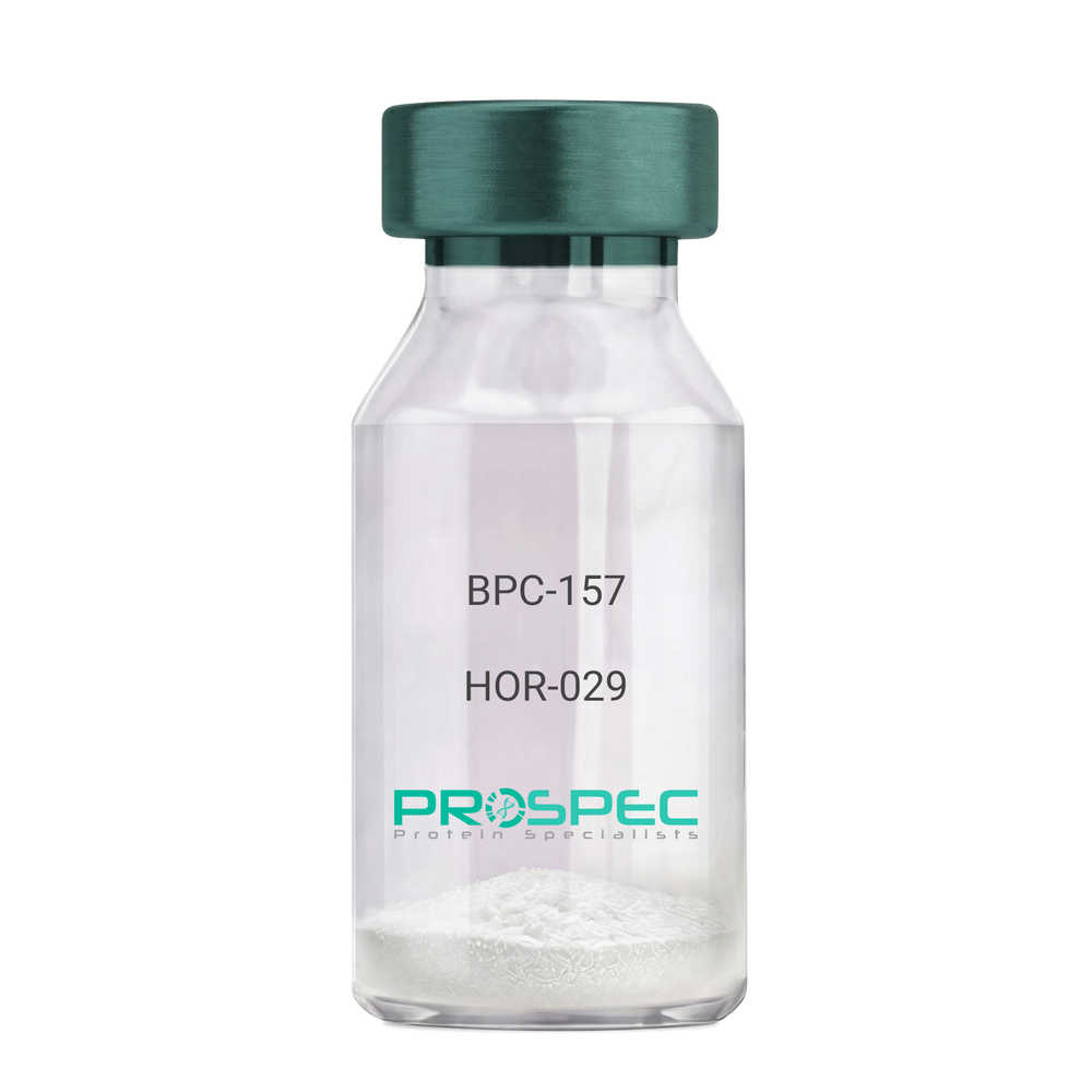
BPC-157Description:
BPC-157 Pentadecapeptide
Product # :
HOR-029Price :
Quantity :
Shipping Method :
Shipped at Room temp
More Info
- description
- formulation
- purity
- More Info
Description
BPC-157 Synthetic is a single, non-glycosylated polypeptide chain containing 15 amino acids, having a molecular mass of 1419.55 Dalton and a Molecular formula of C62H98N16O22.
Formulation
The protein was lyophilized with no additives.
Purity
Greater than 97.0% as determined by analysis by RP-HPLC.
More Info
-
Physical Appearance
Sterile Filtered White lyophilized (freeze-dried) powder.
-
Stability
Lyophilized BPC-157 although stable at room temperature for 3 weeks, should be stored desiccated below -18°C. Upon reconstitution bpc-157 should be stored at 4°C between 2-7 days and for future use below -18°C. For long term storage it is recommended to add a carrier protein (0.1% HSA or BSA). Please prevent freeze-thaw cycles.
-
Solubility
It is recommended to reconstitute the lyophilized BPC-157 in sterile 18MΩ-cm H2O not less than 100 µg/ml, which can then be further diluted to other aqueous solutions.
-
Amino Acid Sequence
H-Gly-Glu-Pro-Pro-Pro-Gly-Lys-Pro-Ala-Asp-Asp-Ala-Gly-Leu-Val-OH
-
Background
BPC-157, short for Body Protection Compound-157, is a synthetic peptide that has garnered significant attention in the field of regenerative medicine and sports science. This peptide, derived from a portion of the human gastric juice protein known as BPC, exhibits remarkable healing and tissue regeneration properties. BPC-157 has shown promise in various preclinical and clinical studies, demonstrating its potential for the treatment of a wide range of injuries and disorders.
The research on BPC-157 encompasses investigations into its mechanisms of action, efficacy, safety, and potential therapeutic applications. Studies have elucidated the peptide's ability to enhance angiogenesis, promote collagen synthesis, modulate inflammatory responses, and protect against oxidative stress. These properties make BPC-157 an intriguing candidate for accelerating tissue healing, reducing inflammation, and improving overall recovery outcomes.
Preclinical studies have revealed the beneficial effects of BPC-157 in several injury models. For instance, BPC-157 has demonstrated its potential in accelerating tendon and ligament healing, mitigating muscle damage, and promoting bone regeneration. These findings suggest that BPC-157 could be a valuable therapeutic tool in orthopedic medicine and sports-related injuries.
Furthermore, BPC-157 has exhibited promising effects on gastrointestinal health. Studies have highlighted its ability to protect and heal the gut lining, reduce ulcer formation, and alleviate symptoms associated with inflammatory bowel disease. These observations open up avenues for BPC-157 as a potential treatment for gastrointestinal disorders.
In addition to its regenerative properties, BPC-157 has shown potential in neurological and psychiatric conditions. Research has indicated its neuroprotective effects, with implications for the treatment of traumatic brain injury, stroke, and neurodegenerative disorders. Preliminary studies also suggest BPC-157's potential as an antidepressant and anxiolytic agent.
Despite the promising findings, further research is needed to fully understand the mechanisms underlying BPC-157's actions and to assess its long-term safety and efficacy. Clinical trials are underway to explore its potential therapeutic applications in humans, including its use in tendon and ligament repair, inflammatory bowel disease, and neurodegenerative disorders.
This comprehensive review aims to summarize the current state of research on BPC-157, providing an overview of its mechanisms of action and therapeutic potential. By examining relevant studies and findings, we aim to shed light on the diverse applications of BPC-157 and its implications for regenerative medicine, sports science, and various disease
ProSpec's products are furnished for LABORATORY RESEARCH USE ONLY. The product may not be used as drugs, agricultural or pesticidal products, food additives or household chemicals.
- View Data Sheet
Name :
HCG-b core antibodyDescription:
Human Chorionic Gonadotropin beta core, Mouse antibody
Chorionic gonadotropin beta polypeptide, CGB3, hCGB,Choriogonadotropin subunit beta, CG-beta, Chorionic gonadotrophin chain beta.
Product # :
ANT-147Price :
Quantity :
Shipping Method :
Shipped at Room temp
More Info
- formulation
- More Info
Formulation
1mg/ml in PBS (after reconstitution).
More Info
-
Introduction
Human chorionic gonadotropin (hCG) is a peptide hormoneproduced in pregnancy, that is made by the embryosoon after conceptionand later by the syncytiotrophoblast(part of the placenta). Its role is to prevent the disintegration of the corpus luteumof the ovaryand thereby maintain progesteroneproduction that is critical for a pregnancyin humans. hCG may have additional functions, for instance it is thought that it affects the immune tolerance of the pregnancy. Early pregnancy testinggenerally is based on the detection or measurement of hCG.
-
Synonyms
Chorionic gonadotropin beta polypeptide, CGB3, hCGB,Choriogonadotropin subunit beta, CG-beta, Chorionic gonadotrophin chain beta.
-
Solubility
Reconstitute with H20. Mix gently, wash the sides of the vial and wait 30-60 seconds before use.
-
Immunogen
purified hCG beta core.
-
Ig Subclass
mouse IgG2b.
-
Clone
NYRhCG.
-
Note
Minimal cross-reactivity with human Luteinizing Hormone b-Core fragment.
-
Titer
By direct ELISA, 1:10,000 dilution will yield 0.5 O.D using alkaline phosphatase conjugated rabbit anti-mouse Ig (Jackson Laboratories).
-
Shipping Conditions
Antibody is shipped lyophilized at ambient temperature.
-
Type
Mouse Antibody Monoclonal.
-
Storage Procedures
In lyophilized form, for long periods, store at 4°C in a dry environment. After reconstitution, if not intended for use within a month, aliquot and store at -20°C.
-
Purification Method
Ion exchange column.
ProSpec's products are furnished for LABORATORY RESEARCH USE ONLY. The product may not be used as drugs, agricultural or pesticidal products, food additives or household chemicals.
- View Data Sheet
Name :
LGALS14 HumanDescription:
Galectin-14 Human Recombinant
Placental protein 13-like, Charcot-Leyden crystal protein 2, CLC2, Galectin-14, Gal-14, LGALS14, PPL13, MGC22235.
Product # :
CYT-003Price :
Quantity :
Shipping Method :
Shipped with Ice Packs
More Info
- description
- source
- formulation
- purity
- More Info
Description
LGALS14 Human Recombinant fused with a 23 amino acid His tag at N-terminus produced in E.Coli is a single, non-glycosylated, polypeptide chain containing 162 amino acids (1-139 a.a.) and having a molecular mass of 18.5kDa. The LGALS14 is purified by proprietary chromatographic techniques.
Source
Escherichia Coli.
Formulation
The LGALS14 solution (0.5mg/ml) contains 20mM Tris-HCl buffer (pH8.0), 20% glycerol, 0.1M NaCl and 1mM DTT.
Purity
Greater than 90.0% as determined by SDS-PAGE.
More Info
-
Introduction
Galectin14, aka LGALS14, is a member of the galectin family of carbohydrate binding proteins. Galectin family members contain one or two carbohydrate recognition domains, which can bind beta-galactoside. The LGALS14 gene is predominantly expressed in the placenta. LGALS14 is expressed intracellularly in the placenta and eosinophils and is released by eosinophils following allergen stimulation. LGALS14 may be involved in the development of allergic inflammation.
-
Synonyms
Placental protein 13-like, Charcot-Leyden crystal protein 2, CLC2, Galectin-14, Gal-14, LGALS14, PPL13, MGC22235.
-
Physical Appearance
Sterile Filtered colorless solution.
-
Stability
Store at 4°C if entire vial will be used within 2-4 weeks. Store, frozen at -20°C for longer periods of time. For long term storage it is recommended to add a carrier protein (0.1% HSA or BSA).Avoid multiple freeze-thaw cycles.
-
Amino Acid Sequence
MGSSHHHHHH SSGLVPRGSH MGSMSSLPVP YTLPVSLPVG SCVIITGTPI LTFVKDPQLE VNFYTGMDED SDIAFQFRLH FGHPAIMNSR VFGIWRYEEK CYYLPFEDGK PFELCIYVRH KEYKVMVNGQ RIYNFAHRFP PASVKMLQVL RDISLTRVLI SD.
-
Background
What is the molecular weight/Mw of LGALS14 HUMAN Protein?
LGALS14 HUMAN Protein has a total Mw of 18.5kDa.
What is the source or expression system of LGALS14 HUMAN Protein?
Escherichia Coli.
What is the Purity of LGALS14 HUMAN Protein?
LGALS14 HUMAN Protein is >90% pure as determined by SDS-PAGE.
What is the Biological Activity of LGALS14 HUMAN Protein?
The biological functionality of LGALS14 HUMAN Protein will be determined in the future.
What is the amino acid sequence of LGALS14 HUMAN Protein?
MGSSHHHHHH SSGLVPRGSH MGSMSSLPVP YTLPVSLPVG SCVIITGTPI LTFVKDPQLE VNFYTGMDED SDIAFQFRLH FGHPAIMNSR VFGIWRYEEK CYYLPFEDGK PFELCIYVRH KEYKVMVNGQ RIYNFAHRFP PASVKMLQVL RDISLTRVLI SD.
What applications can LGALS14 HUMAN Protein be used in?
LGALS14 HUMAN Protein can probably be used in western blot, ELISA and Lateral Flow.
What is the endotoxin level for LGALS14 HUMAN Protein?
The endotoxin level is minimal, LGALS14 HUMAN Protein was purified using conventional chromatography techniques.
ProSpec's products are furnished for LABORATORY RESEARCH USE ONLY. The product may not be used as drugs, agricultural or pesticidal products, food additives or household chemicals.
- View Data Sheet
Name :
MMP 3 Human, HEKDescription:
Matrix Metalloproteinase-3 Human Recombinant, HEK
Stromelysin-1, EC 3.4.24.17, Matrix metalloproteinase-3, MMP-3, Transin-1, SL-1, STMY, STR1, STMY1, MGC126102, MGC126103, MGC126104.
Product # :
ENZ-284Price :
Quantity :
Shipping Method :
Shipped with Ice Packs
More Info
- description
- source
- formulation
- purity
- biological activity
- More Info
- sds-page
Description
MMP-3 Human Recombinant produced in HEK293 cells is a proform of the Human MMP3 [Tyr18-Cys477 (Lys45Glu)] and fused with a ployhistide tag at the C-terminus, having an Mw of 52kDa. MMP-3 is purified by proprietary chromatographic techniques.
Source
HEK293 cells.
Formulation
The MMP-3 is supplied as a 0.2µm filtered solution in 20mM Tris-HCl, 150mM NaCl and 0.05% Brij35, pH 7.5.
Purity
Greater than 95% as determined by SDS-PAGE.
Biological Activity
The activity was measured by its ability to cleave the fluorogenic peptide substrate, Mca-RPKPVE-Nval-WRK(Dnp)-NH2. The specific activity is > 150 pmoles/min/µg.
Recombinant Human MMP-3 protein pro form needs to be activated with Chymotrypsin.
Activation Protocol:
1. Dilute MMP3 to 20µg/ml in the Assay Buffer: 50mM Tris, 10mM CaCl2, 150mM NaCl, 0.05% (w/v) and Brij 35, pH 7.5.
2. Activate MMP3 by adding Chymotrypsin(Sigma, Catalog#C3142,1mg/ml stock in 1mM HCl) to a final concentration of 5ug/ml.
3. Incubate at 37°C for 30 minutes.
4. Stop activation with 2mM PMSF. Pre-warm the PMSF to 37°C prior to adding to sample.sds-page
More Info
-
Introduction
MMP-3 enzyme is also known as Stromelysin-1or as Transin-1 which hydrolyzes natural collagen at physiological pH and temperature. It dissolves the intervertebral nucleus pulposus and annulus fibrosus of Herniated Lumbar Intervertebral Disk . MMP-3 hydrolyzes components of the extracellular matrix like proteoglycan, laminin, fibronectin, gelatin and collagen types III, IV and IX. It also activates pro-MMP-9 and pro-MMP-8 and superactivates plasmin activated MMP-1. MMP-3 is secreted as a latent proenzyme and is activated by a variety of proteinases, e.g. plasmin, trypsin, chymotrypsin, cathepsin G or human neutrophil elastase. MMP-3 was found to be capable of activating the precursor of IL1-beta.
-
Synonyms
Stromelysin-1, EC 3.4.24.17, Matrix metalloproteinase-3, MMP-3, Transin-1, SL-1, STMY, STR1, STMY1, MGC126102, MGC126103, MGC126104.
-
Physical Appearance
Sterile Filtered colorless solution.
-
Stability
Store at 4°C if entire vial will be used within 2-4 weeks. Store, frozen at -20°C for longer periods of time.Avoid multiple freeze-thaw cycles.
ProSpec's products are furnished for LABORATORY RESEARCH USE ONLY. The product may not be used as drugs, agricultural or pesticidal products, food additives or household chemicals.