Search results
1000 results found for “killer cell”
Name
Description
Product #
Price
Quantity
Shipping Method
- View Data Sheet
Name :
NRG1 B1 HumanDescription:
Neuregulin-1/Heregulin-b1 Human Recombinant
Neuregulin-1, Heregulin-b1, NRG1-B1, NRG1 B1.
Product # :
CYT-733Price :
Quantity :
Shipping Method :
Shipped at Room temp
More Info
- description
- source
- formulation
- purity
- biological activity
- More Info
Description
Recombinant Human Neuregulin-1/Heregulin-b1 produced in E.Coli is a single, non-glycosylated, polypeptide chain (a.a 177-241) containing 65 amino acids and having a total molecular mass of 7.5kDa. NRG1-B1 is purified by proprietary chromatographic techniques.
Source
Escherichia Coli.
Formulation
Lyophilized from a 0.2µm filtered solution in 1×PBS, pH 7.4 and 5% trehalose.
Purity
Greater than 97.0% as determined by:
(a) Analysis by RP-HPLC.
(b) Analysis by SDS-PAGE.Biological Activity
The ED50 was determined by the dose-dependent stimulation of the proliferation of human MCF-7 cells is less than 0.5ng/ml, corresponding to a specific activity of >2.0×10⁶ units/mg.More Info
-
Introduction
Neuregulin/Heregulin is a family of structurally related polypeptide growth factors which are stemmed from alternatively spliced genes (NRG1, NRG2, NRG3 and NRG4). Thus far, there are more than 14 soluble and transmembrane proteins derived from the NRG1 gene. Proteolytic processing of the extracellular domain of the transmembrane NRG1 isoforms release soluble growth factors. HRG1-b1 is comprised of an Ig domain and an EGF-like domain which is necessary for direct binding to receptor tyrosine kinases erb3 and erb4. This binding stimulates erb3 and erb4 heterodimerization with erb2, promoting intrinsic kinase activity, which results in tyrosine phosphorylation. Even though HRG1-b1 biological effects are still unclear, it has been discovered to advance motility and invasiveness of breast cancer cells which in addition might entail up-regulation of expression and function of the autocrine motility-promoting factor (AMF).
-
Synonyms
Neuregulin-1, Heregulin-b1, NRG1-B1, NRG1 B1.
-
Physical Appearance
Sterile Filtered White lyophilized (freeze-dried) powder.
-
Stability
Lyophilized NRG1-B1 although stable at room temperature for 3 weeks, should be stored desiccated below -18°C. Upon reconstitution NRG1-B1 should be stored at 4°C between 2-7 days and for future use below -18°C. Please prevent freeze-thaw cycles.
-
Solubility
It is recommended to reconstitute the lyophilized NRG1-B1 in sterile 18M-cm H2O not less than 100µg/ml, which can then be further diluted to other aqueous solutions.
-
Amino Acid Sequence
SHLVKCAEKE KTFCVNGGEC FMVKDLSNPS RYLCKCPNEF TGDRCQNYVM ASFYKHLGIE FMEAE.
ProSpec's products are furnished for LABORATORY RESEARCH USE ONLY. The product may not be used as drugs, agricultural or pesticidal products, food additives or household chemicals.
- View Data Sheet
Name :
OPG Human, Hi-5Description:
Osteoprotegerin Human Recombinant, Hi-5
TNFRSF11B, OPG, OCIF, Osteoclastogenesis inhibitory factor, Osteoprotegerin, TR1, MGC29565.
Product # :
CYT-633Price :
Quantity :
Shipping Method :
Shipped with Ice Packs
More Info
- description
- source
- formulation
- purity
- biological activity
- More Info
Description
Recombinant Osteoprotegerin produced in baculovirus is a signle, glycosylated polypeptide chain containing 389 amino acid residues (22-401 a.a.), having a calculated molecular mass of 44.7 kDa. The Osteoprotegerin is fused to a 6 amino acid His tag at C-terminus and purified by proprietary chromatographic techniques.
Source
Hi-5.
Formulation
Osteoprotegerin protein contains PBS pH-7.4 and 10% glycerol.
Purity
Greater than 90.0% as determined by SDS-PAGE.
Biological Activity
The ED50 range ≤ 8 ng/ml and is measured by its ability to inhibit cytotoxicity using Jurkat human acute T cell leukemia cells in the presence of 2ng/ml of human TRAIL (CAT# cyt-1014).
More Info
-
Introduction
Osteoprotegerin acts as decoy receptor for rankl and thereby neutralizes its function in osteoclastogenesis. OPG inhibits the activation of osteoclasts and promotes osteoclast apoptosis in vitro. Bone homeostasis seems to depend on the local rankl/opg ratio. Osteoprotegerin may also play a role in preventing arterial calcification. May act as decoy receptor for trail and protect against apoptosis. Trail binding blocks the inhibition of osteoclastogenesis.
-
Synonyms
TNFRSF11B, OPG, OCIF, Osteoclastogenesis inhibitory factor, Osteoprotegerin, TR1, MGC29565.
-
Physical Appearance
Sterile Filtered colorless clear solution.
-
Stability
Store at 4°C if entire vial will be used within 2-4 weeks. Store, frozen at -20°C for longer periods of time. Please avoid freeze thaw cycles.
-
Amino Acid Sequence
ADPETFPPKY LHYDEETSHQ LLCDKCPPGT YLKQHCTAKW KTVCAPCPDH YYTDSWHTSD ECLYCSPVCK ELQYVKQECN RTHNRVCECK EGRYLEIEFC LKHRSCPPGF GVVQAGTPER NTVCKRCPDG FFSNETSSKA PCRKHTNCSV FGLLLTQKGN ATHDNICSGN SESTQKCGID VTLCEEAFFR FAVPTKFTPN WLSVLVDNLP GTKVNAESVE RIKRQHSSQE QTFQLLKLWK HQNKDQDIVK KIIQDIDLCE NSVQRHIGHA NLTFEQLRSL MESLPGKKVG AEDIEKTIKA CKPSDQILKL LSLWRIKNGD QDTLKGLMHA LKHSKTYHFP KTVTQSLKKT IRFLHSFTMY KLYQKLFLEM IGNQVQSVKI SCLSHHHHHH.
ProSpec's products are furnished for LABORATORY RESEARCH USE ONLY. The product may not be used as drugs, agricultural or pesticidal products, food additives or household chemicals.
- View Data Sheet
Name :
KXD1 HumanDescription:
KxDL Motif Containing 1 Human Recombinant
C19orf50, KXDL, MST096, MSTP096, KXD1.
Product # :
PRO-1656Price :
Quantity :
Shipping Method :
Shipped with Ice Packs
More Info
- description
- source
- formulation
- purity
- More Info
Description
KXD1 Human Recombinant produced in E.Coli is a single, non-glycosylated polypeptide chain containing 199 amino acids (1-176 a.a.) and having a molecular mass of 22.1kDa.KXD1 is fused to a 23 amino acid His-tag at N-terminus & purified by proprietary chromatographic techniques.
Source
Escherichia Coli.
Formulation
KXD1 protein solution (0.5mg/ml) contains 20mM Tris-HCl buffer (pH 8.0), 0.15M NaCl, 10% glycerol and 1mM DTT.
Purity
Greater than 85.0% as determined by SDS-PAGE.
More Info
-
Introduction
KxDL Motif Containing 1 (KXD1) is a member of the KXD1 family. KXD1 is participating in endosomal cargo sorting. KXD1 contacts with the BLOC-1 complex and Interacts with BLOC1S1. KXD1 Interacts with DTNBP1/BLOC1S7 through coiled-coil domain.
-
Synonyms
C19orf50, KXDL, MST096, MSTP096, KXD1.
-
Physical Appearance
Sterile Filtered clear solution.
-
Stability
Store at 4°C if entire vial will be used within 2-4 weeks. Store, frozen at -20°C for longer periods of time. For long term storage it is recommended to add a carrier protein (0.1% HSA or BSA).Avoid multiple freeze-thaw cycles.
-
Amino Acid Sequence
MGSSHHHHHH SSGLVPRGSH MGSMDLPDSA SRVFCGRILS MVNTDDVNAI ILAQKNMLDR FEKTNEMLLN FNNLSSARLQ QMSERFLHHT RTLVEMKRDL DSIFRRIRTL KGKLARQHPE AFSHIPEASF LEEEDEDPIP PSTTTTIATS EQSTGSCDTS PDTVSPSLSP GFEDLSHVQP GSPAINGRSQ TDDEEMTGE.
ProSpec's products are furnished for LABORATORY RESEARCH USE ONLY. The product may not be used as drugs, agricultural or pesticidal products, food additives or household chemicals.
- View Data Sheet
Name :
ADH1C Human, sf9Description:
Alcohol Dehydrogenase 1C, Human Recombinant, sf9
ADH1, ADH1C, ADH3, Alcohol dehydrogenase 1C, Alcohol dehydrogenase subunit gamma.
Product # :
ENZ-1010Price :
Quantity :
Shipping Method :
Shipped with Ice Packs
More Info
- description
- source
- formulation
- purity
- More Info
Description
ADH1C Human Recombinant produced in Sf9 Baculovirus cells is a single, glycosylated polypeptide chain containing 381 amino acids (1-375) and having a molecular mass of 40.6kDa. ADH1C is fused to a 6 amino acid IgG His-Tag at C-terminus and purified by proprietary chromatographic techniques.
Source
Sf9, Baculovirus cells.
Formulation
ADH1C protein solution (1mg/ml) containing Phosphate Buffered Saline (pH 7.4) and 10% glycerol.
Purity
Greater than 95.0% as determined by analysis by SDS-PAGE.
More Info
-
Introduction
ADH1C is a member of the zinc-containing alcohol dehydrogenase family which metabolizes a large assortment of substrates, such as ethanol, retinol, other aliphatic alcohols, hydroxysteroids, and lipid peroxidation products. ADH1 is a monomorphic and a key factor in fetal and infant livers, becoming less active in gestation and only weakly active during adulthood.
-
Synonyms
ADH1, ADH1C, ADH3, Alcohol dehydrogenase 1C, Alcohol dehydrogenase subunit gamma.
-
Physical Appearance
Sterile Filtered clear solution.
-
Stability
Store at 4°C if entire vial will be used within 2-4 weeks. Store, frozen at -20°C for longer periods of time. For long term storage it is recommended to add a carrier protein (0.1% HSA or BSA).Avoid multiple freeze-thaw cycles.
-
Amino Acid Sequence
MSTAGKVIKC KAAVLWELKK PFSIEEVEVA PPKAHEVRIK MVAAGICRSD EHVVSGNLVT PLPVILGHEA AGIVESVGEG VTTVKPGDKV IPLFTPQCGK CRICKNPESN YCLKNDLGNP RGTLQDGTRR FTCSGKPIHH FVGVSTFSQY TVVDENAVAK IDAASPLEKV CLIGCGFSTG YGSAVKVAKV TPGSTCAVFG LGGVGLSVVM GCKAAGAARI IAVDINKDKF AKAKELGATE CINPQDYKKP IQEVLKEMTD GGVDFSFEVI GRLDTMMASL LCCHEACGTS VIVGVPPDSQ NLSINPMLLL TGRTWKGAIF GGFKSKESVP KLVADFMAKK FSLDALITNI LPFEKINEGF DLLRSGKSIR
TVLTFHHHHH H.
ProSpec's products are furnished for LABORATORY RESEARCH USE ONLY. The product may not be used as drugs, agricultural or pesticidal products, food additives or household chemicals.
- View Data Sheet
Name :
CSK AntibodyDescription:
C-Src Tyrosine Kinase, Mouse Anti Human
Tyrosine-protein kinase CSK, C-Src kinase, Protein-tyrosine kinase CYL, CSK, C-Src Tyrosine Kinase.
Product # :
ANT-693Price :
Quantity :
Shipping Method :
Shipped with Ice Packs
More Info
- formulation
- More Info
Formulation
1mg/ml containing PBS, pH-7.4, 10% Glycerol and 0.02% Sodium Azide.
More Info
-
Introduction
C-Src Tyrosine Kinase, also known as CSK, regulates cell growth, migration, differentiation, and immune response. CSK Phosphorylates tyrosine residues located in the C-terminal tails of Src-family kinases. CSK is a protein coding gene which suppresses signaling by several surface receptors. Among CSK's related pathways are PI-3K cascade and Signaling by FGFR.
-
Synonyms
Tyrosine-protein kinase CSK, C-Src kinase, Protein-tyrosine kinase CYL, CSK, C-Src Tyrosine Kinase.
-
Physical Appearance
Sterile filtered colorless solution.
-
Immunogen
Anti-human CSK mAb, is derived from hybridization of mouse F0 myeloma cells with spleen cells from BALB/c mice immunized with recombinant human CSK amino acids 1-450 purified from E. coli.
-
Ig Subclass
Mouse IgG2b heavy chain and κ light chain.
-
Clone
PAT7C6AT.
-
Applications
CSK antibody has been tested by ELISA, Western blot analysis and ICC/IF to assure specificity and reactivity. Since application varies, however, each investigation should be titrated by the reagent to obtain optimal results.
-
Type
Mouse Anti Human Monoclonal.
-
Storage Procedures
For periods up to 1 month store at 4°C, for longer periods of time, store at -20°C. Prevent freeze thaw cycles.
-
Purification Method
CSK antibody was purified from mouse ascitic fluids by protein-A affinity chromatography.
ProSpec's products are furnished for LABORATORY RESEARCH USE ONLY. The product may not be used as drugs, agricultural or pesticidal products, food additives or household chemicals.
- View Data Sheet
Name :
AKT1 AntibodyDescription:
Protein Kinase B Alpha, Mouse Anti Human
V-Akt Murine Thymoma Viral Oncogene Homolog 1, Protein Kinase B Alpha, Proto-Oncogene C-Akt, RAC-PK-Alpha, EC 2.7.11.1, PKB Alpha, CWS6, PKB, RAC, RAC-Alpha Serine/Threonine-Protein Kinase, Rac Protein Kinase Alpha, Protein Kinase B, PKB-ALPHA, RAC-ALPHA, EC 2.7.11, AKT1m, PRKBA, AKT, RAC-alpha serine/threonine-protein kinase.
Product # :
ANT-710Price :
Quantity :
Shipping Method :
Shipped with Ice Packs
More Info
- formulation
- More Info
Formulation
1mg/ml containing PBS, pH-7.4, 10% Glycerol and 0.02% Sodium Azide.
More Info
-
Introduction
Akt1, also known as Akt or else protein kinase B (PKB) is an important molecule in mammaliancellular signaling. In humans, there are three genes in the "Akt family": Akt1, Akt2, and Akt3. Moreover, these enzymes are members of the serine/threonine-specific protein kinase family (EC2.7.11.1). Akt1 is involved in cellular survival pathways, by inhibiting apoptoticprocesses. Akt1 is also able to induce protein synthesis pathways, and is therefore a key signaling protein in the cellular pathways which lead to skeletal muscle hypertrophy, and general tissue growth. Since it can block apoptosis, and thereby promote cell survival, Akt1 has been implicated as a most important factor in numerous types of cancer. Akt (now also called Akt1) was at first identified as the oncogenein the transforming retrovirus, AKT8.
-
Synonyms
V-Akt Murine Thymoma Viral Oncogene Homolog 1, Protein Kinase B Alpha, Proto-Oncogene C-Akt, RAC-PK-Alpha, EC 2.7.11.1, PKB Alpha, CWS6, PKB, RAC, RAC-Alpha Serine/Threonine-Protein Kinase, Rac Protein Kinase Alpha, Protein Kinase B, PKB-ALPHA, RAC-ALPHA, EC 2.7.11, AKT1m, PRKBA, AKT, RAC-alpha serine/threonine-protein kinase.
-
Physical Appearance
Sterile filtered colorless solution.
-
Immunogen
Anti-human AKT1 mAb, is derived from hybridization of mouse F0 myeloma cells with spleen cells from BALB/c mice immunized with recombinant human AKT1 amino acids 1-480 purified from E. coli.
-
Ig Subclass
Mouse IgG2a heavy chain and κ light chain.
-
Clone
PAT6D5AT.
-
Applications
AKT1 antibody has been tested by ELISA and Western blot analysis to assure specificity and reactivity. Since application varies, however, each investigation should be titrated by the reagent to obtain optimal results.
-
Type
Mouse Anti Human Monoclonal.
-
Storage Procedures
For periods up to 1 month store at 4°C, for longer periods of time, store at -20°C. Prevent freeze thaw cycles.
-
Purification Method
AKT1 antibody was purified from mouse ascetic fluids by protein-A affinity chromatography.
ProSpec's products are furnished for LABORATORY RESEARCH USE ONLY. The product may not be used as drugs, agricultural or pesticidal products, food additives or household chemicals.
- View Data Sheet
Name :
STK16 AntibodyDescription:
Serine/Threonine Kinase 16, Mouse Anti Human
Serine/threonine-protein kinase 16, Myristoylated and palmitoylated serine/threonine-protein kinase, MPSK, Protein kinase PKL12, TGF-beta-stimulated factor 1, TSF-1, Tyrosine-protein kinase STK16, hPSK, STK16, MPSK1, PKL12, TSF1, KRCT.
Product # :
ANT-521Price :
Quantity :
Shipping Method :
Shipped with Ice Packs
More Info
- formulation
- More Info
Formulation
1mg/ml containing PBS, pH-7.4, 10% Glycerol and 0.02% Sodium Azide.
More Info
-
Introduction
Serine/threonine-protein kinase 16 (STK16) is a membrane-associated protein kinase which phosphorylates on serine and threonine residues. STK16 is involved in secretory vesicle trafficking or intracellular signaling. Furthermore, the STK16 protein may have a role in regulating stromal-epithelial interactions which occur during ductal morphogenesis in the mammary gland. STK16 can autophosphorylate on Tyr residue; it is however unclear whether STK16 has tyrosine-protein kinase toward other proteins. STK16 may also be involved in TGF-beta signaling.
-
Synonyms
Serine/threonine-protein kinase 16, Myristoylated and palmitoylated serine/threonine-protein kinase, MPSK, Protein kinase PKL12, TGF-beta-stimulated factor 1, TSF-1, Tyrosine-protein kinase STK16, hPSK, STK16, MPSK1, PKL12, TSF1, KRCT.
-
Physical Appearance
Sterile filtered colorless solution.
-
Immunogen
Anti-human STK16 mAb, clone PAT4A1AT, is derived from hybridization of mouse F0 myeloma cells with spleen cells from BALB/c mice immunized with a recombinant human STK16 protein 1-305 amino acids purified from E. coli.
-
Ig Subclass
Mouse IgG1 heavy chain and k light chain.
-
Clone
PAT4A1AT.
-
Applications
The antibody has been tested by ELISA, Western blot analysis to assure specificity and reactivity. Since application varies, however, each investigation should be titrated by the reagent to obtain optimal results. Recommended starting dilution is 1:1000.
-
Type
Mouse Anti Human Monoclonal.
-
Storage Procedures
For periods up to 1 month store at 4°C, for longer periods of time, store at -20°C. Prevent freeze thaw cycles.
-
Purification Method
STK16 antibody was purified from mouse ascitic fluids by protein-A affinity chromatography.
ProSpec's products are furnished for LABORATORY RESEARCH USE ONLY. The product may not be used as drugs, agricultural or pesticidal products, food additives or household chemicals.
- View Data Sheet
Name :
TNFRSF10C AntibodyDescription:
TRAIL Receptor-3, Mouse Anti Human
Tumor Necrosis Factor Receptor Superfamily, Member 10c, Decoy Without An , Intracellular Domain, TRAILR3, DCR1, TRID, TNF-Related Apoptosis-Inducing Ligand Receptor, Antagonist Decoy Receptor For TRAIL/Apo-2L, Decoy TRAIL Receptor Without Death Domain, Lymphocyte Inhibitor Of TRAIL, Decoy Receptor,TRAIL-R, LIT,Tumor Necrosis Factor Receptor Superfamily Member 10C, TRAIL Receptor Without an Intracellular Domain, Cytotoxic TRAIL Receptor, TRAIL Receptor, CD263 Antigen, DCR1-TNFR, CD263.
Product # :
ANT-741Price :
Quantity :
Shipping Method :
Shipped with Ice Packs
More Info
- formulation
- More Info
Formulation
1mg/ml containing PBS, pH-7.4, 10% Glycerol and 0.02% Sodium Azide.
More Info
-
Introduction
TRAIL Receptor-3, also known as TNFRSF10C belongs to theTNF-receptor super family. TNFRSF10C contains an extracellular TRAIL-binding domain as well as a transmembranedomain, however no cytoplasmic death domain. Furthermore, TNFRSF10C is not capable of inducing apoptosis, and is considered toact as an antagonistic receptor which protects cells from TRAIL-induced apoptosis.
-
Synonyms
Tumor Necrosis Factor Receptor Superfamily, Member 10c, Decoy Without An , Intracellular Domain, TRAILR3, DCR1, TRID, TNF-Related Apoptosis-Inducing Ligand Receptor, Antagonist Decoy Receptor For TRAIL/Apo-2L, Decoy TRAIL Receptor Without Death Domain, Lymphocyte Inhibitor Of TRAIL, Decoy Receptor,TRAIL-R, LIT,Tumor Necrosis Factor Receptor Superfamily Member 10C, TRAIL Receptor Without an Intracellular Domain, Cytotoxic TRAIL Receptor, TRAIL Receptor, CD263 Antigen, DCR1-TNFR, CD263.
-
Physical Appearance
Sterile filtered colorless solution.
-
Immunogen
Anti-human TNFRSF10C mAb, is derived from hybridization of mouse SP2/0 myeloma cells with spleen cells from BALB/c mice immunized with Recombinant human TNFRSF10C amino acids 26-236 purified from E. coli.
-
Ig Subclass
Mouse IgG1 heavy chain and κ light chain.
-
Clone
PAT1E12AT.
-
Applications
The antibody has been tested by ELISA, Western blot analysis, Flow cytometry and ICC/IF to assure specificity and reactivity. Since application varies, however, each investigation should be titrated by the reagent to obtain optimal results.
-
Type
Mouse Anti Human Monoclonal.
-
Storage Procedures
For periods up to 1 month store at 4°C, for longer periods of time, store at -20°C. Prevent freeze thaw cycles.
-
Purification Method
TNFRSF10C antibody was purified from mouse ascitic fluids by protein-A affinity chromatography.
ProSpec's products are furnished for LABORATORY RESEARCH USE ONLY. The product may not be used as drugs, agricultural or pesticidal products, food additives or household chemicals.
- View Data Sheet
Name :
PPIF AntibodyDescription:
Cyclophilin-F, Mouse Anti Human
Oeptidylprolyl Isomerase F, PPIF, CYP-D, CYP3, SnuCyp-20, Peptidyl-prolyl cis-trans isomerase mitochondrial, Cyclophilin F, FLJ90798, MGC117207, peptidylprolyl isomerase F.
Product # :
ANT-389Price :
Quantity :
Shipping Method :
Shipped with Ice Packs
More Info
- formulation
- More Info
Formulation
1mg/ml containing PBS, pH-7.4, & 0.1% Sodium Azide.
More Info
-
Introduction
PPIF is a part of the peptidyl-prolyl cis-trans isomerase (PPIase) family. PPIF accelerates the folding of proteins. It catalyzes the cis-trans isomerization of proline imidic peptide bonds in oligopeptides. PPIF is key component of the mitochondrial permeability transition pore in the inner mitochondrial membrane. Activation of this pore is thought to be involved in the induction of apoptotic and necrotic cell death.
-
Synonyms
Oeptidylprolyl Isomerase F, PPIF, CYP-D, CYP3, SnuCyp-20, Peptidyl-prolyl cis-trans isomerase mitochondrial, Cyclophilin F, FLJ90798, MGC117207, peptidylprolyl isomerase F.
-
Immunogen
Anti-human Cyclophilin-F mAb is derived from hybridization of mouse F0 myeloma cells with spleen cells from BALB/c mice immunized with recombinant human Cyclophilin-F amino acids 30-207 purified from E. coli.
-
Ig Subclass
Mouse IgG2b heavy chain and κ light chain.
-
Clone
PAT1F5AT.
-
Applications
Cyclophilin-F antibody has been tested by ELISA, Western blot and immunohistochemistry analysis to assure specificity and reactivity. Since application varies, however, each investigation should be titrated by the reagent to obtain optimal results. Recommended dilution range for Western blot is 1:500~1:1,000 and immunohistochemistry analysis is 1:50~100. Recommended starting dilution for Western blot is 1:500 and Immunohistochemistry is 1:50.
-
Type
Mouse Anti Human Monoclonal.
-
Storage Procedures
For periods up to 1 month store at 4°C, for longer periods of time, store at -20°C. Prevent freeze thaw cycles.
-
Purification Method
Cyclophilin-F antibody was purified from mouse ascitic fluids by protein-G affinity chromatography.
ProSpec's products are furnished for LABORATORY RESEARCH USE ONLY. The product may not be used as drugs, agricultural or pesticidal products, food additives or household chemicals.
- View Data Sheet
Name :
PSMD11 PAT2C7AT AntibodyDescription:
26S proteasome non-ATPase regulatory subunit 11 clone PAT2C7AT Mouse Anti Human
26S proteasome non-ATPase regulatory subunit 11, 26S proteasome regulatory subunit S9, 26S proteasome regulatory subunit RPN6, 26S proteasome regulatory subunit p44.5, PSMD11, S9, Rpn6, p44.5, MGC3844.
Product # :
ANT-675Price :
Quantity :
Shipping Method :
Shipped with Ice Packs
More Info
- formulation
- More Info
Formulation
1mg/ml containing PBS, pH-7.4, 10% Glycerol and 0.02% Sodium Azide.
More Info
-
Introduction
The 26S proteasome is a multicatalytic proteinase complex with a well ordered structure composed of two complexes- a 20S core and a 19S regulator. PSMD11 is a non-ATPase subunit of the 19S regulator. The 20S core is composed of four rings of 28 non-identical subunits; two rings are composed of 7 alpha subunits and two rings are composed of 7 beta subunits. The 19S regulator is composed of a base that contains six ATPase subunits and two non-ATPase subunits, and a lid that contains up to ten non-ATPase subunits. Proteasomes are spread throughout eukaryotic cells at a high concentration and cleave peptides in an ATP/ubiquitin-dependent process in a non-lysosomal pathway. An indispensable function of a modified proteasome, the immunoproteasome, is the processing of class I MHC peptides.
-
Synonyms
26S proteasome non-ATPase regulatory subunit 11, 26S proteasome regulatory subunit S9, 26S proteasome regulatory subunit RPN6, 26S proteasome regulatory subunit p44.5, PSMD11, S9, Rpn6, p44.5, MGC3844.
-
Physical Appearance
Sterile filtered colorless solution.
-
Immunogen
Anti-human PSMD11 mAb, is derived from hybridization of mouse F0 myeloma cells with spleen cells from BALB/c mice immunized with recombinant human PSMD11 PAT2C7AT amino acids 1-422 purified from E. coli.
-
Ig Subclass
Mouse IgG2b heavy chain and ? light chain.
-
Clone
PAT2C7AT.
-
Applications
PSMD11 antibody has been tested by ELISA, Western blot analysis, Flow cytometry and ICC/IF to assure specificity and reactivity. Since application varies, however, each investigation should be titrated by the reagent to obtain optimal results.
-
Type
Mouse Anti Human Monoclonal.
-
Storage Procedures
For periods up to 1 month store at 4°C, for longer periods of time, store at -20°C. Prevent freeze thaw cycles.
-
Purification Method
PSMD11 antibody was purified from mouse ascitic fluids by protein-A affinity chromatography.
ProSpec's products are furnished for LABORATORY RESEARCH USE ONLY. The product may not be used as drugs, agricultural or pesticidal products, food additives or household chemicals.
- View Data Sheet
Name :
Vimentin PAT5H8AT AntibodyDescription:
Vimentin PAT5H8AT, Mouse Anti Human
Vimentin, Vim, FLJ36605.
Product # :
ANT-682Price :
Quantity :
Shipping Method :
Shipped with Ice Packs
More Info
- formulation
- More Info
Formulation
1mg/ml containing PBS, pH-7.4, 10% Glycerol and 0.02% Sodium Azide.
More Info
-
Introduction
Vimentin expression in human malignant glioma cells depends on cellular density, algorithms of drug delivery and chemo/radio treatment. Vimentin and detyrosinated microtubules provide structural support for the extensive microtentacles observed in detached tumor cells and a mechanism to promote successful metastatic spread. Primary colorectal carcinomas display aberrant expression of vimentin, and have activated Notch and TGFbeta signaling pathways. Vimentin is a strong arterial substrate for transglutaminases. Transglutaminase-mediated vimentin dimerization results in a novel unifying pathway by which vasodilatory and remodeling responses may be regulated. Ablation of vimentin expression inhibits migration and invasion of colon and breast cancer cell lines. Vimentin is the main intermediate filament protein in mesenchymal cells and is therefore of value in the differential diagnosis of undifferentiated neoplasms.
-
Synonyms
Vimentin, Vim, FLJ36605.
-
Physical Appearance
Sterile filtered colorless solution.
-
Immunogen
Anti-human Vimentin mAb, is derived from hybridization of mouse F0 myeloma cells with spleen cells from BALB/c mice immunized with a recombinant human Vimentin protein 1-466 amino acids purified from E. coli.
-
Ig Subclass
Mouse IgG1 heavy chain and k light chain.
-
Clone
PAT5H8AT.
-
Applications
Vimentin antibody has been tested by ELISA, Western blot analysis, Flow cytometry and ICC/IF to assure specificity and reactivity. Since application varies, however, each investigation should be titrated by the reagent to obtain optimal results.
-
Type
Mouse Anti Human Monoclonal.
-
Storage Procedures
For periods up to 1 month store at 4°C, for longer periods of time, store at -20°C. Prevent freeze thaw cycles.
-
Purification Method
Vimentin antibody was purified from mouse ascitic fluids by protein-A affinity chromatography.
ProSpec's products are furnished for LABORATORY RESEARCH USE ONLY. The product may not be used as drugs, agricultural or pesticidal products, food additives or household chemicals.
- View Data Sheet
Name :
IL 13 Human, HisDescription:
Interleukin-13 Human Recombinant, His Tag
NC30, ALRH, BHR1, P600, IL-13, MGC116786, MGC116788, MGC116789.
Product # :
CYT-489Price :
Quantity :
Shipping Method :
Shipped with Ice Packs
More Info
- description
- source
- formulation
- purity
- More Info
Description
Interleukin-13 His Human Recombinant produced in E.Coli is a single, non-glycosylated, Polypeptide chain containing 112 amino acids fragment (21-132) having a molecular weight of 12.84kDa with a 4kDa amino-terminal hexahistidine tag (total weight of 16.84kDa). The IL-13 His is purified by proprietary chromatographic techniques.
Source
Escherichia Coli.
Formulation
Interleukin -13 His-Tag protein is supplied in 25mM Na-Acetate (pH 4.8) and 50% glycerol.
Purity
Greater than 95.0% as determined by SDS-PAGE.
More Info
-
Introduction
Interleukin-13 is a pleiotropic regulatory cytokine, which shares many of the properties of Interleukin-4. IL-13 is produced primarily by activated T lymphocytes and acts via specific cell surface receptor complexes. Its biological activities include the stimulation of B-cell proliferation and differentiation and the regulation of inflammatory and immune responses.
-
Synonyms
NC30, ALRH, BHR1, P600, IL-13, MGC116786, MGC116788, MGC116789.
-
Physical Appearance
Sterile Filtered clear solution.
-
Stability
Store at 4°C if entire vial will be used within 2-4 weeks. Store, frozen at -20°C for longer periods of time. Please avoid freeze thaw cycles.
ProSpec's products are furnished for LABORATORY RESEARCH USE ONLY. The product may not be used as drugs, agricultural or pesticidal products, food additives or household chemicals.
- View Data Sheet
Name :
CD9 HumanDescription:
CD9 Human Recombinant
CD9 antigen, 5H9 antigen, Cell growth-inhibiting gene 2 protein, Leukocyte antigen MIC3, Motility-related protein, CD9, MRP-1, Tetraspanin-29, Tspan-29, p24, MIC3, TSPAN29, GIG2, BTCC-1, DRAP-27, TSPAN-29.
Product # :
PRO-2175Price :
Quantity :
Shipping Method :
Shipped with Ice Packs
More Info
- description
- source
- formulation
- purity
- More Info
Description
CD9 Human Recombinant produced in E. coli is a single polypeptide chain containing 107 amino acids (112-195) and having a molecular mass of 12kDa.CD9 is fused to a 23 amino acid His-tag at N-terminus & purified by proprietary chromatographic techniques.
Source
Escherichia Coli.
Formulation
The CD9 solution (0.25mg/1ml) contains Phosphate buffer saline (pH 7.4), 20% glycerol and 1mM DTT.
Purity
Greater than 85.0% as determined by SDS-PAGE.
More Info
-
Introduction
CD9 which is found on the surface of exosomes is a cell surface glycoprotein which is interacts with integrins and various transmembrane superfamily proteins. CD9 is involved in platelet activation and aggregation and also regulates paranodal junction formation. CD9 takes part in cell adhesion and migration and also promotes muscle cell fusion. CD9 is necessary for the egg-sperm fusion during mammalian fertilization.
-
Synonyms
CD9 antigen, 5H9 antigen, Cell growth-inhibiting gene 2 protein, Leukocyte antigen MIC3, Motility-related protein, CD9, MRP-1, Tetraspanin-29, Tspan-29, p24, MIC3, TSPAN29, GIG2, BTCC-1, DRAP-27, TSPAN-29.
-
Physical Appearance
Sterile Filtered colorless solution.
-
Stability
Store at 4°C if entire vial will be used within 2-4 weeks. Store, frozen at -20°C for longer periods of time. For long term storage it is recommended to add a carrier protein (0.1% HSA or BSA).Avoid multiple freeze-thaw cycles.
-
Amino Acid Sequence
MGSSHHHHHH SSGLVPRGSH MGSSHKDEVI KEVQEFYKDT YNKLKTKDEP QRETLKAIHY ALNCCGLAGG VEQFISDICP KKDVLETFTV KSCPDAIKEV FDNKFHI.
ProSpec's products are furnished for LABORATORY RESEARCH USE ONLY. The product may not be used as drugs, agricultural or pesticidal products, food additives or household chemicals.
- View Data Sheet
Name :
HERV-KDescription:
Endogenous retrovirus K Envelope Human Recombinant
Product # :
HER-001Price :
Quantity :
Shipping Method :
Shipped with Ice Packs
More Info
- description
- source
- formulation
- purity
- More Info
Description
The E.Coli derived HERV-K recombinant truncated protein is fused to a Six histidine tag at C-terminus and has a MW of 51.5kDa (pI 9.06).
Source
Escherichia Coli.
Formulation
10mM Tris-HCl, pH 7.2.
Purity
Protein is >90% pure as determined by SDS PAGE.
More Info
-
Introduction
HERVK is related with several tumors such as passenger virus, and anti-Retroviral therapy against amyotrophic lateral sclerosis/ALS has exhibited to decrease the symptoms of amyotrophic lateral sclerosis/ALS. High counts of viral sequences exist in the human genome but stay silent. Though, in pathological conditions, these viruses are produced. Human Endogenous Retrovirus-K, is produced in neurons of a subpopulation of patients with amyotrophic lateral sclerosis/ALS, a progressive neurodegenerative disease. The envelope protein of this HERV results in degeneration of neurons, and transgenic animals producing this protein advance an ALS-like syndrome triggered by nucleolar dysfunction in motor neurons. Reactivation of the HERVK is controlled by the transcription factor TDP-43. Therefore, therapeutic tactics against this virus alter the course of the disease.
-
Physical Appearance
Sterile Filtered clear solution.
-
Stability
HERV-K although stable at 4°C for 1 week, should be stored below -18°C. Please prevent freeze thaw cycles.
-
Amino Acid Sequence
MWTVPSFTND SYQVYNVFST NSFQLLTVKR TPHEAWRVPL TTKTNKTKGL PDCPKKPTNG PFIVTSILWD NCNAPKAVVL QTLAMGIVID WAPKGHYWQD CSSKNTLCSE FIYSLDYIEH GWQSYTMRQR VSPYPFKWMD TGIAPPRPKI IHPFFTPEHP ELWKLAAALS GIKIWNTTYQ LLRTKTKTPT FNITLISEWV IPIRSCVKPP YMLLVGNIIM MPDAQTIECH NCKLFTCIDA TFNPTTSILL VRAREGVWIP VSLHRPWESS PSIHIVNEVL KDILKRTKRF IFTLIAVLAG LLAVTATAAT AGVAIRSSVQ TAHYVEACQK NSSRLWNSQA QIDQKLANQI NDLRQSVTWL GDRVMNLQHR MQLQCDWNTS DYCITPYAYN QDQHSWENVS RHLKAWDDNL TLDISQLKEQIFEASQAHLS TVPGSHIFEG ITKQLPDFNP FKWLKPVRGS LLLLALLILV CLCCLLLVCRCL.
-
Applications
Detection of Human Endogenious Retrovirus K in individuals is untested.
ProSpec's products are furnished for LABORATORY RESEARCH USE ONLY. The product may not be used as drugs, agricultural or pesticidal products, food additives or household chemicals.
- View Data Sheet
Name :
Dengue NS1 AntibodyDescription:
Monoclonal Mouse Anti Dengue NS1
Product # :
ANT-495Price :
Quantity :
Shipping Method :
Shipped with Ice Packs
More Info
- description
- formulation
- purity
- More Info
Description
Paired monoclonal antibodies developed for dengue NS1 antigen test by lateral flow immunoassay device. First antibody is for conjugation and the second antibody is for membrane coating. Please note that when ordering for example: 100µg antibody we ship 50µg from each of the antibody solutions (100µg in total).
Formulation
*Dengue NS1 Antibody solution for gold conjugation (3.6mg/ml) in PBS and Sodium nitrate.
*Dengue NS1 Antibody solution for membrane coating (2.2mg/ml) in PBS and Sodium nitrate.
Purity
95% pure (12% SDS-PAGE and Coomassie staining).
More Info
-
Physical Appearance
2 vials of sterile Filtered clear colorless solution.
-
Stability
Store at 4°C if entire vial will be used within 2-4 weeks. Store, frozen at -20°C for longer periods of time. For long term storage it is recommended to add a carrier protein (0.1% HSA or BSA).Avoid multiple freeze-thaw cycles.
-
Applications
Lateral flow immunoassay.
-
Assay Conditions
1. 1-2 days fresh prepared gold particle with maximal homogeneity in gold particle size, λ 525-529, OD 1.2-1.4. 2. Conjugation at pH 8.5, conjugation time should be 6-8 hours. 3. Blocking condition: remove supernatant and re-suspend gold pellet bound with anti-dengue NS1 to OD 8.0- 8.2, add casein to the final 0.3-0.4%. 4. Treat sample pad with 50mM sodium borate, 0.5% mouse serum and 0.5% Triton X-100, pH8.0.
-
Purification Method
Purified monoclonal IgG by protein A chromatography.
ProSpec's products are furnished for LABORATORY RESEARCH USE ONLY. The product may not be used as drugs, agricultural or pesticidal products, food additives or household chemicals.
- View Data Sheet
Name :
KLF7 HumanDescription:
Kruppel-Like Factor 7 Human Recombinant
UKLF, Krueppel-like factor 7, Ubiquitous krueppel-like factor, KLF7.
Product # :
PRO-1527Price :
Quantity :
Shipping Method :
Shipped with Ice Packs
More Info
- description
- source
- formulation
- purity
- More Info
Description
KLF7 Human Recombinant produced in E. coli is a single polypeptide chain containing 325 amino acids (1-302) and having a molecular mass of 35.8kDa. KLF7 is fused to a 23 amino acid His-tag at N-terminus & purified by proprietary chromatographic techniques.
Source
Escherichia Coli.
Formulation
The KLF7 solution (1mg/ml) contains 20mM Tris-HCl buffer (pH 8.0), 0.4M urea and 10% glycerol.
Purity
Greater than 85% as determined by SDS-PAGE.
More Info
-
Introduction
Kruppel-Like Factor 7 (KLF7) is a part of the Kruppel-like transcriptional regulator family whose members regulate cell proliferation, differentiation and survival and contain 3 C2H2 zinc fingers at the C-terminus that mediate binding to GC-rich sites. KLF7 contributes to the progression of type 2 diabetes by inhibiting hormone expression and secretion in pancreatic beta-cells and also by deregulating adipocytokine secretion in adipocytes.
-
Synonyms
UKLF, Krueppel-like factor 7, Ubiquitous krueppel-like factor, KLF7.
-
Physical Appearance
Sterile Filtered clear solution.
-
Stability
Store at 4°C if entire vial will be used within 2-4 weeks. Store, frozen at -20°C for longer periods of time. For long term storage it is recommended to add a carrier protein (0.1% HSA or BSA).Avoid multiple freeze-thaw cycles.
-
Amino Acid Sequence
MGSSHHHHHH SSGLVPRGSH MGSMDVLASY SIFQELQLVH DTGYFSALPS LEETWQQTCL ELERYLQTEP RRISETFGED LDCFLHASPP PCIEESFRRL DPLLLPVEAA ICEKSSAVDI LLSRDKLLSE TCLSLQPASS SLDSYTAVNQ AQLNAVTSLT PPSSPELSRH LVKTSQTLSA VDGTVTLKLV AKKAALSSVK VGGVATAAAA VTAAGAVKSG QSDSDQGGLG AEACPENKKR VHRCQFNGCR KVYTKSSHLK AHQRTHTGEK PYKCSWEGCE WRFARSDELT RHYRKHTGAK PFKCNHCDRC FSRSDHLALH MKRHI.
ProSpec's products are furnished for LABORATORY RESEARCH USE ONLY. The product may not be used as drugs, agricultural or pesticidal products, food additives or household chemicals.
- View Data Sheet
Name :
I TAC HumanDescription:
I-TAC Human Recombinant (CXCL11)
Small inducible cytokine B11, CXCL11, I-TAC, IP-9, H174, Beta-R1, chemokine (C-X-C motif) ligand 11, IP9, b-R1, SCYB11, SCYB9B, MGC102770.
Product # :
CHM-334Price :
Quantity :
Shipping Method :
Shipped at Room temp
More Info
- description
- source
- formulation
- purity
- biological activity
- More Info
Description
I-TAC Human Recombinant produced in E.Coli is a single, non-glycosylated, polypeptide chain containing 73 amino acids and having a molecular mass of 8300 Dalton. The I-TAC is purified by proprietary chromatographic techniques.
Source
Escherichia Coli.
Formulation
Lyophilized from a 0.2?m filtered concentrated (0.5mg/ml) solution in 20mM PB, pH 7.4, 100mM NaCl.
Purity
Greater than 97.0% as determined by:
(a) Analysis by RP-HPLC.
(b) Analysis by SDS-PAGE.Biological Activity
Determined by its ability to chemoattract human IL-2 activated T-Lymphocytes using a concentration range of 0.1-10.0 ng/ml.More Info
-
Introduction
Chemokine (C-X-C motif) ligand 11 (CXCL11) is a small cytokine belonging to the CXC chemokinen family that is also called inducible T-cell alpha chemoattractant (I-TAC) and (IP-9). I-TAC is highly expressed in peripheral blood leukocytes, pancreas and liver, with moderate levels in thymus, spleen and lung and low expression levels were in small intestine, placenta and prostate. Gene expression of CXCL11 is strongly induced by IFN-g and IFN-b, and weakly induced by IFN-a. The I-TAC chemokine elicits its effects on its target cells by interacting with the cell surface chemokine receptor CXCR3, with a higher affinity than do the other ligands for this receptor, CXCL9 and CXCL10. I-TAC is chemotactic for activated T cells. The CXCL11 gene is located on human chromosome 4 along with many other members of the CXC chemokine family.
-
Synonyms
Small inducible cytokine B11, CXCL11, I-TAC, IP-9, H174, Beta-R1, chemokine (C-X-C motif) ligand 11, IP9, b-R1, SCYB11, SCYB9B, MGC102770.
-
Physical Appearance
Sterile Filtered White lyophilized (freeze-dried) powder.
-
Stability
Lyophilized I-TAC although stable at room temperature for 3 weeks, should be stored desiccated below -18°C. Upon reconstitution I-TAC should be stored at 4°C between 2-7 days and for future use below -18°C. For long term storage it is recommended to add a carrier protein (0.1% HSA or BSA).Please prevent freeze-thaw cycles.
-
Solubility
It is recommended to reconstitute the lyophilized I-TAC in sterile 18MΩ-cm H2O not less than 100µg/ml, which can then be further diluted to other aqueous solutions.
-
Amino Acid Sequence
FPMFKRGRCLCIGPGVKAVKVADIEKASIMYPSNNCDKIEVIITLKENKGQRCLNPKSKQARLIIKKVERKNF.
ProSpec's products are furnished for LABORATORY RESEARCH USE ONLY. The product may not be used as drugs, agricultural or pesticidal products, food additives or household chemicals.
- View Data Sheet
Name :
IL-1 Alpha Rat, His ActiveDescription:
Interleukin-1 alpha Rat Recombinant, His Tag Active
Interleukin-1 alpha, IL-1 alpha.
Product # :
CYT-1100Price :
Quantity :
Shipping Method :
Shipped with Ice Packs
More Info
- description
- source
- formulation
- purity
- biological activity
- More Info
Description
IL1A Rat Recombinant produced in E.Coli is a single, non-glycosylated polypeptide chain containing 179 amino acids (115-270 a.a) and having a molecular mass of 20.2kDa.IL1A is fused to a 23 amino acid His-tag at N-terminus & purified by proprietary chromatographic techniques.
Source
E.coli.
Formulation
IL1A protein solution (1mg/ml) contains 10% glycerol & Phosphate Buffered Saline (pH 7.4).
Purity
Greater than 95% as determined by SDS-PAGE.
Biological Activity
More Info
-
Introduction
IL-1 alpha is produced by activated macrophages.IL-1 alpha stimulates thymocyte proliferation by inducing il-2 release, b-cell maturation and proliferation, and fibroblast growth factor activity. IL1A proteins take part in the inflammatory response, being identified as endogenous pyrogens, and stimulate the release of prostaglandin and collagenase from synovial cells.
-
Synonyms
Interleukin-1 alpha, IL-1 alpha.
-
Physical Appearance
Sterile Filtered colorless solution.
-
Stability
Store at 4°C if entire vial will be used within 2-4 weeks. Store, frozen at -20°C for longer periods of time. For long term storage it is recommended to add a carrier protein (0.1% HSA or BSA).Avoid multiple freeze-thaw cycles.
-
Amino Acid Sequence
MGSSHHHHHH SSGLVPRGSH MGSSAPHSFQ NNLRYKLIRI VKQEFIMNDS LNQNIYVDMD
RIHLKAASLN DLQLEVKFDM YAYSSGGDDSKYPVTLKVSN TQLFVSAQGE DKPVLLKEIP
ETPKLITGSE TDLIFFWEKI NSKNYFTSAA FPELLIATKE QSQVHLARGL PSMIDFQIS.
ProSpec's products are furnished for LABORATORY RESEARCH USE ONLY. The product may not be used as drugs, agricultural or pesticidal products, food additives or household chemicals.
- View Data Sheet
Name :
NAP 2 AntibodyDescription:
Natural anti-proliferative protein, Mouse-Anti Human
Platelet basic protein, PBP, Small inducible cytokine B7, CXCL7, Leukocyte-derived growth factor, LDGF, Macrophage-derived growth factor, MDGF, pro-platelet basic protein (chemokine (C-X-C motif) ligand 7), TC1, TC2, TGB, TGB1, B-TG1, CTAP3, NAP-2, SCYB7, THBGB, LA-PF4, THBGB1, Beta-TG, CTAPIII, CTAP-III.
Product # :
ANT-170Price :
Quantity :
Shipping Method :
Shipped at Room temp
More Info
- formulation
- More Info
Formulation
1mg/ml in PBS (after reconstitution).
More Info
-
Introduction
Chemokine (C-X-C motif) ligand (CXCL7) is a small cytokine belonging to the CXC chemokine family. It is a protein that is released in large amounts from platelets following their activation. It stimulates various processes including mitogenesis, synthesis of extracellular matrix, glucose metabolism and synthesis of plasminogen activator.
-
Synonyms
Platelet basic protein, PBP, Small inducible cytokine B7, CXCL7, Leukocyte-derived growth factor, LDGF, Macrophage-derived growth factor, MDGF, pro-platelet basic protein (chemokine (C-X-C motif) ligand 7), TC1, TC2, TGB, TGB1, B-TG1, CTAP3, NAP-2, SCYB7, THBGB, LA-PF4, THBGB1, Beta-TG, CTAPIII, CTAP-III.
-
Solubility
Reconstitute with sterile H20. Mix gently, wash the sides of the vial and wait 30-60 seconds before use.
-
Immunogen
r.Human NAP-2.
-
Ig Subclass
Mouse IgG1.
-
Clone
NYRhNAP-2.
-
Applications
Direct ELISA, Western Blot, Immuneprecipitation.
-
Note
This antibody will bind very well to protein A in a buffer (PBS) containing high salt concentration (3M Nacl).
-
Titer
In direct ELISA, using alkaline phosphatase goat anti-mouse Ig (Jackson Laboratories) 1:10,000 dilution will yield 0.5 O.D within 10 minutes.
-
Shipping Conditions
Antibody is shipped lyophilized at ambient temperature.
-
Type
Mouse Anti Human Monoclonal.
-
Storage Procedures
In lyophilized form, for long periods, store at 4oC in a dry environment. After reconstitution, if not intended for use within a month, aliquot and store at -20oC.
-
Purification Method
ion exchange.
ProSpec's products are furnished for LABORATORY RESEARCH USE ONLY. The product may not be used as drugs, agricultural or pesticidal products, food additives or household chemicals.
- View Data Sheet
Name :
CD57 Biotin AntibodyDescription:
CD57 Biotinylated, Mouse Anti-Human
NK1, CD57, HNK1, LEU7, NK-1, GLCATP, GLCUATP, B3GAT1.
Product # :
ANT-288Price :
Quantity :
Shipping Method :
Shipped at Room temp
More Info
- formulation
- More Info
Formulation
1mg/ml in PBS (after reconstitution).
More Info
-
Introduction
CD57 also called B3GAT1is part of the glucuronyltransferase gene family. CD57 exhibits strict acceptor specificity, recognizing nonreducing terminal sugars and their anomeric linkages. CD57 takes part in the glucuronyl transfer reaction during the biosynthesis of the carbohydrate epitope HNK-1.
-
Synonyms
NK1, CD57, HNK1, LEU7, NK-1, GLCATP, GLCUATP, B3GAT1.
-
Solubility
Reconstitute with of H20. Mix gently, wash the sides of the vial and wait 30-60 seconds before use.
-
Immunogen
Human CD57 Positive Cells.
-
Ig Subclass
Mouse IgM.
-
Clone
hCD57.
-
Applications
Staining antibody. For staining, use 10µl/1,000,000 cells.
-
Available Conjugates
This antibody is also available unconjugated and conjugated to FITC. For staining with biotin or FITC-conjugated antibody use 5-10µl/1,000,000 cells.
-
Type
Mouse Anti Human Monoclonal.
-
Storage Procedures
Lyophilized: store at 4°C. After reconstitution, if not intended for use within a month, aliquot and store at -20°C.
-
Purification Method
Ion-Exchange Column.
ProSpec's products are furnished for LABORATORY RESEARCH USE ONLY. The product may not be used as drugs, agricultural or pesticidal products, food additives or household chemicals.
- View Data Sheet
Name :
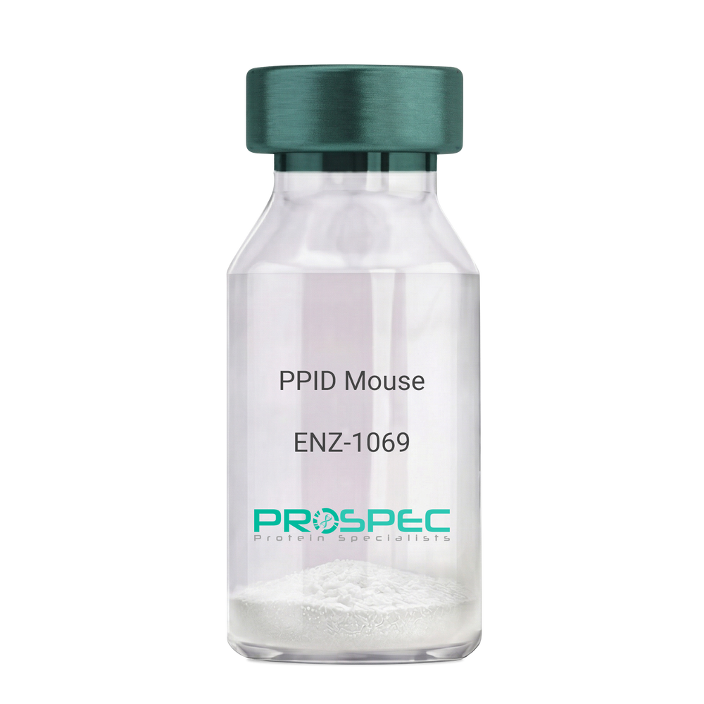
PPID MouseDescription:
Peptidylprolyl Isomerase D Mouse Recombinant
Peptidyl-prolyl cis-trans isomerase D, PPIase D, 40 kDa peptidyl-prolyl cis-trans isomerase, Cyclophilin-40, CYP-40, Cyclophilin-related protein, CYP40, CYPD, PPID, Peptidylprolyl Isomerase D.
Product # :
ENZ-1069Price :
Quantity :
Shipping Method :
Shipped with Ice Packs
More Info
- description
- source
- formulation
- purity
- biological activity
- More Info
Description
PPID Mouse Recombinant produced in E.Coli is a single, non-glycosylated polypeptide chain containing 395 amino acids (1-370a.a.) and having a molecular mass of 43.4kDa. PPID is fused to a 25 amino acid His-tag at N-terminus & purified by proprietary chromatographic techniques.
Source
Escherichia Coli.
Formulation
PPID protein solution (1mg/ml) containing 20mM Tris-Hcl buffer (pH8.0), 10% glycerol and 1mM DTT.
Purity
Greater than 90.0% as determined by SDS-PAGE.
Biological Activity
Specific activity is > 700nmol/min/mg, and is defined as the amount of enzyme that cleaves 1umole of suc-AAFP-PNA per minute at 37°C in Tris–HCl pH 8.0 using chymotrypsin.
More Info
-
Introduction
Cyclophilin-D is a member of the peptidyl-prolyl cis-trans isomerase (PPIase) family. PPIases catalyze the cis-trans isomerization of proline imidic peptide bonds in oligopeptides and speeds up the protein folding. Cyclophilin-D possess PPIase activity and binds to the immunosuppressant cyclosporin-A. Cyclophilin-D is very well known that its overexpression suppresses the apoptosis in cancer cell. Cyclophilin-D suppresses apoptotic cell death by the use of mitochondrial hexokinase-2 dependent mechanism in cancer cells.
-
Synonyms
Peptidyl-prolyl cis-trans isomerase D, PPIase D, 40 kDa peptidyl-prolyl cis-trans isomerase, Cyclophilin-40, CYP-40, Cyclophilin-related protein, CYP40, CYPD, PPID, Peptidylprolyl Isomerase D.
-
Physical Appearance
Sterile Filtered colorless solution.
-
Stability
Store at 4°C if entire vial will be used within 2-4 weeks. Store, frozen at -20°C for longer periods of time. For long term storage it is recommended to add a carrier protein (0.1% HSA or BSA). Please prevent freeze-thaw cycles.
-
Amino Acid Sequence
MGSSHHHHHH SSGLVPRGSH MGSEFMSHAS PAAKPSNSKN PRVFFDVDIG GERVGRIVLE LFADIVPKTA ENFRALCTGE KGTGSTTGKP LHFKGCPFHR IIKKFMIQGG DFSNQNGTGG ESIYGEKFED ENFHYKHDRE GLLSMANAGP NTNGSQFFIT TVPTPHLDGK HVVFGQVIKG LGVARTLENV EVNGEKPAKL CVIAECGELK EGDDWGIFPK DGSGDSHPDF PEDADIDLKD VDKILLISED LKNIGNTFFK SQNWEMAIKK YAKVLRYVDS SKAVIEKADR SRLQPIALSC VLNIGACKLK MSNWQGAIDS CLEALEMDPS NTKALYRKAQ GWQGLKEYDQ ALADLKKAQE IAPGDKAIQA ELLKVKQMIK AQKDKEKAVY AKMFA.
ProSpec's products are furnished for LABORATORY RESEARCH USE ONLY. The product may not be used as drugs, agricultural or pesticidal products, food additives or household chemicals.
- View Data Sheet
Name :
MSP1 AntibodyDescription:
Malaria Falciparum MSP1 , antibody
Product # :
ANT-792Price :
Quantity :
Shipping Method :
Shipped with Ice Packs
More Info
- description
- formulation
- purity
- More Info
Description
Merozoite surface antigen is a protein located on the outside of the merozoite, playing an imperative role in immune reaction. About 55% cases of malaria are infected by Plasmodium falciparum (Pf). Pf MSP1 has to be used with Plasmodium vivax (Pv) together for ELISA and rapid diagnostic test, Plasmodium falciparum and vivax infection takes about 95% of Plasmodium caused infection.
Purification Method: MSP1 Antibody is the purified IgG of mouse monoclonal antibody targeting to merozoite surface protein 1 of Plasmodium falciparum for research.
Formulation
PBS, 0.05% sodium azide and 25mM K2CO3.
Purity
Protein is >90% pure.
Purified by protein A chromatography.
More Info
-
Physical Appearance
Sterile Filtered solution.
-
Stability
Pf. MSP1 although stable at 4°C for 1 week, should be stored below -18°C. Please prevent freeze thaw cycles.
-
Applications
ELISA.
-
Background
Mab-Pf-MSP1 is a monoclonal antibody that recognizes the Plasmodium falciparum Merozoite Surface Protein 1 (MSP1), a major surface antigen expressed during the blood stage of malaria infection. It is widely used for immunological assays such as ELISA, Western blotting, immunofluorescence, and studies investigating parasite invasion, MSP1 processing, and malaria vaccine development.
1. What is MSP1 antibody?
Mab-Pf-MSP1 is a purified mouse monoclonal IgG antibody that specifically targets the merozoite surface protein 1 (MSP1) of Plasmodium falciparum.
2. What species is MSP1 antibody derived from?
Mab-Pf-MSP1 is produced in mouse and is supplied as a purified mouse IgG monoclonal antibody.
3. What antigen does Pf-MSP1 antibody recognize?
The antibody specifically recognizes merozoite surface protein 1 (MSP1) from Plasmodium falciparum.
4. Does MSP1 antibody cross-react with Plasmodium vivax MSP1?
No. Mab-Pf-MSP1 has no cross-reactivity with merozoite surface protein 1 (MSP1) from Plasmodium vivax.
5. What is the format of MSP1 antibody?
The antibody is supplied as purified IgG in liquid form, making it ready for use or further dilution.
6. How is MSP1 antibody purified?
The antibody is purified from mouse ascites fluid using Protein A chromatography and has a purity greater than 90%.
7. What is the concentration of MSP1 antibody?
Pf-MSP1 antibody is supplied at a concentration that ranges between 0.5mg-2mg/mL
8. What buffer is MSP1 antibody supplied in?
The antibody is provided in PBS containing 25 mM potassium carbonate (K₂CO₃).
9. Does MSP1 antibody contain a preservative?
Yes. The formulation contains 0.05% sodium azide as a preservative.
10. What applications is MSP1 antibody recommended for?
MSP1 antibody is recommended for ELISA applications and is suggested to be diluted more than 1:750 for indirect ELISA.
11. What dilution is recommended for indirect ELISA?
For indirect ELISA, a dilution greater than 1:750 is recommended. Users may optimize the dilution depending on their assay conditions.
12. How should MSP1 antibody be stored?
Store the antibody at 4°C for short-term use. For long-term storage, keep it at −20°C.
13. Why should the vial be centrifuged before opening?
The vial should be centrifuged before opening to ensure complete recovery of the contents and minimize product loss.
14. What is the purity of MSP1 Antibody?
The antibody has a purity of greater than 90%, as determined following Protein A chromatography purification.
15. Is MSP1 antibody supplied in liquid or lyophilized form?
Mab-Pf-MSP1 is supplied in liquid form as purified IgG.
16. Can MSP1 antibody be used to distinguish Plasmodium falciparum from Plasmodium vivax?
Yes. Since MSP1 antibody specifically recognizes P. falciparum MSP1 and does not cross-react with P. vivax MSP1, it can be useful in studies requiring species-specific detection.
ProSpec's products are furnished for LABORATORY RESEARCH USE ONLY. The product may not be used as drugs, agricultural or pesticidal products, food additives or household chemicals.
- View Data Sheet
Name :
BCAM HumanDescription:
Basal Cell Adhesion Molecule (CD239) Human Recombinant
Basal cell adhesion molecule isoform 1, BCAM, AU, CD239, LU, MSK19, Auberger B antigen, B-CAM cell surface glycoprotein, F8/G253 antigen, Lutheran antigen, Lutheran blood group glycoprotein, CD_antigen: CD239, LU, MSK19.
Product # :
PRO-2429Price :
Quantity :
Shipping Method :
Shipped with Ice Packs
More Info
- description
- source
- formulation
- purity
- More Info
Description
BCAM produced in Sf9 Baculovirus cells is a single, glycosylated polypeptide chain containing 755 amino acids (32-547a.a.) and having a molecular mass of 83.2kDa. (Molecular size on SDS-PAGE will appear at approximately 70-100kDa). BCAM is expressed with a 239 amino acid hIgG-His tag at C-Terminus and purified by proprietary chromatographic techniques.
Source
Sf9, Baculovirus cells.
Formulation
BCAM protein solution (0.5mg/ml) containing Phosphate Buffered Saline (pH 7.4), and 10% glycerol.
Purity
Greater than 90% as determined by SDS-PAGE.
More Info
-
Introduction
Basal Cell Adhesion Molecule (BCAM) which is a product of alternate splicing of the Lutheran blood group molecule is a part of the immunoglobulin superfamily. BCAM contains five extracellular immunoglobulin domains, a single transmembrane domain, and a short C-terminal cytoplasmic tail. BCAM protein is upregulated following malignant transformation of some cell types in vivo and in vitro. Furthermore, BCAM interacts with integrin in sickle red cells, and participates in vasoocclusive episodes.
-
Synonyms
Basal cell adhesion molecule isoform 1, BCAM, AU, CD239, LU, MSK19, Auberger B antigen, B-CAM cell surface glycoprotein, F8/G253 antigen, Lutheran antigen, Lutheran blood group glycoprotein, CD_antigen: CD239, LU, MSK19.
-
Physical Appearance
Sterile Filtered colorless solution.
-
Stability
Store at 4°C if entire vial will be used within 2-4 weeks. Store, frozen at -20°C for longer periods of time. For long term storage it is recommended to add a carrier protein (0.1% HSA or BSA).Avoid multiple freeze-thaw cycles.
-
Amino Acid Sequence
EVRLSVPPLV EVMRGKSVIL DCTPTGTHDH YMLEWFLTDR SGARPRLASA EMQGSELQVT MHDTRGRSPP YQLDSQGRLV LAEAQVGDER DYVCVVRAGA AGTAEATARL NVFAKPEATE VSPNKGTLSV MEDSAQEIAT CNSRNGNPAP KITWYRNGQR LEVPVEMNPE GYMTSRTVRE ASGLLSLTST LYLRLRKDDR DASFHCAAHY SLPEGRHGRL DSPTFHLTLH YPTEHVQFWV GSPSTPAGWV REGDTVQLLC RGDGSPSPEY TLFRLQDEQE EVLNVNLEGN LTLEGVTRGQ SGTYGCRVED YDAADDVQLS KTLELRVAYL DPLELSEGKV LSLPLNSSAV VNCSVHGLPT PALRWTKDST PLGDGPMLSL SSITFDSNGT YVCEASLPTV PVLSRTQNFT LLVQGSPELK TAEIEPKADG SWREGDEVTL ICSARGHPDP KLSWSQLGGS PAEPIPGRQG WVSSSLTLKV TSALSRDGIS CEASNPHGNK RHVFHFGTVS PQTSQAVEPK SCDKTHTCPP CPAPELLGGP SVFLFPPKPK DTLMISRTPE VTCVVVDVSH EDPEVKFNWY VDGVEVHNAK TKPREEQYNS TYRVVSVLTV LHQDWLNGKE YKCKVSNKAL PAPIEKTISK AKGQPREPQV YTLPPSRDEL TKNQVSLTCL VKGFYPSDIA VEWESNGQPE NNYKTTPPVL DSDGSFFLYS KLTVDKSRWQ QGNVFSCSVM HEALHNHYTQ KSLSLSPGKH HHHHH.
ProSpec's products are furnished for LABORATORY RESEARCH USE ONLY. The product may not be used as drugs, agricultural or pesticidal products, food additives or household chemicals.
- View Data Sheet
Name :
TEK HumanDescription:
TEK Tyrosine Kinase Endothelial Human Recombinant
TEK Receptor Tyrosine Kinase, Tyrosine Kinase With Ig And EGF Homology Domains-2, Tunica Interna Endothelial Cell Kinase, Tyrosine-Protein Kinase Receptor TIE-2, Tyrosine-Protein Kinase Receptor TEK, TEK Tyrosine Kinase, Endothelial, Endothelial Tyrosine Kinase, EC 2.7.10.1, VMCM1, VMCM, TIE2, Venous Malformations, Multiple Cutaneous And Mucosal, Angiopoietin-1 Receptor, CD202b Antigen, EC 2.7.10, P140 TEK, CD202B, GLC3E, TIE-2, HTIE2.
Product # :
PKA-110Price :
Quantity :
Shipping Method :
Shipped with Ice Packs
More Info
- description
- source
- formulation
- purity
- biological activity
- More Info
Description
TEK produced in Sf9 Baculovirus cells is a single, glycosylated polypeptide chain containing 965 amino acids (23-748 a.a.) and having a molecular mass of 107.9kDa. (Molecular size on SDS-PAGE will appear at approximately 100-150 kDa).TEK is expressed with a 239 amino acid hIgG-His-tag at C-Terminus and purified by proprietary chromatographic techniques.
Source
Sf9, Baculovirus cells.
Formulation
TEK protein solution (0.5mg/ml) contains 10% glycerol & Phosphate Buffered Saline (pH 7.4).
Purity
Greater than 90.0% as determined by SDS-PAGE.
Biological Activity
Measured by the ability of the binding activity in a functional ELISA. The ED50 range ≤1ug/ml.
More Info
-
Introduction
TIE-1 (tyrosine kinase with Ig and EGF homology domains 1) and TIE-2/Tek comprise a receptor tyrosine kinase (RTK) subfamily with unique structural characteristics: two immunoglobulin-like domains flanking three epidermal growth factor (EGF)-like domains and followed by three fibronectin type III-like repeats in the extracellular region and a split tyrosine kinase domain in the cytoplasmic region. These receptors are expressed primarily on endothelial and hematopoietic progenitor cells and play critical roles in angiogenesis, vasculogenesis and hematopoiesis. Human TIE-1 cDNA encodes a 1122 amino acid (aa) residue precursor protein with an 18 residue putative signal peptide, a 726 residue extracellular domain and a 353 residue cytoplasmic domain. Two ligands, angiopoietin-1 (Ang1) and angiopoietin-2 (Ang2), which bind TIE-2 with high-affinity have been identified. Ang2 has been reported to act as an antagonist for Ang1. Mice engineered to overexpress Ang2 or to lack Ang1 or Tie-1 display similar angiogenic defects.
-
Synonyms
TEK Receptor Tyrosine Kinase, Tyrosine Kinase With Ig And EGF Homology Domains-2, Tunica Interna Endothelial Cell Kinase, Tyrosine-Protein Kinase Receptor TIE-2, Tyrosine-Protein Kinase Receptor TEK, TEK Tyrosine Kinase, Endothelial, Endothelial Tyrosine Kinase, EC 2.7.10.1, VMCM1, VMCM, TIE2, Venous Malformations, Multiple Cutaneous And Mucosal, Angiopoietin-1 Receptor, CD202b Antigen, EC 2.7.10, P140 TEK, CD202B, GLC3E, TIE-2, HTIE2.
-
Physical Appearance
Sterile Filtered colorless solution.
-
Stability
Store at 4°C if entire vial will be used within 2-4 weeks. Store, frozen at -20°C for longer periods of time. For long term storage it is recommended to add a carrier protein (0.1% HSA or BSA). Avoid multiple freeze-thaw cycles.
-
Amino Acid Sequence
AMDLILINSL PLVSDAETSL TCIASGWRPH EPITIGRDFE ALMNQHQDPL EVTQDVTREW AKKVVWKREK ASKINGAYFC EGRVRGEAIR IRTMKMRQQA SFLPATLTMT VDKGDNVNIS FKKVLIKEED AVIYKNGSFI HSVPRHEVPD ILEVHLPHAQ PQDAGVYSAR YIGGNLFTSA FTRLIVRRCE AQKWGPECNH LCTACMNNGV CHEDTGECIC PPGFMGRTCE KACELHTFGR TCKERCSGQE GCKSYVFCLP DPYGCSCATG WKGLQCNEAC HPGFYGPDCK LRCSCNNGEM CDRFQGCLCS PGWQGLQCER EGIPRMTPKI VDLPDHIEVN SGKFNPICKA SGWPLPTNEE MTLVKPDGTV LHPKDFNHTD HFSVAIFTIH RILPPDSGVW VCSVNTVAGM VEKPFNISVK VLPKPLNAPN VIDTGHNFAV INISSEPYFG DGPIKSKKLL YKPVNHYEAW QHIQVTNEIV TLNYLEPRTE YELCVQLVRR GEGGEGHPGP VRRFTTASIG LPPPRGLNLL PKSQTTLNLT WQPIFPSSED DFYVEVERRS VQKSDQQNIK VPGNLTSVLL NNLHPREQYV VRARVNTKAQ GEWSEDLTAW TLSDILPPQP ENIKISNITH SSAVISWTIL DGYSISSITI RYKVQGKNED QHVDVKIKNA TITQYQLKGL EPETAYQVDI FAENNIGSSN PAFSHELVTL PESQAPADLG GGKMLLLEPK SCDKTHTCPP CPAPELLGGP SVFLFPPKPK DTLMISRTPE VTCVVVDVSH EDPEVKFNWY VDGVEVHNAK TKPREEQYNS TYRVVSVLTV LHQDWLNGKE YKCKVSNKAL PAPIEKTISK AKGQPREPQV YTLPPSRDEL TKNQVSLTCL VKGFYPSDIA VEWESNGQPE NNYKTTPPVL DSDGSFFLYS KLTVDKSRWQ QGNVFSCSVM HEALHNHYTQ KSLSLSPGKH HHHHH.
ProSpec's products are furnished for LABORATORY RESEARCH USE ONLY. The product may not be used as drugs, agricultural or pesticidal products, food additives or household chemicals.