Search results
865 results found for “Serine Peptidase Inhibitor”
Name
Description
Product #
Price
Quantity
Shipping Method
- View Data Sheet
Name :
CYTH3 HumanDescription:
Cytohesin 3 Human Recombinant
Cytohesin 3, PSCD3, ARNO3, GRP1, PH SEC7 and Coiled-Coil Domain-Containing Protein 3 , Pleckstrin Homology Sec7 and Coiled-Coil Domains 3, General Receptor of Phosphoinositides 1 , ARF Nucleotide-Binding Site Opener 3 , Protein ARNO3 , Cytohesin-3, CYTH3.
Product # :
PRO-1951Price :
Quantity :
Shipping Method :
Shipped with Ice Packs
More Info
- description
- source
- formulation
- purity
- More Info
Description
CYTH3 Human Recombinant produced in E.coli is a single, non-glycosylated polypeptide chain containing 422 amino acids (1-399) and having a molecular mass of 48.7 kDa.CYTH3 is fused to a 23 amino acid His-tag at N-terminus & purified by proprietary chromatographic techniques.
Source
E.coli.
Formulation
The CYTH3 solution (0.25mg/ml) contains 20mM Tris-HCl buffer (pH 8.0), 0.15M NaCl, 20% glycerol and 1mM DTT.
Purity
Greater than 90% as determined by SDS-PAGE.
More Info
-
Introduction
Cytohesin 3 (CYTH3) belongs to the PSCD family, whose members have identical structural organization which consists of an N-terminal coiled-coil motif, a central Sec7 domain, and a C-terminal pleckstrin homology (PH) domain, and seem to mediate the regulation of protein sorting and membrane trafficking. CYTH3 is involved in the regulation of Golgi structure and function, and it might have a physiological role in regulating ADP-ribosylation factor protein 6 (ARF) functions, in addition to acting on ARF1.
-
Synonyms
Cytohesin 3, PSCD3, ARNO3, GRP1, PH SEC7 and Coiled-Coil Domain-Containing Protein 3 , Pleckstrin Homology Sec7 and Coiled-Coil Domains 3, General Receptor of Phosphoinositides 1 , ARF Nucleotide-Binding Site Opener 3 , Protein ARNO3 , Cytohesin-3, CYTH3.
-
Physical Appearance
Sterile Filtered colorless solution.
-
Stability
Store at 4°C if entire vial will be used within 2-4 weeks. Store, frozen at -20°C for longer periods of time. For long term storage it is recommended to add a carrier protein (0.1% HSA or BSA).Avoid multiple freeze-thaw cycles.
-
Amino Acid Sequence
MGSSHHHHHH SSGLVPRGSH MGSMDEDGGG EGGGVPEDLS LEEREELLDI RRRKKELIDD IERLKYEIAE VMTEIDNLTS VEESKTTQRN KQIAMGRKKF NMDPKKGIQF LIENDLLQSS PEDVAQFLYK GEGLNKTVIG DYLGERDEFN IKVLQAFVEL HEFADLNLVQ ALRQFLWSFR LPGEAQKIDR MMEAFASRYC LCNPGVFQST DTCYVLSFAI IMLNTSLHNH NVRDKPTAER FIAMNRGINE GGDLPEELLR NLYESIKNEP FKIPEDDGND LTHTFFNPDR EGWLLKLGGR VKTWKRRWFI LTDNCLYYFE YTTDKEPRGI IPLENLSIRE VEDPRKPNCF ELYNPSHKGQ VIKACKTEAD GRVVEGNHVV YRISAPSPEE KEEWMKSIKA SISRDPFYDM LATRKRRIAN KK.
ProSpec's products are furnished for LABORATORY RESEARCH USE ONLY. The product may not be used as drugs, agricultural or pesticidal products, food additives or household chemicals.
- View Data Sheet
Name :
B3GNT2 HumanDescription:
Beta-1,3-N-Acetylglucosaminyltransferase 2 Human Recombinant
B3GNT2, B3GN-T2, B3GNT, B3GNT-2, B3GNT1, BETA3GNT, BGnT-2, BGNT2, N-acetyllactosaminide beta-1,3-N-acetylglucosaminyltransferase 2, Beta-1,3-N-acetylglucosaminyltransferase 1, BGnT-1, Beta-1,3-Gn-T1, Beta3Gn-T1, Beta-1,3-galactosyltransferase 7, Beta-1,3-GalTase 7, Beta3Gal-T7, Beta3GalT7, b3Gal-T7, Beta-3-Gx-T7, beta-GlcNAc beta-1,3-galactosyltransferase 7, betaGal beta-1,3-N-acetylglucosaminyltransferase 2, BGnT-2, Beta-1,3-Gn-T2, Beta-1,3-N-acetylglucosaminyltransferase 2, Beta3Gn-T2, beta-N-acetylglucosamine beta-1,3-galactosyltransferase 7.
Product # :
ENZ-973Price :
Quantity :
Shipping Method :
Shipped with Ice Packs
More Info
- description
- source
- formulation
- purity
- More Info
Description
B3GNT2 produced in Sf9 Baculovirus cells is a single, glycosylated polypeptide chain containing 375 amino acids (29-397a.a.) and having a molecular mass of 43.5kDa (Molecular size on SDS-PAGE will appear at approximately 40-57kDa). B3GNT2 is expressed with a 6 amino acid His tag at C-Terminus and purified by proprietary chromatographic techniques.
Source
Sf9, Baculovirus cells.
Formulation
B3GNT2 protein solution (0.5mg/ml) contains Phosphate Buffered Saline (pH 7.4) and 10% glycerol.
Purity
Greater than 90.0% as determined by SDS-PAGE.
More Info
-
Introduction
Beta-1,3-N-Acetylglucosaminyltransferase 2 (B3GNT2) is a part of the beta-1,3-N-acetylglucosaminyltransferase family which takes part in the synthesis of poly-N-acetyllactosamine. B3GNT2 is a type II transmembrane protein which prefers the substrate of lacto-N-neotetraose. B3GNT2 catalyzes the initiation and elongation of poly-N-acetyllactosamine chains and comprises the main polylactosamine synthase.
-
Synonyms
B3GNT2, B3GN-T2, B3GNT, B3GNT-2, B3GNT1, BETA3GNT, BGnT-2, BGNT2, N-acetyllactosaminide beta-1,3-N-acetylglucosaminyltransferase 2, Beta-1,3-N-acetylglucosaminyltransferase 1, BGnT-1, Beta-1,3-Gn-T1, Beta3Gn-T1, Beta-1,3-galactosyltransferase 7, Beta-1,3-GalTase 7, Beta3Gal-T7, Beta3GalT7, b3Gal-T7, Beta-3-Gx-T7, beta-GlcNAc beta-1,3-galactosyltransferase 7, betaGal beta-1,3-N-acetylglucosaminyltransferase 2, BGnT-2, Beta-1,3-Gn-T2, Beta-1,3-N-acetylglucosaminyltransferase 2, Beta3Gn-T2, beta-N-acetylglucosamine beta-1,3-galactosyltransferase 7.
-
Physical Appearance
Sterile Filtered colorless solution.
-
Stability
Store at 4°C if entire vial will be used within 2-4 weeks. Store, frozen at -20°C for longer periods of time. For long term storage it is recommended to add a carrier protein (0.1% HSA or BSA). Avoid multiple freeze-thaw cycles.
-
Amino Acid Sequence
ADPKSSSQEK NGKGEVIIPK EKFWKISTPP EAYWNREQEK LNRQYNPILS MLTNQTGEAG RLSNISHLNY CEPDLRVTSV VTGFNNLPDR FKDFLLYLRC RNYSLLIDQP DKCAKKPFLL LAIKSLTPHF ARRQAIRESW GQESNAGNQT VVRVFLLGQT PPEDNHPDLS DMLKFESEKH QDILMWNYRD TFFNLSLKEV LFLRWVSTSC PDTEFVFKGD DDVFVNTHHI LNYLNSLSKT KAKDLFIGDV IHNAGPHRDK KLKYYIPEVV YSGLYPPYAG GGGFLYSGHL ALRLYHITDQ VHLYPIDDVY TGMCLQKLGL VPEKHKGFRT FDIEEKNKNN ICSYVDLMLV HSRKPQEMID IWSQLQSAHL KCHHHHHH.
ProSpec's products are furnished for LABORATORY RESEARCH USE ONLY. The product may not be used as drugs, agricultural or pesticidal products, food additives or household chemicals.
- View Data Sheet
Name :
BD 3 HumanDescription:
Beta Defensin-3 Human Recombinant
HBD3, HBP3, DEFB3, HBD-3, HBP-3, DEFB103.
Product # :
CYT-461Price :
Quantity :
Shipping Method :
Shipped at Room temp
More Info
- description
- source
- formulation
- purity
- More Info
Description
Beta Defensin-3 Human Recombinant produced in E.Coli is a single, non-glycosylated polypeptide chain containing 45 amino acids and having a molecular mass of 5161.2 Dalton. The BD-3 is purified by proprietary chromatographic techniques.
Source
Escherichia Coli.
Formulation
The HBD-3 was lyophilized without additives.
Purity
Greater than 95.0% as determined by(a) Analysis by RP-HPLC.
(b) Analysis by SDS-PAGE.More Info
-
Introduction
Defensins form a family of microbicidal and cytotoxic peptides made by neutrophils. Members of the defensin family are highly similar in protein sequence. This gene encodes defensin, beta 103A, which has broad spectrum antimicrobial activity and may play an important role in innate epithelial defense.
-
Synonyms
HBD3, HBP3, DEFB3, HBD-3, HBP-3, DEFB103.
-
Physical Appearance
Sterile Filtered lyophilized (freeze-dried) powder.
-
Stability
Lyophilized Beta Defensin-3 Recombinant although stable at room temperature for 3 weeks, should be stored desiccated below -18°C. Upon reconstitution BD-3 should be stored at 4°C between 2-7 days and for future use below -18°C.Please prevent freeze-thaw cycles.
-
Solubility
It is recommended to reconstitute the lyophilized Beta Defensin-3 in sterile 18MΩ-cm H2O not less than 100µg/ml, which can then be further diluted to other aqueous solutions.
-
Amino Acid Sequence
GIINTLQKYY CRVRGGRCAV LSCLPKEEQI GKCSTRGRKC CRRKK.
-
Background
Beta Defensin-3 Human Recombinant: Advancements in Antimicrobial Peptide Therapy
Abstract:
Beta Defensin-3 (hBD-3) human recombinant is a promising antimicrobial peptide with broad-spectrum activity against bacteria, viruses, and fungi. This research paper provides an overview of hBD-3, including its properties, mode of action, and potential applications. Additionally, novel methodologies for the production and optimization of hBD-3 human recombinant are discussed, highlighting its future implications in the field of infectious disease management.Introduction:
The rise of drug-resistant pathogens necessitates exploring alternative therapeutic approaches, such as antimicrobial peptides. Beta Defensin-3 (hBD-3) human recombinant has emerged as a potent candidate due to its broad-spectrum antimicrobial activity. This paper aims to examine the unique features of hBD-3 and propose innovative methodologies for its production and optimization.Properties and Mode of Action:
hBD-3 possesses a distinct structural composition consisting of 45 amino acids, including an N-terminal loop, three antiparallel β-strands, and a C-terminal α-helix. These structural elements contribute to its ability to disrupt microbial membranes and target selectivity. The mode of action involves electrostatic interactions with negatively charged microbial membranes, leading to membrane disruption and subsequent cell death. Furthermore, hBD-3 exhibits immunomodulatory functions by promoting chemotaxis, enhancing phagocytic activity, and modulating the release of pro-inflammatory cytokines.Production of hBD-3 Human Recombinant:
Various expression systems, such as bacterial, yeast, and mammalian cell-based platforms, have been explored for the efficient production of hBD-3 human recombinant. Each system offers distinct advantages and challenges, requiring careful selection to achieve high yields and desired protein quality. Optimization strategies, including codon optimization, fusion protein tags, and appropriate growth conditions, have been employed to enhance production efficiency. Purification techniques, such as chromatography and ultrafiltration, have been optimized to isolate high-quality hBD-3 recombinant.Applications and Future Perspectives:
hBD-3 human recombinant exhibits significant therapeutic potential against drug-resistant pathogens, making it a promising alternative to conventional antibiotics. It also demonstrates promise in wound healing and tissue regeneration by stimulating angiogenesis, extracellular matrix production, and keratinocyte migration. Moreover, the unique physicochemical properties of hBD-3 open avenues for its utilization in nanomedicine, enabling targeted therapy and improved drug delivery.Conclusion:
hBD-3 human recombinant represents a potent antimicrobial peptide with broad-spectrum activity against diverse pathogens. The optimization of production methodologies and further exploration of its mechanisms of action will contribute to its clinical utility. With its potential applications in infectious disease management, wound healing, and nanomedicine, hBD-3 human recombinant holds promise as a versatile therapeutic agent.What is the molecular weight/Mw of BD3 Protein?
BD3 Protein has a total Mw of 5.1kDa.
What is the source or expression system of BD3 Protein?
Escherichia Coli.
What is the Purity of BD3 Protein?
BD3 Protein is >95% pure as determined by SDS-PAGE.
What is the Biological Activity of BD3 Protein?
The biological functionality of BD3 Protein will be determined in the future.
What is the amino acid sequence of BD3 Protein?
GIINTLQKYY CRVRGGRCAV LSCLPKEEQI GKCSTRGRKC CRRKK.
What applications can BD3 Protein be used in?
BD3 Protein can probably be used in western blot, ELISA and Lateral Flow.
What is the endotoxin level for BD3 Protein?
The endotoxin level is minimal, BD3 Protein was purified using conventional chromatography techniques.
ProSpec's products are furnished for LABORATORY RESEARCH USE ONLY. The product may not be used as drugs, agricultural or pesticidal products, food additives or household chemicals.
- View Data Sheet
Name :
SRSF1 HumanDescription:
Serine/arginine-Rich Splicing Factor 1 Human Recombinant
ASF, MGC5228, SF2, SF2p33, SRp30a, Serine/arginine-rich splicing factor 1, SRSF1, splicing factor 1, ASF-1, Splicing factor, arginine/serine-rich 1, pre-mRNA-splicing factor SF2, P33 subunit, OK/SW-cl.3.
Product # :
PRO-1352Price :
Quantity :
Shipping Method :
Shipped with Ice Packs
More Info
- description
- source
- formulation
- purity
- More Info
Description
SRSF1 Human Recombinant produced in E.coli is a single, non-glycosylated polypeptide chain containing 268 amino acids (1-248) and having a molecular mass of 29.9 kDa.SRSF1 is fused to a 20 amino acid His-tag at N-terminus & purified by proprietary chromatographic techniques.
Source
Escherichia Coli.
Formulation
The SRSF1 solution (1mg/ml) contains 20mM Tris-HCl buffer (pH 8.0), 0.4M urea and 10% glycerol.
Purity
Greater than 90% as determined by SDS-PAGE.
More Info
-
Introduction
Serine/arginine-rich splicing factor 1 (SFRS1) belongs to the arginine/serine-rich splicing factor protein family, and functions in both constitutive and alternative pre-mRNA splicing. SFRS1 binds to pre-mRNA transcripts and components of the spliceosome, and can either initiate or inhibit splicing depending on the position of the pre-mRNA binding site. The ability of SFRS1 to activate splicing is controlled by phosphorylation and interactions with other splicing factor associated proteins.
-
Synonyms
ASF, MGC5228, SF2, SF2p33, SRp30a, Serine/arginine-rich splicing factor 1, SRSF1, splicing factor 1, ASF-1, Splicing factor, arginine/serine-rich 1, pre-mRNA-splicing factor SF2, P33 subunit, OK/SW-cl.3.
-
Physical Appearance
Sterile Filtered colorless solution.
-
Stability
Store at 4°C if entire vial will be used within 2-4 weeks. Store, frozen at -20°C for longer periods of time. For long term storage it is recommended to add a carrier protein (0.1% HSA or BSA).Avoid multiple freeze-thaw cycles.
-
Amino Acid Sequence
MGSSHHHHHH SSGLVPRGSH MSGGGVIRGP AGNNDCRIYV GNLPPDIRTK DIEDVFYKYG AIRDIDLKNR RGGPPFAFVE FEDPRDAEDA VYGRDGYDYD GYRLRVEFPR SGRGTGRGGG GGGGGGAPRG RYGPPSRRSE NRVVVSGLPP SGSWQDLKDH MREAGDVCYA DVYRDGTGVV EFVRKEDMTY AVRKLDNTKF RSHEGETAYI RVKVDGPRSP SYGRSRSRSR SRSRSRSRSN SRSRSYSPRR SRGSPRYSPR HSRSRSRT.
ProSpec's products are furnished for LABORATORY RESEARCH USE ONLY. The product may not be used as drugs, agricultural or pesticidal products, food additives or household chemicals.
- View Data Sheet
Name :
ANGPTL2 HumanDescription:
Angiopoietin-like Protein 2 Human Recombinant
Angiopoietin-related protein 2, Angiopoietin-like protein 2, ANGPTL2, ARP2, HARP.
Product # :
CYT-765Price :
Quantity :
Shipping Method :
Shipped with Ice Packs
More Info
- description
- source
- formulation
- purity
- More Info
- sds-page
Description
ANGPTL2 Human Recombinant produced in E.Coli is a single, non-glycosylated, polypeptide chain containing 493 amino acids ( 22-493 a.a.) including a 20 a.a N-terminal His tag. The total molecular mass is 57.1kDa (calculated).
Source
Escherichia Coli.
Formulation
ANGPTL2 protein solution (0.5mg/ml) contains 20mM Tris HCL (pH7-8) and 10% glycerol.
Purity
Greater than 85.0% as determined by SDS-PAGE.
sds-page
More Info
-
Introduction
Angiopoietins belong to the vascular endothelial growth factor family and the only known growth factors largely specific for vascular endothelium. Angiopoietins-1, 2 and 4 partake in the formation of blood vessels. ANGPTL2 displays angiogenic effects. Angiopoietin-like Protein 2 (ANGPTL2) is an anti-diabetic factor. ANGPTL2 induces sprouting in endothelial cells through an autocrine and paracrine action. ANGPTL2 increases insulin sensitivity in adipocytes. In addition, ANGPTL2 is a mediator of chronic adipose tissue inflammation. ANGPTL2 is widely expressed in the heart, small intestine, spleen and stomach. ANGPTL2 is also found in lower levels in the colon, ovary, adrenal gland, skeletal muscle and in prostate.
-
Synonyms
Angiopoietin-related protein 2, Angiopoietin-like protein 2, ANGPTL2, ARP2, HARP.
-
Physical Appearance
Sterile filtered colorless solution.
-
Stability
Store at 4°C if entire vial will be used within 2-4 weeks.Store, frozen at -20°C for longer periods of time.For long term storage it is recommended to add a carrier protein (0.1% HSA or BSA).Avoid multiple freeze-thaw cycles.
-
Amino Acid Sequence
MGSSHHHHHH SSGLVPRGSH MGQEDGFEGT EEGSPREFIY LNRYKRAGES QDKCTYTFIV PQQRVTGAIC VNSKEPEVLL ENRVHKQELE LLNNELLKQK RQIETLQQLV EVDGGIVSEV KLLRKESRNM NSRVTQLYMQ LLHEIIRKRD NALELSQLEN RILNQTADML QLASKYKDLE HKYQHLATLA HNQSEIIAQL EEHCQRVPSA RPVPQPPPAA PPRVYQPPTY NRIINQISTN EIQSDQNLKV LPPPLPTMPT LTSLPSSTDK PSGPWRDCLQ ALEDGHDTSS IYLVKPENTN RLMQVWCDQR HDPGGWTVIQ RRLDGSVNFF RNWETYKQGF GNIDGEYWLG LENIYWLTNQ GNYKLLVTME DWSGRKVFAE YASFRLEPES EYYKLRLGRY HGNAGDSFTW HNGKQFTTLD RDHDVYTGNC AHYQKGGWWY NACAHSNLNG VWYRGGHYRS RYQDGVYWAE FRGGSYSLKK VVMMIRPNPN TFH
-
Background
Angiopoietin-like Protein 2 Human Recombinant: A Potential Therapeutic Target for Metabolic and Cardiovascular Disorders
Abstract:
Angiopoietin-like protein 2 (ANGPTL2) has emerged as a crucial regulator in metabolic and cardiovascular disorders. This multifunctional protein is involved in various biological processes, including angiogenesis, adipose tissue function, and inflammation. The availability of human recombinant ANGPTL2 protein has provided researchers with a valuable tool to explore its therapeutic potential. This concise review provides an overview of the role of ANGPTL2 in metabolic and cardiovascular health and discusses the potential of ANGPTL2 human recombinant protein as a therapeutic target.
Introduction:
Metabolic disorders, such as obesity and type 2 diabetes, are closely associated with cardiovascular diseases and pose significant global health challenges. ANGPTL2, a member of the angiopoietin-like protein family, has gained attention for its involvement in metabolic regulation and cardiovascular homeostasis. Through interactions with various receptors and signaling pathways, ANGPTL2 influences lipid metabolism, insulin sensitivity, inflammation, and vascular integrity.
Mechanisms of ANGPTL2 Action:
ANGPTL2 acts through binding to integrins, toll-like receptors (TLRs), and other receptors on different cell types, including adipocytes, endothelial cells, and immune cells. By influencing angiogenesis, inflammation, and extracellular matrix remodeling, ANGPTL2 affects adipose tissue function, lipid metabolism, and insulin signaling.
Role of ANGPTL2 in Metabolic Regulation:
ANGPTL2 plays a critical role in metabolic regulation and the development of metabolic disorders. It promotes adipose tissue inflammation, impairs adipogenesis, and alters adipokine secretion, contributing to metabolic dysfunction. Furthermore, ANGPTL2 modulates lipid metabolism by regulating lipoprotein lipase activity, affecting triglyceride clearance, and promoting hepatic lipid accumulation.
ANGPTL2 in Cardiovascular Health and Disease:
Increasing evidence suggests that ANGPTL2 is implicated in cardiovascular diseases, including atherosclerosis and heart failure. ANGPTL2 promotes vascular inflammation, endothelial dysfunction, and smooth muscle cell proliferation, which contribute to atherosclerotic plaque progression and vascular remodeling. Additionally, ANGPTL2 influences cardiac remodeling and fibrosis, impacting heart failure development.
Therapeutic Potential of ANGPTL2 Human Recombinant Protein:
The availability of ANGPTL2 human recombinant protein opens avenues for therapeutic interventions targeting metabolic and cardiovascular disorders. Preclinical studies employing ANGPTL2 blockade or supplementation have shown promising results in improving metabolic parameters, reducing atherosclerosis, and preserving cardiac function. However, further research is needed to optimize the clinical application of ANGPTL2 human recombinant protein, including dosage, timing, and delivery methods.
Conclusion:
ANGPTL2 holds promise as a therapeutic target for metabolic and cardiovascular disorders. Its involvement in key biological processes makes it an attractive candidate for interventions aiming to improve metabolic health and prevent cardiovascular complications. The development of ANGPTL2 human recombinant protein provides a valuable tool for investigating its therapeutic potential further.
What is the molecular weight/Mw of ANGPTL2 Protein?
ANGPTL2 Protein has a total Mw of 57.1kDa.
What is the source or expression system of ANGPTL2 Protein?
Escherichia Coli.
What is the Purity of ANGPTL2 Protein?
ANGPTL2 Protein is >85% pure as determined by SDS-PAGE.
What is the Biological Activity of ANGPTL2 Protein?
The biological functionality of ANGPTL2 Protein will be determined in the future.
What is the amino acid sequence of ANGPTL2 Protein?
MGSSHHHHHH SSGLVPRGSH MGQEDGFEGT EEGSPREFIY LNRYKRAGES QDKCTYTFIV PQQRVTGAIC VNSKEPEVLL ENRVHKQELE LLNNELLKQK RQIETLQQLV EVDGGIVSEV KLLRKESRNM NSRVTQLYMQ LLHEIIRKRD NALELSQLEN RILNQTADML QLASKYKDLE HKYQHLATLA HNQSEIIAQL EEHCQRVPSA RPVPQPPPAA PPRVYQPPTY NRIINQISTN EIQSDQNLKV LPPPLPTMPT LTSLPSSTDK PSGPWRDCLQ ALEDGHDTSS IYLVKPENTN RLMQVWCDQR HDPGGWTVIQ RRLDGSVNFF RNWETYKQGF GNIDGEYWLG LENIYWLTNQ GNYKLLVTME DWSGRKVFAE YASFRLEPES EYYKLRLGRY HGNAGDSFTW HNGKQFTTLD RDHDVYTGNC AHYQKGGWWY NACAHSNLNG VWYRGGHYRS RYQDGVYWAE FRGGSYSLKK VVMMIRPNPN TFH
What applications can ANGPTL2 Protein be used in?
ANGPTL2 Protein can probably be used in western blot, ELISA and Lateral Flow.
What is the endotoxin level for ANGPTL2 Protein?
The endotoxin level is minimal, ANGPTL2 Protein was purified using conventional chromatography techniques.
ProSpec's products are furnished for LABORATORY RESEARCH USE ONLY. The product may not be used as drugs, agricultural or pesticidal products, food additives or household chemicals.
- View Data Sheet
Name :
ANGPTL3 (17-460) HumanDescription:
Angiopoietin-like Protein 3 (17-460 a.a.) Human Recombinant
Angiopoietin Like 3, Angiopoietin 5, ANGPT5, ANG-5, Angiopoietin-Related Protein 3 , Angiopoietin-Like Protein 3, Angiopoietin-Like 3, Angiopoietin-5, FHBL2, ANL3.
Product # :
CYT-986Price :
Quantity :
Shipping Method :
Shipped with Ice Packs
More Info
- description
- source
- formulation
- purity
- More Info
- sds-page
Description
ANGPTL3 produced in Sf9 Baculovirus cells is a single, glycosylated polypeptide chain containing 453 amino acids (17-460 a.a.) and having a molecular mass of 52.9kDa (Migrates at 25-70kDa on SDS-PAGE under reducing conditions). ANGPTL3 is expressed with an 6 amino acid His tag at C-Terminus and purified by proprietary chromatographic techniques.
Source
Sf9, Baculovirus cells.
Formulation
ANGPTL3 protein solution (0.25mg/ml) contains Buffered Saline (pH 7.4),30% glycerol And 1mM DTT.
Purity
Greater than 90.0% as determined by SDS-PAGE.
sds-page
More Info
-
Introduction
ANGPTL3 and ANGPTL4 are angiopoietin-like proteins secreted and expressed mainly by the liver, their role being the regulation of triglyceride metabolism by inhibiting the lipolysis of triglyceride-rich lipoproteins. During different nutritional states (feeding/fasting) the levels of the circulating triglycerides are regulated by Angptl3 and Angptl4 through differential inhibition of Lipoprotein lipase (LPL) as shown by the experimental data. The molecular structure of ANGPTL3 is similar to that of the angiopoietins (vascular endothelial growth factors). Deletion mutants of human Angiopoietin 5 were used in order to demonstrate that the N-terminal domain (fragment 17-207) and not the C-terminal fibrinogen-like domain (fragment 207-460) increased the plasma triglyceride levels in mice.
-
Synonyms
Angiopoietin Like 3, Angiopoietin 5, ANGPT5, ANG-5, Angiopoietin-Related Protein 3 , Angiopoietin-Like Protein 3, Angiopoietin-Like 3, Angiopoietin-5, FHBL2, ANL3.
-
Physical Appearance
Sterile Filtered colorless solution.
-
Stability
Store at 4°C if entire vial will be used within 2-4 weeks. Store, frozen at -20°C for longer periods of time. For long term storage it is recommended to add a carrier protein (0.1% HSA or BSA). Avoid multiple freeze-thaw cycles.
-
Amino Acid Sequence
ADPSRIDQDN SSFDSLSPEP KSRFAMLDDV KILANGLLQL GHGLKDFVHK TKGQINDIFQ KLNIFDQSFY DLSLQTSEIK EEEKELRRTT YKLQVKNEEV KNMSLELNSK LESLLEEKIL LQQKVKYLEE QLTNLIQNQP ETPEHPEVTS LKTFVEKQDN SIKDLLQTVE DQYKQLNQQH SQIKEIENQL RRTSIQEPTE ISLSSKPRAP RTTPFLQLNE IRNVKHDGIP AECTTIYNRG EHTSGMYAIR PSNSQVFHVY CDVISGSPWT LIQHRIDGSQ NFNETWENYK YGFGRLDGEF WLGLEKIYSI VKQSNYVLRI ELEDWKDNKH YIEYSFYLGN HETNYTLHLV AITGNVPNAI PENKDLVFST WDHKAKGHFN CPEGYSGGWW WHDECGENNL NGKYNKPRAK SKPERRRGLS WKSQNGRLYS IKSTKMLIHP TDSESFEHHH HHH.
-
Background
What is the molecular weight/Mw of ANGPTL3 Protein?
ANGPTL3 Protein has a total Mw of 52.9kDa.
What is the source or expression system of ANGPTL3 Protein?
Sf9, Baculovirus cells.
What is the Purity of ANGPTL3 Protein?
ANGPTL3 Protein is >90% pure as determined by SDS-PAGE.
What is the Biological Activity of ANGPTL3 Protein?
The biological functionality of ANGPTL3 Protein will be determined in the future.
What is the amino acid sequence of ANGPTL3 Protein?
ADPSRIDQDN SSFDSLSPEP KSRFAMLDDV KILANGLLQL GHGLKDFVHK TKGQINDIFQ KLNIFDQSFY DLSLQTSEIK EEEKELRRTT YKLQVKNEEV KNMSLELNSK LESLLEEKIL LQQKVKYLEE QLTNLIQNQP ETPEHPEVTS LKTFVEKQDN SIKDLLQTVE DQYKQLNQQH SQIKEIENQL RRTSIQEPTE ISLSSKPRAP RTTPFLQLNE IRNVKHDGIP AECTTIYNRG EHTSGMYAIR PSNSQVFHVY CDVISGSPWT LIQHRIDGSQ NFNETWENYK YGFGRLDGEF WLGLEKIYSI VKQSNYVLRI ELEDWKDNKH YIEYSFYLGN HETNYTLHLV AITGNVPNAI PENKDLVFST WDHKAKGHFN CPEGYSGGWW WHDECGENNL NGKYNKPRAK SKPERRRGLS WKSQNGRLYS IKSTKMLIHP TDSESFEHHH HHH.
What applications can ANGPTL3 Protein be used in?
ANGPTL3 Protein can probably be used in western blot, ELISA and Lateral Flow.
What is the endotoxin level for ANGPTL3 Protein?
The endotoxin level is minimal, ANGPTL3 Protein was purified using conventional chromatography techniques.
ProSpec's products are furnished for LABORATORY RESEARCH USE ONLY. The product may not be used as drugs, agricultural or pesticidal products, food additives or household chemicals.
- View Data Sheet
Name :
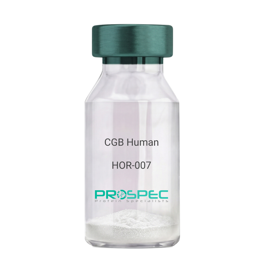
CGB HumanDescription:
CGB Human Recombinant
CGB3, CGB5, CGB7, CGB8, hCGB, CG-beta, CGB.
Product # :
HOR-007Price :
Quantity :
Shipping Method :
Shipped with Ice Packs
More Info
- description
- source
- formulation
- purity
- More Info
Description
CGB Human Recombinant produced in E.Coli is a single, non-glycosylated polypeptide chain containing 168 amino acids (21-165 a.a.) and having a molecular mass of 17.9kDa. CGB is fused to a 23 amino acid His-tag at N-terminus & purified by proprietary chromatographic techniques.
Source
Escherichia Coli.
Formulation
CGB protein solution (0.5mg/ml) containing 20mM Tris-HCl buffer (pH 8.0), 0.4M Urea and 10% glycerol.
Purity
Greater than 85.0% as determined by SDS-PAGE.
More Info
-
Introduction
Glycoprotein hormones are heterodimers consisting of a common alpha subunit and a unique beta subunit which confers biological specificity. CGB is a member of the glycoprotein hormone beta chain family and is the beta 3 subunit of (CG). CG is produced by the trophoblastic cells of the placenta and stimulates the ovaries. The beta subunit of CG is encoded by 6 genes which are arranged in tandem and inverted pairs on chromosome 19q13.3 and contiguous with the luteinizing hormone beta subunit gene.
-
Synonyms
CGB3, CGB5, CGB7, CGB8, hCGB, CG-beta, CGB.
-
Physical Appearance
Sterile filtered colorless solution.
-
Stability
Store at 4°C if entire vial will be used within 2-4 weeks. Store, frozen at -20°C for longer periods of time. For long term storage it is recommended to add a carrier protein (0.1% HSA or BSA).Avoid multiple freeze-thaw cycles.
-
Amino Acid Sequence
MGSSHHHHHH SSGLVPRGSH MGSSKEPLRP RCRPINATLA VEKEGCPVCI TVNTTICAGY CPTMTRVLQG VLPALPQVVC NYRDVRFESI RLPGCPRGVN PVVSYAVALS CQCALCRRST TDCGGPKDHP LTCDDPRFQD SSSSKAPPPS LPSPSRLPGP SDTPILPQ.
ProSpec's products are furnished for LABORATORY RESEARCH USE ONLY. The product may not be used as drugs, agricultural or pesticidal products, food additives or household chemicals.
- View Data Sheet
Name :
FLAG PAT3G6AT antibodyDescription:
FLAG peptide Clone PAT3G6AT, Mouse Anti Human
Product # :
ANT-736Price :
Quantity :
Shipping Method :
Shipped with Ice Packs
More Info
- formulation
- More Info
Formulation
1mg/ml containing PBS, pH-7.4, 10% Glycerol and 0.02% Sodium Azide.
More Info
-
Introduction
A fusion tag called FLAG consists of eight amino acids (AspTyrLysAspAspAspAspLys) including an enterokinase-cleavage site. FLAG is specifically designed for immunoaffinity chromatography. It allows elution under non-denaturing conditions. Several antibodies against this peptide have been developed. One antibody denoted as M1 binds the peptide in the presence of bivalent metal cations preferably Ca(+). Elution is effected by chelating agents. Another strategy is competitive elution with excess of free Flag Peptide. The FLAG peptide purifies and detects recombinant fusion proteins. FLAG peptide is useful in Western blotting, immunocytochemistry, immunoprecipitation, flow cytometry, protein purification, and in the study of protein-protein interactions, cell ultrastructure, and protein localization. Flag peptide is a hydrophilic tag which significantly improves the detection and purification of recombinant fusion proteins.
-
Physical Appearance
Sterile filtered colorless solution.
-
Immunogen
Anti-human FLAG mAb, is derived from hybridization of mouse F0 myeloma cells with spleen cells from BALB/c mice immunized with a Synthetic peptide (DYKDDDDKC)-KLH.
-
Ig Subclass
Mouse IgG1 heavy chain and k light chain.
-
Clone
PAT3G6AT.
-
Applications
The antibody has been tested by ELISA, Western blot analysis, Flow cytometry and ICC/IF to assure specificity and reactivity. Since application varies, however, each investigation should be titrated by the reagent to obtain optimal results.
-
Type
Mouse Anti Human Monoclonal.
-
Storage Procedures
For periods up to 1 month store at 4°C, for longer periods of time, store at -20°C. Prevent freeze thaw cycles.
-
Purification Method
FLAG antibody was purified from mouse ascitic fluids by protein-A affinity chromatography.
ProSpec's products are furnished for LABORATORY RESEARCH USE ONLY. The product may not be used as drugs, agricultural or pesticidal products, food additives or household chemicals.
- View Data Sheet
Name :
HPRT AntibodyDescription:
Hypoxanthine-Guanine Phosphoribosyltransferase, Mouse Anti Human
Hypoxanthine-Guanine Phosphoribosyltransferase, EC 2.4.2.8, HGPRT, HGPRTase, HPRT, HPRT1.
Product # :
ANT-046Price :
Quantity :
Shipping Method :
Shipped with Ice Packs
More Info
- formulation
- More Info
Formulation
1mg/ml containing PBS, pH-7.4, & 0.1% Sodium Azide.
More Info
-
Introduction
HPRT1 has a main part in the generation of purine nucleotides through the purine salvage pathway. HPRT1 primarily functions to salvage purines from degraded DNA to renewed purine synthesis. Therefore, it performs as a catalyst in the reaction between guanine and phosphoribosyl pyrophosphate to form GMP.
-
Synonyms
Hypoxanthine-Guanine Phosphoribosyltransferase, EC 2.4.2.8, HGPRT, HGPRTase, HPRT, HPRT1.
-
Immunogen
Anti-human HPRT mAb, is derived from hybridization of mouse FO myeloma cells with spleen cells from BALB/c mice immunized with recombinant human HPRT amino acids 1-218 purified from E. coli.
-
Ig Subclass
Mouse IgG1 heavy chain and k light chain.
-
Clone
PAT2G8AT.
-
Applications
HPRT antibody has been tested by ELISA and Western blot analysis to assure specificity and reactivity. Since application varies, however, each investigation should be titrated by the reagent to obtain optimal results. Recommended dilution range for Western blot analysis is 1:500~1:5000.
Recommended starting dilution is 1:1000. -
Type
Mouse Anti Human Monoclonal.
-
Storage Procedures
For periods up to 1 month store at 4°C, for longer periods of time, store at -20°C. Prevent freeze thaw cycles.
-
Purification Method
HPRT antibody was purified from mouse ascitic fluids by protein-G affinity chromatography.
ProSpec's products are furnished for LABORATORY RESEARCH USE ONLY. The product may not be used as drugs, agricultural or pesticidal products, food additives or household chemicals.
- View Data Sheet
Name :
ATG3 AntibodyDescription:
Autophagy Related 3, Mouse Anti Human
APG3, APG3-LIKE, APG3L, PC3-96, Ubiquitin-like-conjugating enzyme ATG3, Autophagy-related protein 3, hApg3, PC3-96, ATG3.
Product # :
ANT-534Price :
Quantity :
Shipping Method :
Shipped with Ice Packs
More Info
- formulation
- More Info
Formulation
1mg/ml containing PBS, pH-7.4, 10% Glycerol and 0.02% Sodium Azide.
More Info
-
Introduction
Autophagy Related 3 (ATG3) is a conjugating enzyme essential for the cytoplasm to vacuole transport (Cvt), autophagy, and mitochondrial homeostasis. ATG3 is localizes to the cytoplasm and is expressed in tissues with predominant levels found in kidney, placenta, liver, heart and skeletal muscle. ATG3 catalyzes a reaction which is necessary for autophagy. That reaction is the formation of the Atg8-phosphatidylethanolamine (ATG-PE) conjugate.
-
Synonyms
APG3, APG3-LIKE, APG3L, PC3-96, Ubiquitin-like-conjugating enzyme ATG3, Autophagy-related protein 3, hApg3, PC3-96, ATG3.
-
Physical Appearance
Sterile filtered colorless solution.
-
Immunogen
Anti-human ATG3mAb, clone PAT15F6AT, is derived from hybridization of mouse F0 myeloma cells with spleen cells from BALB/c mice immunized with a recombinant human ATG3 protein (1-314 amino acids purified from E. coli.
-
Ig Subclass
Mouse IgG1 k.
-
Clone
PAT15F6AT.
-
Applications
The antibody has been tested by ELISA, Western blot analysis to assure specificity and reactivity. Since application varies, however, each investigation should be titrated by the reagent to obtain optimal results. Recommended starting dilution is 1:1000.
-
Type
Mouse Anti Human Monoclonal.
-
Storage Procedures
For periods up to 1 month store at 4°C, for longer periods of time, store at -20°C. Prevent freeze thaw cycles.
-
Purification Method
ATG3 antibody was purified from mouse ascitic fluids by protein-A affinity chromatography.
ProSpec's products are furnished for LABORATORY RESEARCH USE ONLY. The product may not be used as drugs, agricultural or pesticidal products, food additives or household chemicals.
- View Data Sheet
Name :
BID AntibodyDescription:
BH3 Interacting Domain Death Agonist , Mouse Anti Human
BH3-interacting domain death agonist, p22 BID, BID, FP497, MGC15319, MGC42355.
Product # :
ANT-353Price :
Quantity :
Shipping Method :
Shipped with Ice Packs
More Info
- formulation
- More Info
Formulation
1mg/ml containing PBS, pH-7.4, & 0.1% Sodium Azide.
More Info
-
Introduction
BID accession number NP_001187 is a pro-apoptotic Bcl-2 protein having only the BH3 domain. In reaction to apoptotic signaling, BID interacts with another Bcl-2 family of cell death regulators, called Bax, they form a heterodimer resulting to the insertion of Bax into the outer mitochondrial membrane. Bax induces the opening of the mitochondrial voltage-dependent anion channel which leads to the release of cytochrome c and other pro-apoptotic factors from the mitochondria resulting in activation of caspases. BID is a mediator of mitochondrial damage induced by caspase-8 (CASP8). CASP8 cleaves BID, and the COOH-terminal part translocates to mitochondria where it triggers cytochrome c release. The major proteolytic product p15 BID releasea cytochrome c. Isoform 1, Isoform 2 and Isoform 4 induce ice-like proteases and apoptosis while Isoform 3 does not induce apoptosis.
-
Synonyms
BH3-interacting domain death agonist, p22 BID, BID, FP497, MGC15319, MGC42355.
-
Immunogen
Anti-human BID mAb is derived from hybridization of mouse F0 myeloma cells with spleen cells from BALB/c mice immunized with recombinant human BID amino acids 1-195 purified from E. coli.
-
Ig Subclass
Mouse IgG1 heavy chain and κ light chain.
-
Clone
P4D3AT.
-
Applications
BID antibody has been tested by ELISA and Western blot analysis to assure specificity and reactivity. Since application varies, however, each investigation should be titrated by the reagent to obtain optimal results. Recommended dilution range for Western blot analysis is 1:1,000 ~ 2,000.Recommended starting dilution is 1:1,000.
-
Type
Mouse Anti Human Monoclonal.
-
Storage Procedures
For periods up to 1 month store at 4°C, for longer periods of time, store at -20°C. Prevent freeze thaw cycles.
-
Purification Method
BID antibody was purified from mouse ascitic fluids by protein-G affinity chromatography.
ProSpec's products are furnished for LABORATORY RESEARCH USE ONLY. The product may not be used as drugs, agricultural or pesticidal products, food additives or household chemicals.
- View Data Sheet
Name :
CST3 Mouse, ActiveDescription:
Cystatin-C Mouse Recombinant, Active
Cystatin-C, Cystatin-3, Cst3, CST3
Product # :
PRO-2433Price :
Quantity :
Shipping Method :
Shipped with Ice Packs
More Info
- description
- source
- formulation
- purity
- biological activity
- More Info
Description
CST3 Mouse produced in Sf9 Insect cells is a single, glycosylated polypeptide chain containing 126 amino acids (21-140 a.a.) and having a molecular mass of 14.2kDa (Molecular size on SDS-PAGE will appear at approximately 13.5-18kDa).CST3 is expressed with a 6 amino acid His tag at C-Terminus and purified by proprietary chromatographic techniques.
Source
Sf9, Baculovirus cells.
Formulation
CST3 protein solution (1mg/ml) contains Phosphate Buffered Saline (pH 7.4) and 10% glycerol.
Purity
Greater than 95% as determined by SDS-PAGE.
Biological Activity
The IC50 value is < 1.0nM. The inhibitory function of Cystatin 3 on protease activity of papain was measured by a fluorometric assay using Z-FR-AMC at pH 7.5 at 25°C.
More Info
-
Introduction
Cystatins are a superfamily of cysteine proteinase inhibitors found in both plants and animals. They comprise a group of proteinase inhibitors, widely distributed in tissues and body fluids, and form tight complexes with cysteine proteases such as cathepsin B, H, L and S. Cystatin C, a secreted molecule of this family, is of interest from biochemical, medicine and evolutionary points of view. Cystatin C, with molecular weight of 13260 Da, is composed of 120 amino acids, lacks carbohydrate and has two disulfide bridges located near the carboxyl terminus. Cystatin C is increased in patients with malignant diseases, and is related to the insufficiency of renal function and appears to be a better marker than creatinine. On the other hand, low levels of cystatin C involve cause the breakdown of the elastic laminae and, subsequently, the atherosclerosis and abdominal aortic aneurysm.
-
Synonyms
Cystatin-C, Cystatin-3, Cst3, CST3
-
Physical Appearance
Sterile Filtered colorless solution.
-
Stability
Store at 4°C if entire vial will be used within 2-4 weeks. Store, frozen at -20°C for longer periods of time. For long term storage it is recommended to add a carrier protein (0.1% HSA or BSA). Avoid multiple freeze-thaw cycles.
-
Amino Acid Sequence
ATPKQGPRML GAPEEADANE EGVRRALDFA VSEYNKGSND AYHSRAIQVV RARKQLVAGV NYFLDVEMGR TTCTKSQTNL TDCPFHDQPH LMRKALCSFQ IYSVPWKGTH SLTKFSCKNA HHHHHH
ProSpec's products are furnished for LABORATORY RESEARCH USE ONLY. The product may not be used as drugs, agricultural or pesticidal products, food additives or household chemicals.
- View Data Sheet
Name :
SSX2 HumanDescription:
Synovial Sarcoma, X Breakpoint 2 Human Recombinant
Cancer/testis antigen family 5, member 2b, CT5.2b, Protein SSX2, Cancer/testis antigen 5.2, Synovial sarcoma, X breakpoint 2, CT5.2, Tumor antigen HOM-MEL-40, SSX2, SSX2A, SSX2B.
Product # :
PRO-1996Price :
Quantity :
Shipping Method :
Shipped with Ice Packs
More Info
- description
- source
- formulation
- purity
- More Info
Description
SSX2 Human Recombinant produced in E.Coli is a single, non-glycosylated polypeptide chain containing 210 amino acids (1-188a.a) and having a molecular mass of 24.0kDa. SSX2 is fused to a 22 amino acid His-tag at N-terminus & purified by proprietary chromatographic techniques.
Source
Escherichia Coli.
Formulation
SSX2 protein solution (0.25mg/ml) containing 20mM Tris-HCl buffer (pH 8.0), 10% glycerol and 0.4M Urea.
Purity
Greater than 85.0% as determined by SDS-PAGE.
More Info
-
Introduction
Synovial Sarcoma, X Breakpoint 2 (SSX2) is a part of the family of highly homologous synovial sarcoma X breakpoint proteins which play a role as transcriptional repressors. Homologous synovial sarcoma X breakpoint proteins are also able to bring out spontaneous humoral and cellular immune responses in cancer patients, and are effective targets in cancer vaccine-based immunotherapy.
-
Synonyms
Cancer/testis antigen family 5, member 2b, CT5.2b, Protein SSX2, Cancer/testis antigen 5.2, Synovial sarcoma, X breakpoint 2, CT5.2, Tumor antigen HOM-MEL-40, SSX2, SSX2A, SSX2B.
-
Physical Appearance
Sterile Filtered colorless solution.
-
Stability
Store at 4°C if entire vial will be used within 2-4 weeks. Store, frozen at -20°C for longer periods of time. For long term storage it is recommended to add a carrier protein (0.1% HSA or BSA).Avoid multiple freeze-thaw cycles.
-
Amino Acid Sequence
MGSSHHHHHH SSGLVPRGSH RSMNGDDAFA RRPTVGAQIP EKIQKAFDDI AKYFSKEEWE KMKASEKIFY VYMKRKYEAM TKLGFKATLP PFMCNKRAED FQGNDLDNDP NRGNQVERPQ MTFGRLQGIS PKIMPKKPAE EGNDSEEVPE ASGPQNDGKE LCPPGKPTTS EKIHERSGPK RGEHAWTHRL RERKQLVIYE EISDPEEDDE.
ProSpec's products are furnished for LABORATORY RESEARCH USE ONLY. The product may not be used as drugs, agricultural or pesticidal products, food additives or household chemicals.
- View Data Sheet
Name :
LGALS3 HumanDescription:
Galectin-3 Human Recombinant
Galectin-3, GAL3, MAC2, CBP35, GALB, GALIG, LGALS2, LGALS3, Galactose-specific lectin 3, Mac-2 antigen, IgE-binding protein, 35 kDa lectin, Carbohydrate-binding protein 35, CBP 35, Laminin-binding protein, Lectin L-29, L-31, Galactoside-binding protein, GALBP.
Product # :
CYT-606Price :
Quantity :
Shipping Method :
Shipped at Room temp
More Info
- description
- source
- formulation
- purity
- More Info
Description
LGALS3 Human Recombinant produced in E.Coli is a single, non-glycosylated, polypeptide chain containing 250 amino acids and having a molecular mass of 26.2kDa. The LGALS3 is purified by proprietary chromatographic techniques.
Source
Escherichia Coli.
Formulation
The Galectin-3 protein was lyophilized from a sterile 0.2 micron filtered aqueous solution containing 10 mM sodium phosphate and 50mM sodium chloride, pH 7.5.
Purity
Greater than 95.0% as determined by analysis by SDS-PAGE.
More Info
-
Introduction
Galectin-3 mediates with the alpha-3, beta-1 integrin the stimulation by cspg4 of endothelial cells migration. Galectin-3 plays an necessary part during the acquisition of vasculogenic mimicry and angiogenic properties associated with melanoma progression. LGALS3 overexpression is highly expressed in early stages of papillary carcinoma, and its expression intensity declines during tumor progression. Serum levels of LGALS3 are high in patients with thyroid malignancy but there is considerable overlap in serum LGALS3 concentrations between those with benign and malignant nodular thyroid disease. LGLAS3 takes part as an immune regulator to inhibit T-cell immune responses and promote tumor growth, as a result providing a new mechanism for tumor immune tolerance.
-
Synonyms
Galectin-3, GAL3, MAC2, CBP35, GALB, GALIG, LGALS2, LGALS3, Galactose-specific lectin 3, Mac-2 antigen, IgE-binding protein, 35 kDa lectin, Carbohydrate-binding protein 35, CBP 35, Laminin-binding protein, Lectin L-29, L-31, Galactoside-binding protein, GALBP.
-
Physical Appearance
Sterile Filtered White lyophilized (freeze-dried) powder.
-
Stability
Lyophilized Galectin-3 Recombinant although stable at room temperature for 3 weeks, should be stored desiccated below -18°C. Upon reconstitution Galectin-3 should be stored at 4°C between 2-7 days and for future use below -18°C.For long term storage it is recommended to add a carrier protein (0.1% HSA or BSA).Please prevent freeze-thaw cycles.
-
Solubility
It is recommended to reconstitute the lyophilized LGALS3 in sterile 18MΩ-cm H2O not less than 100µg/ml, which can then be further diluted to other aqueous solutions.
-
Amino Acid Sequence
MADNFSLHDAL SGSGNPNPQG WPGAWGNQPA GAGGYPGASY PGAYPGQAPP GAYPGQAPPG AYPGAPGAYP GAPAPGVYPG PPSGPGAYPS SGQPSATGAY PATGPYGAPA GPLIVPYNLP LPGGVVPRML ITILGTVKPN ANRIALDFQR GNDVAFHFNP RFNENNRRVI VCNTKLDNNW GREERQSVFP FESGKPFKIQ VLVEPDHFKV AVNDAHLLQY NHRVKKLNEI SKLGISGDID LTSASYTMI.
-
Background
What is the molecular weight/Mw of LGALS3 HUMAN Protein?
LGALS3 HUMAN Protein has a total Mw of 26.2kDa.
What is the source or expression system of LGALS3 HUMAN Protein?
Escherichia Coli.
What is the Purity of LGALS3 HUMAN Protein?
LGALS3 HUMAN Protein is >95% pure as determined by SDS-PAGE.
What is the Biological Activity of LGALS3 HUMAN Protein?
The biological functionality of LGALS3 HUMAN Protein will be determined in the future.
What is the amino acid sequence of LGALS3 HUMAN Protein?
MADNFSLHDAL SGSGNPNPQG WPGAWGNQPA GAGGYPGASY PGAYPGQAPP GAYPGQAPPG AYPGAPGAYP GAPAPGVYPG PPSGPGAYPS SGQPSATGAY PATGPYGAPA GPLIVPYNLP LPGGVVPRML ITILGTVKPN ANRIALDFQR GNDVAFHFNP RFNENNRRVI VCNTKLDNNW GREERQSVFP FESGKPFKIQ VLVEPDHFKV AVNDAHLLQY NHRVKKLNEI SKLGISGDID LTSASYTMI.
What applications can LGALS3 HUMAN Protein be used in?
LGALS3 HUMAN Protein can probably be used in western blot, ELISA and Lateral Flow.
What is the endotoxin level for LGALS3 HUMAN Protein?
The endotoxin level is minimal, LGALS3 HUMAN Protein was purified using conventional chromatography techniques.
ProSpec's products are furnished for LABORATORY RESEARCH USE ONLY. The product may not be used as drugs, agricultural or pesticidal products, food additives or household chemicals.
- View Data Sheet
Name :
ACADS AntibodyDescription:
Acyl-Coenzyme A Dehydrogenase, C-2 to C-3, Mouse Anti Human
ACAD3, SCAD, EC 1.3.99.2, Short-chain specific acyl-CoA dehydrogenase, mitochondrial, Butyryl-CoA dehydrogenase, ACADS.
Product # :
ANT-689Price :
Quantity :
Shipping Method :
Shipped with Ice Packs
More Info
- formulation
- More Info
Formulation
1mg/ml containing PBS, pH-7.4, 10% Glycerol and 0.02% Sodium Azide.
More Info
-
Introduction
ACADS is a tetrameric mitochondrial flavoprotein, which is part of the acyl-CoA dehydrogenase family. ACADS catalyzes the first step of the mitochondrial fatty acid beta-oxidation pathway. Mutations in ACADS have been associated with Short Chain Acyl-CoA Dehydrogenase Deficiency.
-
Synonyms
ACAD3, SCAD, EC 1.3.99.2, Short-chain specific acyl-CoA dehydrogenase, mitochondrial, Butyryl-CoA dehydrogenase, ACADS.
-
Physical Appearance
Sterile filtered colorless solution.
-
Immunogen
Anti-human ACADS mAb, is derived from hybridization of mouse F0 myeloma cells with spleen cells from BALB/c mice immunized with recombinant human ACADS amino acids 25-412 purified from E. coli.
-
Ig Subclass
Mouse IgG2a heavy chain and k light chain.
-
Clone
PAT8B10AT.
-
Applications
ACADS antibody has been tested by ELISA, Western blot analysis, Flow cytometry and ICC/IF to assure specificity and reactivity. Since application varies, however, each investigation should be titrated by the reagent to obtain optimal results.
-
Type
Mouse Anti Human Monoclonal.
-
Storage Procedures
For periods up to 1 month store at 4°C, for longer periods of time, store at -20°C. Prevent freeze thaw cycles.
-
Purification Method
ACADS antibody was purified from mouse ascitic fluids by protein-A affinity chromatography.
ProSpec's products are furnished for LABORATORY RESEARCH USE ONLY. The product may not be used as drugs, agricultural or pesticidal products, food additives or household chemicals.
- View Data Sheet
Name :
ACOT11 AntibodyDescription:
Acyl-Coenzyme A Thioesterase 11, Mouse Anti Human
Acyl-coenzyme A thioesterase 11, Acyl-CoA thioesterase 11, Acyl-CoA thioester hydrolase 11, Brown fat-inducible thioesterase, BFIT, Adipose-associated thioesterase, ACOT11, BFIT, KIAA0707, THEA, BFIT1, BFIT2, THEM1, STARD14.
Product # :
ANT-424Price :
Quantity :
Shipping Method :
Shipped with Ice Packs
More Info
- formulation
- More Info
Formulation
1mg/ml containing PBS, pH-7.4, 0.02% Sodium Azide & 10% Glycerol.
More Info
-
Introduction
ACOT11 is a protein with acyl-CoA thioesterase activity towards medium (C12) and long-chain (C18) fatty acyl-CoA substrates. Expression of a similar murine protein in brown adipose tissue is induced by cold exposure and repressed by warmth. Expression of the mouse ACOT11 has been linked to obesity, with higher expression found in obesity-resistant mice compared with obesity-prone mice.
-
Synonyms
Acyl-coenzyme A thioesterase 11, Acyl-CoA thioesterase 11, Acyl-CoA thioester hydrolase 11, Brown fat-inducible thioesterase, BFIT, Adipose-associated thioesterase, ACOT11, BFIT, KIAA0707, THEA, BFIT1, BFIT2, THEM1, STARD14.
-
Immunogen
Anti-human ACOT11 mAb, is derived from hybridization of mouse F0 myeloma cells with spleen cells from BALB/c mice immunized with recombinant human ACOT11 amino acids 19-250 purified from E. coli.
-
Ig Subclass
Mouse IgG2b heavy chain and κ light chain.
-
Clone
PJ4B2AT.
-
Applications
ACOT11 antibody has been tested by ELISA, Western blot and immunohistochemistry analysis to assure specificity and reactivity. Since application varies, however, each investigation should be titrated by the reagent to obtain optimal results. Recommended dilution range for Western blot is 1:500 ~ 1:2,000 and immunohistochemistry analysis is 1:50~100. Recommended starting dilution for Western blot is 1:1,000 and Immunohistochemistry is 1:100.
-
Type
Mouse Anti Human Monoclonal.
-
Storage Procedures
For periods up to 1 month store at 4°C, for longer periods of time, store at -20°C. Prevent freeze thaw cycles.
-
Purification Method
ACOT11 antibody was purified from mouse ascitic fluids by protein-G affinity chromatography.
ProSpec's products are furnished for LABORATORY RESEARCH USE ONLY. The product may not be used as drugs, agricultural or pesticidal products, food additives or household chemicals.
- View Data Sheet
Name :
Activin-A AntibodyDescription:
Activin-A, Polyclonal Rabbit Anti-Human Antibody
Inhba, Inhibin beta A, FSH releasing protein.
Product # :
ANT-029Price :
Quantity :
Shipping Method :
Shipped at Room temp
More Info
- formulation
- More Info
Formulation
Lyophilized from a sterile filtered (0.2µm) solution containing phosphate buffered saline.
More Info
-
Introduction
Activins are homodimers or heterodimers of the different β subunit isoforms, part of the TGFβ family. Mature Activin A has two 116 amino acids residues βA subunits (βA-βA). Activin displays an extensive variety of biological activities, including mesoderm induction, neural cell differentiation, bone remodelling, haematopoiesis, and reproductive physiology. Activins takes part in the production and regulation of hormones such as FSH, LH, GnRH and ACTH. Cells that are identified to express Activin A include fibroblasts, endothelial cells, hepatocytes, vascular smooth muscle cells, macrophages, keratinocytes, osteoclasts, bone marrow monocytes, prostatic epithelium, neurons, chondrocytes, osteoblasts, Leydig cells, Sertoli cells, and ovarian granulosa cells.
-
Synonyms
Inhba, Inhibin beta A, FSH releasing protein.
-
Physical Appearance
Sterile Filtered White lyophilized (freeze-dried) powder.
-
Stability
Store at -20ºC. For long term storage freezes in working aliquots at -20ºC. Repeated freezing and thawing is not recommended.
-
Solubility
Add 0.1 ml of distilled water and let the lyophilized pellet dissolve completely.
-
Immunogen
Recombinant human Activin-A produced in plants.
-
Applications
Activin-A antibody has been tested by ELISA and Western blot analysis to assure specificity and reactivity. Since application varies, however, each investigation should be titrated by the reagent to obtain optimal results. In order to detect Human Activin A by indirect ELISA a dilution of at least 1:1,000 of the Activin A antibody is required. Activin A antibody, in conjunction with compatible secondary reagents (anti rabbit AP conjugated), allows the detection of 0.2-1 ng /well of Human Activin A. In order to detect human Activin A by WB analysis this IgG can be used in a dilution of 1:1,000.
-
Neutralization
To yield one-half maximal inhibition (ND50) of the biological activity of Activin A (7.5ng/ml), a concentration of 60-200ng/ml of the Activin-A antibody is required.
-
Type
Polyclonal Rabbit Antibody.
-
Purification Method
Purified IgG prepared by affinity chromatography on protein G.
ProSpec's products are furnished for LABORATORY RESEARCH USE ONLY. The product may not be used as drugs, agricultural or pesticidal products, food additives or household chemicals.
- View Data Sheet
Name :
CoV-2-S1 (319-541)Description:
Coronavirus 2019 Spike Glycoprotein-S1 Receptor Binding Domain (319-541 a.a), Recombinant
Product # :
SARS-021Price :
Quantity :
Shipping Method :
Shipped with Ice Packs
More Info
- description
- source
- formulation
- purity
- biological activity
- More Info
Description
The HEK293 derived recombinant protein contains the Coronavirus 2019 Spike Glycoprotein S1 Receptor Binding Domain [ RBD ], Wuhan-Hu-1 strain, amino acids 319-541 fused to His tag at C-terminal.
Source
HEK293 Cells.
Formulation
CoV-S1 RBD protein is 1mg/ml contains 20mM sodium phosphate (NaPP), 300mM NaCl, pH7.2
Purity
Protein is >90% pure as determined SDS-PAGE.
Biological Activity
SARS CoV-2 Spike Glycoprotein S1 RBD was tested for its biological functionality by its binding to recombinant ACE2 receptor protein confirmed by structural complementation reporter assay (NanoBiT®).
More Info
-
Introduction
A human infecting coronavirus (viral pneumonia) called 2019 novel coronavirus, 2019-nCoV was found in the fish market at the city of Wuhan, Hubei province of China on December 2019.
The 2019-nCoV shares an 87% identity to the 2 bat-derived severe acute respiratory syndrome 2018 SARS-CoV-2 located in Zhoushan of eastern China. 2019-nCoV has an analogous receptor-BD-structure to that of 2018 SARS-CoV, even though there is a.a. diversity so thus the 2019-nCoV might bind to ACE2 receptor protein (angiotensin-converting enzyme 2) in humans.
While bats are possibly the host of 2019-nCoV, researchers suspect that animal from the ocean sold at the seafood market was an intermediate host. RSCU analysis proposes that the 2019-nCoV is a recombinant within the viral spike glycoprotein between the bat coronavirus and an unknown coronavirus.
-
Physical Appearance
Sterile Filtered clear solution.
-
Stability
CoV-2 S1 RBD Protein is shipped on ice packs. Upon arrival, Store at -20°C.
-
Purification Method
Purified by Metal-Afinity chromatographic technique.
ProSpec's products are furnished for LABORATORY RESEARCH USE ONLY. The product may not be used as drugs, agricultural or pesticidal products, food additives or household chemicals.
- View Data Sheet
Name :
CTSB Mouse, ActiveDescription:
Cathepsin-B Mouse Recombinant, Active
Cathepsin B, Cathepsin B1, CTSB.
Product # :
PRO-2377Price :
Quantity :
Shipping Method :
Shipped with Ice Packs
More Info
- description
- source
- formulation
- purity
- biological activity
- More Info
Description
CTSB Mouse Recombinant produced in Sf9 Baculovirus cells is a single, glycosylated polypeptide chain containing 330 amino acids (18-339a.a.) and having a molecular mass of 36.4kDa (Molecular size on SDS-PAGE will appear at approximately 28-40kDa). CTSB is expressed with an 8 amino acid His tag at C-Terminus and purified by proprietary chromatographic techniques.
Source
Sf9, Baculovirus cells.
Formulation
CTSB protein solution (0.5mg/ml) containing Phosphate Buffered Saline (pH 7.4) and 10% glycerol.
Purity
Greater than 90% as determined by SDS-PAGE.
Biological Activity
Specific activity is > 2,000 pmol/min/ug and is defined as the amount of enzyme that hydrolyze 1pmole of Z-Arg-Arg-AMC to Z-Arg-Arg and AMC per minute at pH 6.0 at 37C.More Info
-
Introduction
Cathepsin B preproprotein (CTSB) is a papain-family cysteine protease which is normally located in lysosomes. CTSB is able to degrade several extracellular matrix components at both neutral and acidic pH and has been implicated in the progression of some human and rodent tumors progression and arthritis.
-
Synonyms
Cathepsin B, Cathepsin B1, CTSB.
-
Physical Appearance
Sterile Filtered clear solution.
-
Stability
Store at 4°C if entire vial will be used within 2-4 weeks. Store, frozen at -20°C for longer periods of time.For long term storage it is recommended to add a carrier protein (0.1% HSA or BSA).Please avoid freeze thaw cycles.
-
Amino Acid Sequence
HDKPSFHPLS DDLINYINKQ NTTWQAGRNF YNVDISYLKK LCGTVLGGPK LPGRVAFGED IDLPETFDAR EQWSNCPTIG QIRDQGSCGS CWAFGAVEAI SDRTCIHTNG RVNVEVSAED LLTCCGIQCG DGCNGGYPSG AWSFWTKKGL VSGGVYNSHV GCLPYTIPPC EHHVNGSRPP CTGEGDTPRC NKSCEAGYSP SYKEDKHFGY TSYSVSNSVK EIMAEIYKNG PVEGAFTVFS DFLTYKSGVY KHEAGDMMGG HAIRILGWGV ENGVPYWLAA NSWNLDWGDN GFFKILRGEN HCGIESEIVA GIPRTDQYWG RFLEHHHHHH.
ProSpec's products are furnished for LABORATORY RESEARCH USE ONLY. The product may not be used as drugs, agricultural or pesticidal products, food additives or household chemicals.
- View Data Sheet
Name :
CTSL MouseDescription:
Cathepsin-L Mouse Recombinant
Cathepsin L1, Cathepsin L, Major excreted protein, MEP, p39 cysteine proteinase.
Product # :
ENZ-944Price :
Quantity :
Shipping Method :
Shipped with Ice Packs
More Info
- description
- source
- formulation
- purity
- More Info
Description
CTSL Mouse Recombinant produced in Sf9 Baculovirus cells is a single, glycosylated polypeptide chain containing 325 amino acids (18-334a.a.) and having a molecular mass of 36.8kDa (Molecular size on SDS-PAGE will appear at approximately 40-57kDa). CTSL is expressed with an 8 amino acid His tag at C-Terminus and purified by proprietary chromatographic techniques.
Source
Sf9, Baculovirus cells.
Formulation
CTSL protein solution (0.25mg/ml) containing Phosphate Buffered Saline (pH 7.4) and 10% glycerol.
Purity
Greater than 90.0% as determined by SDS-PAGE.
More Info
-
Introduction
Cathepsin-L also known as CTSL is a member of the peptidase C1 family. CTSL, is a dimer composed of disulfide-linked heavy and light chains, both formed from a single protein precursor. Furthermore, CTSL is a lysosomal cysteine proteinase which takes a main part in intracellular protein catabolism. CTSL substrates include collagen and elastin, as well as alpha-1 protease inhibitor, which is the most important controlling element of neutrophil elastase activity. CTSL has been implicated in a number of pathologic processes, including myofibril necrosis in myopathies and in myocardial ischemia, and in the renal tubular response to proteinuria. Multiple alternatively spliced transcript variants have been found for CTSL.
-
Synonyms
Cathepsin L1, Cathepsin L, Major excreted protein, MEP, p39 cysteine proteinase.
-
Physical Appearance
Sterile filtered colorless solution.
-
Stability
Store at 4°C if entire vial will be used within 2-4 weeks. Store, frozen at -20°C for longer periods of time. For long term storage it is recommended to add a carrier protein (0.1% HSA or BSA).Avoid multiple freeze-thaw cycles.
-
Amino Acid Sequence
TPKFDQTFSA EWHQWKSTHR RLYGTNEEEW RRAIWEKNMR MIQLHNGEYS NGQHGFSMEM NAFGDMTNEE FRQVVNGYRH QKHKKGRLFQ EPLMLKIPKS VDWREKGCVT PVKNQGQCGS CWAFSASGCL EGQMFLKTGK LISLSEQNLV DCSHAQGNQG CNGGLMDFAF QYIKENGGLD SEESYPYEAK DGSCKYRAEF AVANDTGFVD IPQQEKALMK AVATVGPISV AMDASHPSLQ FYSSGIYYEP NCSSKNLDHG VLLVGYGYEG TDSNKNKYWL VKNSWGSEWG MEGYIKIAKD RDNHCGLATA ASYPVVNLEH HHHHH.
ProSpec's products are furnished for LABORATORY RESEARCH USE ONLY. The product may not be used as drugs, agricultural or pesticidal products, food additives or household chemicals.
- View Data Sheet
Name :
AMTN HumanDescription:
Amelotin Human Recombinant
UNQ689, Amelotin, PRO1329.
Product # :
PRO-1301Price :
Quantity :
Shipping Method :
Shipped with Ice Packs
More Info
- description
- source
- formulation
- purity
- More Info
Description
AMTN Human Recombinant produced in E.Coli is a single, non-glycosylated polypeptide chain containing 216 amino acids (17-209 a.a.) and having a molecular mass of 22.2kDa.AMTN is fused to a 23 amino acid His-tag at N-terminus & purified by proprietary chromatographic techniques.
Source
Escherichia Coli.
Formulation
AMTN protein solution (0.5mg/ml) contains mM Tris-HCl buffer (pH 8.0), 0.2M NaCl, 20% glycerol and 2mM DTT.
Purity
Greater than 90.0% as determined by SDS-PAGE.
More Info
-
Introduction
AMTN is a member of the amelotin family. AMTN is a lately discovered secreted enamel protein. AMTN mainly expressed during the maturation stage of enamel formation. AMTN gathers in a basal lamina-like structure at the interface between ameloblasts and enamel mineral and it co-localizes with one more lately described enamel protein, odontogenic ameloblast- related protein (ODAM(.
-
Synonyms
UNQ689, Amelotin, PRO1329.
-
Physical Appearance
Sterile Filtered clear solution.
-
Stability
Store at 4°C if entire vial will be used within 2-4 weeks. Store, frozen at -20°C for longer periods of time. For long term storage it is recommended to add a carrier protein (0.1% HSA or BSA).Avoid multiple freeze-thaw cycles.
-
Amino Acid Sequence
MGSSHHHHHH SSGLVPRGSH MGSLPQLKPA LGLPPTKLAP DQGTLPNQQQ SNQVFPSLSL IPLTQMLTLG PDLHLLNPAA GMTPGTQTHP LTLGGLNVQQ QLHPHVLPIF VTQLGAQGTI LSSEELPQIF TSLIIHSLFP GGILPTSQAG ANPDVQDGSL PAGGAGVNPA TQGTPAGRLP TPSGTDDDFA VTTPAGIQRS THAIEEATTE SANGIQ.
ProSpec's products are furnished for LABORATORY RESEARCH USE ONLY. The product may not be used as drugs, agricultural or pesticidal products, food additives or household chemicals.
- View Data Sheet
Name :
PIM2 HumanDescription:
PIM2 Human Recombinant
Pim-2 oncogene, Serine/threonine-protein kinase pim-2, Pim-2h, PIM2.
Product # :
PRO-1977Price :
Quantity :
Shipping Method :
Shipped with Ice Packs
More Info
- description
- source
- formulation
- purity
- More Info
Description
PIM2 Human Recombinant produced in E.Coli is a single, non-glycosylated polypeptide chain containing 334 amino acids (1-311 a.a) and having a molecular mass of 36.6kDa. PIM2 is fused to a 23 amino acid His-tag at N-terminus & purified by proprietary chromatographic techniques.
Source
Escherichia Coli.
Formulation
PIM2 protein solution (0.25mg/ml) containing 20mM Tris-HCl buffer (pH 8.0), 0.1M NaCl and 10% glycerol.
Purity
Greater than 85.0% as determined by SDS-PAGE.
More Info
-
Introduction
PIM2 is a serine/threonine protein kinase which is a part of the CAMK family. PIM2 is expressed in various tissues mainly in spleen, thymus, testis and small intestine. PIM2 is implicated in tumor phenotypes and participates in the formation and preservation of Long-Term Potentiation (LTP). PIM2 is also involved in the positive regulation of chondrocyte survival and autophagy in the epiphyseal growth plate.
-
Synonyms
Pim-2 oncogene, Serine/threonine-protein kinase pim-2, Pim-2h, PIM2.
-
Physical Appearance
Sterile Filtered colorless solution.
-
Stability
Store at 4°C if entire vial will be used within 2-4 weeks. Store, frozen at -20°C for longer periods of time. For long term storage it is recommended to add a carrier protein (0.1% HSA or BSA).Avoid multiple freeze-thaw cycles.
-
Amino Acid Sequence
MGSSHHHHHH SSGLVPRGSH MGSMLTKPLQ GPPAPPGTPT PPPGGKDREA FEAEYRLGPL LGKGGFGTVF AGHRLTDRLQ VAIKVIPRNR VLGWSPLSDS VTCPLEVALL WKVGAGGGHP GVIRLLDWFE TQEGFMLVLE RPLPAQDLFD YITEKGPLGE GPSRCFFGQV VAAIQHCHSR GVVHRDIKDE NILIDLRRGC AKLIDFGSGA LLHDEPYTDF DGTRVYSPPE WISRHQYHAL PATVWSLGIL LYDMVCGDIP FERDQEILEA ELHFPAHVSP DCCALIRRCL APKPSSRPSL EEILLDPWMQ TPAEDVPLNP SKGGPAPLAW SLLP.
ProSpec's products are furnished for LABORATORY RESEARCH USE ONLY. The product may not be used as drugs, agricultural or pesticidal products, food additives or household chemicals.
- View Data Sheet
Name :
PDZK1 AntibodyDescription:
PDZ Domain Containing 1, Mouse Anti Human
PDZ Domain Containing 1, PDZ-Containing Kidney Protein 1, Na(+)/H(+) Exchange Regulatory Cofactor NHE-RF3, Na/Pi Cotransporter C-Terminal-Associated Protein 1, Sodium-Hydrogen Exchanger Regulatory Factor 3, CFTR-Associated Protein Of 70 KDa, NHERF-3, CAP70, PDZD1, CLAMP, naPi-Cap1.
Product # :
ANT-460Price :
Quantity :
Shipping Method :
Shipped with Ice Packs
More Info
- formulation
- More Info
Formulation
1mg/ml containing PBS, pH-7.4, 10% Glycerol and 0.02% Sodium Azide.
More Info
-
Introduction
PDZK1 is a PDZ domain-containing scaffolding protein which facilitates localization of cell surface proteins and has a key part in cholesterol metabolism by regulating the HDL receptor, scavenger receptor class B type 1. Single nucleotide polymorphism in this gene is linked to metabolic syndrome, and overexpression results in drug resistance of multiple myeloma.
-
Synonyms
PDZ Domain Containing 1, PDZ-Containing Kidney Protein 1, Na(+)/H(+) Exchange Regulatory Cofactor NHE-RF3, Na/Pi Cotransporter C-Terminal-Associated Protein 1, Sodium-Hydrogen Exchanger Regulatory Factor 3, CFTR-Associated Protein Of 70 KDa, NHERF-3, CAP70, PDZD1, CLAMP, naPi-Cap1.
-
Physical Appearance
Sterile Filtered colorless solution.
-
Immunogen
Anti-human PDZK1 mAb, clone PAT1A2A, is derived from hybridization of mouse F0 myeloma cells with spleen cells from BALB/c mice immunized with a recombinant human PDZK1 protein 1-519 amino acids purified from E. coli.
-
Ig Subclass
Mouse IgG2b heavy chain and Kappa light chain.
-
Clone
PAT1A2AT.
-
Applications
The antibody has been tested by ELISA and Western blot analysis to assure specificity and reactivity. Since application varies, however, each investigation should be titrated by the reagent to obtain optimal results. Recommended starting dilution is 1:1000.
-
Type
Mouse Anti Human Monoclonal.
-
Storage Procedures
For periods up to 1 month store at 4°C, for longer periods of time, store at -20°C. Prevent freeze thaw cycles.
-
Purification Method
PDZK1 antibody was purified from mouse ascitic fluids by protein-A affinity chromatography.
ProSpec's products are furnished for LABORATORY RESEARCH USE ONLY. The product may not be used as drugs, agricultural or pesticidal products, food additives or household chemicals.
- View Data Sheet
Name :
PPM1A AntibodyDescription:
Protein Phosphatase 1A Alpha Isoform, Mouse Anti Human
Protein phosphatase 1A, EC 3.1.3.16, Protein phosphatase 2C isoform alpha, PP2C-alpha, IA, PPM1A, PP2CA, MGC9201.
Product # :
ANT-309Price :
Quantity :
Shipping Method :
Shipped with Ice Packs
More Info
- formulation
- More Info
Formulation
1mg/ml containing PBS, pH-7.4, & 0.1% Sodium Azide.
More Info
-
Introduction
Protein Phosphatase 2C alpha is a member of the PP2C family of Ser/Thr protein phosphatases. PP2C family members are known to be negative regulators of cell stress response pathways. This phosphatase dephosphorylates, and negatively regulates the activities of, MAP kinases and MAP kinase kinases. It has been shown to inhibit the activation of p38 and JNK kinase cascades induced by environmental stresses. This phosphatase can also dephosphorylate cyclin-dependent kinases, and thus may be involved in cell cycle control. Overexpression of this phosphatase is reported to activate the expression of the tumor suppressor gene TP53/p53, which leads to G2/M cell cycle arrest and apoptosis. Three alternatively spliced transcript variants encoding two distinct isoforms have been described. Protein phosphatase 2C(PP2Cα) is a Mn2+- or Mg2+-dependent protein serine/threonine phosphatase that is essential for regulating cellular stress response in eukaryotes.
-
Synonyms
Protein phosphatase 1A, EC 3.1.3.16, Protein phosphatase 2C isoform alpha, PP2C-alpha, IA, PPM1A, PP2CA, MGC9201.
-
Immunogen
Anti-human PPM1A mAb, is derived from hybridization of mouse SP2/O myeloma cells with spleen cells from BALB/c mice immunized with recombinant human PPM1A amino acids 1-382 purified from E. coli.
-
Ig Subclass
Mouse IgG2b heavy chain and κ light chain.
-
Clone
Pp6c7AT.
-
Applications
PPM1A antibody has been tested by ELISA and Western blot analysis to assure specificity and reactivity. Since application varies, however, each investigation should be titrated by the reagent to obtain optimal results. Recommended dilution range for Western blot analysis is 1:250 ~ 500. Recommended starting dilution is 1:250.
-
Type
Mouse Anti Human Monoclonal.
-
Storage Procedures
For periods up to 1 month store at 4°C, for longer periods of time, store at -20°C. Prevent freeze thaw cycles.
-
Purification Method
PPM1A antibody was purified from mouse ascitic fluids by protein-G affinity chromatography.
ProSpec's products are furnished for LABORATORY RESEARCH USE ONLY. The product may not be used as drugs, agricultural or pesticidal products, food additives or household chemicals.