Search results
1000 results found for “Serglycin”
Name
Description
Product #
Price
Quantity
Shipping Method
- View Data Sheet
Name :
CLEC1B HumanDescription:
C-type Lectin Domain Family 1, Member B Human Recombinant
C-Type Lectin Domain Family 1, Member B, CLEC2, C-Type Lectin-Like Receptor 2, CLEC2B, CLEC-2, C-Type Lectin Domain Family 1 Member B, C-Type Lectin-Like Receptor-2, 1810061I13Rik, PRO1384, QDED721, C-type lectin domain family 1 member B, C-type lectin-like receptor 2.
Product # :
PRO-2185Price :
Quantity :
Shipping Method :
Shipped with Ice Packs
More Info
- description
- source
- formulation
- purity
- More Info
Description
CLEC1B Human Recombinant produced in E.Coli is a single, non-glycosylated polypeptide chain containing 198 amino acids (55-229 a.a) and having a molecular mass of 23.1kDa. CLEC1B is fused to a 23 amino acid His-tag at N-terminus & purified by proprietary chromatographic techniques.
Source
Escherichia Coli.
Formulation
CLEC1B protein solution (1mg/ml) containing 20mM Tris, PH 8.0 and 10% glycerol.
Purity
Greater than 90.0% as determined by SDS-PAGE.
More Info
-
Introduction
C-type Lectin Domain Family 1 Member B, also known as CLEC1B. NK-Natural killer cells express multiple calcium-dependent (C-type) lectin-like receptors, for instance CD94 (KLRD1; MIM 602894) and NKG2D (KLRC4; MIM 602893), which interact with major histocompatibility complex class I molecules and either inhibit or else activate cytotoxicity and cytokine secretion. CLEC1B is a C-type lectin-like receptor which is expressed in myeloid cells as well as in NK cells.
-
Synonyms
C-Type Lectin Domain Family 1, Member B, CLEC2, C-Type Lectin-Like Receptor 2, CLEC2B, CLEC-2, C-Type Lectin Domain Family 1 Member B, C-Type Lectin-Like Receptor-2, 1810061I13Rik, PRO1384, QDED721, C-type lectin domain family 1 member B, C-type lectin-like receptor 2.
-
Physical Appearance
Sterile Filtered colorless solution.
-
Stability
Store at 4°C if entire vial will be used within 2-4 weeks. Store, frozen at -20°C for longer periods of time. For long term storage it is recommended to add a carrier protein (0.1% HSA or BSA).Avoid multiple freeze-thaw cycles.
-
Amino Acid Sequence
MGSSHHHHHH SSGLVPRGSH MGSSVMQRNY LQGENENRTG TLQQLAKRFC QYVVKQSELK GTFKGHKCSP CDTNWRYYGD SCYGFFRHNL TWEESKQYCT DMNATLLKID NRNIVEYIKA RTHLIRWVGL SRQKSNEVWK WEDGSVISEN MFEFLEDGKG NMNCAYFHNG KMHPTFCENK HYLMCERKAG MTKVDQLP.
ProSpec's products are furnished for LABORATORY RESEARCH USE ONLY. The product may not be used as drugs, agricultural or pesticidal products, food additives or household chemicals.
- View Data Sheet
Name :
RELM b MouseDescription:
RELM-Beta Mouse Recombinant
Resistin-like beta, RELM beta, Cysteine-rich secreted protein FIZZ2, Colon and small intestine-specific cysteine-rich protein, Cysteine-rich secreted protein A12-alpha-like 1, Colon carcinoma-related gene protein, RELM-b, XCP2, HXCP2.
Product # :
CYT-413Price :
Quantity :
Shipping Method :
Shipped at Room temp
More Info
- description
- source
- formulation
- purity
- More Info
Description
Mouse RELM-b Recombinant produced in E.Coli is a monomeric, non-glycosylated, polypeptide chain containing 83 amino acids and having a molecular mass of 8.9kDa. The Mouse RETNLB is purified by proprietary chromatographic techniques.
Source
Escherichia Coli.
Formulation
The protein was lyophilized from a concentrated (1mg/ml) protein solution containing 10mM Acetic Acid with 2:1 mannitol to protein.
Purity
Greater than 97% as determined by SDS-PAGE.
More Info
-
Introduction
RELM-beta (Resistin-Like Molecule-beta) is a member of a recently identified family of secreted proteins containing a conserved cystein-rich C-terminus. The RELM family consists of resistin (also called FIZZ3), RELM-alfa (FIZZ1), RELM-beta (FIZZ2) and RELM-gamma. Only resisistin and RELM-beta were found in humans whereas all four RELM family members were identified in rodents.
RELM-beta appears to be produced as a homodimer exclusively by intestinal goblet cells and can be found in high quantities in stool. Remarkably, stool of germ-free mice displaying sterile intestinal tract does not contain RELM-beta until bacterial colonization takes place after pathogen-free mice entered natural environment. Some, but not all, colon carcinoma cell lines secrete RELM-beta into the cell culture supernatant.
The physiological function of RELM-beta is not known. High doses of recombinant RELM-beta showed hyperglycemic effects including lowered glucose disposal and increased hepatic glucose production in mice. -
Synonyms
Resistin-like beta, RELM beta, Cysteine-rich secreted protein FIZZ2, Colon and small intestine-specific cysteine-rich protein, Cysteine-rich secreted protein A12-alpha-like 1, Colon carcinoma-related gene protein, RELM-b, XCP2, HXCP2.
-
Physical Appearance
Brownish lyophilized powder.
-
Stability
Lyophilized RETNLB is stable at -20°C. After reconstitution the protein should be kept at all times at -20°C. It is recommended to add a carrier protein (0.1% HSA or BSA) for long term storage.
-
Solubility
Reconstitute at 0.1 mg/ml with sterile pyrogen free water.
-
Amino Acid Sequence
MQCSFESLVD QRIKEALSRQ EPKTISCTSV TSSGRLASCP AGMVVTGCAC GYGCGSWDIR NGNTCHCQCS VMDWASARCC RMA.
ProSpec's products are furnished for LABORATORY RESEARCH USE ONLY. The product may not be used as drugs, agricultural or pesticidal products, food additives or household chemicals.
- View Data Sheet
Name :
Resistin Human, HisDescription:
Resistin Human Recombinant, His Tag
Cysteine-rich secreted protein FIZZ3, Adipose tissue-specific secretory factor, ADSF, C/EBP-epsilon-regulated myeloid-specific secreted cysteine-rich protein, Cysteine-rich secreted protein A12-alpha-like 2, RSTN, XCP1, RETN1, MGC126603, MGC126609.
Product # :
CYT-256Price :
Quantity :
Shipping Method :
Shipped with Ice Packs
More Info
- description
- source
- formulation
- purity
- More Info
Description
Resistin Human Recombinant produced in E.Coli is a single, non-glycosylated, Polypeptide chain containing a 92 amino acids fragment (17-108) of the mature Human Resistin, having a total molecular mass of 14.23kDa and fused with a 4.5kDa amino-terminal hexahistidine tag. The Resistin is purified by proprietary chromatographic techniques.
Source
Escherichia Coli.
Formulation
Resistin protein solution is supplied in 20mM Tris-HCl pH 8.0, 5mM EDTA and 50% glycerol.
Purity
Greater than 95% as determined by SDS-PAGE.
More Info
-
Introduction
Resistin, a product of the RSTN gene, is a peptide hormone belonging to the class of cysteine-rich secreted proteins which is termed the RELM family, and is also described as ADSF (Adipose Tissue- Specific Secretory Factor) and FIZZ3 (Found in Inflammatory Zone). Human resistin contains 108 amino acids as a prepeptide, and its hydrofobic signal peptide is cleaved before its secretion. Resistin circulates in human blood as a dimeric protein consisting of two 92 amino acid polypeptides, which are disulfide-linked via Cys26.
Resistin may be an important link between obesity and insulin resistance. Mouse resistin, specifically produced and secreted by adipocyte, acts on skeletal muscle myocytes, hepatocytes and adipocytes themselves so that it reduces their sensitivity to insulin. Steppan et al. have suggested that resistin suppresses the ability of insulin to stimulate glucose uptake. They have also suggested that resistin is present at elevated levels in blood of obese mice, and is down regulated by fasting and antidiabetic drugs. Way et al., on the other hand, have found that resistin expression is severly suppressed in obesity and is stimulated by several antidiabetic drugs.
Other studies have shown that mouse resistin increases during the differentiation of adipocytes, but it also seems to inhibit adipogenesis. In contrast, the human adipogenic differentiation is likely to be associated with a down regulation of resistin gene expression. -
Synonyms
Cysteine-rich secreted protein FIZZ3, Adipose tissue-specific secretory factor, ADSF, C/EBP-epsilon-regulated myeloid-specific secreted cysteine-rich protein, Cysteine-rich secreted protein A12-alpha-like 2, RSTN, XCP1, RETN1, MGC126603, MGC126609.
-
Physical Appearance
Sterile filtered colorless solution.
-
Stability
Store at 4°C if entire vial will be used within 2-4 weeks. Store, frozen at -20°C for longer periods of time.Please avoid freeze thaw cycles.
ProSpec's products are furnished for LABORATORY RESEARCH USE ONLY. The product may not be used as drugs, agricultural or pesticidal products, food additives or household chemicals.
- View Data Sheet
Name :
XPA AntibodyDescription:
Xeroderma Pigmentosum Complementation Group A, Mouse Anti Human
XP1, XPAC, DNA repair protein complementing XP-A cells.
Product # :
ANT-504Price :
Quantity :
Shipping Method :
Shipped with Ice Packs
More Info
- formulation
- More Info
Formulation
1mg/ml containing PBS, pH-7.4, 10% Glycerol and 0.02% Sodium Azide.
More Info
-
Introduction
DNA repair protein complementing XP-A cells, (XPA), is a member of the XPA family. XPA protein takes a part in DNA excision repair. It Inductees repair by binding to damaged sites with different affinities depending on the photoproduct and the transcriptional state of the region. Defects in XPA is the reason of xeroderma pigmentosum complementation group A (XP-A), which is infrequent human autosomal recessive disease which characterized by solar sensitivity, high predisposition for developing cancers on areas exposed to sunlight, also may cause to neurological abnormalities.
-
Synonyms
XP1, XPAC, DNA repair protein complementing XP-A cells.
-
Physical Appearance
Sterile filtered colorless solution.
-
Immunogen
Anti-human XPA mAb, clone PAT71H3, is derived from hybridization of mouse F0 myeloma cells with spleen cells from BALB/c mice immunized with a recombinant human XPA protein 1-273 amino acids purified from E. coli.
-
Ig Subclass
Mouse IgG1 heavy chain and k light chain.
-
Clone
PAT71H3AT.
-
Applications
The antibody has been tested by ELISA, Western blot analysis, ICC/IF to assure specificity and reactivity. Since application varies, however, each investigation should be titrated by the reagent to obtain optimal results.
-
Type
Mouse Anti Human Monoclonal.
-
Storage Procedures
For periods up to 1 month store at 4°C, for longer periods of time, store at -20°C. Prevent freeze thaw cycles.
-
Purification Method
XPA antibody was purified from mouse ascitic fluids by protein-A affinity chromatography.
ProSpec's products are furnished for LABORATORY RESEARCH USE ONLY. The product may not be used as drugs, agricultural or pesticidal products, food additives or household chemicals.
- View Data Sheet
Name :
CD68 Human, 38kDaDescription:
CD68 Human Recombinant, 38kDa
Macrosialin, Gp110, CD68 antigen, CD68, SCARD1, DKFZp686M18236.
Product # :
PRO-293Price :
Quantity :
Shipping Method :
Shipped with Ice Packs
More Info
- description
- source
- formulation
- More Info
Description
CD68 Human Recombinant full length expressed in E.coli, shows a 38 kDa band on SDS-PAGE.CD68 is purified by proprietary chromatographic techniques.
Source
Escherichia Coli.
Formulation
CD68 at 100µg/1ml in 50mM Tris-Acetate, pH7.5, 1mM EDTA, 20% Glycerol.
More Info
-
Introduction
CD68 encodes a 110-kD transmembrane glycoprotein that is highly expressed by human monocytes and tissue macrophages. It is a member of the lysosomal/endosomal-associated membrane glycoprotein (LAMP) family. The protein primarily localizes to lysosomes and endosomes with a smaller fraction circulating to the cell surface. It is a type I integral membrane protein with a heavily glycosylated extracellular domain and binds to tissue- and organ-specific lectins or selectins. The protein is also a member of the scavenger receptor family. Scavenger receptors typically function to clear cellular debris, promote phagocytosis, and mediate the recruitment and activation of macrophages. Alternative splicing results in multiple transcripts encoding different isoforms.
-
Synonyms
Macrosialin, Gp110, CD68 antigen, CD68, SCARD1, DKFZp686M18236.
-
Physical Appearance
Sterile Filtered clear solution.
-
Stability
Store vial at -20°C to -80°C. When stored at the recommended temperature, this protein is stable for 12 months.Please prevent freeze-thaw cycles.
ProSpec's products are furnished for LABORATORY RESEARCH USE ONLY. The product may not be used as drugs, agricultural or pesticidal products, food additives or household chemicals.
- View Data Sheet
Name :
SARS Core (340-390)Description:
SARS-Associated Coronavirus Nucleocapsid Core Recombinant, (340-390 a.a)
Product # :
SARS-254Price :
Quantity :
Shipping Method :
Shipped with Ice Packs
More Info
- description
- source
- formulation
- purity
- More Info
Description
The E.Coli derived 32kDa recombinant protein contains the Nucleocapsid core protein 340-390 amino acids immunodominant regions.
Source
Escherichia Coli.
Formulation
50mM Tris-HCl, 60mM NaCl, pH 8 and 50% glycerol.
Purity
SARS Core protein is >95% pure as determined by 10% PAGE (coomassie staining).
More Info
-
Introduction
SARS Coronavirus is an enveloped virus containing three outer structural proteins, namely the membrane (M), envelope (E), and spike (S) proteins. Spike (S)-glycoprotein of the virus interacts with a cellular receptor and mediates membrane fusion to allow viral entry into susceptible target cells. Accordingly, S-protein plays an important role in virus infection cycle and is the primary target of neutralizing antibodies.
-
Physical Appearance
Sterile Filtered clear solution.
-
Stability
Protein is shipped at ambient temperature. Upon arrival, Store at -20°C.
-
Applications
SARS Core antigen is suitable for ELISA and Western blots, excellent antigen for detection of SARS with minimal specificity problems.
-
Specificity
SARS Core protein is Immunoreactive with sera of SARS-infected individuals.
-
Purification Method
SARS Core protein was purified by proprietary chromatographic technique.
ProSpec's products are furnished for LABORATORY RESEARCH USE ONLY. The product may not be used as drugs, agricultural or pesticidal products, food additives or household chemicals.
- View Data Sheet
Name :
EledoisinDescription:
Eledoisin
Product # :
PRO-281Price :
Quantity :
Shipping Method :
Shipped at Room temp
More Info
- description
- formulation
- purity
- More Info
Description
Eledoisin’s molecular weight is 1188.4 having an amino acid sequence of Glp-Pro-Ser-Lys-Asp-Ala-Phe-Ile-Gly-Leu-Met-NH2 and a molecular formula of C54H85N13O15S.
Formulation
The protein (1mg/ml) was lyophilized with no additives.
Purity
Greater than 98.0% as determined by:
(a) Analysis by RP-HPLC.
(b) Analysis by SDS-PAGE.More Info
-
Introduction
Eledoisin is an undecapeptide formed in the venom gland of several species of octopuses and is used as a vasodilator and a contraction agent of extravascular smooth muscle.
-
Physical Appearance
Sterile Filtered White lyophilized (freeze-dried) powder.
-
Stability
Lyophilized Eledoisin although stable at room temperature for 3 weeks, should be stored desiccated below -18°C. Upon reconstitution Eledoisin should be stored at 4°C between 2-7 days and for future use below -18°C.For long term storage it is recommended to add a carrier protein (0.1% HSA or BSA).Please prevent freeze-thaw cycles.
-
Solubility
It is recommended to reconstitute the lyophilized Eledoisin in sterile 18MΩ-cm H2O not less than 100µg/ml, which can then be further diluted to other aqueous solutions.
ProSpec's products are furnished for LABORATORY RESEARCH USE ONLY. The product may not be used as drugs, agricultural or pesticidal products, food additives or household chemicals.
- View Data Sheet
Name :
GHRP1Description:
Growth Hormone Releasing Peptide-1
GHRP-1, GHRP1.
Product # :
HOR-021Price :
Quantity :
Shipping Method :
Shipped at Room temp
More Info
- description
- formulation
- purity
- More Info
Description
Growth Hormone Releasing Peptide-1 Synthetic is a single, non-glycosylated polypeptide chain containing 5 amino acids, having a molecular mass of 955.11 Dalton and a Molecular formula of C51H62N12O7.
Formulation
The protein was lyophilized with no additives.
Purity
Greater than 97.0% as determined by analysis by RP-HPLC.
More Info
-
Introduction
GH-releasing peptides (GHRPs) are synthetic peptides that like GHRH effect on pituitary somatotrophs to stimulate GH release. The GHRP1 is one of several synthetic met-enkephalin analogs that owns unnatural D-amino acids. They were developed for their growth hormone (GH) releasing activity and called GH secretatogues. They lack opioid activity but are potent stimulators of GH release. These secretatogues are distinct from the growth hormone releasing hormone (GHRH or GHRF) as they share no sequence relation and derive their function through action at a completely different receptor, the ghrelin receptor.
-
Synonyms
GHRP-1, GHRP1.
-
Physical Appearance
Sterile Filtered White lyophilized (freeze-dried) powder.
-
Stability
Lyophilized Growth Hormone Releasing Peptide-1 although stable at room temperature for 3 weeks, should be stored desiccated below -18°C. Upon reconstitution GHRP1 should be stored at 4°C between 2-7 days and for future use below -18°C. For long term storage it is recommended to add a carrier protein (0.1% HSA or BSA). Please prevent freeze-thaw cycles.
-
Solubility
It is recommended to reconstitute the lyophilized GHRP1 in sterile 18MΩ-cm H2O not less than 100 µg/ml, which can then be further diluted to other aqueous solutions.
-
Amino Acid Sequence
Ala-His-(D-β-Nal)-Ala-Trp-(D-Phe)-Lys-NH2.
ProSpec's products are furnished for LABORATORY RESEARCH USE ONLY. The product may not be used as drugs, agricultural or pesticidal products, food additives or household chemicals.
- View Data Sheet
Name :
F8 ProteinDescription:
Coagulation Factor-VIII Human Recombinant
Coagulation factor VIII, Procoagulant component, Antihemophilic factor, AHF, F8, F8C, F8B, HEMA, FVIII, DXS1253E, F8 protein.
Product # :
PRO-318Price :
Quantity :
Shipping Method :
Shipped at Room temp
More Info
- description
- source
- formulation
- purity
- biological activity
- More Info
Description
Antihemophilic Facor Human Recombinant produced in CHO is a glycosylated polypeptide chain having 2332 amino acids. The Factor-VIII is purified by proprietary chromatographic techniques.
Source
CHO cells (Chinese Hamster Ovarian Cells).
Formulation
Each 250IU vial was lyophilized from a solution containing 8mg Tween-80, 112mM NaCl, 40mg Mannitol, 10mg Trehalose, 1ng VWF and 4.2mM CaCl2.
Purity
Greater than 97.0% as determined by SDS-PAGE.
Biological Activity
The specific activity was found to be 7,058 IU/mg.More Info
-
Introduction
Coagulation factor VIII participates in the intrinsic pathway of blood coagulation; factor VIII is a cofactor for factor IXa which, in the presence of Ca+2 and phospholipids, converts factor X to the activated form Xa. This gene produces two alternatively spliced transcripts. Transcript variant 1 encodes a large glycoprotein, isoform a, which circulates in plasma and associates with von Willebrand factor in a noncovalent complex. This protein undergoes multiple cleavage events. Transcript variant 2 encodes a putative small protein, isoform b, which consists primarily of the phospholipid binding domain of factor VIIIc. This binding domain is essential for coagulant activity. Defects in this gene results in hemophilia A, a common recessive X-linked coagulation disorder.
-
Synonyms
Coagulation factor VIII, Procoagulant component, Antihemophilic factor, AHF, F8, F8C, F8B, HEMA, FVIII, DXS1253E, F8 protein.
-
Physical Appearance
Sterile Filtered White lyophilized (freeze-dried) powder.
-
Stability
Lyophilized Factor-VIII although stable at room temperature for 3 weeks, should be stored desiccated below -18°C. Upon reconstitution Factor-VIII should be stored at 4°C between 2-7 days and for future use below -18°C.Please prevent freeze-thaw cycles.
-
Solubility
It is recommended to reconstitute 250IU lyophilized Factor-VIII in 5ml sterile 18M-cm H2O, which can then be further diluted to other aqueous solutions.
ProSpec's products are furnished for LABORATORY RESEARCH USE ONLY. The product may not be used as drugs, agricultural or pesticidal products, food additives or household chemicals.
- View Data Sheet
Name :
EGF Mouse, BiotinDescription:
Epidermal Growth Factor Mouse Recombinant, Biotin
Urogastrone, URG, EGF.
Product # :
CYT-841Price :
Quantity :
Shipping Method :
Shipped with Ice Packs
More Info
- description
- source
- formulation
- purity
- biological activity
- More Info
Description
EGF Mouse Recombinant, Biotin produced in E.Coli is a non-glycosylated polypeptide chain containing 61 amino acids and having a total molecular mass of 7.0kDa. This version of EGF has a N terminal leader sequence hosting a biotin conjugation. There are 0.5 biotins for each EGF protein.
Source
Escherichia Coli.
Formulation
The protein (0.5mg/ml) solution contains sterile PBS.
Purity
Greater than 95.0% as determined by analysis by SDS-PAGE.
Biological Activity
The ED50 as determined by the dose-dependent proliferation of mouse BALB/c 3T3 cells is 0.14-0.2ng/ml, corresponding to a specific activity of 7.1x10⁶units/mg.More Info
-
Introduction
Epidermal growth factor has a profound effect on the differentiation of specific cells in vivo and is a potent mitogenic factor for a variety of cultured cells of both ectodermal and mesodermal origin. The EGF precursor is believed to exist as a membrane-bound molecule which is proteolytically cleaved to generate the 53-amino acid peptide hormone that stimulates cells to divide. EGF stimulates the growth of various epidermal and epithelial tissues in vivo and in vitro and of some fibroblasts in cell culture.
-
Synonyms
Urogastrone, URG, EGF.
-
Physical Appearance
Sterile Filtered clear solution.
-
Stability
Should be stored at 4°C.Please do not freeze.
-
Amino Acid Sequence
MKKIDDDKNS YPGCPSSYDG YCLNGGVCMH IESLDSYTCN CVIGYSGDRC QTRDLEWWEL R.
-
Background
Synergistic Explorations: Epidermal Growth Factor Mouse Recombinant and Biotin Conjugation for Enhanced Therapeutic Potential
Abstract:
This research paper delves into the innovative convergence of Epidermal Growth Factor Mouse Recombinant (EGF-MR) and biotin conjugation, unraveling their intricate interplay, molecular attributes, and therapeutic implications. By employing cutting-edge methodologies involving protein engineering, conjugation chemistry, and cellular assays, this study uncovers the augmented cellular responses driven by EGF-MR-biotin complex. The findings highlight a novel avenue for tailored regenerative medicine and targeted therapy.
Introduction:
Epidermal Growth Factor (EGF) governs pivotal cellular processes. This paper navigates the unexplored realm of Epidermal Growth Factor Mouse Recombinant (EGF-MR) in synergy with biotin conjugation, elucidating their combined molecular attributes and therapeutic potential.
Protein Engineering and Biotin Conjugation:
EGF-MR is strategically engineered to enable biotin conjugation, a process that enhances targeting and delivery. This paper delves into site-specific modification approaches, ensuring precise and controlled conjugation of biotin moieties to EGF-MR.
Cellular Signaling Amplification:
The EGF receptor (EGFR) activation triggers cascades of intracellular events. Structural studies and binding kinetics illuminate how the biotin-conjugated EGF-MR modulates EGFR interactions, amplifying downstream signaling pathways like the MAPK and PI3K/Akt cascades.
Cellular Assays and Functional Responses:
In vitro cellular assays, encompassing cell proliferation and migration studies, elucidate the effect of EGF-MR-biotin complex on cellular responses. Live-cell imaging techniques reveal enhanced cell motility and survival, underpinning the potential therapeutic impact.
Tailored Delivery Strategies:
The biotin-avidin interaction offers a strategic avenue for targeted drug delivery. Employing this interaction, EGF-MR-biotin complex can be directed to specific cell types, revolutionizing precision medicine and enabling tailored therapeutic interventions.
Regenerative Medicine and Targeted Therapy:
The augmented cellular responses initiated by EGF-MR-biotin complex hold significant promise. In regenerative medicine, the complex's potential to accelerate tissue regeneration becomes evident. Furthermore, in targeted therapy, the complex's enhanced cellular uptake offers a novel approach to modulate tumor microenvironments.
Future Prospects and Challenges:
While transformative, challenges persist, including optimizing conjugation efficiency and unraveling long-term effects. Future research should focus on refining delivery strategies and conducting comprehensive long-term studies to harness the full therapeutic potential.
Conclusion:
In a convergence of ingenious methodologies and visionary therapeutic approaches, the synergy between Epidermal Growth Factor Mouse Recombinant and biotin emerges as a captivating frontier. The molecular marriage between EGF-MR and biotin not only amplifies cellular responses but also opens doors for targeted interventions and precision therapies, revolutionizing the landscape of medical advancements.
What is the molecular weight/Mw of MEGF, BIOTIN Protein?
MEGF, BIOTIN Protein has a total Mw of 7kDa.
What is the source or expression system of MEGF, BIOTIN Protein?
Escherichia Coli.
What is the Purity of MEGF, BIOTIN Protein?
MEGF, BIOTIN Protein is >95% pure as determined by SDS-PAGE.
What is the Biological Activity of MEGF, BIOTIN Protein?
The ED50 as determined by the dose-dependent proliferation of mouse BALB/c 3T3 cells is 0.14-0.2ng/ml, corresponding to a specific activity of 7.1x10⁶units/mg.
What is the amino acid sequence of MEGF, BIOTIN Protein?
MKKIDDDKNS YPGCPSSYDG YCLNGGVCMH IESLDSYTCN CVIGYSGDRC QTRDLEWWEL R.
What applications can MEGF, BIOTIN Protein be used in?
MEGF, BIOTIN Protein can probably be used in western blot, ELISA and Lateral Flow.
What is the endotoxin level for MEGF, BIOTIN Protein?
The endotoxin level is minimal, MEGF, BIOTIN Protein was purified using conventional chromatography techniques.
ProSpec's products are furnished for LABORATORY RESEARCH USE ONLY. The product may not be used as drugs, agricultural or pesticidal products, food additives or household chemicals.
- View Data Sheet
Name :
GDF6 HumanDescription:
Bone Morphogenetic protein-13 Human Recombinant
Growth Differentiation Factor 6, Growth/Differentiation Factor 16, Bone Morphogenetic Protein 13, BMP-13, BMP13, GDF-6, Klippel-Feil Malformation, Segmentation Syndrome 1, Klip-Feil Malformation, Klippel-Feil Syndrome, MCOPCB6, SCDO4, CDMP2, LCA17, MCOP4, GDF16, KFS1, KFSL, SGM1, KFM, KFS, GDF6.
Product # :
CYT-938Price :
Quantity :
Shipping Method :
Shipped at Room temp
More Info
- description
- source
- formulation
- purity
- biological activity
- More Info
Description
BMP13 Human Recombinant produced in E.coli is a non-glycosylated disulfide linked homodimer containing 2 chains of 120 amino acids and having a molecular mass of 27.1kDa.The BMP-13 is purified by proprietary chromatographic techniques.
Source
Escherichia Coli.
Formulation
BMP-13 protein was lyophilized from a 0.2µm filtered concentrated solution in 30% Acetonitrile and 0.1% TFA.
Purity
Greater than 95.0% as determined by: (a) Analysis by HPLC. (b) Analysis by SDS-PAGE.
Biological Activity
The ED50 as determined by inducing alkaline phosphatase production of murine ATDC5 cells is less than 2.0µg/ml, corresponding to a specific activity of > 500IU/mg.More Info
-
Introduction
Growth/differentiation factors (GDF1-GDF15) belong to the BMP family of TGF-beta superfamily proteins. These factors are produced as inactive preproproteins which are subsequently cleaved and assembled into active secreted homodimers. BMP13 is a growth factor which controls proliferation and cellular differentiation in the retina and bone formation. BMP13 has a central role in regulating apoptosis during retinal development. GDF proteins are vital during embryonic development, particularly in the skeletal, nervous, and muscular systems. BMP13 gene mutations result in colobomata, which are congenital abnormalities in ocular development, and in Klippel-Feil syndrome (KFS), which is a congenital disorder of spinal segmentation.
-
Synonyms
Growth Differentiation Factor 6, Growth/Differentiation Factor 16, Bone Morphogenetic Protein 13, BMP-13, BMP13, GDF-6, Klippel-Feil Malformation, Segmentation Syndrome 1, Klip-Feil Malformation, Klippel-Feil Syndrome, MCOPCB6, SCDO4, CDMP2, LCA17, MCOP4, GDF16, KFS1, KFSL, SGM1, KFM, KFS, GDF6.
-
Physical Appearance
Sterile Filtered White lyophilized (freeze-dried) powder.
-
Stability
Lyophilized BMP13 although stable at room temperature for 3 weeks, should be stored desiccated below -18°C. Upon reconstitution BMP-13 should be stored at 4°C between 2-7 days and for future use below -18°C. For long term storage it is recommended to add a carrier protein (0.1% HSA or BSA). Please prevent freeze-thaw cycles.
-
Solubility
It is recommended to reconstitute the lyophilized BMP13 in sterile 18M-cm H2O not less than 100µg/ml, which can then be further diluted to other aqueous solutions.
-
Amino Acid Sequence
TAFASRHGKR HGKKSRLRCS KKPLHVNFKE LGWDDWIIAP LEYEAYHCEG VCDFPLRSHL EPTNHAIIQT LMNSMDPGST PPSCCVPTKL TPISILYIDA GNNVVYKQYE DMVVESCGCR.
-
Background
Bone Morphogenetic Protein-13 Human Recombinant: Unraveling its Potential in Tissue Engineering and Regenerative Medicine
Abstract:
Bone Morphogenetic Protein-13 (BMP-13) human recombinant is a pivotal member of the bone morphogenetic protein family, known for its crucial role in tissue development, regeneration, and repair. This research paper aims to provide a comprehensive analysis of BMP-13, including its characteristics, signaling pathways, and potential therapeutic applications. Furthermore, innovative methodologies for the production and optimization of BMP-13 human recombinant are proposed, shedding light on its future implications in the field of tissue engineering and regenerative medicine.
Introduction:
Tissue engineering and regenerative medicine hold great promise in addressing tissue repair and regeneration challenges. BMP-13, a prominent member of the BMP family, plays a vital role in orchestrating cellular responses during tissue development and healing. This paper explores the distinctive features of BMP-13 and presents novel approaches for the production and optimization of BMP-13 human recombinant, aiming to unlock its therapeutic potential in various regenerative contexts.
Characteristics and Signaling Pathways:
BMP-13 is a secreted growth factor belonging to the transforming growth factor-beta (TGF-β) superfamily. It exerts its biological effects by binding to specific cell surface receptors, initiating intricate intracellular signaling cascades. BMP-13 signaling pathways, including Smad-dependent and Smad-independent pathways, regulate critical processes such as cell differentiation, proliferation, and extracellular matrix synthesis, influencing tissue development and repair.
Production of BMP-13 Human Recombinant:
Efficient production methodologies are crucial for harnessing the therapeutic potential of BMP-13 human recombinant. Various recombinant protein expression systems, such as mammalian cells or baculovirus-insect cell systems, have been utilized for the production of functional BMP-13. Optimization strategies, including codon optimization, signal peptide engineering, and protein folding enhancement, have been employed to improve the yield and bioactivity of BMP-13 recombinant protein.
Potential Therapeutic Applications:
BMP-13 human recombinant holds immense promise in the field of tissue engineering and regenerative medicine. Its involvement in cartilage formation, osteogenesis, and tissue repair makes it a potential candidate for the treatment of musculoskeletal disorders, joint injuries, and cartilage defects. Furthermore, the ability of BMP-13 to modulate cell behavior and tissue remodeling indicates its wider therapeutic applications in diverse regenerative processes.
Conclusion:
BMP-13 human recombinant emerges as a crucial regulator in tissue engineering and regenerative medicine, offering significant potential for tissue repair and regeneration. Optimizing production methodologies and further unraveling its signaling mechanisms will undoubtedly enhance its therapeutic applications. Given its involvement in cartilage and bone formation, as well as tissue repair, BMP-13 human recombinant represents a valuable tool for promoting tissue regeneration and addressing the unmet clinical needs in regenerative medicine.
What is the molecular weight/Mw of GDF6 Protein?
GDF6 Protein has a total Mw of 27.1kDa.
What is the source or expression system of GDF6 Protein?
Escherichia Coli.
What is the Purity of GDF6 Protein?
GDF6 Protein is >95% pure as determined by SDS-PAGE.
What is the Biological Activity of GDF6 Protein?
The ED50 as determined by inducing alkaline phosphatase production of murine ATDC5 cells is less than 2.0µg/ml, corresponding to a specific activity of > 500IU/mg.
What is the amino acid sequence of GDF6 Protein?
TAFASRHGKR HGKKSRLRCS KKPLHVNFKE LGWDDWIIAP LEYEAYHCEG VCDFPLRSHL EPTNHAIIQT LMNSMDPGST PPSCCVPTKL TPISILYIDA GNNVVYKQYE DMVVESCGCR.
What applications can GDF6 Protein be used in?
GDF6 Protein can probably be used in western blot, ELISA and Lateral Flow.
What is the endotoxin level for GDF6 Protein?
The endotoxin level is minimal, GDF6 Protein was purified using conventional chromatography techniques.
ProSpec's products are furnished for LABORATORY RESEARCH USE ONLY. The product may not be used as drugs, agricultural or pesticidal products, food additives or household chemicals.
- View Data Sheet
Name :
FGF1 Human, 154 a.a.Description:
Fibroblast Growth Factor-acidic (154 a.a.) Human Recombinant
HBGF-1, ECGF-beta, FIBP, FGFIBP, FIBP-1, ECGF, ECGFA, GLIO703, FGF1, FGF-a.
Product # :
CYT-1112Price :
Quantity :
Shipping Method :
Shipped at Room temp
More Info
- description
- source
- formulation
- purity
- biological activity
- More Info
Description
Fibroblast Growth Factor-acidic Human Recombinant produced in E.Coli is a single, non-glycosylated polypeptide chain containing 154 amino acids and having a molecular mass of 17.3kDa. The FGF1 is purified by proprietary chromatographic techniques.
Source
Escherichia Coli.
Formulation
Lyophilized from a 0.2μm filtered concentrated solution in PBS, pH 7.4, with 0.5mM DTT, 2mM EDTA, and 5 % Trehalose.
Purity
Greater than 97.0% as determined by:
(a) Analysis by RP-HPLC.
(b) Analysis by SDS-PAGE.
Biological Activity
The ED50 as determined by a cell proliferation assay using murine balb/c 3T3 cells is < than 0.5 ng/ml, corresponding to a specific activity of > 2.0 ×10⁶ IU/mg in the presence of 10µg/ml Heparin.
More Info
-
Introduction
Acidic fibroblast growth factor is a member of the fibroblast growth factor (FGF) family. FGF family members possess broad mitogenic and cell survival activities, and are involved in a variety of biological processes, including embryonic development, cell growth, morphogenesis, tissue repair, tumor growth and invasion. FGF1 functions as a modifier of endothelial cell migration and proliferation, as well as an angiogenic factor.FGF1 acts as a mitogen for a variety of mesoderm- and neuroectoderm-derived cells in vitro, thus is thought to be involved in organogenesis. Three alternatively spliced variants encoding different isoforms have been described. The binding growth factors are angiogenic agents in vivo and are potent mitogens for a variety of cell types in vitro. There are differences in the tissue distribution and concentration of these 2 growth factors.
-
Synonyms
HBGF-1, ECGF-beta, FIBP, FGFIBP, FIBP-1, ECGF, ECGFA, GLIO703, FGF1, FGF-a.
-
Physical Appearance
Sterile Filtered White lyophilized (freeze-dried) powder.
-
Stability
Lyophilized FGF1 although stable at room temperature for 3 weeks, should be stored desiccated below -18°C. Upon reconstitution Fibroblast Growth Factor-acidic should be stored at 4°C between 2-7 days and for future use below -18°C. Please prevent freeze-thaw cycles.
-
Solubility
It is recommended to reconstitute the lyophilized Fibroblast Growth Factor-acidic Recombinant in sterile 18MΩ-cm H2O not less than 100µg/ml, which can then be further diluted to other aqueous solutions.
-
Amino Acid Sequence
AEGEITTFTA LTEKFNLPPG NYKKPKLLYC SNGGHFLRIL PDGTVDGTRD RSDQHIQLQL SAESVGEVYI KSTETGQYLA MDTDGLLYGS QTPNEECLFL ERLEENHYNT YISKKHAEKN WFVGLKKNGS CKRGPRTHYG QKAILFLPLP VSSD.
-
Background
What is the molecular weight/Mw of FGF 1 Protein?
FGF 1 Protein has a total Mw of 17.3kDa.
What is the source or expression system of FGF 1 Protein?
Escherichia Coli.
What is the Purity of FGF 1 Protein?
FGF 1 Protein is >97% pure as determined by SDS-PAGE.
What is the Biological Activity of FGF 1 Protein?
The ED50 as determined by a cell proliferation assay using murine balb/c 3T3 cells is < than 0.5 ng/ml, corresponding to a specific activity of > 2.0 ×10⁶ IU/mg in the presence of 10µg/ml Heparin.
What is the amino acid sequence of FGF 1 Protein?
AEGEITTFTA LTEKFNLPPG NYKKPKLLYC SNGGHFLRIL PDGTVDGTRD RSDQHIQLQL SAESVGEVYI KSTETGQYLA MDTDGLLYGS QTPNEECLFL ERLEENHYNT YISKKHAEKN WFVGLKKNGS CKRGPRTHYG QKAILFLPLP VSSD.
What applications can FGF 1 Protein be used in?
FGF 1 Protein can probably be used in western blot, ELISA and Lateral Flow.
What is the endotoxin level for FGF 1 Protein?
The endotoxin level is minimal, FGF 1 Protein was purified using conventional chromatography techniques.
ProSpec's products are furnished for LABORATORY RESEARCH USE ONLY. The product may not be used as drugs, agricultural or pesticidal products, food additives or household chemicals.
- View Data Sheet
Name :
Vimentin BovineDescription:
Bovine Vimentin
Vimentin, Vim, FLJ36605.
Product # :
PRO-525Price :
Quantity :
Shipping Method :
Shipped at Room temp
More Info
- description
- source
- formulation
- purity
- More Info
Description
Bovine Vimentin protein produced in Bovine Lens, having a molecular weight of 57 kDa.
Source
Bovine Lens.
Formulation
The protein (1mg/ml) was lyophilized from a 1mg/ml solution containing 10mM sodium phosphate pH 7.5, 6M urea, 2mM DTT, 1mM EDTA and 10mM methylammonium chloride.
Purity
Greater than 98.0% as determined by SDS-PAGE.
More Info
-
Introduction
Vimentin expression in human malignant glioma cells depends on cellular density, algorithms of drug delivery and chemo/radio treatment. Vimentin and detyrosinated microtubules provide structural support for the extensive microtentacles observed in detached tumor cells and a mechanism to promote successful metastatic spread. Primary colorectal carcinomas display aberrant expression of vimentin, and have activated Notch and TGFbeta signaling pathways. Vimentin is a strong arterial substrate for transglutaminases. Transglutaminase-mediated vimentin dimerization results in a novel unifying pathway by which vasodilatory and remodeling responses may be regulated. Ablation of vimentin expression inhibits migration and invasion of colon and breast cancer cell lines. Vimentin is the main intermediate filament protein in mesenchymal cells and is therefore of value in the differential diagnosis of undifferentiated neoplasms.
-
Synonyms
Vimentin, Vim, FLJ36605.
-
Physical Appearance
Sterile Filtered White lyophilized (freeze-dried) powder.
-
Stability
Lyophilized Bovine Vimentin although stable at room temperature for 3 weeks, should be stored desiccated below -18°C. Upon reconstitution BovineVimentin should be stored at 4°C between 2-7 days and for future use below -18°C.For long term storage it is recommended to add a carrier protein (0.1% HSA or BSA).Please prevent freeze-thaw cycles.
-
Solubility
It is recommended to reconstitute the Bovine Vimentin in sterile 18MΩ-cm H2O not less than 100µg/ml, which can then be further diluted to other aqueous solutions.
-
Reconstitution to filaments
After vimentin is dissolved in 6M urea buffer (see above), protofilaments and filament complexes are obtained by dialyzing the resulting polypeptide solution stepwise to a concentration of 4M urea and then to low salt condition (50mM NaCl, 2mM dithiothreitol, 10mM Sodium Phosphate, pH 7.4). For immunization purposes, the solution can be further dialyzed against PBS (phosphate buffered saline, e.g. Dulbecco''s PBS).- Hatzfeld M. and Franke W.W. (1985). J. Cell Biol. 101, 1826-1841.- Hatzfeld M. et al. (1987). J. Mol. Biol. 197, 237-255.
ProSpec's products are furnished for LABORATORY RESEARCH USE ONLY. The product may not be used as drugs, agricultural or pesticidal products, food additives or household chemicals.
- View Data Sheet
Name :
DEFB116 HumanDescription:
Beta Defensin 116 Human Recombinant
Beta-Defensin 16, DEFB-16, Beta 16, defensin, Beta-Defensin 116, Defensin, Beta 16, DEFB16.
Product # :
CYT-713Price :
Quantity :
Shipping Method :
Shipped with Ice Packs
More Info
- description
- source
- formulation
- purity
- More Info
- sds-page
Description
DEFB116 Human Recombinant produced in E.Coli is a single, non-glycosylated polypeptide chain containing 102 amino acids (24-102 a.a) and having a molecular mass of 11.5kDa.DEFB116 is fused to a 23 amino acid His-tag at N-terminus & purified by proprietary chromatographic techniques.
Source
Escherichia Coli.
Formulation
DEFB116 protein solution (0.5mg/ml) containing 20mM Tris-HCl buffer (pH8.0), 10% glycerol and 0.4M Urea.
Purity
Greater than 80.0% as determined by SDS-PAGE.
sds-page
More Info
-
Introduction
Beta Defensin 116, also known as DEFB116 is a member of the beta-defensin family.DEFB116 has antibacterial activity. The innate immune system includes antimicrobial peptides that protect multicellular organisms from a diverse spectrum of microorganisms. In addition, Beta-Defensins contain one important family of mammalian antimicrobial peptides.
-
Synonyms
Beta-Defensin 16, DEFB-16, Beta 16, defensin, Beta-Defensin 116, Defensin, Beta 16, DEFB16.
-
Physical Appearance
Sterile Filtered clear solution.
-
Stability
Store at 4°C if entire vial will be used within 2-4 weeks. Store, frozen at -20°C for longer periods of time. For long term storage it is recommended to add a carrier protein (0.1% HSA or BSA).Avoid multiple freeze-thaw cycles.
-
Amino Acid Sequence
MGSSHHHHHH SSGLVPRGSH MGSGLFRSHN GKSREPWNPC ELYQGMCRNA CREYEIQYLT CPNDQKCCLK LSVKITSSKN VKEDYDSNSN LSVTNSSSYS HI.
-
Background
Title: Beta Defensin 116 Human Recombinant: An Insight into its Antimicrobial Properties and Therapeutic Applications
Abstract:
Beta defensin 116 (BD116) is a key member of the beta defensin family, known for its potent antimicrobial activity against various pathogens. This research paper provides an in-depth analysis of human recombinant BD116, focusing on its production, characterization, and potential applications in antimicrobial therapy. The paper highlights the significance of BD116 in innate immunity and its role in combating microbial infections. Furthermore, it explores ongoing research and clinical trials investigating the therapeutic potential of recombinant BD116 in various infectious diseases. The information presented in this paper aims to enhance our understanding of human recombinant BD116 and its utility as a research tool and a potential antimicrobial agent.Introduction:
Beta defensin 116 (BD116) is a small cationic peptide that plays a crucial role in the innate immune response against microbial pathogens. Human recombinant BD116, produced through genetic engineering techniques, offers a valuable tool for studying its antimicrobial properties and exploring its therapeutic potential.Production and Characterization:
Recombinant BD116 is typically generated using expression systems such as bacteria or yeast. The protein is then purified and characterized to ensure its structural integrity and antimicrobial activity. Rigorous quality control measures are implemented to confirm the specificity and potency of the recombinant BD116.Antimicrobial Properties:
BD116 exhibits broad-spectrum antimicrobial activity against bacteria, fungi, and viruses. It functions by disrupting the microbial cell membrane and interfering with essential cellular processes. Recombinant BD116 serves as a valuable tool for investigating the mechanisms underlying its antimicrobial action and exploring its potential as an antimicrobial agent.Therapeutic Implications:
The emergence of multidrug-resistant pathogens poses a significant challenge in the treatment of infectious diseases. Recombinant BD116 holds promise as an alternative therapeutic option due to its potent antimicrobial properties. Ongoing research and clinical trials are investigating the therapeutic applications of recombinant BD116 in various infectious diseases, including bacterial skin infections and respiratory tract infections.Conclusion:
Human recombinant BD116 is a valuable research tool and a potential antimicrobial agent. Its production, characterization, and applications in antimicrobial therapy contribute to our understanding of innate immunity and the development of novel therapeutic interventions. Continued research and clinical trials exploring the therapeutic potential of recombinant BD116 offer promising prospects for combating multidrug-resistant pathogens and improving outcomes in infectious diseases.What is the molecular weight/Mw of DEFB116 Protein?
DEFB116 Protein has a total Mw of 11.5kDa.
What is the source or expression system of DEFB116 Protein?
Escherichia Coli.
What is the Purity of DEFB116 Protein?
DEFB116 Protein is >80% pure as determined by SDS-PAGE.
What is the Biological Activity of DEFB116 Protein?
The biological functionality of DEFB116 Protein will be determined in the future.
What is the amino acid sequence of DEFB116 Protein?
MGSSHHHHHH SSGLVPRGSH MGSGLFRSHN GKSREPWNPC ELYQGMCRNA CREYEIQYLT CPNDQKCCLK LSVKITSSKN VKEDYDSNSN LSVTNSSSYS HI.
What applications can DEFB116 Protein be used in?
DEFB116 Protein can probably be used in western blot, ELISA and Lateral Flow.
What is the endotoxin level for DEFB116 Protein?
The endotoxin level is minimal, DEFB116 Protein was purified using conventional chromatography techniques.
ProSpec's products are furnished for LABORATORY RESEARCH USE ONLY. The product may not be used as drugs, agricultural or pesticidal products, food additives or household chemicals.
- View Data Sheet
Name :
SSX1 HumanDescription:
Synovial Sarcoma, X Breakpoint 1 Human Recombinant
Synovial Sarcoma X Breakpoint 1, Cancer/Testis Antigen 5.1, CT5.1, SSRC Sarcoma Synovial X-Chromosome-Related 1, Cancer/Testis Antigen Family 5 Member 1, Cancer/Testis Antigen Family 5, Protein SSX1, Member 1, SSX1.
Product # :
PRO-1379Price :
Quantity :
Shipping Method :
Shipped with Ice Packs
More Info
- description
- source
- formulation
- purity
- More Info
Description
SSX1 Human Recombinant produced in E.Coli is a single, non-glycosylated polypeptide chain containing 211 amino acids (1-188 a.a) and having a molecular mass of 24.3kDa.SSX1 is fused to a 23 amino acid His-tag at N-terminus & purified by proprietary chromatographic techniques.
Source
Escherichia Coli.
Formulation
SSX1 protein solution (1mg/ml) containing 20mM Tris-HCl buffer (pH 8.0), 0.4M Urea and 10% glycerol.
Purity
Greater than 85.0% as determined by SDS-PAGE.
More Info
-
Introduction
Synovial Sarcoma, X Breakpoint 1 (SSX1) is a member of a family of highly homologous synovial sarcoma X (SSX) breakpoint proteins. SSX proteins are localized to the nucleus and expressed in testis and some types of cancers and, hence, they are classified as C/T (cancer/testis) antigens. These proteins may serve as transcriptional repressors. SSX1 genes are involved in the t (X;18) translocation typically found in all synovial sarcomas. This translocation leads to the fusion of the synovial sarcoma translocation gene on chromosome 18 to one of the SSX genes on chromosome X. These hybrid proteins are most likely responsible for transforming activity.
-
Synonyms
Synovial Sarcoma X Breakpoint 1, Cancer/Testis Antigen 5.1, CT5.1, SSRC Sarcoma Synovial X-Chromosome-Related 1, Cancer/Testis Antigen Family 5 Member 1, Cancer/Testis Antigen Family 5, Protein SSX1, Member 1, SSX1.
-
Physical Appearance
Sterile Filtered colorless solution.
-
Stability
Store at 4°C if entire vial will be used within 2-4 weeks. Store, frozen at -20°C for longer periods of time. For long term storage it is recommended to add a carrier protein (0.1% HSA or BSA).Avoid multiple freeze-thaw cycles.
-
Amino Acid Sequence
MGSSHHHHHH SSGLVPRGSH MGSMNGDDTF AKRPRDDAKA SEKRSKAFDD IATYFSKKEW KKMKYSEKIS YVYMKRNYKA MTKLGFKVTL PPFMCNKQAT DFQGNDFDND HNRRIQVEHP QMTFGRLHRI IPKIMPKKPA EDENDSKGVS EASGPQNDGK QLHPPGKANI SEKINKRSGP KRGKHAWTHR LRERKQLVIY EEISDPEEDD E.
ProSpec's products are furnished for LABORATORY RESEARCH USE ONLY. The product may not be used as drugs, agricultural or pesticidal products, food additives or household chemicals.
- View Data Sheet
Name :
GPC3 HumanDescription:
Glypican-3 Human Recombinant
Glypican 3, Intestinal Protein OCI-5, Glypican Proteoglycan 3, GTR2-2, MXR7, Heparan Sulphate Proteoglycan, Secreted Glypican-3, Glypican-3, OCI-5, SGBS1, DGSX, SGBS, SDYS, OCI5, SGB, GPC3.
Product # :
PRO-2423Price :
Quantity :
Shipping Method :
Shipped with Ice Packs
More Info
- description
- source
- formulation
- purity
- More Info
Description
GPC3 Human Recombinant produced in Sf9 Baculovirus cells is a single, glycosylated polypeptide chain containing 544 amino acids (25-559a.a.) and having a molecular mass of 61.8kDa (Molecular size on SDS-PAGE will appear at approximately 50-70kDa). GPC3 is expressed with a 6 amino acids His tag at C-Terminus and purified by proprietary chromatographic techniques.
Source
Sf9, Baculovirus cells.
Formulation
GPC3 protein solution (0.5mg/ml) contains Phosphate Buffered Saline (pH 7.4) and 10% glycerol.
Purity
Greater than 90.0% as determined by SDS-PAGE.
More Info
-
Introduction
Glypican-3 (GPC3) belongs to the glypican family, and is highly expressed in the lung, liver, and the kidney. In some tissues, GPC3 functionss as a tumor suppressor gene and an oncofetal protein. The Glypican-3 protein is currently considered as a tumor marker and potential target for immunotherapy. Glypican-3 binds to and inhibits the dipeptidyl peptidase activity of CD26, and it can also induce apoptosis in certain cell types. Deletion mutations in the GPC3 gene are linked with Simpson-Golabi-Behmel syndrome, aka Simpson dysmorphia syndrome.
-
Synonyms
Glypican 3, Intestinal Protein OCI-5, Glypican Proteoglycan 3, GTR2-2, MXR7, Heparan Sulphate Proteoglycan, Secreted Glypican-3, Glypican-3, OCI-5, SGBS1, DGSX, SGBS, SDYS, OCI5, SGB, GPC3.
-
Physical Appearance
Sterile filtered colorless solution.
-
Stability
Store at 4°C if entire vial will be used within 2-4 weeks. Store, frozen at -20°C for longer periods of time. For long term storage it is recommended to add a carrier protein (0.1% HSA or BSA).Avoid multiple freeze-thaw cycles.
-
Amino Acid Sequence
ADPQPPPPPP DATCHQVRSF FQRLQPGLKW VPETPVPGSD LQVCLPKGPT CCSRKMEEKY QLTARLNMEQ LLQSASMELK FLIIQNAAVF QEAFEIVVRH AKNYTNAMFK NNYPSLTPQA FEFVGEFFTD VSLYILGSDI NVDDMVNELF DSLFPVIYTQ LMNPGLPDSA LDINECLRGA RRDLKVFGNF PKLIMTQVSK SLQVTRIFLQ ALNLGIEVIN TTDHLKFSKD CGRMLTRMWY CSYCQGLMMV KPCGGYCNVV MQGCMAGVVE IDKYWREYIL SLEELVNGMY RIYDMENVLL GLFSTIHDSI QYVQKNAGKL TTTIGKLCAH SQQRQYRSAY YPEDLFIDKK VLKVAHVEHE ETLSSRRREL IQKLKSFISF YSALPGYICS HSPVAENDTL CWNGQELVER YSQKAARNGM KNQFNLHELK MKGPEPVVSQ IIDKLKHINQ LLRTMSMPKG RVLDKNLDEE GFESGDCGDD EDECIGGSGD GMIKVKNQLR FLAELAYDLD VDDAPGNSQQ ATPKDNEIST FHNLGNVHHH HHHH.
ProSpec's products are furnished for LABORATORY RESEARCH USE ONLY. The product may not be used as drugs, agricultural or pesticidal products, food additives or household chemicals.
- View Data Sheet
Name :
SARS Spike (306-527)Description:
SARS Spike Receptor Binding Domain(306-527 a.a.), Recombinant
Product # :
SARS-032Price :
Quantity :
Shipping Method :
Shipped at Room temp
More Info
- description
- source
- formulation
- purity
- More Info
Description
The HEK293 derived recombinant protein contains the SARS Coronavirus spike S glycoprotein Receptor Binding Domain, amino acids 306-527 fused to His tag at C-terminal.
Source
HEK293
Formulation
SARS Spike S glycoprotein RBD is lyophilized from 1x PBS pH-7.4 + 5% trehalose.
Purity
Protein is >90% pure as determined SDS-PAGE.
More Info
-
Introduction
SARS Coronavirus is an enveloped virus containing three outer structural proteins, namely the membrane (M), envelope (E), and spike (S) proteins. Spike (S)-glycoprotein of the virus interacts with a cellular receptor and mediates membrane fusion to allow viral entry into susceptible target cells. Accordingly, S-protein plays an important role in virus infection cycle and is the primary target of neutralizing antibodies.
-
Physical Appearance
Lyophilized freezed dried powder.
-
Stability
SARS Spike S1 although stable at room temperature for 3 weeks, should be stored desiccated below -18°C. Upon reconstitution SARS Spike protein should be stored at 4°C between 2-7 days and for future use below -18°C.For long term storage it is recommended to add a carrier protein (0.1% HSA or BSA).Please prevent freeze-thaw cycles.
-
Purification Method
Purified by immobilized metal affinity chromatographic technique.
ProSpec's products are furnished for LABORATORY RESEARCH USE ONLY. The product may not be used as drugs, agricultural or pesticidal products, food additives or household chemicals.
- View Data Sheet
Name :
DeslorelinDescription:
Deslorelin
Product # :
HOR-240Price :
Quantity :
Shipping Method :
Shipped at Room temp
More Info
- description
- formulation
- purity
- More Info
Description
Deslorelin is a potent LHRH/GnRH agonist has a molecular formula of C64H83N17O12, pGlu-His-Trp-Ser-Tyr-D-Trp-Leu-Arg-Pro-NHC2H5 having an Mw of 1284.4 Dalton.
Formulation
The Deslorelin peptide was lyophilized with no additives.
Purity
Greater than 99.0% as determined by RP-HPLC.
More Info
-
Introduction
Deslorelin is being studied in the treatment of cancer as a way to block sex hormones made by the ovaries or testicles. It belongs to the family of drugs called gonadotropin-releasing hormone analogs. It is used for the induction of ovulation in mares. Deslorelin binds to and activates pituitary gonadotropin releasing hormone (GnRH) receptors. Continuous, prolonged administration of goserelin in males results in pituitary GnRH receptor desensitization and inhibition of pituitary secretion of follicle stimulating hormone (FSH) and luteinizing hormone (LH), leading to a significant decline in testosterone production; in females, prolonged administration results in a decrease in estradiol production.
-
Physical Appearance
Sterile Filtered White lyophilized (freeze-dried) powder.
-
Stability
Lyophilized Deslorelin although stable at room temperature for 3 weeks, should be stored desiccated below -18°C. Upon reconstitution Deslorelin should be stored at 4°C between 2-7 days and for future use below -18°C. For long term storage it is recommended to add a carrier protein (0.1% HSA or BSA).Please prevent freeze-thaw cycles.
-
Solubility
It is recommended to reconstitute the lyophilized Deslorelin in sterile 18MΩ-cm H2O not less than 100µg/ml, which can then be further diluted to other aqueous solutions.
ProSpec's products are furnished for LABORATORY RESEARCH USE ONLY. The product may not be used as drugs, agricultural or pesticidal products, food additives or household chemicals.
- View Data Sheet
Name :
Midkine HumanDescription:
Midkine Human Recombinant
NEGF-2, Neurite Growth-Promoting Factor 2, MK, Neurite outgrowth-promoting protein, Midgestation and kidney protein, Amphiregulin-associated protein, ARAP, Neurite outgrowth-promoting factor 2, FLJ27379, Midkine, MK1, NEGF2.
Product # :
CYT-192Price :
Quantity :
Shipping Method :
Shipped at Room temp
More Info
- description
- source
- formulation
- purity
- biological activity
- More Info
Description
Midkine Human Recombinant produced in E.Coli is a single, non-glycosylated polypeptide chain containing 123 amino acids and having a molecular mass of 13.4kDa. The Midkine is purified by proprietary chromatographic techniques.
Source
Escherichia Coli.
Formulation
Lyophilized from a 0.2µm filtered concentrated solution in 1×PBS, pH 7.4.
Purity
Greater than 95.0% as determined by:
(a) Analysis by RP-HPLC.
(b) Analysis by SDS-PAGE.Biological Activity
Determined by its ability to chemoattract human neutrophils using a concentration range of 0.1-10 ng/ml corresponding to a specific activity of 100,000-10,000,000IU/mg.More Info
-
Introduction
Midkine (MK) is the product of a retinoic acid responsive gene. It contains 121 amino acid residues including 10 conserved cysteine residues, all of which appear to be disulphide linked.
Midkine is expressed during embryogenesis, showing an expression pattern that suggests functions in neurogenesis, cell migration, secondary organogenetic induction, and mesoderm-epithelial interaction.
The widespread downregulation of MK in the adult human is reverted in a number of cancers, in which polypeptides are able to act as both transforming growth factors and promoters of angiogenesis.
Midkine (MK), induces chemotaxis of human neutrophils and was found to trigger mobilization of intracellular calcium of these cells.
Midkine induces histamine release from rat peritoneal mast cells with a rapid response in a dose dependent manner.
Midkine is also a potent stimulator of collagen and glycosaminoglycan synthesis. -
Synonyms
NEGF-2, Neurite Growth-Promoting Factor 2, MK, Neurite outgrowth-promoting protein, Midgestation and kidney protein, Amphiregulin-associated protein, ARAP, Neurite outgrowth-promoting factor 2, FLJ27379, Midkine, MK1, NEGF2.
-
Physical Appearance
Sterile Filtered White lyophilized (freeze-dried) powder.
-
Stability
Lyophilized Midkine although stable at room temperature for 3 weeks, should be stored desiccated below -18°C. Upon reconstitution Midkine should be stored at 4°C between 2-7 days and for future use below -18°C. For long term storage it is recommended to add a carrier protein (0.1% HSA or BSA).Please prevent freeze-thaw cycles.
-
Solubility
It is recommended to reconstitute the lyophilized Midkine in sterile 18M-cm H2O not less than 100µg/ml, which can then be further diluted to other aqueous solutions.
-
Amino Acid Sequence
VAKKKDKVKK GGPGSECAEW AWGPCTPSSK DCGVGFREGT CGAQTQRIRC RVPCNWKKEF GADCKYKFEN WGACDGGTGT KVRQGTLKKA RYNAQCQETI RVTKPCTPKT KAKAKAKKGK GKD.
ProSpec's products are furnished for LABORATORY RESEARCH USE ONLY. The product may not be used as drugs, agricultural or pesticidal products, food additives or household chemicals.
- View Data Sheet
Description:
Hepatocyte Growth Factor Human Recombinant, HEK
Scatter Factor (SF), Hepatopoietin (HPTA), HGF, HGFB, F-TCF.
Product # :
CYT-090Price :
Quantity :
Shipping Method :
Shipped at Room temp
More Info
- description
- source
- formulation
- purity
- biological activity
- More Info
Description
HGF Human Recombinant produced in HEK cells is a glycosylated disulfide-linked heterodimer, containing 697 a.a. (Gln-32 to Ser-728) having a total molecular weight of 80kDa. The HGF is purified by proprietary chromatographic techniques.
Source
HEK.
Formulation
The HGF was lyophilized from a solution (1mg/ml) containing 10mM Sodium phosphate, 150mM NaCl, 0.01% Tween 80 and 100mM L-Arginine, pH 6.5.
Purity
Greater than 90.0% as determined by SDS-PAGE.
Biological Activity
The activity was measured in a cell proliferation assay using 4MBr-5 rhesus monkey epithelial cells (ATCC CCL-208). The EC50 for this effect is typically 20-40ng/ml.
More Info
-
Introduction
Hepatocyte Growth Factor (HGF) is a multifunctional growth factor which regulates both cell growth and cell motility. It exerts a strong mitogenic effect on hepatocytes and primary epithelial cells. HGF synergizes with Interleukin-3 and GM-CSF to stimulate colony formation of hematopoietic progenitor cells in vitro and may, therefore, also modulate hematopoiesis.
-
Synonyms
Scatter Factor (SF), Hepatopoietin (HPTA), HGF, HGFB, F-TCF.
-
Physical Appearance
Sterile Filtered White lyophilized (freeze-dried) powder.
-
Stability
Lyophilized HGF although stable at room temperature for 3 weeks, should be stored desiccated below -18°C. Upon reconstitution HGF should be stored at 4°C between 2-7 days and for future use below -18°C.For long term storage it is recommended to add a carrier protein (0.1% HSA or BSA).Please prevent freeze-thaw cycles.
-
Solubility
It is recommended to reconstitute the lyophilized Hepatocyte Growth Factor in sterile 18MΩ-cm H2O not less than 100µg/ml, which can then be further diluted to other aqueous solutions.
-
Amino Acid Sequence
32-QRKRRNTIHE FKKSAKTTLI KIDPALKIKT KKVNTADQCA NRCTRNKGLP FTCKAFVFDK ARKQCLWFPF NSMSSGVKKE FGHEFDLYEN KDYIRNCIIG KGRSYKGTVS ITKSGIKCQP WSSMIPHEHS FLPSSYRGKD LQENYCRNPR GEEGGPWCFT SNPEVRYEVC DIPQCSEVEC MTCNGESYRG LMDHTESGKI CQRWDHQTPH RHKFLPERYP DKGFDDNYCR NPDGQPRPWC YTLDPHTRWE YCAIKTCADN TMNDTDVPLE TTECIQGQGE GYRGTVNTIW NGIPCQRWDS QYPHEHDMTP ENFKCKDLRE NYCRNPDGSE SPWCFTTDPN IRVGYCSQIP NCDMSHGQDC YRGNGKNYMG NLSQTRSGLT CSMWDKNMED LHRHIFWEPD ASKLNENYCR NPDDDAHGPW CYTGNPLIPW DYCPISRCEG DTTPTIVNLD HPVISCAKTK QLRVVNGIPT RTNIGWMVSL RYRNKHICGG SLIKESWVLT ARQCFPSRDL KDYEAWLGIH DVHGRGDEKC KQVLNVSQLV YGPEGSDLVL MKLARPAVLD DFVSTIDLPN YGCTIPEKTS CSVYGWGYTG LINYDGLLRV AHLYIMGNEK CSQHHRGKVT LNESEICAGA EKIGSGPCEG DYGGPLVCEQ HKMRMVLGVI VPGRGCAIPN RPGIFVRVAY YAKWIHKIIL TYKVPQS-728
-
Background
What is the molecular weight/Mw of HEPATOCYTE GROWTH FACTOR HUMAN,HEK Protein?
HEPATOCYTE GROWTH FACTOR HUMAN,HEK Protein has a total Mw of 80kDa.
What is the source or expression system of HEPATOCYTE GROWTH FACTOR HUMAN,HEK Protein?
HEK.
What is the Purity of HEPATOCYTE GROWTH FACTOR HUMAN,HEK Protein?
HEPATOCYTE GROWTH FACTOR HUMAN,HEK Protein is >90% pure as determined by SDS-PAGE.
What is the Biological Activity of HEPATOCYTE GROWTH FACTOR HUMAN,HEK Protein?
The activity was measured in a cell proliferation assay using 4MBr-5 rhesus monkey epithelial cells (ATCC CCL-208). The EC50 for this effect is typically 20-40ng/ml.
What is the amino acid sequence of HEPATOCYTE GROWTH FACTOR HUMAN,HEK Protein?
32-QRKRRNTIHE FKKSAKTTLI KIDPALKIKT KKVNTADQCA NRCTRNKGLP FTCKAFVFDK ARKQCLWFPF NSMSSGVKKE FGHEFDLYEN KDYIRNCIIG KGRSYKGTVS ITKSGIKCQP WSSMIPHEHS FLPSSYRGKD LQENYCRNPR GEEGGPWCFT SNPEVRYEVC DIPQCSEVEC MTCNGESYRG LMDHTESGKI CQRWDHQTPH RHKFLPERYP DKGFDDNYCR NPDGQPRPWC YTLDPHTRWE YCAIKTCADN TMNDTDVPLE TTECIQGQGE GYRGTVNTIW NGIPCQRWDS QYPHEHDMTP ENFKCKDLRE NYCRNPDGSE SPWCFTTDPN IRVGYCSQIP NCDMSHGQDC YRGNGKNYMG NLSQTRSGLT CSMWDKNMED LHRHIFWEPD ASKLNENYCR NPDDDAHGPW CYTGNPLIPW DYCPISRCEG DTTPTIVNLD HPVISCAKTK QLRVVNGIPT RTNIGWMVSL RYRNKHICGG SLIKESWVLT ARQCFPSRDL KDYEAWLGIH DVHGRGDEKC KQVLNVSQLV YGPEGSDLVL MKLARPAVLD DFVSTIDLPN YGCTIPEKTS CSVYGWGYTG LINYDGLLRV AHLYIMGNEK CSQHHRGKVT LNESEICAGA EKIGSGPCEG DYGGPLVCEQ HKMRMVLGVI VPGRGCAIPN RPGIFVRVAY YAKWIHKIIL TYKVPQS-728
What applications can HEPATOCYTE GROWTH FACTOR HUMAN,HEK Protein be used in?
HEPATOCYTE GROWTH FACTOR HUMAN,HEK Protein can probably be used in western blot, ELISA and Lateral Flow.
What is the endotoxin level for HEPATOCYTE GROWTH FACTOR HUMAN,HEK Protein?
The endotoxin level is minimal, HEPATOCYTE GROWTH FACTOR HUMAN,HEK Protein was purified using conventional chromatography techniques.
ProSpec's products are furnished for LABORATORY RESEARCH USE ONLY. The product may not be used as drugs, agricultural or pesticidal products, food additives or household chemicals.
- View Data Sheet
Name :
PON1 Human, HEKDescription:
Paraoxonase-1 Human Recombinant, HEK
Serum paraoxonase/arylesterase 1, Serum aryldialkylphosphatase 1, Aromatic esterase 1, A-esterase 1 , Serum aryldialkylphosphatase 1, paraoxonase 1, K-45, ESA, PON, MVCD5
Product # :
ENZ-1154Price :
Quantity :
Shipping Method :
Shipped with Ice Packs
More Info
- description
- source
- formulation
- purity
- biological activity
- More Info
Description
PON1 Human Recombinant produced in HEK cells is a single, glycosylated, polypeptide chain (16-355 a.a) containing a total of 346 amino acids, having a molecular mass of 39.0kDa. PON1 is fused to a 6 amino acid His-tag at C-terminus,and is purified by proprietary chromatographic techniques.
Source
HEK293 Cells.
Formulation
The PON1 solution (0.25mg/ml) contains 20% Glycerol and Phosphate-Buffered Saline (pH 7.4).
Purity
Greater than 90.0% as determined by SDS-PAGE.
Biological Activity
Specific activity is > 2,500 pmol/min/ug. Defined by the amount of enzyme that hydrolyzes 1pmole of pnitrophenyl acetate to p-nitrophenol per minute at pH 7.5 at 37˚C.
More Info
-
Introduction
Paraoxonase-1 or PON1 is part of the paraoxonase group of proteins. PON1 is an enzyme, responsible to the toxic metabolites of a different of organophosphorus insecticides hydrolyzation. Furthermore, PON1 is a dominant anti-atherosclerotic part of HDL. The enzyme needs PPAR-gamma for activation, leading to synthesis and release of paraoxonase 1 from the liver tissue, resulting in atherosclerosis reduction. PON1 has many qualities for atheroprotective through inflammatory lipid peroxides metabolism. This enzyme can hydrolyze a large number of substrates, for example cyclic carbonates, lactones, nerve gases etc.
-
Synonyms
Serum paraoxonase/arylesterase 1, Serum aryldialkylphosphatase 1, Aromatic esterase 1, A-esterase 1 , Serum aryldialkylphosphatase 1, paraoxonase 1, K-45, ESA, PON, MVCD5
-
Physical Appearance
Sterile Filtered colorless solution.
-
Stability
Store at 4°C if entire vial will be used within 2-4 weeks. Store, frozen at -20°C for longer periods of time. For long term storage it is recommended to add a carrier protein (0.1% HSA or BSA).Avoid multiple freeze-thaw cycles.
-
Amino Acid Sequence
LFRNHQSSYQ TRLNALREVQ PVELPNCNLV KGIETGSEDL EILPNGLAFI SSGLKYPGIK SFNPNSPGKI LLMDLNEEDP TVLELGITGS KFDVSSFNPH GISTFTDEDN AMYLLVVNHP DAKSTVELFK FQEEEKSLLH LKTIRHKLLP NLNDIVAVGP EHFYGTNDHY FLDPYLQSWE MYLGLAWSYV VYYSPSEVRV VAEGFDFANG INISPDGKYV YIAELLAHKI HVYEKHANWT LTPLKSLDFN TLVDNISVDP ETGDLWVGCH PNGMKIFFYD SENPPASEVL RIQNILTEEP KVTQVYAENG TVLQGSTVAS VYKGKLLIGT VFHKALYCEL HHHHHH
ProSpec's products are furnished for LABORATORY RESEARCH USE ONLY. The product may not be used as drugs, agricultural or pesticidal products, food additives or household chemicals.
- View Data Sheet
Name :
Activin-A, CHO HumanDescription:
Activin-A Human Recombinant, CHO
Inhba, Inhibin beta A, FSH releasing protein.
Product # :
CYT-1258Price :
Quantity :
Shipping Method :
Shipped at Room temp
More Info
- description
- source
- formulation
- purity
- biological activity
- More Info
Description
Activin-A Human Recombinant is a homodimeric, glycosylated, polypeptide chain containing 2 x 116 amino acids and having a molecular weight of 26.0kDa. The Active form Activin-A is purified by standard chromatographic techniques.
Source
CHO cells.
Formulation
Human Activin-A was lyophilized from a concentrated filtered solution in 35% (v/v) Acetonitrile and 0.1% (v/v) TFA.
Purity
Greater than 97.0% as determined by SDS-PAGE and SEC-HPLC analyses.
Biological Activity
Assessed by the ability to inhibit the proliferation of mouse MPC-11 cells. The expected ED50 for this effect is < 2.0 ng/ml, corresponding to a specific activity of ≥5.0 × 10⁵ units/mg.
More Info
-
Synonyms
Inhba, Inhibin beta A, FSH releasing protein.
-
Physical Appearance
Lyophilized freeze dried powder.
-
Stability
Lyophilized Activin-A although stable at room temperature for 3 weeks, should be stored desiccated below -18°C. Upon reconstitution Activin-A should be stored at 4°C between 2-7 days and for future use below -18°C.For long term storage it is recommended to add a carrier protein (0.1% HSA or BSA).Please prevent freeze-thaw cycles.
-
Solubility
Human INHBA protein should be reconstituted in sterile 4mM HCl to a concentration of 0.1-1.0 mg/mL. Stock solutions should be apportioned into working aliquots and stored at ≤-20°C. Further dilutions should be made in appropriate buffered solutions.
-
Amino Acid Sequence
GLECDGKVNI CCKKQFFVSF KDIGWNDWII APSGYHANYC EGECPSHIAG TSGSSLSFHS TVINHYRMRG HSPFANLKSC CVPTKLRPMS MLYYDDGQNI IKKDIQNMIV EECGCS.
-
Background
What is the molecular weight / Mw of Activin A Protein?
Activin A Protein has a total Mw of 26 kDa.
What is the source or expression system of Activin A Protein?
CHO Cells.
What is the Purity of Activin A Protein?
Activin A Protein is >97% pure as determined by SDS-PAGE.
What is the Biological Activity of Activin A Protein?
Assessed by the ability to inhibit the proliferation of mouse MPC-11 cells. The expected ED50 for this effect is < 2.0 ng/ml, corresponding to a specific activity of ≥5.0 × 10⁵ units/mg.
What is the endotoxin level for Activin A Protein?
The endotoxin level is minimal, ACTIVIN A Protein was purified using conventional chromatography techniques.
What is the amino acid sequence of ACTIVIN A Protein?
GLECDGKVNI CCKKQFFVSF KDIGWNDWII APSGYHANYC EGECPSHIAG TSGSSLSFHS TVINHYRMRG HSPFANLKSC CVPTKLRPMS MLYYDDGQNI IKKDIQNMIV EECGCS
What applications can ACTIVIN A Protein be used in?
ACTIVIN A Protein can probably be used in western blot, ELISA and Lateral Flow.
ProSpec's products are furnished for LABORATORY RESEARCH USE ONLY. The product may not be used as drugs, agricultural or pesticidal products, food additives or household chemicals.
- View Data Sheet
Name :
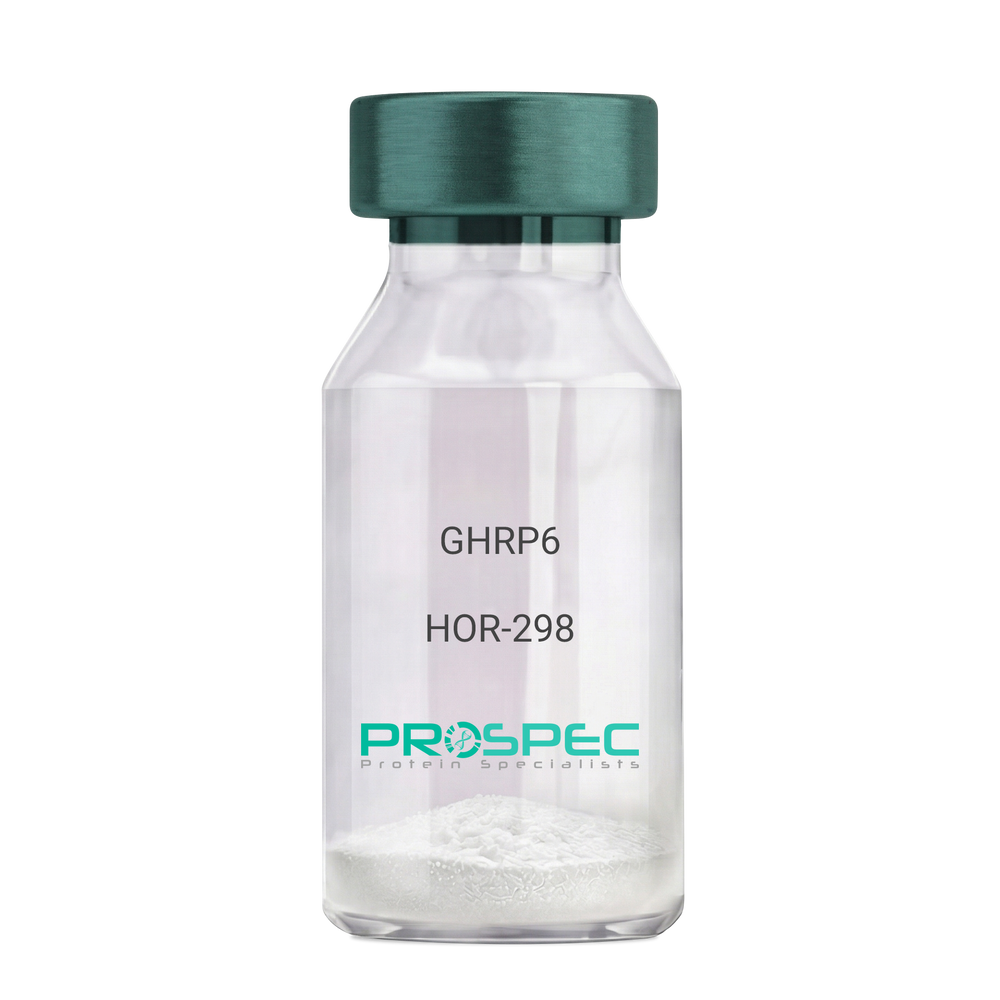
GHRP6Description:
Growth Hormone Releasing Peptide-6
GHRP-6, GHRP6
Product # :
HOR-298Price :
Quantity :
Shipping Method :
Shipped at Room temp
More Info
- description
- formulation
- purity
- More Info
Description
Growth Hormone Releasing Peptide-6 Synthetic is a single, non-glycosylated polypeptide chain containing 6 amino acids, having a molecular mass of 873 Dalton and a Molecular formula of C46H56N12O6.
Formulation
The GHRP-6 peptide was lyophilized with no additives.
Purity
Greater than 98.0% as determined by analysis by RP-HPLC.
More Info
-
Introduction
GH-releasing peptides (GHRPs) are synthetic peptides that like GHRH act directly on pituitary somatotrophs to stimulate GH release. Growth hormone (GH) release is stimulated by a variety of synthetic secretagogues, of which growth hormone-releasing hexapeptide (GHRP-6) has been most thoroughly studied; it is thought to have actions at both pituitary and hypothalamic site.
-
Synonyms
GHRP-6, GHRP6
-
Physical Appearance
Sterile Filtered White lyophilized (freeze-dried) powder.
-
Stability
Lyophilized Growth Hormone Releasing Peptide-6 although stable at room temperature for 3 weeks, should be stored desiccated below -18°C. Upon reconstitution GHRP-6 should be stored at 4°C between 2-7 days and for future use below -18°C. For long term storage it is recommended to add a carrier protein (0.1% HSA or BSA).Please prevent freeze-thaw cycles.
-
Solubility
It is recommended to reconstitute the lyophilized GHRP-6 in sterile 18MΩ-cm H2O not less than 100 µg/ml, which can then be further diluted to other aqueous solutions.
-
Amino Acid Sequence
His-D-Trp-Ala-Trp-D-Phe-Lys-NH2.
ProSpec's products are furnished for LABORATORY RESEARCH USE ONLY. The product may not be used as drugs, agricultural or pesticidal products, food additives or household chemicals.
- View Data Sheet
Name :
GLYATL2 HumanDescription:
Glycine-N-Acyltransferase-Like 2 Human Recombinant
BXMAS2-10, GATF-B, Glycine N-acyltransferase-like protein 2, Acyl-CoA:glycine N-acyltransferase-like protein 2, GLYATL2.
Product # :
ENZ-770Price :
Quantity :
Shipping Method :
Shipped with Ice Packs
More Info
- description
- source
- formulation
- purity
- More Info
Description
GLYATL2 Human Recombinant produced in E.coli is a single, non-glycosylated polypeptide chain containing 317 amino acids (1-294a.a) and having a molecular mass of 36.7kDa. GLYATL2 is fused to a 23 amino acid His-tag at N-terminus & purified by proprietary chromatographic techniques.
Source
Escherichia Coli.
Formulation
The GLYATL2 solution (0.5mg/ml) contains 20mM Tris-HCl buffer (pH 8.0), 0.1M NaCl, 50% glycerol and 2mM DTT.
Purity
Greater than 85% as determined by SDS-PAGE.
More Info
-
Introduction
Glycine-N-Acyltransferase-Like 2 (GLYATL2) is a part of the glycine N-acyltransferase family expressed mainly in salivary gland and trachea. GLYATL2 is a mitochondrial acyltransferase that transfers the acyl group to the N-terminus of glycine. GLYATL2 conjugates numerous substrates, like arachidonoyl-CoA and saturated medium and longchain acyl-CoAs ranging from chain-length C8:0-CoA to C18:0-CoA, to form a variety of N-acylglycines. GLYATL2 also shows a preference for monounsaturated fatty acid oleoyl-CoA (C18:1-CoA) as an acyl donor.
-
Synonyms
BXMAS2-10, GATF-B, Glycine N-acyltransferase-like protein 2, Acyl-CoA:glycine N-acyltransferase-like protein 2, GLYATL2.
-
Physical Appearance
Sterile Filtered colorless solution.
-
Stability
Store at 4°C if entire vial will be used within 2-4 weeks. Store, frozen at -20°C for longer periods of time. For long term storage it is recommended to add a carrier protein (0.1% HSA or BSA). Avoid multiple freeze-thaw cycles.
-
Amino Acid Sequence
MGSSHHHHHH SSGLVPRGSH MGSMLVLHNS QKLQILYKSL EKSIPESIKV YGAIFNIKDK NPFNMEVLVD AWPDYQIVIT RPQKQEMKDD QDHYTNTYHI FTKAPDKLEE VLSYSNVISW EQTLQIQGCQ EGLDEAIRKV ATSKSVQVDY MKTILFIPEL PKKHKTSSND KMELFEVDDD NKEGNFSNMF LDASHAGLVN EHWAFGKNER SLKYIERCLQ DFLGFGVLGP EGQLVSWIVM EQSCELRMGY TVPKYRHQGN MLQIGYHLEK YLSQKEIPFY FHVADNNEKS LQALNNLGFK ICPCGWHQWK CTPKKYC.
ProSpec's products are furnished for LABORATORY RESEARCH USE ONLY. The product may not be used as drugs, agricultural or pesticidal products, food additives or household chemicals.