Skip to content

High-quality research proteins.

  • 1-800-805-6531

  • Mon - Sat 8.00-18.00, Sun - Closed

  • info@prospecbio.com

  • PO Box 6591 East Brunswick NJ 08816

  • Products
    • Cytokines
    • Growth factors
    • Chemokines
    • Cd antigens
    • Neurotrophins
    • Hormones
    • Enzymes
    • Viral antigens
    • Recombinant proteins
    • Natural proteins
    • Monoclonal antibodies
    • Polyclonal antibodies
    • Thermal Packaging
  • About
    • Company
    • Founder
    • Contribution
  • Customers
    • Eastern Asia
    • Eastern Europe
    • North America
    • South America
    • South East Asia
    • Southern Asia
    • Western Europe
  • Ordering
    • Ordering Method
    • Ordering - Credit Card
    • Ordering - PO
    • Shipping Fees
    • FAQs
  • Services
    • DNA Cloning
    • Protein Expression
    • Monoclonal Antibody
    • Fermentation
    • Technology License
  • Contact us
Log in

Country/region

  • USD $ | Albania
  • USD $ | Andorra
  • USD $ | Argentina
  • USD $ | Armenia
  • USD $ | Australia
  • USD $ | Austria
  • USD $ | Azerbaijan
  • USD $ | Belarus
  • USD $ | Belgium
  • USD $ | Bolivia
  • USD $ | Brazil
  • USD $ | Bulgaria
  • USD $ | Cambodia
  • USD $ | Canada
  • USD $ | Chile
  • USD $ | China
  • USD $ | Colombia
  • USD $ | Costa Rica
  • USD $ | Croatia
  • USD $ | Cyprus
  • USD $ | Czechia
  • USD $ | Denmark
  • USD $ | Estonia
  • USD $ | Finland
  • USD $ | France
  • USD $ | Georgia
  • USD $ | Germany
  • USD $ | Greece
  • USD $ | Hong Kong SAR
  • USD $ | Hungary
  • USD $ | Iceland
  • USD $ | India
  • USD $ | Ireland
  • USD $ | Israel
  • USD $ | Italy
  • USD $ | Japan
  • USD $ | Kazakhstan
  • USD $ | Latvia
  • USD $ | Liechtenstein
  • USD $ | Lithuania
  • USD $ | Madagascar
  • USD $ | Malta
  • USD $ | Mexico
  • USD $ | Moldova
  • USD $ | Monaco
  • USD $ | Netherlands
  • USD $ | New Zealand
  • USD $ | North Macedonia
  • USD $ | Norway
  • USD $ | Panama
  • USD $ | Paraguay
  • USD $ | Peru
  • USD $ | Philippines
  • USD $ | Poland
  • USD $ | Portugal
  • USD $ | Romania
  • USD $ | Singapore
  • USD $ | Slovakia
  • USD $ | Slovenia
  • USD $ | South Africa
  • USD $ | South Korea
  • USD $ | Spain
  • USD $ | Sri Lanka
  • USD $ | Sweden
  • USD $ | Switzerland
  • USD $ | Taiwan
  • USD $ | Thailand
  • USD $ | Türkiye
  • USD $ | Ukraine
  • USD $ | United Kingdom
  • USD $ | United States
  • USD $ | Uruguay
  • USD $ | Vatican City
  • USD $ | Venezuela
  • USD $ | Vietnam
  • Facebook
  • Instagram
logoProSpec - Recombinant Protein Manufacturer for academic and R&D industry
Become a distributor
Account Log in
Wishlist
Cart Cart(0)
  • Products
    +
    • Cytokines
    • Growth factors
    • Chemokines
    • Cd antigens
    • Neurotrophins
    • Hormones
    • Enzymes
    • Viral antigens
    • Recombinant proteins
    • Natural proteins
    • Monoclonal antibodies
    • Polyclonal antibodies
    • Thermal Packaging
  • About
    +
    • Company
    • Founder
    • Contribution
  • Customers
    +
    • Eastern Asia
    • Eastern Europe
    • North America
    • South America
    • South East Asia
    • Southern Asia
    • Western Europe
  • Ordering
    +
    • Ordering Method
    • Ordering - Credit Card
    • Ordering - PO
    • Shipping Fees
    • FAQs
  • Services
    +
    • DNA Cloning
    • Protein Expression
    • Monoclonal Antibody
    • Fermentation
    • Technology License
  • Contact us

Item added to your cart

View cart
View All
  • Cytokines
  • Growth factors
  • Chemokines
  • Cd antigens
  • Neurotrophins
  • Hormones
  • Enzymes
  • Viral antigens
  • Recombinant proteins
  • Natural proteins
  • Monoclonal antibodies
  • Polyclonal antibodies
  • Thermal Packaging
  • Tumor Necrosis Factor

    Tumor Necrosis Factor

    View All
  • 4-1BB

    4-1BB

    View All
  • Adiponectin

    Adiponectin

    View All
  • AIF1

    AIF1

    View All
  • Other Cytokines

    Other Cytokines

    View All
  • AITRL

    AITRL

    View All
  • Angiopoietin

    Angiopoietin

    View All
  • Apolipoprotein

    Apolipoprotein

    View All
  • B-Cell Activating Factor

    B-Cell Activating Factor

    View All
  • Beta Defensin

    Beta Defensin

    View All
  • Bone Morphogenetic Protein

    Bone Morphogenetic Protein

    View All
  • B type Natriuretic Peptide

    B type Natriuretic Peptide

    View All
  • BST

    BST

    View All
  • Betacellulin

    Betacellulin

    View All
  • Cardiotrophin

    Cardiotrophin

    View All
  • Activin

    Activin

    View All
  • Fibroblast Growth Factor

    Fibroblast Growth Factor

    View All
  • Other Growth Factors

    Other Growth Factors

    View All
  • CSF

    CSF

    View All
  • CTGF

    CTGF

    View All
  • IGFBP

    IGFBP

    View All
  • VEGF Protein

    VEGF Protein

    View All
  • EGF

    EGF

    View All
  • Epigen

    Epigen

    View All
  • Erythropoietin

    Erythropoietin

    View All
  • Insulin-Like Growth Factor

    Insulin-Like Growth Factor

    View All
  • Growth Hormone

    Growth Hormone

    View All
  • HDGF

    HDGF

    View All
  • Hepatocyte Growth Factor

    Hepatocyte Growth Factor

    View All
  • Insulin

    Insulin

    View All
  • BCA-1/ BLC (CXCL13)

    BCA-1/ BLC (CXCL13)

    View All
  • BRAK (CXCL14)

    BRAK (CXCL14)

    View All
  • C-10 (CCL6)

    C-10 (CCL6)

    View All
  • Other Chemokines

    Other Chemokines

    View All
  • MEC (CCL28)

    MEC (CCL28)

    View All
  • LD78-beta (CCL3L1)

    LD78-beta (CCL3L1)

    View All
  • CTACK (CCL27)

    CTACK (CCL27)

    View All
  • CXCL16

    CXCL16

    View All
  • CXCL17

    CXCL17

    View All
  • Platelet Factor-4 (CXCL4)

    Platelet Factor-4 (CXCL4)

    View All
  • ENA-78 (CXCL5)

    ENA-78 (CXCL5)

    View All
  • Eotaxin (CCL11,24,26)

    Eotaxin (CCL11,24,26)

    View All
  • Exodus-2 (CCL21)

    Exodus-2 (CCL21)

    View All
  • Fractalkine (CX3CL1)

    Fractalkine (CX3CL1)

    View All
  • CXCL6

    CXCL6

    View All
  • Other CD Antigens

    Other CD Antigens

    View All
  • CD14

    CD14

    View All
  • CD164

    CD164

    View All
  • CD1

    CD1

    View All
  • CD2

    CD2

    View All
  • CD200

    CD200

    View All
  • CD207

    CD207

    View All
  • CD226

    CD226

    View All
  • CD23

    CD23

    View All
  • CD244

    CD244

    View All
  • CD247

    CD247

    View All
  • CD27

    CD27

    View All
  • CD274

    CD274

    View All
  • CD300

    CD300

    View All
  • CD33

    CD33

    View All
  • Other Neurotrophins

    Other Neurotrophins

    View All
  • Beta-NGF

    Beta-NGF

    View All
  • BDNF

    BDNF

    View All
  • CDNF

    CDNF

    View All
  • CNTF

    CNTF

    View All
  • GDNF

    GDNF

    View All
  • Glia Maturation Factor

    Glia Maturation Factor

    View All
  • MANF

    MANF

    View All
  • Midkine

    Midkine

    View All
  • Neuroglobin

    Neuroglobin

    View All
  • Neurotrophic factors

    Neurotrophic factors

    View All
  • Neuregulin

    Neuregulin

    View All
  • Neuropilin

    Neuropilin

    View All
  • Pigment Epithelium-Derived Factor

    Pigment Epithelium-Derived Factor

    View All
  • Pleiotrophin

    Pleiotrophin

    View All
  • Peptide Hormones

    Peptide Hormones

    View All
  • Other Hormones

    Other Hormones

    View All
  • HCG

    HCG

    View All
  • Endothelin

    Endothelin

    View All
  • Exendin

    Exendin

    View All
  • FSH

    FSH

    View All
  • Glucagon

    Glucagon

    View All
  • GHRP

    GHRP

    View All
  • GLP

    GLP

    View All
  • Thyrostimulin

    Thyrostimulin

    View All
  • Inhibin A

    Inhibin A

    View All
  • LHRH

    LHRH

    View All
  • Melanotan

    Melanotan

    View All
  • Procalcitonin

    Procalcitonin

    View All
  • PTH

    PTH

    View All
  • Peroxiredoxin

    Peroxiredoxin

    View All
  • Peptidase

    Peptidase

    View All
  • Synthetase

    Synthetase

    View All
  • Transferase

    Transferase

    View All
  • Hydrolase

    Hydrolase

    View All
  • Dehydrogenase

    Dehydrogenase

    View All
  • ACE2

    ACE2

    View All
  • Esterase

    Esterase

    View All
  • Protein Kinases

    Protein Kinases

    View All
  • Other Enzymes

    Other Enzymes

    View All
  • Phosphatase

    Phosphatase

    View All
  • Deaminase

    Deaminase

    View All
  • Oxygenase

    Oxygenase

    View All
  • Adenylate Kinase

    Adenylate Kinase

    View All
  • Lyase

    Lyase

    View All
  • Chagas

    Chagas

    View All
  • SARS

    SARS

    View All
  • Influenza

    Influenza

    View All
  • ASFV

    ASFV

    View All
  • Astrovirus

    Astrovirus

    View All
  • Borrelia

    Borrelia

    View All
  • Helicobacter Pylori

    Helicobacter Pylori

    View All
  • Chikungunya

    Chikungunya

    View All
  • Chlamydia

    Chlamydia

    View All
  • Cytomegalo

    Cytomegalo

    View All
  • Dengue

    Dengue

    View All
  • EBV

    EBV

    View All
  • Ebola

    Ebola

    View All
  • Feline Leukemia Virus

    Feline Leukemia Virus

    View All
  • Hepatitis A

    Hepatitis A

    View All
  • Other

    Other

    View All
  • Actin

    Actin

    View All
  • ADAM

    ADAM

    View All
  • Complement Component

    Complement Component

    View All
  • Ag85

    Ag85

    View All
  • Eukaryotic Translation Initiation Factor

    Eukaryotic Translation Initiation Factor

    View All
  • Anterior Gradient Protein

    Anterior Gradient Protein

    View All
  • Heat Shock Protein

    Heat Shock Protein

    View All
  • Alpha-2-HS-Glycoprotein

    Alpha-2-HS-Glycoprotein

    View All
  • Hemoglobin

    Hemoglobin

    View All
  • Allergy

    Allergy

    View All
  • Anaplasma

    Anaplasma

    View All
  • Angiogenin

    Angiogenin

    View All
  • Ankyrin Repeat Domain

    Ankyrin Repeat Domain

    View All
  • Annexin

    Annexin

    View All
  • Other Natural Proteins

    Other Natural Proteins

    View All
  • Aprotinin

    Aprotinin

    View All
  • Transferrin

    Transferrin

    View All
  • Avidin

    Avidin

    View All
  • Anti Coagulation Factors

    Anti Coagulation Factors

    View All
  • Natural Albumin

    Natural Albumin

    View All
  • Natural Coagulation Factors

    Natural Coagulation Factors

    View All
  • Fibronectin

    Fibronectin

    View All
  • Thrombin

    Thrombin

    View All
  • Anti Human Enzyme

    Anti Human Enzyme

    View All
  • Other Monoclonal Antibodies

    Other Monoclonal Antibodies

    View All
  • Anti Human Cytokine

    Anti Human Cytokine

    View All
  • Anti Human Heat Shock Protein

    Anti Human Heat Shock Protein

    View All
  • Anti Mouse Lymphocyte

    Anti Mouse Lymphocyte

    View All
  • Anti Human Chemokine

    Anti Human Chemokine

    View All
  • Anti Human Lymphocyte

    Anti Human Lymphocyte

    View All
  • Anti Viral Monoclonal

    Anti Viral Monoclonal

    View All
  • Anti Mouse Cytokine

    Anti Mouse Cytokine

    View All
  • Anti-GST Monoclonal

    Anti-GST Monoclonal

    View All
  • Other Polyclonal Antibodies

    Other Polyclonal Antibodies

    View All
  • Anti Viral

    Anti Viral

    View All
  • Anti-GST Monoclonal

    Anti-GST Monoclonal

    View All
Back

Search results

1000 results found for “Fibrinogen”

Name

Description

Product #

Price

Quantity

Shipping Method

  • View Data Sheet

    Name :

    PTX3 Human

    Description:

    Pentraxin-3 Human Recombinant

    TSG-14, TNFAIP5, PTX3, Pentraxin-related protein PTX3, Pentaxin-related protein PTX3, Tumor necrosis factor-inducible gene 14 protein, TSG14, pentraxin-related gene rapidly induced by IL-1 beta.

    Product # :

    PRO-694

    Price :

    Quantity :

    Shipping Method :

    Ice Icon

    Shipped with Ice Packs

    Add To Cart

    More Info

    • description
    • source
    • formulation
    • purity
    • More Info

    Description

    Recombinant Human PTX3 produced in E.Coli is a single, non-glycosylated polypeptide chain containing 401 amino acids (18-381 a.a) and having a molecular mass of 44.4 kDa. PTX3 is fused to a 37 amino acid His Tag at N-terminus and purified by proprietary chromatographic techniques.

    Source

    Escherichia Coli.

    Formulation

    The PTX3 protein contains 20mM Tris-HCl buffer pH-8, 1mM DTT and 10% glycerol.

    Purity

    Greater than 90% as determined by Analysis by SDS-PAGE.

    More Info

    • Introduction

      PTX3 is part of the pentraxin family sharing the C-terminal domain with short pentraxins and containing a unique N-terminal domain. PTX3 is produced and released at inflammatory sites by various cell types including monocytes/macrophages, endothelial cells, vascular smooth muscle cells, fibroblasts, and adipocytes. PTX3 is involved in the regulation of innate resistance to pathogens, inflammatory reactions, possibly clearance of self-components and female fertility. PTX3 is used as a marker for disease activity of psoriasis. High serum PTX3 levels are associated with the disease severity of systemic sclerosis. Elevated serum PTX3 is associated with pulmonary fungal infections.

    • Synonyms

      TSG-14, TNFAIP5, PTX3, Pentraxin-related protein PTX3, Pentaxin-related protein PTX3, Tumor necrosis factor-inducible gene 14 protein, TSG14, pentraxin-related gene rapidly induced by IL-1 beta.

    • Physical Appearance

      Sterile filtered colorless solution.

    • Stability

      Store at 4°C if entire vial will be used within 2-4 weeks. Store, frozen at -20°C for longer periods of time. For long term storage it is recommended to add a carrier protein (0.1% HSA or BSA). Avoid multiple freeze-thaw cycles.

    • Amino Acid Sequence

      MRGSHHHHHH GMASMTGGQQ MGRDLYDDDD KDRWGSMENS DDYDLMYVNL DNEIDNGLHP TEDPTPCDCG QEHSEWDKLF IMLENSQMRE RMLLQATDDV LRGELQRLRE ELGRLAESLA RPCAPGAPAE ARLTSALDEL LQATRDAGRR LARMEGAEAQ RPEEAGRALA AVLEELRQTR ADLHAVQGWA ARSWLPAGCE TAILFPMRSK KIFGSVHPVR PMRLESFSAC IWVKATDVLN KTILFSYGTK RNPYEIQLYL SYQSIVFVVG GEENKLVAEA MVSLGRWTHL CGTWNSEEGL TSLWVNGELA ATTVEMATGH IVPEGGILQI GQEKNGCCVG GGFDETLAFS GRLTGFNIWD SVLSNEEIRE TGGAESCHIR GNIVGWGVTE IQPHGGAQYV S.

    ProSpec's products are furnished for LABORATORY RESEARCH USE ONLY. The product may not be used as drugs, agricultural or pesticidal products, food additives or household chemicals.

    Ptx3 Human
  • View Data Sheet

    Name :

    SAA1 Human

    Description:

    Serum Amyloid A (APO-SAA1) Human Recombinant

    Serum amyloid A protein, SAA, Amyloid protein A, Amyloid fibril protein AA, SAA1, SAA2, PIG4, TP53I4, MGC111216.

    Product # :

    CYT-787

    Price :

    Quantity :

    Shipping Method :

    Room Temp Icon

    Shipped at Room temp

    Add To Cart

    More Info

    • description
    • source
    • formulation
    • purity
    • biological activity
    • More Info

    Description

    SAA1 Human Recombinant produced in E.Coli is a single, non-glycosylated polypeptide chain containing 104 amino acids and having a molecular mass of 11.7kDa. The SAA1 is purified by proprietary chromatographic techniques.

    Source

    Escherichia Coli.

    Formulation

    SAA1 was lyophilized from a 0.2µm filtered concentrated solution in 20mM Tris-HCl, pH 9.0 and 150mM NaCl.

    Purity

    Greater than 98.0% as determined by:
    (a) Analysis by RP-HPLC.
    (b) Analysis by SDS-PAGE.

    Biological Activity

    The biological activity determined by a chemoattract bioassay using human monocytes is in a concentration range of 10-100 ng/ml.

    More Info

    • Introduction

      SAA1 protein is an acute phase apolipoprotein reactant which is produced mostly by hepatocytes and under regulation of inflammatory cytokines. SAA1 (Serum amyloid A1) protein is produced mainly in the liver and circulates in low levels in the blood. The SAA1 seems to have a role in the immune system. SAA1 protein levels increase in the blood and other tissues under conditions of inflammation. SAA1 may facilitate the repair of injured tissues; it also acts as an antibacterial agent, and signals the migration of germ-fighting cells to sites of infection. SAA1 also functions as an apolipoprotein of the HDL complex.
      Elevated levels of SAA1 ultimately affect secondary amyloidosis, extracellular amassing of amyloid fibrils, resulting from a circulating precursor, in a variety of tissues and organs. The most widespread type of amyloidosis appears secondary to chronic inflammatory disease, mainly rheumatoid arthritis. The SAA1 cleavage product a designated amyloid protein A is deposited systemically as amyloid in vital organs such as the liver, spleen, and kidneys in chronic inflammatory diseases patients. These deposits are extremely insoluble and resistant to proteolysis; they disrupt tissue structure and compromise performance.

    • Synonyms

      Serum amyloid A protein, SAA, Amyloid protein A, Amyloid fibril protein AA, SAA1, SAA2, PIG4, TP53I4, MGC111216.

    • Physical Appearance

      Sterile Filtered White lyophilized (freeze-dried) powder.

    • Stability

      Lyophilized SAA1 although stable at room temperature for 3 weeks, should be stored desiccated below -18°C. Upon reconstitution SAA1 should be stored at 4°C between 2-7 days and for future use below -18°C. For long term storage it is recommended to add a carrier protein (0.1% HSA or BSA). Please prevent freeze-thaw cycles.

    • Solubility

      It is recommended to reconstitute the lyophilized SAA1 in sterile 18MΩ-cm H2O not less than 100µg/ml, which can then be further diluted to other aqueous solutions.

    • Amino Acid Sequence

      RSFFSFLGEA FDGARDMWRA YSDMREANYI GSDKYFHARG NYDAAKRGPG GVWAAEAISD ARENIQRFFG HGAEDSLADQ AANEWGRSGK DPNHFRPAGL PEKY.

    ProSpec's products are furnished for LABORATORY RESEARCH USE ONLY. The product may not be used as drugs, agricultural or pesticidal products, food additives or household chemicals.

    Human Saa1
  • View Data Sheet

    Name :

    SSSCA1 Human

    Description:

    Sjogren Syndrome/Scleroderma Autoantigen 1 Human Recombinant

    Sjogren Syndrome/Scleroderma Autoantigen 1, Sjogren'S Syndrome/Scleroderma Autoantigen 1, Autoantigen P27, Centromeric Autoantigen (27kD), p27, Sjoegren Syndrome/Scleroderma Autoantigen 1.

    Product # :

    PRO-1743

    Price :

    Quantity :

    Shipping Method :

    Ice Icon

    Shipped with Ice Packs

    Add To Cart

    More Info

    • description
    • source
    • formulation
    • purity
    • More Info

    Description

    SSSCA1 Human Recombinant produced in E.Coli is a single, non-glycosylated polypeptide chain containing 222 amino acids (1-199aa) and having a molecular mass of 23.9kDa. SSSCA1 is fused to a 23 amino acid His-tag at N-terminus & purified by proprietary chromatographic techniques.

    Source

    Escherichia Coli.

    Formulation

    SSSCA1 protein solution (0.5mg/ml) containing 20mM Tris-HCl buffer (pH 8.0), 0.4M urea and 10% glycerol.

    Purity

    Greater than 90.0% as determined by SDS-PAGE.

    More Info

    • Introduction

      Sjogren syndrome/scleroderma autoantigen 1, SSSCA1 is involved in mitosis. SSSCA1 is a centromere-associated protein which can induce anti-centromere antibodies. In addition SSSCA1 is recognized by a subset of anti-centromere antibodies from patients with sclerodermaand Sjogren's syndrome.

    • Synonyms

      Sjogren Syndrome/Scleroderma Autoantigen 1, Sjogren'S Syndrome/Scleroderma Autoantigen 1, Autoantigen P27, Centromeric Autoantigen (27kD), p27, Sjoegren Syndrome/Scleroderma Autoantigen 1.

    • Physical Appearance

      Sterile Filtered clear solution.

    • Stability

      Store at 4°C if entire vial will be used within 2-4 weeks. Store, frozen at -20°C for longer periods of time.For long term storage it is recommended to add a carrier protein (0.1% HSA or BSA).Please avoid freeze thaw cycles.

    • Amino Acid Sequence

      MGSSHHHHHH SSGLVPRGSH MGSMALNGAE VDDFSWEPPT EAETKVLQAR RERQDRISRL MGDYLLRGYR MLGETCADCG TILLQDKQRK IYCVACQELD SDVDKDNPAL NAQAALSQAR EHQLASASEL PLGSRPAPQP PVPRPEHCEG AAAGLKAAQG PPAPAVPPNT DVMACTQTAL LQKLTWASAE LGSSTSLETS IQLCGLIRAC AEALRSLQQL QH

    ProSpec's products are furnished for LABORATORY RESEARCH USE ONLY. The product may not be used as drugs, agricultural or pesticidal products, food additives or household chemicals.

    Sssca1 Human
  • View Data Sheet

    Name :

    FABP4 Human

    Description:

    Fatty Acid Binding Protein 4 Human

    Fatty acid-binding protein adipocyte, AFABP, Fatty acid-binding protein 4, Adipocyte lipid-binding protein, ALBP, A-FABP, FABP4.

    Product # :

    PRO-1568

    Price :

    Quantity :

    Shipping Method :

    Room Temp Icon

    Shipped at Room temp

    Add To Cart

    More Info

    • description
    • source
    • formulation
    • purity
    • More Info

    Description

    The Human FABP4 produced from Human Adipose Tissue has a molecular mass of 14.587kDa (calculated without glycosylation) containing 131 amino acid residues.

    Source

    Human Adipose Tissue.

    Formulation

    FABP4 protein filtered (0.4µm) and lyophilized in 0.5mg/ml in 0.05M phosphate buffer and 0.075M NaCl, pH 6.5.

    Purity

    Greater than 85.0% as determined by SDS-PAGE.

    More Info

    • Introduction

      Adipocyte fatty acid binding protein FABP4 is a 15 kDa member of the intracellular fatty acid binding protein (FABP) family, which is known for the ability to bind fatty acids and related compounds (bile acids or retinoids) in an internal cavity. FABP4 is expressed in a differentiation-dependent fashion in adipocytes and is a critical gene in the regulation of the biological function of these cells.
      In mice, targeted mutations in FABP4 provide significant protection from IR in the context of both dietary and genetic obesity. Adipocytes obtained from FABP4-deficient mice also have reduced efficiency of ipolysis in vitro and in vivo, and these mice exhibited moderately improved systemic dyslipidemia. Recent studies also demonstrated FABP4 expression in macrophages upon differentiation and activation. In these cells, FABP4 modulates inflammatory responses and cholesterol ester accumulation, and total or macrophage-specific FABP4 deficiency confers dramatic protection against atherosclerosis in the apoE-/- mice. These results indicate a central role for FABP4 in the development of major components of the metabolic syndrome through its distinct actions in adipocytes and macrophages.

    • Synonyms

      Fatty acid-binding protein adipocyte, AFABP, Fatty acid-binding protein 4, Adipocyte lipid-binding protein, ALBP, A-FABP, FABP4.

    • Physical Appearance

      Filtered White lyophilized (freeze-dried) powder.

    • Stability

      Store lyophilized protein at -20°C. Aliquot the product after reconstitution to avoid repeated freezing/thawing cycles. Reconstituted protein can be stored at 4°C for a limited period of time; it does not show any change after two weeks at 4°C.

    • Solubility

      It is recommended to add deionized water to prepare a working stock solution of approximately 0.5 mg/ml and let the lyophilized pellet dissolve completely. Product is not sterile! Please filter the product by an appropriate sterile filter before using it in the cell culture.

    • Amino Acid Sequence

      CDAFVGTWKL VSSENFDDYM KEVGVGFATR KVAGMAKPNM IISVNGDVIT IKSESTFKNT EISFILGQEF DEVTADDRKV KSTITLDGGV LVHVQKWDGK STTIKRKRED DKLVVECVMK GVTSTRVYER A.

    ProSpec's products are furnished for LABORATORY RESEARCH USE ONLY. The product may not be used as drugs, agricultural or pesticidal products, food additives or household chemicals.

    Human Fabp4
  • View Data Sheet

    Name :

    TROVE2 Human

    Description:

    TROVE Domain Family Member 2 Human Recombinant

    60 kDa SS-A/Ro ribonucleoprotein, 60 kDa ribonucleoprotein Ro, RoRNP, 60 kDa Ro protein, Ro 60 kDa autoantigen, TROVE domain family member 2, Sjoegren syndrome type A antigen, SS-A, Sjoegren syndrome antigen A2, TROVE2, RO60, SSA2, RO-60.

    Product # :

    PRO-329

    Price :

    Quantity :

    Shipping Method :

    Ice Icon

    Shipped with Ice Packs

    Add To Cart

    More Info

    • description
    • source
    • formulation
    • purity
    • More Info

    Description

    RO-60 Human Recombinant produced in E.Coli is a single, non-glycosylated, polypeptide chain having a molecular mass of 60kDa. RO60 is fused to a 6 amino acid His-Tag at C-terminus and purified by proprietary chromatographic techniques.

    Source

    Escherichia Coli.

    Formulation

    The protein solution contains 6M Urea, 500mM NaCl, 500mM imidazole and 10mM Tris pH-6.

    Purity

    Greater than 90.0% as determined by SDS-PAGE.

    More Info

    • Introduction

      Ro 60 kDa autoantigen is an RNA-binding protein that binds to several small cytoplasmic RNA molecules known as Y RNAs. SSA2 may stabilize these RNAs from degradation. Sera taken from patients with Systemic Lupus Erythematosus (SLE) often contains antibodies that react with the normal cellular SSA2 protein as if this antigen was foreign.

    • Synonyms

      60 kDa SS-A/Ro ribonucleoprotein, 60 kDa ribonucleoprotein Ro, RoRNP, 60 kDa Ro protein, Ro 60 kDa autoantigen, TROVE domain family member 2, Sjoegren syndrome type A antigen, SS-A, Sjoegren syndrome antigen A2, TROVE2, RO60, SSA2, RO-60.

    • Physical Appearance

      Sterile Filtered clear solution.

    • Stability

      RO-60 although stable at 4°C for 3 weeks, should be stored below -18°C. Please prevent freeze-thaw cycles.

    ProSpec's products are furnished for LABORATORY RESEARCH USE ONLY. The product may not be used as drugs, agricultural or pesticidal products, food additives or household chemicals.

    Trove2 Human
  • View Data Sheet

    Name :

    CGB3 Human

    Description:

    CGB3 Human Recombinant

    CGB3 CGB5 CGB8, CGB5, CGB7, CGB8, HCGB, CG-beta.

    Product # :

    PRO-2398

    Price :

    Quantity :

    Shipping Method :

    Ice Icon

    Shipped with Ice Packs

    Add To Cart

    More Info

    • description
    • source
    • formulation
    • purity
    • More Info

    Description

    CGB3 Human Recombinant produced in Sf9 Baculovirus cells is a single, glycosylated polypeptide chain containing 154 amino acids (21-165.a.) and having a molecular mass of 16.6kDa (Molecular size on SDS-PAGE will appear at approximately 18-28kDa). CGB3 is expressed with a 6 amino acid His tag at C-Terminus and purified by proprietary chromatographic techniques.

    Source

    Sf9, Baculovirus cells.

    Formulation

    CGB3 protein solution (0.25mg/ml) contains Phosphate Buffered Saline (pH 7.4) and 10% glycerol.

    Purity

    Greater than 90.0% as determined by SDS-PAGE.

    More Info

    • Introduction

      CGB3, belongs to the glycoprotein hormone beta chain family. CGB3 encodes the beta 3 subunit. CGB3 stimulates the expression of GCM1 target genes, as well as the fusogenic protein syncytin-1, to promote placental cell fusion. Furthermore, CGB3 is synthesized in large quantities by the growing placenta, reaching peak concentrations in maternal blood throughout the late first trimester and early mid-trimester of pregnancy. CGB3 is expressed solely by trophoblasts. CGB3 is a significant biomarker in pregnancy and oncology, where it is routinely detected and quantified by specific immunoassays.

    • Synonyms

      CGB3 CGB5 CGB8, CGB5, CGB7, CGB8, HCGB, CG-beta.

    • Physical Appearance

      Sterile Filtered clear solution.

    • Stability

      Store at 4°C if entire vial will be used within 2-4 weeks. Store, frozen at -20°C for longer periods of time. For long term storage it is recommended to add a carrier protein (0.1% HSA or BSA).Avoid multiple freeze-thaw cycles.

    • Amino Acid Sequence

      ADPSKEPLRP RCRPINATLA VEKEGCPVCI TVNTTICAGY CPTMTRVLQG VLPALPQVVC NYRDVRFESI RLPGCPRGVN PVVSYAVALS CQCALCRRST TDCGGPKDHP LTCDDPRFQD SSSSKAPPPS LPSPSRLPGP SDTPILPQHH HHHH.

    ProSpec's products are furnished for LABORATORY RESEARCH USE ONLY. The product may not be used as drugs, agricultural or pesticidal products, food additives or household chemicals.

    Cgb3 Human 2
  • View Data Sheet

    Name :

    XG Human

    Description:

    XG Blood Group Human Recombinant

    Glycoprotein Xg isoform 1 precursor, PBDX, Glycoprotein Xg, PBDX, XG.

    Product # :

    PRO-1395

    Price :

    Quantity :

    Shipping Method :

    Ice Icon

    Shipped with Ice Packs

    Add To Cart

    More Info

    • description
    • source
    • formulation
    • purity
    • More Info

    Description

    XG Human Recombinant produced in E.Coli is a single, non-glycosylated polypeptide chain containing 144 amino acids (22-142a.a) and having a molecular mass of 15.5kDa. XG is fused to a 23 amino acid His-tag at N-terminus & purified by proprietary chromatographic techniques.

    Source

    E.coli.

    Formulation

    XG protein solution (0.25mg/ml) containing 20mM Tris-HCl buffer (pH 8.0), 0.15M NaCl, 10% glycerol and 1mM DTT.

    Purity

    Greater than 90% as determined by SDS-PAGE.

    More Info

    • Introduction

      XG is the XG blood group antigen, and is at the pseudoautosomal boundary on the short (p) arm of chromosome X. The three 5' exons are located in the pseudoautosomal area and the remaining exons within the X-specific end. A shorted copy of XG is found on the Y chromosome at the pseudoautosomal boundary. XG is transcribed, although not expected to make a Y-chromosome specific gene product.

    • Synonyms

      Glycoprotein Xg isoform 1 precursor, PBDX, Glycoprotein Xg, PBDX, XG.

    • Physical Appearance

      Sterile Filtered colorless solution.

    • Stability

      Store at 4°C if entire vial will be used within 2-4 weeks. Store, frozen at -20°C for longer periods of time. For long term storage it is recommended to add a carrier protein (0.1% HSA or BSA).Avoid multiple freeze-thaw cycles.

    • Amino Acid Sequence

      MGSSHHHHHH SSGLVPRGSH MGSQRDFDLA DALDDPEPTK KPNSDIYPKP KPPYYPQPEN PDSGGNIYPR PKPRPQPQPG NSGNSGGYFN DVDRDDGRYP PRPRPRPPAG GGGGGYSSYG NSDNTHGGDH HSTYGNPEGN MVAK.

    ProSpec's products are furnished for LABORATORY RESEARCH USE ONLY. The product may not be used as drugs, agricultural or pesticidal products, food additives or household chemicals.

    Xg Human
  • View Data Sheet

    Name :

    sRANKL Human, GST

    Description:

    Soluble RANK Ligand Human Recombinant, GST tag

    Soluble Receptor Activator of NFkB Ligand, TNFSF11, TRANCE, TNF-related activation-induced cytokine, OPGL, ODF, Osteoclast differentiation factor, Tumor necrosis factor ligand superfamily member 11, Receptor activator of nuclear factor kappa B ligand, RANKL, Osteoprotegerin ligand, CD254 antigen, sRANKL, sOdf, hRANKL2.

    Product # :

    CYT-631

    Price :

    Quantity :

    Shipping Method :

    Ice Icon

    Shipped with Ice Packs

    Add To Cart

    More Info

    • description
    • source
    • formulation
    • purity
    • More Info

    Description

    RANKL Human Recombinant fused to GST tag produced in E.Coli is a single, non-glycosylated polypeptide having a molecular mass of 47 kDa. RANKL is purified by proprietary chromatographic techniques.

    Source

    Escherichia Coli.

    Formulation

    The soluble RANKL protein solution contains 1mM EDTA and PBS pH-7.4.

    Purity

    Greater than 95.0% as determined by SDS-PAGE.

    More Info

    • Introduction

      RANKL binds to tnfrsf11b/opg and to tnfrsf11a/rank. Osteoclast differentiation and activation factor. Augments the ability of dendritic cells to stimulate naive t-cell proliferation. May be an important regulator of interactions between t-cells and dendritic cells and may play a role in the regulation of the t-cell-dependent immune response. sRANKL may also play an important role in enhanced bone-resorption in humoral hypercalcemia of malignancy. By injecting soluble RANKL, novel osteopenia model mice were established in only 50 hours. Degree of bone loss can be controlled by changing doses of sRANKL. RANKL can be used in establishing osteopenia model for in vivo screening of drugs for osteoporosis, determination of effect and mechanism, evaluation of bone anabolic drugs.

    • Synonyms

      Soluble Receptor Activator of NFkB Ligand, TNFSF11, TRANCE, TNF-related activation-induced cytokine, OPGL, ODF, Osteoclast differentiation factor, Tumor necrosis factor ligand superfamily member 11, Receptor activator of nuclear factor kappa B ligand, RANKL, Osteoprotegerin ligand, CD254 antigen, sRANKL, sOdf, hRANKL2.

    • Physical Appearance

      Sterile Filtered colorelss clear solution.

    • Stability

      Store at 4°C if entire vial will be used within 2-4 weeks. Store, frozen at -20°C for longer periods of time. Please avoid freeze thaw cycles.

    ProSpec's products are furnished for LABORATORY RESEARCH USE ONLY. The product may not be used as drugs, agricultural or pesticidal products, food additives or household chemicals.

    Rankl Human Gst
  • View Data Sheet

    Name :

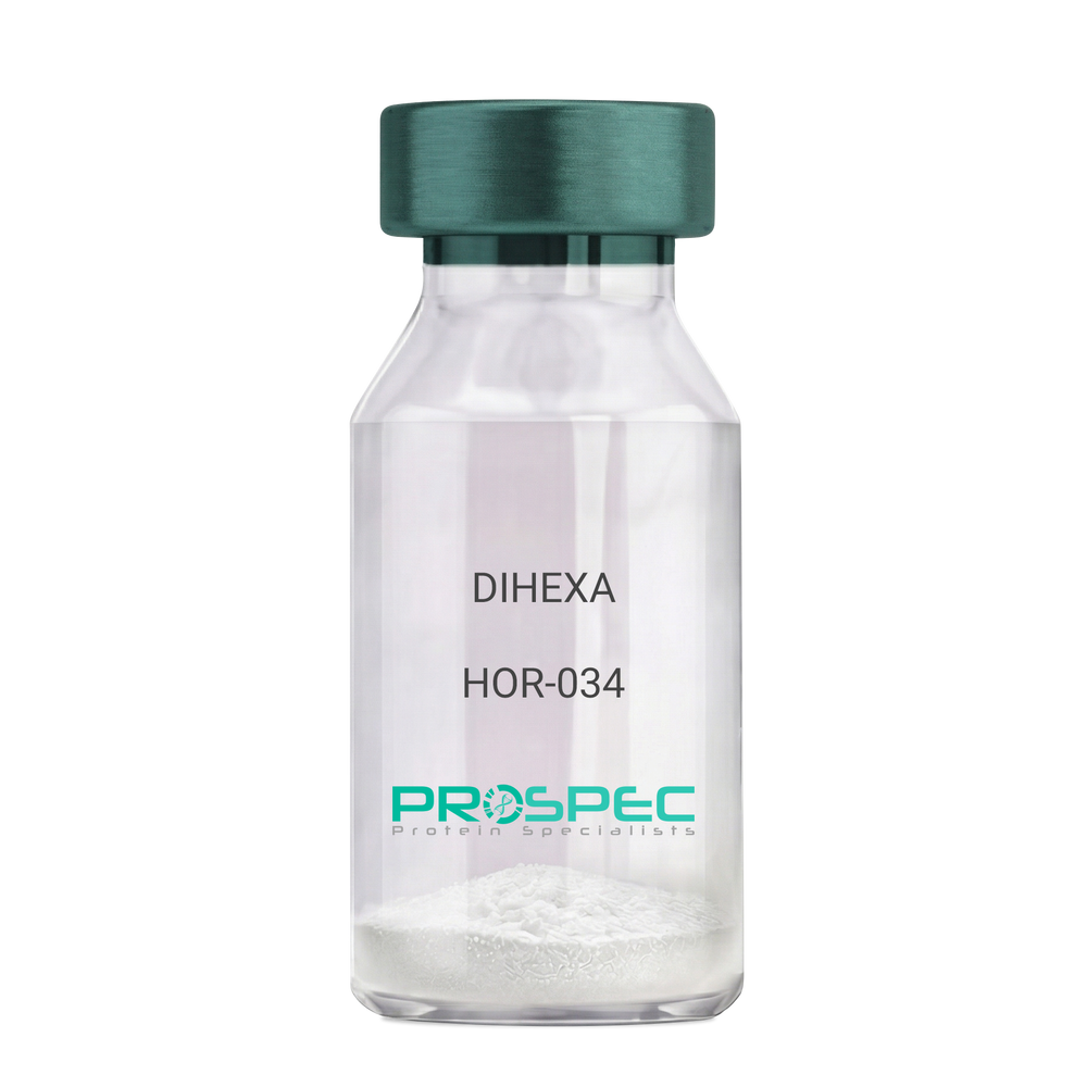
    DIHEXA

    Description:

    DIHEXA

    Product # :

    HOR-034

    Price :

    Quantity :

    Shipping Method :

    Room Temp Icon

    Shipped at Room temp

    Add To Cart

    More Info

    • description
    • formulation
    • purity
    • More Info

    Description

    DIHEXA Synthetic is a single, non-glycosylated polypeptide chain containing 3 amino acids, having a molecular mass of 504.28 Dalton and a Molecular formula of C27H44N4O5.

    Formulation

    The protein was lyophilized with no additives.

    Purity

    Greater than 97.0% as determined by analysis by RP-HPLC.

    More Info

    • Physical Appearance

      Sterile Filtered White lyophilized (freeze-dried) powder.

    • Stability

      Lyophilized DIHEXA although stable at room temperature for 3 weeks, should be stored desiccated below -18°C. Upon reconstitution DIHEXA should be stored at 4°C between 2-7 days and for future use below -18°C. For long term storage it is recommended to add a carrier protein (0.1% HSA or BSA). Please prevent freeze-thaw cycles.

    • Solubility

      It is recommended to reconstitute the lyophilized DIHEXA in sterile 18MΩ-cm H2O not less than 100 µg/ml, which can then be further diluted to other aqueous solutions.

    • Amino Acid Sequence

      Hexanoyl-Tyr-Ile-Ahx-NH2.

    • Background

      Dihexa, also known as N-hexanoic-Tyr-Ile-(6) aminohexanoic amide, is a potent and orally active small peptide that has been the focus of significant research due to its potential neurogenic and neuroprotective effects. This compound, derived from angiotensin IV, has been shown to possess a wide range of biological activities, including enhancing cognitive function, promoting neurogenesis, and potentially mitigating the effects of neurodegenerative diseases.

      Dihexa's primary mechanism of action involves its interaction with hepatocyte growth factor (HGF) and its receptor, c-Met. By mimicking the effects of HGF, Dihexa can stimulate the c-Met receptor, leading to a cascade of events that promote neurogenesis and synaptic plasticity. Studies by Benoist et al. (2014) have demonstrated that Dihexa can enhance cognitive function in rats, suggesting potential applications in cognitive enhancement and the treatment of cognitive disorders.

      In addition to its neurogenic effects, Dihexa has been shown to possess neuroprotective properties. Research by Kawas et al. (2017) found that Dihexa could protect neurons from apoptosis, suggesting potential applications in the treatment of neurodegenerative diseases such as Alzheimer's and Parkinson's disease.

      Given its neurogenic and neuroprotective effects, Dihexa has been proposed as a potential therapeutic agent for a variety of conditions, including cognitive disorders, neurodegenerative diseases, and stroke. For instance, a study by Harding et al. (2018) found that Dihexa could improve outcomes in animal models of stroke, indicating its potential as a therapeutic agent in stroke recovery.

      While research on Dihexa is promising, it is important to note that most studies have been conducted in animals or in vitro. More research is needed to fully understand the potential effects and applications of Dihexa in humans. However, the existing body of research suggests that Dihexa could be a promising tool in the treatment of cognitive disorders and neurodegenerative diseases.

    ProSpec's products are furnished for LABORATORY RESEARCH USE ONLY. The product may not be used as drugs, agricultural or pesticidal products, food additives or household chemicals.

    Dihexa
  • View Data Sheet

    Name :

    hCG Protein

    Description:

    Chorionic Gonadotropin Human

    Chorionic gonadotropin, hCG, CG.

    Product # :

    HOR-250

    Price :

    Quantity :

    Shipping Method :

    Room Temp Icon

    Shipped at Room temp

    Add To Cart

    More Info

    • description
    • source
    • formulation
    • biological activity
    • More Info
    • Activity Assay

    Description

    Human Chorionic Gonadotropin is produced from a sterile preparation of placental glucoprotein urine of pregnant women having a total molecular mass of 36,700 Dalton. The hCG consists of 237 amino acids, a chain-92 amino acids and b chain-145 amino acids. The hCG is purified by proprietary chromatographic techniques.

    Source

    Urine of pregnant women.

    Formulation

    The hCG was lyophilized with no additives.

    Biological Activity

    The activity was found to be 5212IU/mg.

    Activity Assay

    hcg activity assay - Product image 1
    hcg bioactivity assay - Product image 2

    More Info

    • Introduction

      Human chorionic gonadotropin (hCG) is a peptide hormone produced in pregnancy, that is made by the embryosoon after conception and later by the syncytiotrophoblast(part of the placenta). Its role is to prevent the disintegration of the corpus luteumof the ovaryand thereby maintain progesterone production that is critical for a pregnancy in humans. hCG may have additional functions, for instance it is thought that it affects the immune tolerance of the pregnancy. Early pregnancy testing generally is based on the detection or measurement of hCG.

    • Synonyms

      Chorionic gonadotropin, hCG, CG.

    • Physical Appearance

      Sterile Filtered White lyophilized (freeze-dried) powder.

    • Stability

      Lyophilized hCG although stable at room temperature for 3 weeks, should be stored desiccated below -18°C. Upon reconstitution CG-beta should be stored at 4°C between 2-7 days and for future use below -18°C. For long term storage it is recommended to add a carrier protein (0.1% HSA or BSA).Please prevent freeze-thaw cycles.

    • Solubility

      It is recommended to reconstitute the lyophilized Human Chorionic Gonadotropin in sterile 18MΩ-cm H2O not less than 100 µg/ml, which can then be further diluted to other aqueous solutions.

    • Contaminants

      Free of: HbsAg and antibodies to HIV and HCV.

    ProSpec's products are furnished for LABORATORY RESEARCH USE ONLY. The product may not be used as drugs, agricultural or pesticidal products, food additives or household chemicals.

    Hcg Human
  • View Data Sheet

    Name :

    BMP8B Antibody

    Description:

    Bone Morphogenetic protein-8b, Mouse Anti Human

    Bone morphogenetic protein 8B, BMP-8, BMP-8B, Osteogenic protein 2, OP-2, BMP8B, BMP8, Bone Morphogenetic protein-8b, OP2.

    Product # :

    ANT-558

    Price :

    Quantity :

    Shipping Method :

    Ice Icon

    Shipped with Ice Packs

    Add To Cart

    More Info

    • formulation
    • More Info

    Formulation

    1mg/ml containing PBS, pH-7.4, 10% Glycerol and 0.02% Sodium Azide.

    More Info

    • Introduction

      Bone Morphogenetic protein-8b (BMP8B) belongs to a family of secreted signaling molecules which can induce ectopic bone growth. BMP8B is known for having a possible bone inductive activity as it is related to BMP5 and BMP7. BMP8B is the osteoinductive factor accountable for epithelial osteogenesis.

    • Synonyms

      Bone morphogenetic protein 8B, BMP-8, BMP-8B, Osteogenic protein 2, OP-2, BMP8B, BMP8, Bone Morphogenetic protein-8b, OP2.

    • Physical Appearance

      Sterile filtered colorless solution.

    • Immunogen

      Anti-human BMP8B mAb, is derived from hybridization of mouse F0 myeloma cells with spleen cells from BALB/c mice immunized with a recombinant human BMP8B protein 264-402 amino acids purified from E. coli.

    • Ig Subclass

      Mouse IgG2a heavy chain and k light chain.

    • Clone

      PAT13E6AT.

    • Applications

      BMP8B antibody has been tested by ELISA, Western blot analysis to assure specificity and reactivity. Since application varies, however, each investigation should be titrated by the reagent to obtain optimal results. Recommended starting dilution is 1:1000.

    • Type

      Mouse Anti Human Monoclonal.

    • Storage Procedures

      For periods up to 1 month store at 4°C, for longer periods of time, store at -20°C. Prevent freeze thaw cycles.

    • Purification Method

      BMP8B antibody was purified from mouse ascitic fluids by protein-A affinity chromatography.

    ProSpec's products are furnished for LABORATORY RESEARCH USE ONLY. The product may not be used as drugs, agricultural or pesticidal products, food additives or household chemicals.

    Bmp8B Antibody
  • View Data Sheet

    Name :

    IL 1 Beta Antibody, Biotin

    Description:

    Interleukin-1b, Mouse Anti-Human Biotin

    Catabolin, Lymphocyte-activating factor (LAF), Endogenous Pyrogen (EP), Leukocyte Endogenous Mediator (LEM), Mononuclear Cell Factor (MCF), IL1F2, IL-1 beta.

    Product # :

    ANT-249

    Price :

    Quantity :

    Shipping Method :

    Room Temp Icon

    Shipped at Room temp

    Add To Cart

    More Info

    • formulation
    • More Info

    Formulation

    1 mg/ml in PBS (after reconstitution).

    More Info

    • Introduction

      Interleukin-1b is produced by activated macrophages, IL-1B stimulates thymocyte proliferation by inducing il-2 release, b-cell maturation and proliferation, and fibroblast growth factor activity. IL1B proteins are involved in the inflammatory response, being identified as endogenous pyrogens, and are reported to stimulate the release of prostaglandin and collagenase from synovial cells.

    • Synonyms

      Catabolin, Lymphocyte-activating factor (LAF), Endogenous Pyrogen (EP), Leukocyte Endogenous Mediator (LEM), Mononuclear Cell Factor (MCF), IL1F2, IL-1 beta.

    • Solubility

      Reconstitute with sterile H20. Mix gently, wash the sides of the vial and wait 30-60 seconds before use.

    • Immunogen

      r.Human IL-1b.

    • Ig Subclass

      Mouse IgG2b.

    • Clone

      NYR-hIL1b.

    • Titer

      For intra-cellular staining, 10µl per 1,000,000 cells. By direct ELISA, 1:10,000 dilution will yield >1.0 O.D using alkaline phosphatase conjugated rabbit anti-mouse Ig (Jackson Laboratories).

    • Shipping Conditions

      Antibody is shipped lyophilized at ambient temperature.

    • Type

      Mouse Anti Human Monoclonal.

    • Storage Procedures

      In lyophilized form, for long periods, store at 4°C in a dry environment. After reconstitution, if not intended for use within a month, aliquot and store at -20°C.

    • Purification Method

      Ion Exchange.

    ProSpec's products are furnished for LABORATORY RESEARCH USE ONLY. The product may not be used as drugs, agricultural or pesticidal products, food additives or household chemicals.

    Il 1 Beta Antibody Biotin
  • View Data Sheet

    Name :

    Leptin Antibody

    Description:

    Leptin, Mouse Anti-Human

    OB Protein, Obesity Protein, OBS, Obesity factor.

    Product # :

    ANT-172

    Price :

    Quantity :

    Shipping Method :

    Room Temp Icon

    Shipped at Room temp

    Add To Cart

    More Info

    • More Info

    More Info

    • Introduction

      A 16-kDa peptide hormone secreted from white adipocytes and implicated in the regulation of food intake and energy balance. Leptin provides the key afferent signal from fat cells in the feedback system that controls body fat stores.

    • Synonyms

      OB Protein, Obesity Protein, OBS, Obesity factor.

    • Solubility

      Reconstitute with sterile H20. Mix gently, wash the sides of the vial and wait 30-60 seconds before use.

    • Immunogen

      r.Human Leptin.

    • Ig Subclass

      Mouse IgG1.

    • Clone

      YD-HLEP4B6.

    • Applications

      Direct ELISA, Western Blot, Immuneprecipitation.

    • Titer

      By direct ELISA, 1:10,000 dilution will yield 0.8 O.D using alkaline phosphatase conjugated rabbit anti-mouse Ig (Jackson Laboratories).

    • Shipping Conditions

      Antibody is shipped lyophilized at ambient temperature.

    • Type

      Mouse Anti Human Monoclonal.

    • Storage Procedures

      In lyophilized form, for long periods, store at 4C in a dry environment. After reconstitution, if not intended for use within a month, aliquot and store at -20C.

    • Purification Method

      Ion exchange.

    ProSpec's products are furnished for LABORATORY RESEARCH USE ONLY. The product may not be used as drugs, agricultural or pesticidal products, food additives or household chemicals.

    Leptin Antibody
  • View Data Sheet

    Name :

    AGXT Antibody

    Description:

    Serine-Pyruvate Aminotransferase, Mouse Anti Human

    Serine-pyruvate aminotransferase, Alanine-glyoxylate aminotransferase, SPT, AGT, AGXT, AGT1, SPAT, PH1, TLH6, AGXT1.

    Product # :

    ANT-449

    Price :

    Quantity :

    Shipping Method :

    Ice Icon

    Shipped with Ice Packs

    Add To Cart

    More Info

    • formulation
    • More Info

    Formulation

    1mg/ml containing PBS, pH-7.4, & 0.1% Sodium Azide.

    More Info

    • Introduction

      AGXT is expressed only in the liver and its protein is localized mostly in the peroxisomes, where it is involved in glyoxylate detoxification. Mutations in the AGXT gene, some of which alter subcellular targetting, have been linked to type I primary hyperoxaluria.

    • Synonyms

      Serine-pyruvate aminotransferase, Alanine-glyoxylate aminotransferase, SPT, AGT, AGXT, AGT1, SPAT, PH1, TLH6, AGXT1.

    • Immunogen

      Anti-human AGXT mAb, is derived from hybridization of mouse F0 myeloma cells with spleen cells from BALB/c mice immunized with recombinant human AGXT amino acids 330-392 purified from E. coli.

    • Ig Subclass

      Mouse IgG2b heavy chain and κ light chain.

    • Clone

      PAT2T4AT.

    • Applications

      AGXT antibody has been tested by ELISA and Immunofluorescence analysis to assure specificity and reactivity. Since application varies, however, each investigation should be titrated by the reagent to obtain optimal results. Recommended dilution range for Immunofluorescence analysis is 1:500 ~ 1000. Recommended starting dilution is 1:500.

    • Type

      Mouse Anti Human Monoclonal.

    • Storage Procedures

      For periods up to 1 month store at 4°C, for longer periods of time, store at -20°C. Prevent freeze thaw cycles.

    • Purification Method

      AGXT antibody was purified from mouse ascitic fluids by protein-G affinity chromatography.

    ProSpec's products are furnished for LABORATORY RESEARCH USE ONLY. The product may not be used as drugs, agricultural or pesticidal products, food additives or household chemicals.

    Agxt Antibody
  • View Data Sheet

    Name :

    CAPG Antibody

    Description:

    Capping Protein Gelsolin-Like, Mouse Anti Human

    AFCP, CAPG, Macrophage-capping protein, Actin regulatory protein CAP-G, MCP.

    Product # :

    ANT-643

    Price :

    Quantity :

    Shipping Method :

    Ice Icon

    Shipped with Ice Packs

    Add To Cart

    More Info

    • formulation
    • More Info

    Formulation

    1mg/ml containing PBS, pH-7.4, 10% Glycerol and 0.02% Sodium Azide.

    More Info

    • Introduction

      CAPG is part of the gelsolin/villin family of actin-regulatory proteins. CAPG reversibly blocks the barbed ends of F-actin filaments in a Ca2+ and phosphoinositide-regulated method, though it does not separate preformed actin filaments. By capping the barbed ends of actin filaments, CAPG contributes to the control of actin-based motility in non-muscle cells. CAPG is involved in macrophage function. CAPG is involved in regulating cytoplasmic and/or nuclear structures via possible interactions with actin. CAPG binds DNA. CAPG lacks a nuclear export sequence present in structurally related proteins. CAPG is a tumor suppressor protein that plays a role in the tumorigenic progression of certain cancers. Dysregulated expression of CAPG was found in premalignant and malignant oral carcinogenesis.

    • Synonyms

      AFCP, CAPG, Macrophage-capping protein, Actin regulatory protein CAP-G, MCP.

    • Physical Appearance

      Sterile filtered colorless solution.

    • Immunogen

      Anti-human CAPG mAb, is derived from hybridization of mouse F0 myeloma cells with spleen cells from BALB/c mice immunized with recombinant human CAPG amino acids 1-348 purified from E. coli.

    • Ig Subclass

      Mouse IgG2b heavy chain and k light chain.

    • Clone

      PAT1D10AT.

    • Applications

      CAPG antibody has been tested by ELISA, Western blot analysis and ICC/IF to assure specificity and reactivity. Since application varies, however, each investigation should be titrated by the reagent to obtain optimal results.

    • Type

      Mouse Anti Human Monoclonal.

    • Storage Procedures

      For periods up to 1 month store at 4°C, for longer periods of time, store at -20°C. Prevent freeze thaw cycles.

    • Purification Method

      CAPG antibody was purified from mouse ascitic fluids by protein-A affinity chromatography.

    ProSpec's products are furnished for LABORATORY RESEARCH USE ONLY. The product may not be used as drugs, agricultural or pesticidal products, food additives or household chemicals.

    Capg Antibody
  • View Data Sheet

    Name :

    JAM2 antibody

    Description:

    Mouse Anti Human Junctional Adhesion Molecule 2

    Junctional adhesion molecule B, JAM-B, Junctional adhesion molecule 2, JAM-2, Vascular endothelial junction-associated molecule, VE-JAM, CD322.

    Product # :

    ANT-738

    Price :

    Quantity :

    Shipping Method :

    Ice Icon

    Shipped with Ice Packs

    Add To Cart

    More Info

    • formulation
    • More Info

    Formulation

    1mg/ml containing PBS, pH-7.4, 10% Glycerol and 0.02% Sodium Azide.

    More Info

    • Introduction

      Junctional Adhesion Molecule 2 (JAM2) is a member of the immunoglobulin superfamily, and the junctional adhesion molecule (JAM) family. JAM2 is a type I membrane protein which is localized at the tight junctions of both epithelial and endothelial cells. JAM2 functions as an adhesive ligand for interacting with various immune cell types, and may have a role in lymphocyte homing to secondary lymphoid organs.

    • Synonyms

      Junctional adhesion molecule B, JAM-B, Junctional adhesion molecule 2, JAM-2, Vascular endothelial junction-associated molecule, VE-JAM, CD322.

    • Physical Appearance

      Sterile filtered colorless solution.

    • Immunogen

      Anti-human JAM2 mAb, is derived from hybridization of mouse F0 myeloma cells with spleen cells from BALB/c mice immunized with a recombinant human JAM2 protein 21-238 amino acids purified from E. coli.

    • Ig Subclass

      Mouse IgG2a heavy chain and k light chain.

    • Clone

      PAT21D7AT.

    • Applications

      JAM2 antibody has been tested by ELISA and Western blot analysis to assure specificity and reactivity. Since application varies, however, each investigation should be titrated by the reagent to obtain optimal results.

    • Type

      Mouse Anti Human Monoclonal.

    • Storage Procedures

      For periods up to 1 month store at 4°C, for longer periods of time, store at -20°C. Prevent freeze thaw cycles.

    • Purification Method

      JAM2 antibody was purified from mouse ascitic fluids by protein-A affinity chromatography.

    ProSpec's products are furnished for LABORATORY RESEARCH USE ONLY. The product may not be used as drugs, agricultural or pesticidal products, food additives or household chemicals.

    Jam2 Antibody
  • View Data Sheet

    Name :

    Eptifibatide Human

    Description:

    Eptifibatide Human

    Product # :

    PRO-358

    Price :

    Quantity :

    Shipping Method :

    Room Temp Icon

    Shipped at Room temp

    Add To Cart

    More Info

    • description
    • formulation
    • purity
    • More Info

    Description

    Synthetic Eptifibatide is a 6 amino acid peptide having a MW of 832.5 Dalton the following sequence Mpr-Har-Gly-Asp-Trp-Pro-Cys-NH2.

    Formulation

    Eptifibatide was lyophilized with no additives.

    Purity

    Greater than 98.0% as determined by:
    (a) Analysis by RP-HPLC.
    (b) Analysis by SDS-PAGE.

    More Info

    • Introduction

      Eptifibatide binds to the platelet receptor glycoprotein (GP) IIb/IIIa of human platelets and inhibits platelet aggregation. Eptifibatide is a cyclic peptide derived from a protein discovered in the venom of the southeastern pygmy rattlesnake (Sistrurus miliarus barbouri). Eptifibatide belongs to the arginin-glycin-aspartat-mimetics and reversibly binds to platelets.

    • Physical Appearance

      Sterile Filtered White lyophilized (freeze-dried) powder.

    • Stability

      Lyophilized Eptifibatide although stable at room temperature for 3 weeks, should be stored desiccated below -18°C. Upon reconstitution Eptifibatide should be stored at 4°C between 2-7 days and for future use below -18°C.For long term storage it is recommended to add a carrier protein (0.1% HSA or BSA).Please prevent freeze-thaw cycles.

    • Solubility

      It is recommended to reconstitute the lyophilized Eptifibatide in sterile 18MΩ-cm H2O not less than 100µg/ml, which can then be further diluted to other aqueous solutions.

    ProSpec's products are furnished for LABORATORY RESEARCH USE ONLY. The product may not be used as drugs, agricultural or pesticidal products, food additives or household chemicals.

    Eptifibatide
  • View Data Sheet

    Name :

    ATF Bovine

    Description:

    Apo Transferrin Bovine

    Serotransferrin, Transferrin, Siderophilin, Beta-1-metal-binding globulin, TF, PRO1557, PRO2086, DKFZp781D0156, Apo Transferrin, ATF.

    Product # :

    PRO-511

    Price :

    Quantity :

    Shipping Method :

    Room Temp Icon

    Shipped at Room temp

    Add To Cart

    More Info

    • description
    • source
    • formulation
    • purity
    • More Info

    Description

    Bovine Apo Transferrin is a glycoprotein of approximately 77kDa.

    Source

    Bovine Serum.

    Formulation

    The protein (1mg/ml) was lyophilized with no additives.

    Purity

    Greater than 95.0% as determined by SDS-PAGE.

    More Info

    • Introduction

      Transferrin is the iron-transport protein of vertebrate serum and donates iron to cells through interaction with a specific membrane receptor, CD71. Transferrin appears to be indispensable for most cells growing in tissue culture.
      It is referred to frequently as a growth factor because, in analogy to other growth factor-receptor interactions, proliferating cells express high numbers of transferrin receptors, and the binding of transferrin to their receptors is needed for cells to initiate and maintain their DNA synthesis. Apart from its role as an iron transport protein transferrin acts as a cytokine and has functions that may not be related to its iron-carrying capacity.
      Bovine Transferrin is a crucial component for the cultivation of mammalian cells in- vitro. Bovine Transferrin is Critical for long-term cells growth in-vitro. Bovine Transferrin is used as detoxificant in media by binding contaminating metal ions. Bovine Transferrin is often used as a nutrient in fermentation media for recombinant protein and biopharmaceutical production. Additional common uses of Bovine Transferrin are Molecular weight, Affinity purification of anti-human transferrin antibodies and also as receptor mediated transfection of molecules such as DNA, into cells.

    • Synonyms

      Serotransferrin, Transferrin, Siderophilin, Beta-1-metal-binding globulin, TF, PRO1557, PRO2086, DKFZp781D0156, Apo Transferrin, ATF.

    • Physical Appearance

      Sterile Filtered off-white lyophilized (freeze-dried) powder.

    • Stability

      Store the lyophilized Apo Transferrin between 2-8°C, do not freeze.

    • Solubility

      It is recommended to reconstitute the lyophilized Apo Transferrin in sterile 18MΩ-cm H2O not less than 1gr/30ml in 20min. at 20-25C.

    • Iron Content

      The Iron content was estimated by ICP-OES and was found to be less than 40 ppm.

    ProSpec's products are furnished for LABORATORY RESEARCH USE ONLY. The product may not be used as drugs, agricultural or pesticidal products, food additives or household chemicals.

    Apo Transferrin Bovine
  • View Data Sheet

    Name :

    HSA Protein

    Description:

    HSA Human Protein

    HSA, ALB, PRO0883, PRO0903, PRO1341, DKFZp779N1935, GIG20, GIG42, PRO1708, PRO2044, PRO2619, PRO2675, UNQ696, SA, HSA.

    Product # :

    PRO-354

    Price :

    Quantity :

    Shipping Method :

    Ice Icon

    Shipped with Ice Packs

    Add To Cart

    More Info

    • description
    • source
    • formulation
    • purity
    • More Info

    Description

    HSA contains 584 amino acid residues derived from the prototypicalHSA sequence.

    Source

    Human Serum.

    Formulation

    0.2gr/ml solution containing no additives.

    Purity

    Greater than 97.0% as determined by SDS-PAGE.

    More Info

    • Introduction

      HSA is synthesized in the liver as preproalbumin which has an N-terminal peptide that is removed before the nascent protein is released from the rough endoplasmic reticulum. The product, proalbumin, is in turn cleaved in the Golgi vesicles to produce the secreted HSA. HSA is a soluble, monomeric protein which comprises about one-half of the blood serum protein. HSA functions primarily as a carrier protein for steroids, fatty acids, and thyroid hormones and plays a role in stabilizing extracellular fluid volume. Mutations in this gene on chromosome 4 result in various anomalous proteins. HSA is a globular unglycosylated serum protein of molecular weight 65,000. The human HSA gene is 16,961 nucleotides long from the putative 'cap' site to the first poly (A) addition site. It is split into 15 exons which are symmetrically placed within the 3 domains that are thought to have arisen by triplication of a single primordial domain.
      HSA is widely used to stabilize blood volume generally from donors but the fear of contamination such as HIV & Hepatitis has enticed great interest in the recombinant form which is identical to the natural blood.
      Suitable for use in biochemical, excipient (an inert substance used as a diluent or vehicle for a drug), culture media and chromatographic applications.

    • Synonyms

      HSA, ALB, PRO0883, PRO0903, PRO1341, DKFZp779N1935, GIG20, GIG42, PRO1708, PRO2044, PRO2619, PRO2675, UNQ696, SA, HSA.

    • Physical Appearance

      Sterile Filtered clear yellowish solution.

    • Stability

      HSA although stable at room temperature for 2 weeks should be stored at 4°C.

    ProSpec's products are furnished for LABORATORY RESEARCH USE ONLY. The product may not be used as drugs, agricultural or pesticidal products, food additives or household chemicals.

    Human Serum Albumin
  • View Data Sheet

    Name :

    CoV-2-Spike (1-260)

    Description:

    Coronavirus 2019 Spike (1-260 a.a.), Recombinant

    Product # :

    SARS-022

    Price :

    Quantity :

    Shipping Method :

    Ice Icon

    Shipped with Ice Packs

    Add To Cart

    More Info

    • description
    • source
    • formulation
    • purity
    • More Info

    Description

    The HEK293 derived recombinant protein contains the Coronavirus 2019-Spike N-Terminal Domain, Wuhan-Hu-1 strain, amino acids 1-260 fused to Fc tag at C-terminal.

    Source

    HEK293

    Formulation

    CoV-2 spike protein solution is supplied in DPBS.

    Purity

    Protein is >95% pure as determined SDS-PAGE.

    More Info

    • Introduction

      A human infecting coronavirus (viral pneumonia) called 2019 novel coronavirus, 2019-nCoV was found in the fish market at the city of Wuhan, Hubei province of China on December 2019.

      The 2019-nCoV shares an 87% identity to the 2 bat-derived severe acute respiratory syndrome 2018 SARS-CoV-2 located in Zhoushan of eastern China. 2019-nCoV has an analogous receptor-BD-structure to that of 2018 SARS-CoV, even though there is a.a. diversity so thus the 2019-nCoV might bind to ACE2 receptor protein (angiotensin-converting enzyme 2) in humans.

      While bats are possibly the host of 2019-nCoV, researchers suspect that animal from the ocean sold at the seafood market was an intermediate host. RSCU analysis proposes that the 2019-nCoV is a recombinant within the viral spike glycoprotein between the bat coronavirus and an unknown coronavirus.

    • Physical Appearance

      Sterile Filtered clear solution.

    • Stability

      CoV-2 Spike Protein is shipped on ice packs. Upon arrival, Store at -20°C.

    • Purification Method

      Purified by Protein-G chromatographic technique.

    ProSpec's products are furnished for LABORATORY RESEARCH USE ONLY. The product may not be used as drugs, agricultural or pesticidal products, food additives or household chemicals.

    product_image.jpg
  • View Data Sheet

    Name :

    PRDX1 Antibody

    Description:

    Peroxiredoxin-1, Mouse Anti Human

    Peroxiredoxin-1, EC 1.11.1.15, Thioredoxin peroxidase 2, Thioredoxin-dependent peroxide reductase 2, Proliferation-associated gene protein, Natural killer cell-enhancing factor A, NKEF-A, PRDX1, TDPX2, PRDX-1, PAG, PAGA, PRX1, PAGB, PRXI, MSP23, NKEFA.

    Product # :

    ANT-394

    Price :

    Quantity :

    Shipping Method :

    Ice Icon

    Shipped with Ice Packs

    Add To Cart

    More Info

    • formulation
    • More Info

    Formulation

    1mg/ml containing PBS, pH-7.4, & 0.1% Sodium Azide.

    More Info

    • Introduction

      PRDX1 is part of the peroxiredoxin family of antioxidant enzymes, which reduce hydrogen peroxide and alkyl hydroperoxides. PRDX1 is an important protector of red blood cells against reactive oxygen species and in tumor prevention. PRDX1 is antioxidant protective in cells, and contributes to the antiviral activity of CD8(+) T-cells. PRDX1 has a proliferative effect and is involved in cancer development or progression. Peroxiredoxin-1 is plays a role in redox regulation of the cell. Peroxiredoxin decreases peroxides with reducing equivalents provided through the thioredoxin system but not from glutaredoxin. Peroxiredoxin is involved in eliminating peroxides generated during metabolism. Peroxiredoxin participates in the signaling cascades of growth factors and TNF-alpha by regulating the intracellular concentrations of h(2)o(2).

    • Synonyms

      Peroxiredoxin-1, EC 1.11.1.15, Thioredoxin peroxidase 2, Thioredoxin-dependent peroxide reductase 2, Proliferation-associated gene protein, Natural killer cell-enhancing factor A, NKEF-A, PRDX1, TDPX2, PRDX-1, PAG, PAGA, PRX1, PAGB, PRXI, MSP23, NKEFA.

    • Immunogen

      Anti-human PRDX1 mAb is derived from hybridization of mouse F0 myeloma cells with spleen cells from BALB/c mice immunized with recombinant human PRDX1 amino acids 1-199 purified from E. coli.

    • Ig Subclass

      Mouse IgG1 heavy chain and κ light chain.

    • Clone

      PAT1D8AT.

    • Applications

      PRDX1 antibody has been tested by ELISA and Western blot analysis to assure specificity and reactivity. Since application varies, however, each investigation should be titrated by the reagent to obtain optimal results. Recommended dilution range for Western blot analysis is 1:1000 ~ 2000. Recommended starting dilution is 1:1000.

    • Type

      Mouse Anti Human Monoclonal.

    • Storage Procedures

      For periods up to 1 month store at 4°C, for longer periods of time, store at -20°C. Prevent freeze thaw cycles.

    • Purification Method

      PRDX1 antibody was purified from mouse ascitic fluids by protein-G affinity chromatography.

    ProSpec's products are furnished for LABORATORY RESEARCH USE ONLY. The product may not be used as drugs, agricultural or pesticidal products, food additives or household chemicals.

    Prdx1 Antibody
  • View Data Sheet

    Name :

    Clusterin

    Description:

    Human Clusterin

    CLI, AAG4, KUB1, SGP2, SGP-2, SP-40, TRPM2, MGC24903, Clusterin, Apolipoprotein J, Apo-J.

    Product # :

    CYT-548

    Price :

    Quantity :

    Shipping Method :

    Room Temp Icon

    Shipped at Room temp

    Add To Cart

    More Info

    • source
    • formulation
    • purity
    • More Info

    Source

    Plasma.

    Formulation

    Human native Clusterin was filtered (0.4µm) and lyophilized from 0.5mg/ml in 0.1M phosphate buffer, 0.15M NaCl pH 7.5.

    Purity

    Greater than 95% as determined by SDS PAGE.

    More Info

    • Introduction

      Clusterin also named Apolipoprotein J (APO-J) is a 75-80 kD disulfide-linked heterodimeric protein containing about 30% of N-linked carbohydrate rich in sialic acid but truncated forms targeted to the nucleus have also been identified.
      The precursor polypeptide chain is cleaved proteolytically to remove the 22-mer secretory signal peptide and subsequently between residues 227/228 to generate the a and b chains. These are assembled in anti-parallel to give a heterodimeric molecule in which the cysteine-rich centers are linked by five disulfide bridges and are flanked by two predicted coiled-coil a-helices and three predicted amphipathic a-helices.
      Across a broad range of species clusterin shows a high degree of sequence homology ranging from 70% to 80%. It is nearly ubiquitously expressed in most mammalian tissues and can be found in plasma, milk, urine, cerebrospinal fluid and semen.
      It is able to bind and form complexes with numerous partners such as immunoglobulins, lipids, heparin, bacteria, complement components, paraoxonase, beta amyloid, leptin and others. Clusterin has been ascribed a plethora of functions such as phagocyte recruitment, aggregation induction, complement attack prevention, apoptosis inhibition, membrane remodeling, lipid transport, hormone transport and/or scavenging, matrix metalloproteinase inhibition.
      A genuine function of clusterin has not been defined. One tempting hypothesis says that clusterin is an extracellular chaperone protecting cells from stress induced insults caused by degraded and misfolded protein precipitates.
      Clusterin is up- or down regulated on the mRNA or protein level in many pathological and clinically relevant situations including cancer, organ regeneration, infection, Alzheimer disease, retinitis pigmentosa, myocardial infarction, renal tubular damage, autoimmunity and others.

    • Synonyms

      CLI, AAG4, KUB1, SGP2, SGP-2, SP-40, TRPM2, MGC24903, Clusterin, Apolipoprotein J, Apo-J.

    • Physical Appearance

      Filtered White lyophilized (freeze-dried) powder.

    • Stability

      Store lyophilized Clusterin at -20°C. Aliquot the product after reconstitution to avoid repeated freezing/thawing cycles. Reconstituted protein can be stored at 4°C for a limited period of time; it does not show any change after two weeks at 4°C.

    • Solubility

      It is recommended to add deionized H2O to prepare a working stock solution of 0.5mg/ml and let the lyophilized pellet dissolve completely. Product is not sterile! Please filter the product by an appropriate sterile filter before using it in the cell culture.

    ProSpec's products are furnished for LABORATORY RESEARCH USE ONLY. The product may not be used as drugs, agricultural or pesticidal products, food additives or household chemicals.

    Clusterin Human
  • View Data Sheet

    Name :

    Noggin Human

    Description:

    Noggin Human Recombinant

    SYM1, SYNS1, NOG.

    Product # :

    CYT-475

    Price :

    Quantity :

    Shipping Method :

    Room Temp Icon

    Shipped at Room temp

    Add To Cart

    More Info

    • description
    • source
    • formulation
    • purity
    • biological activity
    • More Info

    Description

    Noggin Human Recombinant produced in E.Coli is a non-glycosylated, non-disulfide-linked homodimer consisting of two 206 amino acid polypeptide chains, having a total molecular mass of approximately 46.3kDa. Noggin is purified by proprietary chromatographic techniques.

    Source

    Escherichia Coli.

    Formulation

    Lyophilized from a 0.2μm filtered solution in 30% CH3CN, 0.1% TFA.

    Purity

    Greater than 95.0% as determined by SDS-PAGE.

    Biological Activity

    The ED50 was determined by its ability to inhibit 5.0ng/ml of BMP-4 induced alkaline phosphatase production by murine ATDC-5 cells. The expected ED50 for this effect  is < 3ng/ml of Noggin, corresponding to a Specific Activity of 3.3x10⁵units/mg.

    More Info

    • Introduction

      The secreted polypeptide noggin, encoded by the NOG gene, binds and inactivates members of the transforming growth factor-beta (TGF-beta) superfamily signaling proteins, such as bone morphogenetic protein-4 (BMP4). By diffusing through extracellular matrices more efficiently than members of the TGF-beta superfamily, noggin may have a principal role in creating morphogenic gradients. Noggin appears to have pleiotropic effect, both early in development as well as in later stages. It was originally isolated from Xenopus based on its ability to restore normal dorsal-ventral body axis in embryos that had been artificially ventralized by UV treatment. The results of the mouse knockout of noggin suggest that it is involved in numerous developmental processes, such as neural tube fusion and joint formation. Recently, several dominant human NOG mutations in unrelated families with proximal symphalangism (SYM1) and multiple synostoses syndrome (SYNS1) were identified; both SYM1 and SYNS1 have multiple joint fusion as their principal feature, and map to the same region (17q22) as NOG. All NOG mutations altered evolutionarily conserved amino acid residues. The amino acid sequence of human noggin is highly homologous to that of Xenopus, rat and mouse.

    • Synonyms

      SYM1, SYNS1, NOG.

    • Physical Appearance

      Sterile Filtered White lyophilized (freeze-dried) powder.

    • Stability

      Lyophilized Noggin although stable at room temperature for 3 weeks, should be stored desiccated below -18°C. Upon reconstitution Noggin should be stored at 4°C between 2-7 days and for future use below -18°C.For long term storage it is recommended to add a carrier protein (0.1% HSA or BSA).Please prevent freeze-thaw cycles.

    • Solubility

      It is recommended to be briefly centrifuged prior to opening to bring the contents to the bottom. Reconstitute in 10mM HCl to a concentration of 0.1-1.0 mg/ml. Further dilutions should be made in appropriate buffered solutions.

    • Amino Acid Sequence

      MQHYLHIRPAPSDNLPLVDLIEHPDPIFDPKEKDLNETLLRSLLGGHYDPGFMATSPP
      EDRPGGGGGAAGGAEDLAELDQLLRQRPSGAMPSEIKGLEFSEGLAQGKKQRLSKKLR
      RKLQMWLWSQTFCPVLYAWNDLGSRFWPRYVKVGSCFSKRSCSVPEGMVCKPSKSVHL
      TVLRWRCQRRGGQRCGWIPIQYPIISECKCSC.

    • Background

      Recombinant Human Noggin Growth Beta Factor: A Potent Inhibitor of Bone Morphogenetic Protein Signaling.

      Abstract:

      Recombinant human Noggin Growth Beta Factor (Noggin) is a highly conserved protein that acts as a potent antagonist of the Bone Morphogenetic Protein (BMP) signaling pathway.

      Noggin plays a critical role in embryonic development, tissue homeostasis, and disease processes.

      This research paper provides a comprehensive analysis of the molecular characteristics, signaling mechanisms, and diverse physiological functions of recombinant human Noggin.

      Additionally, it explores the therapeutic implications of Noggin in various disorders. Synonyms such as SYM1, SYNS1, and NOG associated with Noggin are discussed throughout the paper to highlight their relevance in scientific literature.

      Introduction:

      1. Recombinant human Noggin Growth Beta Factor (Noggin) is a protein with multifaceted roles in development, tissue homeostasis, and disease. This section introduces Noggin and its synonyms, including SYM1, SYNS1, and NOG, emphasizing their significance and relevance in scientific research.

      Molecular Characteristics of Noggin :

      1. This section explores the molecular characteristics of Noggin, including its primary amino acid sequence, protein structure, and post-translational modifications. The interactions of Noggin with BMPs and other regulatory molecules are also discussed, highlighting the importance of these interactions in modulating BMP signaling.

      Inhibition of BMP Signaling by Noggin:

      1. Noggin acts as a potent inhibitor of BMP signaling by binding to BMP ligands and preventing their interaction with BMP receptors. This section delves into the mechanisms through which Noggin interferes with BMP signaling, including competition for receptor binding and sequestration of BMPs in extracellular spaces. The implications of Noggin-mediated inhibition of BMP signaling in development and tissue homeostasis are also discussed.

      Physiological Functions of Noggin:

      1. Noggin plays critical roles in various physiological processes, including embryonic development, neurogenesis, skeletal development, and joint formation. This section provides an in-depth analysis of Noggin's contributions to these processes, highlighting its role in maintaining proper tissue patterning, cell fate determination, and morphogenesis.

      Therapeutic Implications of Noggin:

      1. The unique inhibitory properties of Noggin make it an attractive therapeutic candidate for various disorders. This section discusses the potential applications of Noggin in bone and joint diseases, neurological disorders, and cancer. Additionally, it explores the challenges and future prospects of utilizing Noggin as a therapeutic agent.

      Clinical Studies and Translational Research:

      1. This section reviews clinical studies and translational research involving Noggin, emphasizing its potential in regenerative medicine and tissue engineering. It highlights ongoing efforts to develop Noggin-based therapeutics and discusses the promising results observed in preclinical and clinical studies.

    ProSpec's products are furnished for LABORATORY RESEARCH USE ONLY. The product may not be used as drugs, agricultural or pesticidal products, food additives or household chemicals.

    Noggin Human
  • View Data Sheet

    Name :

    IL 1 alpha Rat

    Description:

    Interleukin-1 alpha Rat Recombinant

    Hematopoietin-1, Lymphocyte-activating factor (LAF), Endogenous Pyrogen (EP), Leukocyte Endogenous Mediator (LEM), Mononuclear Cell Factor (MCF), IL-1 alpha,IL1, IL-1A, IL1F1.

    Product # :

    CYT-381

    Price :

    Quantity :

    Shipping Method :

    Room Temp Icon

    Shipped at Room temp

    Add To Cart

    More Info

    • description
    • source
    • formulation
    • purity
    • biological activity
    • More Info

    Description

    Interleukin-1A Rat Recombinant produced in E.Coli is single, a non-glycosylated, Polypeptide chain containing 155 amino acids and having a molecular mass of 17703 Dalton. The IL-1A is purified by proprietary chromatographic techniques.

    Source

    Escherichia Coli.

    Formulation

    The protein was lyophilized from a concentrated (1mg/ml) sterile solution containing 50mM Tris-HCl, pH=8.

    Purity

    Greater than 98.0% as determined by:
    (a) Analysis by RP-HPLC.
    (b) Analysis by SDS-PAGE.

    Biological Activity

    The ED50 as determined by the dose-dependant stimulation of murine D10S cells is < 0.005 ng/ml, corresponding to a Specific Activity of 200,000,000IU/mg.

    More Info

    • Introduction

      Interleukin-1 alpha is a proinflammatory cytokine produced by a wide variety of cell types, including macrophages, osteoblasts, monocytes and hepatocytes. Circulating levels of are normally low and only rise after stimulation by agents such as those produced byinflammation, infection or microbial endotoxins. IL-1 alpha possesses a wide variety of biological activities and exerts its effects by binding to specific cell surface receptors.

    • Synonyms

      Hematopoietin-1, Lymphocyte-activating factor (LAF), Endogenous Pyrogen (EP), Leukocyte Endogenous Mediator (LEM), Mononuclear Cell Factor (MCF), IL-1 alpha,IL1, IL-1A, IL1F1.

    • Physical Appearance

      Sterile Filtered White lyophilized (freeze-dried) powder.

    • Stability

      Lyophilized Interleukin-1a although stable at room temperature for 3 weeks, should be stored desiccated below -18°C. Upon reconstitution IL1A should be stored at 4°C between 2-7 days and for future use below -18°C. For long term storage it is recommended to add a carrier protein (0.1% HSA or BSA).Please prevent freeze-thaw cycles.

    • Solubility

      It is recommended to reconstitute the lyophilized Interleukin 1a in sterile 18MΩ-cm H2O not less than 100µg/ml, which can then be further diluted to other aqueous solutions.

    • Amino Acid Sequence

      The sequence of the first five N-terminal amino acids was determined and was found to be Ala-Pro-His-Ser-Phe.

    ProSpec's products are furnished for LABORATORY RESEARCH USE ONLY. The product may not be used as drugs, agricultural or pesticidal products, food additives or household chemicals.

    Il 1 Alpha Rat
  • 1
  • …
  • 33
  • 34
  • 35
  • 36
  • 37
  • …
  • 42
Your browser does not support the video tag.

Products

  • Cytokines
  • Growth Factors
  • Chemokines
  • CD Antigens
  • Neurotrophins
  • Hormones
  • Enzymes
  • Viral Antigens
  • Recombinant Proteins
  • Natural Proteins
  • Monoclonal Antibodies
  • Polyclonal Antibodies
  • Thermal Packaging

About

  • Company
  • Founder
  • Contribution
  • Contact Us
  • My Account

Ordering

  • Ordering Method
  • Ordering - Credit Card
  • Ordering PO
  • Shipping Fees
  • FAQ's

Legal

  • Terms & Conditions
  • Privacy
  • Site Map
  • info@prospecbio.com
  • Facebook
  • Instagram
  • Linkedin Profile

© 2026 ProSpec-Tany TechnoGene Ltd. All Rights Reserved.

  • Choosing a selection results in a full page refresh.
  • Opens in a new window.