Search results
1000 results found for “transferrin”
Name
Description
Product #
Price
Quantity
Shipping Method
- View Data Sheet
Name :
REG3A HumanDescription:
Regenerating Islet-Derived 3 Alpha Human Recombinant
Regenerating islet-derived protein 3 alpha, Reg III-alpha, Pancreatitis-associated protein 1, REG3A, HIP, PAP, PAP1, REG3, INGAP, PAP-H, PBCGF, REG-III.
Product # :
PRO-421Price :
Quantity :
Shipping Method :
Shipped at Room temp
More Info
- description
- source
- formulation
- purity
- More Info
Description
The Recombinant Human REG3A protein is produced with N-terminal fusion of His Tag. The REG3A His -Tagged Fusion Protein is an 18.4 kDa protein containing 149 amino acid residues of the Human REG3A and 16 additional amino acid residues- His Tag (underlined).
Source
Escherichia Coli.
Formulation
Filtered (0.4µm) and lyophilized from 0.5 mg/ml in 0.05M Acetate buffer pH-4.
Purity
Greater than 95.0% as determined by SDS-PAGE.
More Info
-
Introduction
Pancreatitis-associated protein (PAP) is a secretory protein not normally expressed in healthy pancreas but highly induced during acute pancreatitis. While PAP has been shown to be anti-bacterial and antiapoptotic in vitro, its definitive biological function in vivo is not clear. Using antisepse oligonucleotides, inhibition of PAP expression significantly worsened pancreatitis in a rat model. During pancreatitis, PAP released by the pancreas could mediate lung inflammation through induction of hepatic TNF- alpha expression and subsequent increase in circulating TNF-alpha.
PAP is activated in primary liver cancers. In normal liver, the protein is undetectable in normal mature hepatocytes and found only in some ductular cells, representing potential hepatic progenitor cells. PAP can be considered hepatic cytokine that combines mitogenic and anti-apoptotic functions regarding hepatocytes, and consequently acts as a growth factor in vivo to enhance liver regeneration. In pancreatic cancor, PAP was overexpressed in 79% (30 of 38) of pancreatic ductal adenocarcinoma, 19% (7 of 36) of chronic pancreatitis, and 29% (2 of 7) of mucinous cystadenoma. PAP was found in malignant ductular structures in pancreatic carcinomas as well as in benign proliferating ductules and acinar cells in chronic pancreatitis. Elevation of PAP in patients with pancreatic cancer is not merely explainable by concomitant pancreatitis, but seems to be due to increased PAP production by the cancer cells and is also correlated to tumour load as expressed by the UICC stages.
Epithelial expression of PAP was induced under intestinal mucosal inflammation initiated by exposure to commensal bacteria or DSS as well as inflamed IBD colon. Increased serum level of PAP diagnosed ileal location in active Crohn disease with a sensitivity of 60%, a specificity of 94%, a positive predictive value of 84% and a negative predictive value of 81%. Elevated serum PAP (> 50 ng/mL) is significantly associated with disease activity and ileal location of Crohn disease. -
Synonyms
Regenerating islet-derived protein 3 alpha, Reg III-alpha, Pancreatitis-associated protein 1, REG3A, HIP, PAP, PAP1, REG3, INGAP, PAP-H, PBCGF, REG-III.
-
Physical Appearance
Filtered White lyophilized (freeze-dried) powder.
-
Stability
Store lyophilized protein at -20°C. Aliquot the product after reconstitution to avoid repeated freezing/thawing cycles. Reconstituted protein can be stored at 4°C for a limited period of time; it does not show any change after two weeks at 4°C.
-
Solubility
It is recommended to add 0.1M Acetate buffer pH-4 to prepare a working stock solution of approximately 0.5mg/ml and let the lyophilized pellet dissolve completely. For conversion into higher pH value, we recommend intensive dilution by relevant buffer to a concentration of 10μg/ml. In higher concentrations the solubility of this antigen is limited. Product is not sterile! Please filter the product by an appropriate sterile filter before using it in the cell culture.
-
Amino Acid Sequence
MRGSHHHHHH GMASHMEEPQ RELPSARIRC PKGSKAYGSH CYALFLSPKS WTDADLACQK RPSGNLVSVL SGAEGSFVSS LVKSIGNSYS YVWIGLHDPT QGTEPNGEGW EWSSSDVMNY FAWERNPSTI SSPGHCASLS RSTAFLRWKD YNCNVRLPYV CKFTD.
-
Applications
Western blotting, ELISA.
ProSpec's products are furnished for LABORATORY RESEARCH USE ONLY. The product may not be used as drugs, agricultural or pesticidal products, food additives or household chemicals.
- View Data Sheet
Name :
CD40 Human, HEKDescription:
CD40 Human Recombinant, HEK
CD40 Molecule, TNF Receptor Superfamily Member 5, TNFRSF5, Tumor Necrosis Factor Receptor Superfamily, Member 5, Bp50, B-Cell Surface Antigen CD40, CD40L Receptor, CDW40, B Cell Surface Antigen CD40, B Cell-Associated Molecule, CD40 Antigen (TNF Receptor Superfamily Member 5), CD40 Type II Isoform, Nerve Growth Factor Receptor-Related B-Lymphocyte Activation Molecule, p50, Tumor Necrosis Factor Receptor Superfamily Member 5, CDw40, CD40 Antigen
Product # :
PRO-2719Price :
Quantity :
Shipping Method :
Shipped with Ice Packs
More Info
- description
- source
- formulation
- purity
- More Info
Description
CD40 Human Recombinant produced in HEK cells is a single, glycosylated, polypeptide chain containing 412 amino acids (21-193 a.a.) and having a molecular mass of 46.1kDa.CD40 is expressed with a 239 amino acid hIgG-His-Tag at C-Terminus and purified by proprietary chromatographic techniques.
Source
HEK 293.
Formulation
CD40 protein solution (1mg/ml) contains 10% glycerol and Phosphate-Buffered Saline (pH 7.4).
Purity
Greater than 95.0% as determined by SDS-PAGE.
More Info
-
Introduction
CD40 belongs to the TNF-receptor super family. CD40 has been found to be vital in mediating a wide range of immune and inflammatory responses including T cell-dependent immunoglobulin class switching, memory B cell development, and germinal center formation. AT-hook transcription factor AKNA is accounted to coordinately regulate the expression of CD40 and its ligand, which is significant for homotypic cell interactions. Adaptor protein TNFR2 interacts with CD40 and functions as a mediator of the signal transduction. The interaction of CD40 and its ligand is found to be essential for amyloid-beta-induced microglial activation, and therefore is considered to be an early event in Alzheimer disease pathogenesis.
-
Synonyms
CD40 Molecule, TNF Receptor Superfamily Member 5, TNFRSF5, Tumor Necrosis Factor Receptor Superfamily, Member 5, Bp50, B-Cell Surface Antigen CD40, CD40L Receptor, CDW40, B Cell Surface Antigen CD40, B Cell-Associated Molecule, CD40 Antigen (TNF Receptor Superfamily Member 5), CD40 Type II Isoform, Nerve Growth Factor Receptor-Related B-Lymphocyte Activation Molecule, p50, Tumor Necrosis Factor Receptor Superfamily Member 5, CDw40, CD40 Antigen
-
Physical Appearance
Sterile filtered colorless solution.
-
Stability
Store at 4°C if entire vial will be used within 2-4 weeks.Store, frozen at -20°C for longer periods of time.For long term storage it is recommended to add a carrier protein (0.1% HSA or BSA).Avoid multiple freeze-thaw cycles.
-
Amino Acid Sequence
EPPTACREKQ YLINSQCCSL CQPGQKLVSD CTEFTETECL PCGESEFLDT WNRETHCHQH KYCDPNLGLR VQQKGTSETD TICTCEEGWH CTSEACESCV LHRSCSPGFG VKQIATGVSD TICEPCPVGF FSNVSSAFEK CHPWTSCETK DLVVQQAGTN KTDVVCGPQD RLRLEPKSCD KTHTCPPCPA PELLGGPSVF LFPPKPKDTL MISRTPEVTC VVVDVSHEDP EVKFNWYVDG VEVHNAKTKP REEQYNSTYR VVSVLTVLHQ DWLNGKEYKC KVSNKALPAP IEKTISKAKG QPREPQVYTL PPSRDELTKN QVSLTCLVKG FYPSDIAVEW ESNGQPENNY KTTPPVLDSD GSFFLYSKLT VDKSRWQQGN VFSCSVMHEA LHNHYTQKSL SLSPGKHHHH HH
ProSpec's products are furnished for LABORATORY RESEARCH USE ONLY. The product may not be used as drugs, agricultural or pesticidal products, food additives or household chemicals.
- View Data Sheet
Name :
EGFR Antibody (PAT2H8AT)Description:
Epidermal Growth Factor Receptor Clone PAT2H8AT, Mouse Anti Human
ERBB, ERBB1, Epidermal growth factor receptor, HER1, PIG61, epidermal growth factor receptor, Urogastrone, Proto-oncogene c-ErbB-1, Oncogene ERBB, Cell proliferation inducing protein 61, Cell growth inhibiting protein 40, Avian erythroblastic leukemia viral (verbb) oncogene homolog.
Product # :
ANT-751Price :
Quantity :
Shipping Method :
Shipped with Ice Packs
More Info
- formulation
- More Info
Formulation
1mg/ml containing PBS, pH-7.4, 10% Glycerol and 0.02% Sodium Azide.
More Info
-
Introduction
Epidermal growth factor receptor or EGFR or Erb-1 is a membranal protein that acts as a receptor for the extracellular EGF proteins family. This protein is a part of a family of receptors called ErbB family which are all tyrosine kinases receptors. Mutations in the gene of this receptors can cause cancer.
-
Synonyms
ERBB, ERBB1, Epidermal growth factor receptor, HER1, PIG61, epidermal growth factor receptor, Urogastrone, Proto-oncogene c-ErbB-1, Oncogene ERBB, Cell proliferation inducing protein 61, Cell growth inhibiting protein 40, Avian erythroblastic leukemia viral (verbb) oncogene homolog.
-
Physical Appearance
Sterile filtered colorless solution.
-
Immunogen
Anti-human EGFR mAb, is derived from hybridization of mouse F0 myeloma cells with spleen cells from BALB/c mice immunized with recombinant human EGFR amino acids 424-605 purified from E. coli.
-
Ig Subclass
Mouse IgG2b heavy chain and κ light chain.
-
Clone
PAT2H8AT.
-
Applications
EGFR antibody has been tested by ELISA, Western blot analysis and ICC/IF to assure specificity and reactivity. Since application varies, however, each investigation should be titrated by the reagent to obtain optimal results.
-
Type
Mouse Anti Human Monoclonal.
-
Storage Procedures
For periods up to 1 month store at 4°C, for longer periods of time, store at -20°C. Prevent freeze thaw cycles.
-
Purification Method
EGFR antibody was purified from mouse ascitic fluids by protein-G affinity chromatography.
ProSpec's products are furnished for LABORATORY RESEARCH USE ONLY. The product may not be used as drugs, agricultural or pesticidal products, food additives or household chemicals.
- View Data Sheet
Name :
BCAT1 AntibodyDescription:
Branched Chain Amino-Acid Transaminase 1, Mouse Anti Human
Branched chain amino-acid transaminase 1 cytosolic, BCT1, BCATC, Protein ECA39, placental protein 18, PP18, PNAS121, MECA39, EC 2.6.1.42.
Product # :
ANT-579Price :
Quantity :
Shipping Method :
Shipped with Ice Packs
More Info
- formulation
- More Info
Formulation
1mg/ml containing PBS, pH-7.4, 10% Glycerol and 0.02% Sodium Azide.
More Info
-
Introduction
BCAT1 catalyzes the leading reaction in the catabolism of the indispensable branched chain amino acids isoleucine, leucine and valine.
-
Synonyms
Branched chain amino-acid transaminase 1 cytosolic, BCT1, BCATC, Protein ECA39, placental protein 18, PP18, PNAS121, MECA39, EC 2.6.1.42.
-
Physical Appearance
Sterile filtered colorless solution.
-
Immunogen
Anti-human BCAT1 mAb, is derived from hybridization of mouse F0 myeloma cells with spleen cells from BALB/c mice immunized with recombinant human BCAT1 amino acids 1-386 purified from E. coli.
-
Ig Subclass
Mouse IgG1 heavy chain and k light chain.
-
Clone
PAT3C8AT.
-
Applications
BCAT1 antibody has been tested by ELISA, Western blot analysis to assure specificity and reactivity. Since application varies, however, each investigation should be titrated by the reagent to obtain optimal results.
-
Type
Mouse Anti Human Monoclonal.
-
Storage Procedures
For periods up to 1 month store at 4°C, for longer periods of time, store at -20°C. Prevent freeze thaw cycles.
-
Purification Method
BCAT1 antibody was purified from mouse ascitic fluids by protein-A affinity chromatography.
ProSpec's products are furnished for LABORATORY RESEARCH USE ONLY. The product may not be used as drugs, agricultural or pesticidal products, food additives or household chemicals.
- View Data Sheet
Name :
PTH (1-34) HumanDescription:
Parathyroid Hormone (1-34) Human
Parathyrin, PTH, Parathormone.
Product # :
HOR-290Price :
Quantity :
Shipping Method :
Shipped at Room temp
More Info
- description
- formulation
- purity
- More Info
Description
Parathyroid Hormone Human Synthetic (C181H291N55O51S2) contains 34 amino acids and has a molecular mass of 4117.72 Dalton. The PTH is purified by proprietary chromatographic techniques.
Formulation
The protein (1mg/ml) was lyophilized without any additives.
Purity
Greater than 99.0% as determined by RP-HPLC.
More Info
-
Introduction
Parathyroid hormone (PTH), or parathormone, is secreted by the parathyroid glands as a polypeptide containing 84 amino acids. It acts to increase the concentration of calciumin the blood, whereas calcitonin (a hormone produced by the parafollicular cells of the thyroid gland) acts to decrease calcium concentration. PTH acts to increase the concentration of calcium in the blood by acting upon parathyroid hormone receptorin three parts of the body: In the bones- It enhances the release of calcium from the large reservoir contained in the bones. Bone resorption is the normal destruction of bone by osteoclasts, which are indirectly stimulated by PTH. Stimulation is indirect since osteoclasts do not have a receptor for PTH; rather, PTH binds to osteoblasts, the cells responsible for creating bone. Binding stimulates osteoblasts to increase their expression of RANKL, which can bind to osteoclast precursors containing RANK, a receptor for RANKL. The binding of RANKL to RANK stimulates these precursors to fuse, forming new osteoclasts which ultimately enhances the resorption of bone.
In the kidney- It enhances active reabsorption of calcium from distal tubules and the thick ascending limb.
In the intestine- It enhances the absorption of calcium in the intestine by increasing the production of vitamin D and upregulating the enzyme responsible for 1-alpha hydroxylationof 25-hydroxy vitamin D, converting vitamin D to its active form (1,25-dihydroxy vitamin D) which effects the actual absorption of calcium (as Ca2+ ions) by the intestine via calbindin.
Recombinant Human full length PTH 1-84 has potential as an anti-osteoporotic agent, due to its properties as a bone formation stimulant, it increases bone turnover, stimulating osteoblasts and reducing both vertebral and non vertebral fractures. -
Synonyms
Parathyrin, PTH, Parathormone.
-
Physical Appearance
Sterile Filtered White lyophilized (freeze-dried) powder.
-
Stability
Lyophilized Parathyrin although stable at room temperature for 3 weeks should be stored desiccated below -18°C. Upon reconstitution PTH should be stored at 4°C between 2-7 days and for future use below -18°C.For long term storage it is recommended to add a carrier protein (0.1% HSA or BSA).Please prevent freeze-thaw cycles.
-
Solubility
It is recommended to reconstitute the lyophilized Parathormone in sterile 18MΩ-cm H2O not less than 100 µg/ml, which can then be further diluted to other aqueous solutions.
-
Amino Acid Sequence
H-Ser-Val-Ser-Glu-Ile-Gln-Leu-Met-His-Asn-Leu-Gly-Lys-His-Leu-Asn-Ser-Met-Glu-Arg-Val-Glu-Trp-Leu-Arg-Lys-Lys-Leu-Gln-Asp-Val-His-Asn-Phe-OH.
ProSpec's products are furnished for LABORATORY RESEARCH USE ONLY. The product may not be used as drugs, agricultural or pesticidal products, food additives or household chemicals.
- View Data Sheet
Name :
LypressinDescription:
Lypressin
Product # :
HOR-278Price :
Quantity :
Shipping Method :
Shipped at Room temp
More Info
- description
- formulation
- purity
- More Info
Description
Lypressin is a hormone used to prevent or control the frequent urination, increased thirst, and loss of water associated with diabetes insipidus (water diabetes). Its molecular weight is 1056.2 Dalton having an amino acid sequence of Cys-Tyr-Phe-Gln-Asn-Cys-Pro-Lys-Gly-NH2. The Molecular Formula is: C46H65N13O12S2. The CAS Number is: 50-57-7.
Formulation
The protein (1mg/ml) was lyophilized with no additives.
Purity
Greater than 99.0% as determined by RP-HPLC.
More Info
-
Physical Appearance
Sterile Filtered White lyophilized (freeze-dried) powder.
-
Stability
Store the lyophilized Lypressin between 2-8°C, do not freeze. Upon reconstitution Lypressin should be stored at 4°C between 2-7 days and for future use below -18°C. Please prevent freeze-thaw cycles.
-
Solubility
It is recommended to reconstitute the lyophilized Lypressin in sterile 18MΩ-cm H2O not less than 100 µg/ml, which can then be further diluted to other aqueous solutions.
Lypressin is also soluble in 1% Acetic Acid.
ProSpec's products are furnished for LABORATORY RESEARCH USE ONLY. The product may not be used as drugs, agricultural or pesticidal products, food additives or household chemicals.
- View Data Sheet
Name :
Axin1 AntibodyDescription:
Axin-1, Mouse Anti Human
Axin-1, Axis inhibition protein 1, hAxin, AXIN1, AXIN, MGC52315.
Product # :
ANT-021Price :
Quantity :
Shipping Method :
Shipped with Ice Packs
More Info
- formulation
- More Info
Formulation
1mg/ml containing PBS, pH-7.4, 10% Glycerol and 0.02% Sodium Azide.
More Info
-
Introduction
AXIN1 is a cytoplasmic protein, which contains a regulation of G-protein signaling (RGS) domain and a dishevelled and axin (DIX) domain. Axin1 is has been characterized as a tumor suppressor. In addition, Axin1 has an essential role in the formation of the embryonic neural axis. It has also been reported that Axin is found in mitotic spindles and centrosomes. AXIN1 interacts with adenomatosis polyposis coli, catenin beta-1, glycogen synthase kinase 3 beta, protein phosphate 2, and itself. AXIN1 functions as a negative regulator of the WNT1 signaling pathway and is able to induce apoptosis. Axin can be identified in a mouse mutant strain, created by random integration of a transgene. Mutations in the AXIN1 gene are linked to hepatocellular carcinoma, hepatoblastomas, ovarian endometriod adenocarcinomas, and medullablastomas.
-
Synonyms
Axin-1, Axis inhibition protein 1, hAxin, AXIN1, AXIN, MGC52315.
-
Immunogen
Anti-human Axin1 mAb, is derived from hybridization of mouse F0 myeloma cells with spleen cells from BALB/c mice immunized with recombinant human Axin1 amino acids 546-752 purified from E. coli.
-
Ig Subclass
Mouse IgG2b heavy chain and κ light chain.
-
Clone
PAT1A4AT.
-
Applications
Axin1 antibody has been tested by ELISA, Western blot, Immunofluorescence, and Immunoprecipitation analyses to assure specificity and reactivity. Since applications vary, however, each investigation should be titrated by a reagent to obtain optimal results. Recommended dilution range for Western blot analysis and Immunofluorescence is 1:250 ~ 500.
Recommended starting dilution is 1:500. -
Type
Mouse Anti Human Monoclonal.
-
Storage Procedures
For periods up to 1 month store at 4°C, for longer periods of time, store at -20°C. Prevent freeze thaw cycles.
-
Purification Method
Axin1 antibody was purified from mouse ascitic fluids by protein-G affinity chromatography.
ProSpec's products are furnished for LABORATORY RESEARCH USE ONLY. The product may not be used as drugs, agricultural or pesticidal products, food additives or household chemicals.
- View Data Sheet
Name :
PramlintideDescription:
Pramlintide
Product # :
HOR-300Price :
Quantity :
Shipping Method :
Shipped at Room temp
More Info
- description
- formulation
- purity
- More Info
Description
Pramlintide Synthetic is a single, non-glycosylated polypeptide chain containing 37 amino acids, having a molecular mass of 3949.4 Dalton and a Molecular formula of C171H267N51O53S2.
Formulation
The protein was lyophilized with no additives.
Purity
Greater than 98.0% as determined by:
(a) Analysis by RP-HPLC.
(b) Analysis by SDS-PAGE.More Info
-
Introduction
Pramlintide acetate is a hormone that is released into the bloodstream, in a similar pattern as insulin. Pramlintide aids in the absorption of glucose by slowing gastric emptying, promoting satiety, and inhibiting inappropriate secretion of glucagon, a catabolic hormone that opposes the effects of insulin.
-
Physical Appearance
Sterile Filtered White lyophilized (freeze-dried) powder.
-
Stability
Lyophilized Pramlintide although stable at room temperature for 3 weeks, should be stored desiccated below -18°C. Upon reconstitution Pramlintide should be stored at 4°C between 2-7 days and for future use below -18°C.For long term storage it is recommended to add a carrier protein (0.1% HSA or BSA).Please prevent freeze-thaw cycles.
-
Solubility
It is recommended to reconstitute the lyophilized Pramlintide in sterile 18MΩ-cm H2O not less than 100 µg/ml, which can then be further diluted to other aqueous solutions. The Pramlintide is also soluble in 1% Acetic Acid.
-
Amino Acid Sequence
KCNTATCATNRLANFLVHSSNNFGPILPPTNVGSNTY-NH2.
ProSpec's products are furnished for LABORATORY RESEARCH USE ONLY. The product may not be used as drugs, agricultural or pesticidal products, food additives or household chemicals.
- View Data Sheet
Name :
sIL6R Human, Sf9Description:
IL-6 Soluble Receptor Human Recombinant, Sf9
IL6R-alpha, CD126, IL-6R 1, CD126 antigen, IL6RA, Interleukin 6 receptor, Interleukin 6 receptor alpha subunit, Interleukin-6 receptor alpha chain precursor, B cell stimulatory factor-2, Membrane glycoprotein 80, gp80, IL-6R, MGC104991.
Product # :
CYT-1077Price :
Quantity :
Shipping Method :
Shipped with Ice Packs
More Info
- description
- source
- formulation
- purity
- biological activity
- More Info
Description
sIL6R produced in Sf9 Baculovirus cells is a single, glycosylated polypeptide chain (20-365 a.a.) and fused to a 6 aa His Tag at C-terminus containing a total of 355 amino acids and having a molecular mass of 39.6kDa.sIL6R purified by proprietary chromatographic techniques.
Source
Sf9, Baculovirus cells.
Formulation
sIL6R protein solution (0.25mg/ml) contains 10% glycerol & Phosphate buffered saline (pH7.4).
Purity
Greater than 90.0% as determined by SDS-PAGE.
Biological Activity
Measured by its ability to inhibit proliferation using M1 mouse myeloid leukemia cells. The ED50 range ≤ 20 ng/ml with Human IL-6.
More Info
-
Introduction
The IL-6 receptor complex is composed of two membrane glycoproteins: the low affinity receptor and the signaltransducing component. The soluble form of IL6R is found in the urine of healthy adult humans and the serum of HIV positive individuals, as well as in the cell culture supernatants of stimulated PBMC’s. This soluble form of IL6R results from either proteolytic cleavage from the membrane, or an isoform derived splice variant.
-
Synonyms
IL6R-alpha, CD126, IL-6R 1, CD126 antigen, IL6RA, Interleukin 6 receptor, Interleukin 6 receptor alpha subunit, Interleukin-6 receptor alpha chain precursor, B cell stimulatory factor-2, Membrane glycoprotein 80, gp80, IL-6R, MGC104991.
-
Physical Appearance
Sterile filtered colorless solution.
-
Stability
Store at 4°C if entire vial will be used within 2-4 weeks. Store, frozen at -20°C for longer periods of time. For long term storage it is recommended to add a carrier protein (0.1% HSA or BSA).Avoid multiple freeze-thaw cycles.
-
Amino Acid Sequence
ADPLAPRRCP AQEVARGVLT SLPGDSVTLT CPGVEPEDNA TVHWVLRKPA AGSHPSRWAG MGRRLLLRSV QLHDSGNYSC YRAGRPAGTV HLLVDVPPEE PQLSCFRKSP LSNVVCEWGP RSTPSLTTKA VLLVRKFQNS PAEDFQEPCQ YSQESQKFSC QLAVPEGDSS FYIVSMCVAS
SVGSKFSKTQ TFQGCGILQP DPPANITVTA VARNPRWLSV TWQDPHSWNS SFYRLRFELR YRAERSKTFT TWMVKDLQHH CVIHDAWSGL RHVVQLRAQE EFGQGEWSEW SPEAMGTPWT ESRSPPAENE VSTPMQALTT NKDDDNILFR DSANATSLPV QDSSSVPLPH HHHHH
ProSpec's products are furnished for LABORATORY RESEARCH USE ONLY. The product may not be used as drugs, agricultural or pesticidal products, food additives or household chemicals.
- View Data Sheet
Name :
AFP HumanDescription:
Alpha-Fetoprotein Human
Alpha-fetoprotein, Alpha-fetoglobulin, Alpha-1-fetoprotein, AFP, FETA, HPAFP.
Product # :
PRO-406Price :
Quantity :
Shipping Method :
Shipped with Ice Packs
More Info
- description
- source
- formulation
- purity
- More Info
Description
Human alpha-fetoprotein purified from pooled human cord serum.
Source
Human cord serum.
Formulation
AFP protein filtered (0.2µm) solution in Tris buffered saline pH 7.5 and less than 0.1% NaN3.
Purity
Greater than 95%.
More Info
-
Introduction
AFP is normally synthesized in the liver, intestinal tract, and yolk sac of the fetus. Antibody to AFP has been shown to be useful in detecting hepatocellular carcinomas (HCC) and germ cell neoplasms, especially yolk sac tumors.
-
Synonyms
Alpha-fetoprotein, Alpha-fetoglobulin, Alpha-1-fetoprotein, AFP, FETA, HPAFP.
-
Physical Appearance
Sterile Filtered clear solution.
-
Stability
Human Alpha-Fetoprotein should be stored at 2-8°C.
ProSpec's products are furnished for LABORATORY RESEARCH USE ONLY. The product may not be used as drugs, agricultural or pesticidal products, food additives or household chemicals.
- View Data Sheet
Name :
NAIF1 HumanDescription:
Nuclear Apoptosis Inducing Factor 1 Human Recombinant
bA379C10.2, C9orf90, RP11-379C10.2, Nuclear apoptosis-inducing factor 1, NAIF1.
Product # :
PRO-1978Price :
Quantity :
Shipping Method :
Shipped with Ice Packs
More Info
- description
- source
- formulation
- purity
- More Info
Description
NAIF1 Human Recombinant produced in E.Coli is a single, non-glycosylated polypeptide chain containing 350 amino acids (1-327 a.a) and having a molecular mass of 37.6kDa. NAIF1 is fused to a 23 amino acid His-tag at N-terminus & purified by proprietary chromatographic techniques.
Source
Escherichia Coli.
Formulation
NAIF1 protein solution (0.25mg/ml) containing 20mM Tris-HCl buffer (pH 8.0), 0.15M NaCl, 30% glycerol and 1mM DTT.
Purity
Greater than 90.0% as determined by SDS-PAGE.
More Info
-
Introduction
Nuclear Apoptosis Inducing Factor 1 (NAIF1) is a part of the NAIF1 family. NAIF1 Interacts with HARBI1 and Induces apoptosis.
-
Synonyms
bA379C10.2, C9orf90, RP11-379C10.2, Nuclear apoptosis-inducing factor 1, NAIF1.
-
Physical Appearance
Sterile Filtered colorless solution.
-
Stability
Store at 4°C if entire vial will be used within 2-4 weeks. Store, frozen at -20°C for longer periods of time. For long term storage it is recommended to add a carrier protein (0.1% HSA or BSA).Avoid multiple freeze-thaw cycles.
-
Amino Acid Sequence
MGSSHHHHHH SSGLVPRGSH MGSMAVPAKK RKMNFSEREV EIIVEELELK KHLLVNHFNA GVPLAAKSAA WHGILRRVNA VATCRRELPE VKKKWSDLKT EVRRKVAQVR AAVEGGEAPG PTEEDGAGGP GTGGGSGGGG PAVAPVLLTP MQQRICNLLG EATIISLPST TEIHPVALGP SATAAAATVT LTQIPTETTY HTLEEGVVEY CTAEAPPPLP PETPVDMMAQ HADTSVKPQA LKSRIALNSA KLIQEQRVTN LHVKEIAQHL EQQNDLLQMI RRSQEVQACA QERQAQAMEG TQAALSVLIQ VLRPMIKDFR RYLQSNTANP APASDPGQVA QNGQPDSIIQ.
ProSpec's products are furnished for LABORATORY RESEARCH USE ONLY. The product may not be used as drugs, agricultural or pesticidal products, food additives or household chemicals.
- View Data Sheet
Name :
TLR3 HumanDescription:
Toll Like Receptor 3 Human Recombinant
Toll Like Receptor 3, Toll-Like Receptor 3, CD283 Antigen, CD283, IIAE2, Toll-like receptor 3.
Product # :
PRO-2511Price :
Quantity :
Shipping Method :
Shipped with Ice Packs
More Info
- description
- source
- formulation
- purity
- More Info
Description
TLR3 produced in Sf9 Baculovirus cells is a single, glycosylated polypeptide chain containing 690 amino acids (23-704 a.a.) and having a molecular mass of 78.5kDa. (Molecular size on SDS-PAGE will appear at approximately 70-100kDa).TLR3 is expressed with an 8 amino acid His tag at C-Terminus and purified by proprietary chromatographic techniques.
Source
Sf9, Baculovirus cells.
Formulation
TLR3 protein solution (0.5 mg/ml) contains 20% glycerol, 20mM Tris-HCl Buffer (pH 8.0), 0.1M NaCl & 1mM DTT.
Purity
Greater than 90.0% as determined by SDS-PAGE.
More Info
-
Introduction
TLR3 (Toll-like receptor 3) belongs to the toll-like receptor family which takes an essential part in pathogen recognition as well as activation of innate immunity. TLR3 has innate immune receptors for sensing microbial molecules in addition to damage-associated molecular patterns released from host cells. TLR3 recognizes dsRNA and activation of the receptor which induces the activation of NF-kappaB and the production of type I interferons. TLR3 signaling activation is related with ischemic preconditioning-induced protection against brain ischemia and attenuation of reactive astrogliosis. TLR3 is most plentifully expressed in the placenta & pancreas, and is limited to the dendritic subpopulation of the leukocytes.
-
Synonyms
Toll Like Receptor 3, Toll-Like Receptor 3, CD283 Antigen, CD283, IIAE2, Toll-like receptor 3.
-
Physical Appearance
Sterile Filtered colorless solution.
-
Stability
Store at 4°C if entire vial will be used within 2-4 weeks. Store, frozen at -20°C for longer periods of time. For long term storage it is recommended to add a carrier protein (0.1% HSA or BSA).Avoid multiple freeze-thaw cycles.
-
Amino Acid Sequence
SSTTKCTVSH EVADCSHLKL TQVPDDLPTN ITVLNLTHNQ LRRLPAANFT RYSQLTSLDV GFNTISKLEP ELCQKLPMLK VLNLQHNELS QLSDKTFAFC TNLTELHLMS NSIQKIKNNP FVKQKNLITL DLSHNGLSST KLGTQVQLEN LQELLLSNNK IQALKSEELD IFANSSLKKL ELSSNQIKEF SPGCFHAIGR LFGLFLNNVQ LGPSLTEKLC LELANTSIRN LSLSNSQLST TSNTTFLGLK WTNLTMLDLS YNNLNVVGND SFAWLPQLEY FFLEYNNIQH LFSHSLHGLF NVRYLNLKRS FTKQSISLAS LPKIDDFSFQ WLKCLEHLNM EDNDIPGIKS NMFTGLINLK YLSLSNSFTS LRTLTNETFV SLAHSPLHIL NLTKNKISKI ESDAFSWLGH LEVLDLGLNE IGQELTGQEW RGLENIFEIY LSYNKYLQLT RNSFALVPSL QRLMLRRVAL KNVDSSPSPF QPLRNLTILD LSNNNIANIN DDMLEGLEKL EILDLQHNNL ARLWKHANPG GPIYFLKGLS HLHILNLESN GFDEIPVEVF KDLFELKIID LGLNNLNTLP ASVFNNQVSL KSLNLQKNLI TSVEKKVFGP AFRNLTELDM RFNPFDCTCE SIAWFVNWIN ETHTNIPELS SHYLCNTPPH YHGFPVRLFD TSSCKDSAPF ELLEHHHHHH.
ProSpec's products are furnished for LABORATORY RESEARCH USE ONLY. The product may not be used as drugs, agricultural or pesticidal products, food additives or household chemicals.
- View Data Sheet
Name :
FSTL1 HumanDescription:
Follistatin Like 1 Human Recombinant
Follistatin-related protein 1, Follistatin-like protein 1, FSTL1, FRP, Follistatin Like 1, FSL1.
Product # :
CYT-792Price :
Quantity :
Shipping Method :
Shipped with Ice Packs
More Info
- description
- source
- formulation
- purity
- More Info
- sds-page
Description
FSTL1 Human Recombinant produced in E. coli is a single polypeptide chain containing 309 amino acids (21-308) and having a molecular mass of 34.9 kDa.FSTL1 is fused to a 21 amino acid His-tag at N-terminus & purified by proprietary chromatographic techniques.
Source
E.coli.
Formulation
The FSTL1 solution (0.5mg/1ml) contains 20mM Tris-HCl buffer (pH 8.0), 0.15M NaCl, 20% glycerol and 1mM DTT.
Purity
Greater than 90% as determined by SDS-PAGE.
sds-page
More Info
-
Introduction
FSTL1 protein resembles follistatin, an ACTV-binding protein. FSTL1 is an autoantigen associated with rheumatoid arthritis and it holds an FS section, a follistatin-like sequence having 10 conserved cysteine residues.
-
Synonyms
Follistatin-related protein 1, Follistatin-like protein 1, FSTL1, FRP, Follistatin Like 1, FSL1.
-
Physical Appearance
Sterile Filtered colorless solution.
-
Stability
Store at 4°C if entire vial will be used within 2-4 weeks. Store, frozen at -20°C for longer periods of time. For long term storage it is recommended to add a carrier protein (0.1% HSA or BSA).Avoid multiple freeze-thaw cycles.
-
Amino Acid Sequence
MGSSHHHHHH SSGLVPRGSH MEEELRSKSK ICANVFCGAG RECAVTEKGE PTCLCIEQCK PHKRPVCGSN GKTYLNHCEL HRDACLTGSK IQVDYDGHCK EKKSVSPSAS PVVCYQSNRD ELRRRIIQWL EAEIIPDGWF SKGSNYSEIL DKYFKNFDNG DSRLDSSEFL KFVEQNETAI NITTYPDQEN NKLLRGLCVD ALIELSDENA DWKLSFQEFL KCLNPSFNPP EKKCALEDET YADGAETEVD CNRCVCACGN WVCTAMTCDG KNQKGAQTQT EEEMTRYVQE LQKHQETAEK TKRVSTKEI.
-
Background
What is the molecular weight/Mw of FSTL1 HUMAN Protein?
FSTL1 HUMAN Protein has a total Mw of 34.9kDa.
What is the source or expression system of FSTL1 HUMAN Protein?
Escherichia Coli.
What is the Purity of FSTL1 HUMAN Protein?
FSTL1 HUMAN Protein is >90% pure as determined by SDS-PAGE.
What is the Biological Activity of FSTL1 HUMAN Protein?
The biological functionality of FSTL1 HUMAN Protein will be determined in the future.
What is the amino acid sequence of FSTL1 HUMAN Protein?
MGSSHHHHHH SSGLVPRGSH MEEELRSKSK ICANVFCGAG RECAVTEKGE PTCLCIEQCK PHKRPVCGSN GKTYLNHCEL HRDACLTGSK IQVDYDGHCK EKKSVSPSAS PVVCYQSNRD ELRRRIIQWL EAEIIPDGWF SKGSNYSEIL DKYFKNFDNG DSRLDSSEFL KFVEQNETAI NITTYPDQEN NKLLRGLCVD ALIELSDENA DWKLSFQEFL KCLNPSFNPP EKKCALEDET YADGAETEVD CNRCVCACGN WVCTAMTCDG KNQKGAQTQT EEEMTRYVQE LQKHQETAEK TKRVSTKEI.
What applications can FSTL1 HUMAN Protein be used in?
FSTL1 HUMAN Protein can probably be used in western blot, ELISA and Lateral Flow.
What is the endotoxin level for FSTL1 HUMAN Protein?
The endotoxin level is minimal, FSTL1 HUMAN Protein was purified using conventional chromatography techniques.
ProSpec's products are furnished for LABORATORY RESEARCH USE ONLY. The product may not be used as drugs, agricultural or pesticidal products, food additives or household chemicals.
- View Data Sheet
Name :
GOT2 MouseDescription:
Glutamic-Oxaloacetic Transaminase 2 Mouse Recombinant
Transaminase A, KAT4, KATIV, KAT-4, KAT-IV,Kynurenine Aminotransferase 4.
Product # :
ENZ-1095Price :
Quantity :
Shipping Method :
Shipped with Ice Packs
More Info
- description
- source
- formulation
- purity
- biological activity
- More Info
Description
GOT2 Recombinant Mouse produced in E.Coli is a single, non-glycosylated polypeptide chain containing 424 amino acids (30-430 a.a.) and having a molecular mass of 46.8kDa. Mouse GOT2 is fused to a 21 amino acid His-tag at N-terminus & purified by proprietary chromatographic techniques.
Source
Escherichia Coli.
Formulation
The GOT2 solution (0.5mg/ml) contains PBS, pH 7.4 and 10% glycerol.
Purity
Greater than 90.0% as determined by SDS-PAGE.
Biological Activity
Activity is greater than 20 units/mg, and is defined as the amount of enzyme that converts 1umole of alpha-ketoglutarate to L-Glutamate per minute at pH 8.0 at 25℃.
More Info
-
Introduction
GOT2 is a pyridoxal phosphate-dependent enzyme which is found in cytoplasmic and inner-membrane mitochondrial forms, GOT1 and GOT2. GOT2 takes part in amino acid metabolism and the urea and tricarboxylic acid cycles. Both enzymes are homodimeric and demonstrate close homology.
-
Synonyms
Transaminase A, KAT4, KATIV, KAT-4, KAT-IV,Kynurenine Aminotransferase 4.
-
Physical Appearance
Sterile filtered colorless solution.
-
Stability
Store at 4°C if entire vial will be used within 2-4 weeks. Store, frozen at -20°C for longer periods of time. For long term storage it is recommended to add a carrier protein (0.1% HSA or BSA).Avoid multiple freeze-thaw cycles.
-
Amino Acid Sequence
MGSSHHHHHH SSGLVPRGSH MSSWWTHVEM GPPDPILGVT EAFKRDTNSK KMNLGVGAYR DDNGKPYVLP SVRKAEAQIA AKNLDKEYLP IGGLAEFCKA SAELALGENN EVLKSGRFVT VQTISGTGAL RVGASFLQRF FKFSRDVFLP KPSWGNHTPI FRDAGMQLQG YRYYDPKTCG FDFSGALEDI SKIPEQSVLL LHACAHNPTG VDPRPEQWKE IASVVKKKNL FAFFDMAYQG FASGDGDKDA WAVRHFIEQG INVCLCQSYA KNMGLYGERV GAFTVVCKDA EEAKRVESQL KILIRPLYSN PPLNGARIAA TILTSPDLRK QWLQEVKGMA DRIISMRTQL VSNLKKEGSS HNWQHITDQI GMFCFTGLKP EQVERLTKEF SVYMTKDGRI SVAGVTSGNV GYLAHAIHQV.
ProSpec's products are furnished for LABORATORY RESEARCH USE ONLY. The product may not be used as drugs, agricultural or pesticidal products, food additives or household chemicals.
- View Data Sheet
Name :
CCL13 Human, HisDescription:
Monocyte Chemotactic Protein-4 Human Recombinant (CCL13), His-Tag
CKb10, MCP-4, NCC-1, NCC1, SCYA1, SCYL1, CK-beta-10, Small-inducible cytokine A13.
Product # :
CHM-275Price :
Quantity :
Shipping Method :
Shipped with Ice Packs
More Info
- description
- source
- formulation
- purity
- More Info
Description
MCP4 Human Recombinant produced in E.Coli is a non-glycosylated, Polypeptide chain containing 96 amino acids (24-98) and having a molecular mass of 10.8 kDa. The MCP-4 is fused to 21 amino acids His-Tag at N-terminus and purified by proprietary chromatographic techniques.
Source
Escherichia Coli.
Formulation
The His Tag 0.25mg/ml MCP4 protein solution contains PBS pH-7.4, 1mM DTT, 0.1M NaCl and 10% glycerol.
Purity
Greater than 90.0% as determined by SDS-PAGE.
More Info
-
Introduction
CCL13 chemokine is part of the CC chemokine family (TARC). CCL13 protein induces chemotaxis in monocytes, eosinophils, T lymphocytes, and basophils by binding cell surface G-protein linked chemokine receptors such as CCR2, CCR3 and CCR5. CCL13 is induced by the inflammatory cytokines IL1 & TNFA.
-
Synonyms
CKb10, MCP-4, NCC-1, NCC1, SCYA1, SCYL1, CK-beta-10, Small-inducible cytokine A13.
-
Physical Appearance
Sterile Filtered colorless clear solution.
-
Stability
Store at 4°C if entire vial will be used within 2-4 weeks. Store, frozen at -20°C for longer periods of time. Please avoid freeze thaw cycles.
-
Amino Acid Sequence
MGSSHHHHHH SSGLVPRGSH MQPDALNVPS TCCFTFSSKK ISLQRLKSYV ITTSRCPQKA VIFRTKLGKE ICADPKEKWV QNYMKHLGRK
.AHTLKT -
Background
What is the molecular weight/Mw of CCL13 HUMAN, HIS Protein?
CCL13 HUMAN, HIS Protein has a total Mw of 10.8kDa.
What is the source or expression system of CCL13 HUMAN, HIS Protein?
Escherichia Coli.
What is the Purity of CCL13 HUMAN, HIS Protein?
CCL13 HUMAN, HIS Protein is > 90% pure as determined by SDS-PAGE.
What is the Biological Activity of CCL13 HUMAN, HIS Protein?
The biological functionality of CCL13 HUMAN, HIS Protein will be determined in the future.
What is the amino acid sequence of CCL13 HUMAN, HIS Protein?
MGSSHHHHHH SSGLVPRGSH MQPDALNVPS TCCFTFSSKK ISLQRLKSYV ITTSRCPQKA VIFRTKLGKE ICADPKEKWV QNYMKHLGRK
.AHTLKT
What applications can CCL13 HUMAN, HIS Protein be used in?
CCL13 HUMAN, HIS Protein can probably be used in western blot, ELISA and Lateral Flow.
What is the endotoxin level for CCL13 HUMAN, HIS Protein?
The endotoxin level is minimal, CCL13 HUMAN, HIS Protein was purified using conventional chromatography techniques.
ProSpec's products are furnished for LABORATORY RESEARCH USE ONLY. The product may not be used as drugs, agricultural or pesticidal products, food additives or household chemicals.
- View Data Sheet
Name :
F7 HumanDescription:
Coagulation Factor VIIa Human Recombinant
Coagulation factor VII, EC 3.4.21.21, Serum prothrombin conversion accelerator, SPCA, Proconvertin, Eptacog alfa, F7.
Product # :
PRO-331Price :
Quantity :
Shipping Method :
Shipped at Room temp
More Info
- description
- source
- formulation
- purity
- biological activity
- More Info
Description
Factor VIIa Human Recombinant produced in BHK is a glycosylated polypeptide two-chain dimer consisting of 406 amino acids with a molecular weight of 50kD.The Factor-VIIa is purified by proprietary chromatographic techniques.
Source
BHK cells (Baby Hamster Kidney Cells).
Formulation
The protein 1 mg/ml was lyophilized after from a sterile solution containing 10mg sucrose pH-6.
Purity
Greater than 98.0% as determined by analysis by SDS-PAGE.
Biological Activity
The potency per mg was tested and found to be 50,000Units/mg.More Info
-
Introduction
Coagulation factor VII is a vitamin K-dependent factor which is essential for hemostasis. It circulates in the blood as a zymogen which is later converted to an active form by factor IXa, factor Xa, factor XIIa, or thrombin by minor proteolysis. Upon activation of factor VII, a heavy chain with a catalytic domain and a light chain with 2 EGF-like domains are generated, and the two chains are held together by a disulfide bond. The presence of factor III and calcium ions further activates the coagulation cascade by converting factor IX to factor IXa and/or factor X to factor Xa. Alternative splicing of factor VII results in 2 transcripts. Defects in coagulation factor VII can cause coagulopathy. Coagulation factor VII initiates the extrinsic pathway of blood coagulation. Minor proteolysis converts factor VII to factor VIIa by factors Xa, XIIa, IXa, or thrombin. Factor VIIa also converts factor IX to factor IXa in the presence of tissue factor and calcium.
-
Synonyms
Coagulation factor VII, EC 3.4.21.21, Serum prothrombin conversion accelerator, SPCA, Proconvertin, Eptacog alfa, F7.
-
Physical Appearance
Sterile Filtered White lyophilized (freeze-dried) powder.
-
Stability
Lyophilized Factor-VIIa although stable at room temperature for 3 weeks, should be stored desiccated below -18°C. Upon reconstitution Factor-VIIa should be stored at 4°C between 2-7 days and for future use below -18°C.Please prevent freeze-thaw cycles.
-
Solubility
It is recommended to reconstitute the lyophilized Factor-VIIa in sterile 18MΩ-cm H2O not less than 100µg/ml, which can then be further diluted to other aqueous solutions.
ProSpec's products are furnished for LABORATORY RESEARCH USE ONLY. The product may not be used as drugs, agricultural or pesticidal products, food additives or household chemicals.
- View Data Sheet
Name :
ESR2 HumanDescription:
Estrogen Receptor 2 Human Recombinant
Estrogen Receptor Beta.
Product # :
PRO-2838Price :
Quantity :
Shipping Method :
Shipped at Room temp
More Info
- description
- source
- formulation
- purity
- More Info
Description
The ESR2 Human is created as a recombinant protein with a 4kda N-terminal fusion of His Tag. The ESR2 His-Tagged Fusion Protein, produced in E. coli, is a 10kDa protein containing 101 amino acid residues of the ESR2 Human, 216-316 amino acids.
Source
Escherichia Coli.
Formulation
Each mg was lyophilized with 1xPBS, 0.4% SDS and 4mM DTT.
Purity
Greater than 90% as determined by SDS-PAGE.
More Info
-
Synonyms
Estrogen Receptor Beta.
-
Physical Appearance
Filtered White lyophilized (freeze-dried) powder.
-
Stability
Store lyophilized ESR2 at -20°C. Aliquot the product after reconstitution to avoid repeated freezing/thawing cycles. Reconstituted protein can be stored at 4°C for a limited period of time; it does not show any change after two weeks at 4°C.
-
Solubility
It is recommended to add deionized water to prepare a working stock solution of approximately 0.5mg/ml and let the lyophilized pellet dissolve completely. Product is not sterile! Please filter the product by an appropriate sterile filter before using it on cell culture.
-
Amino Acid Sequence
CGSR RERCGYRLVR RQRSADEQLH CAGKAKRSGGHAPRVRELLL DALSPEQLVL TLLEAEPPHV LISRPSAPFTEASMMMSLTKLADKELVHMI SWAKKI.
-
Background
Estrogen Receptor 2 also known as ESR2 takes part in mediating the biological effects of estrogen, influencing various physiological and pathological processes. ESR2 is found in different tissues including the brain, reproductive organs, bones, cardiovascular system, and immune cells and has distinct functions. ESR2 is involved in various biological processes by responding to estrogen which is an important hormone in regulating reproductive and metabolic functions.
ProSpec's products are furnished for LABORATORY RESEARCH USE ONLY. The product may not be used as drugs, agricultural or pesticidal products, food additives or household chemicals.
- View Data Sheet
Name :
LLODescription:
Listeriolysin-O Recombinant
Listeriolysin-O, LLO, hlyA.
Product # :
PRO-320Price :
Quantity :
Shipping Method :
Shipped with Ice Packs
More Info
- description
- source
- formulation
- purity
- biological activity
- More Info
Description
LLO is a single, non-glycosylated polypeptide chain containing 529 amino acids and having a molecular mass of 58kDa. (accession number: AAF64524).
Source
Escherichia Coli.
Formulation
The protein contains 50mM NaH2PO4, 1mM EDTA, 2.7mM KCl, pH 6.4, 1mM DTT, 5% (v/v) glycerol and 0.5M NaCl.
Purity
Greater than 90.0% as determined by SDS-PAGE.
Biological Activity
Hemolytic activity is 8,27E+05 HU/mg of protein where HU means hemolytic activity unit that is the amount of toxin needed to release half the hemoglobin (50% lysis) of the erythrocytes as determined by hemolysin assay.
More Info
-
Introduction
Listeriolysin O (aka LLO) is a hemolysin produced by Listeria monocytogenes bacteria, the pathogen responsible for causing listeriosis. The toxin may be regarded as a virulence factor, since it is crucial for the virulence of L. monocytogenes. LLO is a single polypeptide protein encoded by the hlyA gene and composed of 529 residues. LLO is a thiol-activated cholesterol-dependent pore forming toxin protein; therefore, it is activated by reducing agents and inhibited by oxidizing agents. Still, LLO differs from other thiol-activated toxins, as its cytolytic activity is maximized at a pH of 5.5. Inside the acidic phagosomes (average pH ~ 5.9) of cells that have phagocytosed L. monocytogenes, LLO is selectively activated by maximizing activity at a pH of 5.5. Following the phagosome lysis by LLO, the bacterium breaks out into the cytosol, where it is able to grow intracellularly, and the toxin has reduced activity in the more basic cytosol. Thus, LLO permits L. monocytogenes to break out from the phagosomes into the cytosol without harming the plasma membrane of the infected cell, which allows the bacteria to live intracellularly, where they are sheltered from extracellular immune system factors such as the complement system and antibodies. LLO also brings about dephosphorylation of histone H3 and deacetylation of histone H4 in the early phases of infection, before entry of L. monocytogenes into the host cell. The pore-forming activity is not implicated in causing the histone modifications. The modifications of the histones affect the down regulation of genes encoding proteins involved in the inflammatory response. Therefore, LLO may be significant in subverting the host immune response to L. monocytogenes. At its NH2-terminus it possesses a 25 residues long typical signal sequence excited during the secretion process. Moreover, in its NH2-terminus there is also a 19 amino acids PEST- like sequence that may target this toxin for degradation. The PEST-like sequence found in LLO and is considered crucial for virulence, given that mutants lacking the sequence lysed the host cell. Nevertheless, contrary to PEST's supposed role in protein degradation, evidence implies that the PEST-like sequence may control LLO production in the cytosol rather than increase degradation of LLO.
-
Synonyms
Listeriolysin-O, LLO, hlyA.
-
Physical Appearance
Sterile Filtered clear solution.
-
Stability
Store at 4°C if entire vial will be used within 2-4 weeks. Store, frozen at -20°C for longer periods of time. Avoid multiple freeze-thaw cycles.
ProSpec's products are furnished for LABORATORY RESEARCH USE ONLY. The product may not be used as drugs, agricultural or pesticidal products, food additives or household chemicals.
- View Data Sheet
Name :
MET HumanDescription:
Met Proto-Oncogene Human Recombinant
Hepatocyte growth factor receptor, HGF receptor, HGF/SF receptor, Proto-oncogene c-Met, Scatter factor receptor, SF receptor, Tyrosine-protein kinase Met, MET,HGFR, AUTS9, RCCP2.
Product # :
PRO-207Price :
Quantity :
Shipping Method :
Shipped with Ice Packs
More Info
- description
- source
- formulation
- purity
- More Info
Description
Met Proto-Oncogene Human Recombinant produced in Insect cells amino acids 1039-1345, having a molecular weight of 34.6kDa.MET is purified by proprietary chromatographic techniques.
Source
Insect cells.
Formulation
MET protein (1mg/ml) is supplied in 50mM Tris, 300mM NaCl, 10% Glycerol, pH 7.5.
Purity
Greater than 90.0% as determined by SDS-PAGE.
More Info
-
Introduction
Mesenchymal epithelial transition factor (c-MET) is a proto-oncogenic receptor tyrosine kinase. The endogenous ligand for c-MET is HGF (hepatocyte growth factor), which is a disulfide-linked heterodimeric molecule produced predominantly by mesenchymal cells. In the adult, c-MET protein expression is limited to stem and progenitor cells and is required for wound healing and hepatocyte regeneration. In the embryo, c-MET receptors are expressed on cells of epithelial origin, which are vital for invasive growth and mediate epithelial-mesenchymal transition (EMT). Abnormal activation of the HGF/MET pathway leads to a variety of cancers. c-MET mutation is linked with a poor prognosis since it can trigger tumor growth, angiogenesis and metastasis.
-
Synonyms
Hepatocyte growth factor receptor, HGF receptor, HGF/SF receptor, Proto-oncogene c-Met, Scatter factor receptor, SF receptor, Tyrosine-protein kinase Met, MET,HGFR, AUTS9, RCCP2.
-
Physical Appearance
Sterile Filtered clear colorless solution.
-
Stability
Store at 4°C if entire vial will be used within 2-4 weeks. Store, frozen at -20°C for longer periods of time.Please avoid freeze thaw cycles.
-
Amino Acid Sequence
DSDISSPLLQNTVHIDLSALNPELVQAVQHVVIGPSSLIVHFNEVIGRGHFGCVYHGTL
LDNDGKKIHCAVKSLNRITDIGEVSQFLTEGIIMKDFSHPNVLSLLGICLRSEGSPLVVL
PYMKHGDLRNFIRNETHNPTVKDLIGFGLQVAKGMKYLASKKFVHRDLAARNCMLDE
KFTVKVADFGLARDMYDKEYYSVHNKTGAKLPVKWMALESLQTQKFTTKSDVWSFG
VLLWELMTRGAPPYPDVNTFDITVYLLQGRRLLQPEYCPDPLYEVMLKCWHPKAEM
RPSFSELVSRISAIFSTFI.
ProSpec's products are furnished for LABORATORY RESEARCH USE ONLY. The product may not be used as drugs, agricultural or pesticidal products, food additives or household chemicals.
- View Data Sheet
Name :
LHRH HumanDescription:
Luteinizing Hormone Releasing Hormone Human
Progonadoliberin-1, Progonadoliberin I, LHRH, GRH, GNRH, LNRH.
Product # :
HOR-261Price :
Quantity :
Shipping Method :
Shipped at Room temp
More Info
- description
- formulation
- purity
- More Info
Description
Lutenizing Hormone Releasing Hormone Human Synthetic is a single, non-glycosylated, polypeptide chain containing 10 amino acids and having a molecular mass of 1182.2 Dalton. The molecular formula is C55H75N17O13.C2H4O2. The CAS Number is 71447-49-9.
Formulation
The LHRH was lyophilized from a concentrated (1 mg/ml) solution with no additives.
Purity
Greater than 98.0% as determined by RP-HPLC.
More Info
-
Introduction
Gonadotropin-releasing hormone 1 (GNRH1), also known as Luteinising-hormone releasing hormone (LHRH), is a peptide hormone responsible for the release of FSH and LH from the anterior pituitary. GNRH1 is synthesized and released by the hypothalamus.
At the pituitary, GNRH1 stimulates the synthesis and secretion of the gonadotropins follicle-stimulating hormone (FSH) and luteinizing hormone (LH). These processes are controlled by the size and frequency of GNRH1 pulses, as well as by feedback from androgens and estrogens. Low requency GNRH1 pulses lead to FSH release, whereas high frequency GNRH1 pulses stimulate LH release.
There are differences in GNRH1 secretion between males and females. In males, GNRH1 is secreted in pulses at a constant frequency, but in females the frequency of the pulses varies during the menstrual cycle and there is a large surge of GNRH1 just before ovulation.
GNRH1 secretion is pulsatile in all vertebrates, and is necessary for correct reproductive function. Thus, a single hormone, GNRH1, controls a complex process of follicular growth, ovulation, and corpus luteum maintenance in the female, and spermatogenesis in the male. -
Synonyms
Progonadoliberin-1, Progonadoliberin I, LHRH, GRH, GNRH, LNRH.
-
Physical Appearance
Sterile Filtered White lyophilized (freeze-dried) powder.
-
Stability
Lyophilized LHRH although stable at room temperature for 3 weeks, should be stored desiccated below -18°C. Upon reconstitution LNRH should be stored at 4°C between 2-7 days and for future use below -18°C.For long term storage it is recommended to add a carrier protein (0.1% HSA or BSA).Please prevent freeze-thaw cycles.
-
Solubility
It is recommended to reconstitute the lyophilized LHRH in sterile 18MΩ-cm H2O not less than 100µg/ml, which can then be further diluted to other aqueous solutions. The LHRH is also soluble in a 1% glacial acetic acid.
-
Amino Acid Sequence
Pyr-His-Trp-Ser-Tyr-Gly-Leu-Arg-Pro-Gly-NH2.
ProSpec's products are furnished for LABORATORY RESEARCH USE ONLY. The product may not be used as drugs, agricultural or pesticidal products, food additives or household chemicals.
- View Data Sheet
Name :
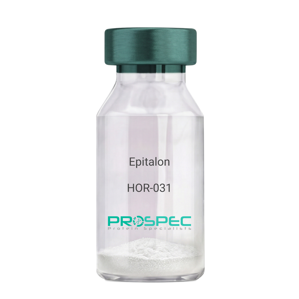
EpitalonDescription:
Epitalon
Product # :
HOR-031Price :
Quantity :
Shipping Method :
Shipped at Room temp
More Info
- description
- formulation
- purity
- More Info
Description
Epithalon Synthetic is a single, non-glycosylated polypeptide chain containing 4 amino acids, having a molecular mass of 390.35 Dalton and a Molecular formula of C14H22N4O9.
Formulation
The protein was lyophilized with no additives.
Purity
Greater than 97.0% as determined by analysis by RP-HPLC.
More Info
-
Physical Appearance
Sterile Filtered White lyophilized (freeze-dried) powder.
-
Stability
Lyophilized Epithalon although stable at room temperature for 3 weeks, should be stored desiccated below -18°C. Upon reconstitution Epithalon should be stored at 4°C between 2-7 days and for future use below -18°C.For long term storage it is recommended to add a carrier protein (0.1% HSA or BSA). Please prevent freeze-thaw cycles.
-
Solubility
It is recommended to reconstitute the lyophilized Epithalon in sterile 18MΩ-cm H2O not less than 100 µg/ml, which can then be further diluted to other aqueous solutions.
-
Amino Acid Sequence
H-Ala-Glu-Asp-Gly-OH .
-
Background
Epitalon, also known as Epithalon, is a synthetic tetrapeptide that has been a focal point in the field of aging and longevity research. This peptide, consisting of four amino acids, is derived from the pineal gland and has been associated with a variety of biological effects, most notably its potential role in extending the lifespan of cells.
Epitalon's primary function is its interaction with telomeres, the protective caps at the ends of chromosomes that shorten as cells divide. By stimulating the production of telomerase, an enzyme that can repair and lengthen telomeres, Epitalon may slow down the aging process at a cellular level.
Numerous studies have explored the potential of Epitalon in extending the lifespan of organisms. For instance, Khavinson and colleagues (2003) found that Epitalon increased the lifespan of fruit flies, while Anisimov et al. (2003) reported similar results in mice. These findings suggest that Epitalon could potentially have similar effects in humans.
Epitalon has also been studied for its role in regulating circadian rhythms. Korkushko et al. (2011) found that Epitalon could restore disrupted circadian rhythms in elderly people, suggesting potential applications in sleep disorders and other conditions related to circadian rhythm disruption.
Beyond its potential role in aging and longevity, Epitalon has been explored for its potential therapeutic applications. Anisimov et al. (2011) found that Epitalon could reduce the incidence of spontaneous tumors in mice, suggesting potential applications in cancer prevention.
While research on Epitalon is promising, it is important to note that most studies have been conducted in animals or in vitro. More research is needed to fully understand the potential effects and applications of Epitalon in humans. However, the existing body of research suggests that Epitalon could be a promising tool in the fight against aging and age-related diseases.
ProSpec's products are furnished for LABORATORY RESEARCH USE ONLY. The product may not be used as drugs, agricultural or pesticidal products, food additives or household chemicals.
- View Data Sheet
Name :
PEDF PAT13D9AT AntibodyDescription:
Mouse Anti Human Pigment Epithelium-Derived Factor Clone PAT13D9AT
Pigment epithelium-derived factor,PEDF, Serpin-F1, SerpinF1, EPC-1, EPC1, PIG35.
Product # :
ANT-714Price :
Quantity :
Shipping Method :
Shipped with Ice Packs
More Info
- formulation
- More Info
Formulation
1mg/ml containing PBS, pH-7.4, 10% Glycerol and 0.02% Sodium Azide.
More Info
-
Introduction
PEDF is a noninhibitory serpin with neurotrophic, anti-angiogenic, and anti-tumorigenic properties. PEDF is a 50,000 dalton glycoprotein created and secreted in many tissues all the way through the body. A key component of the anti-angiogenic action of PEDF is the induction of apoptosis in proliferating endothelial cells. Additionally, PEDF is capable to inhibit the activity of angiogenic factors such as VEGF and FGF-2. The neuro-protective effects of PEDF are achieved through suppression of neuronal apoptosis induced by peroxide, glutamate, or other neurotoxins. The recognition of a lipase-linked cell membrane receptor for PEDF (PEDF-R) that binds to PEDF with high affinity should facilitate further elucidation of the underlying mechanisms of this pluripotent serpin. To date, PEDF-R is the only signaling receptor known to be used by a serpin family member. The unique range of PEDF activities associate it as a potential therapeutic agent for the treatment of vasculature related neurodegenerative diseases such as age-related macular degeneration (AMD) and proliferative diabetic retinopathy (PDR). PEDF in addition has the potential to be functional in the treatment of various angiogenesis-related diseases including a number of cancers.
-
Synonyms
Pigment epithelium-derived factor,PEDF, Serpin-F1, SerpinF1, EPC-1, EPC1, PIG35.
-
Physical Appearance
Sterile filtered colorless solution.
-
Immunogen
Anti-human PEDF mAb, is derived from hybridization of mouse F0 myeloma cells with spleen cells from BALB/c mice immunized with a recombinant human PEDF protein 20-418 amino acids purified from E. coli.
-
Ig Subclass
Mouse IgG1 heavy chain and κ light chain.
-
Clone
PAT13D9AT.
-
Applications
PEDF antibody has been tested by ELISA, Western blot analysis and ICC/IF to assure specificity and reactivity. Since application varies, however, each investigation should be titrated by the reagent to obtain optimal results.
-
Type
Mouse Anti Human Monoclonal.
-
Storage Procedures
For periods up to 1 month store at 4°C, for longer periods of time, store at -20°C. Prevent freeze thaw cycles.
-
Purification Method
PEDF antibody was purified by protein-A affinity chromatography.
ProSpec's products are furnished for LABORATORY RESEARCH USE ONLY. The product may not be used as drugs, agricultural or pesticidal products, food additives or household chemicals.
- View Data Sheet
Name :
IL-6 PAT1F10AT AntibodyDescription:
Interleukin-6 Clone PAT1F10AT, Mouse Anti Human
IFN-b2, B cell differentiation factor, BCDF, BSF-2, HPGF, HSF, MGI-2, B-cell stimulatory factor 2, Interferon beta-2, Hybridoma growth factor, CTL differentiation factor, CDF, IL-6, HGF.
Product # :
ANT-540Price :
Quantity :
Shipping Method :
Shipped with Ice Packs
More Info
- formulation
- More Info
Formulation
1mg/ml containing PBS, pH-7.4, 10% Glycerol and 0.02% Sodium Azide.
More Info
-
Introduction
Il-6 is a cytokine with a wide variety of biological functions: it plays an essential role in the final differentiation of b-cells into ig-secreting cells, it induces myeloma and plasmacytoma growth, it induces nerve cells differentiation, in hepatocytes it induces acute phase reactants.
-
Synonyms
IFN-b2, B cell differentiation factor, BCDF, BSF-2, HPGF, HSF, MGI-2, B-cell stimulatory factor 2, Interferon beta-2, Hybridoma growth factor, CTL differentiation factor, CDF, IL-6, HGF.
-
Physical Appearance
Sterile filtered colorless solution.
-
Immunogen
Anti-human IL-6, clone PAT1F10AT mAb, is derived from hybridization of mouse F0 myeloma cells with spleen cells from BALB/c mice immunized with a recombinant human IL-6 protein 30-212 amino acids purified from E. coli.
-
Ig Subclass
Mouse IgG1 heavy chain and k light chain.
-
Clone
PAT1F10AT.
-
Applications
The antibody has been tested by ELISA, Western blot analysis to assure specificity and reactivity. Since application varies, however, each investigation should be titrated by the reagent to obtain optimal results. Recommended starting dilution is 1:1000.
-
Background
Research Paper on Interleukin-6 Clone PAT1F10AT, Mouse Anti-Human
Abstract:
Interleukin-6 (IL-6) Clone PAT1F10AT, a Mouse Anti-Human monoclonal antibody, plays a pivotal role in deciphering the complexities of immune modulation. This research paper provides an insightful exploration of its molecular attributes and its profound implications in immunological research. We examine its interactions, functions, and synonyms, including DIF, TNFA, and TNFSF2, shedding light on its significance in understanding immune responses.
Introduction:
IL-6 Clone PAT1F10AT, a Mouse Anti-Human antibody, has emerged as a cornerstone in immunology research. This paper delves into its unique characteristics and its critical role in unraveling immune mechanisms.
Molecular Precision and Specificity:
The exceptional affinity and specificity of IL-6 Clone PAT1F10AT for human IL-6 distinguish it as an invaluable tool for immunodetection and functional studies. Its targeted binding facilitates accurate identification of IL-6 in various experimental contexts.
Unveiling Immune Dynamics:
IL-6 is a central player in immune orchestration, and IL-6 Clone PAT1F10AT offers a window into its world. By investigating its involvement in immune responses, inflammation, and signaling pathways, researchers gain insights into immune regulation.
Synonyms and Interactions:
Understanding IL-6 synonyms, such as DIF, TNFA, and TNFSF2, underscores its intricate web of interactions. IL-6 Clone PAT1F10AT's ability to decipher these connections enhances our understanding of cytokine-mediated immune modulation.
Disease Implications and Therapeutic Avenues:
With its relevance in diseases like autoimmune disorders and inflammatory conditions, IL-6 Clone PAT1F10AT assumes significance in disease-focused investigations. It enables researchers to dissect disease mechanisms and evaluate potential therapeutic interventions.
Clinical Utility and Diagnostic Precision:
IL-6 Clone PAT1F10AT's utility transcends research settings, finding application in clinical diagnostics. Its role in immunoassays facilitates accurate disease diagnosis and monitoring, contributing to improved patient management.
Conclusion:
In the realm of immunology, IL-6 Clone PAT1F10AT stands as a potent ally in unraveling immune intricacies. Its molecular precision, diverse functions, and potential therapeutic implications make it a cornerstone in advancing our understanding of immune responses and diseases.
-
Type
Mouse Anti Human Monoclonal.
-
Storage Procedures
For periods up to 1 month store at 4°C, for longer periods of time, store at -20°C. Prevent freeze thaw cycles.
-
Purification Method
IL-6 antibody was purified from mouse ascitic fluids by protein-A affinity chromatography.
ProSpec's products are furnished for LABORATORY RESEARCH USE ONLY. The product may not be used as drugs, agricultural or pesticidal products, food additives or household chemicals.
- View Data Sheet
Name :
Frataxin AntibodyDescription:
Frataxin, Mouse Anti Human
FXN, Friedreich ataxia protein, Frataxin mitochondrial, FRDA, X25, FA, CyaY, FARR, MGC57199, Frataxin.
Product # :
ANT-671Price :
Quantity :
Shipping Method :
Shipped with Ice Packs
More Info
- formulation
- More Info
Formulation
1mg/ml containing PBS, pH-7.4, 10% Glycerol and 0.02% Sodium Azide.
More Info
-
Introduction
Frataxin is a mitochondrial iron binding protein that is part of the FRATAXIN family. Frataxin functions in regulating mitochondrial iron transport and respiration. The expansion of intronic trinucleotide repeat GAA results in Friedreich ataxia. Frataxin plays a role in iron homeostasis. Frataxin is an anti-apoptotic protein which prevents mitochondrial damage and reactive oxygen species (ROS) production.
-
Synonyms
FXN, Friedreich ataxia protein, Frataxin mitochondrial, FRDA, X25, FA, CyaY, FARR, MGC57199, Frataxin.
-
Physical Appearance
Sterile filtered colorless solution.
-
Immunogen
Anti-human Frataxin mAb, is derived from hybridization of mouse F0 myeloma cells with spleen cells from BALB/c mice immunized with a recombinant human Frataxin protein 42-210 amino acids purified from E. coli.
-
Ig Subclass
Mouse IgG1 heavy chain and k light chain.
-
Clone
PAT10E11AT.
-
Applications
The antibody has been tested by ELISA, Western blot analysis and ICC/IF to assure specificity and reactivity. Since application varies, however, each investigation should be titrated by the reagent to obtain optimal results.
-
Type
Mouse Anti Human Monoclonal.
-
Storage Procedures
For periods up to 1 month store at 4°C, for longer periods of time, store at -20°C. Prevent freeze thaw cycles.
-
Purification Method
Frataxin antibody was purified from mouse ascitic fluids by protein-A affinity chromatography.
ProSpec's products are furnished for LABORATORY RESEARCH USE ONLY. The product may not be used as drugs, agricultural or pesticidal products, food additives or household chemicals.