Search results
1000 results found for “paraoxonase”
Name
Description
Product #
Price
Quantity
Shipping Method
- View Data Sheet
Name :
ANXA5 HumanDescription:
Annexin A5 Human Recombinant
PP4, ANX5, ENX2, ANXA5, Annexin A5, Annexin-5, Annexin V, Lipocortin V, Endonexin II, Calphobindin I, CBP-I, Placental anticoagulant protein I, PAP-I, Placental anticoagulant protein 4, Thromboplastin inhibitor, Vascular anticoagulant-alpha, VAC-alpha, Anchorin CII.
Product # :
PRO-732Price :
Quantity :
Shipping Method :
Shipped with Ice Packs
More Info
- description
- source
- formulation
- purity
- More Info
Description
ANXA5 produced in E.Coli is a single, non-glycosylated polypeptide chain containing 320 amino acids (1-320 a.a.) and having a molecular mass of 35.9 kDa.ANXA5 is purified by proprietary chromatographic techniques.
Source
Escherichia Coli.
Formulation
The ANXA5 protein solution contains 20mM Tris-HCl, pH-8, 1mM DTT and 10% glycerol.
Purity
Greater than 95.0% as determined by SDS-PAGE.
More Info
-
Introduction
ANXA5 is a member of the annexin family of calcium-dependent phospholipid binding proteins which are involved in membrane-related activity along exocytotic and endocytotic pathways. ANXA5 is a phospholipase A2 and protein kinase C inhibitory protein with calcium channel properties and takes part in cellular signal transduction, inflammation, growth and differentiation. ANXA5 is an anticoagulant protein that acts as an indirect inhibitor of the thromboplastin-specific complex, which is involved in the blood coagulation cascade. ANXA5 regulates coagulability in the blood stream by binding to phosphatidylserine and sulfatide. ANXA5 protects sinsuoidal endothelial cells from ischemia reperfusion damage. ANXA5 is necessary for normal CFTR chloride channel activity.
-
Synonyms
PP4, ANX5, ENX2, ANXA5, Annexin A5, Annexin-5, Annexin V, Lipocortin V, Endonexin II, Calphobindin I, CBP-I, Placental anticoagulant protein I, PAP-I, Placental anticoagulant protein 4, Thromboplastin inhibitor, Vascular anticoagulant-alpha, VAC-alpha, Anchorin CII.
-
Physical Appearance
Sterile filtered colorless solution.
-
Stability
Store at 4°C if entire vial will be used within 2-4 weeks. Store, frozen at -20°C for longer periods of time. For long term storage it is recommended to add a carrier protein (0.1% HSA or BSA).Avoid multiple freeze-thaw cycles.
-
Amino Acid Sequence
MAQVLRGTVT DFPGFDERAD AETLRKAMKG LGTDEESILT LLTSRSNAQR QEISAAFKTL FGRDLLDDLK SELTGKFEKL IVALMKPSRL YDAYELKHAL KGAGTNEKVL TEIIASRTPE ELRAIKQVYE EEYGSSLEDD VVGDTSGYYQ RMLVVLLQAN RDPDAGIDEA QVEQDAQALF QAGELKWGTD EEKFITIFGT RSVSHLRKVF DKYMTISGFQ IEETIDRETS GNLEQLLLAV VKSIRSIPAY LAETLYYAMK GAGTDDHTLI RVMVSRSEID LFNIRKEFRK NFATSLYSMI KGDTSGDYKK ALLLLCGEDD.
ProSpec's products are furnished for LABORATORY RESEARCH USE ONLY. The product may not be used as drugs, agricultural or pesticidal products, food additives or household chemicals.
- View Data Sheet
Name :
KLK2 Human, sf9Description:
Kallikrein-2 Human Recombinant, sf9
hK2, KLK2A2, Kallikrein-2, Glandular kallikrein-1, hGK-1, issue kallikrein-2, KLK2.
Product # :
ENZ-971Price :
Quantity :
Shipping Method :
Shipped with Ice Packs
More Info
- description
- source
- formulation
- purity
- More Info
Description
KLK2 Human Recombinant produced in Sf9 Baculovirus cells is a single, glycosylated polypeptide chain containing 246 amino acids (25-261 aa) and having a molecular mass of 27.2kDa (Migrates at 28-40kDa on SDS-PAGE under reducing conditions).KLK2 is expressed with a 6 amino acid His tag at C-Terminus and purified by proprietary chromatographic techniques.
Source
Sf9, Baculovirus cells.
Formulation
KLK2 protein solution (0.25mg/ml) containing Phosphate Buffered Saline (pH 7.4) and 10% glycerol.
Purity
Greater than 90.0% as determined by SDS-PAGE.
More Info
-
Introduction
KLK2 is a part of the grandular kallikrein protein family whose Members are engaged in a diverse array of biological functions. Kallikreins are a subgroup of serine proteases which are clustered on chromosome 19. KLK2 is a highly active trypsin-like serine protease which selectively cleaves at arginine remains. KLK2 is mostly expressed in prostatic tissue and is accountable for cleaving pro-prostate-specific antigen into its enzymatically active form. KLK2 is greatly expressed in prostate tumor cells and may possibly be a prognostic maker for prostate cancer risk.
-
Synonyms
hK2, KLK2A2, Kallikrein-2, Glandular kallikrein-1, hGK-1, issue kallikrein-2, KLK2.
-
Physical Appearance
Sterile Filtered colorless solution.
-
Stability
Store at 4°C if entire vial will be used within 2-4 weeks. Store, frozen at -20°C for longer periods of time. For long term storage it is recommended to add a carrier protein (0.1% HSA or BSA).Avoid multiple freeze-thaw cycles.
-
Amino Acid Sequence
ADPIVGGWEC EKHSQPWQVA VYSHGWAHCG GVLVHPQWVL TAAHCLKKNS QVWLGRHNLF EPEDTGQRVP VSHSFPHPLY NMSLLKHQSL RPDEDSSHDL MLLRLSEPAK ITDVVKVLGL PTQEPALGTT CYASGWGSIE PEEFLRPRSL QCVSLHLLSN DMCARAYSEK VTEFMLCAGL WTGGKDTCGG DSGGPLVCNG VLQGITSWGP EPCALPEKPA VYTKVVHYRK WIKDTIAANP HHHHHH
ProSpec's products are furnished for LABORATORY RESEARCH USE ONLY. The product may not be used as drugs, agricultural or pesticidal products, food additives or household chemicals.
- View Data Sheet
Name :
HAV P2CDescription:
Hepatitis A Virus P2C Recombinant
Product # :
HAV-229Price :
Quantity :
Shipping Method :
Shipped with Ice Packs
More Info
- description
- formulation
- purity
- More Info
Description
The E.Coli derived recombinant 38.5 kDa protein contains the P2C immunodominant regions, amino acids 1121-1234.
Formulation
10mM CBB, pH9.6, 0.1% SDS and 50% glycerol.
Purity
Protein is >90% pure as determined by 10% PAGE (coomassie staining).
More Info
-
Introduction
Forty-two antigenic domains were identified across the hepatitis A virus (HAV) polyprotein by using a set of 237 overlapping 20-mer synthetic peptides spanning the entire HAV polyprotein. Nineteen antigenic domains were found within the structural proteins, and 22 were found within the nonstructural proteins, with 1 domain spanning the junction of VP1 and P2A proteins. Five of these domains were considered immunodominant, as judged by both the breadth and the strength of their immunoreactivity. One domain is located within the VP2 protein at position 57-90 aa. A second domain, located at position 767-842 aa, contains the C-terminal part of the VP1 protein and the entire P2A protein. A third domain, located at position 1403-1456 aa, comprises the C-terminal part of the P2C protein and the N-terminal half of the P3A protein. The fourth domain, located at position 1500-1519 aa, includes almost the entire P3B, and the last domain, located at position 1719-1764 aa, contains the C-terminal region of the P3C protein and the N-terminal region of the P3D protein. Four of the five most immunoreactive domains are derived from small HAV proteins and/or encompass protein cleavage sites separating different HAV proteins.
-
Stability
HAV P2C protein although stable at 4°C for 1 week, should be stored below -18°C. Please prevent freeze thaw cycles.
-
Applications
HAV P2C antigen is suitable for ELISA and Western blots, excellent antigen for detection of HAV with minimal specificity problems.
-
Specificity
Immunoreactive with sera HAV-infected individuals.
-
Purification Method
HAV P2C protein was purified by proprietary chromatographic technique.
ProSpec's products are furnished for LABORATORY RESEARCH USE ONLY. The product may not be used as drugs, agricultural or pesticidal products, food additives or household chemicals.
- View Data Sheet
Name :
PTGR1 HumanDescription:
Prostaglandin Reductase 1 Human Recombinant
Prostaglandin reductase 1, PRG-1, 15-oxoprostaglandin 13-reductase, NADP-dependent leukotriene B4 12-hydroxydehydrogenase, PTGR1, LTB4DH, PGR1, ZADH3.
Product # :
ENZ-633Price :
Quantity :
Shipping Method :
Shipped with Ice Packs
More Info
- description
- source
- formulation
- purity
- More Info
Description
PTGR1 Human Recombinant produced in E. coli is a single polypeptide chain containing 354 amino acids (1-329) and having a molecular mass of 38.6kDa.PTGR1 is fused to a 25 amino acid His-tag at N-terminus & purified by proprietary chromatographic techniques.
Source
Escherichia Coli.
Formulation
The PTGR1 solution (0.5mg/ml) contains 20mM Tris-HCl buffer, pH7.5, 10% glycerol, 1mM DTT and 200mM NaCl.
Purity
Greater than 90.0% as determined by SDS-PAGE.
More Info
-
Introduction
Prostaglandin Reductase 1 (PTGR1) is a member of the NADP-dependent oxidoreductase L4BD family. PTGR1 catalyzes the conversion of leukotriene B4 into its biologically less active metabolite, 12-oxo-leukotriene B4, thus being a primary step of metabolic inactivation of leukotriene B4. PTGR1 is highly expressed in the kidney, liver, and intestine but not in leukocytes.
-
Synonyms
Prostaglandin reductase 1, PRG-1, 15-oxoprostaglandin 13-reductase, NADP-dependent leukotriene B4 12-hydroxydehydrogenase, PTGR1, LTB4DH, PGR1, ZADH3.
-
Physical Appearance
Sterile filtered colorless solution.
-
Stability
Store at 4°C if entire vial will be used within 2-4 weeks. Store, frozen at -20°C for longer periods of time. For long term storage it is recommended to add a carrier protein (0.1% HSA or BSA).Avoid multiple freeze-thaw cycles.
-
Amino Acid Sequence
MGSSHHHHHH SSGLVPRGSH MGSEFMVRTK TWTLKKHFVG YPTNSDFELK TAELPPLKNG EVLLEALFLT VDPYMRVAAK RLKEGDTMMG QQVAKVVESK NVALPKGTIV LASPGWTTHS ISDGKDLEKL LTEWPDTIPL SLALGTVGMP GLTAYFGLLE ICGVKGGETV MVNAAAGAVG SVVGQIAKLK GCKVVGAVGS DEKVAYLQKL GFDVVFNYKT VESLEETLKK ASPDGYDCYF DNVGGEFSNT VIGQMKKFGR IAICGAISTY NRTGPLPPGP PPEIVIYQEL RMEAFVVYRW QGDARQKALK DLLKWVLEGK IQYKEYIIEG FENMPAAFMG MLKGDNLGKT IVKA.
ProSpec's products are furnished for LABORATORY RESEARCH USE ONLY. The product may not be used as drugs, agricultural or pesticidal products, food additives or household chemicals.
- View Data Sheet
Name :
UGDH HumanDescription:
UDP-Glucose Dehydrogenase Human Recombinant
GDH, UDP-GlcDH, UDPGDH, UGD, EC=1.1.1.22, UDP-Glc dehydrogenase, UDP-glucose 6-dehydrogenase, UGDH.
Product # :
ENZ-535Price :
Quantity :
Shipping Method :
Shipped with Ice Packs
More Info
- description
- source
- formulation
- purity
- biological activity
- More Info
Description
UGDH Recombinant Human produced in E.Coli is a single, non-glycosylated polypeptide chain containing 533 amino acids (1-494 a.a.) and having a molecular mass of 59.5 kDa. The UGDH is fused to a 39 amino acid His-Tag at N-terminus and purified by proprietary chromatographic techniques.
Source
Escherichia Coli.
Formulation
UGDH Human solution (0.5mg/ml) containing 20mM Tris-HCl pH-8, 1mM DTT, 1mM EDTA, 0.1M NaCl & 20% glycerol.
Purity
Greater than 95.0% as determined by SDS-PAGE.
Biological Activity
Specific activity is > 2500pmol/min/ug, and is defined as the amount of enzyme that convert 1.0pmole of UDPglucose to UDP-glucuronate per minute at pH 8.7 at 37C.
More Info
-
Introduction
UGDH is part of the UDP-glucose/GDP-mannose dehydrogenase family and is a widely expressed enzyme localized in the liver. UGDH transfers UDP-glucose to UDP-glucuronate and thus takes part in the biosynthesis of glycosaminoglycans such as hyaluronan, chondroitin sulfate, and heparan sulfate. These glycosylated products are ordinary molecules of the extracellular matrix and participate in signal transduction, cell migration, and cancer growth and metastasis.
-
Synonyms
GDH, UDP-GlcDH, UDPGDH, UGD, EC=1.1.1.22, UDP-Glc dehydrogenase, UDP-glucose 6-dehydrogenase, UGDH.
-
Physical Appearance
Sterile filtered colorless solution.
-
Stability
Store at 4°C if entire vial will be used within 2-4 weeks. Store, frozen at -20°C for longer periods of time. For long term storage it is recommended to add a carrier protein (0.1% HSA or BSA).Avoid multiple freeze-thaw cycles.
-
Amino Acid Sequence
MRGSHHHHHH GMASMTGGQQ MGRDLYDDDD KDRWGSELEM FEIKKICCIG AGYVGGPTCS VIAHMCPEIR VTVVDVNESR INAWNSPTLP IYEPGLKEVV ESCRGKNLFF STNIDDAIKE ADLVFISVNT PTKTYGMGKG RAADLKYIEA CARRIVQNSN GYKIVTEKST VPVRAAESIR RIFDANTKPN LNLQVLSNPE FLAEGTAIKD LKNPDRVLIG GDETPEGQRA VQALCAVYEH WVPREKILTT NTWSSELSKL AANAFLAQRI SSINSISALC EATGADVEEV ATAIGMDQRI GNKFLKASVG FGGSCFQKDV LNLVYLCEAL NLPEVARYWQ QVIDMNDYQR RRFASRIIDS LFNTVTDKKI AILGFAFKKD TGDTRESSSI YISKYLMDEG AHLHIYDPKV PREQIVVDLS HPGVSEDDQV SRLVTISKDP YEACDGAHAV VICTEWDMFK ELDYERIHKK MLKPAFIFDG RRVLDGLHNE LQTIGFQIET IGKKVSSKRI PYAPSGEIPK FSLQDPPNKK PKV.
ProSpec's products are furnished for LABORATORY RESEARCH USE ONLY. The product may not be used as drugs, agricultural or pesticidal products, food additives or household chemicals.
- View Data Sheet
Name :
BLVRB HumanDescription:
Biliverdin Reductase B Human Recombinant
FLR, BVRB, SDR43U1, MGC117413, BLVRB, Flavin reductase, FR, NADPH-dependent diaphorase, NADPH-flavin reductase, Biliverdin reductase B, BVR-B, Biliverdin-IX beta-reductase, Green heme-binding protein, GHBP.
Product # :
ENZ-387Price :
Quantity :
Shipping Method :
Shipped with Ice Packs
More Info
- description
- source
- formulation
- purity
- More Info
Description
BLVRB Human Recombinant amino produced in E.Coli is a single, non-glycosylated polypeptide chain containing 206 amino acids having a molecular mass of 22.1 kDa.The BLVRB is purified by proprietary chromatographic techniques.
Source
Escherichia Coli.
Formulation
The protein contains 20mM Tris-HCl buffer pH 8.5, 10% glycerol, and 1mM DTT.
Purity
Greater than 95.0% as determined by SDS-PAGE.
More Info
-
Introduction
BLVRB (EC 1.3.1.24) catalyzes electron transfer from reduced pyridine nucleotides to flavins as well as methylene blue, pyrroloquinoline quinone, riboflavin, or methemoglobin. BLVRB is involved in protecting cells from oxidative damage or in regulating iron metabolism. BLVRB converts biliverdin to bilirubin in the liver, converting a double-bond between the second and third pyrrole ring into a single-bond. BLVRB plays a role as in human erythrocytic heme catabolic pathway and most mammalian species. Biliverdin reductase is abundantly expressed in kidney, spleen, liver and brain as well as at lower levels in the thymus and minimal levels being detected in testis.
-
Synonyms
FLR, BVRB, SDR43U1, MGC117413, BLVRB, Flavin reductase, FR, NADPH-dependent diaphorase, NADPH-flavin reductase, Biliverdin reductase B, BVR-B, Biliverdin-IX beta-reductase, Green heme-binding protein, GHBP.
-
Physical Appearance
Sterile filtered colorless solution.
-
Stability
Store at 4°C if entire vial will be used within 2-4 weeks. Store, frozen at -20°C for longer periods of time. For long term storage it is recommended to add a carrier protein (0.1% HSA or BSA).Avoid multiple freeze-thaw cycles.
-
Amino Acid Sequence
MAVKKIAIFG ATGQTGLTTL AQAVQAGYEV TVLVRDSSRL PSEGPRPAHV VVGDVLQAAD VDKTVAGQDA VIVLLGTRND LSPTTVMSEG ARNIVAAMKA HGVDKVVACT SAFLLWDPTK VPPRLQAVTD DHIRMHKVLR ESGLKYVAVM PPHIGDQPLT GAYTVTLDGR GPSRVISKHD LGHFMLRCLT TDEYDGHSTY PSHQYQ.
ProSpec's products are furnished for LABORATORY RESEARCH USE ONLY. The product may not be used as drugs, agricultural or pesticidal products, food additives or household chemicals.
- View Data Sheet
Name :
FAP HumanDescription:
Fibroblast Activation Protein Alpha Human Recombinant
Prolyl endopeptidase FAP, 170 kDa melanoma membrane-bound gelatinase, Dipeptidyl peptidase FAP, Fibroblast activation protein alpha, FAPalpha, Gelatine degradation protease FAP, Integral membrane serine protease, Post-proline cleaving enzyme, Serine integral membrane protease, Surface-expressed protease, Seprase, SIMP, FAP
Product # :
ENZ-1160Price :
Quantity :
Shipping Method :
Shipped with Ice Packs
More Info
- description
- source
- formulation
- purity
- biological activity
- More Info
Description
FAP Human produced in Sf9 Baculovirus cells is a single, glycosylated polypeptide chain containing 744 amino acids (26-760aa) and having a molecular mass of 86.1 kDa.FAP is fused to a 6 amino acid His tag at C-terminus and purified by proprietary chromatographic techniques.
Source
Sf9, Baculovirus cells.
Formulation
The FAP solution (0.25mg/ml) contains 20% Glycerol and Phosphate-Buffered Saline (pH 7.4).
Purity
Greater than 90.0% as determined by SDS-PAGE.
Biological Activity
Specific activity > 5,000 pmol/min/ug. It is defined by the amount of enzyme that hydrolyzes 1.0 pmole of ZGP-AMC per minute at pH 7.5, at 37˚C.
More Info
-
Introduction
DPP4 also called adenosine deaminase complexing protein-2, and T-cell activation antigen CD26 is a serine exopeptidase and complex enzyme that is expressed on the surface of most cell types. DPPIV is an intrinsic membrane glycoprotein and a serine exopeptidase that cleaves X-proline dipeptides from the N-terminus of polypeptides. DPP4 plays a role in t-cell activation. DPP4 is associated with intracellular signal transduction, apoptosis and involved in tumor biology. There are at least 63 substrates which can bind specifically to DPP4 enzyme including growth factors, chemokines, neuro peptides. Furthermore, DPP4 plays a major role in glucose metabolism by cleaving incretins such as glucose-dependent insulinotropic polypeptide (GIP) and GLP-1.
-
Synonyms
Prolyl endopeptidase FAP, 170 kDa melanoma membrane-bound gelatinase, Dipeptidyl peptidase FAP, Fibroblast activation protein alpha, FAPalpha, Gelatine degradation protease FAP, Integral membrane serine protease, Post-proline cleaving enzyme, Serine integral membrane protease, Surface-expressed protease, Seprase, SIMP, FAP
-
Physical Appearance
Sterile Filtered colorless solution.
-
Stability
Store at 4°C if entire vial will be used within 2-4 weeks. Store, frozen at -20°C for longer periods of time. For long term storage it is recommended to add a carrier protein (0.1% HSA or BSA).Avoid multiple freeze-thaw cycles.
-
Amino Acid Sequence
ADPLRPSRVH NSEENTMRAL TLKDILNGTF SYKTFFPNWI SGQEYLHQSA DNNIVLYNIE TGQSYTILSN RTMKSVNASN YGLSPDRQFV YLESDYSKLW RYSYTATYYI YDLSNGEFVR GNELPRPIQY LCWSPVGSKL AYVYQNNIYL KQRPGDPPFQ ITFNGRENKI FNGIPDWVYE EEMLATKYAL WWSPNGKFLA YAEFNDTDIP VIAYSYYGDE QYPRTINIPY PKAGAKNPVV RIFIIDTTYP AYVGPQEVPV PAMIASSDYY FSWLTWVTDE RVCLQWLKRV QNVSVLSICD FREDWQTWDC PKTQEHIEES RTGWAGGFFV STPVFSYDAI SYYKIFSDKD GYKHIHYIKD TVENAIQITS GKWEAINIFR VTQDSLFYSS NEFEEYPGRR NIYRISIGSY PPSKKCVTCH LRKERCQYYT ASFSDYAKYY ALVCYGPGIP ISTLHDGRTD QEIKILEENK ELENALKNIQ LPKEEIKKLE VDEITLWYKM ILPPQFDRSK KYPLLIQVYG GPCSQSVRSV FAVNWISYLA SKEGMVIALV DGRGTAFQGD KLLYAVYRKL GVYEVEDQIT AVRKFIEMGF IDEKRIAIWG WSYGGYVSSL ALASGTGLFK CGIAVAPVSS WEYYASVYTE RFMGLPTKDD NLEHYKNSTV MARAEYFRNV DYLLIHGTAD DNVHFQNSAQ IAKALVNAQV DFQAMWYSDQ NHGLSGLSTN HLYTHMTHFL KQCFSLSDHH HHHH
ProSpec's products are furnished for LABORATORY RESEARCH USE ONLY. The product may not be used as drugs, agricultural or pesticidal products, food additives or household chemicals.
- View Data Sheet
Name :
MMP 3 Human, GSTDescription:
Matrix Metalloproteinase-3 Human Recombinant, GST Tag
Stromelysin-1, EC 3.4.24.17, Matrix metalloproteinase-3, MMP-3, Transin-1, SL-1, STMY, STR1, STMY1, MGC126102, MGC126103, MGC126104.
Product # :
ENZ-455Price :
Quantity :
Shipping Method :
Shipped with Ice Packs
More Info
- description
- source
- formulation
- More Info
Description
MMP-3 Human Recombinant produced in E.Coli is a single, non-glycosylated, Polypeptide chain fused to a GST tag containing 228 amino acids (251-478) and having a total molecular mass of 51kDa. MMP-3 is purified by proprietary chromatographic techniques.
Source
Escherichia Coli.
Formulation
MMP-3 is supplied in 50mM Tris-Acetate, pH-7.5, 1mM EDTA and 20% Glycerol.
More Info
-
Introduction
MMP-3 enzyme is also known as Stromelysin-1or as Transin-1 which hydrolyzes natural collagen at physiological pH and temperature. It dissolves the intervertebral nucleus pulposus and annulus fibrosus of Herniated Lumbar Intervertebral Disk . MMP-3 hydrolyzes components of the extracellular matrix like proteoglycan, laminin, fibronectin, gelatin and collagen types III, IV and IX. It also activates pro-MMP-9 and pro-MMP-8 and superactivates plasmin activated MMP-1. MMP-3 is secreted as a latent proenzyme and is activated by a variety of proteinases, e.g. plasmin, trypsin, chymotrypsin, cathepsin G or human neutrophil elastase. MMP-3 was found to be capable of activating the precursor of IL1-beta.
-
Synonyms
Stromelysin-1, EC 3.4.24.17, Matrix metalloproteinase-3, MMP-3, Transin-1, SL-1, STMY, STR1, STMY1, MGC126102, MGC126103, MGC126104.
-
Physical Appearance
Sterile Filtered clear solution.
-
Stability
Store at 4°C if entire vial will be used within 2-4 weeks. Store, frozen at -20°C for longer periods of time. Please avoid freeze thaw cycles.
ProSpec's products are furnished for LABORATORY RESEARCH USE ONLY. The product may not be used as drugs, agricultural or pesticidal products, food additives or household chemicals.
- View Data Sheet
Name :
ACADVL HumanDescription:
Acyl-CoA Dehydrogenase, Very Long Chain Human Recombinant
ACAD6, LCACD, VLCAD.
Product # :
ENZ-250Price :
Quantity :
Shipping Method :
Shipped with Ice Packs
More Info
- description
- source
- formulation
- purity
- More Info
Description
ACADVL Human Recombinant produced in E.Coli is a non-glycosylated, polypeptide chain containing 636 amino acids (41-655 a.a.) and having a molecular mass of 68.5 kda. ACADVL contains 21 amino acid His-Tag at the N-terminus and is purified by proprietary chromatographic techniques.
Source
Escherichia Coli.
Formulation
The 0.5mg/ml protein solution contains 20mM Tris-HCl buffer pH-8, 1mM DTT, 1mM EDTA, 10% glycerol and 100mM NaCl.
Purity
Greater than 90.0% as determined by SDS-PAGE.
More Info
-
Introduction
ACADVL is an inner mitochondrial membrane enzyme that is part of the family of acyl-CoA dehydrogenases. ACADVL protein participates in lipid metabolism and has catalytic activity toward esters of long chain and very long chain fatty acids such as palmitoyl-CoA and stearoyl-CoA, and is involved in the first step of the fatty acid β-oxidation pathway. ACADVL deficiency in reduces myocardial fatty acid beta-oxidation and is related with cardiomyopathy.
-
Synonyms
ACAD6, LCACD, VLCAD.
-
Physical Appearance
Sterile Filtered clear solution.
-
Stability
Store ACADVL at 4°C if entire vial will be used within 2-4 weeks. Store, frozen at -20°C for longer periods of time. For long term storage it is recommended to add a carrier protein (0.1% HSA or BSA). Avoid multiple freeze-thaw cycles.
-
Amino Acid Sequence
MGSSHHHHHH SSGLVPRGSH MAGGAAQLAL DKSDSHPSDA LTRKKPAKAE SKSFAVGMFK GQLTTDQVFP YPSVLNEEQT QFLKELVEPV SRFFEEVNDP AKNDALEMVE ETTWQGLKEL GAFGLQVPSE LGGVGLCNTQ YARLVEIVGM HDLGVGITLG AHQSIGFKGI LLFGTKAQKE KYLPKLASGE TVAAFCLTEP SSGSDAASIR TSAVPSPCGK YYTLNGSKLW ISNGGLADIF TVFAKTPVTD PATGAVKEKI TAFVVERGFG GITHGPPEKK MGIKASNTAE VFFDGVRVPS ENVLGEVGSG FKVAMHILNN GRFGMAAALA GTMRGIIAKA VDHATNRTQF GEKIHNFGLI QEKLARMVML QYVTESMAYM VSANMDQGAT DFQIEAAISK IFGSEAAWKV TDECIQIMGG MGFMKEPGVE RVLRDLRIFR IFEGTNDILR LFVALQGCMD KGKELSGLGS ALKNPFGNAG LLLGEAGKQL RRRAGLGSGL SLSGLVHPEL SRSGELAVRA LEQFATVVEA KLIKHKKGIV NEQFLLQRLA DGAIDLYAMV VVLSRASRSL SEGHPTAQHE KMLCDTWCIE AAARIREGMA ALQSDPWQQE LYRNFKSISK ALVERGGVVT SNPLGF.
ProSpec's products are furnished for LABORATORY RESEARCH USE ONLY. The product may not be used as drugs, agricultural or pesticidal products, food additives or household chemicals.
- View Data Sheet
Name :
DHODH HumanDescription:
Dihydroorotate Dehydrogenase Human Recombinant
Dihydroorotate dehydrogenase (quinone), Dihydroorotate oxidase, human complement of yeast URA1, DHOdehase, POADS, EC 1.3.5.2, EC 1.3.3.1.
Product # :
ENZ-642Price :
Quantity :
Shipping Method :
Shipped with Ice Packs
More Info
- description
- source
- formulation
- purity
- More Info
Description
DHODH Human Recombinant produced in E. coli is a single polypeptide chain containing 390 amino acids (31-395) and having a molecular mass of 42.3 kDa.DHODH is fused to a 25 amino acid His-tag at N-terminus & purified by proprietary chromatographic techniques.
Source
E.coli.
Formulation
The DHODH solution (0.5mg/1ml) contains 20mM Tris-HCl buffer (pH 8.0), 100mM NaCl, 1mM DTT and 20% glycerol.
Purity
Greater than 90% as determined by SDS-PAGE.
More Info
-
Introduction
Dihydroorotate dehydrogenase quinone (DHODH) is a member of the dihydroorotate dehydrogenase family. DHODH is a mitochondrial protein found on the outer surface of the inner mitochondrial membrane. DHODH catalyzes the 4th enzymatic step, the ubiquinone-mediated oxidation of dihydroorotate to orotate (with quinone as electron acceptor), in de novo pyrimidine biosynthesis. DHODH gene defects cause the postaxial acrofacial dysostosis (POADS), also known as Miller syndrome.
-
Synonyms
Dihydroorotate dehydrogenase (quinone), Dihydroorotate oxidase, human complement of yeast URA1, DHOdehase, POADS, EC 1.3.5.2, EC 1.3.3.1.
-
Physical Appearance
Sterile Filtered colorless solution.
-
Stability
Store at 4°C if entire vial will be used within 2-4 weeks. Store, frozen at -20°C for longer periods of time. For long term storage it is recommended to add a carrier protein (0.1% HSA or BSA).Avoid multiple freeze-thaw cycles.
-
Amino Acid Sequence
GSSHHHHHH SSGLVPRGSH MGSHMTGDER FYAEHLMPTL QGLLDPESAH RLAVRFTSLG LLPRARFQDS DMLEVRVLGH KFRNPVGIAA GFDKHGEAVD GLYKMGFGFV EIGSVTPKPQ EGNPRPRVFR LPEDQAVINR YGFNSHGLSV VEHRLRARQQ KQAKLTEDGL PLGVNLGKNK TSVDAAEDYA EGVRVLGPLA DYLVVNVSSP NTAGLRSLQG KAELRRLLTK VLQERDGLRR VHRPAVLVKI APDLTSQDKE DIASVVKELG IDGLIVTNTT VSRPAGLQGA LRSETGGLSG KPLRDLSTQT IREMYALTQG RVPIIGVGGV SSGQDALEKI RAGASLVQLY TALTFWGPPV VGKVKRELEA LLKEQGFGGV TDAIGADHRR.
ProSpec's products are furnished for LABORATORY RESEARCH USE ONLY. The product may not be used as drugs, agricultural or pesticidal products, food additives or household chemicals.
- View Data Sheet
Name :
NT5M HumanDescription:
5',3'-Nucleotidase, Mitochondrial Human Recombinant
5'(3')-deoxyribonucleotidase mitochondrial, 5',3'-nucleotidase, mitochondrial, Deoxy-5'-nucleotidase 2, dNT-2, NT5M, DNT2.
Product # :
ENZ-160Price :
Quantity :
Shipping Method :
Shipped with Ice Packs
More Info
- description
- source
- formulation
- purity
- More Info
Description
NT5M Human Recombinant produced in E.Coli is a single, non-glycosylated polypeptide chain containing 218 amino acids (32-228 a.a.) and having a molecular mass of 25.1kDa.NT5M is fused to a 21 amino acid His-tag at N-terminus & purified by proprietary chromatographic techniques.
Source
Escherichia Coli.
Formulation
NT5M protein solution (0.5mg/ml) containing 20mM Tris-HCl buffer (pH8.0), 20% glycerol, 0.1M NaCl and 1mM DTT.
Purity
Greater than 95% as determined by SDS-PAGE.
More Info
-
Introduction
NT5M is a member of the 5'(3')-deoxyribonucleotidase family. NT5M localizes to the mitochondrial matrix. The NT5M enzyme dephosphorylates the 5'- and 2'(3')-phosphates of uracil and thymine deoxyribonucleotides. The NT5M gene is located on chromosome 17 p11.2 in the critical region deleted in the Smith-Magenis syndrome (SMS), a genetic disease of unidentified etiology.
-
Synonyms
5'(3')-deoxyribonucleotidase mitochondrial, 5',3'-nucleotidase, mitochondrial, Deoxy-5'-nucleotidase 2, dNT-2, NT5M, DNT2.
-
Physical Appearance
Sterile Filtered clear solution.
-
Stability
Store at 4°C if entire vial will be used within 2-4 weeks. Store, frozen at -20°C for longer periods of time. For long term storage it is recommended to add a carrier protein (0.1% HSA or BSA).Avoid multiple freeze-thaw cycles.
-
Amino Acid Sequence
MGSSHHHHHH SSGLVPRGSH MGGRALRVLV DMDGVLADFE GGFLRKFRAR FPDQPFIALE DRRGFWVSEQ YGRLRPGLSE KAISIWESKN FFFELEPLPG AVEAVKEMAS LQNTDVFICT SPIKMFKYCP YEKYAWVEKY FGPDFLEQIV LTRDKTVVSA DLLIDDRPDI TGAEPTPSWE HVLFTACHNQ HLQLQPPRRR LHSWADDWKA ILDSKRPC.
ProSpec's products are furnished for LABORATORY RESEARCH USE ONLY. The product may not be used as drugs, agricultural or pesticidal products, food additives or household chemicals.
- View Data Sheet
Name :
LDHA MouseDescription:
Lactate Dehydrogenase A Mouse Recombinant
L-lactate dehydrogenase A chain, LDH-A, LDH muscle subunit, LDH-M.
Product # :
ENZ-952Price :
Quantity :
Shipping Method :
Shipped with Ice Packs
More Info
- description
- source
- formulation
- purity
- biological activity
- More Info
Description
LDHA Mouse Recombinant produced in Sf9 Baculovirus cells is a single, glycosylated polypeptide chain containing 340 amino acids (1-332a.a.) and having a molecular mass of 37.5kDa (Molecular size on SDS-PAGE will appear at approximately 28-40kDa). LDHA is expressed with an 8 amino acid His tag at C-Terminus and purified by proprietary chromatographic techniques.
Source
Sf9, Baculovirus cells.
Formulation
LDHA protein solution (0.5mg/ml) containing Phosphate Buffered Saline (pH 7.4) and 10% glycerol.
Purity
Greater than 95.0% as determined by SDS-PAGE.
Biological Activity
Specific activity is > 250 units/mg, and is defined as the Amount of enzyme that convert 1.0 umole of pyruvate to L-lactate and per minute at pH 7.5 at 37C.More Info
-
Introduction
LDHA catalyzes the conversion of L-lactate and NAD to pyruvate and NADH in the final step of anaerobic glycolysis. LDHA is localized primarily in muscle tissue and is part of the lactate dehydrogenase family. Mutations in LDHA have been linked to exertional myoglobinuria. LDH1 is decreased in essential thrombocythemia. LDHA is induced through a non-genomic pathway of estrogen action. Reduction in LDH-A activity results in stimulation of mitochondrial respiration and decrease of mitochondrial membrane potential.
-
Synonyms
L-lactate dehydrogenase A chain, LDH-A, LDH muscle subunit, LDH-M.
-
Physical Appearance
Sterile Filtered colorless solution.
-
Stability
Store at 4°C if entire vial will be used within 2-4 weeks. Store, frozen at -20°C for longer periods of time. For long term storage it is recommended to add a carrier protein (0.1% HSA or BSA).Avoid multiple freeze-thaw cycles.
-
Amino Acid Sequence
MATLKDQLIV NLLKEEQAPQ NKITVVGVGA VGMACAISIL MKDLADELAL VDVMEDKLKG EMMDLQHGSL FLKTPKIVSS KDYCVTANSK LVIITAGARQ QEGESRLNLV QRNVNIFKFI IPNIVKYSPH CKLLIVSNPV DILTYVAWKI SGFPKNRVIG SGCNLDSARF RYLMGERLGV HALSCHGWVL GEHGDSSVPV WSGVNVAGVS LKSLNPELGT DADKEQWKEV HKQVVDSAYE VIKLKGYTSW AIGLSVADLA ESIMKNLRRV HPISTMIKGL YGINEDVFLS VPCILGQNGI SDVVKVTLTP EEEARLKKSA DTLWGIQKEL QFLEHHHHHH.
ProSpec's products are furnished for LABORATORY RESEARCH USE ONLY. The product may not be used as drugs, agricultural or pesticidal products, food additives or household chemicals.
- View Data Sheet
Name :
ADH5 HumanDescription:
Alcohol Dehydrogenase 5 Human Recombinant
Alcohol dehydrogenase 5 (class III) chi polypeptide, Alcohol dehydrogenase class chi chain, Glutathione-dependent formaldehyde dehydrogenase, S-(hydroxymethyl) glutathione dehydrogenase, FDH, ADHX, ADH-3, FALDH, GSH-FDH, GSNOR, EC 1.1.1.1, EC 1.1.1.284.
Product # :
ENZ-595Price :
Quantity :
Shipping Method :
Shipped with Ice Packs
More Info
- description
- source
- formulation
- purity
- More Info
Description
ADH5 Recombinant produced in E. coli is a single polypeptide chain containing 398 amino acids (1-374) and having a molecular mass of 42.3kDa.ADH5 is fused to a 24 amino acid His-tag at N-terminus & purified by proprietary chromatographic techniques.
Source
E.coli.
Formulation
The ADH5 solution (1mg/ml) contains 20mM Tris-HCl buffer (pH 8.0), 100mM NaCl, 1mM DTT and 20% glycerol.
Purity
Greater than 90% as determined by SDS-PAGE.
More Info
-
Introduction
ADH5 belongs to the alcohol dehydrogenase family which metabolizes a large selection of substrates, such as retinol, ethanol, other aliphatic alcohols, hydroxysteroids, and lipid peroxidation products. ADH5 has practically no activity for ethanol oxidation, but displays high activity for oxidation of long-chain primary alcohols and for oxidation of S-hydroxymethyl-glutathione, a spontaneous adduct between glutathione and formaldehyde. ADH5 enzyme is a key factor of cellular metabolism for the removal of formaldehyde, a powerful aggravating and alerting mediator which causes pharyngitis, lacrymation, rhinitis and contact dermatitis.
-
Synonyms
Alcohol dehydrogenase 5 (class III) chi polypeptide, Alcohol dehydrogenase class chi chain, Glutathione-dependent formaldehyde dehydrogenase, S-(hydroxymethyl) glutathione dehydrogenase, FDH, ADHX, ADH-3, FALDH, GSH-FDH, GSNOR, EC 1.1.1.1, EC 1.1.1.284.
-
Physical Appearance
Sterile Filtered colorless solution.
-
Stability
Store at 4°C if entire vial will be used within 2-4 weeks. Store, frozen at -20°C for longer periods of time. For long term storage it is recommended to add a carrier protein (0.1% HSA or BSA).Avoid multiple freeze-thaw cycles.
-
Amino Acid Sequence
MGSSHHHHHH SSGLVPRGSH MGSHMANEVI KCKAAVAWEA GKPLSIEEIE VAPPKAHEVR IKIIATAVCH TDAYTLSGAD PEGCFPVILG HEGAGIVESV GEGVTKLKAG DTVIPLYIPQ CGECKFCLNP KTNLCQKIRV TQGKGLMPDG TSRFTCKGKT ILHYMGTSTF SEYTVVADIS VAKIDPLAPL DKVCLLGCGI STGYGAAVNT AKLEPGSVCA VFGLGGVGLA VIMGCKVAGA SRIIGVDINK DKFARAKEFG ATECINPQDF SKPIQEVLIE MTDGGVDYSF ECIGNVKVMR AALEACHKGW GVSVVVGVAA SGEEIATRPF QLVTGRTWKG TAFGGWKSVE SVPKLVSEYM SKKIKVDEFV THNLSFDEIN KAFELMHSGK SIRTVVKI
ProSpec's products are furnished for LABORATORY RESEARCH USE ONLY. The product may not be used as drugs, agricultural or pesticidal products, food additives or household chemicals.
- View Data Sheet
Name :
HS3ST1 HumanDescription:
Heparan Sulfate 3-O-Sulfotransferase 1 Human Recombinant
Heparan sulfate glucosamine 3-O-sulfotransferase 1, Heparan sulfate D-glucosaminyl 3-O-sulfotransferase 1, 3-OST-1, Heparan sulfate 3-O-sulfotransferase 1, h3-OST-1, HS3ST1, 3OST, 3OST1, HS3S1.
Product # :
ENZ-744Price :
Quantity :
Shipping Method :
Shipped with Ice Packs
More Info
- description
- source
- formulation
- purity
- More Info
Description
HS3ST1 Human Recombinant produced in E.Coli is a single, non-glycosylated polypeptide chain containing 310 amino acids (21-307 a.a) and having a molecular mass of 36.2kDa.HS3ST1 is fused to a 23 amino acid His-tag at N-terminus & purified by proprietary chromatographic techniques.
Source
Escherichia Coli.
Formulation
HS3ST1 protein solution (0.25mg/ml) containing 20mM Tris-HCl buffer (pH 8.0), 0.2M NaCl, 40% glycerol and 2mM DTT.
Purity
Greater than 90.0% as determined by SDS-PAGE.
More Info
-
Introduction
Heparan Sulfate 3-O-Sulfotransferase 1 (HS3ST1), is sulfotransferase which uses 3'-phospho-5'-adenylyl sulfate (PAPS) to catalyze the transfer of a sulfo group to position 3 of glucosamine residues in heparan. HS3ST1 catalyzes the rate limiting step in the biosynthesis of heparan sulfate (HSact). This modification is a vital part in the biosynthesis of anticoagulant heparan sulfate since it concludes the structure of the antithrombin pentasaccharide binding site.
-
Synonyms
Heparan sulfate glucosamine 3-O-sulfotransferase 1, Heparan sulfate D-glucosaminyl 3-O-sulfotransferase 1, 3-OST-1, Heparan sulfate 3-O-sulfotransferase 1,
h3-OST-1, HS3ST1, 3OST, 3OST1, HS3S1. -
Physical Appearance
Sterile Filtered colorless solution.
-
Stability
Store at 4°C if entire vial will be used within 2-4 weeks. Store, frozen at -20°C for longer periods of time. For long term storage it is recommended to add a carrier protein (0.1% HSA or BSA).Avoid multiple freeze-thaw cycles.
-
Amino Acid Sequence
MGSSHHHHHH SSGLVPRGSH MGSRPAELGQ QELLRKAGTL QDDVRDGVAP NGSAQQLPQT IIIGVRKGGT RALLEMLSLH PDVAAAENEV HFFDWEEHYS HGLGWYLSQM PFSWPHQLTV EKTPAYFTSP KVPERVYSMN PSIRLLLILR DPSERVLSDY TQVFYNHMQK HKPYPSIEEF LVRDGRLNVD YKALNRSLYH VHMQNWLRFF PLRHIHIVDG DRLIRDPFPE IQKVERFLKL SPQINASNFY FNKTKGFYCL RDSGRDRCLH ESKGRAHPQV DPKLLNKLHE YFHEPNKKFF ELVGRTFDWH.
ProSpec's products are furnished for LABORATORY RESEARCH USE ONLY. The product may not be used as drugs, agricultural or pesticidal products, food additives or household chemicals.
- View Data Sheet
Name :
BCOADC-E2 HumanDescription:
2-Oxo-Acid Dehydrogenase Complex E2 Human Recombinant
Product # :
ENZ-084Price :
Quantity :
Shipping Method :
Shipped with Ice Packs
More Info
- description
- source
- formulation
- purity
- More Info
Description
BCOADC-E2 is a Full-length cDNA coding for the mature form of the human BCOADC-E2 protein having a molecular mass of 47,321 Dalton(pH 6.5). BCOADC-E2 protein is fused to a hexa-histidine purification tag.
Source
Sf9 insect cells.
Formulation
DLST (0.91mg/ml) is supplied in 20mM HEPES buffer pH-8.0, 200mM NaCl and 20% glycerol.
Purity
Greater than 75% as determined by SDS-PAGE.
More Info
-
Stability
Store at 4°C if entire vial will be used within 2-4 weeks. Store, frozen at -20°C for longer periods of time. Avoid multiple freeze-thaw cycles.
-
Immunological Functions
1. Binds IgG-type human auto-antibodies. 2. Standard ELISA test (checkerboard analysis of positive/negative sera panels); immunodot test with positive/negative sera panels.
-
coating concentration
0.4-0.8 µg/ml (depending on the type of ELISA plate and coating buffer). Suitable for biotinylation and iodination.
-
Applications
Western blot with anti-M2-Antigen autoantibody-positive patient sera or monoclonal
anti-hexa-His-tag antibody.
ProSpec's products are furnished for LABORATORY RESEARCH USE ONLY. The product may not be used as drugs, agricultural or pesticidal products, food additives or household chemicals.
- View Data Sheet
Name :
Enterokinase BovineDescription:
Enteropeptidase/ Enterokinase Light Chain Bovine Recombinant
Enteropeptidase, EC 3.4.21.9, Enterokinase, Serine protease 7, ENTK, MGC133046.
Product # :
ENZ-311Price :
Quantity :
Shipping Method :
Shipped with Ice Packs
More Info
- description
- source
- formulation
- purity
- More Info
Description
Enterokinase (rEK) Bovine Recombinant is the catalytic subunit of bovine enterokinase, which is expressed by E. Coli and purified to yield a high enzyme activity preparation. EK recognizes the sequence Asp-Asp-Asp-Asp-Lys and cleaves the peptide bond after the lysine residue. The enzyme can be used to cleave any fusion protein that carries this sequence. Recombinant Bovine Enterokinase is a single glycosylated polypeptide chain containing 235 amino acids and having an MW of ~28kDa.
Source
E. Coli.
Formulation
Bovine EK in 50mM Tris-HCl, pH 8.0, 0.5M NaCl and 50% glycerol.
Purity
Greater than 95.0% as determined by SDS-PAGE.
More Info
-
Introduction
Enteropeptidase or enterokinase is an enzyme involved in human digestion. It is produced by cells in the duodenum wall, and is secreted from duodenum's glands, the crypts of Lieberk?hn, whenever ingested food enters the duodenum from the stomach. Enteropeptidase has the critical job of turning trypsinogen (a zymogen) to trypsin, indirectly activating a number of pancreatic digestive enzymes. Enteropeptidase is a serine protease enzyme (EC 3.4.21.9). Enteropeptidase is a part of the Chymotrypsin-clan of serine proteases, and is structurally similar to these proteins.
-
Synonyms
Enteropeptidase, EC 3.4.21.9, Enterokinase, Serine protease 7, ENTK, MGC133046.
-
Physical Appearance
Sterile liquid solution.
-
Stability
One year when stored at –20°C. Please avoid freeze-thaw cycles.
-
Unit Definition
One unit is defined as the amount of enzyme needed to cleave 50µg of fusion protein in 16 hours to 95% completion at 25°C in a buffer containing 25mM Tris-HCl, pH 7.6, 50mM NaCl, and 2mM CaCl2.
ProSpec's products are furnished for LABORATORY RESEARCH USE ONLY. The product may not be used as drugs, agricultural or pesticidal products, food additives or household chemicals.
- View Data Sheet
Name :
Clusterin Human, HisDescription:
Apolipoprotein-J Human Recombinant, His Tag
CLI, AAG4, APOJ, KUB1, SGP2, SGP-2, SP-40, TRPM2, TRPM-2, MGC24903, Clusterin, ging-associated gene 4 protein, Apolipoprotein J,Complement cytolysis inhibitor, Complement-associated protein SP-40,40, Ku70-binding protein 1, NA1/NA2, Testosterone-repressed prostate message 2, CLU.
Product # :
CYT-814Price :
Quantity :
Shipping Method :
Shipped with Ice Packs
More Info
- description
- source
- formulation
- purity
- More Info
- sds-page
Description
Clusterin Human Recombinant produced in E.Coli is a single, non-glycosylated polypeptide chain containing 463 amino acids (23-449 a.a.) and having a molecular mass of 54.1kDa. Clusterin is fused to a 36 amino acid His-tag at N-terminus & purified by proprietary chromatographic techniques.
Source
Escherichia Coli.
Formulation
Clusterin protein solution (0.5mg/ml) contains 20mM Tris-HCl buffer (pH 8.0), 0.15M NaCl, 10% glycerol and 1mM DTT.
Purity
Greater than 85% as determined by SDS-PAGE.
sds-page
More Info
-
Introduction
Clusterin also named Apolipoprotein J (APO-J) is a 75-80 kD disulfide-linked heterodimeric protein containing about 30% of N-linked carbohydrate rich in sialic acid but truncated forms targeted to the nucleus have also been identified.
The precursor polypeptide chain is cleaved proteolytically to remove the 22-mer secretory signal peptide and subsequently between residues 227/228 to generate the a and b chains. These are assembled in anti-parallel to give a heterodimeric molecule in which the cysteine-rich centers are linked by five disulfide bridges and are flanked by two predicted coiled-coil a-helices and three predicted amphipathic a-helices.
Across a broad range of species clusterin shows a high degree of sequence homology ranging from 70% to 80%. It is nearly ubiquitously expressed in most mammalian tissues and can be found in plasma, milk, urine, cerebrospinal fluid and semen.
It is able to bind and form complexes with numerous partners such as immunoglobulins, lipids, heparin, bacteria, complement components, paraoxonase, beta amyloid, leptin and others. Clusterin has been ascribed a plethora of functions such as phagocyte recruitment, aggregation induction, complement attack prevention, apoptosis inhibition, membrane remodeling, lipid transport, hormone transport and/or scavenging, matrix metalloproteinase inhibition.
A genuine function of clusterin has not been defined. One tempting hypothesis says that clusterin is an extracellular chaperone protecting cells from stress induced insults caused by degraded and misfolded protein precipitates.
Clusterin is up- or down regulated on the mRNA or protein level in many pathological and clinically relevant situations including cancer, organ regeneration, infection, Alzheimer disease, retinitis pigmentosa, myocardial infarction, renal tubular damage, autoimmunity and others. -
Synonyms
CLI, AAG4, APOJ, KUB1, SGP2, SGP-2, SP-40, TRPM2, TRPM-2, MGC24903, Clusterin, ging-associated gene 4 protein, Apolipoprotein J,Complement cytolysis inhibitor, Complement-associated protein SP-40,40, Ku70-binding protein 1, NA1/NA2, Testosterone-repressed prostate message 2, CLU.
-
Physical Appearance
Sterile filtered colorless solution.
-
Stability
Store at 4°C if entire vial will be used within 2-4 weeks. Store, frozen at -20°C for longer periods of time. For long term storage it is recommended to add a carrier protein (0.1% HSA or BSA).Avoid multiple freeze-thaw cycles.
-
Amino Acid Sequence
MRGSHHHHHH GMASMTGGQQ MGRDLYDDDD KDRWGSDQTV SDNELQEMSN QGSKYVNKEI QNAVNGVKQI KTLIEKTNEE RKTLLSNLEE AKKKKEDALN ETRESETKLK ELPGVCNETM MALWEECKPC LKQTCMKFYA RVCRSGSGLV GRQLEEFLNQ SSPFYFWMNG DRIDSLLEND RQQTHMLDVM QDHFSRASSI IDELFQDRFF TREPQDTYHY LPFSLPHRRP HFFFPKSRIV RSLMPFSPYE PLNFHAMFQP FLEMIHEAQQ AMDIHFHSPA FQHPPTEFIR EGDDDRTVCR EIRHNSTGCL RMKDQCDKCR EILSVDCSTN NPSQAKLRRE LDESLQVAER LTRKYNELLK SYQWKMLNTS SLLEQLNEQF NWVSRLANLT QGEDQYYLRV TTVASHTSDS DVPSGVTEVV VKLFDSDPIT VTVPVEVSRK NPKFMETVAE KALQEYRKKH REE.
-
Background
What is the molecular weight/Mw of CLUSTERIN Protein?
CLUSTERIN Protein has a total Mw of 54.1kDa.
What is the source or expression system of CLUSTERIN Protein?
Escherichia Coli.
What is the Purity of CLUSTERIN Protein?
CLUSTERIN Protein is >85% pure as determined by SDS-PAGE.
What is the Biological Activity of CLUSTERIN Protein?
The biological functionality of CLUSTERIN Protein will be determined in the future.
What is the amino acid sequence of CLUSTERIN Protein?
MRGSHHHHHH GMASMTGGQQ MGRDLYDDDD KDRWGSDQTV SDNELQEMSN QGSKYVNKEI QNAVNGVKQI KTLIEKTNEE RKTLLSNLEE AKKKKEDALN ETRESETKLK ELPGVCNETM MALWEECKPC LKQTCMKFYA RVCRSGSGLV GRQLEEFLNQ SSPFYFWMNG DRIDSLLEND RQQTHMLDVM QDHFSRASSI IDELFQDRFF TREPQDTYHY LPFSLPHRRP HFFFPKSRIV RSLMPFSPYE PLNFHAMFQP FLEMIHEAQQ AMDIHFHSPA FQHPPTEFIR EGDDDRTVCR EIRHNSTGCL RMKDQCDKCR EILSVDCSTN NPSQAKLRRE LDESLQVAER LTRKYNELLK SYQWKMLNTS SLLEQLNEQF NWVSRLANLT QGEDQYYLRV TTVASHTSDS DVPSGVTEVV VKLFDSDPIT VTVPVEVSRK NPKFMETVAE KALQEYRKKH REE.
What applications can CLUSTERIN Protein be used in?
CLUSTERIN Protein can probably be used in western blot, ELISA and Lateral Flow.
What is the endotoxin level for CLUSTERIN Protein?
The endotoxin level is minimal, CLUSTERIN Protein was purified using conventional chromatography techniques.
ProSpec's products are furnished for LABORATORY RESEARCH USE ONLY. The product may not be used as drugs, agricultural or pesticidal products, food additives or household chemicals.
- View Data Sheet
Name :
PARK7 P1E12AT AntibodyDescription:
Parkinson Disease Protein 7 Clone P1E12AT, Mouse Anti Human
PARK7, Parkinson disease protein 7, Oncogene DJ-1, Parkinson disease (autosomal recessive, early onset) 7.
Product # :
ANT-515Price :
Quantity :
Shipping Method :
Shipped with Ice Packs
More Info
- formulation
- More Info
Formulation
1mg/ml containing PBS, pH-7.4, 10% Glycerol and 0.02% Sodium Azide.
More Info
-
Introduction
The PARK7 is a ubiquitously expressed protein involved in various cellular processes including spermatogenesis and fertilization, cancer, RNA-binding, androgen-receptor signaling and oxidative stress. Mutations in the PARK7 are the cause of autosomal recessive early-onset Parkinson’s disease 7 (Park7).
-
Synonyms
PARK7, Parkinson disease protein 7, Oncogene DJ-1, Parkinson disease (autosomal recessive, early onset) 7.
-
Physical Appearance
Sterile filtered colorless solution.
-
Immunogen
Anti-human PARK7 mAb, clone P1E12AT, is derived from hybridization of mouse F0 myeloma cells with spleen cells from BALB/c mice immunized with a recombinant human PARK7 protein 1-189 amino acids purified from E. coli
-
Ig Subclass
Mouse IgG2b heavy chain and k light chain.
-
Clone
P1E12AT.
-
Applications
The antibody has been tested by ELISA, Western blot analysis to assure specificity and reactivity. Since application varies, however, each investigation should be titrated by the reagent to obtain optimal results. Recommended starting dilution is 1:1000.
-
Type
Mouse Anti Human Monoclonal.
-
Storage Procedures
For periods up to 1 month store at 4°C, for longer periods of time, store at -20°C. Prevent freeze thaw cycles.
-
Purification Method
PARK7 antibody was purified from mouse ascitic fluids by protein-A affinity chromatography.
ProSpec's products are furnished for LABORATORY RESEARCH USE ONLY. The product may not be used as drugs, agricultural or pesticidal products, food additives or household chemicals.
- View Data Sheet
Name :
ELANE MouseDescription:
Elastase-2 Mouse Recombinant
Elane, Ela2, F430011M15Rik, NE, Neutrophil elastase, Elastase-2, Leukocyte elastase.
Product # :
ENZ-1105Price :
Quantity :
Shipping Method :
Shipped with Ice Packs
More Info
- description
- source
- formulation
- purity
- More Info
Description
ELANE Mouse produced in Sf9 Insect cells is a single, glycosylated polypeptide chain (27-265a.a.) fused to a 6 aa His Tag at C-terminus containing 245 amino acids and having a molecular mass of 26.8kDa.ELANE shows multiple bands between 28-40kDa on SDS-PAGE, reducing conditions and purified by proprietary chromatographic techniques.
Source
Sf9, Insect cells.
Formulation
ELANE protein solution (0.25mg/ml) contains 20mM Tris-HCl buffer (pH 8.0), 30% Glycerol and 0.1M NaCl.
Purity
Greater than 90.0% as determined by SDS-PAGE.
More Info
-
Introduction
Elastase-2 (ELANE) is a serine protease that belongs to the chymotrypsin family. ELANE breaks down elastin, an elastic fiber together with collagen and controls the mechanical properties of connective tissue. The neutrophil form breaks down the Outer membrane protein A (OmpA) of E. coli and other Gram-negative bacteria. ELANEis inhibited by the acute-phase protein alpha 1-antitrypsin (A1AT), which binds almost irreversibly to the active site of elastase and trypsin.A1AT is usually secreted by the liver cells into the serum. A1AD leads to uninhibited destruction of elastic fiber by elastase.
-
Synonyms
Elane, Ela2, F430011M15Rik, NE, Neutrophil elastase, Elastase-2, Leukocyte elastase.
-
Physical Appearance
Sterile Filtered colorless solution.
-
Stability
Store at 4°C if entire vial will be used within 2-4 weeks. Store, frozen at -20°C for longer periods of time. For long term storage it is recommended to add a carrier protein (0.1% HSA or BSA).Avoid multiple freeze-thaw cycles.
-
Amino Acid Sequence
SEIVGGRPAR PHAWPFMASL QRRGGHFCGA TLIARNFVMS AAHCVNGLNF RSVQVVLGAH
DLRRQERTRQ TFSVQRIFEN GFDPSQLLND IVIIQLNGSA TINANVQVAQ LPAQGQGVGD
RTPCLAMGWG RLGTNRPSPS VLQELNVTVV TNMCRRRVNV CTLVPRRQAG ICFGDSGGPL
VCNNLVQGID SFIRGGCGSG LYPDAFAPVA EFADWINSII RSHNDHLLTH PKDREGRTNH HHHHH
ProSpec's products are furnished for LABORATORY RESEARCH USE ONLY. The product may not be used as drugs, agricultural or pesticidal products, food additives or household chemicals.
- View Data Sheet
Name :
PSMA8 HumanDescription:
Proteasome Subunit Alpha Type 8 Human Recombinant
Proteasome subunit alpha type-7-like, PSMA8, PSMA7L.
Product # :
ENZ-203Price :
Quantity :
Shipping Method :
Shipped with Ice Packs
More Info
- description
- source
- formulation
- purity
- More Info
Description
PSMA8 Human Recombinant produced in E.coli is a single, non-glycosylated polypeptide chain containing 270 amino acids (1-250) and having a molecular mass of 30kDa.PSMA8 is fused to a 20 amino acid His-tag at N-terminus & purified by proprietary chromatographic techniques.
Source
E.coli.
Formulation
The PSMA8 solution (0.5mg/ml) contains 20mM Tris-HCl buffer (pH 8.0), 1mM DTT, 10% glycerol and 0.1M NaCl.
Purity
Greater than 90% as determined by SDS-PAGE.
More Info
-
Introduction
Proteasome subunit alpha type-8 (PSMA8) is a member of the peptidase T1A family. PSMA8 is a multicatalytic proteinase complex which is distinguished by its ability to cleave peptides with Arg, Phe, Tyr, Leu, and Glu adjacent to the leaving group at neutral or slightly basic pH. The proteasome has an ATP-dependent proteolytic activity. The PSMA8 subunit may be testis-specific.
-
Synonyms
Proteasome subunit alpha type-7-like, PSMA8, PSMA7L.
-
Physical Appearance
Sterile Filtered colorless solution.
-
Stability
Store at 4°C if entire vial will be used within 2-4 weeks. Store, frozen at -20°C for longer periods of time. For long term storage it is recommended to add a carrier protein (0.1% HSA or BSA).Avoid multiple freeze-thaw cycles.
-
Amino Acid Sequence
MGSSHHHHHH SSGLVPRGSH MASRYDRAIT VFSPDGHLFQ VEYAQEAVKK GSTAVGIRGT NIVVLGVEKK SVAKLQDERT VRKICALDDH VCMAFAGLTA DARVVINRAR VECQSHKLTV EDPVTVEYIT RFIATLKQKY TQSNGRRPFG ISALIVGFDD DGISRLYQTD PSGTYHAWKA
NAIGRSAKTV REFLEKNYTE DAIASDSEAI KLAIKALLEV VQSGGKNIEL AIIRRNQPLK MFSAKEVELY VTEIEKEKEE AEKKKSKKSV.
ProSpec's products are furnished for LABORATORY RESEARCH USE ONLY. The product may not be used as drugs, agricultural or pesticidal products, food additives or household chemicals.
- View Data Sheet
Name :
G6PD HumanDescription:
Glucose-6-Phosphate Dehydrogenase Human Recombinant
G6PD, G6PD1, Glucose-6-phosphate 1-dehydrogenase.
Product # :
ENZ-608Price :
Quantity :
Shipping Method :
Shipped with Ice Packs
More Info
- description
- source
- formulation
- purity
- biological activity
- More Info
Description
G6PD Human Recombinant produced in Hi-5 cells is a single polypeptide chain containing 535 amino acids (1-515) and having a molecular mass of 61.4kDa.G6PD is fused to a 20 amino acid His-tag at N-terminus & purified by proprietary chromatographic techniques.
Source
Hi-5 cells.
Formulation
The G6PD solution (0.5mg/1ml) contains 20mM Tris-HCl buffer (pH 8.0), 0.1mM PMSF, 2mM EDTA, 2mM DTT, 200mM NaCl and 20% glycerol.
Purity
Greater than 95% as determined by SDS-PAGE.
Biological Activity
Specific activity is > 45 units/ml obtained by measuring the increase of beta-NADPH in absorbance at 340 nm resulting from the reduction of beta NADP. One unit oxidizes 1.0 umole D-glucose-6-phosphate to 6-phospho-D-gluconate per min in the presence of beta-NADP at pH 7.4 at 25C.
More Info
-
Introduction
G6PD is the rate-limiting enzyme of the pentose phosphate pathway, a metabolic pathway that supplies reducing energy to cells by maintaining the level of NADPH. G6PD converts glucose-6-phosphate into 6-phosphoglucono-?-lactone and at the same time produces NADPH. The NADPH maintains the level of glutathione in these cells that helps protect the red blood cells against oxidative damage. G6PD deficiency causes acute hemolytic anemia, neonatal jaundice or acute hemolysis. G6PD is a cytosolic enzyme encoded by an X-linked gene whose main function is to produce NADPH, a crucial electron donor in the defense against oxidizing agents and in reductive biosynthetic reactions. G6PD produces pentose sugars for nucleic acid synthesis and is a main producer of NADPH reducing power.
-
Synonyms
G6PD, G6PD1, Glucose-6-phosphate 1-dehydrogenase.
-
Physical Appearance
Sterile Filtered colorless solution.
-
Stability
Store at 4°C if entire vial will be used within 2-4 weeks. Store, frozen at -20°C for longer periods of time. For long term storage it is recommended to add a carrier protein (0.1% HSA or BSA).Avoid multiple freeze-thaw cycles.
-
Amino Acid Sequence
MGSSHHHHHH SSGLVPRGSH MAEQVALSRT QVCGILREEL FQGDAFHQSD THIFIIMGAS GDLAKKKIYP TIWWLFRDGL LPENTFIVGY ARSRLTVADI RKQSEPFFKA TPEEKLKLED FFARNSYVAG QYDDAASYQR LNSHMNALHL GSQANRLFYL ALPPTVYEAV TKNIHESCMS QIGWNRIIVE KPFGRDLQSS DRLSNHISSL FREDQIYRID HYLGKEMVQN LMVLRFANRI FGPIWNRDNI ACVILTFKEP FGTEGRGGYF DEFGIIRDVM QNHLLQMLCL VAMEKPASTN SDDVRDEKVK VLKCISEVQA NNVVLGQYVG NPDGEGEATK GYLDDPTVPR GSTTATFAAV VLYVENERWD GVPFILRCGK ALNERKAEVR LQFHDVAGDI FHQQCKRNEL VIRVQPNEAV YTKMMTKKPG MFFNPEESEL DLTYGNRYKN VKLPDAYERL ILDVFCGSQM HFVRSDELRE AWRIFTPLLH QIELEKPKPI PYIYGSRGPT EADELMKRVG FQYEGTYKWV NPHKL
ProSpec's products are furnished for LABORATORY RESEARCH USE ONLY. The product may not be used as drugs, agricultural or pesticidal products, food additives or household chemicals.
- View Data Sheet
Name :
CKBB Human HisDescription:
Creatine Kinase Brain Human Recombinant, His Tag
EC 2.7.3.2, Creatine kinase B chain, Creatine kinase B type, CKB, CKBBB, B-CK, Creatine Kinase Brain.
Product # :
CKI-274Price :
Quantity :
Shipping Method :
Shipped with Ice Packs
More Info
- description
- source
- formulation
- purity
- More Info
Description
CKBB Recombinant Human produced in E.Coli is a single, non-glycosylated polypeptide chain containing 401 amino acids (1-381 a.a.) and having a molecular mass of 44.8 kDa. The CKB is fused to 20 amino acid His-Tag at N-terminus and purified by proprietary chromatographic techniques.
Source
Escherichia Coli.
Formulation
CKBB Human solution containing 20mM Trsi pH-8, 1mM DTT, & 10% glycerol.
Purity
Greater than 95.0% as determined by SDS-PAGE.
More Info
-
Introduction
CKB is a cytoplasmic enzyme that takes part in energy homeostasis. CKB enzyme reversibly catalyzes the transfer of phosphate among ATP and various phosphogens such as creatine phosphate. CKB functions as a homodimer in the brain as well as in different tissues, and as a heterodimer with a similar muscle isozyme in heart. Creatine kinases supply the energy of phosphate hydrolysis essential to drive the normal role of many cellular systems including muscle, tumor and cancer cells.
-
Synonyms
EC 2.7.3.2, Creatine kinase B chain, Creatine kinase B type, CKB, CKBBB, B-CK, Creatine Kinase Brain.
-
Physical Appearance
Sterile filtered colorless solution.
-
Stability
CKBB Human although stable at 4°C for 1 week, should be stored below -18°C. Please prevent freeze thaw cycles.
-
Amino Acid Sequence
MGSSHHHHHH SSGLVPRGSH MPFSNSHNAL KLRFPAEDEF PDLSAHNNHM AKVLTPELYA ELRAKSTPSG FTLDDVIQTG VDNPGHPYIM TVGCVAGDEE SYEVFKDLFD PIIEDRHGGY KPSDEHKTDL NPDNLQGGDD LDPNYVLSSR VRTGRSIRGF CLPPHCSRGE RRAIEKLAVE ALSSLDGDLA GRYYALKSMT EAEQQQLIDD HFLFDKPVSP LLLASGMARD WPDARGIWHN DNKTFLVWVN EEDHLRVISM QKGGNMKEVF TRFCTGLTQI ETLFKSKDYE FMWNPHLGYI LTCPSNLGTG LRAGVHIKLP NLGKHEKFSE VLKRLRLQKR GTGGVDTAAV GGVFDVSNAD
RLGFSEVELV QMVVDGVKLL IEMEQRLEQG QAIDDLMPAQ K.
ProSpec's products are furnished for LABORATORY RESEARCH USE ONLY. The product may not be used as drugs, agricultural or pesticidal products, food additives or household chemicals.
- View Data Sheet
Name :
ME2 HumanDescription:
Malic Enzyme 2 Human Recombinant
Malic enzyme 2 NAD(+)-dependent mitochondrial, NAD-ME, ODS1, Malate Dehydrogenase, NAD-dependent malic enzyme mitochondrial, pyruvic-malic carboxylase, Malic enzyme 2, EC 1.1.1.38, EC 1.1.1.
Product # :
ENZ-376Price :
Quantity :
Shipping Method :
Shipped at Room temp
More Info
- description
- source
- formulation
- purity
- More Info
Description
ME2 Human Recombinant produced in E.coli is a single, non-glycosylated polypeptide chain containing 573 amino acids and having a total molecular mass of 64.4kDa.ME2 is purified by proprietary chromatographic techniques.
Source
Escherichia Coli.
Formulation
The protein was Lyophilized from a 0.2µm filtered concentrated solution in 20mM Tris, 150mM NaCl, 1mM b-mercaptoethanol, 1mM EDTA, pH8.0.
Purity
Greater than 95.0% as determined by
(a) Analysis by HPLC.
(b) Analysis by SDS-PAGE.More Info
-
Introduction
ME2 catalyzes the oxidative decarboxylation of malate to pyruvate, malat + NAD(P)+? pyruvate + CO2 + NAD(P)H+, and is found both in eukaryotic and prokaryotic cells. Three different isoforms of ME are known to be in mammalian tissues: a strictly cytosolic NADP+-dependent enzyme, an NADP+-dependent mitochondriail isoform, and a mitochondrial isoenzyme that is able to use both NAD+ and NADP+ but is more effective with NAD+. The mammalian isoforms size is about 62-64 kDa. A native size of 240,000 Da proposes a tetrameric structure for the active enzyme.
Mitochondrial NAD+-dependent ME 2 activity is seen in tissues that experience many cell divisions, like spleen, thymus, and the basal cells of the small intestinal mucosa. ME2 is also expressed all through the rapid cleavage stages of early Xenopus development. Activity for this isoform is low or nonexistent in brain, muscle, and normal and regenerating liver tissue from rat but was observed in rat adrenal cortex, pigeon and human skeletal muscle, and in heart muscle of some species. In addition, it is expressed in mitochondria of all tumor cells inspected to detain ascites tumors, hepatoma cells, and a variety of other tumors and transformed cell lines. -
Synonyms
Malic enzyme 2 NAD(+)-dependent mitochondrial, NAD-ME, ODS1, Malate Dehydrogenase, NAD-dependent malic enzyme mitochondrial, pyruvic-malic carboxylase, Malic enzyme 2, EC 1.1.1.38, EC 1.1.1.
-
Physical Appearance
Sterile Filtered White lyophilized (freeze-dried) powder.
-
Stability
Lyophilized ME2 although stable at room temperature for 3 weeks, should be stored desiccated below -18°C. Upon reconstitution ME2 should be stored at 4°C between 2-7 days and for future use below -18°C.Please prevent freeze-thaw cycles.
-
Solubility
It is recommended to reconstitute the lyophilized ME2 in sterile 18M-cm H2O not less than 100µg/ml, which can then be further diluted to other aqueous solutions.
-
Amino Acid Sequence
MLHIKEKGKPLMLNPRTNKGMAFTLQERQMLGLQGLLPPKIETQDIQALRFHRNLK
KMTSPLEKYIYIMGIQERNEKLFYRILQDDIESLMPIVYTPTVGLACSQYGHIFRRPKGL
FISISDRGHVRSIVDNWPENHVKAVVVTDGERILGLGDLGVYGMGIPVGKLCLYTAC
AGIRPDRCLPVCIDVGTDNIALLKDPFYMGLYQKRDRTQQYDDLIDEFMKAITDRYG
RNTLIQFEDFGNHNAFRFLRKYREKYCTFNDDIQGTAAVALAGLLAAQKVISKPISEH
KILFLGAGEAALGIANLIVMSMVENGLSEQEAQKKIWMFDKYGLLVKGRKAKIDSYQ
EPFTHSAPESIPDTFEDAVNILKPSTIIGVAGAGRLFTPDVIRAMASINERPVIFALSNPT
AQAECTAEEAYTLTEGRCLFASGSPFGPVKLTDGRVFTPGQGNNVYIFPGVALAVILC
NTRHISDSVFLEAAKALTSQLTDEELAQGRLYPPLANIQEVSINIAIKVTEYLYANKMAF
RYPEPEDKAKYVKERTWRSEYDSLLPDVYEWPESASSPPVITEHHHHHH.
ProSpec's products are furnished for LABORATORY RESEARCH USE ONLY. The product may not be used as drugs, agricultural or pesticidal products, food additives or household chemicals.
- View Data Sheet
Name :
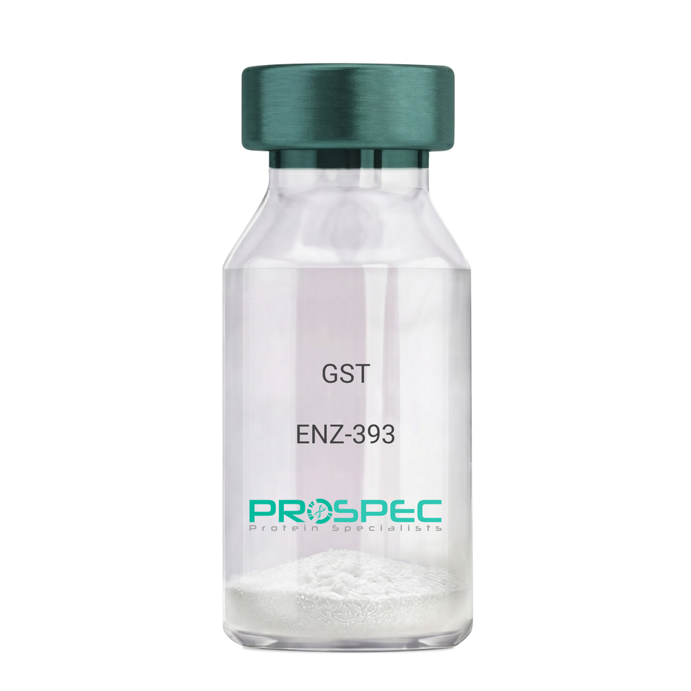
GSTDescription:
Glutathione S-Transferase Recombinant
Glutathione S-Transferase, GST, Glutathione S-transferase class-mu 26 kDa isozyme, GST 26, Sj26 antigen, SjGST.
Product # :
ENZ-393Price :
Quantity :
Shipping Method :
Shipped with Ice Packs
More Info
- description
- source
- formulation
- purity
- biological activity
- More Info
Description
Recombinant Glutathione S-Transferase full length protein (1-218a.a.) expressed in E.coli, having a molecular mass of 26kDa. GST was isolated from an E. coli strain that carries the coding sequence for Schistosoma japonicum GST under the control of a T7 promoter. The GST is purified by proprietary chromatographic techniques.
Source
Escherichia Coli.
Formulation
GST supplied in Phosphate Buffered Saline pH 7.4.
Purity
Greater than 95% as determined by SDS-PAGE.
Biological Activity
Specific activity is >20 units/mg. A unit is defined as the amount of enzyme that conjugate 1.0 u mole of 1-chloro-2,4-dinitrobenzene (CDNB) with reduced glutathione per minute at pH 6.5 at 25°C.More Info
-
Introduction
Antioxidant enzyme Glutathione S- Transferase (GST) is thought to do the primary cellular defense mechanism against reactive oxygen species. GST reduces lipid hydroperoxides through its Se-independent glutathione peroxidase activity. The enzyme also detoxifies lipid peroxidation end products such as 4-hydroxynonenal (4-HNE).
The soluble GST is a 26 kDa protein which occurs as a dimer in all aerobic organisms. Each monomer has two domains, one that binds GSH and is an /-structure similar to thioredoxin and the other, all helical, that binds the hydrophobic substrate. The GST -fusion protein expression system is a widely used recombinant protein expression system that allows a peptide or a regulatory protein domain to be expressed as a fusion to the C-terminus of Schistosoma japonicum GST. Fusion proteins also possess GST -enzymatic activity and can undergo dimerization similar to in vivo. The fusion protein can be purified via GST -affinity column chromatography. In most cases, the desired peptides or domains are removed from GST by applying a specific protease that recognizes and cleaves the linker between the protein domain and GST. The technique has been widely used to generate different kinds of proteins for crystallization, molecular immunology studies, the production of vaccines and studies involving protein-protein and protein-DNA interactions. -
Synonyms
Glutathione S-Transferase, GST, Glutathione S-transferase class-mu 26 kDa isozyme, GST 26, Sj26 antigen, SjGST.
-
Physical Appearance
Sterile Filtered clear solution.
-
Stability
Store at 4°C if entire vial will be used within 2-4 weeks. Store, frozen at -20°C for longer periods of time. For long term storage it is recommended to add a carrier protein (0.1% HSA or BSA).Avoid multiple freeze-thaw cycles.
-
Amino Acid Sequence
MSPILGYWKI KGLVQPTRLL LEYLEEKYEE HLYERDEGDK WRNKKFELGL EFPNLPYYID GDVKLTQSMA IIRYIADKHN MLGGCPKERA EISMLEGAVL DIRYGVSRIA YSKDFETLKV DFLSKLPEML KMFEDRLCHK TYLNGDHVTH PDFMLYDALD VVLYMDPMCL DAFPKLVCFK KRIEAIPQID KYLKSSKYIA WPLQGWQATF GGGDHPPKSD LVPR.
ProSpec's products are furnished for LABORATORY RESEARCH USE ONLY. The product may not be used as drugs, agricultural or pesticidal products, food additives or household chemicals.