Search results
1000 results found for “mumps”
Name
Description
Product #
Price
Quantity
Shipping Method
- View Data Sheet
Name :
GREM1 HumanDescription:
GREM1 Human Recombinant
Gremlin-1 isoform 1, CKTSF1B1, DAND2, DRM, GREMLIN, IHG-2, PIG2, GREM1, Cell proliferation-inducing gene 2 protein, Cysteine knot superfamily 1, BMP antagonist 1, DAN domain family member 2, Down-regulated in Mos-transformed cells protein, Increased in high glucose protein 2.
Product # :
PRO-1359Price :
Quantity :
Shipping Method :
Shipped with Ice Packs
More Info
- description
- source
- formulation
- purity
- More Info
Description
GREM1 Human Recombinant produced in E.coli is a single, non-glycosylated polypeptide chain containing 183 amino acids (25-184) and having a molecular mass of 20.7 kDa. GREM1 is fused to a 23 amino acid His-tag at N-terminus & purified by proprietary chromatographic techniques.
Source
Escherichia Coli.
Formulation
The GREM1 solution (0.5mg/ml) contains 20mM Tris-HCl buffer (pH 8.0), 0.4M Urea and 10% glycerol.
Purity
Greater than 90.0% as determined by SDS-PAGE.
More Info
-
Introduction
GREM1 belongs to the BMP (bone morphogenic protein) antagonist family. Like BMPs, BMP antagonists comprise cystine knots and usually form homo- and heterodimers. The CAN (cerberus and dan) subfamily of BMP antagonists, to which GREM1 belongs, is characterized by a C-terminal cystine knot with an eight-membered ring. The antagonistic effect of the secreted glycosylated protein is because of its direct binding to BMP proteins. As an antagonist of BMP, GREM1 takes a place in regulating organogenesis, body patterning, and tissue differentiation. In mouse, GREM1 has been shown to convey the sonic hedgehog (SHH) signal from the polarizing region to the apical ectodermal ridge during limb bud outgrowth. Alternatively merged transcript variants encoding different isoforms have been found for this gene.
-
Synonyms
Gremlin-1 isoform 1, CKTSF1B1, DAND2, DRM, GREMLIN, IHG-2, PIG2, GREM1, Cell proliferation-inducing gene 2 protein, Cysteine knot superfamily 1, BMP antagonist 1, DAN domain family member 2, Down-regulated in Mos-transformed cells protein, Increased in high glucose protein 2.
-
Physical Appearance
Sterile Filtered colorless solution.
-
Stability
Store at 4°C if entire vial will be used within 2-4 weeks. Store, frozen at -20°C for longer periods of time. For long term storage it is recommended to add a carrier protein (0.1% HSA or BSA).Avoid multiple freeze-thaw cycles.
-
Amino Acid Sequence
MGSSHHHHHH SSGLVPRGSH MGSKKKGSQG AIPPPDKAQH NDSEQTQSPQ QPGSRNRGRG QGRGTAMPGE EVLESSQEAL HVTERKYLKR DWCKTQPLKQ TIHEEGCNSR TIINRFCYGQ CNSFYIPRHI RKEEGSFQSC SFCKPKKFTT MMVTLNCPEL QPPTKKKRVT RVKQCRCISI DLD.
ProSpec's products are furnished for LABORATORY RESEARCH USE ONLY. The product may not be used as drugs, agricultural or pesticidal products, food additives or household chemicals.
- View Data Sheet
Name :
MAPKAPK3 HumanDescription:
Mitogen-Activated Protein Kinase-Activated Protein Kinase 3 Human Recombinant
3PK, MAPKAP-K3, MAPKAP3, MAPKAPK-3, MK-3, MAP kinase-activated protein kinase 3, MAPK-activated protein kinase 3, MAPKAP kinase 3,MAPKAPK-3, MK-3,MAPKAPK3.
Product # :
PKA-045Price :
Quantity :
Shipping Method :
Shipped with Ice Packs
More Info
- description
- source
- formulation
- purity
- More Info
Description
MAPKAPK3 Human Recombinant produced in E.Coli is a single, non-glycosylated polypeptide chain containing 405 amino acids (1-382 a.a) and having a molecular mass of 45.4kDa. MAPKAPK3 is fused to a 23 amino acid His-tag at N-terminus & purified by proprietary chromatographic techniques.
Source
E.coli.
Formulation
MAPKAPK3 protein solution (1mg/ml) containing 20mM Tris-HCl buffer (pH 8.0), 0.2M NaCl, 20% glycerol and 1mM DTT.
Purity
Greater than 95% as determined by SDS-PAGE.
More Info
-
Introduction
MAP kinase-activated protein kinase 3 (MAPKAPK3) is involved in inflammatory Reaction by regulating tumor necrosis factor (TNF) and IL6 production post-transcriptionally. MAPKAPK3 phosphorylates AU-rich elements (AREs)-binding proteins, like TTP/ZFP36, leading to control of stability and translation of TNF and IL6 mRNAs. Phosphorylation of TTP/ZFP36 (a major post-transcriptional regulator of TNF), promotes its binding to 14-3-3 proteins and reduces its ARE mRNA affinity resulting in inhibition of dependent degradation of ARE-containing transcript. MAPKAPK3 is activated by growth inducers and stress stimulation of cells.
-
Synonyms
3PK, MAPKAP-K3, MAPKAP3, MAPKAPK-3, MK-3, MAP kinase-activated protein kinase 3, MAPK-activated protein kinase 3, MAPKAP kinase 3,MAPKAPK-3, MK-3,MAPKAPK3.
-
Physical Appearance
Sterile Filtered colorless solution.
-
Stability
Store at 4°C if entire vial will be used within 2-4 weeks. Store, frozen at -20°C for longer periods of time. For long term storage it is recommended to add a carrier protein (0.1% HSA or BSA).Avoid multiple freeze-thaw cycles.
-
Amino Acid Sequence
MGSSHHHHHH SSGLVPRGSH MGSMDGETAE EQGGPVPPPV APGGPGLGGA PGGRREPKKY AVTDDYQLSK QVLGLGVNGK VLECFHRRTG QKCALKLLYD SPKARQEVDH HWQASGGPHI VCILDVYENM HHGKRCLLII MECMEGGELF SRIQERGDQA FTEREAAEIM RDIGTAIQFL HSHNIAHRDV KPENLLYTSK EKDAVLKLTD FGFAKETTQN ALQTPCYTPY YVAPEVLGPE KYDKSCDMWS LGVIMYILLC GFPPFYSNTG QAISPGMKRR IRLGQYGFPN PEWSEVSEDA KQLIRLLLKT DPTERLTITQ FMNHPWINQS MVVPQTPLHT ARVLQEDKDH WDEVKEEMTS ALATMRVDYD QVKIKDLKTS NNRLLNKRRK KQAGSSSASQ GCNNQ.
ProSpec's products are furnished for LABORATORY RESEARCH USE ONLY. The product may not be used as drugs, agricultural or pesticidal products, food additives or household chemicals.
- View Data Sheet
Name :
MCP 2 HumanDescription:
Monocyte Chemotactic Protein-2 Human Recombinant (CCL8)
Small inducible cytokine A8, CCL8, Monocyte chemotactic protein 2, MCP-2, Monocyte chemoattractant protein 2, HC14, chemokine (C-C motif) ligand 8, MCP2, SCYA8, SCYA10.
Product # :
CHM-316Price :
Quantity :
Shipping Method :
Shipped at Room temp
More Info
- description
- source
- formulation
- purity
- biological activity
- More Info
Description
Monocyte Chemotactic Protein-2 Human Recombinant produced in E.Coli is a non-glycosylated, Polypeptide chain containing 76 amino acids and having a molecular mass of 8904 Dalton. The MCP2 is purified by proprietary chromatographic techniques.
Source
Escherichia Coli.
Formulation
The protein was lyophilized from a concentrated (1mg/ml) sterile solution containing no additives.
Purity
Greater than 95.0% as determined by:
(a) Analysis by RP-HPLC.
(b) Analysis by SDS-PAGE.Biological Activity
The biological activity was determined by measuring the dose dependent mobilization of intracellular calcium (calcium flux) with human THP-1 cells. Significant calcium mobilization is observed with 500ng/mL of recombinant human MCP-2. Human MCP-2 also induces dose dependent chemotaxis of human THP-1 cells with an ED50=30-100 ng/mL corresponding to a Specific Activity of 10,000-33,334IU/mg.More Info
-
Introduction
Chemokine (C-C motif) ligand 8 (CCL8) is a small cytokine belonging to the CC chemokine family that was once called monocyte chemotactic protein-2 (MCP-2). The CCL8 protein is produced as a precursor containing 109 amino acids, which is cleaved to produce mature CCL8 containing 75 amino acids. The gene for CCL8 is encoded by 3 exons and is located within a large cluster of CC chemokines on chromosome 17q11.2 in humans. MCP-2 is chemotactic for and activates a many different immune cells, including mast cells, eosinophils and basophils, (that are implicated in allergic responses), and monocytes, T cells, and NK cells that are involved in the inflammatory response. CCL8 elicits its effects by binding to several different cell surface receptors called chemokine receptors. These receptors include CCR1, CCR2B and CCR5.
-
Synonyms
Small inducible cytokine A8, CCL8, Monocyte chemotactic protein 2, MCP-2, Monocyte chemoattractant protein 2, HC14, chemokine (C-C motif) ligand 8, MCP2, SCYA8, SCYA10.
-
Physical Appearance
Sterile Filtered White lyophilized (freeze-dried) powder.
-
Stability
Lyophilized MCP2 although stable at room temperature for 3 weeks, should be stored desiccated below -18°C. Upon reconstitution CCL8should be stored at 4°C between 2-7 days and for future use below -18°C. For long term storage it is recommended to add a carrier protein (0.1% HSA or BSA).Please prevent freeze-thaw cycles.
-
Solubility
It is recommended to reconstitute the lyophilized Monocyte Chemotactic Protein-2in sterile 18MΩ-cm H2O not less than 100µg/ml, which can then be further diluted to other aqueous solutions.
-
Amino Acid Sequence
The sequence of the first five N-terminal amino acids was determined and was found to be Gln-Pro-Asp-Ser-Val.
ProSpec's products are furnished for LABORATORY RESEARCH USE ONLY. The product may not be used as drugs, agricultural or pesticidal products, food additives or household chemicals.
- View Data Sheet
Name :
HIV-1 gp41 LongDescription:
HIV-1 gp41 Long Recombinant
Product # :
HIV-113Price :
Quantity :
Shipping Method :
Shipped with Ice Packs
More Info
- description
- source
- formulation
- purity
- More Info
Description
The E.Coli derived protein contains the full-length sequence of N-terminal epitopes of HIV-I gp41 395 amino acids (444-833) immunodominant regions gp41L. The protein is fused to b-galactosidase (114 kDa) at N-Terminus.
Source
Escherichia Coli.
Formulation
8M Urea, 20mM Tris-HCl pH 8.0, 10mM b-mercaptoethanol.
Purity
Greater than 95.0% as determined by HPLC analysis & SDS-PAGE.
More Info
-
Introduction
Human immunodeficiency virus (HIV) is a retrovirusthat can lead to a condition in which the immune systembegins to fail, leading to opportunistic infections. HIV primarily infects vital cells in the humanimmune systemsuch as helper T cells(specifically CD4+ T cells), macrophagesand dendritic cells. HIV infection leads to low levels of CD4+ T cells through three main mechanisms: firstly, direct viral killing of infected cells; secondly, increased rates of apoptosisin infected cells; and thirdly, killing of infected CD4+ T cells by CD8 cytotoxic lymphocytesthat recognize infected cells. When CD4+ T cell numbers decline below a critical level, cell-mediated immunityis lost, and the body becomes progressively more susceptible to opportunistic infections. HIV was classified as a member of the genus Lentivirus, part of the family of Retroviridae. Lentiviruses have many common morphologies and biological properties. Many species are infected by lentiviruses, which are characteristically responsible for long-duration illnesses with a long incubation period. Lentiviruses are transmitted as single-stranded, positive-sense, enveloped RNA viruses. Upon entry of the target cell, the viral RNA genomeis converted to double-stranded DNAby a virally encoded reverse transcriptasethat is present in the virus particle. This viral DNA is then integrated into the cellular DNA by a virally encoded integraseso that the genome can be transcribed. Once the virus has infected the cell, two pathways are possible: either the virus becomes latentand the infected cell continues to function, or the virus becomes active and replicates, and a large number of virus particles are liberated that can then infect other cells.
-
Physical Appearance
Sterile filtered colorless clear solution.
-
Stability
HIV-1 gp41 Long although stable at 4°C for 1 week, should be stored below -18°C. Please prevent freeze thaw cycles.
-
Amino Acid Sequence
IEFPGIFRPGGGDMRDNWRSELYKYKVVKIEPLGVAPTKAKRRVVQ REKRAVGIGALFLGFLGAAGSTMGAASMTLTVQARQLLSGIVQQQNNLLR AIEAQQHLLQLTVWGIKQLQARILAVERYLKDQQLLGIWGCSGKLICTTAVPWNASWSN KSLEQIWNNMTWMEWDREINNYTSLIHSLIEESQNQQEKNEQELLELDKWASLWNWFNITNWLWYIKLFIMIVGGLVGLRIVFAVLSVVNRVRQGYSPLSFQTHLPIPRGPDRPEGIEEEGGERDRDRSIRLVNGSLALIWDDLRSLCLFSYHRLRDLLLIVTRIVELLGRRGWEALKYWWNLLQYWSQELKNSAVSLLNATAIAVAEGTDRVIEVVQGAYRAIRHIPRRIRQGLERILL
-
Specificity
Immunoreactive with all sera of HIV-1 infected individuals.
ProSpec's products are furnished for LABORATORY RESEARCH USE ONLY. The product may not be used as drugs, agricultural or pesticidal products, food additives or household chemicals.
- View Data Sheet
Name :
HPV 18Description:
Human Papillomavirus 18 Recombinant
Papillomavirus, HPV, Papilloma Virus.
Product # :
HPV-002Price :
Quantity :
Shipping Method :
Shipped with Ice Packs
More Info
- description
- source
- formulation
- purity
- More Info
Description
The Recombinant HPV-18 is a full length large capsid protein having an Mw of 55kDa expressed in E. coli and fused to a GST-Tag at n-terminal, having a total Mw of 78kDa, purified by standard chromatography.
Source
E.Coli
Formulation
Recombinant HPV-18 solution in PBS and 2M Urea.
Purity
Protein is >90% pure as determined by 10% PAGE (Coomassie staining).
More Info
-
Introduction
Human papillomavirus family consist of over 200 types. Over than 30 to 40 types of HPV are transferred via sexual contact and infect the anogenital region initiating genital warts. Persistent infection with "high-risk" HPV types results in skin warts and leads to precancerous lesions and invasive cancer. HPV infection is considered as a source for all incidents of cervical cancer. E2, E6, and E7 proteins of HPV-16 and 18 are considered the main viral oncoproteins that take part in cervical cancer. The type-specific antigen epitopes of E2, E6, and E7 proteins of HPV-16 are fused together and expressed in E. coli for diagnostic purpose.
-
Synonyms
Papillomavirus, HPV, Papilloma Virus.
-
Physical Appearance
Sterile filtered clear liquid formulation.
-
Amino Acid Sequence
VDVYLPPPSVARVVNTDDYVTPTSIFYHAGSSRLLTVGNPYFRVPAGGG
NKQDIPKVSAYQYRVFRVQLPDPNKFGLPDTSIYNPETQRLVWACAGVE
IGRGQPLGVGLSGHPFYNKLDDTESSHAATSNVSEDVRDNVSVDYKQTQ
LCILGCAPAIGEHWAKGTACKSRPLSQGDCPPLELKNTVLEDGDMVDTG
YGAMDFSTLQDTKCEVPLDICQSICKYPDYLQMSADPYGDSMFFCLRRE
QLFARHFWNRAGTMGDTVPQSLYIKGTGMPASPGSCVYSPSPSGSIVTS
DSQLFNKPYWLHKAQGHNNGVCWHNQLFVTVVDTTPSTNLTICASTQSP
VPGQYDATKFKQYSRHVEEYDLQFIFQLCTITLTADVMSYIHSMNSSIL
EDWNFGVPPPPTTSLVDTYRFVQSVAITCQKDAAPAENKDPYDKLKFWN
VDLKEKFSLDLDQYPLGRKFLVQAGLRRKPTIGPRKRSAPSATTSSKPA.
ProSpec's products are furnished for LABORATORY RESEARCH USE ONLY. The product may not be used as drugs, agricultural or pesticidal products, food additives or household chemicals.
- View Data Sheet
Name :
T.gondii p40Description:
Toxoplasma Gondii p40 Recombinant
Product # :
TOX-268Price :
Quantity :
Shipping Method :
Shipped with Ice Packs
More Info
- description
- source
- formulation
- purity
- More Info
Description
The E.Coli derived recombinant Toxoplasma Gondii p40 having a Mw of 35kDa and fused to a 6 His tag at C-terminus can be used to test the specific IgG and IgM antibody for the diagnosis of Toxoplasma gondii infection.
Source
Escherichia Coli.
Formulation
1x PBS and 25mM arginine.
Purity
Toxoplasma protein is >95% pure as determined by 10% PAGE (coomassie staining).
More Info
-
Introduction
The life cycle of Toxoplasma gondii has two phases. The coccidia like takes place only in members of the Felidae family which makes these animals the parasite's primary host. The asexual part of the life cycle can take place in any warm-blooded animal, like other mammals(including felines) and birds. T. gondii constructing daughter scaffolds within the mother cell. In the intermediate hosts (including felines), the parasite invades cells, forming intracellular so-called parasitophorous vacuoles containing bradyzoites, the slowly replicating form of the parasit. Vacuoles form tissue cysts mainly within the muscles and brain. Since they are within cells, the host's immune system does not detect these cysts. Resistance to antibiotics varies, but the cysts are very difficult to eradicate entirely. Within these vacuoles T. gondii propagates by a series of binary fissions until the infected cell eventually bursts and tachyzoites are released. Tachyzoites are the motile, asexually reproducing form of the parasite. Unlike the bradyzoites, the free tachyzoites are usually efficiently cleared by the host's immune response, although some manage to infect cells and form bradyzoites, thus maintaining the infection.
-
Physical Appearance
Sterile Filtered clear solution.
-
Stability
T.gondii p40 although stable at 4°C for 1 week, should be stored below -18°C. Please prevent freeze thaw cycles.
-
Purification Method
Purified by proprietary chromatographic technique.
ProSpec's products are furnished for LABORATORY RESEARCH USE ONLY. The product may not be used as drugs, agricultural or pesticidal products, food additives or household chemicals.
- View Data Sheet
Name :
PODXL Human (335-387)Description:
Podocalyxin-Like (335-387 a.a) Human Recombinant
Product # :
PRO-2840Price :
Quantity :
Shipping Method :
Shipped at Room temp
More Info
- description
- source
- formulation
- purity
- More Info
Description
The PODXL Human is created as a recombinant protein with a 4kda N-terminal fusion of His Tag. The PODXL His-Tagged Fusion Protein, produced in E. coli, is a 11kDa protein containing 53 amino acid residues of the PODXL Human, 335-387 amino acids.
Source
Escherichia Coli.
Formulation
Each mg was lyophilized with 1xPBS, 0.4% SDS and 4mM DTT.
Purity
Greater than 90% as determined by SDS-PAGE.
More Info
-
Physical Appearance
Filtered White lyophilized (freeze-dried) powder.
-
Stability
Store lyophilized PODXLat -20°C. Aliquot the product after reconstitution to avoid repeated freezing/thawing cycles. Reconstituted protein can be stored at 4°C for a limited period of time; it does not show any change after two weeks at 4°C.
-
Solubility
It is recommended to add deionized water to prepare a working stock solution of approximately 0.5mg/ml and let the lyophilized pellet dissolve completely. Product is not sterile! Please filter the product by an appropriate sterile filter before using it on cell culture.
-
Amino Acid Sequence
AGGASDEKLI SLICRAVKAT FNPAQDKCGI RLASVPGSQT VVVKEITIHS.
-
Background
Podocalyxin-Like also known as PODXL is a glycosylated transmembrane sialoprotein in the CD34 and endoglycan family. The PODXL protein takes part in the regulation of both adhesion and cell morphology and cancer progression. PODXL plays a role as an anti-adhesive molecule, which retains an open filtration pathway between neighboring foot processes in the podocyte by charge repulsion. PODXL also functions as a pro-adhesive molecule, enhancing the adherence of cells to immobilized ligand which leads to rate increase of migration and cell-cell contacts in an integrin-dependent manner.
ProSpec's products are furnished for LABORATORY RESEARCH USE ONLY. The product may not be used as drugs, agricultural or pesticidal products, food additives or household chemicals.
- View Data Sheet
Name :
Toxoplasma P32Description:
Toxoplasma Gondii P32 (GRA6) Recombinant
Product # :
TOX-267Price :
Quantity :
Shipping Method :
Shipped with Ice Packs
More Info
- description
- source
- formulation
- purity
- More Info
Description
Recombinant Toxoplasma Gondii P32 (GRA6) produced in E.coli containing 180 amino acids of the Dense Granule Antigen 6 (GRA6) and fused with a 6xHis tag at C- terminus. This antigen forms dimers on SDS-PAGE under denatured conditions, compared to other toxoplasma monomer antigens, it has better immunoreactions.
Source
Escherichia Coli.
Formulation
Sterile Filtered solution containing containing PBS and 25mM arginine.
Purity
Toxoplasma protein is >95% pure as determined by 10% PAGE (coomassie staining).
More Info
-
Introduction
The life cycle of Toxoplasma gondii has two phases. The coccidia like takes place only in members of the Felidae family which makes these animals the parasite's primary host. The asexual part of the life cycle can take place in any warm-blooded animal, like other mammals(including felines) and birds. T. gondii constructing daughter scaffolds within the mother cell. In the intermediate hosts (including felines), the parasite invades cells, forming intracellular so-called parasitophorous vacuoles containing bradyzoites, the slowly replicating form of the parasit. Vacuoles form tissue cysts mainly within the muscles and brain. Since they are within cells, the host's immune system does not detect these cysts. Resistance to antibiotics varies, but the cysts are very difficult to eradicate entirely. Within these vacuoles T. gondii propagates by a series of binary fissions until the infected cell eventually bursts and tachyzoites are released. Tachyzoites are the motile, asexually reproducing form of the parasite. Unlike the bradyzoites, the free tachyzoites are usually efficiently cleared by the host's immune response, although some manage to infect cells and form bradyzoites, thus maintaining the infection.
-
Physical Appearance
Sterile Filtered solution.
-
Stability
Toxoplasma P32 although stable at 4°C for 1 week, should be stored below -18°C. Please prevent freeze thaw cycles.
-
Applications
Immunoassay.
-
Purification Method
Toxoplasma protein was purified by proprietary chromatographic technique.
ProSpec's products are furnished for LABORATORY RESEARCH USE ONLY. The product may not be used as drugs, agricultural or pesticidal products, food additives or household chemicals.
- View Data Sheet
Name :
HIV-1 nefDescription:
HIV-1 nef Recombinant
Product # :
HIV-124Price :
Quantity :
Shipping Method :
Shipped with Ice Packs
More Info
- description
- source
- formulation
- purity
- More Info
Description
The E.coli derived 20kDa recombinant protein genotype IIIB is a non-glycosylated polypeptide chain, containing the HIV-1 nef immunodominant regions, 3-190 amino acids. The HIV-1 nef is fused to beta-galactosidase (114 kDa) at the N-terminus.
Source
Escherichia Coli.
Formulation
HIV-1 nef protein solution in 8M urea, 20mM Tris-HCl pH 8.0, 10mM b-mercaptoethanol.
Purity
Greater than 95.0% as determined by HPLC analysis and SDS-PAGE.
More Info
-
Introduction
Human immunodeficiency virus (HIV) is a retrovirus that can lead to a condition in which the immune system begins to fail, leading to opportunistic infections. HIV primarily infects vital cells in the human immune system such as helper T cells (specifically CD4+ T cells), macrophages and dendritic cells. HIV infection leads to low levels of CD4+ T cells through three main mechanisms: firstly, direct viral killing of infected cells; secondly, increased rates of apoptosis in infected cells; and thirdly, killing of infected CD4+ T cells by CD8 cytotoxic lymphocytes that recognize infected cells. When CD4+ T cell numbers decline below a critical level, cell-mediated immunity is lost, and the body becomes progressively more susceptible to opportunistic infections. HIV was classified as a member of the genus Lentivirus, part of the family of Retroviridae. Lentiviruses have many common morphologies and biological properties. Many species are infected by lentiviruses, which are characteristically responsible for long-duration illnesses with a long incubation period. Lentiviruses are transmitted as single-stranded, positive-sense, enveloped RNA viruses. Upon entry of the target cell, the viral RNA genome is converted to double-stranded DNA by a virally encoded reverse transcriptase that is present in the virus particle. This viral DNA is then integrated into the cellular DNA by a virally encoded integrase so that the genome can be transcribed. Once the virus has infected the cell, two pathways are possible: either the virus becomes latent and the infected cell continues to function, or the virus becomes active and replicates, and a large number of virus particles are liberated that can then infect other cells.
-
Physical Appearance
Sterile filtered colorless clear solution.
-
Stability
HIV-1 nef although stable at 4°C for 1 week, should be stored below -18°C. Please prevent freeze thaw cycles.
-
Amino Acid Sequence
kwskssv igwptvrerm rraepaadgv gaasqdlekh gaitssntaa tnadcawlea
qeeeevgfpv tpqvplrpmt ykaavdlshf lkekgglegl ihsqrrqdil dlwiyhtqgy
fpdwqnytpg pgirypltfg wcyklvpvep ekleeankge ntsllhpvsl hgmddperev
lewrfdsrla.
ProSpec's products are furnished for LABORATORY RESEARCH USE ONLY. The product may not be used as drugs, agricultural or pesticidal products, food additives or household chemicals.
- View Data Sheet
Name :
HIV-1 p24 PairedDescription:
Mouse Anti HIV-1 p24 Paired
Product # :
ANT-772Price :
Quantity :
Shipping Method :
Shipped with Ice Packs
More Info
- description
- formulation
- purity
- More Info
Description
HIV-1 p24 Paired antibodies coating and conjugating are used for lateral flow immunoassay and having a Mw of 24kDa. Please note that when ordering for example: 100µg antibody we ship 50µg from each of the antibodies (100µg in total).
Formulation
*HIV-1 p24 Label antibody contains PBS, pH 7.4.
* HIV-1 p24 Capture antibody contains PBS, pH 7.4.
Purity
Greater than 95%.
More Info
-
Introduction
Human immunodeficiency virus (HIV) is a retrovirus that can lead to a condition in which the immune system begins to fail, leading to opportunistic infections. HIV primarily infects vital cells in the human immune system such as helper T cells (specifically CD4+ T cells), macrophage sand dendritic cells. HIV infection leads to low levels of CD4+ T cells through three main mechanisms: firstly, direct viral killing of infected cells; secondly, increased rates of apoptosis in infected cells; and thirdly, killing of infected CD4+ T cells by CD8 cytotoxic lymphocytes that recognize infected cells. When CD4+ T cell numbers decline below a critical level, cell-mediated immunity is lost, and the body becomes progressively more susceptible to opportunistic infections. HIV was classified as a member of the genus Lentivirus, part of the Retroviridae family. Lentiviruses have many common morphologies and biological properties. Many species are infected by lentiviruses, which are characteristically responsible for long-duration illnesses with a long incubation period. Lentiviruses are transmitted as single-stranded, positive-sense, enveloped RNA viruses. Upon entry of the target cell, the viral RNA genome is converted to double-stranded DNA by a virally encoded reverse transcriptase that is present in the virus particle. This viral DNA is then integrated into the cellular DNA by a virally encoded integrase so that the genome can be transcribed. Once the virus has infected the cell, two pathways are possible: either the virus becomes latent and the infected cell continues to function, or the virus becomes active and replicates, and a large number of virus particles are liberated that can then infect other cells. The p24 protein is detected in patient blood as early as 2 weeks after HIV infection, further reducing the window period necessary to accurately detect the HIV status of the patient.
-
Physical Appearance
2 vials of sterile filtered clear colorless solution.
-
Stability
HIV-1 p24 antibody although stable at 4°C for 1 week, should be stored below -18°C. For long term storage it is recommended to add a carrier protein (0.1% HSA or BSA). Please prevent freeze-thaw cycles.
-
Applications
Lateral flow immunoassay.
-
Type
Mouse Anti Human Monoclonal.
-
Purification Method
Purified by protein A affinity column.
ProSpec's products are furnished for LABORATORY RESEARCH USE ONLY. The product may not be used as drugs, agricultural or pesticidal products, food additives or household chemicals.
- View Data Sheet
Name :
HIV-1 p24 CoreDescription:
HIV-1 p24 Core Recombinant
Product # :
HIV-103Price :
Quantity :
Shipping Method :
Shipped with Ice Packs
More Info
- description
- source
- formulation
- purity
- More Info
Description
HIV-1 p24 Core is a 24kDa non-glycosylated polypeptide chain, containing the full length sequence of the HIV-1 p24.
Source
Escherichia Coli.
Formulation
50mM Tris, pH 8.5 and 8M urea.
Purity
Greater than 95.0% as determined by HPLC analysis and SDS-PAGE.
More Info
-
Introduction
Human immunodeficiency virus (HIV) is a retrovirusthat can lead to a condition in which the immune systembegins to fail, leading to opportunistic infections. HIV primarily infects vital cells in the humanimmune systemsuch as helper T cells(specifically CD4+ T cells), macrophagesand dendritic cells. HIV infection leads to low levels of CD4+ T cells through three main mechanisms: firstly, direct viral killing of infected cells; secondly, increased rates of apoptosisin infected cells; and thirdly, killing of infected CD4+ T cells by CD8 cytotoxic lymphocytesthat recognize infected cells. When CD4+ T cell numbers decline below a critical level, cell-mediated immunityis lost, and the body becomes progressively more susceptible to opportunistic infections. HIV was classified as a member of the genus Lentivirus, part of the family of Retroviridae. Lentiviruses have many common morphologies and biological properties. Many species are infected by lentiviruses, which are characteristically responsible for long-duration illnesses with a long incubation period. Lentiviruses are transmitted as single-stranded, positive-sense, enveloped RNA viruses. Upon entry of the target cell, the viral RNA genomeis converted to double-stranded DNAby a virally encoded reverse transcriptasethat is present in the virus particle. This viral DNA is then integrated into the cellular DNA by a virally encoded integraseso that the genome can be transcribed. Once the virus has infected the cell, two pathways are possible: either the virus becomes latentand the infected cell continues to function, or the virus becomes active and replicates, and a large number of virus particles are liberated that can then infect other cells.
-
Physical Appearance
Sterile filtered colorless clear solution.
-
Stability
HIV-1 p24 core although stable at 4°C for 1 week, should be stored below -18°C. Please prevent freeze thaw cycles.
-
Specificity
Immunoreactive with all sera of HIV-1 infected individuals.
ProSpec's products are furnished for LABORATORY RESEARCH USE ONLY. The product may not be used as drugs, agricultural or pesticidal products, food additives or household chemicals.
- View Data Sheet
Name :
LLODescription:
Listeriolysin-O Recombinant
Listeriolysin-O, LLO, hlyA.
Product # :
PRO-320Price :
Quantity :
Shipping Method :
Shipped with Ice Packs
More Info
- description
- source
- formulation
- purity
- biological activity
- More Info
Description
LLO is a single, non-glycosylated polypeptide chain containing 529 amino acids and having a molecular mass of 58kDa. (accession number: AAF64524).
Source
Escherichia Coli.
Formulation
The protein contains 50mM NaH2PO4, 1mM EDTA, 2.7mM KCl, pH 6.4, 1mM DTT, 5% (v/v) glycerol and 0.5M NaCl.
Purity
Greater than 90.0% as determined by SDS-PAGE.
Biological Activity
Hemolytic activity is 8,27E+05 HU/mg of protein where HU means hemolytic activity unit that is the amount of toxin needed to release half the hemoglobin (50% lysis) of the erythrocytes as determined by hemolysin assay.
More Info
-
Introduction
Listeriolysin O (aka LLO) is a hemolysin produced by Listeria monocytogenes bacteria, the pathogen responsible for causing listeriosis. The toxin may be regarded as a virulence factor, since it is crucial for the virulence of L. monocytogenes. LLO is a single polypeptide protein encoded by the hlyA gene and composed of 529 residues. LLO is a thiol-activated cholesterol-dependent pore forming toxin protein; therefore, it is activated by reducing agents and inhibited by oxidizing agents. Still, LLO differs from other thiol-activated toxins, as its cytolytic activity is maximized at a pH of 5.5. Inside the acidic phagosomes (average pH ~ 5.9) of cells that have phagocytosed L. monocytogenes, LLO is selectively activated by maximizing activity at a pH of 5.5. Following the phagosome lysis by LLO, the bacterium breaks out into the cytosol, where it is able to grow intracellularly, and the toxin has reduced activity in the more basic cytosol. Thus, LLO permits L. monocytogenes to break out from the phagosomes into the cytosol without harming the plasma membrane of the infected cell, which allows the bacteria to live intracellularly, where they are sheltered from extracellular immune system factors such as the complement system and antibodies. LLO also brings about dephosphorylation of histone H3 and deacetylation of histone H4 in the early phases of infection, before entry of L. monocytogenes into the host cell. The pore-forming activity is not implicated in causing the histone modifications. The modifications of the histones affect the down regulation of genes encoding proteins involved in the inflammatory response. Therefore, LLO may be significant in subverting the host immune response to L. monocytogenes. At its NH2-terminus it possesses a 25 residues long typical signal sequence excited during the secretion process. Moreover, in its NH2-terminus there is also a 19 amino acids PEST- like sequence that may target this toxin for degradation. The PEST-like sequence found in LLO and is considered crucial for virulence, given that mutants lacking the sequence lysed the host cell. Nevertheless, contrary to PEST's supposed role in protein degradation, evidence implies that the PEST-like sequence may control LLO production in the cytosol rather than increase degradation of LLO.
-
Synonyms
Listeriolysin-O, LLO, hlyA.
-
Physical Appearance
Sterile Filtered clear solution.
-
Stability
Store at 4°C if entire vial will be used within 2-4 weeks. Store, frozen at -20°C for longer periods of time. Avoid multiple freeze-thaw cycles.
ProSpec's products are furnished for LABORATORY RESEARCH USE ONLY. The product may not be used as drugs, agricultural or pesticidal products, food additives or household chemicals.
- View Data Sheet
Name :
SARS Spike (14-667)Description:
SARS Spike (14-667 a.a.), Recombinant
Product # :
SARS-031Price :
Quantity :
Shipping Method :
Shipped with Ice Packs
More Info
- description
- source
- formulation
- purity
- biological activity
- More Info
Description
The HEK293 derived recombinant protein contains the SARS Coronavirus Spike S1 Gycoprotein, amino acids 14-667 fused to His tag at C-terminal.
Source
HEK293
Formulation
SARS Spike S1 glycoprotein is lyophilized from 1x PBS pH-7.4 + 10% Glycerol.
Purity
Protein is >90% pure as determined SDS-PAGE.
Biological Activity
Measured by its binding ability in a functional ELISA with Human ACE-2 (CAT# enz-1125)
More Info
-
Introduction
SARS Coronavirus is an enveloped virus containing three outer structural proteins, namely the membrane (M), envelope (E), and spike (S) proteins. Spike (S)-glycoprotein of the virus interacts with a cellular receptor and mediates membrane fusion to allow viral entry into susceptible target cells. Accordingly, S-protein plays an important role in virus infection cycle and is the primary target of neutralizing antibodies.
-
Physical Appearance
Sterile Filtered clear solution.
-
Stability
Protein is shipped on ice packs. Upon arrival, Store at -20°C.
-
Purification Method
Purified by immobilized metal affinity chromatographic technique.
ProSpec's products are furnished for LABORATORY RESEARCH USE ONLY. The product may not be used as drugs, agricultural or pesticidal products, food additives or household chemicals.
- View Data Sheet
Name :
IAV-NPDescription:
Influenza A Virus Nucleoprotein Recombinant
Product # :
IHA-035Price :
Quantity :
Shipping Method :
Shipped with Ice Packs
More Info
- description
- source
- formulation
- purity
- More Info
- sds-page
Description
Recombinant Influenza A Virus Nucleoprotein produced in E. coli having a Mw of 66.6kDa. IAV-NP is fused to a 6xHis tag at its C terminal is and purified by proprietary chromatographic technique.
Source
E. coli.
Formulation
IAV-NP protein solution contains 0.25% sodium azide, 10mM K2CO3 and PBS.
Purity
Protein is >90% pure as determined by 10% PAGE (coomassie staining).
sds-page
More Info
-
Physical Appearance
Sterile Filtered clear solution.
-
Stability
Store at 4°C if entire vial will be used within 2-4 weeks. Store, frozen at -20°C for longer periods of time. For long term storage it is recommended to add a carrier protein (0.1% HSA or BSA). Avoid multiple freeze-thaw cycles.
-
Amino Acid Sequence
MASQGTKRSYEQMETDGERQNATEIRASVGKMIDGIGRFYIQMCTELKLSDYEGR LIQNSLTIERMVLSAFDERRNRYLEEHPSAGKDPKKTGGPIYKRVDGKWMRELVL YDKEEIRRIWR QANNGDDATRGLTHMMIWHSNLNDTTYQRTRALVRTGMDPRMC SLMQGSTLPRRSGAAGAAVKGIGTMVMELIRMIKRGINDRNFWRGENGRKTRSAY ERMCNILKGKFQTAAQRAMMDQVRESRNPGNAEIEDLIFSARSALILRGSVAHKS CLPACVYGPAVSSGYDFEKEGYSLVGIDPFKLLQNSQVYSLIRPNENPAHKSQLVWM ACHSAAFEDLRLLSFIRGTKVCPRGKLSTRGVQIASNENMDNMESSTLELRSRYWAIR TRSGGNTNQQRASAGQISVQPTFSVQRNLPFEKSTVMAAFTGNTEGRTSDMRAEIIRMM
-
Background
Influenza A virus, notorious for its ability to cause seasonal epidemics and occasional pandemics, poses a significant threat to global public health. The pursuit of effective countermeasures has led to the exploration of Influenza A Virus Nucleoprotein Recombinant as a key molecular protagonist in our defense against this elusive virus. This research endeavors to unravel the intricacies of the Nucleoprotein Recombinant, shedding light on its structural features, immunogenic prowess, and applications in vaccine development and antiviral therapies. By dissecting the properties of the Nucleoprotein Recombinant, scientists aim to fortify our defenses against Influenza A, potentially paving the way for a new era in influenza prevention and control.
Structural Insights into Nucleoprotein Recombinant:
The Influenza A Virus Nucleoprotein Recombinant, engineered to mirror crucial viral components, serves as a versatile tool for studying the virus's genetic material. A nuanced understanding of its three-dimensional structure and conformational intricacies is pivotal, not only for deciphering the virus's replication mechanisms but also for designing targeted interventions, such as vaccines and antiviral drugs.
Immunogenic Potential and Vaccine Development:
The quest for an effective influenza vaccine has been fueled by the virus's notorious mutability. Nucleoprotein Recombinant, designed to elicit strong immune responses, stands as a promising candidate for vaccine development. By presenting conserved viral elements to the immune system, the recombinant protein aims to induce robust and lasting immunity, thwarting the influenza virus's ability to evade immune detection through antigenic drift.
Application in Antiviral Therapies:
Beyond vaccination, Influenza A Virus Nucleoprotein Recombinant holds promise in the realm of antiviral therapies. The recombinant protein's ability to stimulate immune responses can be harnessed in the development of immunotherapies. Monoclonal antibodies derived from the Nucleoprotein Recombinant may offer targeted treatments, neutralizing the virus and potentially shortening the duration and severity of influenza infections.
Challenges and Future Directions:
While the Nucleoprotein Recombinant stands at the forefront of influenza research, challenges persist. The virus's ability to rapidly mutate demands ongoing vigilance and adaptability in recombinant strategies. Furthermore, considerations of vaccine safety, efficacy, and public acceptance necessitate continuous exploration to optimize the translational success of Nucleoprotein Recombinant-based interventions.
Influenza A Virus Nucleoprotein Recombinant emerges as a sentinel in the scientific arsenal against influenza, epitomizing the quest for innovative solutions in the face of viral challenges. Its structural insights, immunogenic potential, and applications in vaccine development and antiviral therapies position it as a pivotal player. As researchers continue to decipher the molecular intricacies of Nucleoprotein Recombinant, they not only fortify our defenses against influenza but potentially pave the way for a paradigm shift in our approach to influenza prevention and control, shaping the landscape of global health security.
ProSpec's products are furnished for LABORATORY RESEARCH USE ONLY. The product may not be used as drugs, agricultural or pesticidal products, food additives or household chemicals.
- View Data Sheet
Name :
GM-CSF Poricne, HisDescription:
Granulocyte Macrophage-Colony Stimulating Factor Porcine Recombinant, His Tag
CSF-2, MGI-1GM, GM-CSF, Pluripoietin-alpha, Molgramostin, Sargramostim
Product # :
CYT-1160Price :
Quantity :
Shipping Method :
Shipped with Ice Packs
More Info
- description
- source
- formulation
- purity
- biological activity
- More Info
Description
GMCSF Poricne Recombinant produced in E.Coli is a single, non-glycosylated polypeptide chain containing 148 amino acids (18-144a.a.) and having a molecular mass of 16.6kDa.GMCSF is fused to a 21 amino acid His tag at N-Terminus and purified by proprietary chromatographic techniques.
Source
Escherichia Coli.
Formulation
GMCSF protein solution (0.5mg/ml) containing Phosphate-Buffered Saline (pH7.4).
Purity
Greater than 95.0% as determined by SDS-PAGE.
Biological Activity
The ED50 range is ≤ 40 ng/ml, and is measured in a cell proliferation assay using TF-1 human erythroleukemic cells.
More Info
-
Introduction
The hematopoietic growth factor GM-CSF or granulocyte macrophage colony-stimulating factor, stimulates the development of neutrophils & macrophages, enhance proliferation and development of early erythroid megakaryocytic & eosinophilic progenitor cells. GM-CSF is secreted from the fibroblasts, monocytes, T-lymphocytes & endothelial cells. This protein blocks the migration of neutrophils & induces the biological activity of mature end-cells.
-
Synonyms
CSF-2, MGI-1GM, GM-CSF, Pluripoietin-alpha, Molgramostin, Sargramostim
-
Physical Appearance
Sterile Filtered colorless solution.
-
Stability
Store at 4°C if entire vial will be used within 2-4 weeks. Store, frozen at -20°C for longer periods of time. For long term storage it is recommended to add a carrier protein (0.1% HSA or BSA).Avoid multiple freeze-thaw cycles.
-
Amino Acid Sequence
MGSSHHHHHH SSGLVPRGSH MAPTRPPSPV TRPWQHVDAI KEALSLLNNS NDTAAVMNET VDVVCEMFDP QEPTCVQTRL NLYKQGLRGS LTRLKSPLTL LAKHYEQHCP LTEETSCETQ SITFKSFKDS LNKFLFTIPF DCWGPVKK
-
Background
What is the molecular weight/Mw of GM-CSF PORCINE, HIS Protein?
GM-CSF PORCINE, HIS Protein has a total Mw of 16.6kDa.
What is the source or expression system of GM-CSF PORCINE, HIS Protein?
Escherichia Coli.
What is the Purity of GM-CSF PORCINE, HIS Protein?
GM-CSF PORCINE, HIS Protein is >95% pure as determined by SDS-PAGE.
What is the Biological Activity of GM-CSF PORCINE, HIS Protein?
The ED50 range is ≤ 40 ng/ml, and is measured in a cell proliferation assay using TF-1 human erythroleukemic cells.
What is the amino acid sequence of GM-CSF PORCINE, HIS Protein?
MGSSHHHHHH SSGLVPRGSH MAPTRPPSPV TRPWQHVDAI KEALSLLNNS NDTAAVMNET VDVVCEMFDP QEPTCVQTRL NLYKQGLRGS LTRLKSPLTL LAKHYEQHCP LTEETSCETQ SITFKSFKDS LNKFLFTIPF DCWGPVKK
What applications can GM-CSF PORCINE, HIS Protein be used in?
GM-CSF PORCINE, HIS Protein can probably be used in western blot, ELISA and Lateral Flow.
What is the endotoxin level for GM-CSF PORCINE, HIS Protein?
The endotoxin level is minimal, GM-CSF PORCINE, HIS Protein was purified using conventional chromatography techniques.
ProSpec's products are furnished for LABORATORY RESEARCH USE ONLY. The product may not be used as drugs, agricultural or pesticidal products, food additives or household chemicals.
- View Data Sheet
Name :
Recombinant TNF-a AntibodyDescription:
Recombinant Anti Human Tumor Necrosis Factor-Alpha
Product # :
ANT-599Price :
Quantity :
Shipping Method :
Shipped with Ice Packs
More Info
- description
- source
- formulation
- purity
- biological activity
- More Info
Description
Recombinant TNF-a Antibody is a recombinant human IgG1 monoclonal antibody specific for human tumor necrosis factor (TNF). Recombinant TNF-a Antibody is produced by recombinant DNA technology in a Chinese Hamster Ovary mammalian cell expression system in a serum-free medium and has a molecular weight of approximately 148 kDa.
Source
CHO.
Formulation
The Recombinant TNF-a Antibody 53mg/ml solution contains 6.16 mg/ml of sodium chloride, 0.86 mg/ml of monobasic sodium phosphate dihydrate, 1.53 mg/ml of dibasic sodium phosphate dihydrate, 0.3 mg/ml of sodium citrate, 1.30 mg/ml of citric acidmonohydrate, 12 mg/ml of mannitol, 1mg/ml of polysorbate 80, pH-5.
Purity
Greater than 98.0% as determined by:
(a) Analysis by SEC-HPLC.
(b) Analysis by SDS-PAGE.Biological Activity
The EC50 as determined by L929 cell proliferation assay for neutralization reaction between TNF-a Antibody and TNFA, Perform a comparison of a dilution series of the Sample solution with a dilution series of the Standard solution, measured potency was found to be 1.2 X 10⁴EU/mg.More Info
-
Introduction
Tumor necrosis factor is a cytokine involved in systemic inflammation and is a member of a group of cytokines that all stimulate the acute phase reaction. TNF is mainly secreted by macrophages.
TNF causes apoptotic cell death, cellular proliferation, differentiation, inflammation, tumorigenesisand viral replication, TNF is also involved in lipid metabolism, and coagulation. TNF's primary role is in the regulation of immune cells.
Dysregulation and, in particular, overproduction of TNF have been implicated in a variety of human diseases- autoimmune diseases, insulin resistance, and cancer. -
Physical Appearance
Clear and colorless solution.
-
Stability
Recombinant TNF-a Antibody should be stored between 2-8°C and should be protected from light. DO NOT FREEZE. DO NOT SHAKE.
-
Amino Acid Sequence
LIGHT CHAIN
DIQMTQSPSSLSASVGDRVTITCRASQGIRNYLAWYQQKPGKAPKLLIYAASTLQSGVPSRFSGSGSGTDF
TLTISSLQPEDVATYYCQRYNRAPYTFGQGTKVEIKRTVAAPSVFIFPPSDEQLKSGTASVVCLLNNFYPR
EAKVQWKVDNALQSGNSQESVTEQDSKDSTYSLSSTLTLSKADYEKHKVYACEVTHQGLSSPVTKSFNRGEC
HEAVY CHAIN
EVQLVESGGGLVQPGRSLRLSCAASGFTFDDYAMHWVRQAPGKGLEWVSAITWNSGHIDYADSVEGRFTISR
DNAKNSLYLQMNSLRAEDTAVYYCAKVSYLSTASSLDYWGQGTLVTVSSASTKGPSVFPLAPSSKSTSGGTA
ALGCLVKDYFPEPVTVSWNSGALTSGVHTFPAVLQSSGLYSLSSVVTVPSSSLGTQTYICNVNHKPSNTKVD
KKVEPKSCDKTHTCPPCPAPELLGGPSVFLFPPKPKDTLMISRTPEVTCVVVDVSHEDPEVKFNWYVDGVEV
HNAKTKPREEQYNSTYRVVSVLTVLHQDWLNGKEYKCKVSNKALPAPIEKTISKAKGQPREPQVYTLPPSRD
ELTKNQVSLTCLVKGFYPSDIAVEWESNGQPENNYKTTPPVLDSDGSFFLYSKLTVDKSRWQQGNVFSCSVM
HEALHNHYTQKSLSLSPGK.
ProSpec's products are furnished for LABORATORY RESEARCH USE ONLY. The product may not be used as drugs, agricultural or pesticidal products, food additives or household chemicals.
- View Data Sheet
Name :
DUSP23 Human, ActiveDescription:
Dual Specificity Phosphatase 23 Human Recombinant, Active
Dual specificity protein phosphatase 23, Low molecular mass dual specificity phosphatase 3, LDP-3, VH1-like phosphatase Z, DUSP23, LDP3, VHZ, VH1-Like Member Z, EC 3.1.3.16, EC 3.1.3.48, DUSP25, MOSP, LDP3, Dual Specificity Phosphatase 23, VH1-Like Phosphatase Z, LDP-3, VHZ, Low-Molecular-Mass Dual-Specificity Phosphatase 3, Low Molecular Mass Dual Specificity Phosphatase 3, Dual Specificity Protein, Phosphatase 23, Testicular Tissue Protein Li 59.
Product # :
ENZ-1043Price :
Quantity :
Shipping Method :
Shipped with Ice Packs
More Info
- description
- source
- formulation
- purity
- biological activity
- More Info
Description
DUSP23 Human Recombinant produced in E.coli is a single, non-glycosylated polypeptide chain containing 170 amino acids (1-150 a.a) and having a molecular mass of 18.8kDa.DUSP23 is fused to a 20 amino acid His-tag at N-terminus & purified by proprietary chromatographic techniques.
Source
Escherichia Coli.
Formulation
DUSP23 protein solution (1mg/ml) containing 20mM Tris-HCl buffer (pH 8.0), 2mM DTT, 10% glycerol and 100mM NaCl.
Purity
Greater than 90% as determined by SDS-PAGE.
Biological Activity
Specific activity is > 200 units/mg, and is defined as the amount of enzyme that hydrolyzes 1.0 nmole of p-nitrophenyl phosphate (pNPP) per minute at pH 7.5 at 37°C.
More Info
-
Introduction
DUSP23 is a member of the protein-tyrosine phosphatase family. DUSP23 is a protein phosphatase which facilitates dephosphorylation of phosphorylated proteins on Tyr and Ser/Thr residues. In vitro, DUSP23 dephosphorylate p44-ERK1 (MAPK3) but not p54 SAPK-beta (MAPK10). In addition, DUSP23 enhances activation of JNK and p38(MAPK14).
-
Synonyms
Dual specificity protein phosphatase 23, Low molecular mass dual specificity phosphatase 3, LDP-3, VH1-like phosphatase Z, DUSP23, LDP3, VHZ, VH1-Like Member Z, EC 3.1.3.16, EC 3.1.3.48, DUSP25, MOSP, LDP3, Dual Specificity Phosphatase 23, VH1-Like Phosphatase Z, LDP-3, VHZ, Low-Molecular-Mass Dual-Specificity Phosphatase 3, Low Molecular Mass Dual Specificity Phosphatase 3, Dual Specificity Protein, Phosphatase 23, Testicular Tissue Protein Li 59.
-
Physical Appearance
Sterile Filtered clear solution.
-
Stability
Store at 4°C if entire vial will be used within 2-4 weeks. Store, frozen at -20°C for longer periods of time. For long term storage it is recommended to add a carrier protein (0.1% HSA or BSA).Avoid multiple freeze-thaw cycles.
-
Amino Acid Sequence
MGSSHHHHHH SSGLVPRGSH MGVQPPNFSW VLPGRLAGLA LPRLPAHYQF LLDLGVRHLV SLTERGPPHS DSCPGLTLHR LRIPDFCPPA PDQIDRFVQI VDEANARGEA VGVHCALGFG RTGTMLACYL VKERGLAAGD AIAEIRRLRP GSIETYEQEK AVFQFYQRTK.
ProSpec's products are furnished for LABORATORY RESEARCH USE ONLY. The product may not be used as drugs, agricultural or pesticidal products, food additives or household chemicals.
- View Data Sheet
Name :
CAMP HumanDescription:
Cathelicidin Antimicrobial Peptide Human Recombinant
CAP-18, CAP18, CRAMP, FALL-39, FALL39, HSD26, LL37, 18 kDa cationic antimicrobial protein, FALL-39 peptide antibiotic, Cathelicidin antimicrobial peptide.
Product # :
PRO-1405Price :
Quantity :
Shipping Method :
Shipped with Ice Packs
More Info
- description
- source
- formulation
- purity
- More Info
Description
CAMP Human Recombinant produced in E.Coli is a single, non-glycosylated polypeptide chain containing 163 amino acids (34-173 a.a.) and having a molecular mass of 18.4kDa.CAMP is fused to a 23 amino acid His-tag at N-terminus.
Source
Escherichia Coli.
Formulation
CAMP protein solution (0.5mg/ml) contains 20mM Tris-HCl buffer (pH 8.0), 0.4M UREA and 10% glycerol.
Purity
Greater than 85.0% as determined by SDS-PAGE.
More Info
-
Introduction
CAMP belongs to the antimicrobial peptide family, contains highly conserved N-terminal signal peptide, a cathelin domain and a structurally variable cationic antimicrobial peptide that produced by extracellular proteolysis from the C-terminus. CAMP has numerous functions besides the antimicrobial activity such as: cell chemotaxis, immune mediator induction and inflammatory response regulation.
-
Synonyms
CAP-18, CAP18, CRAMP, FALL-39, FALL39, HSD26, LL37, 18 kDa cationic antimicrobial protein, FALL-39 peptide antibiotic, Cathelicidin antimicrobial peptide.
-
Physical Appearance
Sterile Filtered clear solution.
-
Stability
Store at 4°C if entire vial will be used within 2-4 weeks. Store, frozen at -20°C for longer periods of time. For long term storage it is recommended to add a carrier protein (0.1% HSA or BSA).Avoid multiple freeze-thaw cycles.
-
Amino Acid Sequence
MGSSHHHHHH SSGLVPRGSH MGSQVLSYKE AVLRAIDGIN QRSSDANLYR LLDLDPRPTM DGDPDTPKPV SFTVKETVCP RTTQQSPEDC DFKKDGLVKR CMGTVTLNQA RGSFDISCDK DNKRFALLGD FFRKSKEKIG KEFKRIVQRI KDFLRNLVPR TES.
ProSpec's products are furnished for LABORATORY RESEARCH USE ONLY. The product may not be used as drugs, agricultural or pesticidal products, food additives or household chemicals.
- View Data Sheet
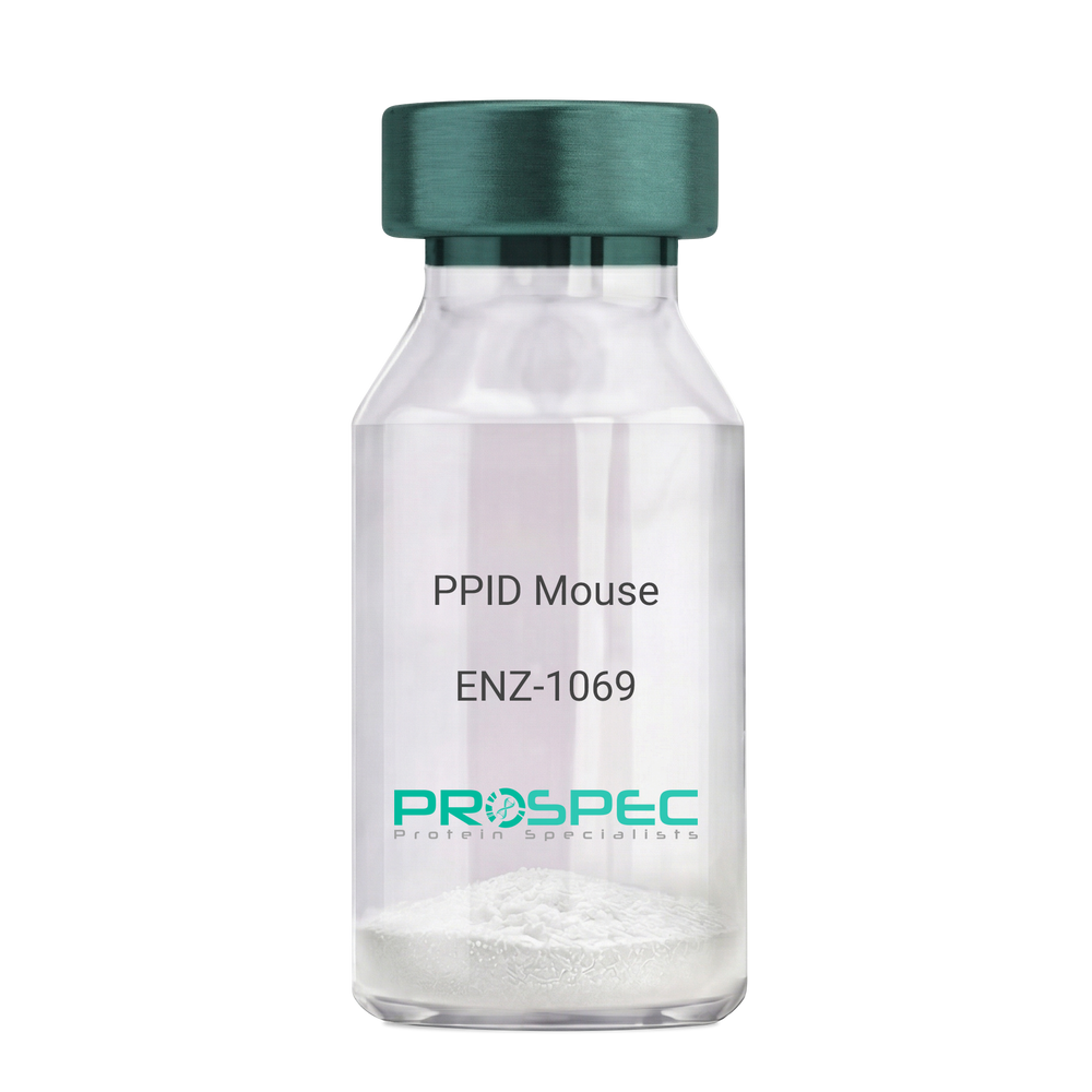
Name :
PPID MouseDescription:
Peptidylprolyl Isomerase D Mouse Recombinant
Peptidyl-prolyl cis-trans isomerase D, PPIase D, 40 kDa peptidyl-prolyl cis-trans isomerase, Cyclophilin-40, CYP-40, Cyclophilin-related protein, CYP40, CYPD, PPID, Peptidylprolyl Isomerase D.
Product # :
ENZ-1069Price :
Quantity :
Shipping Method :
Shipped with Ice Packs
More Info
- description
- source
- formulation
- purity
- biological activity
- More Info
Description
PPID Mouse Recombinant produced in E.Coli is a single, non-glycosylated polypeptide chain containing 395 amino acids (1-370a.a.) and having a molecular mass of 43.4kDa. PPID is fused to a 25 amino acid His-tag at N-terminus & purified by proprietary chromatographic techniques.
Source
Escherichia Coli.
Formulation
PPID protein solution (1mg/ml) containing 20mM Tris-Hcl buffer (pH8.0), 10% glycerol and 1mM DTT.
Purity
Greater than 90.0% as determined by SDS-PAGE.
Biological Activity
Specific activity is > 700nmol/min/mg, and is defined as the amount of enzyme that cleaves 1umole of suc-AAFP-PNA per minute at 37°C in Tris–HCl pH 8.0 using chymotrypsin.
More Info
-
Introduction
Cyclophilin-D is a member of the peptidyl-prolyl cis-trans isomerase (PPIase) family. PPIases catalyze the cis-trans isomerization of proline imidic peptide bonds in oligopeptides and speeds up the protein folding. Cyclophilin-D possess PPIase activity and binds to the immunosuppressant cyclosporin-A. Cyclophilin-D is very well known that its overexpression suppresses the apoptosis in cancer cell. Cyclophilin-D suppresses apoptotic cell death by the use of mitochondrial hexokinase-2 dependent mechanism in cancer cells.
-
Synonyms
Peptidyl-prolyl cis-trans isomerase D, PPIase D, 40 kDa peptidyl-prolyl cis-trans isomerase, Cyclophilin-40, CYP-40, Cyclophilin-related protein, CYP40, CYPD, PPID, Peptidylprolyl Isomerase D.
-
Physical Appearance
Sterile Filtered colorless solution.
-
Stability
Store at 4°C if entire vial will be used within 2-4 weeks. Store, frozen at -20°C for longer periods of time. For long term storage it is recommended to add a carrier protein (0.1% HSA or BSA). Please prevent freeze-thaw cycles.
-
Amino Acid Sequence
MGSSHHHHHH SSGLVPRGSH MGSEFMSHAS PAAKPSNSKN PRVFFDVDIG GERVGRIVLE LFADIVPKTA ENFRALCTGE KGTGSTTGKP LHFKGCPFHR IIKKFMIQGG DFSNQNGTGG ESIYGEKFED ENFHYKHDRE GLLSMANAGP NTNGSQFFIT TVPTPHLDGK HVVFGQVIKG LGVARTLENV EVNGEKPAKL CVIAECGELK EGDDWGIFPK DGSGDSHPDF PEDADIDLKD VDKILLISED LKNIGNTFFK SQNWEMAIKK YAKVLRYVDS SKAVIEKADR SRLQPIALSC VLNIGACKLK MSNWQGAIDS CLEALEMDPS NTKALYRKAQ GWQGLKEYDQ ALADLKKAQE IAPGDKAIQA ELLKVKQMIK AQKDKEKAVY AKMFA.
ProSpec's products are furnished for LABORATORY RESEARCH USE ONLY. The product may not be used as drugs, agricultural or pesticidal products, food additives or household chemicals.
- View Data Sheet
Name :
TBEV gEDescription:
Tick-Borne Encephalitis Virus gE Recombinant
Product # :
TBE-281Price :
Quantity :
Shipping Method :
Shipped with Ice Packs
More Info
- description
- source
- formulation
- purity
- More Info
Description
The E.coli derived recombinant protein contains the Tick-borne Encephalitis Virus glycoprotein E regions, 95-229 amino acids.
Source
Escherichia Coli.
Formulation
20mM Tris-MES pH 6.5, 8M urea, 200mM NaCl and 0.05% Tween-20.
Purity
Protein is >90% pure as determined by SDS- PAGE.
More Info
-
Introduction
TBE is caused by tick-borne encephalitis virus (TBEV), a member of the family Flaviviridae.
A closely related virus in Far Eastern Eurasia, Russian spring-summer encephalitis virus (RSSEV).
The family Flaviviridae includes other tick-borne viruses are closely related to TBEV and RSSEV, such as Omsk hemorrhagic fever virus & Kyasanur Forest virus.
Louping ill virus is also a member of this family. -
Stability
Encephalitis protein although stable at 4°C for 1 week, should be stored below -18°C. Please prevent freeze thaw cycles.
-
Applications
Encephalitis antigen in ELISA and Western blots, excellent antigen for detection of Tick-borne encephalitis virus with minimal specificity problems.
-
Specificity
Immunoreactive with sera of encephalitis virus infected individuals.
-
Purification Method
Encephalitis protein was purified by proprietary chromatographic technique.
ProSpec's products are furnished for LABORATORY RESEARCH USE ONLY. The product may not be used as drugs, agricultural or pesticidal products, food additives or household chemicals.
- View Data Sheet
Name :
S.Typhi HylEDescription:
Salmonella Typhi Haemolysin E Recombinant
Product # :
STY-004Price :
Quantity :
Shipping Method :
Shipped at Room temp
More Info
- description
- source
- formulation
- purity
- More Info
Description
Recombinant S.Typhi HylE produced in E.coli is a non-glycosylated polypeptide chain having a molecular mass of 34 kDa and fused to a His tag at N-terminus.
Source
Escherichia Coli.
Formulation
Lyophilized from 1mg/ml in 20mM sodium carbonate pH-9.6.
Purity
Greater than 95.0% as determined by SDS-PAGE.
More Info
-
Introduction
Salmonella Typhi is a pathogen causing typhoid fever, affecting over 17 million people with approximately 600,000 deaths annually worldwide. If untreated, typhoid fever cases result in mortality rates ranging from 12-30%.
-
Physical Appearance
Sterile Filtered White lyophilized (freeze-dried) powder.
-
Stability
S.Typhi HylE although stable at room temperature for 4 weeks, should be stored below -18°C. Please prevent freeze thaw cycles.
-
Solubility
It is recommended to reconstitute the lyophilized S.Typhi HylE in sterile 18M-cm H2O not less than 100µg/ml, which can then be further diluted to other aqueous solutions.
ProSpec's products are furnished for LABORATORY RESEARCH USE ONLY. The product may not be used as drugs, agricultural or pesticidal products, food additives or household chemicals.
- View Data Sheet
Name :
Anhui H7N9Description:
Hemagglutinin-Influenza A Virus H7N9 Anhui 2013 Recombinant
Hemagglutinin, Influenza A virus (A/Anhui/1-BALF_RG45/2013(H7N9) hemagglutinin, HA, Hemagglutinin HA1 chain, Hemagglutinin HA2 chain
Product # :
IHA-037Price :
Quantity :
Shipping Method :
Shipped with Ice Packs
More Info
- description
- source
- formulation
- purity
- More Info
Description
Anhui H7N9 produced in Sf9 Baculovirus cells is a single, glycosylated polypeptide chain containing 330 amino acids (19-339 aa) and having a molecular mass of 36 kDa.Anhui H7N9 is fused to a 6 amino acid His tag at C-terminus and purified by proprietary chromatographic techniques.
Source
Sf9, Baculovirus cells.
Formulation
The Anhui H7N9 solution (1mg/ml) contains 10% Glycerol and Phosphate-Buffered Saline (pH 7.4).
Purity
Greater than 95.0% as determined by SDS-PAGE.
More Info
-
Introduction
Hemagglutinin-Influenza A Virus H7N9 Anhui 2013 (Anhui H7N9) is a part of the influenza viruses hemagglutinin family. H7N9 Anhui is anantigenic glycoprotein which is responsible for binding the virus to the infected cell. An H7N9 virus was firstreported to have infected humans in March 2013, in China. Although the risk is low, The H7N9 virus has thegreatest potential compared with other influenza A viruses to cause a pandemicbecause like other type A viruses, it is not easily transmitted between people in its current form.
-
Synonyms
Hemagglutinin, Influenza A virus (A/Anhui/1-BALF_RG45/2013(H7N9) hemagglutinin, HA, Hemagglutinin HA1 chain, Hemagglutinin HA2 chain
-
Physical Appearance
Sterile Filtered colorless solution.
-
Stability
Store at 4°C if entire vial will be used within 2-4 weeks. Store, frozen at -20°C for longer periods of time. For long term storage it is recommended to add a carrier protein (0.1% HSA or BSA). Avoid multiple freeze-thaw cycles.
-
Amino Acid Sequence
ADLDKICLGH HAVSNGTKVN TLTERGVEVV NATETVERTN IPRICSKGKR TVDLGQCGLL GTITGPPQCD QFLEFSADLI IERREGSDVC YPGKFVNEEA LRQILRESGG IDKEAMGFTY SGIRTNGATS ACRRSGSSFY AEMKWLLSNT DNAAFPQMTK SYKNTRKSPA LIVWGIHHSV STAEQTKLYG SGNKLVTVGS SNYQQSFVPS PGARPQVNGL SGRIDFHWLM LNPNDTVTFS FNGAFIAPDR ASFLRGKSMG IQSGVQVDAN CEGDCYHSGG TIISNLPFQN IDSRAVGKCP RYVKQRSLLL ATGMKNVPEI PKGRHHHHHH
ProSpec's products are furnished for LABORATORY RESEARCH USE ONLY. The product may not be used as drugs, agricultural or pesticidal products, food additives or household chemicals.
- View Data Sheet
Name :
HCV NS3 Genotype-1b C33CDescription:
Hepatitis C Virus NS3 Genotype-1b C33C Recombinant
Product # :
HCV-271Price :
Quantity :
Shipping Method :
Shipped with Ice Packs
More Info
- description
- source
- formulation
- purity
- More Info
Description
Recombinant Hepatitis C Virus NS3 Genotype-1b C33C (40-315 aa) produced in E. coli having 226 aa.Recombinant Hepatitis C Virus NS3 Genotype-1b C33C is fused to a 6xHis tag at C-Terminus and purified by proprietary chromatographic technique.
Source
Escherichia Coli.
Formulation
HCV NS3 Genotype-1b C33C solution contains 50mM potassium phosphate buffer.
Purity
Protein is >90% pure as determined by 10% PAGE (coomassie staining).
More Info
-
Introduction
HCV is a small 50nm, enveloped, single-stranded, positive sense RNAvirus in the family Flaviviridae. HCV has a high rate of replication with approximately one trillion particles produced each day in an infected individual. Due to lack of proofreading by the HCV RNA polymerase, the HCV has an exceptionally high mutation rate, a factor that may help it elude the host's immune response. Hepatitis C virus is classified into six genotypes(1-6) with several subtypes within each genotype. The preponderance and distribution of HCV genotypes varies globally. Genotype is clinically important in determining potential response to interferon-based therapy and the required duration of such therapy. Genotypes 1 and 4 are less responsive to interferon-based treatment than are the other genotypes (2, 3, 5 and 6). HCV NS3 (genotype 1b) contains 2 domains which are serine protinase and helicase. C33c is a fragment of NS3 containing the most important epitopes recognized by HCV antibody.
-
Physical Appearance
Sterile Filtered clear solution.
-
Stability
Store at 4°C if entire vial will be used within 2-4 weeks.Store, frozen at -20°C for longer periods of time.For long term storage it is recommended to add a carrier protein (0.1% HSA or BSA).Avoid multiple freeze-thaw cycles.
-
Applications
ELISA and lateral flow assay.
ProSpec's products are furnished for LABORATORY RESEARCH USE ONLY. The product may not be used as drugs, agricultural or pesticidal products, food additives or household chemicals.
- View Data Sheet
Name :
ALPL MouseDescription:
Alkaline Phosphatase Liver/Bone/Kidney Mouse Recombinant
Alpl, Akp-2, Akp2, ALP, APTNAP, TNAP, TNSALP, HOPS, AP-TNAP, Alkaline phosphatase 2.
Product # :
ENZ-1008Price :
Quantity :
Shipping Method :
Shipped with Ice Packs
More Info
- description
- source
- formulation
- purity
- biological activity
- More Info
Description
ALPL produced in Sf9 Baculovirus cells is a single, glycosylated polypeptide chain containing 493 amino acids (19-503 a.a.) and having a molecular mass of 54.5kDa (Molecular size on SDS-PAGE will appear at approximately 50-70kDa). ALPL is expressed with an 8 amino acid His tag at C-Terminus and purified by proprietary chromatographic techniques.
Source
Sf9, Baculovirus cells.
Formulation
ALPL protein solution (0.5mg/ml) contains Phosphate Buffered Saline (pH 7.4) and 10% glycerol.
Purity
Greater than 95.0% as determined by SDS-PAGE.
Biological Activity
Specific activity is > 46,000 pmol/min/ug and is defined as the amount of enzyme that hydrolyze 1pmole of 4-Methylumbelliferyl phosphate to phosphate and 4-Methylumbelliferone per minute at pH 8.8 at 25C.More Info
-
Introduction
Alkaline Phosphatase Liver/Bone/Kidney (Alpl) is a part of the alkaline phosphatases family which comprises 4 related alkaline phosphatases. Alpl is a membrane-bound glycosylated enzyme which is not expressed in any particular tissue. Alpl takes part in skeletal mineralization.
-
Synonyms
Alpl, Akp-2, Akp2, ALP, APTNAP, TNAP, TNSALP, HOPS, AP-TNAP, Alkaline phosphatase 2.
-
Physical Appearance
Sterile Filtered colorless solution.
-
Stability
Store at 4°C if entire vial will be used within 2-4 weeks. Store, frozen at -20°C for longer periods of time. For long term storage it is recommended to add a carrier protein (0.1% HSA or BSA). Avoid multiple freeze-thaw cycles.
-
Amino Acid Sequence
VPEKERDPSY WRQQAQETLK NALKLQKLNT NVAKNVIMFL GDGMGVSTVT AARILKGQLH HNTGEETRLE MDKFPFVALS KTYNTNAQVP DSAGTATAYL CGVKANEGTV GVSAATERTR CNTTQGNEVT SILRWAKDAG KSVGIVTTTR VNHATPSAAY AHSADRDWYS DNEMPPEALS QGCKDIAYQL MHNIKDIDVI MGGGRKYMYP KNRTDVEYEL DEKARGTRLD GLDLISIWKS FKPRHKHSHY VWNRTELLAL DPSRVDYLLG LFEPGDMQYE LNRNNLTDPS LSEMVEVALR ILTKNLKGFF LLVEGGRIDH GHHEGKAKQA LHEAVEMDQA IGKAGAMTSQ KDTLTVVTAD HSHVFTFGGY TPRGNSIFGL APMVSDTDKK PFTAILYGNG PGYKVVDGER ENVSMVDYAH NNYQAQSAVP LRHETHGGED VAVFAKGPMA HLLHGVHEQN YIPHVMAYAS CIGANLDHCA WAGSGLEHHH HHH.
ProSpec's products are furnished for LABORATORY RESEARCH USE ONLY. The product may not be used as drugs, agricultural or pesticidal products, food additives or household chemicals.