Skip to content

High-quality research proteins.

  • 1-800-805-6531

  • Mon - Sat 8.00-18.00, Sun - Closed

  • info@prospecbio.com

  • PO Box 6591 East Brunswick NJ 08816

  • Products
    • Cytokines
    • Growth factors
    • Chemokines
    • Cd antigens
    • Neurotrophins
    • Hormones
    • Enzymes
    • Viral antigens
    • Recombinant proteins
    • Natural proteins
    • Monoclonal antibodies
    • Polyclonal antibodies
    • Thermal Packaging
  • About
    • Company
    • Founder
    • Contribution
  • Customers
    • Eastern Asia
    • Eastern Europe
    • North America
    • South America
    • South East Asia
    • Southern Asia
    • Western Europe
  • Ordering
    • Ordering Method
    • Ordering - Credit Card
    • Ordering - PO
    • Shipping Fees
    • FAQs
  • Services
    • DNA Cloning
    • Protein Expression
    • Monoclonal Antibody
    • Fermentation
    • Technology License
  • Contact us
Log in

Country/region

  • USD $ | Albania
  • USD $ | Andorra
  • USD $ | Argentina
  • USD $ | Armenia
  • USD $ | Australia
  • USD $ | Austria
  • USD $ | Azerbaijan
  • USD $ | Belarus
  • USD $ | Belgium
  • USD $ | Bolivia
  • USD $ | Brazil
  • USD $ | Bulgaria
  • USD $ | Cambodia
  • USD $ | Canada
  • USD $ | Chile
  • USD $ | China
  • USD $ | Colombia
  • USD $ | Costa Rica
  • USD $ | Croatia
  • USD $ | Cyprus
  • USD $ | Czechia
  • USD $ | Denmark
  • USD $ | Estonia
  • USD $ | Finland
  • USD $ | France
  • USD $ | Georgia
  • USD $ | Germany
  • USD $ | Greece
  • USD $ | Hong Kong SAR
  • USD $ | Hungary
  • USD $ | Iceland
  • USD $ | India
  • USD $ | Ireland
  • USD $ | Israel
  • USD $ | Italy
  • USD $ | Japan
  • USD $ | Kazakhstan
  • USD $ | Latvia
  • USD $ | Liechtenstein
  • USD $ | Lithuania
  • USD $ | Madagascar
  • USD $ | Malta
  • USD $ | Mexico
  • USD $ | Moldova
  • USD $ | Monaco
  • USD $ | Netherlands
  • USD $ | New Zealand
  • USD $ | North Macedonia
  • USD $ | Norway
  • USD $ | Panama
  • USD $ | Paraguay
  • USD $ | Peru
  • USD $ | Philippines
  • USD $ | Poland
  • USD $ | Portugal
  • USD $ | Romania
  • USD $ | Singapore
  • USD $ | Slovakia
  • USD $ | Slovenia
  • USD $ | South Africa
  • USD $ | South Korea
  • USD $ | Spain
  • USD $ | Sri Lanka
  • USD $ | Sweden
  • USD $ | Switzerland
  • USD $ | Taiwan
  • USD $ | Thailand
  • USD $ | Türkiye
  • USD $ | Ukraine
  • USD $ | United Kingdom
  • USD $ | United States
  • USD $ | Uruguay
  • USD $ | Vatican City
  • USD $ | Venezuela
  • USD $ | Vietnam
  • Facebook
  • Instagram
logoProSpec - Recombinant Protein Manufacturer for academic and R&D industry
Become a distributor
Account Log in
Wishlist
Cart Cart(0)
  • Products
    +
    • Cytokines
    • Growth factors
    • Chemokines
    • Cd antigens
    • Neurotrophins
    • Hormones
    • Enzymes
    • Viral antigens
    • Recombinant proteins
    • Natural proteins
    • Monoclonal antibodies
    • Polyclonal antibodies
    • Thermal Packaging
  • About
    +
    • Company
    • Founder
    • Contribution
  • Customers
    +
    • Eastern Asia
    • Eastern Europe
    • North America
    • South America
    • South East Asia
    • Southern Asia
    • Western Europe
  • Ordering
    +
    • Ordering Method
    • Ordering - Credit Card
    • Ordering - PO
    • Shipping Fees
    • FAQs
  • Services
    +
    • DNA Cloning
    • Protein Expression
    • Monoclonal Antibody
    • Fermentation
    • Technology License
  • Contact us

Item added to your cart

View cart
View All
  • Cytokines
  • Growth factors
  • Chemokines
  • Cd antigens
  • Neurotrophins
  • Hormones
  • Enzymes
  • Viral antigens
  • Recombinant proteins
  • Natural proteins
  • Monoclonal antibodies
  • Polyclonal antibodies
  • Thermal Packaging
  • Tumor Necrosis Factor

    Tumor Necrosis Factor

    View All
  • 4-1BB

    4-1BB

    View All
  • Adiponectin

    Adiponectin

    View All
  • AIF1

    AIF1

    View All
  • Other Cytokines

    Other Cytokines

    View All
  • AITRL

    AITRL

    View All
  • Angiopoietin

    Angiopoietin

    View All
  • Apolipoprotein

    Apolipoprotein

    View All
  • B-Cell Activating Factor

    B-Cell Activating Factor

    View All
  • Beta Defensin

    Beta Defensin

    View All
  • Bone Morphogenetic Protein

    Bone Morphogenetic Protein

    View All
  • B type Natriuretic Peptide

    B type Natriuretic Peptide

    View All
  • BST

    BST

    View All
  • Betacellulin

    Betacellulin

    View All
  • Cardiotrophin

    Cardiotrophin

    View All
  • Activin

    Activin

    View All
  • Fibroblast Growth Factor

    Fibroblast Growth Factor

    View All
  • Other Growth Factors

    Other Growth Factors

    View All
  • CSF

    CSF

    View All
  • CTGF

    CTGF

    View All
  • IGFBP

    IGFBP

    View All
  • VEGF Protein

    VEGF Protein

    View All
  • EGF

    EGF

    View All
  • Epigen

    Epigen

    View All
  • Erythropoietin

    Erythropoietin

    View All
  • Insulin-Like Growth Factor

    Insulin-Like Growth Factor

    View All
  • Growth Hormone

    Growth Hormone

    View All
  • HDGF

    HDGF

    View All
  • Hepatocyte Growth Factor

    Hepatocyte Growth Factor

    View All
  • Insulin

    Insulin

    View All
  • BCA-1/ BLC (CXCL13)

    BCA-1/ BLC (CXCL13)

    View All
  • BRAK (CXCL14)

    BRAK (CXCL14)

    View All
  • C-10 (CCL6)

    C-10 (CCL6)

    View All
  • Other Chemokines

    Other Chemokines

    View All
  • MEC (CCL28)

    MEC (CCL28)

    View All
  • LD78-beta (CCL3L1)

    LD78-beta (CCL3L1)

    View All
  • CTACK (CCL27)

    CTACK (CCL27)

    View All
  • CXCL16

    CXCL16

    View All
  • CXCL17

    CXCL17

    View All
  • Platelet Factor-4 (CXCL4)

    Platelet Factor-4 (CXCL4)

    View All
  • ENA-78 (CXCL5)

    ENA-78 (CXCL5)

    View All
  • Eotaxin (CCL11,24,26)

    Eotaxin (CCL11,24,26)

    View All
  • Exodus-2 (CCL21)

    Exodus-2 (CCL21)

    View All
  • Fractalkine (CX3CL1)

    Fractalkine (CX3CL1)

    View All
  • CXCL6

    CXCL6

    View All
  • Other CD Antigens

    Other CD Antigens

    View All
  • CD14

    CD14

    View All
  • CD164

    CD164

    View All
  • CD1

    CD1

    View All
  • CD2

    CD2

    View All
  • CD200

    CD200

    View All
  • CD207

    CD207

    View All
  • CD226

    CD226

    View All
  • CD23

    CD23

    View All
  • CD244

    CD244

    View All
  • CD247

    CD247

    View All
  • CD27

    CD27

    View All
  • CD274

    CD274

    View All
  • CD300

    CD300

    View All
  • CD33

    CD33

    View All
  • Other Neurotrophins

    Other Neurotrophins

    View All
  • Beta-NGF

    Beta-NGF

    View All
  • BDNF

    BDNF

    View All
  • CDNF

    CDNF

    View All
  • CNTF

    CNTF

    View All
  • GDNF

    GDNF

    View All
  • Glia Maturation Factor

    Glia Maturation Factor

    View All
  • MANF

    MANF

    View All
  • Midkine

    Midkine

    View All
  • Neuroglobin

    Neuroglobin

    View All
  • Neurotrophic factors

    Neurotrophic factors

    View All
  • Neuregulin

    Neuregulin

    View All
  • Neuropilin

    Neuropilin

    View All
  • Pigment Epithelium-Derived Factor

    Pigment Epithelium-Derived Factor

    View All
  • Pleiotrophin

    Pleiotrophin

    View All
  • Peptide Hormones

    Peptide Hormones

    View All
  • Other Hormones

    Other Hormones

    View All
  • HCG

    HCG

    View All
  • Endothelin

    Endothelin

    View All
  • Exendin

    Exendin

    View All
  • FSH

    FSH

    View All
  • Glucagon

    Glucagon

    View All
  • GHRP

    GHRP

    View All
  • GLP

    GLP

    View All
  • Thyrostimulin

    Thyrostimulin

    View All
  • Inhibin A

    Inhibin A

    View All
  • LHRH

    LHRH

    View All
  • Melanotan

    Melanotan

    View All
  • Procalcitonin

    Procalcitonin

    View All
  • PTH

    PTH

    View All
  • Peroxiredoxin

    Peroxiredoxin

    View All
  • Peptidase

    Peptidase

    View All
  • Synthetase

    Synthetase

    View All
  • Transferase

    Transferase

    View All
  • Hydrolase

    Hydrolase

    View All
  • Dehydrogenase

    Dehydrogenase

    View All
  • ACE2

    ACE2

    View All
  • Esterase

    Esterase

    View All
  • Protein Kinases

    Protein Kinases

    View All
  • Other Enzymes

    Other Enzymes

    View All
  • Phosphatase

    Phosphatase

    View All
  • Deaminase

    Deaminase

    View All
  • Oxygenase

    Oxygenase

    View All
  • Adenylate Kinase

    Adenylate Kinase

    View All
  • Lyase

    Lyase

    View All
  • Chagas

    Chagas

    View All
  • SARS

    SARS

    View All
  • Influenza

    Influenza

    View All
  • ASFV

    ASFV

    View All
  • Astrovirus

    Astrovirus

    View All
  • Borrelia

    Borrelia

    View All
  • Helicobacter Pylori

    Helicobacter Pylori

    View All
  • Chikungunya

    Chikungunya

    View All
  • Chlamydia

    Chlamydia

    View All
  • Cytomegalo

    Cytomegalo

    View All
  • Dengue

    Dengue

    View All
  • EBV

    EBV

    View All
  • Ebola

    Ebola

    View All
  • Feline Leukemia Virus

    Feline Leukemia Virus

    View All
  • Hepatitis A

    Hepatitis A

    View All
  • Other

    Other

    View All
  • Actin

    Actin

    View All
  • ADAM

    ADAM

    View All
  • Complement Component

    Complement Component

    View All
  • Ag85

    Ag85

    View All
  • Eukaryotic Translation Initiation Factor

    Eukaryotic Translation Initiation Factor

    View All
  • Anterior Gradient Protein

    Anterior Gradient Protein

    View All
  • Heat Shock Protein

    Heat Shock Protein

    View All
  • Alpha-2-HS-Glycoprotein

    Alpha-2-HS-Glycoprotein

    View All
  • Hemoglobin

    Hemoglobin

    View All
  • Allergy

    Allergy

    View All
  • Anaplasma

    Anaplasma

    View All
  • Angiogenin

    Angiogenin

    View All
  • Ankyrin Repeat Domain

    Ankyrin Repeat Domain

    View All
  • Annexin

    Annexin

    View All
  • Other Natural Proteins

    Other Natural Proteins

    View All
  • Aprotinin

    Aprotinin

    View All
  • Transferrin

    Transferrin

    View All
  • Avidin

    Avidin

    View All
  • Anti Coagulation Factors

    Anti Coagulation Factors

    View All
  • Natural Albumin

    Natural Albumin

    View All
  • Natural Coagulation Factors

    Natural Coagulation Factors

    View All
  • Fibronectin

    Fibronectin

    View All
  • Thrombin

    Thrombin

    View All
  • Anti Human Enzyme

    Anti Human Enzyme

    View All
  • Other Monoclonal Antibodies

    Other Monoclonal Antibodies

    View All
  • Anti Human Cytokine

    Anti Human Cytokine

    View All
  • Anti Human Heat Shock Protein

    Anti Human Heat Shock Protein

    View All
  • Anti Mouse Lymphocyte

    Anti Mouse Lymphocyte

    View All
  • Anti Human Chemokine

    Anti Human Chemokine

    View All
  • Anti Human Lymphocyte

    Anti Human Lymphocyte

    View All
  • Anti Viral Monoclonal

    Anti Viral Monoclonal

    View All
  • Anti Mouse Cytokine

    Anti Mouse Cytokine

    View All
  • Anti-GST Monoclonal

    Anti-GST Monoclonal

    View All
  • Other Polyclonal Antibodies

    Other Polyclonal Antibodies

    View All
  • Anti Viral

    Anti Viral

    View All
  • Anti-GST Monoclonal

    Anti-GST Monoclonal

    View All
Back

Search results

1000 results found for “lbp”

Name

Description

Product #

Price

Quantity

Shipping Method

  • View Data Sheet

    Name :

    USP14 Human

    Description:

    Ubiquitin Specific Peptidase 14 Human Recombinant

    TGT, Ubiquitin thioesterase 14, Deubiquitinating enzyme 14, Ubiquitin thioesterase 14, Ubiquitin-specific-processing protease 14.

    Product # :

    PRO-016

    Price :

    Quantity :

    Shipping Method :

    Ice Icon

    Shipped with Ice Packs

    Add To Cart

    More Info

    • description
    • source
    • formulation
    • purity
    • More Info

    Description

    USP14 Human Recombinant produced in E.Coli is a single, non-glycosylated polypeptide chain containing 517 amino acids (1-494 a.a) and having a molecular mass of 58.5kDa. USP14 is fused to a 23 amino acid His-tag at N-terminus & purified by proprietary chromatographic techniques.

    Source

    Escherichia Coli.

    Formulation

    USP14 protein solution (1mg/ml) containing 20mM Tris-HCl buffer (pH 8.0), 0.2M NaCl, 20% glycerol and 1mM DTT.

    Purity

    Greater than 90.0% as determined by SDS-PAGE.

    More Info

    • Introduction

      USP14 belongs to the ubiquitin-specific processing (UBP) family of proteases which is a deubiquitinating enzyme (DUB) containing His and Cys domains. USP14 placed in the cytoplasm and cuts the ubiquitin from ubiquitin-fused precursors and ubiquitinylated proteins.a mutation which results in reduced expression of the ortholog of USP14 in mice inhibits growth, develop severe tremors by 2 to 3 weeks of age followed by paralysis and death by 6 to 10 weeks of age.

    • Synonyms

      TGT, Ubiquitin thioesterase 14, Deubiquitinating enzyme 14, Ubiquitin thioesterase 14, Ubiquitin-specific-processing protease 14.

    • Physical Appearance

      Sterile Filtered colorless solution.

    • Stability

      Store at 4°C if entire vial will be used within 2-4 weeks. Store, frozen at -20°C for longer periods of time. For long term storage it is recommended to add a carrier protein (0.1% HSA or BSA).Avoid multiple freeze-thaw cycles.

    • Amino Acid Sequence

      MGSSHHHHHH SSGLVPRGSH MGSMPLYSVT VKWGKEKFEG VELNTDEPPM VFKAQLFALT GVQPARQKVM VKGGTLKDDD WGNIKIKNGM TLLMMGSADA LPEEPSAKTV FVEDMTEEQL ASAMELPCGL TNLGNTCYMN ATVQCIRSVP ELKDALKRYA GALRASGEMA SAQYITAALR DLFDSMDKTS SSIPPIILLQ FLHMAFPQFA EKGEQGQYLQ QDANECWIQM MRVLQQKLEA IEDDSVKETD SSSASAATPS KKKSLIDQFF GVEFETTMKC TESEEEEVTK GKENQLQLSC FINQEVKYLF TGLKLRLQEE ITKQSPTLQR NALYIKSSKI SRLPAYLTIQ MVRFFYKEKE SVNAKVLKDV KFPLMLDMYE LCTPELQEKM VSFRSKFKDL EDKKVNQQPN TSDKKSSPQK EVKYEPFSFA DDIGSNNCGY YDLQAVLTHQ GRSSSSGHYV SWVKRKQDEW IKFDDDKVSI VTPEDILRLS GGGDWHIAYV LLYGPRRVEI MEEESEQ.

    ProSpec's products are furnished for LABORATORY RESEARCH USE ONLY. The product may not be used as drugs, agricultural or pesticidal products, food additives or household chemicals.

    Usp14 Human
  • View Data Sheet

    Name :

    PTPN4 Human

    Description:

    Protein Tyrosine Phosphatase Non Receptor Type-4 Human Recombinant

    Protein Tyrosine Phosphatase, Non-Receptor Type 4 (Megakaryocyte), Protein-Tyrosine Phosphatase MEG1, PTPase-MEG1, EC 3.1.3.48, MEG, Tyrosine-Protein Phosphatase Non-Receptor Type 4, Megakaryocyte Protein-Tyrosine Phosphatase, Protein Tyrosine Phosphatase MEG1, Megakaryocyte Phosphatase, PTPMEG1, PTPMEG, Tyrosine-protein phosphatase non-receptor type 4, Protein-tyrosine phosphatase MEG1.

    Product # :

    PKA-075

    Price :

    Quantity :

    Shipping Method :

    Ice Icon

    Shipped with Ice Packs

    Add To Cart

    More Info

    • description
    • source
    • formulation
    • purity
    • More Info

    Description

    PTPN4 Human Recombinant produced in E.Coli is a single, non-glycosylated polypeptide chain containing 280 amino acids (655-913 a.a) and having a molecular mass of 32kDa. PTPN4 is fused to a 21 amino acid His-tag at N-terminus & purified by proprietary chromatographic techniques.

    Source

    E.coli.

    Formulation

    PTPN4 protein solution (1mg/ml) containing 20mM Tris buffer (pH 8.0) and 10% glycerol.

    Purity

    Greater than 85% as determined by SDS-PAGE.

    More Info

    • Introduction

      Protein Tyrosine Phosphatase Non Receptor Type-4, also known asPTPN4 belongs to the protein tyrosine phosphatase, PTP family. PTPs are identified as signaling molecules which regulate a variety of cellular processes as well as cell growth, differentiation, mitotic cycle, and oncogenic transformation. PTPN4 is broadly expressed non-receptor protein tyrosine phosphatase. In addition, PTPN4 interacts with glutamate receptor delta 2 & epsilon, and affects glutamate receptors signaling and/or in regulation of their activities through tyrosine dephosphorylation.

    • Synonyms

      Protein Tyrosine Phosphatase, Non-Receptor Type 4 (Megakaryocyte), Protein-Tyrosine Phosphatase MEG1, PTPase-MEG1, EC 3.1.3.48, MEG, Tyrosine-Protein Phosphatase Non-Receptor Type 4, Megakaryocyte Protein-Tyrosine Phosphatase, Protein Tyrosine Phosphatase MEG1, Megakaryocyte Phosphatase, PTPMEG1, PTPMEG, Tyrosine-protein phosphatase non-receptor type 4, Protein-tyrosine phosphatase MEG1.

    • Physical Appearance

      Sterile Filtered colorless solution.

    • Stability

      Store at 4°C if entire vial will be used within 2-4 weeks. Store, frozen at -20°C for longer periods of time. For long term storage it is recommended to add a carrier protein (0.1% HSA or BSA).Avoid multiple freeze-thaw cycles.

    • Amino Acid Sequence

      MGSSHHHHHH SSGLVPRGSH MVLTQFDQLY RKKPGMTMSC AKLPQNISKN RYRDISPYDA TRVILKGNED YINANYINME IPSSSIINQY IACQGPLPHT CTDFWQMTWE QGSSMVVMLT TQVERGRVKC HQYWPEPTGS SSYGCYQVTC HSEEGNTAYI FRKMTLFNQE KNESRPLTQI QYIAWPDHGV PDDSSDFLDF VCHVRNKRAG KEEPVVVHCS AGIGRTGVLI TMETAMCLIE CNQPVYPLDI VRTMRDQRAM MIQTPSQYRF VCEAILKVYE.

    ProSpec's products are furnished for LABORATORY RESEARCH USE ONLY. The product may not be used as drugs, agricultural or pesticidal products, food additives or household chemicals.

    Ptpn4 Human
  • View Data Sheet

    Name :

    MAP1LC3B Mouse

    Description:

    Microtubule-Associated Protein 1 Light Chain 3 Beta Mouse Recombinant

    Microtubule-Associated Protein 1 Light Chain 3 beta, ATG8F, Autophagy-related ubiquitin-like modifier LC3 B, MAP1 light chain 3-like protein 2, MAP1A/MAP1B LC3 B, LC3B, MAP1A/1BLC3, MAP1ALC3.

    Product # :

    PRO-2482

    Price :

    Quantity :

    Shipping Method :

    Ice Icon

    Shipped with Ice Packs

    Add To Cart

    More Info

    • description
    • source
    • formulation
    • purity
    • More Info

    Description

    MAP1LC3B Mouse Recombinant produced in E.Coli is a single, non-glycosylated polypeptide chain containing 144 amino acids (1-120 a.a.) and having a molecular mass of 16.7kDa. MAP1LC3B is fused to a 24 amino acid His-tag at N-terminus & purified by proprietary chromatographic techniques.

    Source

    Escherichia Coli.

    Formulation

    MAP1LC3B protein solution (0.25mg/ml) containing 20mM MES(pH6.0), 0.1M NaCl, 1mM DTT and 30% glycerol.

    Purity

    Greater than 95.0% as determined by SDS-PAGE.

    More Info

    • Introduction

      MAP1LC3B is a member of the MAP1 LC3 family. MAP1LC3B is a subunit of neuronal microtubule-associated MAP1A and MAP1B proteins, that are involved in microtubule assembly and important for neurogenesis. In addition, MAP1LC3B takes part in formation of autophagosomal vacuoles and is expressed mainly in heart, testis, brain and skeletal muscle.

    • Synonyms

      Microtubule-Associated Protein 1 Light Chain 3 beta, ATG8F, Autophagy-related ubiquitin-like modifier LC3 B, MAP1 light chain 3-like protein 2, MAP1A/MAP1B LC3 B, LC3B, MAP1A/1BLC3, MAP1ALC3.

    • Physical Appearance

      Sterile filtered colorless solution.

    • Stability

      Store at 4°C if entire vial will be used within 2-4 weeks. Store, frozen at -20°C for longer periods of time. For long term storage it is recommended to add a carrier protein (0.1% HSA or BSA).Avoid multiple freeze-thaw cycles.

    • Amino Acid Sequence

      MGSSHHHHHH SSGLVPRGSH MGSHMPSEKT FKQRRSFEQR VEDVRLIREQ HPTKIPVIIE RYKGEKQLPV LDKTKFLVPD HVNMSELIKI IRRRLQLNAN QAFFLLVNGH SMVSVSTPIS EVYESERDED GFLYMVYASQ ETFG.

    ProSpec's products are furnished for LABORATORY RESEARCH USE ONLY. The product may not be used as drugs, agricultural or pesticidal products, food additives or household chemicals.

    Map1Lc3B Mouse
  • View Data Sheet

    Name :

    Visfatin Human

    Description:

    Visfatin Human Recombinant

    PBEF, Pre-B cell colony-enhancing factor, Nicotinamide phosphoribosyltransferase NAmPRTase, Nampt, MGC117256, DKFZP666B131, 1110035O14Rik.

    Product # :

    CYT-318

    Price :

    Quantity :

    Shipping Method :

    Room Temp Icon

    Shipped at Room temp

    Add To Cart

    More Info

    • description
    • source
    • formulation
    • purity
    • biological activity
    • More Info

    Description

    Visfatin Human Recombinant produced in E.Coli is a single, non-glycosylated, polypeptide chain containing 466 amino acids. The total molecular mass is 52.6kDa (calculated). The Visfatin is purified by Flag-affinity chromatography.

    Source

    Escherichia Coli.

    Formulation

    Visfatin was lyophilized with no additives.

    Purity

    Greater than 90.0% as determined by SDS-PAGE.

    Biological Activity

    The activity is determined by its ability to induce IL-6, IL-1 beta and TNF alpha production from human PBMCs at 100ng/ml.

    More Info

    • Introduction

      Excess adiposity is the most important risk in the development of type 2 diabetes mellitus (T2DM). Adipose tissue produces several proteins (adipocytokines) such as leptin, adiponectin, resistin, tumor necrosis factor-a, and IL-6, that modulate sensitivity and appear to play an important role in the pathogenesis, diabetes, dyslipidemia, inflammation, and atherosclerosis. Visfatin, also known as pre-B cell colony-enhancing factor (PBEF), is a cytokine that is highly expressed in visceral fat and was originally isolated as a secreted factor that synergizes with IL-7 and stem cell factors to promote the growth of B cell precursors. Visfatin homologs have been identified in carp, invertebrate mollusks, and bacteria, as well as in vertebrates, including humans and the mouse. It has been postulated to play a role in innate immunity.
      Visfatin exerts mimetic effects that are dose-dependent and quantitatively similar to stimulating muscle and adipocyte glucose transport, and in inhibiting hepatocyte glucose production. Intravenous injection of recombinant visfatin in mice decreased plasma glucose in a dose-dependent fashion. In keeping with its mimetic effects, visfatin was as effective in reducing hyperglycemia in deficient diabetic mice. Visfatin was also found to be bound to and activate receptor, causing receptor phosphorylation and the activation of downstream signaling molecules. However, visfatin did not compete for binding to the receptor, indicating that the two proteins were recognized by different regions of the receptor. Thus, visfatin might play a role in glucose homeostasis and dysregulation in biosynthesis or signal transduction, and might contribute to the pathogenesis of diabetes.

    • Synonyms

      PBEF, Pre-B cell colony-enhancing factor, Nicotinamide phosphoribosyltransferase NAmPRTase, Nampt, MGC117256, DKFZP666B131, 1110035O14Rik.

    • Physical Appearance

      Filtered White lyophilized (freeze-dried) powder.

    • Stability

      Lyophilized Visfatin although stable at room temperature for 3 weeks, should be stored desiccated below -18°C. Upon reconstitution Visfatin should be stored at 4°C between 2-7 days and for future use below -18°C.For long term storage it is recommended to add a carrier protein (0.1% HSA or BSA).Please prevent freeze-thaw cycles.

    • Solubility

      Centrifuge vial before opening. When reconstituting the product, gently pipet and wash down the sides of the vial to ensure full recovery of the protein into solution. It is recommended to reconstitute the lyophilized product with 20 mM HCl at a concentration of 0.1 mg/mL, which can be further diluted into other aqueous solutions. Wait several minutes for full reconstitution and solubility.

    • Amino Acid Sequence

      MPPNTSKVYS YFECREKKTE NSKLRKVKYE ETVFYGLQYI LNKYLKGKVV TKEKIQEAKD VYKEHFQDDV FNEKGWNYIL EKYDGHLPIE IKAVPEGFVI PRGNVLFTVE NTDPECYWLT NWIETILVQS WYPITVATNS REQKKILAKY LLETSGNLDG LEYKLHDFGY RGVSSQETAG IGASAHLVNF KGTDTVAGLA LIKKYYGTKD PVPGYSVPAA EHSTITAWGK DHEKDAFEHI VTQFSSVPVS VVSDSYDIYN ACEKIWGEDL RHLIVSRSTQ APLIIRPDSG NPLDTVLKVL EILGKKFPVT ENSKGYKLLP PYLRVIQGDG VDINTLQEIV EGMKQKMWSI ENIAFGSGGG LLQKLTRDLL NCSFKCSYVV TNGLGINVFK DPVADPNKRS KKGRLSLHRT PAGNFVTLEE GKGDLEEYGQ DLLHTVFKNG KVTKSYSFDE IRKNAQLNIE LEAAHH.

    • Background

      About Visfatin Human


      Visfatin is a cytokine expressed in visceral fat that was originally isolated as a secreted
      element that synergized with stem cell factors and IL-7. One of its main functions is to
      enhance the development of B cell precursors.

      The cytokine is also known as the “Pre-B Cell Colony-Enhancing Factor (PBEF).” It has been
      identified in vertebrates, including mice and humans, and it’s being studied due to its link
      to inflammatory conditions, beta cell function, and cardiovascular disease.


      What’s the Function of Visfatin Human Recombinant?

      Visfatin human recombinant is produced in E. Coli. It’s a single, non-glycosylated,
      polypeptide chain that contains 466 amino acids, it’s purified by FLAG-affinity
      chromatography, and it contains a total molecular mass of 52.6 kDa.


      What Are the Main Applications of Visfatin Human Recombinant?

      The cytokine is being researched because of its involvement in glucose homeostasis,
      dysregulation in biosynthesis and signal transduction, and the pathogenesis of diabetes.
      Visfatin human recombinant is tailored exclusively for laboratory research, ensuring
      experts can get further answers regarding the cytokine’s involvement in different
      processes, including pathogenesis, diabetes, inflammation, dyslipidemia, and
      atherosclerosis.

      Findings can also help during the identification of high-risk people for cardiovascular
      disease and diabetes.

    ProSpec's products are furnished for LABORATORY RESEARCH USE ONLY. The product may not be used as drugs, agricultural or pesticidal products, food additives or household chemicals.

    Visfatin Human
  • View Data Sheet

    Name :

    RELM b Mouse

    Description:

    RELM-Beta Mouse Recombinant

    Resistin-like beta, RELM beta, Cysteine-rich secreted protein FIZZ2, Colon and small intestine-specific cysteine-rich protein, Cysteine-rich secreted protein A12-alpha-like 1, Colon carcinoma-related gene protein, RELM-b, XCP2, HXCP2.

    Product # :

    CYT-413

    Price :

    Quantity :

    Shipping Method :

    Room Temp Icon

    Shipped at Room temp

    Add To Cart

    More Info

    • description
    • source
    • formulation
    • purity
    • More Info

    Description

    Mouse RELM-b Recombinant produced in E.Coli is a monomeric, non-glycosylated, polypeptide chain containing 83 amino acids and having a molecular mass of 8.9kDa. The Mouse RETNLB is purified by proprietary chromatographic techniques.

    Source

    Escherichia Coli.

    Formulation

    The protein was lyophilized from a concentrated (1mg/ml) protein solution containing 10mM Acetic Acid with 2:1 mannitol to protein.

    Purity

    Greater than 97% as determined by SDS-PAGE.

    More Info

    • Introduction

      RELM-beta (Resistin-Like Molecule-beta) is a member of a recently identified family of secreted proteins containing a conserved cystein-rich C-terminus. The RELM family consists of resistin (also called FIZZ3), RELM-alfa (FIZZ1), RELM-beta (FIZZ2) and RELM-gamma. Only resisistin and RELM-beta were found in humans whereas all four RELM family members were identified in rodents.
      RELM-beta appears to be produced as a homodimer exclusively by intestinal goblet cells and can be found in high quantities in stool. Remarkably, stool of germ-free mice displaying sterile intestinal tract does not contain RELM-beta until bacterial colonization takes place after pathogen-free mice entered natural environment. Some, but not all, colon carcinoma cell lines secrete RELM-beta into the cell culture supernatant.
      The physiological function of RELM-beta is not known. High doses of recombinant RELM-beta showed hyperglycemic effects including lowered glucose disposal and increased hepatic glucose production in mice.

    • Synonyms

      Resistin-like beta, RELM beta, Cysteine-rich secreted protein FIZZ2, Colon and small intestine-specific cysteine-rich protein, Cysteine-rich secreted protein A12-alpha-like 1, Colon carcinoma-related gene protein, RELM-b, XCP2, HXCP2.

    • Physical Appearance

      Brownish lyophilized powder.

    • Stability

      Lyophilized RETNLB is stable at -20°C. After reconstitution the protein should be kept at all times at -20°C. It is recommended to add a carrier protein (0.1% HSA or BSA) for long term storage.

    • Solubility

      Reconstitute at 0.1 mg/ml with sterile pyrogen free water.

    • Amino Acid Sequence

      MQCSFESLVD QRIKEALSRQ EPKTISCTSV TSSGRLASCP AGMVVTGCAC GYGCGSWDIR NGNTCHCQCS VMDWASARCC RMA.

    ProSpec's products are furnished for LABORATORY RESEARCH USE ONLY. The product may not be used as drugs, agricultural or pesticidal products, food additives or household chemicals.

    Relm Beta Mouse
  • View Data Sheet

    Name :

    ROBLD3 Human

    Description:

    Roadblock Domain Containing 3 Human Recombinant

    Ragulator complex protein LAMTOR2, Endosomal adaptor protein p14, Late endosomal/lysosomal Mp1-interacting protein, Late endosomal/lysosomal adaptor and MAPK and MTOR activator 2, Mitogen-activated protein-binding protein-interacting protein, Roadblock domain-containing protein 3, LAMTOR2, MAPBPIP, ROBLD3, p14, ENDAP, HSPC003, MAPKSP1AP, Ragulator2, RP11-336K24.9.

    Product # :

    PRO-050

    Price :

    Quantity :

    Shipping Method :

    Ice Icon

    Shipped with Ice Packs

    Add To Cart

    More Info

    • description
    • source
    • formulation
    • purity
    • More Info

    Description

    ROBLD3 Human Recombinant fused with a 24 amino acid His tag at N-terminus produced in E.Coli is a single, non-glycosylated, polypeptide chain containing 149 amino acids (1-125 a.a.) and having a molecular mass of 16kDa. The ROBLD3 is purified by proprietary chromatographic techniques.

    Source

    Escherichia Coli.

    Formulation

    The ROBLD3 solution (1 mg/ml) contains 20mM Tris-HCl buffer (pH8.0), 0.2M Nacl, 2mM DTT and 10% glycerol.

    Purity

    Greater than 95.0% as determined by SDS-PAGE.

    More Info

    • Introduction

      ROBLD3 is a member of the GAMAD family. ROBLD3 is an adapter protein which enhances the efficiency of the MAP kinase cascade and facilitates the activation of MAPK2. ROBLD3 compels the recruitment of MP1 to late endosomes where they form a very stable heterodimeric complex required for ERK activation on endosomes. In humans, a mutation in the ROBLD3 gene has been associated with a primary immunodeficiency syndrome, and suggests a role for the ROBLD3 protein in endosomal biogenesis.

    • Synonyms

      Ragulator complex protein LAMTOR2, Endosomal adaptor protein p14, Late endosomal/lysosomal Mp1-interacting protein, Late endosomal/lysosomal adaptor and MAPK and MTOR activator 2, Mitogen-activated protein-binding protein-interacting protein, Roadblock domain-containing protein 3, LAMTOR2, MAPBPIP, ROBLD3, p14, ENDAP, HSPC003, MAPKSP1AP, Ragulator2, RP11-336K24.9.

    • Physical Appearance

      Sterile Filtered colorless solution.

    • Stability

      Store at 4°C if entire vial will be used within 2-4 weeks. Store, frozen at -20°C for longer periods of time. For long term storage it is recommended to add a carrier protein (0.1% HSA or BSA). Avoid multiple freeze-thaw cycles.

    • Amino Acid Sequence

      MGSSHHHHHH SSGLVPRGSH MGSHMLRPKA LTQVLSQANT GGVQSTLLLN NEGSLLAYSG YGDTDARVTA AIASNIWAAY DRNGNQAFNE DNLKFILMDC MEGRVAITRV ANLLLCMYAK ETVGFGMLKA KAQALVQYLE EPLTQVAAS.

    ProSpec's products are furnished for LABORATORY RESEARCH USE ONLY. The product may not be used as drugs, agricultural or pesticidal products, food additives or household chemicals.

    Robld3 Human
  • View Data Sheet

    Name :

    ACPP Human

    Description:

    Acid Phosphatase Prostate Human Recombinant

    PAP, ACP3, ACP-3, Acid phosphatase, prostate, 5'-nucleotidase, 5'-NT, Ecto-5'-nucleotidase, Thiamine monophosphatase, TMPase.

    Product # :

    ENZ-847

    Price :

    Quantity :

    Shipping Method :

    Ice Icon

    Shipped with Ice Packs

    Add To Cart

    More Info

    • description
    • source
    • formulation
    • purity
    • More Info

    Description

    ACPP Human Recombinant produced in E.Coli is a single, non-glycosylated polypeptide chain containing 375 amino acids (33-386 a.a) and having a molecular mass of 43.2kDa.ACPP is fused to a 20 amino acid His-tag at N-terminus & purified by proprietary chromatographic techniques.

    Source

    Escherichia Coli.

    Formulation

    ACPP protein solution (0.5mg/ml) containing 20mM Tris-HCl buffer (pH 8.0) and 10% glycerol.

    Purity

    Greater than 85.0% as determined by SDS-PAGE.

    More Info

    • Introduction

      Acid phosphatase, prostate (ACPP) is a non-specific tyrosine phosphatase, which dephosphorylates a varied number of substrates under acidic conditions (pH 4-6) including alkyl, aryl, and acyl orthophosphate monoesters and phosphorylated proteins. ACPP has lipid phosphatase activity and inactivates lysophosphatidic acid in seminal plasma.

    • Synonyms

      PAP, ACP3, ACP-3, Acid phosphatase, prostate, 5'-nucleotidase, 5'-NT, Ecto-5'-nucleotidase, Thiamine monophosphatase, TMPase.

    • Physical Appearance

      Sterile Filtered colorless solution.

    • Stability

      Store at 4°C if entire vial will be used within 2-4 weeks. Store, frozen at -20°C for longer periods of time. For long term storage it is recommended to add a carrier protein (0.1% HSA or BSA).Avoid multiple freeze-thaw cycles.

    • Amino Acid Sequence

      MGSSHHHHHH SSGLVPRGSH MKELKFVTLV FRHGDRSPID TFPTDPIKES SWPQGFGQLT QLGMEQHYEL GEYIRKRYRK FLNESYKHEQ VYIRSTDVDR TLMSAMTNLA ALVPPEGVSI WNPILLWQPI PVHTVPLSED QLLYLPFRNC PRFQELESET LKSEEFQKRL HPYKDFIATL GKLSGLHGQD LFGIWSKVYD PLYCESVHNF TLPSRATEDT MTKLRELSEL SLLSLYGIHK QKEKSRLQGG VLVNEILNHM KRATQIPSYK KLIMYSAHDT TVSGLQMALD VYNGLLPPYA SCHLTELYFE KGEYFVEMYY RNETQHEPYP LMLPGCSPSC PLERFAELVG PVIPQDWSTE CMTTNSHQGT EDSTD.

    ProSpec's products are furnished for LABORATORY RESEARCH USE ONLY. The product may not be used as drugs, agricultural or pesticidal products, food additives or household chemicals.

    Acpp Human
  • View Data Sheet

    Name :

    BMP 2 Antibody

    Description:

    Bone Morphogenetic protein-2, Mouse Anti-Human

    BMP-2, BMP2A.

    Product # :

    ANT-183

    Price :

    Quantity :

    Shipping Method :

    Room Temp Icon

    Shipped at Room temp

    Add To Cart

    More Info

    • source
    • formulation
    • purity
    • More Info

    Source

    The BMP-2 antibody was prepared by affinity chromatography from ascites of BALB/c mouseinoculated by a hybridoma cell that secreted anti- BMP-2 monoclonal antibodies.

    Formulation

    Samples are lyophilized in sterile PBS buffers.

    Purity

    Greater than 90%, analysed by reducing SDS-PAGE.

    More Info

    • Introduction

      BMP2 belongs to the transforming growth factor-beta (TGFB) superfamily. Bone morphogenic protein induces bone formation. BMP2 is a candidate gene for the autosomal dominant disease of fibrodysplasia (myositis) ossificans progressiva.

    • Synonyms

      BMP-2, BMP2A.

    • Physical Appearance

      Sterile Filtered White lyophilized (freeze-dried) powder.

    • Stability

      For long-term storage, keep aliquot samples at -70°C. Once reconstituted, samples are stable at 4°C for several weeks.

    • Solubility

      Reconstitute with sterile H20. Mix gently, wash the sides of the vial and wait 30-60 seconds before use.

    • Applications

      Applied in detections of antigen or antibody: ELISA or Western blot. For indirect ELISA, dilute antibody solution by 1: 250.

    • Type

      Mouse Anti Human Monoclonal.

    ProSpec's products are furnished for LABORATORY RESEARCH USE ONLY. The product may not be used as drugs, agricultural or pesticidal products, food additives or household chemicals.

    Bmp 2 Antibody
  • View Data Sheet

    Name :

    PPP3CA antibody

    Description:

    Mouse Anti Human Protein Phosphatase 3, Catalytic subunit, Alpha Isozyme

    Serine/threonine-protein phosphatase 2B catalytic subunit alpha isoform, CAM-PRP catalytic subunit, Calmodulin-dependent calcineurin A subunit alpha isoform, PPP3CA, CALNA, CAN, CALN, CCN1, CNA1, PPP2B, CALNA1.

    Product # :

    ANT-737

    Price :

    Quantity :

    Shipping Method :

    Ice Icon

    Shipped with Ice Packs

    Add To Cart

    More Info

    • formulation
    • More Info

    Formulation

    1mg/ml containing PBS, pH-7.4, 10% Glycerol and 0.02% Sodium Azide.

    More Info

    • Introduction

      PPP3CA (aka Calcineurin A) is a major soluble calmodulin binding protein in the brain and a Ca2+/calmodulin dependent serine/threonine protein phosphatase, with a relatively limited substrate specificity. PPP3CA activates the T cells of the immune system and can be blocked by drugs. PPP3CA activates NFATc (a transcription factor) by dephosphorylating it. The activated NFATc is subsequently translocated into the nucleus, where it upregulates the expression of interleukin 2.

    • Synonyms

      Serine/threonine-protein phosphatase 2B catalytic subunit alpha isoform, CAM-PRP catalytic subunit, Calmodulin-dependent calcineurin A subunit alpha isoform, PPP3CA, CALNA, CAN, CALN, CCN1, CNA1, PPP2B, CALNA1.

    • Physical Appearance

      Sterile filtered colorless solution.

    • Immunogen

      Anti-human PPP3CA mAb, is derived from hybridization of mouse F0 myeloma cells with spleen cells from BALB/c mice immunized with a recombinant human PPP3CA protein 1-511 amino acids purified from E. coli.

    • Ig Subclass

      Mouse IgG2b heavy chain and k light chain.

    • Clone

      PAT1E11AT.

    • Applications

      PPP3CA antibody has been tested by ELISA and Western blot analysis to assure specificity and reactivity. Since application varies, however, each investigation should be titrated by the reagent to obtain optimal results.

    • Type

      Mouse Anti Human Monoclonal.

    • Storage Procedures

      For periods up to 1 month store at 4°C, for longer periods of time, store at -20°C. Prevent freeze thaw cycles.

    • Purification Method

      PPP3CA antibody was purified from mouse ascitic fluids by protein-A affinity chromatography.

    ProSpec's products are furnished for LABORATORY RESEARCH USE ONLY. The product may not be used as drugs, agricultural or pesticidal products, food additives or household chemicals.

    Ppp3Ca Antibody
  • View Data Sheet

    Name :

    MEP1B Mouse

    Description:

    Meprin A Beta Mouse Recombinant

    Meprin A subunit beta (EC:3.4.24.63), Endopeptidase-2, Meprin B, Mep1b, Mep-1b.

    Product # :

    PRO-2313

    Price :

    Quantity :

    Shipping Method :

    Ice Icon

    Shipped with Ice Packs

    Add To Cart

    More Info

    • description
    • source
    • formulation
    • purity
    • More Info

    Description

    MEP1B produced in Sf9 Insect cells is a single, glycosylated polypeptide chain containing 642 amino acids (21-654aa) and having a molecular mass of 72.6kDa. (Molecular size on SDS-PAGE will appear at approximately 70-100kDa).MEP1Bis expressed with an 8 amino acid His tag at C-Terminus and purified by proprietary chromatographic techniques.

    Source

    Sf9 Insect cells.

    Formulation

    MEP1B protein solution (0.25mg/ml) contains Phosphate Buffered Saline (pH 7.4) and 10% glycerol.

    Purity

    Greater than 90.0% as determined by SDS-PAGE.

    More Info

    • Introduction

      Meprin A subunit beta (MEP1B) belongs to the astacin family of zinc endopeptidases 1, 2. MEP1B is a disulfide-linked, tetrameric metalloendopeptidase in renal brush border membranes. MEP1B is highly regulated, secreted cell-surface metalloendopeptidase, which is amply expressed in the kidney and intestine.

    • Synonyms

      Meprin A subunit beta (EC:3.4.24.63), Endopeptidase-2, Meprin B, Mep1b, Mep-1b.

    • Physical Appearance

      Sterile filtered colorless solution.

    • Stability

      Store at 4°C if entire vial will be used within 2-4 weeks. Store, frozen at -20°C for longer periods of time. For long term storage it is recommended to add a carrier protein (0.1% HSA or BSA).Avoid multiple freeze-thaw cycles.

    • Amino Acid Sequence

      LPAPEKFVKD IDGGIDQDIF DINQGLGLDL FEGDIKLEAN GKNSIIGDHK RWPHTIPYVL EDSLEMNAKG VILNAFERYR LKTCIDFKPW SGEANYISVF KGSGCWSSVG NIHAGKQELS IGTNCDRIAT VQHEFLHALG FWHEQSRADR DDYVIIVWDR IQPGKEHNFN IYNDSVSDSL NVPYDYTSVM HYSKTAFQNG TESTIVTRIS EFEDVIGQRM DFSDYDLLKL NQLYNCTSSL SFMDSCDFEL ENICGMIQSS GDSADWQRVS QVLSGPESDH SKMGQCKDSG FFMHFNTSIL NEGATAMLES RLLYPKRGFQ CLEFYLYNSG SGNDQLNIYT REYTTGQQGG VLTLQRQIKE VPIGSWQLHY VTLQVTKKFR VVFEGLRGPG TSSGGLSIDD INLSETRCPH HIWHIQNFTQ ILGGQDTSVY SPPFYSSKGY AFQIYMDLRS STNVGIYFHL ISGANDDQLQ WPCPWQQATM TLLDQNPDIR QRMFNQRSIT TDPTMTSDNG SYFWDRPSKV GVTDVFPNGT QFSRGIGYGT TVFITRERLK SREFIKGDDI YILLTVEDIS HLNSTSAVPD PVPTLAVHNA CSEVVCQNGG ICVVQDGRAE CKCPAGEDWW YMGKRCEKRG STRDVEHHHH HH.

    ProSpec's products are furnished for LABORATORY RESEARCH USE ONLY. The product may not be used as drugs, agricultural or pesticidal products, food additives or household chemicals.

    MEP1B Mouse
  • View Data Sheet

    Name :

    CHMP1B Human

    Description:

    Charged Multivesicular Body Protein 1B Human Recombinant

    Charged multivesicular body protein 1b, CHMP1.5, Chromatin-modifying protein 1b, CHMP1b, Vacuolar protein sorting-associated protein 46-2, Vps46-2, hVps46-2, CHMP1B, C18orf2, Vps46B, C10orf2, C18-ORF2.

    Product # :

    PRO-1117

    Price :

    Quantity :

    Shipping Method :

    Ice Icon

    Shipped with Ice Packs

    Add To Cart

    More Info

    • description
    • source
    • formulation
    • purity
    • More Info

    Description

    CHMP1B Human Recombinant produced in E.Coli is a single, non-glycosylated polypeptide chain containing 223 amino acids (1-199 a.a) and having a molecular mass of 24.7kDa (Molecular weight on SDS-PAGE will appear higher).CHMP1B is fused to a 24 amino acid His-tag at N-terminus & purified by proprietary chromatographic techniques.

    Source

    Escherichia Coli.

    Formulation

    CHMP1B protein solution (0.25mg/ml) containing 20mM Tris-HCl buffer (pH8.0), 20% glycerol, 0.1M NaCl and 1mM DTT.

    Purity

    Greater than 85.0% as determined by SDS-PAGE.

    More Info

    • Introduction

      Charged multivesicular body protein 1b (CHMP1B) is a member of the chromatin-modifying protein/charged multivesicular body protein (CHMP) family. CHMP1B is a possible peripherally associated component of the ESCRT-III (endosomal sorting required for transport complex III) which is involved in MVBs (multivesicular bodies) formation and sorting of endosomal cargo proteins into MVBs. The MVBs contain ILVs (intraluminal vesicles) which are created by invagination and scission from the limiting membrane of the endosome and generally are transported to lysosomes enabling degradation of membrane proteins, such as stimulated growth factor receptors, lysosomal enzymes and lipids.

    • Synonyms

      Charged multivesicular body protein 1b, CHMP1.5, Chromatin-modifying protein 1b, CHMP1b, Vacuolar protein sorting-associated protein 46-2, Vps46-2, hVps46-2, CHMP1B, C18orf2, Vps46B, C10orf2, C18-ORF2.

    • Physical Appearance

      Sterile Filtered colorless solution.

    • Stability

      Store at 4°C if entire vial will be used within 2-4 weeks. Store, frozen at -20°C for longer periods of time. For long term storage it is recommended to add a carrier protein (0.1% HSA or BSA).Avoid multiple freeze-thaw cycles.

    • Amino Acid Sequence

      MGSSHHHHHH SSGLVPRGSH MGSHMSNMEK HLFNLKFAAK ELSRSAKKCD KEEKAEKAKI KKAIQKGNME VARIHAENAI RQKNQAVNFL RMSARVDAVA ARVQTAVTMG KVTKSMAGVV KSMDATLKTM NLEKISALMD KFEHQFETLD VQTQQMEDTM SSTTTLTTPQ NQVDMLLQEM ADEAGLDLNM ELPQGQTGSV GTSVASAEQD ELSQRLARLR DQV.

    ProSpec's products are furnished for LABORATORY RESEARCH USE ONLY. The product may not be used as drugs, agricultural or pesticidal products, food additives or household chemicals.

    Chmp1B Human
  • View Data Sheet

    Name :

    GP1BB Human

    Description:

    Glycoprotein Ib Platelet Subunit Beta Human Recombinant

    GPIBB, glycoprotein Ib platelet subunit beta, BDPLT1,GP-Ib beta, BS, CD42C ,GPIbbeta, Antigen CD42b-beta, GPIb-beta, GPIbB, Platelet glycoprotein Ib beta chain.

    Product # :

    PRO-2774

    Price :

    Quantity :

    Shipping Method :

    Ice Icon

    Shipped with Ice Packs

    Add To Cart

    More Info

    • description
    • source
    • formulation
    • purity
    • More Info

    Description

    GP1BB Human Recombinant is a single, glycosylated, polypeptide chain (26-147 a.a) containing a total of 131 amino acids, having a molecular mass of 14.0 kDa. GP1BB is fused to a 6 amino acid His-tag at C-terminus and is purified by proprietary chromatographic techniques.

    Source

    HEK293 Cells.

    Formulation

    The GP1BB solution (0.25mg/ml) contains 10% Glycerol and Phosphate-Buffered Saline (pH 7.4).

    Purity

    Greater than 95.0% as determined by SDS-PAGE.

    More Info

    • Synonyms

      GPIBB, glycoprotein Ib platelet subunit beta, BDPLT1,GP-Ib beta, BS, CD42C ,GPIbbeta, Antigen CD42b-beta, GPIb-beta, GPIbB, Platelet glycoprotein Ib beta chain.

    • Physical Appearance

      Sterile Filtered colorless solution.

    • Stability

      Store at 4°C if entire vial will be used within 2-4 weeks. Store, frozen at -20°C for longer periods of time. For long term storage it is recommended to add a carrier protein (0.1% HSA or BSA). Avoid multiple freeze-thaw cycles.

    • Amino Acid Sequence

      DGSCPAPCSC AGTLVDCGRR GLTWASLPTA FPVDTTELVL TGNNLTALPP GLLDALPALR TAHLGANPWR CDCRLVPLRA WLAGRPERAP YRDLRCVAPP ALRGRLLPYL AEDELRAACA PGPLCHHHHH H.

    • Background

      Glycoprotein 1b beta (GP1BB) is a crucial component of the platelet glycoprotein Ib-IX-V complex, playing a pivotal role in platelet function and hemostasis. This research aims to explore the structure, function, and implications of GP1BB protein in platelet adhesion, aggregation, and the development of thrombotic disorders. Understanding the molecular mechanisms and regulatory roles of GP1BB can provide valuable insights into its potential as a therapeutic target for thrombotic diseases.

      Structure and Expression of GP1BB Protein:

      GP1BB is a transmembrane protein consisting of an extracellular domain, a single transmembrane region, and a short cytoplasmic tail. It is predominantly expressed on the surface of platelets, where it interacts with von Willebrand factor (VWF) and other components of the glycoprotein Ib-IX-V complex. GP1BB is crucial for mediating platelet adhesion to damaged endothelium and the formation of stable platelet aggregates at the site of injury.

      GP1BB and Platelet Adhesion:

      GP1BB interacts with VWF, which is exposed upon vascular injury. This interaction facilitates the initial attachment of platelets to the damaged endothelium. GP1BB also participates in the formation of high-affinity bonds between platelets and VWF, contributing to the stabilization of platelet adhesion and the initiation of platelet aggregation. Dysregulation of GP1BB-mediated platelet adhesion can lead to abnormal clot formation and thrombotic disorders.

      Implications of GP1BB in Thrombotic Disorders:

      Defects or mutations in the GP1BB gene can result in Bernard-Soulier syndrome (BSS), a rare inherited bleeding disorder characterized by giant platelets, thrombocytopenia, and impaired platelet adhesion. BSS patients often experience excessive bleeding and bruising due to defective GP1BB function. On the other hand, abnormal activation or overexpression of GP1BB has been associated with increased risk of thrombotic events, such as myocardial infarction and stroke.

      Therapeutic Strategies Targeting GP1BB:

      Given its crucial role in platelet function and thrombotic disorders, GP1BB represents a potential target for therapeutic interventions. Strategies aimed at modulating GP1BB expression or function have been explored. This includes the development of monoclonal antibodies or small molecule inhibitors that selectively target GP1BB and interfere with its interaction with VWF or other binding partners. Such approaches aim to prevent excessive platelet adhesion and aggregation, reducing the risk of thrombotic events.

      Challenges and Future Directions:

      While targeting GP1BB shows promise, several challenges need to be addressed. The development of specific inhibitors or modulators of GP1BB that do not interfere with its physiological functions is crucial. Additionally, understanding the complex interplay between GP1BB and other platelet receptors and signaling pathways is essential for the development of effective therapeutic strategies.

      Conclusion:

      The study of GP1BB protein provides valuable insights into its central role in platelet adhesion, aggregation, and thrombotic disorders. Understanding the molecular mechanisms and functional implications of GP1BB opens avenues for the development of targeted therapies for thrombotic diseases. Further research on GP1BB protein, its interactions, and its modulation in pathological conditions will contribute to the development of novel therapeutic interventions to mitigate the risk of thrombotic events and improve patient outcomes.

    ProSpec's products are furnished for LABORATORY RESEARCH USE ONLY. The product may not be used as drugs, agricultural or pesticidal products, food additives or household chemicals.

    Gp1Bb Human
  • View Data Sheet

    Name :

    CNDP1 Human, Active

    Description:

    CNDP Dipeptidase 1 Human Recombinant, Active

    Carnosine Dipeptidase 1 (Metallopeptidase M20 Family), Glutamate Carboxypeptidase-Like Protein 2, CNDP Dipeptidase 1, Serum Carnosinase, Carnosinase 1, CPGL2, CN1, Carnosine Dipeptidase 1, EC 3.4.13.20, HsT2308.

    Product # :

    ENZ-1022

    Price :

    Quantity :

    Shipping Method :

    Ice Icon

    Shipped with Ice Packs

    Add To Cart

    More Info

    • description
    • source
    • formulation
    • purity
    • biological activity
    • More Info

    Description

    CNDP1 Human Recombinant produced in Sf9 Baculovirus cells is a single, glycosylated polypeptide chain containing 489 amino acids (27-507 a.a.) and having a molecular mass of 54.9kDa (Migrates at 50-70kDa on SDS-PAGE under reducing conditions).CNDP1 is expressed with a 8 amino acid His tag at C-Terminus and purified by proprietary chromatographic techniques.

    Source

    Sf9, Insect cells.

    Formulation

    CNDP1 protein solution (0.25mg/ml) contains Phosphate Buffered Saline (pH 7.4) and 10% glycerol.

    Purity

    Greater than 95.0% as determined by SDS-PAGE.

    Biological Activity

    Specific activity is >3,000 pmol/min/ug, and as measured by the hydrolysis of carnosine per minute at pH6.8 at 25°C.

    More Info

    • Introduction

      CNDP Dipeptidase 1, also known as CNDP1 is a member of the peptidase M20A family. CNDP1 Mannheim which is the shortest allelic form has been more common in the absence of nephropathy in addition to being associated with lower serum carnosinase levels. Furthermore, Carnosine inhibited the increased production of fibronectin as well as collagen type VI in podocytes and the increased production of TGF-beta in mesangial cells. Diabetic patients with the CNDP1 Mannheim variant are less at risk for nephropathy. In addition, on renal cells carnosine protects against the adverse effects of high glucose levels.

    • Synonyms

      Carnosine Dipeptidase 1 (Metallopeptidase M20 Family), Glutamate Carboxypeptidase-Like Protein 2, CNDP Dipeptidase 1, Serum Carnosinase, Carnosinase 1, CPGL2, CN1, Carnosine Dipeptidase 1, EC 3.4.13.20, HsT2308.

    • Physical Appearance

      Sterile filtered colorless solution.

    • Stability

      Store at 4°C if entire vial will be used within 2-4 weeks.Store, frozen at -20°C for longer periods of time. For long term storage it is recommended to add a carrier protein (0.1% HSA or BSA).Avoid multiple freeze-thaw cycles.

    • Amino Acid Sequence

      SPSPPPALLE KVFQYIDLHQ DEFVQTLKEW VAIESDSVQP VPRFRQELFR MMAVAADTLQ RLGARVASVD MGPQQLPDGQ SLPIPPVILA ELGSDPTKGT VCFYGHLDVQ PADRGDGWLT DPYVLTEVDG KLYGRGATDN KGPVLAWINA VSAFRALEQD LPVNIKFIIE GMEEAGSVAL EELVEKEKDR FFSGVDYIVI SDNLWISQRK PAITYGTRGN SYFMVEVKCR DQDFHSGTFG GILHEPMADL VALLGSLVDS SGHILVPGIY DEVVPLTEEE INTYKAIHLD LEEYRNSSRV EKFLFDTKEE ILMHLWRYPS LSIHGIEGAF DEPGTKTVIP GRVIGKFSIR LVPHMNVSAV EKQVTRHLED VFSKRNSSNK MVVSMTLGLH PWIANIDDTQ YLAAKRAIRT VFGTEPDMIR DGSTIPIAKM FQEIVHKSVV LIPLGAVDDG EHSQNEKINR WNYIEGTKLF AAFFLEMAQL HLEHHHHHH.

    ProSpec's products are furnished for LABORATORY RESEARCH USE ONLY. The product may not be used as drugs, agricultural or pesticidal products, food additives or household chemicals.

    Cndp1 Human Active
  • View Data Sheet

    Name :

    BPGM Antibody

    Description:

    2,3-Bisphosphoglycerate Mutase, Mouse Anti Human

    Bisphosphoglycerate mutase, EC 5.4.2.4, BPGM, 2,3-bisphosphoglycerate mutase erythrocyte, 2,3-bisphosphoglycerate synthase, BPG-dependent PGAM.

    Product # :

    ANT-685

    Price :

    Quantity :

    Shipping Method :

    Ice Icon

    Shipped with Ice Packs

    Add To Cart

    More Info

    • formulation
    • More Info

    Formulation

    1mg/ml containing PBS, pH-7.4, 10% Glycerol and 0.02% Sodium Azide.

    More Info

    • Introduction

      BPGM is found at high concentrations in red blood cells where it binds to and decreases the oxygen affinity of hemoglobin. PGM deficiency increases the oxygen affinity of cells. BPGM is a multifunctional enzyme that catalyzes 2,3-DPG synthesis through its synthetase activity, and 2,3-DPG degradation using its phosphatase activity. BPGM has phosphoglycerate phosphomutase activity. Mutations in BPGM cause hemolytic anemia. BPGM catalyzes the reaction of EC 5.4.2.1 (mutase) and EC 3.1.3.13 (phosphatase), but with a reduced activity.

    • Synonyms

      Bisphosphoglycerate mutase, EC 5.4.2.4, BPGM, 2,3-bisphosphoglycerate mutase erythrocyte, 2,3-bisphosphoglycerate synthase, BPG-dependent PGAM.

    • Physical Appearance

      Sterile filtered colorless solution.

    • Immunogen

      Anti-human BPGM mAb, is derived from hybridization of mouse F0 myeloma cells with spleen cells from BALB/c mice immunized with recombinant human BPGM amino acids 1-259 purified from E. coli.

    • Ig Subclass

      Mouse IgG2a heavy chain and K light chain.

    • Clone

      PA2E11AT.

    • Applications

      BPGM antibody has been tested by ELISA, Western blot analysis, Flow cytometry and ICC/IF to assure specificity and reactivity. Since application varies, however, each investigation should be titrated by the reagent to obtain optimal results.

    • Type

      Mouse Anti Human Monoclonal.

    • Storage Procedures

      For periods up to 1 month store at 4°C, for longer periods of time, store at -20°C. Prevent freeze thaw cycles.

    • Purification Method

      BPGM antibody was purified from mouse ascitic fluids by protein-A affinity chromatography.

    ProSpec's products are furnished for LABORATORY RESEARCH USE ONLY. The product may not be used as drugs, agricultural or pesticidal products, food additives or household chemicals.

    Bpgm Antibody
  • View Data Sheet

    Name :

    PPA1 Antibody

    Description:

    Pyrophosphatase-1, Mouse Anti Human

    Pyrophosphatase (inorganic) 1, PP, PP1, IOPPP, SID6-8061, Pyrophosphate phospho-hydrolase, PPase, cytosolic inorganic pyrophosphatase, diphosphate phosphohydrolase, inorganic diphosphatase, EC 3.6.1.1.

    Product # :

    ANT-658

    Price :

    Quantity :

    Shipping Method :

    Ice Icon

    Shipped with Ice Packs

    Add To Cart

    More Info

    • formulation
    • More Info

    Formulation

    1mg/ml containing PBS, pH-7.4, 10% Glycerol and 0.02% Sodium Azide.

    More Info

    • Introduction

      PPA1 is a member of the PPase family. PPA1 catalyzes the conversion of one molecule of pyrophosphate to two phosphate ions. Many biochemical pathways use the hydrolysis of PPi to two phosphate ions to make the reactions permanently irreversible. A prominent illustration of this phenomenon is the inorganic pyrophosphatase catalyzation in the hydrolysis reaction at the beginning of lipid degradation. Inorganic pyrophosphatase provides the motivation for the activation of fatty acids destined for oxidation by stimulating the rapid hydrolysis of pyrophosphate.

    • Synonyms

      Pyrophosphatase (inorganic) 1, PP, PP1, IOPPP, SID6-8061, Pyrophosphate phospho-hydrolase, PPase, cytosolic inorganic pyrophosphatase, diphosphate phosphohydrolase, inorganic diphosphatase, EC 3.6.1.1.

    • Physical Appearance

      Sterile filtered colorless solution.

    • Immunogen

      Anti-human PPA1 mAb, is derived from hybridization of mouse F0 myeloma cells with spleen cells from BALB/c mice immunized with a recombinant human PPA1 protein 1-289 amino acids purified from E. coli.

    • Ig Subclass

      Mouse IgG2b Lambda.

    • Clone

      PAT4G4AT.

    • Applications

      The antibody has been tested by ELISA, Western blot analysis, Flow cytometry and ICC/IF to assure specificity and reactivity. Since application varies, however, each investigation should be titrated by the reagent to obtain optimal results.

    • Type

      Mouse Anti Human Monoclonal.

    • Storage Procedures

      For periods up to 1 month store at 4°C, for longer periods of time, store at -20°C. Prevent freeze thaw cycles.

    • Purification Method

      PPA1 antibody was purified from mouse ascitic fluids by protein-A affinity chromatography.

    ProSpec's products are furnished for LABORATORY RESEARCH USE ONLY. The product may not be used as drugs, agricultural or pesticidal products, food additives or household chemicals.

    Ppa1 Antibody
  • View Data Sheet

    Name :

    PSMB7 Human

    Description:

    Proteasome Subunit Beta Type 7 Human Recombinant

    Proteasome subunit beta type-7, Macropain chain Z, Multicatalytic endopeptidase complex chain Z, Proteasome subunit Z, PSMB7, Z.

    Product # :

    ENZ-577

    Price :

    Quantity :

    Shipping Method :

    Ice Icon

    Shipped with Ice Packs

    Add To Cart

    More Info

    • description
    • source
    • formulation
    • purity
    • More Info

    Description

    PSMB7 Human Recombinant produced in E.coli is a single, non-glycosylated polypeptide chain containing 255 amino acids (44-277) and having a molecular mass of 27.6kDa (Molecular size on SDS-PAGE will appear higher).PSMB7 is fused to a 21 amino acid His-tag at N-terminus & purified by proprietary chromatographic techniques.

    Source

    Escherichia Coli.

    Formulation

    The PSMB7 solution (0.25mg/ml) contains 20mM Tris-HCl buffer (pH8.0), 20% glycerol and 1mM DTT.

    Purity

    Greater than 80.0% as determined by SDS-PAGE.

    More Info

    • Introduction

      Proteasome subunit beta type-7 (PSMB7) belongs to the proteasome B-type family. PSMB7 is a multicatalytic proteinase complex with an extremely ordered ring-shaped 20S core structure. This core structure is comprised of 4 rings of 28 non-identical subunits; 2 rings are comprised of 7 alpha subunits and 2 rings are comprised of 7 beta subunits. Proteasomes are scattered all over eukaryotic cells at a high concentration and cleave peptides in an ATP/ubiquitin-dependent manner in a non-lysosomal pathway. The processing of class I MHC peptides is a vital function of the immunoproteasome (which is a modified proteasome).

    • Synonyms

      Proteasome subunit beta type-7, Macropain chain Z, Multicatalytic endopeptidase complex chain Z, Proteasome subunit Z, PSMB7, Z.

    • Physical Appearance

      Sterile Filtered colorless solution.

    • Stability

      Store at 4°C if entire vial will be used within 2-4 weeks. Store, frozen at -20°C for longer periods of time. For long term storage it is recommended to add a carrier protein (0.1% HSA or BSA).Avoid multiple freeze-thaw cycles.

    • Amino Acid Sequence

      MGSSHHHHHH SSGLVPRGSH MTTIAGVVYK DGIVLGADTR ATEGMVVADK NCSKIHFISP NIYCCGAGTA ADTDMTTQLI SSNLELHSLS TGRLPRVVTA NRMLKQMLFR YQGYIGAALV LGGVDVTGPH LYSIYPHGST DKLPYVTMGS GSLAAMAVFE DKFRPDMEEE EAKNLVSEAI AAGIFNDLGS GSNIDLCVIS KNKLDFLRPY TVPNKKGTRL GRYRCEKGTT AVLTEKITPL EIEVLEETVQ TMDTS.

    ProSpec's products are furnished for LABORATORY RESEARCH USE ONLY. The product may not be used as drugs, agricultural or pesticidal products, food additives or household chemicals.

    Psmb7 Human
  • View Data Sheet

    Name :

    BMP8B Antibody

    Description:

    Bone Morphogenetic protein-8b, Mouse Anti Human

    Bone morphogenetic protein 8B, BMP-8, BMP-8B, Osteogenic protein 2, OP-2, BMP8B, BMP8, Bone Morphogenetic protein-8b, OP2.

    Product # :

    ANT-558

    Price :

    Quantity :

    Shipping Method :

    Ice Icon

    Shipped with Ice Packs

    Add To Cart

    More Info

    • formulation
    • More Info

    Formulation

    1mg/ml containing PBS, pH-7.4, 10% Glycerol and 0.02% Sodium Azide.

    More Info

    • Introduction

      Bone Morphogenetic protein-8b (BMP8B) belongs to a family of secreted signaling molecules which can induce ectopic bone growth. BMP8B is known for having a possible bone inductive activity as it is related to BMP5 and BMP7. BMP8B is the osteoinductive factor accountable for epithelial osteogenesis.

    • Synonyms

      Bone morphogenetic protein 8B, BMP-8, BMP-8B, Osteogenic protein 2, OP-2, BMP8B, BMP8, Bone Morphogenetic protein-8b, OP2.

    • Physical Appearance

      Sterile filtered colorless solution.

    • Immunogen

      Anti-human BMP8B mAb, is derived from hybridization of mouse F0 myeloma cells with spleen cells from BALB/c mice immunized with a recombinant human BMP8B protein 264-402 amino acids purified from E. coli.

    • Ig Subclass

      Mouse IgG2a heavy chain and k light chain.

    • Clone

      PAT13E6AT.

    • Applications

      BMP8B antibody has been tested by ELISA, Western blot analysis to assure specificity and reactivity. Since application varies, however, each investigation should be titrated by the reagent to obtain optimal results. Recommended starting dilution is 1:1000.

    • Type

      Mouse Anti Human Monoclonal.

    • Storage Procedures

      For periods up to 1 month store at 4°C, for longer periods of time, store at -20°C. Prevent freeze thaw cycles.

    • Purification Method

      BMP8B antibody was purified from mouse ascitic fluids by protein-A affinity chromatography.

    ProSpec's products are furnished for LABORATORY RESEARCH USE ONLY. The product may not be used as drugs, agricultural or pesticidal products, food additives or household chemicals.

    Bmp8B Antibody
  • View Data Sheet

    Name :

    C1QBP Antibody

    Description:

    Complement Component 1, Mouse Anti Human

    p32, HABP1, gC1Qr, GC1QBP, SF2p32, gC1Q-R, Complement component 1 Q subcomponent-binding protein mitochondrial, Glycoprotein gC1qBP, C1qBP, GC1q-R protein, Hyaluronan-binding protein 1, Mitochondrial matrix protein p32, p33, C1QBP.

    Product # :

    ANT-556

    Price :

    Quantity :

    Shipping Method :

    Ice Icon

    Shipped with Ice Packs

    Add To Cart

    More Info

    • formulation
    • More Info

    Formulation

    1mg/ml containing PBS, pH-7.4, 10% Glycerol and 0.02% Sodium Azide.

    More Info

    • Introduction

      C1QBP binds to the globular "heads" of c1q thus inhibiting c1 activation. C1QBP interacts with a wide range of ligands and is implicated in cell signaling. C1QBP associates with C1r and C1s in order to yield the first component of the serum complement system. C1QBP protein has been identified as the p32 subunit of pre-mRNA splicing factor SF2, as well as a hyaluronic acid-binding protein.
      C1QBP is a new marker of tumor cells and tumor-associated macrophages/myeloid cells in hypoxic/metabolically deprived areas of tumors.
      Mitochondrial C1QBP is a critical mediator of p14ARF-induced apoptosis.
      C1QBP functions as a chemotactic factor for immature dendritic cells, and migration is mediated through ligation of both C1QBP and cC1qR/CR.
      C1QBP overexpression successfully blocks mRNA accumulation from the adenovirus major late transcription unit (MLTU) and stimulates RNA polymerase II carboxy-terminal domain phosphorylation in virus-infected cells.
      C1QBP binds with Hepacivirus core protein on CD8+ and CD4+ positive t-cells and inactivates lck and akt.

    • Synonyms

      p32, HABP1, gC1Qr, GC1QBP, SF2p32, gC1Q-R, Complement component 1 Q subcomponent-binding protein mitochondrial, Glycoprotein gC1qBP, C1qBP, GC1q-R protein, Hyaluronan-binding protein 1, Mitochondrial matrix protein p32, p33, C1QBP.

    • Physical Appearance

      Sterile filtered colorless solution.

    • Immunogen

      Anti-human C1QBP mAb, is derived from hybridization of mouse F0 myeloma cells with spleen cells from BALB/c mice immunized with a recombinant human C1QBP protein 74-282 amino acids purified from E. coli.

    • Ig Subclass

      Mouse IgG1 heavy chain and k light chain.

    • Clone

      PAT1G7AT.

    • Applications

      C1QBP antibody has been tested by ELISA, Western blot analysis to assure specificity and reactivity. Since application varies, however, each investigation should be titrated by the reagent to obtain optimal results.

    • Type

      Mouse Anti Human Monoclonal.

    • Storage Procedures

      For periods up to 1 month store at 4°C, for longer periods of time, store at -20°C. Prevent freeze thaw cycles.

    • Purification Method

      C1QBP antibody was purified from mouse ascitic fluids by protein-A affinity chromatography.

    ProSpec's products are furnished for LABORATORY RESEARCH USE ONLY. The product may not be used as drugs, agricultural or pesticidal products, food additives or household chemicals.

    C1Qbp Antibody
  • View Data Sheet

    Name :

    XPA Antibody

    Description:

    Xeroderma Pigmentosum Complementation Group A, Mouse Anti Human

    XP1, XPAC, DNA repair protein complementing XP-A cells.

    Product # :

    ANT-504

    Price :

    Quantity :

    Shipping Method :

    Ice Icon

    Shipped with Ice Packs

    Add To Cart

    More Info

    • formulation
    • More Info

    Formulation

    1mg/ml containing PBS, pH-7.4, 10% Glycerol and 0.02% Sodium Azide.

    More Info

    • Introduction

      DNA repair protein complementing XP-A cells, (XPA), is a member of the XPA family. XPA protein takes a part in DNA excision repair. It Inductees repair by binding to damaged sites with different affinities depending on the photoproduct and the transcriptional state of the region. Defects in XPA is the reason of xeroderma pigmentosum complementation group A (XP-A), which is infrequent human autosomal recessive disease which characterized by solar sensitivity, high predisposition for developing cancers on areas exposed to sunlight, also may cause to neurological abnormalities.

    • Synonyms

      XP1, XPAC, DNA repair protein complementing XP-A cells.

    • Physical Appearance

      Sterile filtered colorless solution.

    • Immunogen

      Anti-human XPA mAb, clone PAT71H3, is derived from hybridization of mouse F0 myeloma cells with spleen cells from BALB/c mice immunized with a recombinant human XPA protein 1-273 amino acids purified from E. coli.

    • Ig Subclass

      Mouse IgG1 heavy chain and k light chain.

    • Clone

      PAT71H3AT.

    • Applications

      The antibody has been tested by ELISA, Western blot analysis, ICC/IF to assure specificity and reactivity. Since application varies, however, each investigation should be titrated by the reagent to obtain optimal results.

    • Type

      Mouse Anti Human Monoclonal.

    • Storage Procedures

      For periods up to 1 month store at 4°C, for longer periods of time, store at -20°C. Prevent freeze thaw cycles.

    • Purification Method

      XPA antibody was purified from mouse ascitic fluids by protein-A affinity chromatography.

    ProSpec's products are furnished for LABORATORY RESEARCH USE ONLY. The product may not be used as drugs, agricultural or pesticidal products, food additives or household chemicals.

    Xpa Antibody
  • View Data Sheet

    Name :

    LY86 Human

    Description:

    Lymphocyte Antigen 86 Human Recombinant

    Lymphocyte Antigen 86, MD-1 RP105-Associated, Protein MD-1, Ly-86, MMD-1, dJ80N2.1.

    Product # :

    PRO-1559

    Price :

    Quantity :

    Shipping Method :

    Ice Icon

    Shipped with Ice Packs

    Add To Cart

    More Info

    • description
    • source
    • formulation
    • purity
    • More Info

    Description

    LY86 Human Recombinant produced in E.coli is a single, non-glycosylated polypeptide chain containing 165 amino acids (21-162) and having a molecular mass of 18.1kDa.LY86 is fused to a 23 amino acid His-tag at N-terminus & purified by proprietary chromatographic techniques.

    Source

    Escherichia Coli.

    Formulation

    The LY86 solution (1mg/ml) contains 20mM Tris-HCl buffer (pH 8.0), 0.4M Urea and 10% glycerol.

    Purity

    Greater than 85% as determined by SDS-PAGE.

    More Info

    • Introduction

      LY86 is vital for effective CD180 cell surface expression and is vastly expressed in B-cells, tonsil and monocytes. LY86 collaborate with TLR4 and CD180 to facilitate the essential immune response to bacterial lipopolysaccharide (LPS) and cytokine production.

    • Synonyms

      Lymphocyte Antigen 86, MD-1 RP105-Associated, Protein MD-1, Ly-86, MMD-1, dJ80N2.1.

    • Physical Appearance

      Sterile Filtered clear solution.

    • Stability

      Store at 4°C if entire vial will be used within 2-4 weeks. Store, frozen at -20°C for longer periods of time. For long term storage it is recommended to add a carrier protein (0.1% HSA or BSA).Avoid multiple freeze-thaw cycles.

    • Amino Acid Sequence

      MGSSHHHHHH SSGLVPRGSH MGSGGGGKAW PTHVVCSDSG LEVLYQSCDP LQDFGFSVEK CSKQLKSNIN IRFGIILRED IKELFLDLAL MSQGSSVLNF SYPICEAALP KFSFCGRRKG EQIYYAGPVN NPEFTIPQGE YQVLLELYTE KRSTVACANA TIMCS

    ProSpec's products are furnished for LABORATORY RESEARCH USE ONLY. The product may not be used as drugs, agricultural or pesticidal products, food additives or household chemicals.

    Ly86 Human
  • View Data Sheet

    Name :

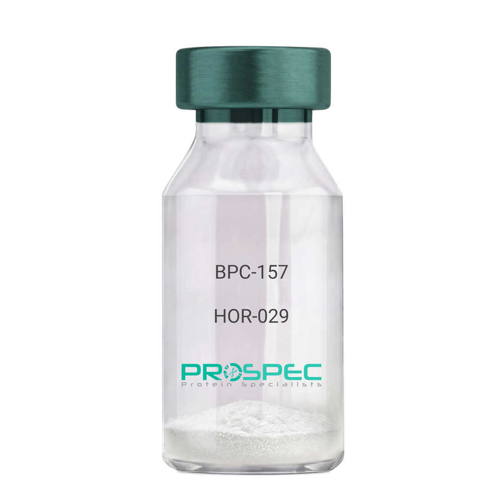
    BPC-157

    Description:

    BPC-157 Pentadecapeptide

    Product # :

    HOR-029

    Price :

    Quantity :

    Shipping Method :

    Room Temp Icon

    Shipped at Room temp

    Add To Cart

    More Info

    • description
    • formulation
    • purity
    • More Info

    Description

    BPC-157 Synthetic is a single, non-glycosylated polypeptide chain containing 15 amino acids, having a molecular mass of 1419.55 Dalton and a Molecular formula of C62H98N16O22.

    Formulation

    The protein was lyophilized with no additives.

    Purity

    Greater than 97.0% as determined by analysis by RP-HPLC.

    More Info

    • Physical Appearance

      Sterile Filtered White lyophilized (freeze-dried) powder.

    • Stability

      Lyophilized BPC-157 although stable at room temperature for 3 weeks, should be stored desiccated below -18°C. Upon reconstitution bpc-157 should be stored at 4°C between 2-7 days and for future use below -18°C. For long term storage it is recommended to add a carrier protein (0.1% HSA or BSA). Please prevent freeze-thaw cycles.

    • Solubility

      It is recommended to reconstitute the lyophilized BPC-157 in sterile 18MΩ-cm H2O not less than 100 µg/ml, which can then be further diluted to other aqueous solutions.

    • Amino Acid Sequence

      H-Gly-Glu-Pro-Pro-Pro-Gly-Lys-Pro-Ala-Asp-Asp-Ala-Gly-Leu-Val-OH

    • Background

      BPC-157, short for Body Protection Compound-157, is a synthetic peptide that has garnered significant attention in the field of regenerative medicine and sports science. This peptide, derived from a portion of the human gastric juice protein known as BPC, exhibits remarkable healing and tissue regeneration properties. BPC-157 has shown promise in various preclinical and clinical studies, demonstrating its potential for the treatment of a wide range of injuries and disorders.

      The research on BPC-157 encompasses investigations into its mechanisms of action, efficacy, safety, and potential therapeutic applications. Studies have elucidated the peptide's ability to enhance angiogenesis, promote collagen synthesis, modulate inflammatory responses, and protect against oxidative stress. These properties make BPC-157 an intriguing candidate for accelerating tissue healing, reducing inflammation, and improving overall recovery outcomes.

      Preclinical studies have revealed the beneficial effects of BPC-157 in several injury models. For instance, BPC-157 has demonstrated its potential in accelerating tendon and ligament healing, mitigating muscle damage, and promoting bone regeneration. These findings suggest that BPC-157 could be a valuable therapeutic tool in orthopedic medicine and sports-related injuries.

      Furthermore, BPC-157 has exhibited promising effects on gastrointestinal health. Studies have highlighted its ability to protect and heal the gut lining, reduce ulcer formation, and alleviate symptoms associated with inflammatory bowel disease. These observations open up avenues for BPC-157 as a potential treatment for gastrointestinal disorders.

      In addition to its regenerative properties, BPC-157 has shown potential in neurological and psychiatric conditions. Research has indicated its neuroprotective effects, with implications for the treatment of traumatic brain injury, stroke, and neurodegenerative disorders. Preliminary studies also suggest BPC-157's potential as an antidepressant and anxiolytic agent.

      Despite the promising findings, further research is needed to fully understand the mechanisms underlying BPC-157's actions and to assess its long-term safety and efficacy. Clinical trials are underway to explore its potential therapeutic applications in humans, including its use in tendon and ligament repair, inflammatory bowel disease, and neurodegenerative disorders.

      This comprehensive review aims to summarize the current state of research on BPC-157, providing an overview of its mechanisms of action and therapeutic potential. By examining relevant studies and findings, we aim to shed light on the diverse applications of BPC-157 and its implications for regenerative medicine, sports science, and various disease

    ProSpec's products are furnished for LABORATORY RESEARCH USE ONLY. The product may not be used as drugs, agricultural or pesticidal products, food additives or household chemicals.

    Bpc 157
  • View Data Sheet

    Name :

    GFP Antibody

    Description:

    Green Fluorescent Protein, Mouse Antibody

    Green fluorescent protein, GFP.

    Product # :

    ANT-359

    Price :

    Quantity :

    Shipping Method :

    Ice Icon

    Shipped with Ice Packs

    Add To Cart

    More Info

    • formulation
    • More Info

    Formulation

    1mg/ml containing PBS, pH-7.4, 10% glycerol & 0.02% Sodium Azide.

    More Info

    • Introduction

      rGFP, also known as Green Fluorescent Protein, is a protein produced by the jellyfish (Aequorea Victoria) that produces bioluminescence in the green zone of the noticeable spectrum. Green Fluorescent Protein is a useful and ubiquitous instrument for producing chimeric proteins, where it functions as a fluorescent protein tag. rGFP is expressed in most known cell types and is used as a noninvasive fluorescent marker in living cells and organisms. Green Fluorescent Protein permits a broad range of applications where it has functioned as a cell lineage tracer, reporter of gene expression, or as a measure of protein-protein interactions.

    • Synonyms

      Green fluorescent protein, GFP.

    • Physical Appearance

      Sterile filtered colorless solution.

    • Immunogen

      Anti GFP mAb is derived from hybridization of mouse F0 myeloma cells with spleen cells from BALB/c mice immunized with recombinant GFP amino acids 1-238 purified from E. coli.

    • Ig Subclass

      Mouse IgG2a heavy chain and κ light chain.

    • Clone

      PAT2G5AT.

    • Applications

      GFP antibody has been tested by ELISA and Western blot analysis to assure specificity and reactivity. Since application varies, however, each investigation should be titrated by the reagent to obtain optimal results.

    • Type

      Mouse Antibody Monoclonal.

    • Storage Procedures

      For periods up to 1 month store at 4°C, for longer periods of time, store at -20°C. Prevent freeze thaw cycles.

    • Purification Method

      GFP antibody was purified from mouse ascitic fluids by protein-G affinity chromatography.

    ProSpec's products are furnished for LABORATORY RESEARCH USE ONLY. The product may not be used as drugs, agricultural or pesticidal products, food additives or household chemicals.

    Gfp Antibody
  • View Data Sheet

    Name :

    CRABP2 Antibody

    Description:

    Cellular Retinoic Acid binding Protein 2, Mouse Anti Human

    RBP6, CRABP-II, CRABP2, RETINOIC ACID-BINDING PROTEIN CELLULAR TYPE II, Cellular retinoic acid-binding protein 2, Cellular retinoic acid-binding protein II.

    Product # :

    ANT-356

    Price :

    Quantity :

    Shipping Method :

    Ice Icon

    Shipped with Ice Packs

    Add To Cart

    More Info

    • formulation
    • More Info

    Formulation

    1mg/ml containing PBS, pH-7.4, & 0.1% Sodium Azide.

    More Info

    • Introduction

      CRABP2 NCBI Accession No: NP_001869 regulates the access of retinoic acid to the nuclear retinoic acid receptors. CRABP2 is involved in a regulatory feedback mechanism that controls the action of retinoic acid on cell differentiation. CRABP2 is involved in the conversion of vitamin A into its intracellular active form retinoic acid, which regulate the genes responsible for lipid metabolism and adipocyte differentiation. CRABP2 gene is located on chromosome 1q21-23 and this region has been linked with related disorders such as familial combined hyperlipidemia (FCHL) and type 2 diabetes mellitus. CRABP proteins are of low molecular weight having an important function in retinoic acid-mediated regulation of human skin growth and differentiation.

    • Synonyms

      RBP6, CRABP-II, CRABP2, RETINOIC ACID-BINDING PROTEIN CELLULAR TYPE II, Cellular retinoic acid-binding protein 2, Cellular retinoic acid-binding protein II.

    • Immunogen

      Anti-human CRABP2 mAb is derived from hybridization of mouse F0 myeloma cells with spleen cells from BALB/c mice immunized with recombinant human CRABP2 amino acids 1-138 purified from E. coli.

    • Ig Subclass

      Mouse IgG2a heavy chain and κ light chain.

    • Clone

      PAT2E11AT.

    • Applications

      CRABP2 antibody has been tested by ELISA and Western blot analysis to assure specificity and reactivity. Since application varies, however, each investigation should be titrated by the reagent to obtain optimal results. Recommended dilution range for Western blot analysis is 1:250 ~ 1000. Recommended starting dilution is 1:250.

    • Type

      Mouse Anti Human Monoclonal.

    • Storage Procedures

      For periods up to 1 month store at 4°C, for longer periods of time, store at -20°C. Prevent freeze thaw cycles.

    • Purification Method

      CRABP2 antibody was purified from mouse ascitic fluids by protein-G affinity chromatography.

    ProSpec's products are furnished for LABORATORY RESEARCH USE ONLY. The product may not be used as drugs, agricultural or pesticidal products, food additives or household chemicals.

    Crabp2 Antibody
  • View Data Sheet

    Name :

    LGALS14 Human

    Description:

    Galectin-14 Human Recombinant

    Placental protein 13-like, Charcot-Leyden crystal protein 2, CLC2, Galectin-14, Gal-14, LGALS14, PPL13, MGC22235.

    Product # :

    CYT-003

    Price :

    Quantity :

    Shipping Method :

    Ice Icon

    Shipped with Ice Packs

    Add To Cart

    More Info

    • description
    • source
    • formulation
    • purity
    • More Info

    Description

    LGALS14 Human Recombinant fused with a 23 amino acid His tag at N-terminus produced in E.Coli is a single, non-glycosylated, polypeptide chain containing 162 amino acids (1-139 a.a.) and having a molecular mass of 18.5kDa. The LGALS14 is purified by proprietary chromatographic techniques.

    Source

    Escherichia Coli.

    Formulation

    The LGALS14 solution (0.5mg/ml) contains 20mM Tris-HCl buffer (pH8.0), 20% glycerol, 0.1M NaCl and 1mM DTT.

    Purity

    Greater than 90.0% as determined by SDS-PAGE.

    More Info

    • Introduction

      Galectin14, aka LGALS14, is a member of the galectin family of carbohydrate binding proteins. Galectin family members contain one or two carbohydrate recognition domains, which can bind beta-galactoside. The LGALS14 gene is predominantly expressed in the placenta. LGALS14 is expressed intracellularly in the placenta and eosinophils and is released by eosinophils following allergen stimulation. LGALS14 may be involved in the development of allergic inflammation.

    • Synonyms

      Placental protein 13-like, Charcot-Leyden crystal protein 2, CLC2, Galectin-14, Gal-14, LGALS14, PPL13, MGC22235.

    • Physical Appearance

      Sterile Filtered colorless solution.

    • Stability

      Store at 4°C if entire vial will be used within 2-4 weeks. Store, frozen at -20°C for longer periods of time. For long term storage it is recommended to add a carrier protein (0.1% HSA or BSA).Avoid multiple freeze-thaw cycles.

    • Amino Acid Sequence

      MGSSHHHHHH SSGLVPRGSH MGSMSSLPVP YTLPVSLPVG SCVIITGTPI LTFVKDPQLE VNFYTGMDED SDIAFQFRLH FGHPAIMNSR VFGIWRYEEK CYYLPFEDGK PFELCIYVRH KEYKVMVNGQ RIYNFAHRFP PASVKMLQVL RDISLTRVLI SD.

    • Background

      What is the molecular weight/Mw of LGALS14 HUMAN Protein?
      LGALS14 HUMAN Protein has a total Mw of 18.5kDa.

      What is the source or expression system of LGALS14 HUMAN Protein?
      Escherichia Coli.

      What is the Purity of LGALS14 HUMAN Protein?
      LGALS14 HUMAN Protein is >90% pure as determined by SDS-PAGE.

      What is the Biological Activity of LGALS14 HUMAN Protein?
      The biological functionality of LGALS14 HUMAN Protein will be determined in the future.

      What is the amino acid sequence of LGALS14 HUMAN Protein?
      MGSSHHHHHH SSGLVPRGSH MGSMSSLPVP YTLPVSLPVG SCVIITGTPI LTFVKDPQLE VNFYTGMDED SDIAFQFRLH FGHPAIMNSR VFGIWRYEEK CYYLPFEDGK PFELCIYVRH KEYKVMVNGQ RIYNFAHRFP PASVKMLQVL RDISLTRVLI SD.
      What applications can LGALS14 HUMAN Protein be used in?
      LGALS14 HUMAN Protein can probably be used in western blot, ELISA and Lateral Flow.

      What is the endotoxin level for LGALS14 HUMAN Protein?
      The endotoxin level is minimal, LGALS14 HUMAN Protein was purified using conventional chromatography techniques.


    ProSpec's products are furnished for LABORATORY RESEARCH USE ONLY. The product may not be used as drugs, agricultural or pesticidal products, food additives or household chemicals.

    Lgals14 Human
  • 1
  • …
  • 32
  • 33
  • 34
  • 35
  • 36
  • …
  • 42
Your browser does not support the video tag.

Products

  • Cytokines
  • Growth Factors
  • Chemokines
  • CD Antigens
  • Neurotrophins
  • Hormones
  • Enzymes
  • Viral Antigens
  • Recombinant Proteins
  • Natural Proteins
  • Monoclonal Antibodies
  • Polyclonal Antibodies
  • Thermal Packaging

About

  • Company
  • Founder
  • Contribution
  • Contact Us
  • My Account

Ordering

  • Ordering Method
  • Ordering - Credit Card
  • Ordering PO
  • Shipping Fees
  • FAQ's

Legal

  • Terms & Conditions
  • Privacy
  • Site Map
  • info@prospecbio.com
  • Facebook
  • Instagram
  • Linkedin Profile

© 2026 ProSpec-Tany TechnoGene Ltd. All Rights Reserved.

  • Choosing a selection results in a full page refresh.
  • Opens in a new window.