Search results
1000 results found for “interferon”
Name
Description
Product #
Price
Quantity
Shipping Method
- View Data Sheet
Name :
ESAT6-CFP10Description:
ESAT6-CFP10 Chimeric Recombinant
Early Secretory Target Mycobacterium Tuberculosis, ESAT-6
Product # :
PRO-2737Price :
Quantity :
Shipping Method :
Shipped with Ice Packs
More Info
- description
- source
- formulation
- purity
- More Info
- Citations
Description
Recombinant Chimeric ESAT6-CFP10 produced in E. coli is fused to a 6 amino acid his tag at its C-terminus and purified by proprietary chromatographic technique.
Source
Escherichia Coli.
Formulation
ESAT6-CFP10 solution contains 25mM K2CO3, 0.02% NaN3 and PBS.
Purity
Protein is >95% pure as determined by 12% PAGE (coomassie staining).
Citations
More Info
-
Introduction
Mycobacterium antigen ESAT6 has been isolated from low molecular weight fractions of the shot-term-culture filtrate (ST-CF) and it can easily be detected in tuberculosis patients. The export of ESAT-6 which is a potent T-cell antigen, and related proteins requires a dedicated secretory apparatus that is encoded by a group of genes, several of which also code for proteins that are recognized strongly by T cells. The ESAT-6 systems can consequently be considered as immunogenicity islands and there is mounting evidence that the equivalent genes are subject to selective pressure imposed by the immune system of the host. This antigen includes many epitopes detectable in the serum of most patients with tuberculosis (more than 90%). By the attempts to obtain the vaccine on the basis of ESAT-6 it was demonstrated that the optimization of adjuvant is very important when using the combination of dioctadecylammonium bromide and monophosphoryllipide. ESAT-6 is very potential as diagnostic for differentiation between the mycobacterial infection and BCG vaccination. The main topic in ESAT-6 using is in antibody production and in test-systems for tuberculosis elaboration. Early secretory antigen 6 (ESAT6) and cell filtrate protein 10 (CFP10) are 2 antigens secreted as a complex by the replicating Mycobacterium tuberculosis complex (MTC).
-
Synonyms
Early Secretory Target Mycobacterium Tuberculosis, ESAT-6
-
Physical Appearance
Sterile Filtered clear solution.
-
Stability
Store at 4°C if entire vial will be used within 2-4 weeks.Store, frozen at -20°C for longer periods of time.For long term storage it is recommended to add a carrier protein (0.1% HSA or BSA).Avoid multiple freeze-thaw cycles.
-
Applications
Immunoassay.
ProSpec's products are furnished for LABORATORY RESEARCH USE ONLY. The product may not be used as drugs, agricultural or pesticidal products, food additives or household chemicals.
- View Data Sheet
Name :
HAV VP1-P2A (669-782 a.a)Description:
Hepatitis A Virus VP1-P2A (669-782 a.a.) Recombinant
Product # :
HAV-265Price :
Quantity :
Shipping Method :
Shipped with Ice Packs
More Info
- description
- source
- formulation
- purity
- More Info
- Citations
Description
The E.Coli derived recombinant protein contains the VP1-P2A immunodominant regions, amino acids 669-782.
Source
Escherichia Coli.
Formulation
10mM Tris-HCl, pH 9.6, 1.5M urea, 50% glycerol.
Purity
HAV VP1-P2A protein is >90% pure as determined by 10% PAGE (coomassie staining).
Citations
More Info
-
Introduction
Forty-two antigenic domains were identified across the hepatitis A virus (HAV) polyprotein by using a set of 237 overlapping 20-mer synthetic peptides spanning the entire HAV polyprotein. Nineteen antigenic domains were found within the structural proteins, and 22 were found within the nonstructural proteins, with 1 domain spanning the junction of VP1 and P2A proteins. Five of these domains were considered immunodominant, as judged by both the breadth and the strength of their immunoreactivity. One domain is located within the VP2 protein at position 57-90 aa. A second domain, located at position 767-842 aa, contains the C-terminal part of the VP1 protein and the entire P2A protein. A third domain, located at position 1403-1456 a.a, comprises the C-terminal part of the P2C protein and the N-terminal half of the P3A protein. The fourth domain, located at position 1500-1519 a.a, includes almost the entire P3B, and the last domain, located at position 1719-1764 aa, contains the C-terminal region of the P3C protein and the N-terminal region of the P3D protein. Four of the five most immunoreactive domains are derived from small HAV proteins and/or encompass protein cleavage sites separating different HAV proteins.
-
Stability
HAV VP1-P2A protein although stable at 4°C for 1 week, should be stored below -18°C. Please prevent freeze thaw cycles.
-
Applications
HAV VP1-P2A antigen is suitable for ELISA and Western blots, excellent antigen for detection of HAV with minimal specificity problems.
-
Specificity
Immunoreactive with sera HAV-infected individuals.
-
Purification Method
HAV VP1-P2A protein was purified by proprietary chromatographic technique.
ProSpec's products are furnished for LABORATORY RESEARCH USE ONLY. The product may not be used as drugs, agricultural or pesticidal products, food additives or household chemicals.
- View Data Sheet
Name :
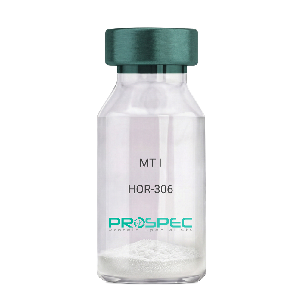
MT IDescription:
Melanotan-I
Melanotan-I, MT-I, Melanotan-1.
Product # :
HOR-306Price :
Quantity :
Shipping Method :
Shipped at Room temp
More Info
- description
- formulation
- purity
- More Info
- Citations
Description
Melanotan-I has the amino acid sequence of Ser-Tyr-Ser-Nle-Glu-His-D-Phe-Arg-Trp-Gly-Lys-Pro-Val and a molecular weight of 1647.4 Dalton.
Formulation
The protein (1mg/ml) was lyophilized with no additives.
Purity
Greater than 98.0% as determined by RP-HPLC.
Citations
More Info
-
Synonyms
Melanotan-I, MT-I, Melanotan-1.
-
Physical Appearance
Sterile Filtered off-White lyophilized (freeze-dried) powder.
-
Stability
Lyophilized Melanotan-I although stable at room temperature for 3 weeks, should be stored desiccated below -18°C. Upon reconstitution MT-I should be stored at 4°C between 2-7 days and for future use below -18°C.For long term storage it is recommended to add a carrier protein (0.1% HSA or BSA).Please prevent freeze-thaw cycles.
-
Solubility
It is recommended to reconstitute the lyophilized Melanotan-I in sterile 1% acetic acid not less than 100 µg/ml, which can then be further diluted to other aqueous solutions.
ProSpec's products are furnished for LABORATORY RESEARCH USE ONLY. The product may not be used as drugs, agricultural or pesticidal products, food additives or household chemicals.
- View Data Sheet
Name :
HAV P2C-P3ADescription:
Hepatitis A Virus P2C-P3A Recombinant
Product # :
HAV-266Price :
Quantity :
Shipping Method :
Shipped with Ice Packs
More Info
- description
- source
- formulation
- purity
- More Info
- Citations
Description
The E.Coli derived recombinant protein contains the P2C-P3A immunodominant regions, amino acids 1392-1521.
Source
Escherichia Coli.
Formulation
10mM CBB, pH9.6, 0.1% SDS and 50% glycerol.
Purity
HAV P2C-P3A protein is >90% pure as determined by 10% PAGE (coomassie staining).
Citations
More Info
-
Introduction
Forty-two antigenic domains were identified across the hepatitis A virus (HAV) polyprotein by using a set of 237 overlapping 20-mer synthetic peptides spanning the entire HAV polyprotein. Nineteen antigenic domains were found within the structural proteins, and 22 were found within the nonstructural proteins, with 1 domain spanning the junction of VP1 and P2A proteins. Five of these domains were considered immunodominant, as judged by both the breadth and the strength of their immunoreactivity. One domain is located within the VP2 protein at position 57-90 aa. A second domain, located at position 767-842 aa, contains the C-terminal part of the VP1 protein and the entire P2A protein. A third domain, located at position 1403-1456 aa, comprises the C-terminal part of the P2C protein and the N-terminal half of the P3A protein. The fourth domain, located at position 1500-1519 aa, includes almost the entire P3B, and the last domain, located at position 1719-1764 aa, contains the C-terminal region of the P3C protein and the N-terminal region of the P3D protein. Four of the five most immunoreactive domains are derived from small HAV proteins and/or encompass protein cleavage sites separating different HAV proteins.
-
Stability
HAV P2C-P3A protein although stable at 4°C for 1 week, should be stored below -18°C. Please prevent freeze thaw cycles.
-
Applications
HAV P2C-P3A antigen is suitable for ELISA and Western blots, excellent antigen for detection of HAV with minimal specificity problems.
-
Specificity
Immunoreactive with sera HAV-infected individuals.
-
Purification Method
HAV P2C-P3A protein was purified by proprietary chromatographic technique.
ProSpec's products are furnished for LABORATORY RESEARCH USE ONLY. The product may not be used as drugs, agricultural or pesticidal products, food additives or household chemicals.
- View Data Sheet
Name :
CMV Pp150Description:
Cytomegalo Virus Pp150 (UL32) Recombinant
Product # :
CMV-216Price :
Quantity :
Shipping Method :
Shipped with Ice Packs
More Info
- description
- source
- formulation
- purity
- More Info
- Citations
Description
The E.Coli derived recombinant protein contains the CMV Pp150 (UL32) immunodominant regions, 1011-1048 amino acids.
Source
Escherichia Coli.
Formulation
50mM Tris pH 8.0, 1mM EDTA and 50% glycerol.
Purity
CMV Pp150 protein is >95% pure as determined by 10% PAGE (coomassie staining).
Citations
More Info
-
Introduction
CMV belongs to the Betaherpesvirinae subfamily of Herpesviridae which includes herpes simplex virustypes 1 and 2, varicella-zoster virus, and Epstein-Barrvirus. The herpesviruses share a characteristic ability to remain latentover long periods. CMV is a double-stranded linear DNA virus with 162 hexagonal protein capsomeres surrounded by a lipid membrane. CMV has the largest genome of the herpes viruses, ranging from 230-240 kilobase pairs. Human CMV is composed of unique and inverted repeats that include the existence of 4 genome isomers caused by inversion of L-S genome components (class E). Replication may be divided into immediate early, delayed early, and late gene expression based on time of synthesis after infection. The DNA is replicated by rolling circles. In vitro, CMV replicates in human fibroblasts.
-
Stability
CMV Pp150 protein although stable at 4°C for 1 week, should be stored below -18°C. Please prevent freeze thaw cycles.
-
Applications
CMV Pp65 antigen is suitable for ELISA and Western blots, excellent antigen for detection of CMV with minimal specificity problems.
-
Specificity
Immunoreactive with sera of CMV-infected individuals.
-
Purification Method
CMV Pp150 was purified by proprietary chromatographic technique.
ProSpec's products are furnished for LABORATORY RESEARCH USE ONLY. The product may not be used as drugs, agricultural or pesticidal products, food additives or household chemicals.
- View Data Sheet
Name :
CMV Pp52Description:
Cytomegalo Virus Pp52 (UL44) Recombinant
Product # :
CMV-214Price :
Quantity :
Shipping Method :
Shipped with Ice Packs
More Info
- description
- source
- formulation
- purity
- More Info
- Citations
Description
The E.coli derived 51 kDa recombinant protein contains the CMV Pp52 (UL44) immunodominant regions, 202-434 amino acids. Recombinant CMV-Pp52 is fused to a 26 kDa GST tag.
Source
Escherichia Coli.
Formulation
50mM Tris pH 7.2, 1mM EDTA and 50% glycerol.
Purity
CMV Pp52 protein is >95% pure as determined by 10% PAGE (coomassie staining).
Citations
More Info
-
Introduction
CMV belongs to the Betaherpesvirinae subfamily of Herpesviridae which includes herpes simplex virustypes 1 and 2, varicella-zoster virus, and Epstein-Barrvirus. The herpesviruses share a characteristic ability to remain latentover long periods. CMV is a double-stranded linear DNA virus with 162 hexagonal protein capsomeres surrounded by a lipid membrane. CMV has the largest genome of the herpes viruses, ranging from 230-240 kilobase pairs. Human CMV is composed of unique and inverted repeats that include the existence of 4 genome isomers caused by inversion of L-S genome components (class E). Replication may be divided into immediate early, delayed early, and late gene expression based on time of synthesis after infection. The DNA is replicated by rolling circles. In vitro, CMV replicates in human fibroblasts.
-
Stability
CMV Pp52 protein although stable at 4°C for 1 week, should be stored below -18°C. Please prevent freeze thaw cycles.
-
Applications
CMV Pp52 antigen is suitable for ELISA and Western blots, excellent antigen for detection of CMV with minimal specificity problems.
-
Specificity
Immunoreactive with sera of CMV-infected individuals.
-
Purification Method
CMV Pp52 protein was purified by proprietary chromatographic technique.
ProSpec's products are furnished for LABORATORY RESEARCH USE ONLY. The product may not be used as drugs, agricultural or pesticidal products, food additives or household chemicals.
- View Data Sheet
Name :
HAV P2CDescription:
Hepatitis A Virus P2C Recombinant
Product # :
HAV-229Price :
Quantity :
Shipping Method :
Shipped with Ice Packs
More Info
- description
- formulation
- purity
- More Info
- Citations
Description
The E.Coli derived recombinant 38.5 kDa protein contains the P2C immunodominant regions, amino acids 1121-1234.
Formulation
10mM CBB, pH9.6, 0.1% SDS and 50% glycerol.
Purity
Protein is >90% pure as determined by 10% PAGE (coomassie staining).
Citations
More Info
-
Introduction
Forty-two antigenic domains were identified across the hepatitis A virus (HAV) polyprotein by using a set of 237 overlapping 20-mer synthetic peptides spanning the entire HAV polyprotein. Nineteen antigenic domains were found within the structural proteins, and 22 were found within the nonstructural proteins, with 1 domain spanning the junction of VP1 and P2A proteins. Five of these domains were considered immunodominant, as judged by both the breadth and the strength of their immunoreactivity. One domain is located within the VP2 protein at position 57-90 aa. A second domain, located at position 767-842 aa, contains the C-terminal part of the VP1 protein and the entire P2A protein. A third domain, located at position 1403-1456 aa, comprises the C-terminal part of the P2C protein and the N-terminal half of the P3A protein. The fourth domain, located at position 1500-1519 aa, includes almost the entire P3B, and the last domain, located at position 1719-1764 aa, contains the C-terminal region of the P3C protein and the N-terminal region of the P3D protein. Four of the five most immunoreactive domains are derived from small HAV proteins and/or encompass protein cleavage sites separating different HAV proteins.
-
Stability
HAV P2C protein although stable at 4°C for 1 week, should be stored below -18°C. Please prevent freeze thaw cycles.
-
Applications
HAV P2C antigen is suitable for ELISA and Western blots, excellent antigen for detection of HAV with minimal specificity problems.
-
Specificity
Immunoreactive with sera HAV-infected individuals.
-
Purification Method
HAV P2C protein was purified by proprietary chromatographic technique.
ProSpec's products are furnished for LABORATORY RESEARCH USE ONLY. The product may not be used as drugs, agricultural or pesticidal products, food additives or household chemicals.
- View Data Sheet
Name :
SARS Nucleocapsid MonoclonalDescription:
SARS-Nucleocapsid, Monoclonal Antibody
Product # :
ANT-759Price :
Quantity :
Shipping Method :
Shipped at Room temp
More Info
- formulation
- More Info
- Citations
Formulation
Lyophilized from 1mg/ml in PBS
Citations
More Info
-
Introduction
SARS Coronavirus is an enveloped virus containing three outer structural proteins, namely the membrane (M), envelope (E), and spike (S) proteins. Spike (S)-glycoprotein of the virus interacts with a cellular receptor and mediates membrane fusion to allow viral entry into susceptible target cells. Accordingly, S-protein plays an important role in virus infection cycle and is the primary target of neutralizing antibodies.It has recently been shown that SARS is caused by a human coronavirus. Human coronaviruses are the major cause of upper respiratory tract illness in humans, such as the common cold. Coronaviruses are positive-stranded RNA viruses, featuring the largest viral RNA genomes known to date (27-31 kb). The first step in coronavirus infection is binding of the viral spike protein, a 139-kDa protein, to certain receptors on host cells. The spike protein is the main surface antigen of the coronavirus. The most prominent protein in the culture supernatants infected with SARS virus is a 46 kDa nucleocapsid protein. This suggests that the nucleocapsid protein is a major immunogen that may be useful for early diagnostics.
-
Physical Appearance
Sterile Filtered White lyophilized (freeze-dried) powder.
-
Stability
Lyophilized SARS-Nucleocapsid Monoclonal Antibody although stable at room temperature for 3 weeks, should be stored desiccated below -18°C. Upon reconstitution store at 4°C between 2-7 days and for future use below -18°C.Please avoid freeze thaw cycles.
-
Solubility
Reconstitute Mouse Anti SARS Nucleocapsid antibody at a conc. of 0.5mg/ml in sterile PBS.
-
Immunogen
SARS Antibody was developed by immunizing mice with a protein fragment amino acids 1-49 from the human SARS Nucleocapsid coronavirus (Genbank accession no. NP_828858). SARS-Nucleocapsid, Monoclonal Antibody is protein-G purified.
-
Applications
Western Blot 1:1000, ELISA, Immunocytochemistry/Immunofluorescence.
SARS-Nucleocapsid, Monoclonal Antibody detects CoV-2 & SARS Coronavirus Nucleoprotein in direct ELISA. S-Species cross reactivity based on immunogen sequence homology: COV-2 (84%)
-
Type
Mouse antibody Monoclonal.
ProSpec's products are furnished for LABORATORY RESEARCH USE ONLY. The product may not be used as drugs, agricultural or pesticidal products, food additives or household chemicals.
- View Data Sheet
Name :
HIV-1 gp41 Long, BiotinDescription:
HIV-1 gp41 Long Recombinant, Biotin Labeled
Product # :
HIV-119Price :
Quantity :
Shipping Method :
Shipped with Ice Packs
More Info
- description
- source
- formulation
- purity
- More Info
- Citations
Description
The E.coli derived biotin labeled recombinant protein is a non-glycosylated polypeptide chain, containing the HIV-1 gag p41 immunodominant regions, amino acids 444-833.The HIV-1 p41 Biotinylated is fused to beta-galactosidase (114 kDa) at the N-terminus.
Source
Escherichia Coli.
Formulation
8M Urea, 20mM Tris-HCl pH 8.0, 10mM b-mercaptoethanol.
Purity
Greater than 95.0% as determined by HPLC analysis & SDS-PAGE.
Citations
More Info
-
Introduction
Human immunodeficiency virus (HIV) is a retrovirusthat can lead to a condition in which the immune systembegins to fail, leading to opportunistic infections. HIV primarily infects vital cells in the humanimmune systemsuch as helper T cells(specifically CD4+ T cells), macrophagesand dendritic cells. HIV infection leads to low levels of CD4+ T cells through three main mechanisms: firstly, direct viral killing of infected cells; secondly, increased rates of apoptosisin infected cells; and thirdly, killing of infected CD4+ T cells by CD8 cytotoxic lymphocytesthat recognize infected cells. When CD4+ T cell numbers decline below a critical level, cell-mediated immunityis lost, and the body becomes progressively more susceptible to opportunistic infections. HIV was classified as a member of the genus Lentivirus, part of the family of Retroviridae. Lentiviruses have many common morphologies and biological properties. Many species are infected by lentiviruses, which are characteristically responsible for long-duration illnesses with a long incubation period. Lentiviruses are transmitted as single-stranded, positive-sense, enveloped RNA viruses. Upon entry of the target cell, the viral RNA genomeis converted to double-stranded DNAby a virally encoded reverse transcriptasethat is present in the virus particle. This viral DNA is then integrated into the cellular DNA by a virally encoded integraseso that the genome can be transcribed. Once the virus has infected the cell, two pathways are possible: either the virus becomes latentand the infected cell continues to function, or the virus becomes active and replicates, and a large number of virus particles are liberated that can then infect other cells.
-
Physical Appearance
Sterile filtered colorless clear solution.
-
Stability
HIV-1 gp41 Long although stable at 4°C for 1 week, should be stored below -18°C. Please prevent freeze thaw cycles.
-
Applications
HIV-I gp41 antigen is suitable for ELISA and Western blots, excellent antigen for early detection of HIV seroconvertors with minimal specificity problems.
-
Specificity
Immunoreactive with all sera of HIV-1 infected individuals.
ProSpec's products are furnished for LABORATORY RESEARCH USE ONLY. The product may not be used as drugs, agricultural or pesticidal products, food additives or household chemicals.
- View Data Sheet
Name :
PECAM1 MouseDescription:
Platelet Endothelial Cell Adhesion Molecule 1 MouseRecombinant
Platelet endothelial cell adhesion molecule, PECAM1, CD31, CD31/EndoCAM, endoCAM, GPIIA', PECA1, PECAM-1, Platelet Endothelial Cell Adhesion Molecule 1.
Product # :
CYT-1211Price :
Quantity :
Shipping Method :
Shipped with Ice Packs
More Info
- description
- source
- formulation
- purity
- More Info
- Citations
Description
PECAM1 Mouse Recombinant produced in HEK293 is a single, non-glycosylated polypeptide chain containing 579 amino acids (18-590 a.a.) and having a molecular mass of 65.3 kDa. PECAM1 Mouse is fused to an 6 amino acid His-tag at C-terminus & purified by proprietary chromatographic techniques.
Source
Sf9, Baculovirus cells.
Formulation
PECAM1 Mouse protein solution (0.25mg/ml) containing Phosphate Buffered Saline (pH 7.4) and 10% glycerol.
Purity
Greater than 90% as determined by SDS-PAGE.
Citations
More Info
-
Introduction
Platelet endothelial cell adhesion molecule (PECAM1) induces susceptibility to atherosclerosis. PECAM1 averts phagocyte ingestion of closely apposed viable cells by transmitting detachment signals, and transforms function upon apoptosis, thus promoting tethering of dying cells to phagocytes. The encounter of a viable cell with a phagocyte via the homophilic interaction of PECAM1 on both cell surfaces as a result causing the viable cell's active repulsion from the phagocyte.
-
Synonyms
Platelet endothelial cell adhesion molecule, PECAM1, CD31, CD31/EndoCAM, endoCAM, GPIIA', PECA1, PECAM-1, Platelet Endothelial Cell Adhesion Molecule 1.
-
Physical Appearance
Sterile filtered colorless solution.
-
Stability
Store at 4°C if entire vial will be used within 2-4 weeks. Store, frozen at -20°C for longer periods of time. For long term storage it is recommended to add a carrier protein (0.1% HSA or BSA).Avoid multiple freeze-thaw cycles.
-
Amino Acid Sequence
EENSFTINSI HMESLPSWEV MNGQQLTLEC LVDISTTSKS RSQHRVLFYK DDAMVYNVTS REHTESYVIP QARVFHSGKY KCTVMLNNKE KTTIEYEVKV HGVSKPKVTL DKKEVTEGGV VTVNCSLQEE KPPIFFKIEK LEVGTKFVKR RIDKTSNENF VLMEFPIEAQ DHVLVFRCQA GILSGFKLQE SEPIRSEYVT VQESFSTPKF EIKPPGMIIE GDQLHIRCIV QVTHLVQEFT EIIIQKDKAI VATSKQSSEA VYSVMAMVEY SGHYTCKVES NRISKASSIM VNITELFPKP KLEFSSSRLD QGELLDLSCS VSGTPVANFT IQKEETVLSQ YQNFSKIAEE SDSGEYSCTA GIGKVVKRSG LVPIQVCEML SKPSIFHDAK SEIIKGHAIG ISCQSENGTA PITYHLMKAK SDFQTLEVTS NDPATFTDKP TRDMEYQCRA DNCHSHPAVF SEILRVRVIA PVDEVVISIL SSNEVQSGSE MVLRCSVKEG TSPITFQFYK EKEDRPFHQA VVNDTQAFWH NKQASKKQEG QYYCTASNRA SSMRTSPRSS TLAVRVFLAP WKK HHHHH
ProSpec's products are furnished for LABORATORY RESEARCH USE ONLY. The product may not be used as drugs, agricultural or pesticidal products, food additives or household chemicals.
- View Data Sheet
Name :
KIR2DL4 HumanDescription:
Killer Cell Immunoglobulin-Like Receptor, 2 Domains Long Cytoplasmic Tail 4 Human Recombinant
Killer Cell Immunoglobulin Like Receptor, Two Ig Domains And Long Cytoplasmic Tail 4,Killer Cell Immunoglobulin-Like Receptor, Two Domains, Long Cytoplasmic Tail 4,Killer Cell Inhibitory Receptor 103AS, MHC Class I NK Cell Receptor KIR103AS,CD158 Antigen-Like Family Member D, KIR-103AS, KIR103AS, CD158D, G9P, Killer Cell Immunoglobulin-Like Receptor 2DL4, CD158d Antigen, KIR-2DL4, KIR103, KIR2DL4.
Product # :
PRO-2460Price :
Quantity :
Shipping Method :
Shipped with Ice Packs
More Info
- description
- source
- formulation
- purity
- More Info
- Citations
Description
KIR2DL4 Human Recombinant produced in Sf9 Insect cells is a single, glycosylated polypeptide chain containing 458 amino acids (24-242 a.a.) and having a molecular mass of 51kDa (Molecular size on SDS-PAGE will appear at approximately 50-70kDa). KIR2DL4 is expressed with a 239 amino acids hIgG-His tag at C-Terminus and purified by proprietary chromatographic techniques.
Source
Sf9, Baculovirus cells.
Formulation
KIR2DL4 protein solution (0.25mg/ml) contains Phosphate buffered saline (pH7.4) and 10% glycerol.
Purity
Greater than 85.0% as determined by SDS-PAGE.
Citations
More Info
-
Introduction
Killer cell immunoglobulin-like receptor 2DL4, also known as KIR2DL4 is part of the killer cell Ig-like receptor (KIR) family. KIR proteins with the long cytoplasmic domain transduce inhibitory signals through an immune tyrosine-based inhibitory motif (ITIM), whereas KIR proteins which contain the short cytoplasmic domain, lack the ITIM motif and as a substitute associate with the TYRO protein tyrosine kinase binding protein to transduce activating signals. KIR2DL4 stimulates NK cells to produce IFN-gamma and stimulation with IL-2 upregulates cell surface expression on CD56dimcells and leads to the inhibition of the cytolytic NK cell function.
-
Synonyms
Killer Cell Immunoglobulin Like Receptor, Two Ig Domains And Long Cytoplasmic Tail 4,Killer Cell Immunoglobulin-Like Receptor, Two Domains, Long Cytoplasmic Tail 4,Killer Cell Inhibitory Receptor 103AS, MHC Class I NK Cell Receptor KIR103AS,CD158 Antigen-Like Family Member D, KIR-103AS, KIR103AS, CD158D, G9P, Killer Cell Immunoglobulin-Like Receptor 2DL4, CD158d Antigen, KIR-2DL4, KIR103, KIR2DL4.
-
Physical Appearance
Sterile Filtered colorless solution.
-
Stability
Store at 4°C if entire vial will be used within 2-4 weeks. Store, frozen at -20°C for longer periods of time. For long term storage it is recommended to add a carrier protein (0.1% HSA or BSA).Avoid multiple freeze-thaw cycles.
-
Amino Acid Sequence
HVGGQDKPFC SAWPSAVVPQ GGHVTLRCHY RRGFNIFTLY KKDGVPVPEL YNRIFWNSFL ISPVTPAHAG TYRCRGFHPH SPTEWSAPSN PLVIMVTGLY EKPSLTARPG PTVRAGENVT LSCSSQSSFD IYHLSREGEA HELRLPAVPS INGTFQADFP LGPATHGETY RCFGSFHGSP
YEWSDPSDPL PVSVTGNPSS SWPSPTEPSF KTGIARHLHL EPKSCDKTHT CPPCPAPELL GGPSVFLFPP KPKDTLMISR TPEVTCVVVD VSHEDPEVKF NWYVDGVEVH NAKTKPREEQ YNSTYRVVSV LTVLHQDWLN GKEYKCKVSN KALPAPIEKT ISKAKGQPRE PQVYTLPPSR
DELTKNQVSL TCLVKGFYPS DIAVEWESNG QPENNYKTTP PVLDSDGSFF LYSKLTVDKS RWQQGNVFSC SVMHEALHNH YTQKSLSLSP GKHHHHHH
ProSpec's products are furnished for LABORATORY RESEARCH USE ONLY. The product may not be used as drugs, agricultural or pesticidal products, food additives or household chemicals.
- View Data Sheet
Name :
INSR HumanDescription:
Insulin Receptor Human Recombinant
Insulin receptor, IR, EC 2.7.10.1, CD220, INSR, HHF5.
Product # :
CYT-777Price :
Quantity :
Shipping Method :
Shipped at Room temp
More Info
- description
- source
- formulation
- purity
- More Info
- Citations
Description
Insulin Receptor Human Recombinant produced in HEK cells is a single, glycosylated, polypeptide chain (aa 28-944 of the short isoform- HIR-A, Uniprot accession # P06213-2 which includes the whole subunit alpha and extracellular domain of subunit beta) containing a total of 927 amino acids, having a molecular mass of 105.9kDa (calculated), though it migrates at approximately 160kDa on SDS PAGE, the INSR is fused to a 2 a.a N-terminal linker, a 2 a.a C-terminal linker and fused to a 6 a.a His tag at C-Terminus.The Human INSR is purified by proprietary chromatographic techniques.
Source
HEK 293.
Formulation
INSR was filtered (0.4µm) and lyophilized from 0.5mg/ml in 0.05M phosphate buffer and 0.075M NaCl, pH 7.4.
Purity
Greater than 95.0% as determined by SDS-PAGE.
Citations
More Info
-
Introduction
Insulin Receptor (INSR) is a receptor tyrosine kinase which mediates the pleiotropic actions of insulin. Binding of insulin to the insulin receptor (INSR) stimulates glucose uptake. Once the precursor signal peptide is removed, the insulin receptor precursor is post-translationally cleaved into 2 chains (alpha and beta) which are covalently linked. Insulin binding initiates phosphorylation of several intracellular substrates, including, insulin receptor substrates (IRS1, 2, 3, 4), SHC, GAB1, CBL and other signaling intermediates. Each of these phosphorylated proteins function as docking proteins for other signaling proteins which contain Src-homology-2 domains (SH2 domain) that specifically recognize different phosphotyrosines residues, including the p85 regulatory subunit of PI3K and SHP2.
-
Synonyms
Insulin receptor, IR, EC 2.7.10.1, CD220, INSR, HHF5.
-
Physical Appearance
Filtered White lyophilized (freeze-dried) powder.
-
Stability
Store lyophilized protein at -20°C. Aliquot the product after reconstitution to avoid repeated freezing/thawing cycles. Reconstituted protein can be stored at 4°C for a limited period of time.
-
Solubility
It is recommended to add deionized water to a working concentration of 0.5mg/ml and let the lyophilized pellet dissolve completely. INSR is not sterile! Please filter the product by an appropriate sterile filter before using it in the cell culture.
-
Amino Acid Sequence
ASHLYPGEVC PGMDIRNNLT RLHELENCSV IEGHLQILLM FKTRPEDFRD LSFPKLIMIT DYLLLFRVYG LESLKDLFPN LTVIRGSRLF FNYALVIFEM VHLKELGLYN LMNITRGSVR IEKNNELCYL ATIDWSRILD SVEDNYIVLN KDDNEECGDI CPGTAKGKTN CPATVINGQF VERCWTHSHC QKVCPTICKS HGCTAEGLCC HSECLGNCSQ PDDPTKCVAC RNFYLDGRCV ETCPPPYYHF QDWRCVNFSF CQDLHHKCKN SRRQGCHQYV IHNNKCIPEC PSGYTMNSSN LLCTPCLGPC PKVCHLLEGE KTIDSVTSAQ ELRGCTVING SLIINIRGGN NLAAELEANL GLIEEISGYL KIRRSYALVS LSFFRKLRLI RGETLEIGNY SFYALDNQNL RQLWDWSKHN LTITQGKLFF HYNPKLCLSE IHKMEEVSGT KGRQERNDIA LKTNGDQASC ENELLKFSYI RTSFDKILLR WEPYWPPDFR DLLGFMLFYK EAPYQNVTEF DGQDACGSNS WTVVDIDPPL RSNDPKSQNH PGWLMRGLKP WTQYAIFVKT LVTFSDERRT YGAKSDIIYV QTDATNPSVP LDPISVSNSS SQIILKWKPP SDPNGNITHY LVFWERQAED SELFELDYCL KGLKLPSRTW SPPFESEDSQ KHNQSEYEDS AGECCSCPKT DSQILKELEE SSFRKTFEDY LHNVVFVPRP SRKRRSLGDV GNVTVAVPTV AAFPNTSSTS VPTSPEEHRP FEKVVNKESL VISGLRHFTG YRIELQACNQ DTPEERCSVA AYVSARTMPE AKADDIVGPV THEIFENNVV HLMWQEPKEP NGLIVLYEVS YRRYGDEELH LCVSRKHFAL ERGCRLRGLS PGNYSVRIRA TSLAGNGSWT EPTYFYVTDY LDVPSNIAKK LHHHHHH.
ProSpec's products are furnished for LABORATORY RESEARCH USE ONLY. The product may not be used as drugs, agricultural or pesticidal products, food additives or household chemicals.
- View Data Sheet
Name :
CD105 Human, Sf9Description:
Endoglin Human Recombinant, Sf9
CD105, ENG, END, ORW, HHT1, ORW1, FLJ41744, Endoglin.
Product # :
CYT-389Price :
Quantity :
Shipping Method :
Shipped at Room temp
More Info
- description
- source
- formulation
- purity
- biological activity
- More Info
- Citations
Description
CD105 Human Recombinant extracellular domain produced in baculovirus is a homodimeric, glycosylated, Polypeptide containing 586 amino acids and having a molecular mass of 61 kDa but as a result of glycosylation, migrates at 90 kDa under reducing conditions in SDS-PAGE. The CD105 is fused to a C-terminal His-tag (6xHis) and purified by proprietary chromatographic techniques.
Source
Sf9 Insect Cells.
Formulation
Endoglin was lyophilized from a concentrated (1mg/ml) sterile solution containing 1xPBS.
Purity
Greater than 95.0% as determined by:
(a) Analysis by RP-HPLC.
(b) Analysis by SDS-PAGE.Biological Activity
Measured by its ability to bind with rhTGF-beta RII/Fc in a functional ELISA. Optimal dilutions should be determined by each laboratory for each application.Citations
More Info
-
Introduction
Endoglin is a type I membrane glycoprotein located on cell surfaces and is part of the TGF beta receptor complex.
The protein consists of a homodimer of 180 kDA with disulfide links. It has been found on endothelial cells, activated macrophages, fibroblasts, and smooth muscle cells. Endoglin has been found to be part of the TGF-beta1 receptor complex. It thus may be involved in the binding of TGF-beta1, TGF-beta3, activin-A, BMP-2, and BMP-7. Beside TGF-beta signaling endoglin may have other functions. It has been postulated that endoglin is involved in the cytoskeletal organization affecting cell morphology and migration. Endoglin has a role in the development of the cardiovascular system and in vascular remodeling. Its expression is regulated during heart development . Experimental mice without the endoglin gene die due to cardiovascular abnormalities. -
Synonyms
CD105, ENG, END, ORW, HHT1, ORW1, FLJ41744, Endoglin.
-
Physical Appearance
Sterile Filtered White lyophilized (freeze-dried) powder.
-
Stability
Lyophilized Endoglin although stable at room temperature for 3 weeks, should be stored desiccated below -18°C. Upon reconstitution CD105 should be stored at 4°C between 2-7 days and for future use below -18°C.For long term storage it is recommended to add a carrier protein (0.1% HSA or BSA).Please prevent freeze-thaw cycles.
-
Solubility
It is recommended to reconstitute the lyophilized CD-105 in sterile PBS not less than 100µg/ml, which can then be further diluted to other aqueous solutions.
-
Amino Acid Sequence
MDRGTLPLAVALLLASCSLSPTSLAETVHCDLQPVGPERGEVTY TTSQVSKGCVAQAPNAILEVHVLFLEFPTGPSQLELTLQASKQNGTWPREVLLVL SVNSSVFLHLQALGIPLHLAYNSSLVTFQEPPGVNTTELPSFPKTQILEWAAERGPI TSAAELNDPQSILLRLGQAQGSLSFCMLEASQDMGRTLEWRPRTPALVRGCHLE GVAGHKEAHILRVLPGHSAGPRTVTVKVELSCAPGDLDAVLILQGPPYVSWLID ANHNMQIWTTGEYSFKIFPEKNIRGFKLPDTPQGLLGEARMLNASIVASFVELPL ASIVSLHASSCGGRLQTSPAPIQTTPPKDTCSPELLMSLIQTKCADDAMTLVLKKE LVAHLKCTITGLTFWDPSCEAEDRGDKFVLRSAYSSCGMQVSASMISNEAVVNI LSSSSPQRKKVHCLNMDSLSFQLGLYLSPHFLQASNTIEPGQQSFVQVRVSPSVSE FLLQLDSCHLDLGPEGGTVELIQGRAAKGNCVSLLSPSPEGDPRFSFLLHFYTVPI PKTGTLSCTVALRPKTGS.
-
Background
What is the molecular weight/Mw of CD105 Protein?
CD105 Protein has a total Mw of 90kDa.
What is the source or expression system of CD105 Protein?
Sf9 Insect Cells.
What is the Purity of CD105 Protein?
CD105 Protein is >95% pure as determined by SDS-PAGE.
What is the Biological Activity of CD105 Protein?
Measured by its ability to bind with rhTGF-beta RII/Fc in a functional ELISA. Optimal dilutions should be determined by each laboratory for each application.
What is the amino acid sequence of CD105 Protein?
MDRGTLPLAVALLLASCSLSPTSLAETVHCDLQPVGPERGEVTY TTSQVSKGCVAQAPNAILEVHVLFLEFPTGPSQLELTLQASKQNGTWPREVLLVL SVNSSVFLHLQALGIPLHLAYNSSLVTFQEPPGVNTTELPSFPKTQILEWAAERGPI TSAAELNDPQSILLRLGQAQGSLSFCMLEASQDMGRTLEWRPRTPALVRGCHLE GVAGHKEAHILRVLPGHSAGPRTVTVKVELSCAPGDLDAVLILQGPPYVSWLID ANHNMQIWTTGEYSFKIFPEKNIRGFKLPDTPQGLLGEARMLNASIVASFVELPL ASIVSLHASSCGGRLQTSPAPIQTTPPKDTCSPELLMSLIQTKCADDAMTLVLKKE LVAHLKCTITGLTFWDPSCEAEDRGDKFVLRSAYSSCGMQVSASMISNEAVVNI LSSSSPQRKKVHCLNMDSLSFQLGLYLSPHFLQASNTIEPGQQSFVQVRVSPSVSE FLLQLDSCHLDLGPEGGTVELIQGRAAKGNCVSLLSPSPEGDPRFSFLLHFYTVPI PKTGTLSCTVALRPKTGS.
What applications can CD105 Protein be used in?
CD105 Protein can probably be used in western blot, ELISA and Lateral Flow.
What is the endotoxin level for CD105 Protein?
The endotoxin level is minimal, CD105 Protein was purified using conventional chromatography techniques.
ProSpec's products are furnished for LABORATORY RESEARCH USE ONLY. The product may not be used as drugs, agricultural or pesticidal products, food additives or household chemicals.
- View Data Sheet
Name :
ING2 HumanDescription:
Inhibitor of Growth Family, Member 2 Human Recombinant
Inhibitor Of Growth Family Member 2, ING1L, Inhibitor Of Growth 1-Like Protein, P33ING2, ING1Lp, P32, Inhibitor Of Growth Family Member 1-Like, Inhibitor Of Growth Protein 2, ING2.
Product # :
PRO-1739Price :
Quantity :
Shipping Method :
Shipped with Ice Packs
More Info
- description
- source
- formulation
- purity
- More Info
- Citations
Description
ING2 Human Recombinant produced in E.Coli is a single, non-glycosylated polypeptide chain containing 303 amino acids (1-280 a.a) and having a molecular mass of 35.2kDa.ING2 is fused to a 23 amino acid His-tag at N-terminus & purified by proprietary chromatographic techniques.
Source
Escherichia Coli.
Formulation
ING2 protein solution (1mg/ml) containing 20mM Tris-HCl buffer (pH 8.0), 0.4M UREA and 10% glycerol.
Purity
Greater than 85.0% as determined by SDS-PAGE.
Citations
More Info
-
Introduction
Inhibitor of Growth Family, Member 2 (ING2) belongs to the inhibitor of growth (ING) family. ING family members associate with and modulate the activity of histone acetyltransferase (HAT) and histone deacetylase (HDAC) complexes and serve in DNA repair and apoptosis. ING2 appears to be involved in p53/TP53 activation and p53/TP53-dependent apoptotic pathways, most likely by enhancing acetylation of p53/TP53. ING2 is a component of an mSin3A-like corepressor complex, which is probably involved in deacetylation of nucleosomal histones. ING2 activity is modulated by binding to phosphoinositides (PtdInsPs).
-
Synonyms
Inhibitor Of Growth Family Member 2, ING1L, Inhibitor Of Growth 1-Like Protein, P33ING2, ING1Lp, P32, Inhibitor Of Growth Family Member 1-Like, Inhibitor Of Growth Protein 2, ING2.
-
Physical Appearance
Sterile Filtered clear solution.
-
Stability
Store at 4°C if entire vial will be used within 2-4 weeks. Store, frozen at -20°C for longer periods of time. For long term storage it is recommended to add a carrier protein (0.1% HSA or BSA).Avoid multiple freeze-thaw cycles.
-
Amino Acid Sequence
MGSSHHHHHH SSGLVPRGSH MGSMLGQQQQ QLYSSAALLT GERSRLLTCY VQDYLECVES LPHDMQRNVS VLRELDNKYQ ETLKEIDDVY EKYKKEDDLN QKKRLQQLLQ RALINSQELG DEKIQIVTQM LELVENRARQ MELHSQCFQD PAESERASDK AKMDSSQPER SSRRPRRQRT SESRDLCHMA NGIEDCDDQP PKEKKSKSAK KKKRSKAKQE REASPVEFAI DPNEPTYCLC NQVSYGEMIG CDNEQCPIEW FHFSCVSLTY KPKGKWYCPK CRGDNEKTMD KSTEKTKKDR RSR.
ProSpec's products are furnished for LABORATORY RESEARCH USE ONLY. The product may not be used as drugs, agricultural or pesticidal products, food additives or household chemicals.
- View Data Sheet
Name :
HBsAg AntibodyDescription:
Hepatitis-B Surface , Mouse antibody
Product # :
ANT-773Price :
Quantity :
Shipping Method :
Shipped with Ice Packs
More Info
- formulation
- More Info
- Citations
Formulation
1mg/ml in PBS.
Citations
More Info
-
Introduction
HBsAg is the surface antigen of the Hepatitis-B-Virus (HBV). The capsid of a virus has different surface proteins from the rest of the virus. The antigen is a protein that binds specifically on one of these surface proteins. It is commonly referred to as the Australian Antigen.
-
Physical Appearance
Sterile Filtered solution.
-
Immunogen
HBsAg purified from human sera pool
-
Ig Subclass
Mouse IgG1.
-
Clone
PS38VN
-
Specificity
HBsAg antigen adw/ayw
-
Shipping Conditions
Antibody is shipped as in liquid form with ice packs.
-
Type
Mouse antibody Monoclonal.
-
Storage Procedures
For periods up to 1 month store at 4°C, for longer periods of time, store at -20°C. Prevent freeze thaw cycles.
ProSpec's products are furnished for LABORATORY RESEARCH USE ONLY. The product may not be used as drugs, agricultural or pesticidal products, food additives or household chemicals.
- View Data Sheet
Name :
HAV VP4-VP2Description:
Hepatitis A Virus VP4-VP2 Recombinant
Product # :
HAV-225Price :
Quantity :
Shipping Method :
Shipped with Ice Packs
More Info
- description
- source
- formulation
- purity
- More Info
- Citations
Description
The E.Coli derived 44 kDa recombinant protein fused to a GST tag contains the VP4-VP2 immunodominant regions, amino acids 55-164.
Source
Escherichia Coli.
Formulation
10mM CBB, pH9.6, 0.1% SDS and 50% glycerol.
Purity
HAV VP4-VP2 protein is >90% pure as determined by 10% PAGE (coomassie staining).
Citations
More Info
-
Introduction
Forty-two antigenic domains were identified across the hepatitis A virus (HAV) polyprotein by using a set of 237 overlapping 20-mer synthetic peptides spanning the entire HAV polyprotein. Nineteen antigenic domains were found within the structural proteins, and 22 were found within the nonstructural proteins, with 1 domain spanning the junction of VP1 and P2A proteins. Five of these domains were considered immunodominant, as judged by both the breadth and the strength of their immunoreactivity. One domain is located within the VP2 protein at position 57-90 aa. A second domain, located at position 767-842 aa, contains the C-terminal part of the VP1 protein and the entire P2A protein. A third domain, located at position 1403-1456 aa, comprises the C-terminal part of the P2C protein and the N-terminal half of the P3A protein. The fourth domain, located at position 1500-1519 aa, includes almost the entire P3B, and the last domain, located at position 1719-1764 aa, contains the C-terminal region of the P3C protein and the N-terminal region of the P3D protein. Four of the five most immunoreactive domains are derived from small HAV proteins and/or encompass protein cleavage sites separating different HAV proteins.
-
Stability
HAV VP4-VP2 protein although stable at 4°C for 1 week, should be stored below -18°C. Please prevent freeze thaw cycles.
-
Applications
HAV VP4-VP2 antigen is suitable for ELISA and Western blots, excellent antigen for detection of HAV with minimal specificity problems.
-
Specificity
Immunoreactive with sera HAV-infected individuals.
-
Purification Method
HAV VP4-VP2 Protein was purified by proprietary chromatographic technique.
ProSpec's products are furnished for LABORATORY RESEARCH USE ONLY. The product may not be used as drugs, agricultural or pesticidal products, food additives or household chemicals.
- View Data Sheet
Description:
Treponema pallidum p15 (Partial) Recombinant, His Tag
Product # :
TRP-247Price :
Quantity :
Shipping Method :
Shipped with Ice Packs
More Info
- description
- source
- formulation
- purity
- More Info
- sds-page
- Citations
Description
The E.Coli derived recombinant 6xHis tag fusion protein is a multimer having a molecular mass of 15kDa and contains the Trp. Pallidum p15 immunodominant regions and six histidines fused at the C- terminus.
Source
Escherichia Coli.
Formulation
70mM Tris-HCl pH 8.0, 50mM NaCl, 50% Glycerol, 1.5 M Urea.
Purity
Treponema Pallidum protein is >95% pure as determined by SDS- PAGE.
sds-page
Citations
More Info
-
Introduction
Treponema pallidum is a gram-negative spirochaete bacterium and is considered to be metabolically crippled. There are at least four known subspecies: T. pallidum pallidum, T. pallidum pertenue, T. pallidum carateum and T. pallidum endemicum. The helical structure of T. pallidum pallidum allows it to move in a corkscrew motion through viscous mediums such as mucus. Treponema pallidum sub sp. pallidum has one of the smallest bacterial genomes at 1.14 million base pairs (Mb) and has limited metabolic capabilities, reflecting its adaptation through genome reduction to the rich environment of mammalian tissue.
-
Stability
Treponema Pallidum protein should be stored at 2-8°C. Do NOT freeze.
-
Specificity
Immunoreactive with sera of Trp. Pallidum infected individuals.
-
Purification Method
Treponema Pallidum protein was purified by proprietary chromatographic technique.
ProSpec's products are furnished for LABORATORY RESEARCH USE ONLY. The product may not be used as drugs, agricultural or pesticidal products, food additives or household chemicals.
- View Data Sheet
Name :
Insulin HumanDescription:
Insulin Human Recombinant
Product # :
CYT-270Price :
Quantity :
Shipping Method :
Shipped at Room temp
More Info
- description
- source
- formulation
- purity
- biological activity
- More Info
- Citations
Description
Insulin Human Recombinant produced in E.Coli is a two chain, non-glycosylated polypeptide chain containing 51 amino acids and having a molecular mass of 5807 Dalton. Insulin is purified by proprietary chromatographic techniques.
Source
Escherichia Coli.
Formulation
The protein was lyophilized from a concentrated (1mg/ml) solution with no additives.
Purity
Greater than 98.0% as determined by RP-HPLC analysis.
Biological Activity
The Biological Activity was determined to be 28 units/mg.Citations
More Info
-
Introduction
Insulin decreases blood glucose concentration. It increases cell permeability to monosaccharides, amino acids and fatty acids. It accelerates glycolysis, the pentose phosphate cycle, and glycogen synthesis in liver.
-
Physical Appearance
Sterile Filtered White lyophilized (freeze-dried) powder.
-
Stability
Lyophilized Insulin although stable at room temperature for 3 weeks, should be stored desiccated below -18°C. Upon reconstitution Insulin should be stored at 4°C between 2-7 days and for future use below -18°C.For long term storage it is recommended to add a carrier protein (0.1% HSA or BSA).Please prevent freeze-thaw cycles.
-
Solubility
It is recommended to reconstitute the lyophilized Insulin in sterile 0.005N HCl not more than 1 mg/ml.
ProSpec's products are furnished for LABORATORY RESEARCH USE ONLY. The product may not be used as drugs, agricultural or pesticidal products, food additives or household chemicals.
- View Data Sheet
Name :
SARS Nucleocapsid PT3851Description:
Mouse Anti SARS Nucleocapsid Clone PT3851
Product # :
ANT-763Price :
Quantity :
Shipping Method :
Shipped with Ice Packs
More Info
- description
- formulation
- purity
- More Info
- Citations
Description
SARS Nucleoprotein antibody clone PT3851 is specific for the nucleocapsid SARS-CoV protein and recognizes the nucleoprotein of SARS-CoV-2 by ELISA.
Formulation
1mg/ml in PBS and 0.09% NaN3.
Purity
Greater than 90% as determined by SDS-PAGE gel.
Citations
More Info
-
Introduction
SARS Coronavirus is an enveloped virus containing three outer structural proteins, namely the membrane (M), envelope (E), and spike (S) proteins. Spike (S)-glycoprotein of the virus interacts with a cellular receptor and mediates membrane fusion to allow viral entry into susceptible target cells. Accordingly, S-protein plays an important role in virus infection cycle and is the primary target of neutralizing antibodies.It has recently been shown that SARS is caused by a human coronavirus. Human coronaviruses are the major cause of upper respiratory tract illness in humans, such as the common cold. Coronaviruses are positive-stranded RNA viruses, featuring the largest viral RNA genomes known to date (27-31 kb). The first step in coronavirus infection is binding of the viral spike protein, a 139-kDa protein, to certain receptors on host cells. The spike protein is the main surface antigen of the coronavirus. The most prominent protein in the culture supernatants infected with SARS virus is a 46 kDa nucleocapsid protein. This suggests that the nucleocapsid protein is a major immunogen that may be useful for early diagnostics.
-
Physical Appearance
Sterile Filtered liquid formulation.
-
Stability
Store at 4°C, stable for 2 weeks. For long-term storage, store at -20°C.
-
Immunogen
Recombinant protein fragment 1-49 a.a. of the SARS nucleoprotein.
-
Clone
PT3851
-
Applications
ELISA.
-
Isotype
Mouse IgG2b.
-
Type
Mouse antibody Monoclonal.
-
Purification Method
Protein A affinity purified.
ProSpec's products are furnished for LABORATORY RESEARCH USE ONLY. The product may not be used as drugs, agricultural or pesticidal products, food additives or household chemicals.
- View Data Sheet
Name :
PECAM1 HumanDescription:
Platelet Endothelial Cell Adhesion Molecule 1 Human Recombinant
Platelet endothelial cell adhesion molecule, PECAM1, CD31, CD31/EndoCAM, endoCAM, GPIIA', PECA1, PECAM-1, Platelet Endothelial Cell Adhesion Molecule 1.
Product # :
CYT-924Price :
Quantity :
Shipping Method :
Shipped with Ice Packs
More Info
- description
- source
- formulation
- purity
- More Info
- Citations
Description
PECAM1 Human Recombinant produced in Sf9 Baculovirus is a single, non-glycosylated polypeptide chain containing 582 amino acids (28-601a.a.) and having a molecular mass of 65.5kDa (Migrates at 70-100kDa on SDS-PAGE under reducing conditions).PECAM1 is fused to an 8 amino acid His-tag at C-terminus & purified by proprietary chromatographic techniques.
Source
Sf9, Baculovirus cells.
Formulation
PECAM1 protein solution (0.25mg/ml) containing Phosphate Buffered Saline (pH 7.4) and 10% glycerol.
Purity
Greater than 95.0% as determined by SDS-PAGE.
Citations
More Info
-
Introduction
Platelet endothelial cell adhesion molecule (PECAM1) induces susceptibility to atherosclerosis. PECAM1 averts phagocyte ingestion of closely apposed viable cells by transmitting detachment signals, and transforms function upon apoptosis, thus promoting tethering of dying cells to phagocytes. The encounter of a viable cell with a phagocyte via the homophilic interaction of PECAM1 on both cell surfaces as a result causing the viable cell's active repulsion from the phagocyte.
-
Synonyms
Platelet endothelial cell adhesion molecule, PECAM1, CD31, CD31/EndoCAM, endoCAM, GPIIA', PECA1, PECAM-1, Platelet Endothelial Cell Adhesion Molecule 1.
-
Physical Appearance
Sterile filtered colorless solution.
-
Stability
Store at 4°C if entire vial will be used within 2-4 weeks. Store, frozen at -20°C for longer periods of time. For long term storage it is recommended to add a carrier protein (0.1% HSA or BSA).Avoid multiple freeze-thaw cycles.
-
Amino Acid Sequence
QENSFTINSV DMKSLPDWTV QNGKNLTLQC FADVSTTSHV KPQHQMLFYK DDVLFYNISS MKSTESYFIP EVRIYDSGTY KCTVIVNNKE KTTAEYQVLV EGVPSPRVTL DKKEAIQGGI VRVNCSVPEE KAPIHFTIEK LELNEKMVKL KREKNSRDQN FVILEFPVEE QDRVLSFRCQ ARIISGIHMQ TSESTKSELV TVTESFSTPK FHISPTGMIM EGAQLHIKCT IQVTHLAQEF PEIIIQKDKA IVAHNRHGNK AVYSVMAMVE HSGNYTCKVE SSRISKVSSI VVNITELFSK PELESSFTHL DQGERLNLSC SIPGAPPANF TIQKEDTIVS QTQDFTKIAS KSDSGTYICT AGIDKVVKKS NTVQIVVCEM LSQPRISYDA QFEVIKGQTI EVRCESISGT LPISYQLLKT SKVLENSTKN SNDPAVFKDN PTEDVEYQCV ADNCHSHAKM LSEVLRVKVI APVDEVQISI LSSKVVESGE DIVLQCAVNE GSGPITYKFY REKEGKPFYQ MTSNATQAFW TKQKASKEQE GEYYCTAFNR ANHASSVPRS KILTVRVILA PWKKVEHHHH HH.
ProSpec's products are furnished for LABORATORY RESEARCH USE ONLY. The product may not be used as drugs, agricultural or pesticidal products, food additives or household chemicals.
- View Data Sheet
Name :
BD 2 MouseDescription:
Beta Defensin-2 Mouse Recombinant
Beta-defensin 2, BD-2, mBD-2, Defensin beta 2, Defb2, MGC129140, MGC129141.
Product # :
CYT-035Price :
Quantity :
Shipping Method :
Shipped at Room temp
More Info
- description
- source
- formulation
- purity
- biological activity
- More Info
- Citations
Description
Beta Defensin-2 Mouse Recombinant produced in E.Coli is a single, non-glycosylated polypeptide chain containing 51 amino acids and having a molecular mass of 5.5kDa. The BD-2 is purified by proprietary chromatographic techniques.
Source
Escherichia Coli.
Formulation
The Mouse BD-2 was lyophilized from a 0.2µm filtered concentrated solution in PBS, pH7.4.
Purity
Greater than 98.0% as determined by:
(a) Analysis by RP-HPLC.
(b) Analysis by SDS-PAGE.Biological Activity
Determined by the ability to chemoattract immature human dendritic cells at a concentration of 10-100ng/ml.Citations
More Info
-
Introduction
The Defensin family are highly similar in their protein sequence and are microbicidal & cytotoxic peptides made by neutrophils. Beta Defensin-1 is an antimicrobial peptide having the resistance of epithelial surfaces to microbial colonization. Beta Defensin-1 has close proximity to Defensin Alpha-1 and has been implicated in the pathogenesis of cystic fibrosis.
Skin of patients having atopic dermatitis patients and mycosis fungoides (non-lesional and lesional) show lower human Beta Defensin-1 mRNA expression and higher human Beta Defensin-2 and human Beta Defensin-3 mRNA expression.
BBeta Defensin is highly expressed by epithelial cells.
Beta-defensin 1 may play a role in the pathogenesis of severe sepsis.
Variation in human Beta Defensin-1 contributes to asthma diagnosis, with apparent gender-specific effects. Human Beta Defensin-3 is a dimer, while Human BD-1 and Human BD-2 are monomeric. The expression of Human BD1 is correlated with induction profiles in gingival keratinocytes.
The level of expression of human DEFB1 mRNA is lower than that of human BD3 and human BD-2 in reconstructed epidermis.
Human BD1 is down-regulated in human prostatic and renal carcinomas. -
Synonyms
Beta-defensin 2, BD-2, mBD-2, Defensin beta 2, Defb2, MGC129140, MGC129141.
-
Physical Appearance
Sterile Filtered White lyophilized (freeze-dried) powder.
-
Stability
Lyophilized Beta Defensin-2 Recombinant although stable at room temperature for 3 weeks, should be stored desiccated below -18°C. Upon reconstitution BD-2 should be stored at 4°C between 2-7 days and for future use below -18°C.Please prevent freeze-thaw cycles.
-
Solubility
It is recommended to reconstitute the lyophilized Beta Defensin-2 in sterile 18M-cm H2O not less than 100µg/ml, which can then be further diluted to other aqueous solutions.
-
Amino Acid Sequence
AVGSLKSIGY EAELDHCHTN GGYCVRAICP PSARRPGSCF PEKNPCCKYM K.
-
Background
What is the molecular weight/Mw of BD2 Protein?
BD2 Protein has a total Mw of 5.5kDa.
What is the source or expression system of BD2 Protein?
Escherichia Coli.
What is the Purity of BD2 Protein?
BD2 Protein is >98% pure as determined by SDS-PAGE.
What is the Biological Activity of BD2 Protein?
Determined by the ability to chemoattract immature human dendritic cells at a concentration of 10-100ng/ml.
What is the amino acid sequence of BD2 Protein?
AVGSLKSIGY EAELDHCHTN GGYCVRAICP PSARRPGSCF PEKNPCCKYM K.
What applications can BD2 Protein be used in?
BD2 Protein can probably be used in western blot, ELISA and Lateral Flow.
What is the endotoxin level for BD2 Protein?
The endotoxin level is minimal, BD2 Protein was purified using conventional chromatography techniques.
ProSpec's products are furnished for LABORATORY RESEARCH USE ONLY. The product may not be used as drugs, agricultural or pesticidal products, food additives or household chemicals.
- View Data Sheet
Name :
IGJ HumanDescription:
Immunoglobulin J Human Recombinant
IGCJ, JCH, Immunoglobulin J chain, IGJ.
Product # :
PRO-1420Price :
Quantity :
Shipping Method :
Shipped with Ice Packs
More Info
- description
- source
- formulation
- purity
- More Info
- Citations
Description
IGJ Human Recombinant produced in E. coli is a single polypeptide chain containing 160 amino acids (23-159) and having a molecular mass of 18kDa. IGJ is fused to a 23 amino acid His-tag at N-terminus & purified by proprietary chromatographic techniques.
Source
E.coli.
Formulation
The IGJ solution (0.5mg/ml) contains 20mM Tris-HCl buffer (pH 8.0), 0.15M NaCl, 10% glycerol and 1mM DTT.
Purity
Greater than 85% as determined by SDS-PAGE.
Citations
More Info
-
Introduction
Immunoglobulin J chain (IGJ) helps to link 2 monomer units of either IgM or IgA and also also helps to bind these immunoglobulins to secretory component. When it comes to IgM, the J chain-joined dimer is a nucleating unit for the IgM pentamer and in the case of IgA it induces larger polymers.
-
Synonyms
IGCJ, JCH, Immunoglobulin J chain, IGJ.
-
Physical Appearance
Sterile Filtered colorless solution.
-
Stability
Store at 4°C if entire vial will be used within 2-4 weeks. Store, frozen at -20°C for longer periods of time. For long term storage it is recommended to add a carrier protein (0.1% HSA or BSA).Avoid multiple freeze-thaw cycles.
-
Amino Acid Sequence
MGSSHHHHHH SSGLVPRGSH MGSQEDERIV LVDNKCKCAR ITSRIIRSSE DPNEDIVERN IRIIVPLNNR ENISDPTSPL RTRFVYHLSD LCKKCDPTEV ELDNQIVTAT QSNICDEDSA TETCYTYDRN KCYTAVVPLV YGGETKMVET ALTPDACYPD.
ProSpec's products are furnished for LABORATORY RESEARCH USE ONLY. The product may not be used as drugs, agricultural or pesticidal products, food additives or household chemicals.
- View Data Sheet
Name :
BD 4 HumanDescription:
Beta Defensin-4 Human Recombinant
HBD-4, DEFB-4, HBD4, DEFB104B, Beta-defensin 4, BD-4.
Product # :
CYT-599Price :
Quantity :
Shipping Method :
Shipped at Room temp
More Info
- description
- source
- formulation
- purity
- biological activity
- More Info
- Citations
Description
Beta Defensin-4 Human Recombinant produced in E.Coli is a single, non-glycosylated polypeptide chain containing 50 amino acids and having a molecular mass of 6 kDa. The BD-4 is purified by proprietary chromatographic techniques.
Source
Escherichia Coli.
Formulation
The DEFB4 (1mg/ml) was lyophilized with 20mM sodium Phosphate buffer pH-7.4 and 130mM NaCl.
Purity
Greater than 98.0% as determined by:
(a) Analysis by RP-HPLC.
(b) Analysis by SDS-PAGE.Biological Activity
Determined by its ability to chemoattract human monocytes using a concentration range of 0.1-50 ng/ml, corresponding to a specific activity of 20,000-10,000,000 units/mg.Citations
More Info
-
Introduction
Defensins are cationic peptides with a large spectrum of antimicrobial activity that comprise an important arm of the innate immune system. The Alpha defensins are differentiated from the Beta-defensins by the pairing of their 3 disulfide bonds.
4 human Beta-defensins have been identified to date; BD-1, BD-2, BD-3 and BD-4.
Beta-defensins are expressed on some leukocytes and at epithelial surfaces.
In addition to their direct antimicrobial activities, they are chemoattractant towards immature dendritic cells and memory T cells. The beta-defensin proteins are expressed as the C-terminal portion of precursors and are released by proteolytic cleavage of a signal sequence and, in the case of BD-1 (36 a.a.), a propeptide region. Beta-defensins contain a six-cysteine motif that forms three intra-molecular disulfide bonds. Beta-Defensins are 3-5 kDa peptides ranging in size from 33-47 amino acid residues. -
Synonyms
HBD-4, DEFB-4, HBD4, DEFB104B, Beta-defensin 4, BD-4.
-
Physical Appearance
Sterile Filtered White lyophilized (freeze-dried) powder.
-
Stability
Lyophilized Beta Defensin-4 Recombinant although stable at room temperature for 3 weeks, should be stored desiccated below -18°C. Upon reconstitution BD-3 should be stored at 4°C between 2-7 days and for future use below -18°C.Please prevent freeze-thaw cycles.
-
Solubility
It is recommended to reconstitute the lyophilized Beta Defensin-4 in sterile 18MΩ-cm H2O not less than 100µg/ml, which can then be further diluted to other aqueous solutions.
-
Amino Acid Sequence
EFELDRICGY GTARCRKKCR SQEYRIGRCP NTYACCLRKW DESLLNRTKP.
-
Background
Beta Defensin-4 Human Recombinant: Exploring the Potential of a Novel Antimicrobial Peptide
Abstract:
Beta Defensin-4 (hBD-4) human recombinant is a promising antimicrobial peptide with unique properties and potential therapeutic applications. This research paper provides an in-depth analysis of hBD-4, including its characteristics, mode of action, and potential uses. Furthermore, novel methodologies for the production and optimization of hBD-4 human recombinant are proposed, shedding light on its future implications in the field of infectious disease management.
Introduction:
In the face of increasing drug-resistant infections, alternative therapeutic strategies are crucial. Antimicrobial peptides, such as hBD-4, have gained attention due to their broad-spectrum activity against pathogens. This paper aims to explore the distinctive features of hBD-4 and propose innovative approaches for its production and optimization.
Characteristics and Mode of Action:
hBD-4 is a cationic peptide comprising 50 amino acids and is characterized by a unique structure that contributes to its antimicrobial properties. The mechanism of action involves the disruption of microbial membranes and subsequent cell death. Additionally, hBD-4 exhibits immunomodulatory effects, including the stimulation of chemotaxis and modulation of the inflammatory response.
Production of hBD-4 Human Recombinant:
Efficient production methodologies for hBD-4 human recombinant are essential for its therapeutic applications. Various expression systems, such as bacterial, yeast, and mammalian cell-based platforms, have been explored. Each system presents advantages and challenges, necessitating careful selection for high yields and protein quality. Optimization strategies, including codon optimization, fusion protein tags, and growth conditions, have been employed to enhance production efficiency. Purification techniques, such as chromatography and ultrafiltration, have been optimized to isolate high-quality hBD-4 recombinant.
Potential Applications:
hBD-4 human recombinant demonstrates potential therapeutic applications in combating drug-resistant pathogens. Its broad-spectrum antimicrobial activity against bacteria, viruses, and fungi positions it as a promising candidate for infectious disease management. Moreover, hBD-4 shows promise in wound healing and tissue regeneration due to its ability to promote angiogenesis and stimulate cell migration. Exploring its potential in combination with drug delivery systems for targeted therapy is an exciting avenue for future research.
Conclusion:
hBD-4 human recombinant represents a novel antimicrobial peptide with diverse potential applications. Optimizing production methodologies and elucidating its mechanisms of action will further enhance its clinical utility. With its broad-spectrum antimicrobial activity and potential implications in wound healing and targeted therapy, hBD-4 human recombinant holds promise as an innovative therapeutic agent.
What is the molecular weight/Mw of BD4 Protein?
BD4 Protein has a total Mw of 6kDa.
What is the source or expression system of BD4 Protein?
Escherichia Coli.
What is the Purity of BD4 Protein?
BD4 Protein is >98% pure as determined by SDS-PAGE.
What is the Biological Activity of BD4 Protein?
Determined by its ability to chemoattract human monocytes using a concentration range of 0.1-50 ng/ml, corresponding to a specific activity of 20,000-10,000,000 units/mg.
What is the amino acid sequence of BD4 Protein?
EFELDRICGY GTARCRKKCR SQEYRIGRCP NTYACCLRKW DESLLNRTKP.
What applications can BD4 Protein be used in?
BD4 Protein can probably be used in western blot, ELISA and Lateral Flow.
What is the endotoxin level for BD4 Protein?
The endotoxin level is minimal, BD4 Protein was purified using conventional chromatography techniques.
ProSpec's products are furnished for LABORATORY RESEARCH USE ONLY. The product may not be used as drugs, agricultural or pesticidal products, food additives or household chemicals.
- View Data Sheet
Name :
TNF a Rhesus MacaqueDescription:
Tumor Necrosis Factor-Alpha Rhesus Macaque Recombinant
Tumor necrosis factor, Cachectin, TNF-alpha, Tumor necrosis factor ligand superfamily member 2, TNF-a, TNF, TNFA, TNFSF2.
Product # :
CYT-737Price :
Quantity :
Shipping Method :
Shipped at Room temp
More Info
- description
- source
- formulation
- purity
- biological activity
- More Info
- Citations
Description
TNF-a Rhesus Macaque Recombinant produced in E.Coli is a single, non-glycosylated, polypeptide chain containing 157 amino acids and having a molecular mass of 17.3kDa.The TNFA Rhesus Macaque is purified by proprietary chromatographic techniques.
Source
Escherichia Coli.
Formulation
Lyophilized from a 0.2µm filtered concentrated solution in PBS, pH 7.4 and 5% trehalose.
Purity
Greater than 97.0% as determined by:
(a) Analysis by RP-HPLC.
(b) Analysis by SDS-PAGE.Biological Activity
The ED50 as determined by a cytotoxicity assay using murine L929 cells is less than 0.05 ng/ml, corresponding to a specific activity of > 2.0 × 10⁷ IU/mg in the presence of actinomycin D.Citations
More Info
-
Introduction
Tumor necrosis factor is a cytokine involved in systemic inflammation and is a member of a group of cytokines that all stimulate the acute phase reaction. TNF is mainly secreted by macrophages.
TNF causes apoptotic cell death, cellular proliferation, differentiation, inflammation, tumorigenesis and viral replication, TNF is also involved in lipid metabolism, and coagulation. TNF's primary role is in the regulation of immune cells.
Dysregulation and, in particular, overproduction of TNF have been implicated in a variety of human diseases- autoimmune diseases, insulin resistance, and cancer. -
Synonyms
Tumor necrosis factor, Cachectin, TNF-alpha, Tumor necrosis factor ligand superfamily member 2, TNF-a, TNF, TNFA, TNFSF2.
-
Physical Appearance
Sterile Filtered White lyophilized (freeze-dried) powder.
-
Stability
Lyophilized Tumor Necrosis Factor-a although stable at room temperature for 3 weeks, should be stored desiccated below -18°C. Upon reconstitution TNF-a should be stored at 4°C between 2-7 days and for future use below -18°C.For long term storage it is recommended to add a carrier protein (0.1% HSA or BSA).Please prevent freeze-thaw cycles.
-
Solubility
It is recommended to reconstitute the lyophilized Tumor Necrosis Factor-alpha in sterile 18M-cm H2O not less than 100µg/ml, which can then be further diluted to other aqueous solutions.
-
Amino Acid Sequence
VRSSSRTPSD KPVAHVVANP QAEGQLQWLN RRANALLANG VELTDNQLVV PSEGLYLIYS QVLFKGQGCP SNHVLLTHTI SRIAVSYQTK VNLLSAIKSP CQRETPEGAE AKPWYEPIYL GGVFQLEKGD RLSAEINLPD YLDFAESGQV YFGIIAL.
ProSpec's products are furnished for LABORATORY RESEARCH USE ONLY. The product may not be used as drugs, agricultural or pesticidal products, food additives or household chemicals.