Search results
1000 results found for “inhibin a”
Name
Description
Product #
Price
Quantity
Shipping Method
- View Data Sheet
Name :
FLAG AntibodyDescription:
FLAG peptide, Mouse Antibody
Product # :
ANT-146Price :
Quantity :
Shipping Method :
Shipped at Room temp
More Info
- formulation
- More Info
- Citations
Formulation
1mg/ml in PBS (after reconstitution).
Citations
More Info
-
Introduction
A fusion tag called FLAG consists of eight amino acids (AspTyrLysAspAspAspAspLys) including an enterokinase-cleavage site. FLAG is specifically designed for immunoaffinity chromatography. It allows elution under non-denaturing conditions. Several antibodies against this peptide have been developed. One antibody. denoted as M1. binds the peptide in the presence of bivalent metal cations. preferably Ca(+). Elution is effected by chelating agents. Another strategy is competitive elution with excess of free Flag Peptide.
The FLAG peptide purifies and detects recombinant fusion proteins. FLAG peptide is useful in Western blotting, immunocytochemistry, immunoprecipitation, flow cytometry, protein purification, and in the study of protein-protein interactions, cell ultrastructure, and protein localization. Flag peptide is a hydrophilic tag which significantly improves the detection and purification of recombinant fusion proteins. -
Solubility
Reconstitute with H20 to get a 1 mg/ml concentration. Mix gently, wash the sides of the vial and wait 30-60 seconds before use.
-
Immunogen
Flag-conjugated proteins.
-
Ig Subclass
Mouse IgG1/kappa.
-
Clone
NYRFLAG.
-
Note
The antibody was produced in BALB/c mice. Will recognize FLAG (DYKDDDDK) conjugated at either the amino or carboxy terminus. Can be used bound to various resins for affinity purifications.
-
Titer
By direct ELISA, 1:20,000 dilution will yield >1.0 O.D using alkaline phosphatase conjugated rabbit anti-mouse Ig (Jackson Laboratories).
-
Shipping Conditions
Antibody is shipped lyophilized at ambient temperature.
-
Type
Mouse Antibody Monoclonal.
-
Storage Procedures
In lyophilized form, for long periods, store at 4oC in a dry environment. After reconstitution, if not intended for use within a month, aliquot and store at -20oC.
-
Purification Method
Protein A & Ion exchange column.
ProSpec's products are furnished for LABORATORY RESEARCH USE ONLY. The product may not be used as drugs, agricultural or pesticidal products, food additives or household chemicals.
- View Data Sheet
Name :
GST antibodyDescription:
Glutathione-S-transferase, Mouse antibody
Product # :
ANT-164Price :
Quantity :
Shipping Method :
Shipped at Room temp
More Info
- formulation
- More Info
- Citations
Formulation
Lyophilized with no salts.
Citations
More Info
-
Introduction
GST family of enzymescomprises a long list of cytosolic, mitochondrial, and microsomalproteinsthat are 45-55 kDa (dimer form) size and are capable of multiple reactions with a multitude of substrates, both endogenousand xenobiotic. GST catalyses the conjugation of reduced glutathione meaning the sulfhydryl group, to electrophilic centers on a wide variety of substrates. This activity is useful in the detoxification of endogenous compounds such as peroxidised lipids, as well as the metabolism of xenobiotics. GST binds toxins and function as transport protein. Glutathione S-transferase is used to create the so-called 'GST gene fusion system'. The GST is used to purify and detect proteins of interest. In a GST gene fusion system, the GST sequence is incorporated into an expression vector alongside the gene sequence encoding the protein of interest. Induction of protein expression from the vector's multiple cloning sites results in expression of a fusion protein - the protein of interest fused to the GST protein. This GST-fusion protein can then be purified from cells via its high affinity for glutathione. Fusion proteins offer an important biological assay for direct protein-to-protein interactions. The GST tag has the size of 220 amino acids, which, compared to other tags like the myc- or the FLAG-tag, is quite big. It is fused to the N-terminusof a protein. However, many commercially-available sources of GST-tagged plasmids include a thrombindomain for cleavage of the GST tag during protein purification. A GST-tag is often used to separate and purify proteins that contain the GST-fusion. GST-fusion proteins can be produced in Escherichia coli, as recombinant proteins.
-
Solubility
Reconstitute with 1.0 ml of PBS. Mix gently, wash the sides of the vial and wait 30-60 seconds before use. Protein after reconstitution is 2 mgs/ml.
-
Immunogen
glutathione-S-transferase (GST) of Schistosoma japonicum.
-
Ig Subclass
Mouse IgG1.
-
Clone
6C7.
-
Specificity
glutathione-S-transferase (GST) of Schistosoma japonicum.
-
Type
Mouse Antibody Monoclonal.
-
Purification Method
Protein A column.
ProSpec's products are furnished for LABORATORY RESEARCH USE ONLY. The product may not be used as drugs, agricultural or pesticidal products, food additives or household chemicals.
- View Data Sheet
Name :
ACE2 (18-615) HumanDescription:
Angiotensin Converting Enzyme 2 (18-615 a.a.), Human Recombinant
Product # :
ENZ-1121Price :
Quantity :
Shipping Method :
Shipped with Ice Packs
More Info
- description
- source
- formulation
- purity
- biological activity
- More Info
- Citations
Description
The CHO derived ACE2 Human recombinant protein contains the extracellular domain amino acids 18- 615 fused to Fc tag at C-terminal and has a molecular weight of ~130 kDa. ACE2 Protein binds to SARS Coronavirus-2 [ CoV-2019 ] Spike receptor binding domain.
Source
CHO Cells
Formulation
ACE2 Human protein solution is supplied in 50mM Tris-HCl, pH7.5, and 90mM glycine.
Purity
Protein is >95% pure as determined SDS-PAGE.
Biological Activity
ACE2 activity was measured by its binding ability in a functional ELISA.
The immobilized Recombinant Human ACE2 protein binds to SARS CoV2 Spike protein Receptor Binding Domain at 2ug per ml.
Citations
More Info
-
Introduction
ACE-2 (Angiotensin converting enzyme 2) an enzyme bound to cell membranes in various organs such as intestines arteries , lungs, heart & kidney. ACE2 an entry receptor of SARS coronaviruses as well as SARS-CoV-2,.The coronavirus spike (S) glycoprotein is a class I viral fusion antigen located on the external envelope of the virion that takes part in a critical part in viral infection by identifying host cell receptors and facilitating fusion of the viral and cellular membranes. 2 main domains in coronavirus S1 have been recognized, the N-terminal domain and C-terminal domain. One or the other and/or both S1 domains function as a receptor-binding domain. SARS-CoV + MERS-CoV equally use C-domain to attach their receptors.ACE2 is a type I transmembrane antigen with an extracellular N-terminal domain having the catalytic site and an intracellular C-terminal tail. ACE2 obtains a signal peptide, a transmembrane domain, and a single metalloproteinase active site containing an HEXXH zinc-binding domain. ACE-2 plays a role as a mono-carboxypeptidase which degrades Ang I to produce the nonapeptide Ang 1–9 and Ang II to create the heptapeptide Ang 1–7.
-
Physical Appearance
Sterile Filtered clear solution.
-
Stability
ACE-2 Human Recombinant Protein is shipped on ice packs. Upon arrival, Store at -20°C.
-
Background
Research on Angiotensin Converting Enzyme 2 Human Recombinant: Structure, Function, and Therapeutic Implications.
Introduction:
Angiotensin Converting Enzyme 2 (ACE2) is a key enzyme involved in the renin-angiotensin system (RAS) and plays a crucial role in cardiovascular homeostasis. In recent years, ACE2 has garnered significant attention due to its pivotal role as the cellular receptor for the severe acute respiratory syndrome coronavirus 2 (SARS-CoV-2), the virus responsible for the global COVID-19 pandemic. This research paper aims to provide an overview of ACE2, including its structure, function, and potential therapeutic implications.ACE2 is a transmembrane protein that belongs to the zinc metalloprotease family. It is expressed in various tissues, including the cardiovascular system, lungs, kidneys, and gastrointestinal tract. ACE2 acts as a counter-regulatory enzyme within the RAS by catalyzing the conversion of angiotensin II (Ang II) to angiotensin-(1-7) (Ang-(1-7)). This enzymatic conversion results in the downregulation of the vasoconstrictor effects of Ang II and the promotion of vasodilation, anti-inflammatory responses, and tissue protection.
The recent emergence of SARS-CoV-2 has highlighted ACE2's role as the primary receptor for viral entry into host cells. The binding of the viral spike protein to ACE2 enables the virus to enter and infect target cells, primarily in the respiratory system. This interaction has raised interest in ACE2 as a potential target for therapeutic interventions against COVID-19.
In addition to its involvement in viral infections, ACE2 has been implicated in various cardiovascular and respiratory disorders. Dysregulation of ACE2 expression and activity has been associated with hypertension, heart failure, acute respiratory distress syndrome (ARDS), and pulmonary fibrosis. Understanding the intricate balance of ACE2 activity and its interaction with the RAS is crucial for elucidating the pathophysiology of these diseases and developing targeted therapeutic approaches.
Several therapeutic strategies targeting ACE2 have been explored, including the development of ACE2-based recombinant proteins. Recombinant ACE2 has shown potential as a therapeutic agent by acting as a decoy receptor for SARS-CoV-2, thereby preventing viral entry into host cells. Moreover, modulation of ACE2 expression and activity holds promise for managing cardiovascular and respiratory diseases.
In conclusion, Angiotensin Converting Enzyme 2 (ACE2) plays a critical role in cardiovascular homeostasis and has emerged as a key player in the context of viral infections, particularly the COVID-19 pandemic. This research aims to provide insights into the structure, function, and potential therapeutic implications of ACE2. By unraveling the complexities of ACE2 biology, we strive to pave the way for the development of innovative therapeutic strategies against COVID-19 and other related diseases.
ProSpec's products are furnished for LABORATORY RESEARCH USE ONLY. The product may not be used as drugs, agricultural or pesticidal products, food additives or household chemicals.
- View Data Sheet
Name :
HIV-1 p24Description:
HIV-1 p24 Recombinant
Product # :
HIV-006Price :
Quantity :
Shipping Method :
Shipped at Room temp
More Info
- description
- source
- formulation
- purity
- More Info
- Citations
Description
Recombinant HIV-1 p24 produced in E.coli is a non-glycosylated polypeptide chain having a molecular mass of 26.7kDa and fused to a His tag at C-terminus.
Source
Escherichia Coli.
Formulation
Lyophilized from 1mg/ml in 20mM sodium carbonate pH-10.
Purity
Greater than 95.0% as determined by SDS-PAGE.
Citations
More Info
-
Introduction
Human immunodeficiency virus (HIV) is a retrovirusthat can lead to a condition in which the immune systembegins to fail, leading to opportunistic infections. HIV primarily infects vital cells in the humanimmune systemsuch as helper T cells(specifically CD4+ T cells), macrophagesand dendritic cells. HIV infection leads to low levels of CD4+ T cells through three main mechanisms: firstly, direct viral killing of infected cells; secondly, increased rates of apoptosisin infected cells; and thirdly, killing of infected CD4+ T cells by CD8 cytotoxic lymphocytesthat recognize infected cells. When CD4+ T cell numbers decline below a critical level, cell-mediated immunityis lost, and the body becomes progressively more susceptible to opportunistic infections. HIV was classified as a member of the genus Lentivirus, part of the family of Retroviridae. Lentiviruses have many common morphologies and biological properties. Many species are infected by lentiviruses, which are characteristically responsible for long-duration illnesses with a long incubation period. Lentiviruses are transmitted as single-stranded, positive-sense, enveloped RNA viruses. Upon entry of the target cell, the viral RNA genomeis converted to double-stranded DNAby a virally encoded reverse transcriptasethat is present in the virus particle. This viral DNA is then integrated into the cellular DNA by a virally encoded integraseso that the genome can be transcribed. Once the virus has infected the cell, two pathways are possible: either the virus becomes latentand the infected cell continues to function, or the virus becomes active and replicates, and a large number of virus particles are liberated that can then infect other cells.
-
Physical Appearance
Sterile Filtered White lyophilized (freeze-dried) powder.
-
Stability
Store the lyophilized HIV-1 p24 between 2-8°C, do not freeze. Upon reconstitution HIV-1 p24 should be stored at 4°C between 2-7 days and for future use below -18°C. Please prevent freeze-thaw cycles.
-
Solubility
It is recommended to reconstitute the lyophilized HIV-1 p24 in sterile 18M-cm H2O not less than 100µg/ml, which can then be further diluted to other aqueous solutions.
ProSpec's products are furnished for LABORATORY RESEARCH USE ONLY. The product may not be used as drugs, agricultural or pesticidal products, food additives or household chemicals.
- View Data Sheet
Name :
HK2 AntibodyDescription:
Hexokinase-2, Mouse Anti Human
Hexokinase-2, EC 2.7.1.1, HK2, Hexokinase type II, HK II, Muscle form hexokinase, HXK2, DKFZp686M1669.
Product # :
ANT-378Price :
Quantity :
Shipping Method :
Shipped with Ice Packs
More Info
- formulation
- More Info
- Citations
Formulation
1mg/ml containing PBS, pH-7.4, & 0.1% Sodium Azide.
Citations
More Info
-
Introduction
Hexokinases phosphorylate glucose to produce glucose-6-phosphate, thus committing glucose to the glycolytic pathway. Hexokinase 2 is the predominant form found in skeletal muscle. It localizes to the outer membrane of mitochondria. Expression of this gene is insulin-responsive, and studies in rat suggest that it is involved in the increased rate of glycolysis seen in rapidly growing cancer cells.
-
Synonyms
Hexokinase-2, EC 2.7.1.1, HK2, Hexokinase type II, HK II, Muscle form hexokinase, HXK2, DKFZp686M1669.
-
Immunogen
Anti-human Hexokinase-2 mAb is derived from hybridization of mouse F0 myeloma cells with spleen cells from BALB/c mice immunized with recombinant human Hexokinase-2 amino acids 1-917 purified from E. coli.
-
Ig Subclass
Mouse IgG1 heavy chain and κ light chain.
-
Clone
P1A7AT.
-
Applications
Hexokinase-2 antibody has been tested by ELISA and Western blot analysis to assure specificity and reactivity. Since application varies, however, each investigation should be titrated by the reagent to obtain optimal results. Recommended dilution range for Western blot analysis is 1:1,000 ~ 3,000. Recommended starting dilution is 1:2,000.
-
Type
Mouse Anti Human Monoclonal.
-
Storage Procedures
For periods up to 1 month store at 4°C, for longer periods of time, store at -20°C. Prevent freeze thaw cycles.
-
Purification Method
Hexokinase-2 antibody was purified from mouse ascitic fluids by protein-G affinity chromatography.
ProSpec's products are furnished for LABORATORY RESEARCH USE ONLY. The product may not be used as drugs, agricultural or pesticidal products, food additives or household chemicals.
- View Data Sheet
Name :
AIMP1 HumanDescription:
Aminoacyl tRNA Synthetase Complex-Interacting Multifunctional Protein 1 Human Recombinant
Aminoacyl tRNA synthase complex-interacting multifunctional protein 1, Multisynthase complex auxiliary component p43, AIMP1, EMAP2, SCYE1, p43, EMAPII.
Product # :
CYT-021Price :
Quantity :
Shipping Method :
Shipped with Ice Packs
More Info
- description
- source
- formulation
- purity
- More Info
- sds-page
- Citations
Description
AIMP1 Human Recombinant fused with a 20 amino acid His tag at N-terminus produced in E.Coli is a single, non-glycosylated, polypeptide chain containing 356 amino acids (1-336 a.a.) and having a molecular mass of 39.2kDa (Molecular size on SDS-PAGE will appear higher). The AIMP1 is purified by proprietary chromatographic techniques.
Source
Escherichia Coli.
Formulation
The AIMP1 solution (0.25mg/ml) 20mM Tris-HCl buffer (pH8.0), 0.2M NaCl, 2mM DTT and 10% glycerol.
Purity
Greater than 85.0% as determined by SDS-PAGE.
sds-page
Citations
More Info
-
Introduction
AIMP1 (EMPA2 or p43) is a cytokine that is specifically induced by apoptosis, and it is involved in the control of angiogenesis, inflammation, and wound healing. The release of the AIMP1 cytokine renders the tumor-associated vasculature sensitive to tumor necrosis factor. Furthermore, AIMP1 is involved in the stimulation of inflammatory responses after proteolytic cleavage in tumor cells.
-
Synonyms
Aminoacyl tRNA synthase complex-interacting multifunctional protein 1, Multisynthase complex auxiliary component p43, AIMP1, EMAP2, SCYE1, p43, EMAPII.
-
Physical Appearance
Sterile Filtered colorless solution.
-
Stability
Store at 4°C if entire vial will be used within 2-4 weeks. Store, frozen at -20°C for longer periods of time.For long term storage it is recommended to add a carrier protein (0.1% HSA or BSA).Please avoid freeze thaw cycles.
-
Amino Acid Sequence
MGSSHHHHHH SSGLVPRGSH MLPAVAVSEP VVLRFMIFCR LLAKMANNDA VLKRLEQKGA EADQIIEYLK QQVSLLKEKA ILQATLREEK KLRVENAKLK KEIEELKQEL IQAEIQNGVK QIPFPSGTPL HANSMVSENV IQSTAVTTVS SGTKEQIKGG TGDEKKAKEK IEKKGEKKEK KQQSIAGSAD SKPIDVSRLD LRIGCIITAR KHPDADSLYV EEVDVGEIAP RTVVSGLVNH VPLEQMQNRM VILLCNLKPA KMRGVLSQAM VMCASSPEKI EILAPPNGSV PGDRITFDAF PGEPDKELNP KKKIWEQIQP DLHTNDECVA TYKGVPFEVK GKGVCRAQTM SNSGIK.
-
Background
What is the molecular weight/Mw of AIMP1 HUMAN Protein?
AIMP1 HUMAN Protein has a total Mw of 39.2kDa.
What is the source or expression system of AIMP1 HUMAN Protein?
Escherichia Coli.
What is the Purity of AIMP1 HUMAN Protein?
AIMP1 HUMAN Protein is >85% pure as determined by SDS-PAGE.
What is the Biological Activity of AIMP1 HUMAN Protein?
The biological functionality of AIMP1 HUMAN Protein will be determined in the future.
What is the amino acid sequence of AIMP1 HUMAN Protein?
MGSSHHHHHH SSGLVPRGSH MLPAVAVSEP VVLRFMIFCR LLAKMANNDA VLKRLEQKGA EADQIIEYLK QQVSLLKEKA ILQATLREEK KLRVENAKLK KEIEELKQEL IQAEIQNGVK QIPFPSGTPL HANSMVSENV IQSTAVTTVS SGTKEQIKGG TGDEKKAKEK IEKKGEKKEK KQQSIAGSAD SKPIDVSRLD LRIGCIITAR KHPDADSLYV EEVDVGEIAP RTVVSGLVNH VPLEQMQNRM VILLCNLKPA KMRGVLSQAM VMCASSPEKI EILAPPNGSV PGDRITFDAF PGEPDKELNP KKKIWEQIQP DLHTNDECVA TYKGVPFEVK GKGVCRAQTM SNSGIK.
What applications can AIMP1 HUMAN Protein be used in?
AIMP1 HUMAN Protein can probably be used in western blot, ELISA and Lateral Flow.
What is the endotoxin level for AIMP1 HUMAN Protein?
The endotoxin level is minimal, AIMP1 HUMAN Protein was purified using conventional chromatography techniques..
ProSpec's products are furnished for LABORATORY RESEARCH USE ONLY. The product may not be used as drugs, agricultural or pesticidal products, food additives or household chemicals.
- View Data Sheet
Name :
NAA30 HumanDescription:
N Alpha-Acetyltransferase 30, NatC Catalytic Subunit Human Recombinant
N(Alpha)-Acetyltransferase 30, NatC Catalytic Subunit, C14orf35, NAT12, N-Acetyltransferase 12 (GCN5-Related, Putative), N-Acetyltransferase 12, NatC Catalytic Subunit, MAK3, N-Acetyltransferase MAK3 Homolog, Chromosome 14 Open Reading Frame 35, Mak3p, NAT12P, N-Alpha-Acetyltransferase 30, N-Alpha-Acetyltransferase 30, NatC Catalytic Subunit, Putative N-Acetyltransferase, EC 2.3.1.88.
Product # :
ENZ-720Price :
Quantity :
Shipping Method :
Shipped with Ice Packs
More Info
- description
- source
- formulation
- purity
- More Info
- Citations
Description
NAA30 Human Recombinant produced in E.Coli is a single, non-glycosylated polypeptide chain containing 385 amino acids (1-362 a.a) and having a molecular mass of 41.7kDa.NAA30 is fused to a 23 amino acid His-tag at N-terminus & purified by proprietary chromatographic techniques.
Source
Escherichia Coli.
Formulation
NAA30 protein solution (0.25mg/ml) containing 20mM Tris-HCl buffer (pH 8.0), 0.15M NaCl, 10% glycerol and 1mM DTT.
Purity
Greater than 90.0% as determined by SDS-PAGE.
Citations
More Info
-
Introduction
N-alpha-acetyltransferase 30 (NAA30) is catalytic subunit of the N-terminal acetyltransferase C (NatC) complex. NAA30 catalyzes acetylation of the N-terminal methionine residues of peptides beginning with Met-Leu-Ala and Met-Leu-Gly. In addition, NAA30 is essential for the lysosomal localization and function of ARL8B. The disease Eastern equine encephalitis has been associated with NAA30.
-
Synonyms
N(Alpha)-Acetyltransferase 30, NatC Catalytic Subunit, C14orf35, NAT12, N-Acetyltransferase 12 (GCN5-Related, Putative), N-Acetyltransferase 12, NatC Catalytic Subunit, MAK3, N-Acetyltransferase MAK3 Homolog, Chromosome 14 Open Reading Frame 35, Mak3p, NAT12P, N-Alpha-Acetyltransferase 30, N-Alpha-Acetyltransferase 30, NatC Catalytic Subunit, Putative N-Acetyltransferase, EC 2.3.1.88.
-
Physical Appearance
Sterile Filtered clear solution.
-
Stability
Store at 4°C if entire vial will be used within 2-4 weeks. Store, frozen at -20°C for longer periods of time. For long term storage it is recommended to add a carrier protein (0.1% HSA or BSA).Avoid multiple freeze-thaw cycles.
-
Amino Acid Sequence
MGSSHHHHHH SSGLVPRGSH MGSMAEVPPG PSSLLPPPAP PAPAAVEPRC PFPAGAALAC CSEDEEDDEE HEGGGSRSPA GGESATVAAK GHPCLRCPQP PQEQQQLNGL ISPELRHLRA AASLKSKVLS VAEVAATTAT PDGGPRATAT KGAGVHSGER PPHSLSSNAR TAVPSPVEAA AASDPAAARN GLAEGTEQEE EEEDEQVRLL SSSLTADCSL RSPSGREVEP GEDRTIRYVR YESELQMPDI MRLITKDLSE PYSIYTYRYF IHNWPQLCFL AMVGEECVGA IVCKLDMHKK MFRRGYIAML AVDSKYRRNG IGTNLVKKAI YAMVEGDCDE VVLETEITNK SALKLYENLG FVRDKRLFRY YLNGVDALRL KLWLR
ProSpec's products are furnished for LABORATORY RESEARCH USE ONLY. The product may not be used as drugs, agricultural or pesticidal products, food additives or household chemicals.
- View Data Sheet
Name :
NAIF1 HumanDescription:
Nuclear Apoptosis Inducing Factor 1 Human Recombinant
bA379C10.2, C9orf90, RP11-379C10.2, Nuclear apoptosis-inducing factor 1, NAIF1.
Product # :
PRO-1978Price :
Quantity :
Shipping Method :
Shipped with Ice Packs
More Info
- description
- source
- formulation
- purity
- More Info
- Citations
Description
NAIF1 Human Recombinant produced in E.Coli is a single, non-glycosylated polypeptide chain containing 350 amino acids (1-327 a.a) and having a molecular mass of 37.6kDa. NAIF1 is fused to a 23 amino acid His-tag at N-terminus & purified by proprietary chromatographic techniques.
Source
Escherichia Coli.
Formulation
NAIF1 protein solution (0.25mg/ml) containing 20mM Tris-HCl buffer (pH 8.0), 0.15M NaCl, 30% glycerol and 1mM DTT.
Purity
Greater than 90.0% as determined by SDS-PAGE.
Citations
More Info
-
Introduction
Nuclear Apoptosis Inducing Factor 1 (NAIF1) is a part of the NAIF1 family. NAIF1 Interacts with HARBI1 and Induces apoptosis.
-
Synonyms
bA379C10.2, C9orf90, RP11-379C10.2, Nuclear apoptosis-inducing factor 1, NAIF1.
-
Physical Appearance
Sterile Filtered colorless solution.
-
Stability
Store at 4°C if entire vial will be used within 2-4 weeks. Store, frozen at -20°C for longer periods of time. For long term storage it is recommended to add a carrier protein (0.1% HSA or BSA).Avoid multiple freeze-thaw cycles.
-
Amino Acid Sequence
MGSSHHHHHH SSGLVPRGSH MGSMAVPAKK RKMNFSEREV EIIVEELELK KHLLVNHFNA GVPLAAKSAA WHGILRRVNA VATCRRELPE VKKKWSDLKT EVRRKVAQVR AAVEGGEAPG PTEEDGAGGP GTGGGSGGGG PAVAPVLLTP MQQRICNLLG EATIISLPST TEIHPVALGP SATAAAATVT LTQIPTETTY HTLEEGVVEY CTAEAPPPLP PETPVDMMAQ HADTSVKPQA LKSRIALNSA KLIQEQRVTN LHVKEIAQHL EQQNDLLQMI RRSQEVQACA QERQAQAMEG TQAALSVLIQ VLRPMIKDFR RYLQSNTANP APASDPGQVA QNGQPDSIIQ.
ProSpec's products are furnished for LABORATORY RESEARCH USE ONLY. The product may not be used as drugs, agricultural or pesticidal products, food additives or household chemicals.
- View Data Sheet
Name :
ARSA Human, SF9Description:
Arylsulfatase A Human Recombinant, Sf9
Arylsulfatase A, Cerebroside-Sulfatase, ASA, Metachromatic Leucodystrophy, MLD, EC 3.1.6.8.
Product # :
ENZ-1087Price :
Quantity :
Shipping Method :
Shipped with Ice Packs
More Info
- description
- source
- formulation
- purity
- biological activity
- More Info
- Citations
Description
ARSA produced in Sf9 Baculovirus cells is a single, glycosylated polypeptide chain containing 498 amino acids (21-509a.a.) and having a molecular mass of 53.0kDa. (Molecular size on SDS-PAGE will appear at approximately 50-70kDa). ARSA is expressed with a 9 amino acid His tag at C-Terminus and purified by proprietary chromatographic techniques.
Source
Sf9, Baculovirus cells.
Formulation
ARSA protein solution (0.25mg/ml) contains Phosphate Buffered Saline (pH 7.4) and 10% glycerol.
Purity
Greater than 95.0% as determined by SDS-PAGE.
Biological Activity
Specific activity is > 2,500 pmol/min/ug, and defined as the amount of enzyme that hydrolyze 4-Nitrocatechol at pH 5.0 at 37C.
Citations
More Info
-
Introduction
The enzyme Arylsulfatase A, also known as cerebroside-sulfatase, is responsible to break down sulfatides. The main molecule that Arylsulfatase A breaks down is cerebroside 3-sulfate into cerebroside and sulfate. The enzyme is encoded by the ARSA gene in humans. Phosphate can form a covalent bond with the Arylsulfatase A’s active site 3-oxoalanine, thus, inhibits the protein.
-
Synonyms
Arylsulfatase A, Cerebroside-Sulfatase, ASA, Metachromatic Leucodystrophy, MLD, EC 3.1.6.8.
-
Physical Appearance
Sterile Filtered clear solution.
-
Stability
Store at 4°C if entire vial will be used within 2-4 weeks. Store, frozen at -20°C for longer periods of time. For long term storage it is recommended to add a carrier protein (0.1% HSA or BSA).Avoid multiple freeze-thaw cycles.
-
Amino Acid Sequence
ADPRPPNIVL IFADDLGYGD LGCYGHPSST TPNLDQLAAG GLRFTDFYVP VSLCTPSRAA LLTGRLPVRM GMYPGVLVPS SRGGLPLEEVTVAEVLAARG YLTGMAGKWH LGVGPEGAFL PPHQGFHRFL GIPYSHDQGP CQNLTCFPPA TPCDGGCDQG LVPIPLLANL SVEAQPPWLP GLEARYMAFA HDLMADAQRQ DRPFFLYYAS HHTHYPQFSG QSFAERSGRG PFGDSLMELD AAVGTLMTAI GDLGLLEETL VIFTADNGPETMRMSRGGCS GLLRCGKGTT YEGGVREPAL AFWPGHIAPG VTHELASSLD LLPTLAALAG APLPNVTLDG FDLSPLLLGT GKSPRQSLFF YPSYPDEVRG VFAVRTGKYK AHFFTQGSAH SDTTADPACH ASSSLTAHEP PLLYDLSKDP GENYNLLGGV AGATPEVLQA LKQLQLLKAQLDAAVTFGPS QVARGEDPAL QICCHPGCTP RPACCHCPDP HAHHHHHH.
ProSpec's products are furnished for LABORATORY RESEARCH USE ONLY. The product may not be used as drugs, agricultural or pesticidal products, food additives or household chemicals.
- View Data Sheet
Name :
IL1A Mouse, Sf9Description:
Interleukin-1 alpha Mouse Recombinant, Sf9
Il1a, Il-1a, Hematopoietin-1, Lymphocyte-activating factor (LAF), Endogenous Pyrogen (EP), Leukocyte Endogenous Mediator (LEM), Mononuclear Cell Factor (MCF), IL-1 alpha,IL1, IL-1A, IL1F1.
Product # :
CYT-1062Price :
Quantity :
Shipping Method :
Shipped with Ice Packs
More Info
- description
- source
- formulation
- purity
- biological activity
- More Info
- Citations
Description
IL1AMouse Recombinant produced in Sf9 Baculovirus cells is a single, glycosylated polypeptide chain containing 165 amino acids (115-270a.a.) and having a molecular mass of 19.0kDa (Molecular size on SDS-PAGE will appear at approximately 18-28kDa). IL1A is expressed with a 9 amino acid His tag at C-Terminus and purified by proprietary chromatographic techniques.
Source
Sf9, Baculovirus cells.
Formulation
IL1A protein solution (0.5mg/ml) contains Phosphate Buffered Saline (pH 7.4) and 10% glycerol.
Purity
Greater than 90% as determined by SDS-PAGE.
Biological Activity
The ED50 as determined in a cell proliferation assay using D10.G4.1 mouse helper T cells is < 0.15 ng/ml.
Citations
More Info
-
Introduction
The cytokine IL-1 alpha or hematopoietin 1 is a member to the interleukin 1 family, encoded by the IL1A gene in humans. Essentially Il-1 is in charge of inflammation and the rising of fever and sepsis. In order to disturb the mentioned processes and treatment for diseases, inhibitors for Interleukin 1 alpha are being developed. Neutrophils and macrophages are the main activators of IL-1 alpha, as well as endothelial and epithelial cells. By binding to the il1 receptor it is a major part in the immune response regulation. In the activation process of tumor necrosis factor-alpha the IL1A has a part as well, and has physiological, metabolic and hematopoietic activities.
-
Synonyms
Il1a, Il-1a, Hematopoietin-1, Lymphocyte-activating factor (LAF), Endogenous Pyrogen (EP), Leukocyte Endogenous Mediator (LEM), Mononuclear Cell Factor (MCF), IL-1 alpha,IL1, IL-1A, IL1F1.
-
Physical Appearance
Sterile Filtered colorless solution.
-
Stability
Store at 4°C if entire vial will be used within 2-4 weeks. Store, frozen at -20°C for longer periods of time. For long term storage it is recommended to add a carrier protein (0.1% HSA or BSA).Avoid multiple freeze-thaw cycles.
-
Amino Acid Sequence
ADPSAPYTYQ SDLRYKLMKL VRQKFVMNDS LNQTIYQDVD KHYLSTTWLN DLQQEVKFDM YAYSSGGDDS KYPVTLKISD SQLFVSAQGE DQPVLLKELP ETPKLITGSE TDLIFFWKSI NSKNYFTSAA YPELFIATKE QSRVHLARGL PSMTDFQISH HHHHH.
-
Background
What is the molecular weight/Mw of IL1A MOUSE, SF9 Protein?
IL1A MOUSE, SF9 Protein has a total Mw of 19kDa.
What is the source or expression system of IL1A MOUSE, SF9 Protein?
Sf9, Baculovirus cells.
What is the Purity of IL1A MOUSE, SF9 Protein?
IL1A MOUSE, SF9 Protein is >90% pure as determined by SDS-PAGE.
What is the Biological Activity of IL1A MOUSE, SF9 Protein?
The ED50 as determined in a cell proliferation assay using D10.G4.1 mouse helper T cells is < 0.15 ng/ml.
What is the amino acid sequence of IL1A MOUSE, SF9 Protein?
ADPSAPYTYQ SDLRYKLMKL VRQKFVMNDS LNQTIYQDVD KHYLSTTWLN DLQQEVKFDM YAYSSGGDDS KYPVTLKISD SQLFVSAQGE DQPVLLKELP ETPKLITGSE TDLIFFWKSI NSKNYFTSAA YPELFIATKE QSRVHLARGL PSMTDFQISH HHHHH.
What applications can IL1A MOUSE, SF9 Protein be used in?
IL1A MOUSE, SF9 Protein can probably be used in western blot, ELISA and Lateral Flow.
What is the endotoxin level for IL1A MOUSE, SF9 Protein?
The endotoxin level is minimal, IL1A MOUSE, SF9 Protein was purified using conventional chromatography techniques.
ProSpec's products are furnished for LABORATORY RESEARCH USE ONLY. The product may not be used as drugs, agricultural or pesticidal products, food additives or household chemicals.
- View Data Sheet
Name :
CDC25A HumanDescription:
Cell Division Cycle 25A Human Recombinant
M-phase inducer phosphatase 1, Dual specificity phosphatase Cdc25A, CDC25A, CDC25A2.
Product # :
ENZ-091Price :
Quantity :
Shipping Method :
Shipped with Ice Packs
More Info
- description
- source
- formulation
- purity
- More Info
- Citations
Description
CDC25A Human Recombinant fused with a 36 amino acid His tag at N-terminus produced in E.Coli is a single, non-glycosylated, polypeptide chain containing 560 amino acids (1-524 a.a.) and having a molecular mass of 63.2kDa. The CDC25A is purified by proprietary chromatographic techniques.
Source
Escherichia Coli.
Formulation
The CDC25A solution (0.25 mg/ml) contains 20mM Tris-HCl buffer (pH 8.0), 5mM DTT, 20% glycerol, 0.2M NaCl and 1mM EDTA.
Purity
Greater than 80.0% as determined by SDS-PAGE.
Citations
More Info
-
Introduction
M-phase inducer phosphatase 1 (CDC25A) belongs to the CDC25 family of phosphatases. CDC25A is essential for progression from G1 to the S phase of the cell cycle. CDC25A activates the cyclin-dependent kinase CDC2 by eliminating 2 phosphate groups. CDC25A is specifically degraded in reaction to DNA damage, which inhibits cells with chromosomal abnormalities from progressing in the course of cell division. CDC25A is an oncogene, though its exact function in oncogenesis has not been determined.
-
Synonyms
M-phase inducer phosphatase 1, Dual specificity phosphatase Cdc25A, CDC25A, CDC25A2.
-
Physical Appearance
Sterile Filtered colorless solution.
-
Stability
Store at 4°C if entire vial will be used within 2-4 weeks. Store, frozen at -20°C for longer periods of time. For long term storage it is recommended to add a carrier protein (0.1% HSA or BSA).Avoid multiple freeze-thaw cycles.
-
Amino Acid Sequence
MRGSHHHHHH GMASMTGGQQ MGRDLYDDDD KDRWGSMELG PEPPHRRRLL FACSPPPASQ PVVKALFGAS AAGGLSPVTN LTVTMDQLQG LGSDYEQPLE VKNNSNLQRM GSSESTDSGF CLDSPGPLDS KENLENPMRR IHSLPQKLLG CSPALKRSHS DSLDHDIFQL IDPDENKENE AFEFKKPVRP VSRGCLHSHG LQEGKDLFTQ RQNSAPARML SSNERDSSEP GNFIPLFTPQ SPVTATLSDE DDGFVDLLDG ENLKNEEETP SCMASLWTAP LVMRTTNLDN RCKLFDSPSL CSSSTRSVLK RPERSQEESP PGSTKRRKSM SGASPKESTN PEKAHETLHQ SLSLASSPKG TIENILDNDP RDLIGDFSKG YLFHTVAGKH QDLKYISPEI MASVLNGKFA NLIKEFVIID CRYPYEYEGG HIKGAVNLHM EEEVEDFLLK KPIVPTDGKR VIVVFHCEFS SERGPRMCRY VRERDRLGNE YPKLHYPELY VLKGGYKEFF MKCQSYCEPP SYRPMHHEDF KEDLKKFRTK SRTWAGEKSK REMYSRLKKL.
ProSpec's products are furnished for LABORATORY RESEARCH USE ONLY. The product may not be used as drugs, agricultural or pesticidal products, food additives or household chemicals.
- View Data Sheet
Name :
HAVCR2 Human, Sf9Description:
Hepatitis A Virus Cellular Receptor 2 Human Recombinant, Sf9
Hepatitis A virus cellular receptor 2, HAVCR2, Hepatitis A virus cellular receptor 2, HAVcr-2, T-cell immunoglobulin and mucin domain-containing protein 3, TIMD-3, T-cell immunoglobulin mucin receptor 3, TIM-3, T-cell membrane protein 3, TIM3, TIMD3, KIM-3.
Product # :
HAV-221Price :
Quantity :
Shipping Method :
Shipped with Ice Packs
More Info
- description
- source
- formulation
- purity
- More Info
- Citations
Description
Recombinant Human HAVCR2 produced in Sf9 Baculovirus cells is a single, glycosylated polypeptide chain containing 423 amino acids (aa 22-202) and having a molecular mass of 47.3kDa. (Molecular size on SDS-PAGE will appear at approximately 40-57kDa). HAVCR2 is fused to a 239 amino acid hIgG-His-tag at C-terminus & purified by proprietary chromatographic techniques.
Source
Sf9, Baculovirus cells.
Formulation
The HAVCR2 solution (0.5mg/ml) contains Phosphate Buffered Saline (pH 7.4) and 10% glycerol.
Purity
Greater than 85% as determined by SDS-PAGE.
Citations
More Info
-
Introduction
Hepatitis A Virus Cellular Receptor 2 (HAVCR2) is a member of the immunoglobulin superfamily. HACVR2 controls macrophage activation. HACVR2 inhibits T-helper type 1 lymphocyte (Th1)-mediated auto- and alloimmune responses and stimulates immunological tolerance.
-
Synonyms
Hepatitis A virus cellular receptor 2, HAVCR2, Hepatitis A virus cellular receptor 2, HAVcr-2, T-cell immunoglobulin and mucin domain-containing protein 3, TIMD-3, T-cell immunoglobulin mucin receptor 3, TIM-3, T-cell membrane protein 3, TIM3, TIMD3, KIM-3.
-
Physical Appearance
Sterile filtered colorless solution.
-
Stability
Store at 4°C if entire vial will be used within 2-4 weeks. Store, frozen at -20°C for longer periods of time. For long term storage it is recommended to add a carrier protein (0.1% HSA or BSA).Avoid multiple freeze-thaw cycles.
-
Amino Acid Sequence
ADLSEVEYRA EVGQNAYLPC FYTPAAPGNL VPVCWGKGAC PVFECGNVVL RTDERDVNYW TSRYWLNGDF RKGDVSLTIE NVTLADSGIY CCRIQIPGIM NDEKFNLKLV IKPAKVTPAP TRQRDFTAAF PRMLTTRGHG PAETQTLGSL PDINLTQIST LANELRDSRL ANDLRDSGAT IRIGLEPKSC DKTHTCPPCP APELLGGPSV FLFPPKPKDT LMISRTPEVT CVVVDVSHED PEVKFNWYVD GVEVHNAKTK PREEQYNSTY RVVSVLTVLH QDWLNGKEYK CKVSNKALPA PIEKTISKAK GQPREPQVYT LPPSRDELTK NQVSLTCLVK GFYPSDIAVE WESNGQPENN YKTTPPVLDS DGSFFLYSKL TVDKSRWQQG NVFSCSVMHE ALHNHYTQKS LSLSPGKHHH HHH.
ProSpec's products are furnished for LABORATORY RESEARCH USE ONLY. The product may not be used as drugs, agricultural or pesticidal products, food additives or household chemicals.
- View Data Sheet
Name :
IL 32A Human, HisDescription:
Interleukin-32 alpha Human Recombinant , His Tag
NK4, TAIF, TAIFa, TAIFb, TAIFc, TAIFd, IL-32beta, IL-32alpha, IL-32delta, IL-32gamma, Interleukin-32, IL-32, Natural killer cells protein 4, Tumor necrosis factor alpha-inducing factor, IL-32a, IL32a, IL32, Interleukin-32 alpha.
Product # :
CYT-648Price :
Quantity :
Shipping Method :
Shipped with Ice Packs
More Info
- description
- source
- formulation
- purity
- More Info
- Citations
Description
Interleukin-32 human recombinant produced in E.Coli is a single, non-glycosylated, polypeptide chain containing 168 amino acids (1-131 a.a.) and having a molecular mass of 19.1 kDa. The IL-32 is fused to a 37 amino acid His Tag at N-terminus and purified by conventional chromatography.
Source
Escherichia Coli.
Formulation
The IL-32 His Tag protein solution contains 20mM Tris-HCl, pH-8, 1mM DTT and 10% glycerol.
Purity
Greater than 95.0% as determined by SDS-PAGE.
Citations
More Info
-
Introduction
IL-32 is part of the cytokine family and contains a tyrosine sulfation site, 3 potential N-myristoylation sites, multiple putative phosphorylation sites, and an RGD cell-attachment sequence. IL-32 expression is elevated after the activation of T-cells by mitogens or the activation of NK cells by IL-2. IL-32 induces the production of TNF-a from macrophage cells. IL-32 pro-inflammatory pathway is activated in response to influenza A virus infection. Dysregulation of IL-32 in myelodysplastic syndrome and chronic myelomonocytic leukemia modulates apoptosis and impairs NK function.
Induction of TNF, IL-1beta, and IL-6 by IL-32 is intervened by p38-MAPK. IL-32 induced monocyte-to-macrophage differentiation is mediated through nonapoptotic, caspase-3-dependent mechanisms. IL32 plays an important role in the pathogenesis of rheumatoid arthritis. IL-32 is involved in activation-induced cell death in T cells, through its intracellular actions. IL-32 is a cell-associated proinflammatory cytokine, which is particularly stimulated by mycobacteria through a caspase-1- and IL-18-dependent production of interferon gamma.
IL-32 is associated with TNF-a, IL-1beta, and IL-18. IL32 is involved in human rheumatoid arthritis and is a novel target in autoimmune diseases. -
Synonyms
NK4, TAIF, TAIFa, TAIFb, TAIFc, TAIFd, IL-32beta, IL-32alpha, IL-32delta, IL-32gamma, Interleukin-32, IL-32, Natural killer cells protein 4, Tumor necrosis factor alpha-inducing factor, IL-32a, IL32a, IL32, Interleukin-32 alpha.
-
Physical Appearance
Sterile filtered colorless solution.
-
Stability
Store at 4°C if entire vial will be used within 2-4 weeks. Store, frozen at -20°C for longer periods of time. For long term storage it is recommended to add a carrier protein (0.1% HSA or BSA).Avoid multiple freeze-thaw cycles.
-
Amino Acid Sequence
MRGSHHHHHH GMASMTGGQQ MGRDLYDDDD KDRWGSHMCF PKVLSDDMKK LKARMHQAIE RFYDKMQNAE SGRGQVMSSL AELEDDFKEGYLETVAAYYE EQHPELTPLL EKERDGLRCR GNRSPVPDVE DPATEEPGES FCDKSYGAPR GDKEELTPQK CSEPQSSK.
ProSpec's products are furnished for LABORATORY RESEARCH USE ONLY. The product may not be used as drugs, agricultural or pesticidal products, food additives or household chemicals.
- View Data Sheet
Name :
H3N2 KievDescription:
H3N2 Influenza-A Virus Kiev/301/94
Product # :
IHA-005Price :
Quantity :
Shipping Method :
Shipped with Ice Packs
More Info
- description
- formulation
- purity
- More Info
- Citations
Description
Allantoic fluid of 10 days old embryonated eggs, inoculated with influenza A virus, strain A/Kiev/301/94 like /Johannesburg/33/94. The Influenza Virus was purified by Ultracentrifugation with 10-40 % sucrose gradient.
Formulation
The H3N2 A/Kiev/301/94 solution contains 0.1M NaCl, 10mM Tris-Hcl, 1mM EDTA pH-8, 0.1% sodium azide (NaN3) and 0.005% thimerosal.
Purity
Greater than 90.0% as determined byAnalysis by SDS-PAGE.
Citations
More Info
-
Introduction
H3N2 is a subtype of the influenza A virus. Its name derives from the forms of the two kinds of proteinson the surface of its coat, hemagglutinin(H) and neuraminidase(N). H3N2 exchanges genes for internal proteins with other influenza subtypes. H3N2 has tended to dominate in prevalence over H1N1, H1N2, and influenza B. H3N2 strain descended from H2N2by antigenic shift, in which genes from multiple subtypes re-assorted to form a new virus. Both the H2N2and H3N2 strains contained genesfrom avian influenzaviruses.
-
Physical Appearance
Sterile Filtered colorless solution.
-
Stability
A/Kiev/301/94 although stable 4°C for 4 weeks, should be stored below -18°C. Please prevent freeze-thaw cycles.
-
Inactivation
Thimerosal and beta propiolactone treatment.This product has been treated in a manner consistent with methods of inactivation. Generally accepted good laboratory practices appropriate to microbiological/viral safe handling practices and techniques are required at work.
-
Immunological Activity
Serological studies of influenza A virus, immunogen for antibody production.Tested with anti-influenza A monoclonal antibodies in ELISA.
ProSpec's products are furnished for LABORATORY RESEARCH USE ONLY. The product may not be used as drugs, agricultural or pesticidal products, food additives or household chemicals.
- View Data Sheet
Name :
HCV Core 16.8kDaDescription:
Hepatitis C Virus Nucleocapsid (core) 16.8kDa Recombinant
Product # :
HCV-011Price :
Quantity :
Shipping Method :
Shipped with Ice Packs
More Info
- description
- source
- formulation
- purity
- More Info
- Citations
Description
Recombinant HCV Core produced in E.coli is a non-glycosylated polypeptide chain having a molecular mass of 16.8 kDa and fused to a His tag at N-terminus.
Source
Escherichia Coli.
Formulation
HCV Core containing PBS, pH-7.4.
Purity
Greater than 95.0% as determined by SDS-PAGE.
Citations
More Info
-
Introduction
HCV is a small 50nm, enveloped, single-stranded, positive sense RNAvirus in the family Flaviviridae.
HCV has a high rate of replication with approximately one trillion particles produced each day in an infected individual. Due to lack of proofreading by the HCV RNA polymerase, the HCV has an exceptionally high mutation rate, a factor that may help it elude the host's immune response. Hepatitis C virus is classified into six genotypes(1-6) with several subtypes within each genotype. The preponderance and distribution of HCV genotypes varies globally. Genotype is clinically important in determining potential response to interferon-based therapy and the required duration of such therapy. Genotypes 1 and 4 are less responsive to interferon-based treatment than are the other genotypes (2, 3, 5 and 6). -
Physical Appearance
Sterile filtered colorless solution.
-
Stability
HCV Core although stable at room temperature for 4 weeks, should be stored below -18°C. Please prevent freeze thaw cycles.
ProSpec's products are furnished for LABORATORY RESEARCH USE ONLY. The product may not be used as drugs, agricultural or pesticidal products, food additives or household chemicals.
- View Data Sheet
Name :
HK1/2/3 AntibodyDescription:
Hexokinase-1/2/3, Mouse Anti Human
Hexokinase-3, EC 2.7.1.1, Hexokinase type III, HK III, HXK3, HK3.
Product # :
ANT-726Price :
Quantity :
Shipping Method :
Shipped with Ice Packs
More Info
- formulation
- More Info
- Citations
Formulation
1mg/ml containing PBS, pH-7.4, 10% Glycerol and 0.02% Sodium Azide.
Citations
More Info
-
Introduction
Hexokinases phosphorylate glucose to produce glucose-6-phosphate, thus committing glucose to the glycolytic pathway. HK3 encodes hexokinase 3. Similar to hexokinases 1 and 2, this allosteric enzyme is inhibited by its product glucose-6-phosphate. Hexokinase3 lacks the hydrophobic N-terminal sequence critical for targeting to mitochondria. Hexpkinase3 may have anabolic functions, providing H6P for glycogen or lipid synthesis.
-
Synonyms
Hexokinase-3, EC 2.7.1.1, Hexokinase type III, HK III, HXK3, HK3.
-
Physical Appearance
Sterile filtered colorless solution.
-
Immunogen
Anti-human HK1/2/3mAb, is derived from hybridization of mouse F0 myeloma cells with spleen cells from BALB/c mice immunized with recombinant human HK1/2/3amino acids 1-923 purified from E. coli.
-
Ig Subclass
Mouse IgG1 heavy chain and κ light chain.
-
Clone
PAT4C12AT.
-
Applications
HK1/2/3antibody has been tested by ELISA and Western blot analysis to assure specificity and reactivity. Since application varies, however, each investigation should be titrated by the reagent to obtain optimal results.
-
Type
Mouse Anti Human Monoclonal.
-
Storage Procedures
For periods up to 1 month store at 4°C, for longer periods of time, store at -20°C. Prevent freeze thaw cycles.
-
Purification Method
HK1/2/3 antibody was purified from mouse ascitic fluids by protein-A affinity chromatography.
ProSpec's products are furnished for LABORATORY RESEARCH USE ONLY. The product may not be used as drugs, agricultural or pesticidal products, food additives or household chemicals.
- View Data Sheet
Name :
EPCAM AntibodyDescription:
Epithelial Cell Adhesion Molecule, Mouse Anti Human
Epithelial cell adhesion molecule, Ep-CAM, Adenocarcinoma-associated antigen, Cell surface glycoprotein Trop-1, Epithelial cell surface antigen, Epithelial glycoprotein, EGP, Epithelial glycoprotein 314, EGP314, hEGP314, KS 1/4 antigen, KSA, Major gastrointestinal tumor-associated protein GA733-2, Tumor-associated calcium signal transducer 1, CD326, EPCAM, GA733-2, M1S2, M4S1, MIC18, TACSTD1, TROP1, ESA, MK-1, DIAR5, EGP-2, EGP40, KS1/4, HNPCC8.
Product # :
ANT-525Price :
Quantity :
Shipping Method :
Shipped with Ice Packs
More Info
- formulation
- More Info
- Citations
Formulation
1mg/ml containing PBS, pH-7.4, 10% Glycerol and 0.02% Sodium Azide.
Citations
More Info
-
Introduction
EPCAM is a carcinoma-associated antigen and belongs to a family which includes at least 2 type I membrane proteins. The EPCAM protein has a role in embryonic stem cells proliferation and differentiation. EPCAM is used as a target for immunotherapy treatment of human carcinomas. EPCAM is expressed on most normal epithelial cells and gastrointestinal carcinomas and acts as a homotypic calcium-independent cell adhesion molecule. Epithelial cell adhesion molecules (EPCAM) can act as a physical homophilic interaction molecule between intestinal epithelial cells (IECs) and intraepithelial lymphocytes (IELs) at the mucosal epithelium for supplying immunological barrier as a first line of defense against mucosal infection. EPCAM gene mutations result in congenital tufting enteropathy.
-
Synonyms
Epithelial cell adhesion molecule, Ep-CAM, Adenocarcinoma-associated antigen, Cell surface glycoprotein Trop-1, Epithelial cell surface antigen, Epithelial glycoprotein, EGP, Epithelial glycoprotein 314, EGP314, hEGP314, KS 1/4 antigen, KSA, Major gastrointestinal tumor-associated protein GA733-2, Tumor-associated calcium signal transducer 1, CD326, EPCAM, GA733-2, M1S2, M4S1, MIC18, TACSTD1, TROP1, ESA, MK-1, DIAR5, EGP-2, EGP40, KS1/4, HNPCC8.
-
Physical Appearance
Sterile filtered colorless solution.
-
Immunogen
Anti-human EPCAM mAb, clone PAT37F9AT, is derived from hybridization of mouse F0 myeloma cells with spleen cells from BALB/c mice immunized with a recombinant human EPCAM protein 24-265 amino acids purified from E. coli.
-
Ig Subclass
Mouse IgG1 heavy chain and k light chain.
-
Clone
PAT36F10AT.
-
Applications
The antibody has been tested by ELISA, Western blot analysis to assure specificity and reactivity. Since application varies, however, each investigation should be titrated by the reagent to obtain optimal results. Recommended starting dilution is 1:500.
-
Type
Mouse Anti Human Monoclonal.
-
Storage Procedures
For periods up to 1 month store at 4°C, for longer periods of time, store at -20°C. Prevent freeze thaw cycles.
-
Purification Method
EPCAM antibody was purified from mouse ascitic fluids by protein-A affinity chromatography.
ProSpec's products are furnished for LABORATORY RESEARCH USE ONLY. The product may not be used as drugs, agricultural or pesticidal products, food additives or household chemicals.
- View Data Sheet
Name :
Kisspeptin-10Description:
Kisspeptin-10
Product # :
HOR-044Price :
Quantity :
Shipping Method :
Shipped at Room temp
More Info
- description
- formulation
- purity
- More Info
- Citations
Description
Kisspeptin-10 Synthetic is a single, non-glycosylated polypeptide chain containing 10 amino acids, having a molecular mass of 1302 Dalton and a Molecular formula of C63H83N17O14 .
Formulation
The protein was lyophilized with no additives.
Purity
Greater than 97.0% as determined by analysis by RP-HPLC.
Citations
More Info
-
Physical Appearance
Sterile Filtered White lyophilized (freeze-dried) powder.
-
Stability
Lyophilized Kisspeptin-10 although stable at room temperature for 3 weeks, should be stored desiccated below -18°C. Upon reconstitution Kisspeptin-10 should be stored at 4°C between 2-7 days and for future use below -18°C. For long term storage it is recommended to add a carrier protein (0.1% HSA or BSA). Please prevent freeze-thaw cycles.
-
Solubility
It is recommended to reconstitute the lyophilized Kisspeptin-10 in sterile 18MΩ-cm H2O not less than 100 µg/ml, which can then be further diluted to other aqueous solutions.
-
Amino Acid Sequence
H-Tyr-Asn-Trp-Asn-Ser-Phe-Gly-Leu-Arg-Phe-NH2.
-
Background
Kisspeptin-10, a peptide derived from the Kisspeptin gene (KISS1), has emerged as a key player in the regulation of reproductive physiology. The peptide is known for its potent ability to stimulate the release of gonadotropin-releasing hormone (GnRH), a crucial factor in the control of the hypothalamic-pituitary-gonadal axis. The pivotal role of kisspeptin-10 in orchestrating the onset of puberty and the regulation of the menstrual cycle highlights its significance in reproductive health. This research aims to delve into the multifaceted functions of kisspeptin-10, shedding light on its physiological roles and potential applications in reproductive disorders.
The primary objective of this study is to comprehensively explore the effects of kisspeptin-10 on the reproductive system. In vitro and in vivo assays will be conducted to investigate the impact of kisspeptin-10 on GnRH release and subsequent gonadotropin secretion. The interactions between kisspeptin-10 and its receptor, G protein-coupled receptor 54 (GPR54), will be explored using binding assays, potentially unraveling novel signaling pathways activated by this interaction.
The second objective is to investigate the potential therapeutic applications of kisspeptin-10 in reproductive disorders. Clinical studies and animal models will be utilized to assess the efficacy of kisspeptin-10 in stimulating ovulation, particularly in cases of infertility caused by hypothalamic dysfunction. Furthermore, the potential of kisspeptin-10 in regulating conditions like polycystic ovary syndrome (PCOS) and hypogonadotropic hypogonadism will be explored.
The third objective is to elucidate the molecular mechanisms underlying the actions of kisspeptin-10. Transcriptomic and proteomic analyses will be employed to identify genes and proteins regulated by kisspeptin-10 stimulation. This information could reveal novel downstream effectors of kisspeptin-10 signaling, contributing to a more comprehensive understanding of its role in reproductive physiology.
By delving into the complexities of kisspeptin-10, this research aims to provide insights into its multifunctional roles in reproductive health and expand its potential therapeutic applications. The findings from this study may pave the way for the development of targeted interventions for reproductive disorders, potentially improving the quality of life for individuals affected by these conditions.
ProSpec's products are furnished for LABORATORY RESEARCH USE ONLY. The product may not be used as drugs, agricultural or pesticidal products, food additives or household chemicals.
- View Data Sheet
Name :
Prolactin Paired AntibodyDescription:
Anti Human Prolactin Paired Antibody Mouse
Product # :
ANT-789Price :
Quantity :
Shipping Method :
Shipped with Ice Packs
More Info
- description
- formulation
- More Info
- Citations
Description
Paired Prolactin monoclonal antibodies are composed of anti-Prolactin α and β subunits, the anti-Prolactin α is used for the coating and antibody to β subunit is used for colloid gold conjugation. Please note that when ordering for example: 100µg paired antibody, you receive 50µg from each antibody (100µg in total).
Formulation
*Prolactin α (coating) antibody in PBS, PH 7.4.
* Prolactin β (conjugation) antibody in PBS, PH 7.4.
Citations
More Info
-
Physical Appearance
2 vials of sterile Filtered clear colorless solution.
-
Stability
For periods up to 1month Prolactin Paired Antibody should be stored at 4°C, for longer periods of time, store at -20°C. Prevent freeze thaw cycles.
-
Applications
Lateral flow immunoassay.
-
Background
Prolactin is a neuroendocrine hormone synthesized primarily by the pituitary gland but also a variety of other cell types including the placenta, brain and uterus. Its primary role is to promote and maintain lactation but has also a part in breast cancer development, regulation of reproductive function and immunoregulation. Prolactin also plays an important role in metabolism, pancreatic development and regulation of the immune system.
-
Type
Mouse antibody Monoclonal.
-
Purification Method
Purified by protein A chromatography.
ProSpec's products are furnished for LABORATORY RESEARCH USE ONLY. The product may not be used as drugs, agricultural or pesticidal products, food additives or household chemicals.
- View Data Sheet
Name :
HTLV 1 TAX antibodyDescription:
HTLV-1 TAX Mouse antibody
Product # :
ANT-290Price :
Quantity :
Shipping Method :
Shipped at Room temp
More Info
- formulation
- More Info
- Citations
Formulation
1mg/ml in PBS (after reconstitution).
Citations
More Info
-
Introduction
HTLV-1 is the etiological agent for Adult T-Cell Leukemia which encodes a 40kDa cell-transforming oncoprotein called Tax. HTLV-1 Tax protein sensitizes cells to apoptotic cell death induced by DNA damaging agents. Constitutive Activation of TAK1 by HTLV-1 Tax-dependent Overexpression of TAB2 Induces Activation of JNK-ATF2 but Not IKK-NF-kB.
-
Solubility
Reconstitute with H20. Mix gently, wash the sides of the vial and wait 30-60 seconds before use.
-
Immunogen
r.HTLV TAX protein.
-
Ig Subclass
mouse IgG2a.
-
Clone
YRHTLV-1tax.
-
Applications
Direct ELISA, Western Blot, Immunoprecipitation.
-
Titer
By direct ELISA (against recombinant gp24), 1:50,000 dilution will yield 0.5 O.D using alkaline phosphatase conjugated rabbit anti-mouse Ig (Jackson Laboratories).
-
Shipping Conditions
Antibody is shipped lyophilized at ambient temperature.
-
Type
Mouse antibody Monoclonal.
-
Storage Procedures
In lyophilized form, for long periods, store at 4°C in a dry environment. After reconstitution, if not intended for use within a month, aliquot and store at -20°C.
-
Purification Method
Ion exchange column.
ProSpec's products are furnished for LABORATORY RESEARCH USE ONLY. The product may not be used as drugs, agricultural or pesticidal products, food additives or household chemicals.
- View Data Sheet
Name :
CD8 Antibody, BiotinDescription:
CD8, Mouse Anti-Human, Biotin
CD8, MAL, p32.
Product # :
ANT-134Price :
Quantity :
Shipping Method :
Shipped at Room temp
More Info
- formulation
- More Info
- Citations
Formulation
1mg/ml in PBS (after reconstitution).
Citations
More Info
-
Introduction
CD8 is a cell surface glycoprotein found on most cytotoxic T lymphocytes that mediates efficient cell-cell interactions within the immune system. CD8 acts as a co-receptor, and the T-cell receptor on the T lymphocyte recognize antigen displayed by an antigen presenting cell (APC) in the context of class I MHC molecules. The functional CD8 is either a homodimer composed of two alpha chains, or a heterodimer composed of one alpha and one beta chain. Both alpha and beta chains share significant homology to immunoglobulin variable light chains.
CD8 identifies cytotoxic/suppressor t-cells that interact with MHC class I bearing targets. CD8 is thought to play a role in the process of t-cell mediated killing. CD8 alpha chains binds to class-I MHC molecules alpha-3 domains. -
Synonyms
CD8, MAL, p32.
-
Solubility
Reconstitute with of H20. Mix gently, wash the sides of the vial and wait 30-60 seconds before use.
-
Immunogen
Purified human PBL CD8+ T cells.
-
Ig Subclass
Mouse IgG2a.
-
Clone
hCD8.
-
Applications
Staining antibody, weakly cytotoxic. For staining, use 10µl/1,000,000 cells. Titer for cytotoxicity should be determined by the investigator.
-
Available Conjugates
This antibody is also available conjugated to FITC.
-
Type
Mouse Anti Human Monoclonal.
-
Storage Procedures
Lyophilized: store at 4C. After reconstitution, if not intended for use within a month, aliquot and store at -20C.
-
Purification Method
Ion exchange column.
ProSpec's products are furnished for LABORATORY RESEARCH USE ONLY. The product may not be used as drugs, agricultural or pesticidal products, food additives or household chemicals.
- View Data Sheet
Name :
BuserelinDescription:
Buserelin
Product # :
HOR-255Price :
Quantity :
Shipping Method :
Shipped at Room temp
More Info
- description
- formulation
- purity
- More Info
- Citations
Description
Buserelin contains 9 amino acids Glu-His-Trp-Ser-Tyr-D-Ser(tBu)-Leu-Arg-Pro-NHEt and having a molecular weight of 1239.44 Dalton.
Formulation
The Buserelin peptide was lyophilized with no additives.
Purity
Greater than 98.0% as determined by:
(a) Analysis by RP-HPLC.
(b) Analysis by SDS-PAGE.Citations
More Info
-
Introduction
Buserelin belongs to the group of gonadotrophin releasing hormone (gonadorelin) analogues (LHRH agonist). It acts on the pituitary gland which controls the amount of many different types of hormones (chemical messengers). It alters the amount of hormones, particularly the oestrogens androgens. This alteration of hormone levels can be exploited to treat cancers of the prostate gland, which are stimulated to grow by testosterone. Buserelin lowers the levels of testosterone, which starves the tumour of testosterone and causes it to shrink.
-
Physical Appearance
Sterile Filtered White lyophilized (freeze-dried) powder.
-
Stability
Lyophilized Buserelin although stable at room temperature for 3 weeks, should be stored desiccated below -18°C. Upon reconstitution Buserelin should be stored at 4°C between 2-7 days and for future use below -18°C. For long term storage it is recommended to add a carrier protein (0.1% HSA or BSA).Please prevent freeze-thaw cycles.
-
Solubility
It is recommended to reconstitute the lyophilized Buserelin in sterile 18MΩ-cm H2O not less than 100 µg/ml, which can then be further diluted to other aqueous solutions.
-
Background
What is the molecular weight/Mw of BUSERELIN Protein?
BUSERELIN Protein has a total Mw of 1.24kDa.
What is the Purity of BUSERELIN Protein?
BUSERELIN Protein is > 98% pure as determined by SDS-PAGE.
What is the Biological Activity of BUSERELIN Protein?
The biological functionality of BUSERELIN Protein will be determined in the future.
What is the amino acid sequence of BUSERELIN Protein?
BUSERELIN Protein is composed from 9 amino acids.
What applications can BUSERELIN Protein be used in?
BUSERELIN Protein can probably be used in western blot, ELISA and Lateral Flow.
What is the endotoxin level for BUSERELIN Protein?
The endotoxin level is minimal, BUSERELIN Protein was purified using conventional chromatography techniques.
Who supplies BUSERELIN that is in stock and available to order?
ProSpec has the protein in stock and available for immediate delivery.
Where can I order bulk mg quantities of BUSERELIN?
ProSpec supplies bulk mg quantities at affordable prices that meets research and academic budgets.
ProSpec's products are furnished for LABORATORY RESEARCH USE ONLY. The product may not be used as drugs, agricultural or pesticidal products, food additives or household chemicals.
- View Data Sheet
Name :
HSV-1 gD (84-175)Description:
Herpes Simplex Virus-1 gD (84-175 a.a.) Recombinant
Product # :
HSV-231Price :
Quantity :
Shipping Method :
Shipped with Ice Packs
More Info
- description
- source
- formulation
- purity
- More Info
- Citations
Description
The E.Coli derived recombinant protein contains the HSV-1 gD (84-175) immunodominant region.
Source
Escherichia Coli.
Formulation
0.1 % SDS, 50% glycerol and 100 mM NaCl.
Purity
Protein is >95% pure as determined by SDS-PAGE
Citations
More Info
-
Introduction
Receptors across the cell membrane interact with some viral glycoproteins therefor enabling HSV to enter the host cell. The HSV enter through pores created by the binding of particular receptors across the cell membrane with the virus's coating envelope, following by a fusion of the HSV and the host cell. HSV enters the host cell through the same mechanism and stages as other viruses do. Initially, matching receptors across the virus's envelope and host cell's membrane interacts and bring the two together. During the transitional stage, begins fusion between the host cell and virus (hemifusion state). The closing stage happens when a steady pore was made; through these pores the virus's particles enter the cell.
-
Physical Appearance
Sterile Filtered clear solution.
-
Stability
HSV-1 gD although stable at 4°C for 1 week, should be stored below -18°C. Please prevent freeze thaw cycles.
ProSpec's products are furnished for LABORATORY RESEARCH USE ONLY. The product may not be used as drugs, agricultural or pesticidal products, food additives or household chemicals.
- View Data Sheet
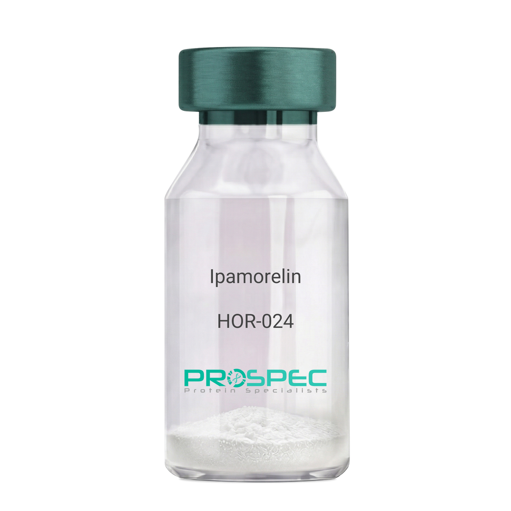
Name :
IpamorelinDescription:
Ipamorelin
Ipamorelin
Product # :
HOR-024Price :
Quantity :
Shipping Method :
Shipped at Room temp
More Info
- description
- formulation
- purity
- More Info
- Citations
Description
Ipamorelin Synthetic is a single, non-glycosylated polypeptide chain containing 4 amino acids, having a molecular mass of 711.85 Dalton and a Molecular formula of C38H49N9O5.
Formulation
The protein was lyophilized with no additives.
Purity
Greater than 97.0% as determined by analysis by RP-HPLC.
Citations
More Info
-
Introduction
Ipamorelin is a peptide selective agonist of the ghrelin/growth hormone secretagogue receptor and a growth hormone secretagogue. Ipamorelin is a pentapeptide that was derived from GHRP1. Ipamorelin significantly increases plasma growth hormone levels in both animals and humans. Like pralmorelin and GHRP-6, ipamorelin does not affect prolactin, FSH, LH or TSH levels. However, unlike GHRP2 and GHRP6, but as growth hormone-releasing hormone (GHRH), ipamorelin does not stimulate the secretion of adrenocorticotropic hormone (ACTH) or cortisol, and is highly selective for inducing the secretion only of GH.
-
Synonyms
Ipamorelin
-
Physical Appearance
Sterile Filtered White lyophilized (freeze-dried) powder.
-
Stability
Lyophilized Ipamorelin although stable at room temperature for 3 weeks, should be stored desiccated below -18°C. Upon reconstitution Ipamorelin should be stored at 4°C between 2-7 days and for future use below -18°C. For long term storage it is recommended to add a carrier protein (0.1% HSA or BSA). Please prevent freeze-thaw cycles.
-
Solubility
It is recommended to reconstitute the lyophilized Ipamorelin in sterile 18MΩ-cm H2O not less than 100 µg/ml, which can then be further diluted to other aqueous solutions.
-
Amino Acid Sequence
Aib-His-D-2-Nal-D-Phe-Lys-NH2.
ProSpec's products are furnished for LABORATORY RESEARCH USE ONLY. The product may not be used as drugs, agricultural or pesticidal products, food additives or household chemicals.