Search results
1000 results found for “gliadin”
Name
Description
Product #
Price
Quantity
Shipping Method
- View Data Sheet
Name :
CD57 Biotin AntibodyDescription:
CD57 Biotinylated, Mouse Anti-Human
NK1, CD57, HNK1, LEU7, NK-1, GLCATP, GLCUATP, B3GAT1.
Product # :
ANT-288Price :
Quantity :
Shipping Method :
Shipped at Room temp
More Info
- formulation
- More Info
Formulation
1mg/ml in PBS (after reconstitution).
More Info
-
Introduction
CD57 also called B3GAT1is part of the glucuronyltransferase gene family. CD57 exhibits strict acceptor specificity, recognizing nonreducing terminal sugars and their anomeric linkages. CD57 takes part in the glucuronyl transfer reaction during the biosynthesis of the carbohydrate epitope HNK-1.
-
Synonyms
NK1, CD57, HNK1, LEU7, NK-1, GLCATP, GLCUATP, B3GAT1.
-
Solubility
Reconstitute with of H20. Mix gently, wash the sides of the vial and wait 30-60 seconds before use.
-
Immunogen
Human CD57 Positive Cells.
-
Ig Subclass
Mouse IgM.
-
Clone
hCD57.
-
Applications
Staining antibody. For staining, use 10µl/1,000,000 cells.
-
Available Conjugates
This antibody is also available unconjugated and conjugated to FITC. For staining with biotin or FITC-conjugated antibody use 5-10µl/1,000,000 cells.
-
Type
Mouse Anti Human Monoclonal.
-
Storage Procedures
Lyophilized: store at 4°C. After reconstitution, if not intended for use within a month, aliquot and store at -20°C.
-
Purification Method
Ion-Exchange Column.
ProSpec's products are furnished for LABORATORY RESEARCH USE ONLY. The product may not be used as drugs, agricultural or pesticidal products, food additives or household chemicals.
- View Data Sheet
Name :
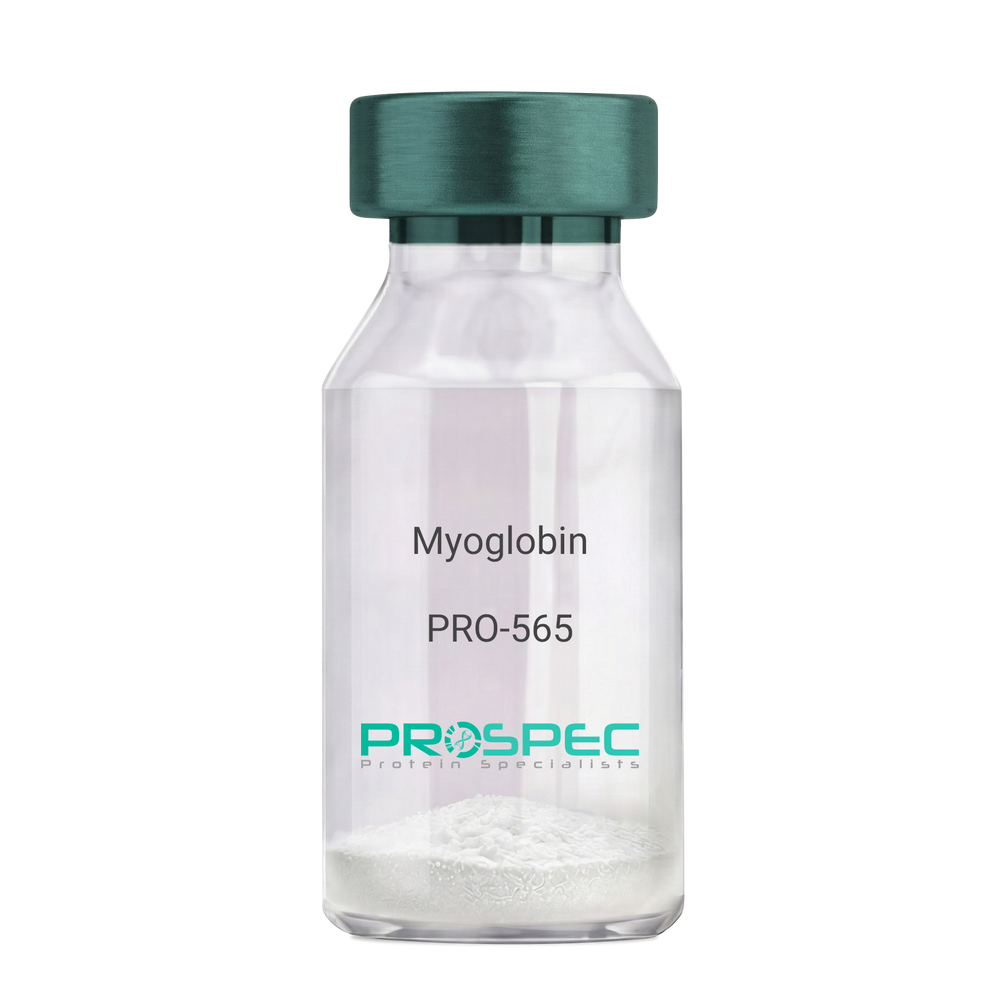
MyoglobinDescription:
Myoglobin Human
Myoglobin, MB, PVALB, MGC13548.
Product # :
PRO-565Price :
Quantity :
Shipping Method :
Shipped with Ice Packs
More Info
- description
- source
- formulation
- purity
- More Info
Description
Human Myoglobin produced in Human Cardiac Tissues having a molecular mass of 17.5kDa. Myoglobin is released from recently injured myocardial cells within a few hours of Infarction. Peak levels are reached more quickly than CK-MB or Troponin complex.
Source
Human Cardiac Tissues.
Formulation
The protein solution is in 0.05M phosphate buffer pH 7.5 containing 0.15M NaCl and 0.09% NaN3. Filtered through a 0.2µM membrane.
Purity
Greater than 96.0%.
More Info
-
Introduction
Myoglobin is a member of the globin superfamily and can be found in skeletal and cardiac muscles. It is a haemoprotein that contributs to intracellular oxygen storage and transcellular facilitated diffusion of oxygen. Myoglobin has a single-chain globular structure of 153 amino acids, containing a heme prosthetic group (iron-containing porphyrin) in the core around which the remaining apoprotein folds. Myoglobin has 8 alpha helices and a hydrophobic core. Myoglobin’s molecular weight is 16.7 kDa, and it is the primary oxygen-carrying pigment of muscle tissues. The binding of oxygen in myoglobin is different from the cooperative oxygen binding in hemoglobin, since positive collaboration is a property of multimeric/oligomeric proteins only. Instead, the binding of oxygen by myoglobin is uninfluenced by the oxygen pressure in the surrounding tissue. Myoglobin is frequently referred to as having an "instant binding tenacity" to oxygen given its hyperbolic oxygen dissociation curve. Different organisms are able to hold their breaths longer due to high concentrations of myoglobin in their muscle cells. Myoglobin is responsible for the pigments that make meat red. The color of the meat is partly determined by the charge of the iron atom in myoglobin and the oxygen attached to it. Myoglobin is found in Type I muscle, Type II A and Type II B, but it is mostly deemed that myoglobin is not found in smooth muscle. Myoglobin is discharged from damaged muscle tissue (rhabdomyolysis), which contains very high concentrations of myoglobin. Even though the released myoglobin is filtered by the kidneys, it is toxic to the renal tubular epithelium and thus may cause acute renal failure.
-
Synonyms
Myoglobin, MB, PVALB, MGC13548.
-
Physical Appearance
Sterile Filtered red solution.
-
Stability
Human Myoglobin should be stored at 2-8°C.
-
Human Virus Test
Starting material donor tested and certified negative for HIV I & II antibodies, Hepatitis B surface antigen, and Hepatitis C antibodies.
ProSpec's products are furnished for LABORATORY RESEARCH USE ONLY. The product may not be used as drugs, agricultural or pesticidal products, food additives or household chemicals.
- View Data Sheet
Name :
ADAM10 HumanDescription:
A Disintegrin and Metalloproteinase Domain 10 Human Recombinant
Kuz, AD10, MADM, CD156c, HsT18717, ADAM metallopeptidase domain 10, A disintegrin and metalloproteinase domain 10, Mammalian disintegrin-metalloprotease, Kuzbanian protein homolog, CDw156, ADAM 10, ADAM10.
Product # :
PRO-476Price :
Quantity :
Shipping Method :
Shipped with Ice Packs
More Info
- description
- source
- formulation
- purity
- More Info
Description
ADAM10 extracellular domain minus the signal peptide and pro-sequence Human Recombinant fused to N-terminal His-Tag produced in E.Coli is a single, non-glycosylated polypeptide chain containing 459 amino acids (214-672) and having a molecular mass of 55.089kDa.
Source
Escherichia Coli.
Formulation
The ADAM10 solution contains 20mM Tris (pH 8), 1mM EDTA and 50% Glycerol.
Purity
Greater than 95.0% as determined by SDS-PAGE.
More Info
-
Introduction
ADAM10 is part of the ADAM family which are cell surface proteins with a distinctive structure possessing both potential adhesion and protease domains. ADAM10 cleaves many proteins including TNF-alpha and E-cadherin. ADAM10 cleaves the membrane-bound precursor of tnf-alpha at 76- ala-|-val-77 to its mature soluble form. ADAM10 is in charge for the proteolytic release of several other cell-surface proteins, including ephrin-a2 and for constitutive and regulated alpha-secretase cleavage of amyloid precursor protein. ADAM10 is involved in the normal cleavage of the cellular prion protein. ADAM10 is involved in the cleavage of the adhesion molecule l1 at the cell surface and in the release of membrane vesicles, suggesting a vesicle-based protease activity. ADAM10 controls the proteolytic processing of notch and mediates lateral inhibition during neurogenesis.
-
Synonyms
Kuz, AD10, MADM, CD156c, HsT18717, ADAM metallopeptidase domain 10, A disintegrin and metalloproteinase domain 10, Mammalian disintegrin-metalloprotease, Kuzbanian protein homolog, CDw156, ADAM 10, ADAM10.
-
Physical Appearance
Sterile Filtered colorless solution.
-
Stability
Store at 4°C if entire vial will be used within 2-4 weeks.Store, frozen at -20°C for longer periods of time. For long term storage it is recommended to add a carrier protein (0.1% HSA or BSA).Avoid multiple freeze-thaw cycles.
ProSpec's products are furnished for LABORATORY RESEARCH USE ONLY. The product may not be used as drugs, agricultural or pesticidal products, food additives or household chemicals.
- View Data Sheet
Name :
HERPUD1 HumanDescription:
HERPUD1 Human Recombinant
Homocysteine-responsive endoplasmic reticulum-resident ubiquitin-like domain member 1 protein, HERPUD1, Methyl methanesulfonate (MMF)-inducible fragment protein 1, HERP, KIAA0025, MIF1, Mif1, SUP.
Product # :
PRO-2105Price :
Quantity :
Shipping Method :
Shipped with Ice Packs
More Info
- description
- source
- formulation
- purity
- More Info
Description
HERPUD1 Human Recombinant produced in E. coli is a single polypeptide chain containing 286 amino acids (1-263) and having a molecular mass of 31.6kDa.HERPUD1 is fused to a 23 amino acid His-tag at N-terminus & purified by proprietary chromatographic techniques.
Source
Escherichia Coli.
Formulation
The HERPUD1 solution (0.5mg/1ml) contains Phosphate buffer saline (pH 7.4) and 10% glycerol.
Purity
Greater than 85.0% as determined by SDS-PAGE.
More Info
-
Introduction
HERPUD1 is a multi-pass membrane protein which is a part of the endoplasmic reticulum quality control system. HERPUD1 owns 1 N-terminal ubiquitin-like domain and is expressed highly in the brain. HERPUD1 is also known as ER-associated degradation (ERAD) which takes part in ubiquitin-dependent degradation of misfolded endoplasmic reticulum proteins.
-
Synonyms
Homocysteine-responsive endoplasmic reticulum-resident ubiquitin-like domain member 1 protein, HERPUD1, Methyl methanesulfonate (MMF)-inducible fragment protein 1, HERP, KIAA0025, MIF1, Mif1, SUP.
-
Physical Appearance
Sterile Filtered clear solution.
-
Stability
Store at 4°C if entire vial will be used within 2-4 weeks. Store, frozen at -20°C for longer periods of time. For long term storage it is recommended to add a carrier protein (0.1% HSA or BSA).Avoid multiple freeze-thaw cycles.
-
Amino Acid Sequence
MGSSHHHHHH SSGLVPRGSH MGSMESETEP EPVTLLVKSP NQRHRDLELS GDRGWSVGHL KAHLSRVYPE RPRPEDQRLI YSGKLLLDHQ CLRDLLPKQE KRHVLHLVCN VKSPSKMPEI NAKVAESTEE PAGSNRGQYP EDSSSDGLRQ REVLRNLSSP GWENISRPEA AQQAFQGLGP GFSGYTPYGW LQLSWFQQIY ARQYYMQYLA ATAASGAFVP PPSAQEIPVV SAPAPAPIHN QFPAENQPAN QNAAPQVVVN PGANQNLRMN AQGGPIVEED DEINRD.
ProSpec's products are furnished for LABORATORY RESEARCH USE ONLY. The product may not be used as drugs, agricultural or pesticidal products, food additives or household chemicals.
- View Data Sheet
Name :
AIF1 HumanDescription:
Allograft Inflammatory Factor 1 Human Recombinant
AIF-1, Allograft inflammatory factor 1, Em:AF129756.17, G1, IBA1, Ionized calcium-binding adapter molecule 1, IRT-1, Protein G1, AIF1.
Product # :
CYT-697Price :
Quantity :
Shipping Method :
Shipped with Ice Packs
More Info
- description
- source
- formulation
- purity
- More Info
- sds-page
Description
AIF1 produced in E.Coli is a single, non-glycosylated polypeptide chain containing 167 amino acids (1-147a.a.) and having a molecular mass of 18.9kDa. AIF1 is fused to a 20 amino acid His-tag at N-terminus & purified by proprietary chromatographic techniques.
Source
E. Coli.
Formulation
The AIF1 protein solution (0.5mg/1ml) is formulated in 20mM Tris-HCl buffer (pH8.0)
2mM DTT, 200mM NaCl and 20% glycerol.Purity
Greater than 95% as determined by SDS PAGE.
sds-page
More Info
-
Introduction
Human AIF1 protein shares 98% homology/identity with that of rat. AIF1 is expressed in macrophages and neutrophils. The expression of AIF1 transcripts is upregulated by IFN-g in rat macrophages. AIF1 is expressed selectively in human macrophage-like cell lines, and in a subset of CD68(+) macrophages in the interstitial and perivascular spaces of human heart allografts. In quiescent cultured human vascular smooth muscle cells synthesis of AIF1 is induced by IFN-g, IL1b, and conditioned medium of T-cells. Overexpression of AIF1 in human VSMCs results in enhanced growth of these cells. AIF1 is expressed during apoptosis rat mammary gland and ventral prostate tissues. Allograft Inflammatory Factor 1 is expressed by several tumor-associated activated macrophages and microglial cells in rat and human gliomas. There is an evident relationship of AIF1-expressing activated macrophages and microglial cells with tumor malignancy in humans.
-
Synonyms
AIF-1, Allograft inflammatory factor 1, Em:AF129756.17, G1, IBA1, Ionized calcium-binding adapter molecule 1, IRT-1, Protein G1, AIF1.
-
Stability
Store AIF1 at 4°C if entire vial will be used within 2-4 weeks. Store, frozen at -20°C for longer periods of time.For long term storage it is recommended to add a carrier protein (0.1% HSA or BSA).Avoid multiple freeze-thaw cycles.
-
Amino Acid Sequence
MGSSHHHHHH SSGLVPRGSH MSQTRDLQGG KAFGLLKAQQ EERLDEINKQ FLDDPKYSSD EDLPSKLEGF KEKYMEFDLN GNGDIDIMSL KRMLEKLGVP KTHLELKKLI GEVSSGSGET FSYPDFLRMM LGKRSAILKM ILMYEEKARE KEKPTGPPAK KAISELP.
-
Background
Allograft Inflammatory Factor 1 Human Recombinant: Uncovering its Role in Immune Responses and Therapeutic Prospects
1. Abstract
This paper explores the Allograft Inflammatory Factor 1 Human Recombinant (AIF-1), a cytoplasmic, IFN-gamma-inducible calcium-binding protein involved in inflammation and immunity. We review the structure, biological roles, and involvement of AIF-1 in disease pathology. The therapeutic potential of AIF-1 in immune-related disorders is also explored.
2. Introduction
AIF-1, also known as IBA1, plays an important role in immune responses. It is associated with various immune cells, particularly macrophages, and has been implicated in numerous inflammatory and immune-related diseases. Understanding the function of AIF-1 could aid the development of novel therapeutic strategies.
3. Structure and Signaling of AIF-1
AIF-1 is a small 17 kDa protein with an EF-hand calcium-binding motif. Although the precise mechanism by which AIF-1 exerts its functions is not entirely clear, it is known to regulate the activation, migration, and proliferation of macrophages, key cells involved in immune responses.
4. Biological Functions of AIF-1
AIF-1 has been shown to play key roles in macrophage activation and function, which are central to inflammation and immunity. It is also implicated in cell survival, proliferation, and differentiation.
5. AIF-1 in Disease Pathology
AIF-1 has been associated with a range of inflammatory and immune-related diseases, including rheumatoid arthritis, atherosclerosis, and multiple sclerosis. It is also implicated in several cancers, further underscoring its broad physiological and pathological relevance.
6. Therapeutic Potential of AIF-1
Given its pivotal role in immune responses, AIF-1 presents an intriguing target for therapeutic interventions in immune-related diseases. Modulating the activity of AIF-1 could potentially alleviate pathological inflammation and autoimmunity.
7. Conclusion and Future Perspectives
Our knowledge of AIF-1 and its functions has significantly improved in recent years, but much remains to be discovered. Further research into AIF-1's exact molecular mechanisms and roles in disease will undoubtedly contribute to the development of novel therapeutic strategies.
What is the molecular weight/Mw of AIF1 Protein?
AIF1 Protein has a total Mw of 18.9kDa.
What is the source or expression system of AIF1 Protein?
Escherichia Coli.
What is the Purity of AIF1 Protein?
AIF1 Protein is >95% pure as determined by SDS-PAGE.
What is the Biological Activity of AIF1 Protein?
The biological functionality of AIF1 Protein will be determined in the future.
What is the amino acid sequence of AIF1 Protein?
MGSSHHHHHH SSGLVPRGSH MSQTRDLQGG KAFGLLKAQQ EERLDEINKQ FLDDPKYSSD EDLPSKLEGF KEKYMEFDLN GNGDIDIMSL KRMLEKLGVP KTHLELKKLI GEVSSGSGET FSYPDFLRMM LGKRSAILKM ILMYEEKARE KEKPTGPPAK KAISELP.
What applications can AIF1 Protein be used in?
AIF1 Protein can probably be used in western blot, ELISA and Lateral Flow.
What is the endotoxin level for AIF1 Protein?
The endotoxin level is minimal, AIF1 Protein was purified using conventional chromatography techniques.
ProSpec's products are furnished for LABORATORY RESEARCH USE ONLY. The product may not be used as drugs, agricultural or pesticidal products, food additives or household chemicals.
- View Data Sheet
Name :
SOST Human, HEKDescription:
Sclerostin Human Recombinant, HEK
Sclerostin, SOST, CDD, VBCH.
Product # :
PRO-2481Price :
Quantity :
Shipping Method :
Shipped at Room temp
More Info
- description
- source
- formulation
- purity
- More Info
Description
SOST Human Recombinant produced in HEK293 cells is a single, glycosylated polypeptide chain (a.a 24-213) containing 196 amino acids including a 6 a.a C-terminal His tag. The total molecular mass is 22.4kDa (calculated).
Source
HEK293 Cells.
Formulation
SOST filtered (0.4 µm) and lyophilized from 0.5mg/ml in PBS and 5 % (w/v) trehalose, pH 7.4.
Purity
Greater than 95.0% as determined by SDS-PAGE.
More Info
-
Introduction
Sclerostin (SOST) is a secreted glycoprotein with a C-terminal cysteine knot-like (CTCK) domain and sequence similarity to the DAN (differential screening-selected gene aberrative in neuroblastoma) family of bone morphogenetic protein (BMP) antagonists. Sclerostin functions as a negative regulator of bone growth, by inhibiting bone formation. SOST is widely expressed at low levels, with highest levels in the bone, cartilage, kidney, liver, bone marrow and primary osteoblasts differentiated for 21 days. SOST gene defects cause sclerosteosis and bone dysplasia.
-
Synonyms
Sclerostin, SOST, CDD, VBCH.
-
Physical Appearance
Filtered White lyophilized (freeze-dried) powder.
-
Stability
Store lyophilized protein at -20°C. Aliquot the product after reconstitution to avoid repeated freezing/thawing cycles. Reconstituted protein can be stored at 4°C for a limited period of time; it does not show any change after one week at 4°C.
-
Solubility
It is recommended to add deionized water to prepare a working stock solution of approximately 0.5mg/ml and let the lyophilized pellet dissolve completely. SOST is not sterile! Please filter the product by an appropriate sterile filter before using it in the cell culture.
-
Amino Acid Sequence
QGWQAFKNDA TEIIPELGEY PEPPPELENN KTMNRAENGG RPPHHPFETK DVSEYSCREL HFTRYVTDGP CRSAKPVTEL VCSGQCGPAR LLPNAIGRGK WWRPSGPDFR CIPDRYRAQR VQLLCPGGEA PRARKVRLVA SCKCKRLTRF HNQSELKDFG TEAARPQKGR KPRPRARSAK ANQAELENAY HHHHHH.
ProSpec's products are furnished for LABORATORY RESEARCH USE ONLY. The product may not be used as drugs, agricultural or pesticidal products, food additives or household chemicals.
- View Data Sheet
Name :
SOST MouseDescription:
Sclerostin Mouse Recombinant
SOST, Sclerostin, 5430411E23Rik.
Product # :
PRO-2670Price :
Quantity :
Shipping Method :
Shipped with Ice Packs
More Info
- description
- source
- formulation
- purity
- More Info
Description
SOST Mouse Recombinant produced in HEK293 cells is a single, glycosylated polypeptide chain (24-211 a.a) containing 194 amino acids and having a molecular mass of 21.9 kDa.SOST is fused to a 6 amino acid His-tag at C-terminus & purified by proprietary chromatographic techniques.
Source
HEK293 cells.
Formulation
SOST protein (0.25mg/ml) contains 10% glycerol and Phosphate-Buffered Saline (pH 7.4).
Purity
Greater than 90.0% as determined by SDS-PAGE.
More Info
-
Introduction
Sclerostin (SOST) is a secreted glycoprotein with a C-terminal cysteine knot-like (CTCK) domain and sequence similarity to the DAN (differential screening-selected gene aberrative in neuroblastoma) family of bone morphogenetic protein antagonists. Sclerostin functions as a negative regulator of bone growth, by inhibiting bone formation. SOST is expressed mainly in the bone, cartilage, kidney, liver, bone marrow and primary osteoblasts differentiated for 21 days. SOST gene defects cause sclerosteosis and bone dysplasia.
-
Synonyms
SOST, Sclerostin, 5430411E23Rik.
-
Physical Appearance
Sterile Filtered colorless solution.
-
Stability
Store at 4°C if entire vial will be used within 2-4 weeks. Store, frozen at -20°C for longer periods of time. For long term storage it is recommended to add a carrier protein (0.1% HSA or BSA).Avoid multiple freeze-thaw cycles.
-
Amino Acid Sequence
QGWQAFRNDA TEVIPGLGEY PEPPPENNQT MNRAENGGRP PHHPYDAKDV SEYSCRELHY TRFLTDGPCR SAKPVTELVC SGQCGPARLL PNAIGRVKWW RPNGPDFRCI PDRYRAQRVQ LLCPGGAAPR SRKVRLVASC KCKRLTRFHN QSELKDFGPE TARPQKGRKP RPGARGAKAN QAELENAYHH HHHH
ProSpec's products are furnished for LABORATORY RESEARCH USE ONLY. The product may not be used as drugs, agricultural or pesticidal products, food additives or household chemicals.
- View Data Sheet
Name :
TADA3 HumanDescription:
Transcriptional Adaptor 3 Human Recombinant
Transcriptional adapter 3, ADA3 homolog, hADA3, STAF54, Transcriptional adapter 3-like, ADA3-like protein, TADA3, ADA3, TADA3L, Transcriptional adapter 3 isoform a, NGG1.
Product # :
PRO-1689Price :
Quantity :
Shipping Method :
Shipped with Ice Packs
More Info
- description
- source
- formulation
- purity
- More Info
Description
TADA3 Human Recombinant produced in E.coli is a single, non-glycosylated polypeptide chain containing 455 amino acids (1-432) and having a molecular mass of 51.3 kDa.TADA3 is fused to a 23 amino acid His-tag at N-terminus & purified by proprietary chromatographic techniques.
Source
Escherichia Coli.
Formulation
The TADA3 solution (0.5mg/ml) contains 20mM Tris-HCl buffer (pH 8.0), 0.2M NaCl, 50% glycerol and 2mM DTT.
Purity
Greater than 80.0% as determined by SDS-PAGE.
More Info
-
Introduction
Transcriptional adapter 3 (TATD3) is a subunit of 2 histone acetyltransferase complexes, which functions as a component of the PCAF complex. TATD3 is a transcriptional activator adaptor that is also associates with the tumor suppressor protein p53 and is essential for full activity of p53 and p53-mediated apoptosis.
-
Synonyms
Transcriptional adapter 3, ADA3 homolog, hADA3, STAF54, Transcriptional adapter 3-like, ADA3-like protein, TADA3, ADA3, TADA3L, Transcriptional adapter 3 isoform a, NGG1.
-
Physical Appearance
Sterile Filtered colorless solution.
-
Stability
Store at 4°C if entire vial will be used within 2-4 weeks. Store, frozen at -20°C for longer periods of time. For long term storage it is recommended to add a carrier protein (0.1% HSA or BSA).Avoid multiple freeze-thaw cycles.
-
Amino Acid Sequence
MGSSHHHHHH SSGLVPRGSH MGSMSELKDC PLQFHDFKSV DHLKVCPRYT AVLARSEDDG IGIEELDTLQ LELETLLSSA SRRLRVLEAE TQILTDWQDK KGDRRFLKLG RDHELGAPPK HGKPKKQKLE GKAGHGPGPG PGRPKSKNLQ PKIQEYEFTD DPIDVPRIPK NDAPNRFWAS VEPYCADITS EEVRTLEELL KPPEDEAEHY KIPPLGKHYS QRWAQEDLLE EQKDGARAAA VADKKKGLMG PLTELDTKDV DALLKKSEAQ HEQPEDGCPF GALTQRLLQA LVEENIISPM EDSPIPDMSG KESGADGAST SPRNQNKPFS VPHTKSLESR IKEELIAQGL LESEDRPAED SEDEVLAELR KRQAELKALS AHNRTKKHDL LRLAKEEVSR QELRQRVRMA DNEVMDAFRK IMAARQKKRT PTKKEKDQAW KTLKERESIL KLLDG.
ProSpec's products are furnished for LABORATORY RESEARCH USE ONLY. The product may not be used as drugs, agricultural or pesticidal products, food additives or household chemicals.
- View Data Sheet
Name :
CALML5 HumanDescription:
Calmodulin Like 5 Human Recombinant
CLSP, Calmodulin-like skin protein, CALML5, Calmodulin-like protein 5.
Product # :
PRO-2475Price :
Quantity :
Shipping Method :
Shipped at Room temp
More Info
- description
- source
- formulation
- purity
- More Info
Description
CALML5 Human Recombinant produced in E.Coli is a single, non-glycosylated, polypeptide chain (a.a 2-146) containing 155 amino acids including a 10 a.a N-terminal His tag. The total molecular mass is 17.0kDa (calculated).
Source
Escherichia Coli.
Formulation
CALML5 filtered (0.4 µm) and lyophilized from 0.5mg/ml in 20 mM Tris buffer, 50 mM NaCl and 5% w/v trehalosa, pH 7.5.
Purity
Greater than 95.0% as determined by SDS-PAGE.
More Info
-
Introduction
Calmodulin Like 5, also known as CALML5 is a part of the calmodulin family of calcium binding proteins. CALML5 undergoes a conformational change as a result of binding calcium. CALML5 is taking part in terminal differentiation of keratinocytes and encodes a calcium binding protein expressed in the epidermis.
-
Synonyms
CLSP, Calmodulin-like skin protein, CALML5, Calmodulin-like protein 5.
-
Physical Appearance
Filtered White lyophilized (freeze-dried) powder.
-
Stability
Store lyophilized protein at -20°C. Aliquot the product after reconstitution to avoid repeated freezing/thawing cycles. Reconstituted protein can be stored at 4°C for a limited period of time; it does not show any change after two weeks at 4°C.
-
Solubility
It is recommended to add deionized water to prepare a working stock solution of approximately 0.5mg/ml and let the lyophilized pellet dissolve completely. CALML5 is not sterile! Please filter the product by an appropriate sterile filter before using it in the cell culture.
-
Amino Acid Sequence
MKHHHHHHAS AGELTPEEEA QYKKAFSAVD TDGNGTINAQ ELGAALKATG KNLSEAQLRK LISEVDSDGD GEISFQEFLT AAKKARAGLE DLQVAFRAFD QDGDGHITVD ELRRAMAGLG QPLPQEELDA MIREADVDQD GRVNYEEFAR MLAQE.
ProSpec's products are furnished for LABORATORY RESEARCH USE ONLY. The product may not be used as drugs, agricultural or pesticidal products, food additives or household chemicals.
- View Data Sheet
Name :
ITGB1 Human, Sf9Description:
Integrin Beta 1 Human Recombinant, Sf9
Integrin beta-1, Fibronectin receptor subunit beta, Glycoprotein IIa, GPIIA, VLA-4 subunit beta, CD29, ITGB1, FNRB, MDF2, MSK12, Integrin beta 1, CD29, VLAB.
Product # :
PRO-2410Price :
Quantity :
Shipping Method :
Shipped with Ice Packs
More Info
- description
- source
- formulation
- purity
- More Info
Description
ITGB1 Human Recombinant produced in Sf9 Baculovirus cells is a single, glycosylated polypeptide chain containing 716 amino acids (1-728) and having a molecular mass of 79.4kDa (Molecular size on SDS-PAGE will appear at approximately 70-100kDa). ITGB1 is fused to an 8 amino acid His-Tag at C-terminus and purified by proprietary chromatographic techniques.
Source
Sf9, Baculovirus cells.
Formulation
ITGB1 protein solution (1.0mg/ml) contains Phosphate Buffered Saline (pH 7.4) and 10% glycerol.
Purity
Greater than 95.0% as determined by SDS-PAGE.
More Info
-
Introduction
Integrins are heterodimeric proteins consist of alpha and beta subunits. There are more than 18 alpha and 8 beta subunits discovered in mammals. Integrin family members are membrane receptors that participates in cell adhesion and recognition in a variety of processes including embryogenesis, hemostasis, tissue repair, immune response and metastatic diffusion of tumor cells. ITGB1 encodes a beta subunit.
-
Synonyms
Integrin beta-1, Fibronectin receptor subunit beta, Glycoprotein IIa, GPIIA, VLA-4 subunit beta, CD29, ITGB1, FNRB, MDF2, MSK12, Integrin beta 1, CD29, VLAB.
-
Physical Appearance
Sterile Filtered colorless solution.
-
Stability
Store at 4°C if entire vial will be used within 2-4 weeks. Store, frozen at -20°C for longer periods of time. For long term storage it is recommended to add a carrier protein (0.1% HSA or BSA). Avoid multiple freeze-thaw cycles.
-
Amino Acid Sequence
QTDENRCLKA NAKSCGECIQ AGPNCGWCTN STFLQEGMPT SARCDDLEAL KKKGCPPDDI ENPRGSKDIK KNKNVTNRSK GTAEKLKPED ITQIQPQQLV LRLRSGEPQT FTLKFKRAED YPIDLYYLMD LSYSMKDDLE NVKSLGTDLM NEMRRITSDF RIGFGSFVEK TVMPYISTTP AKLRNPCTSE QNCTSPFSYK NVLSLTNKGE VFNELVGKQR ISGNLDSPEG GFDAIMQVAV CGSLIGWRNV TRLLVFSTDA GFHFAGDGKL GGIVLPNDGQ CHLENNMYTM SHYYDYPSIA HLVQKLSENN IQTIFAVTEE FQPVYKELKN LIPKSAVGTL SANSSNVIQL IIDAYNSLSS EVILENGKLS EGVTISYKSY CKNGVNGTGE NGRKCSNISI GDEVQFEISI TSNKCPKKDS DSFKIRPLGF TEEVEVILQY ICECECQSEG IPESPKCHEG NGTFECGACR CNEGRVGRHC ECSTDEVNSE DMDAYCRKEN SSEICSNNGE CVCGQCVCRK RDNTNEIYSG KFCECDNFNCDRSNGLICGG NGVCKCRVCE CNPNYTGSAC DCSLDTSTCE ASNGQICNGR GICECGVCKC TDPKFQGQTC EMCQTCLGVC AEHKECVQCR AFNKGEKKDT CTQECSYFNI TKVESRDKLP QPVQPDPVSH CKEKDVDDCW FYFTYSVNGN NEVMVHVVEN PECPTGPDLE HHHHHH
ProSpec's products are furnished for LABORATORY RESEARCH USE ONLY. The product may not be used as drugs, agricultural or pesticidal products, food additives or household chemicals.
- View Data Sheet
Name :
GABARAPL2 HumanDescription:
GABA(A) Receptor-Associated Protein Like 2 Human Recombinant
GABA(A) receptor-associated protein-like 2, GATE-16, GEF2, ATG8, ATG8C, Ganglioside expression factor 2, General protein transport factor p16, Golgi-associated ATPase enhancer of 16 kDa, MAP1 light chain 3-related protein, gamma-aminobutyric acid receptor-associated protein-like 2, MAP1 light chain 3 related protein, FLC3A.
Product # :
PRO-079Price :
Quantity :
Shipping Method :
Shipped with Ice Packs
More Info
- description
- source
- formulation
- purity
- More Info
Description
GABARAPL2 produced in E.Coli is a single, non-glycosylated polypeptide chain containing 137 amino acids (1-117a.a.) and having a molecular mass of 15.8kDa. GABARAPL2 is fused to a 20 amino acid His-tag at N-terminus & purified by proprietary chromatographic techniques.
Source
Escherichia Coli.
Formulation
The GABARAPL2 protein solution (0.5mg/1ml) is formulated in 20mM Tris-HCl buffer (pH8.0), 0.2M NaCl, 5mM DTT and 20% glycerol.
Purity
Greater than 90% as determined by SDS-PAGE.
More Info
-
Introduction
GABARAPL2 takes part in intra Golgi traffic and modulates intra Golgi transport by coupling between NSF activity and SNAREs activation. GABARAPL2 first stimulates the ATPase activity of NSF which in turn stimulates the association with GOSR1.
-
Synonyms
GABA(A) receptor-associated protein-like 2, GATE-16, GEF2, ATG8, ATG8C, Ganglioside expression factor 2, General protein transport factor p16, Golgi-associated ATPase enhancer of 16 kDa, MAP1 light chain 3-related protein, gamma-aminobutyric acid receptor-associated protein-like 2, MAP1 light chain 3 related protein, FLC3A.
-
Physical Appearance
Sterile Filtered clear solution.
-
Stability
Store at 4°C if entire vial will be used within 2-4 weeks. Store, frozen at -20°C for longer periods of time.Please avoid freeze thaw cycles.
-
Amino Acid Sequence
MGSSHHHHHH SSGLVPRGSH MKWMFKEDHS LEHRCVESAK IRAKYPDRVP VIVEKVSGSQ IVDIDKRKYL VPSDITVAQF MWIIRKRIQL PSEKAIFLFV DKTVPQSSLT MGQLYEKEKD EDGFLYVAYS GENTFGF
ProSpec's products are furnished for LABORATORY RESEARCH USE ONLY. The product may not be used as drugs, agricultural or pesticidal products, food additives or household chemicals.
- View Data Sheet
Name :
Procalcitonin RatDescription:
Procalcitonin Rat Recombinant
Calcitonin, Calca, Calc.
Product # :
HOR-019Price :
Quantity :
Shipping Method :
Shipped at Room temp
More Info
- description
- source
- formulation
- purity
- More Info
Description
Procalcitonin Rat Recombinant produced in E.Coli is a single, non-glycosylated, polypeptide chain (Val26-Asn136) containing 121 amino acids including a 10 aa His tag at N-terminus. The total calculated molecular mass is 13.6kDa.
Source
Escherichia Coli.
Formulation
Procalcitonin was filtered (0.4µm) and lyophilized in 20mM TRIS and 50mM NaCl, pH 8.0.
Purity
Greater than 95.0% as determined by SDS-PAGE.
More Info
-
Introduction
Procalcitonin is a peptide hormone mainly produced by the C cells of the thyroid and certain endocrine cells of the lung. Under normal expression conditions, procalcitonin is immediately cleaved into three specific fragments, an N terminal residue, calcitonin and katacalcin. Levels of unprocessed procalcitonin rise significantly after bacterial infection, trauma or shock.
-
Synonyms
Calcitonin, Calca, Calc.
-
Physical Appearance
Filtered White lyophilized (freeze-dried) powder.
-
Stability
Store lyophilized protein at -20°C. Aliquot the product after reconstitution to avoid repeated freezing/thawing cycles. Reconstituted protein can be stored at 4°C for a limited period of time; it does not show any change after two weeks at 4°C.
-
Solubility
It is recommended to add deionized water to prepare a working stock solution of approximately 0.5 mg/ml and let the lyophilized pellet dissolve completely. Procalcitonin is not sterile! Please filter the product by an appropriate sterile filter before using it in the cell culture.
-
Amino Acid Sequence
MKHHHHHHASVPLRSTLESS PGMATLSEEE ARLLAALVQN YMQMKVRELE QEEEQEAEGS SLDSPRSKRC GNLSTCMLGT YTQDLNKFHT FPQTSIGVGA PGKKRDMAKD LETNHHPYFG N.
ProSpec's products are furnished for LABORATORY RESEARCH USE ONLY. The product may not be used as drugs, agricultural or pesticidal products, food additives or household chemicals.
- View Data Sheet
Name :
Procalcitonin RhesusDescription:
Procalcitonin Rhesus Recombinant
Calcitonin.
Product # :
HOR-016Price :
Quantity :
Shipping Method :
Shipped at Room temp
More Info
- description
- source
- formulation
- purity
- More Info
Description
Procalcitonin Rhesus Recombinant produced in E.Coli is a single, non-glycosylated, polypeptide chain (Ala26-Asn140) containing 125 amino acids including a 10 aa His tag at N-terminus. The total calculated molecular mass is 14kDa.
Source
Escherichia Coli.
Formulation
Procalcitonin was filtered (0.4µm) and lyophilized from 0.5mg/ml solution in 20mM Tris buffer and 50mM NaCl, pH 7.5.
Purity
Greater than 95.0% as determined by SDS-PAGE.
More Info
-
Introduction
Procalcitonin is a peptide hormone mainly produced by the C cells of the thyroid and certain endocrine cells of the lung. Under normal expression conditions, procalcitonin is immediately cleaved into three specific fragments, an N terminal residue, calcitonin and katacalcin. Levels of unprocessed procalcitonin rise significantly after bacterial infection, trauma or shock.
-
Synonyms
Calcitonin.
-
Physical Appearance
Filtered White lyophilized (freeze-dried) powder.
-
Stability
Store lyophilized protein at -20°C. Aliquot the product after reconstitution to avoid repeated freezing/thawing cycles. Reconstituted protein can be stored at 4°C for a limited period of time; it does not show any change after two weeks at 4°C.
-
Solubility
It is recommended to add deionized water to prepare a working stock solution of approximately 0.5 mg/ml and let the lyophilized pellet dissolve completely. Procalcitonin is not sterile! Please filter the product by an appropriate sterile filter before using it in the cell culture.
-
Amino Acid Sequence
MKHHHHHHASAPFRSALESS PDPATLSEEE ARLLLAALVQ DYVQMKASEL EQEQETEGSS LDSPRSKRCG NLSTCMLGTY TQDFNKFHTF PQTAIGVGAP GKKRDMSSDL ERNRRRYVSM PQDAN.
ProSpec's products are furnished for LABORATORY RESEARCH USE ONLY. The product may not be used as drugs, agricultural or pesticidal products, food additives or household chemicals.
- View Data Sheet
Name :
GNB1 HumanDescription:
Guanine Nucleotide Binding Protein Beta Polypeptide 1 Human Recombinant
Guanine Nucleotide Binding Protein (G Protein) Beta Polypeptide 1, Transducin Beta Chain 1, GNB1.
Product # :
PRO-1955Price :
Quantity :
Shipping Method :
Shipped with Ice Packs
More Info
- description
- source
- formulation
- purity
- More Info
Description
GNB1 Human Recombinant produced in E.coli is a single, non-glycosylated polypeptide chain containing 363 amino acids (1-340) and having a molecular mass of 39.8 kDa.GNB1 is fused to a 23 amino acid His-tag at N-terminus & purified by proprietary chromatographic techniques.
Source
Escherichia Coli.
Formulation
The GNB1 solution (0.25mg/ml) contains 20mM Tris-HCl buffer (pH 8.0), 0.4M UREA and 10% glycerol.
Purity
Greater than 85.0% as determined by SDS-PAGE.
More Info
-
Introduction
Heterotrimeric guanine nucleotide-binding proteins (G proteins) which integrate signals between receptors and effector proteins, are comprised of an alpha, a beta and a gamma subunit. The alpha, beta and gamma subunits are encoded by families of related genes. GNB1 encodes a beta subunit. Beta subunits are vital regulators of alpha subunits, as well as of certain signal transduction receptors and effectors. G proteins are involved as a modulator or transducer in numerous transmembrane signaling systems. The beta and gamma chains are necessary for the GTPase activity, for replacement of GDP by GTP, and for G protein-effector interaction.
-
Synonyms
Guanine Nucleotide Binding Protein (G Protein) Beta Polypeptide 1, Transducin Beta Chain 1, GNB1.
-
Physical Appearance
Sterile Filtered clear solution.
-
Stability
Store at 4°C if entire vial will be used within 2-4 weeks. Store, frozen at -20°C for longer periods of time. For long term storage it is recommended to add a carrier protein (0.1% HSA or BSA).Avoid multiple freeze-thaw cycles.
-
Amino Acid Sequence
MGSSHHHHHH SSGLVPRGSH MGSMSELDQL RQEAEQLKNQ IRDARKACAD ATLSQITNNI DPVGRIQMRT RRTLRGHLAK IYAMHWGTDS RLLVSASQDG KLIIWDSYTT NKVHAIPLRS SWVMTCAYAP SGNYVACGGL DNICSIYNLK TREGNVRVSR ELAGHTGYLS CCRFLDDNQI VTSSGDTTCA LWDIETGQQT TTFTGHTGDV MSLSLAPDTR LFVSGACDAS AKLWDVREGM CRQTFTGHES DINAICFFPN GNAFATGSDD ATCRLFDLRA DQELMTYSHD NIICGITSVS FSKSGRLLLA GYDDFNCNVW DALKADRAGV LAGHDNRVSC LGVTDDGMAV ATGSWDSFLK IWN.
ProSpec's products are furnished for LABORATORY RESEARCH USE ONLY. The product may not be used as drugs, agricultural or pesticidal products, food additives or household chemicals.
- View Data Sheet
Name :
CXCL3 Human, HisDescription:
GRO-Gamma Human Recombinant (CXCL3), His Tag
Macrophage inflammatory protein 2-beta, MIP2-beta, CXCL3, Growth-regulated protein gamma, GRO-gamma, GRO-gamma(1-73), GRO3, GROg, MIP2B, SCYB3, MIP-2b, CINC-2b, MGSA gamma.
Product # :
CHM-244Price :
Quantity :
Shipping Method :
Shipped with Ice Packs
More Info
- description
- source
- formulation
- purity
- More Info
Description
GRO-Gamma Human Recombinant produced in E.Coli is a single,non-glycosylated, polypeptide chain containing 94 amino acids (35-107) and having a molecular mass of 7902 Dalton. The GRO-g is fused to 20 amino acid His-Tag at N-Terminus and purified by proprietary chromatographic techniques.
Source
Escherichia Coli.
Formulation
The CXCL3 protein is formulated in 20mM Tris-HCl buffer pH-8, 1mM DTT and 20% glycerol.
Purity
Greater than 95.0% as determined by SDS-PAGE.
More Info
-
Introduction
Chemokine (C-X-C motif) ligand 3 (CXCL3) is a small cytokine belonging to the CXC chemokine family that is also known as GRO3 oncogene (GRO3), GRO protein gamma (GROg) and macrophage inflammatory protein-2-beta (MIP2b). CXCL3 controls migration and adhesion of monocytes and mediates it effects on its target cell by interacting with a cell surface chemokine receptor called CXCR2. The gene for CXCL3 is located on chromosome 4 in a cluster of other CXC chemokines.
-
Synonyms
Macrophage inflammatory protein 2-beta, MIP2-beta, CXCL3, Growth-regulated protein gamma, GRO-gamma, GRO-gamma(1-73), GRO3, GROg, MIP2B, SCYB3, MIP-2b, CINC-2b, MGSA gamma.
-
Physical Appearance
Sterile Filtered colorless solution.
-
Stability
Liquid CXCL3 although stable at 10°C for 1 week, should be stored below -18°C. For long term storage it is recommended to add a carrier protein (0.1% HSA or BSA).Please prevent freeze-thaw cycles.
-
Amino Acid Sequence
MGSSHHHHHH SSGLVPRGSH MASVVTELRC QCLQTLQGIH LKNIQSVNVR SPGPHCAQTE VIATLKNGKK ACLNPASPMV QKIIEKILNKGSTN.
-
Background
What is the molecular weight/Mw of CXCL3 HUMAN, HIS Protein?
CXCL3 HUMAN, HIS Protein has a total Mw of 7.9kDa.
What is the source or expression system of CXCL3 HUMAN, HIS Protein?
Escherichia Coli.
What is the Purity of CXCL3 HUMAN, HIS Protein?
CXCL3 HUMAN, HIS Protein is >95% pure as determined by SDS-PAGE.
What is the Biological Activity of CXCL3 HUMAN, HIS Protein?
The biological functionality of CXCL3 HUMAN, HIS Protein will be determined in the future.
What is the amino acid sequence of CXCL3 HUMAN, HIS Protein?
MGSSHHHHHH SSGLVPRGSH MASVVTELRC QCLQTLQGIH LKNIQSVNVR SPGPHCAQTE VIATLKNGKK ACLNPASPMV QKIIEKILNKGSTN.
What applications can CXCL3 HUMAN, HIS Protein be used in?
CXCL3 HUMAN, HIS Protein can probably be used in western blot, ELISA and Lateral Flow.
What is the endotoxin level for CXCL3 HUMAN, HIS Protein?
The endotoxin level is minimal, CXCL3 HUMAN, HIS Protein was purified using conventional chromatography techniques.
ProSpec's products are furnished for LABORATORY RESEARCH USE ONLY. The product may not be used as drugs, agricultural or pesticidal products, food additives or household chemicals.
- View Data Sheet
Name :
PTGDS HumanDescription:
Prostaglandin D2 Synthase Human Recombinant
Prostaglandin-H2 D-isomerase, Beta-trace protein, Cerebrin-28, Glutathione-independent PGD synthase, Lipocalin-type prostaglandin-D synthase, Prostaglandin-D2 synthase, PGD2 synthase, PGDS, PGDS2, PTGDS, PDS, PGD2, LPGDS, L-PGDS.
Product # :
ENZ-109Price :
Quantity :
Shipping Method :
Shipped with Ice Packs
More Info
- description
- source
- formulation
- purity
- More Info
Description
PTGDS produced in E.Coli is a single, non-glycosylated polypeptide chain containing 189 amino acids (23-190 a.a.) and having a molecular mass of 20.9kDa.PTGDS is fused to a 21 amino acid His-tag at N-terminus & purified by proprietary chromatographic techniques.
Source
Escherichia Coli.
Formulation
PTGDS solution (1mg/ml) containing 20mM Tris-HCl buffer (pH 8.0), 1mM DTT, 30% glycerol, 1mM EDTA and 0.1M NaCl.
Purity
Greater than 90.0% as determined by SDS-PAGE.
More Info
-
Introduction
Prostaglandin-H2 D-isomerase (PTGDS) is a glutathione-independent prostaglandin D synthase which catalyzes the conversion of prostaglandin H2 (PGH2) to postaglandin D2 (PGD2). PTGDS is may have vital roles in both maturation and maintenance of the central nervous system and male reproductive system. PTGDS is the most abundant protein in the cerebral spinal fluid and recent evidence suggests that PTGDS acts as a beta-amyloid chaperone and may play a role in the deposition of Ab plaques in Alzheimer’s disease.
-
Synonyms
Prostaglandin-H2 D-isomerase, Beta-trace protein, Cerebrin-28, Glutathione-independent PGD synthase, Lipocalin-type prostaglandin-D synthase, Prostaglandin-D2 synthase, PGD2 synthase, PGDS, PGDS2, PTGDS, PDS, PGD2, LPGDS, L-PGDS.
-
Physical Appearance
Sterile filtered colorless solution.
-
Stability
Store at 4°C if entire vial will be used within 2-4 weeks. Store, frozen at -20°C for longer periods of time. For long term storage it is recommended to add a carrier protein (0.1% HSA or BSA).Avoid multiple freeze-thaw cycles.
-
Amino Acid Sequence
MGSSHHHHHH SSGLVPRGSH MAPEAQVSVQ PNFQQDKFLG RWFSAGLASN SSWLREKKAA LSMCKSVVAP ATDGGLNLTS TFLRKNQCET RTMLLQPAGS LGSYSYRSPH WGSTYSVSVV ETDYDQYALL YSQGSKGPGE DFRMATLYSR TQTPRAELKE KFTAFCKAQG FTEDTIVFLP QTDKCMTEQ.
ProSpec's products are furnished for LABORATORY RESEARCH USE ONLY. The product may not be used as drugs, agricultural or pesticidal products, food additives or household chemicals.
- View Data Sheet
Name :
MAPT Human 383a.a.Description:
Microtubule-Associated Protein Tau 383 a.a. Human Recombinant
Microtubule-associated protein tau, Neurofibrillary tangle protein, Paired helical filament-tau, PHF-tau, MAPT, MAPTL, MTBT1, TAU, MSTD, PPND, DDPAC, MTBT2, FTDP-17, FLJ31424, MGC138549.
Product # :
PRO-012Price :
Quantity :
Shipping Method :
Shipped with Ice Packs
More Info
- description
- source
- formulation
- purity
- More Info
Description
MAPT Human Recombinant (Isoform 3) fused with a 20 amino acid His tag at N-terminus produced in E.Coli is a single, non-glycosylated, polypeptide chain containing 403 amino acids (1-383 a.a.) and having a molecular mass of 42.1kDa (Molecular size on SDS-PAGE will appear higher). The MAPT is purified by proprietary chromatographic techniques.
Source
Escherichia Coli.
Formulation
The MAPT solution (1 mg/ml) contains 20mM Tris-HCl buffer (pH8.0), 10% glycerol and 0.1M NaCl.
Purity
Greater than 90.0% as determined by SDS-PAGE.
More Info
-
Introduction
MAPT is a neuronal microtubule associated protein localized mostly on axons.
MAPT promotes tubulin polymerisation and stabilizes microtubules, however it also serves to connect certain signalling pathways to the cytoskeleton. MAPT, in its hyperphosphorylated form, is the main part of paired helical filaments (PHF) and neurofibrillary lesions in Alzheimer''s disease (AD) brain. -
Synonyms
Microtubule-associated protein tau, Neurofibrillary tangle protein, Paired helical filament-tau, PHF-tau, MAPT, MAPTL, MTBT1, TAU, MSTD, PPND, DDPAC, MTBT2, FTDP-17, FLJ31424, MGC138549.
-
Physical Appearance
Sterile Filtered colorless solution.
-
Stability
Store at 4°C if entire vial will be used within 2-4 weeks. Store, frozen at -20°C for longer periods of time. For long term storage it is recommended to add a carrier protein (0.1% HSA or BSA).Please prevent freeze-thaw cycles.
-
Amino Acid Sequence
MGSSHHHHHH SSGLVPRGSH MAEPRQEFEV MEDHAGTYGL GDRKDQGGYT MHQDQEGDTD AGLKAEEAGI GDTPSLEDEA AGHVTQARMV SKSKDGTGSD DKKAKGADGK TKIATPRGAA PPGQKGQANA TRIPAKTPPA PKTPPSSGEP PKSGDRSGYS SPGSPGTPGS RSRTPSLPTP PTREPKKVAV VRTPPKSPSS AKSRLQTAPV PMPDLKNVKS KIGSTENLKH QPGGGKVQII NKKLDLSNVQ SKCGSKDNIK HVPGGGSVQI VYKPVDLSKV TSKCGSLGNI HHKPGGGQVE VKSEKLDFKD RVQSKIGSLD NITHVPGGGN KKIETHKLTF RENAKAKTDH GAEIVYKSPV VSGDTSPRHL SNVSSTGSID MVDSPQLATL ADEVSASLAK QGL.
ProSpec's products are furnished for LABORATORY RESEARCH USE ONLY. The product may not be used as drugs, agricultural or pesticidal products, food additives or household chemicals.
- View Data Sheet
Name :
Resistin Mouse, FlagDescription:
Resistin Mouse Recombinant, Flag Tag
Cysteine-rich secreted protein FIZZ3, Adipose tissue-specific secretory factor, ADSF, C/EBP-epsilon-regulated myeloid-specific secreted cysteine-rich protein, Cysteine-rich secreted protein A12-alpha-like 2, RSTN, XCP1, RETN1, MGC126603, MGC126609.
Product # :
CYT-457Price :
Quantity :
Shipping Method :
Shipped at Room temp
More Info
- description
- source
- formulation
- purity
- More Info
Description
Resistin Mouse is manufactured with signal sequence of phage fd (21aa) and C-terminal fusion of flagTag (10aa). Resistin Mouse Recombinant Flag-Tagged Fusion Protein is 13.7 kDa protein containing 93 amino acid residues of the Resistin Mouse and 31 additional amino acid residues - signal sequence of phage fd, flagTag (underlined).
Source
Escherichia Coli.
Formulation
Filtered (0.4µm) and lyophilized from 0.5 mg/ml in 0.05M Acetate buffer pH-4.
Purity
Greater than 95% as determined by SDS-PAGE.
More Info
-
Introduction
Resistin, a product of the RSTN gene, is a peptide hormone belonging to the class of cysteine-rich secreted proteins which is termed the RELM family, and is also described as ADSF (Adipose Tissue- Specific Secretory Factor) and FIZZ3 (Found in Inflammatory Zone). Human resistin contains 108 amino acids as a prepeptide, and its hydrofobic signal peptide is cleaved before its secretion. Resistin circulates in human blood as a dimeric protein consisting of two 92 amino acid polypeptides, which are disulfide-linked via Cys26.
Resistin may be an important link between obesity resistance. Mouse resistin, specifically produced and secreted by adipocyte, acts on skeletal muscle myocytes, hepatocytes and adipocytes themselves so that it reduces their sensitivity. They have also suggested that resistin is present at elevated levels in blood of obese mice, and is down regulated by fasting and antidiabetic drugs. Way et al., on the other hand, have found that resistin expression is severly suppressed in obesity and is stimulated by several antidiabetic drugs.
Other studies have shown that mouse resistin increases during the differentiation of adipocytes, but it also seems to inhibit adipogenesis. In contrast, the human adipogenic differentiation is likely to be associated with a down regulation of resistin gene expression. -
Synonyms
Cysteine-rich secreted protein FIZZ3, Adipose tissue-specific secretory factor, ADSF, C/EBP-epsilon-regulated myeloid-specific secreted cysteine-rich protein, Cysteine-rich secreted protein A12-alpha-like 2, RSTN, XCP1, RETN1, MGC126603, MGC126609.
-
Physical Appearance
Filtered White lyophilized (freeze-dried) powder.
-
Stability
Store lyophilized protein at -20°C. Aliquot the product after reconstitution to avoid repeated freezing/thawing cycles. Reconstituted protein can be stored at 4°C for a limited period of time; it does not show any change after two weeks at 4°C.
-
Solubility
It is recommended to add 0.1M Acetate buffer pH4 to prepare a working stock solution of approximately 0.5 mg/mL and let the lyophilized pellet dissolve completely. For conversion into higher pH value, we recommend intensive dilution by relevant buffer to a concentration of 10μg/ml. In higher concentrations the solubility of this antigen is limited. Product is not sterile! Please filter the product by an appropriate sterile filter before using it in the cell culture.
-
Amino Acid Sequence
MKKLLFAIPL VVPFYSHSTM ASMPLCPIDE AIDKKIKQDF NSLFPNAIKN IGLNCWTVSS RGKLASCPEG TAVLSCSCGS ACGSWDIREE KVCHCQCARI DWTAARCCKL QVASLEDYKD DDDK.
ProSpec's products are furnished for LABORATORY RESEARCH USE ONLY. The product may not be used as drugs, agricultural or pesticidal products, food additives or household chemicals.
- View Data Sheet
Name :
IL-12 HumanDescription:
Interleukin-12 Human Recombinant
NKSF, CTL maturation factor (TCMF), Cytotoxic lymphocyte maturation factor (CLMF), TSF, Edodekin-alpha, IL-12.
Product # :
CYT-101Price :
Quantity :
Shipping Method :
Shipped at Room temp
More Info
- description
- source
- formulation
- purity
- biological activity
- More Info
Description
Interleukin-12 Human Recombinant produced in HEK cells is a glycosylated heterodimer, having a total molecular weight of 57kDa.The IL12 is purified by proprietary chromatographic techniques.
Source
HEK.
Formulation
IL12 was lyophilized from a 0.2µm filtered solution containing 1xPBS.
Purity
Greater than 95% as obsereved by SDS-PAGE.
Biological Activity
The specific activity was determined by the dose-dependent release of IFN-gamma from the human NK92 cell line in presence of 20ng/mL rIL-2.
The EC50 is 0.5ng/ml.More Info
-
Introduction
IL-12 is a heterodimeric cytokine that stimulates the production of IFNgamma from T-cells and natural killer cells, and also induces differentiation of Th1 helper cells. IL-12 is an initiator of cell-mediated immunity.
-
Synonyms
NKSF, CTL maturation factor (TCMF), Cytotoxic lymphocyte maturation factor (CLMF), TSF, Edodekin-alpha, IL-12.
-
Physical Appearance
Sterile Filtered White lyophilized (freeze-dried) powder.
-
Stability
Lyophilized IL-12 although stable at room temperature for 3 weeks, should be stored desiccated below -18°C. Upon reconstitution IL12 should be stored at 4°C between 2-7 days and for future use below -18°C.For long term storage it is recommended to add a carrier protein (0.1% HSA or BSA).Please prevent freeze-thaw cycles.
-
Solubility
It is recommended to reconstitute the lyophilized IL12 in sterile PBS containing 0.1% endotoxin-free recombinant HSA.
-
Amino Acid Sequence
IL-12 is a heterodimer of IL-12A and IL-12B linked through a disulfide-bond between cysteines in red in sequences below.
>IL-12 A
RNLPVATPDPGMFPCLHHSQNLLRAVSNMLQKARQTLEFYPCTSEEIDHEDITKDKTSTVEACLP
LELTKNESCLNSRETSFITNGSCLASRKTSFMMALCLSSIYEDLKMYQVEFKTMNAKLLMDPKRQ
IFLDQNMLAVIDELMQALNFNSETVPQKSSLEEPDFYKTKIKLCILLHAFRIRAVTIDRVMSYLNAS.
>IL-12B
IWELKKDVYVVELDWYPDAPGEMVVLTCDTPEEDGITWTLDQSSEVLGSGKTLTIQVKEFGDAG
QYTCHKGGEVLSHSLLLLHKKEDGIWSTDILKDQKEPKNKTFLRCEAKNYSGRFTCWWLTTISTD
LTFSVKSSRGSSDPQGVTCGAATLSAERVRGDNKEYEYSVECQEDSACPAAEESLPIEVMVDAV
HKLKYENYTSSFFIRDIIKPDPPKNLQLKPLKNSRQVEVSWEYPDTWSTPHSYFSLTFCVQVQGK
SKREKKDRVFTDKTSATVICRKNASISVRAQDRYYSSSWSEWASVPCS.
ProSpec's products are furnished for LABORATORY RESEARCH USE ONLY. The product may not be used as drugs, agricultural or pesticidal products, food additives or household chemicals.
- View Data Sheet
Name :
SGTA AntibodyDescription:
Small Glutamine-Rich Tetratricopeptide Repeat-Containing Protein Alpha, Mouse Anti Human
alphaSGT, hSGT, SGT, Vpu-binding protein, UBP, Small glutamine-rich tetratricopeptide repeat-containing protein alpha, Alpha-SGT, SGT1.
Product # :
ANT-631Price :
Quantity :
Shipping Method :
Shipped with Ice Packs
More Info
- formulation
- More Info
Formulation
1mg/ml containing PBS, pH-7.4, 10% Glycerol and 0.02% Sodium Azide.
More Info
-
Introduction
SGTA is a ubiquitously expressed protein that contains three TPR protein-protein interaction duplicates. SGTA takes part as a component of the androgen receptor (AR)-chaperone-cochaperone complex, functions as a cochaperone and participates in androgen signaling. SGTA binds directly to HSC70 and HSP70 and mediates their ATPase activity. SGTA gene encodes a protein that is able to interact with the chief nonstructural protein of parvovirus H-1 and 70-kDa heat shock cognate. In addition, SGTA interacts with Vpu and Gag from HIV-1, SARS-CoV accessory protein 7a, DNAJC5 and DNAJC5B. Since this transcript is expressed universally in several tissues, SGTA serves a housekeeping function. While being involved in apoptosis and androgen signaling, SGTA is a possible molecule for polycystic ovary syndrome, a disorder characterized by androgen excess, obesity and menstrual disturbances.
-
Synonyms
alphaSGT, hSGT, SGT, Vpu-binding protein, UBP, Small glutamine-rich tetratricopeptide repeat-containing protein alpha, Alpha-SGT, SGT1.
-
Physical Appearance
Sterile filtered colorless solution.
-
Immunogen
Anti-human SGTA mAb, is derived from hybridization of mouse F0 myeloma cells with spleen cells from BALB/c mice immunized with a recombinant human SGTA protein 1-313 amino acids purified from E. coli.
-
Ig Subclass
Mouse IgG1 heavy chain and k light chain.
-
Clone
PAT19E8AT.
-
Applications
The antibody has been tested by ELISA, Western blot analysis, Flow cytometry and ICC/IF to assure specificity and reactivity. Since application varies, however, each investigation should be titrated by the reagent to obtain optimal results.
-
Type
Mouse Anti Human Monoclonal.
-
Storage Procedures
For periods up to 1 month store at 4°C, for longer periods of time, store at -20°C. Prevent freeze thaw cycles.
-
Purification Method
SGTA antibody was purified from mouse ascitic fluids by protein-A affinity chromatography.
ProSpec's products are furnished for LABORATORY RESEARCH USE ONLY. The product may not be used as drugs, agricultural or pesticidal products, food additives or household chemicals.
- View Data Sheet
Name :
GALE AntibodyDescription:
UDP-Galactose-4-Epimerase, Mouse Anti Human
UDP-glucose 4-epimerase, EC=5.1.3.2, Galactowaldenase, UDP-galactose 4 epimerase, GALE, SDR1E1, FLJ95174, FLJ97302.
Product # :
ANT-627Price :
Quantity :
Shipping Method :
Shipped with Ice Packs
More Info
- formulation
- More Info
Formulation
1mg/ml containing PBS, pH-7.4, 10% Glycerol and 0.02% Sodium Azide.
More Info
-
Introduction
GALE is an enzyme that participates as the third enzyme in the Leloir pathway of galactose metabolism. GALE is a homodimeric epimerase localized in bacterial, plant, and mammalian cells. GALE inhances the reverse chemical reaction, the conversion of UDP-glucose to UDP-galactose. UDP-galactose builds galactose-containing proteins and fats, which have a crucial part in chemical signaling, building cellular structures, transporting molecules, and producing energy.
-
Synonyms
UDP-glucose 4-epimerase, EC=5.1.3.2, Galactowaldenase, UDP-galactose 4 epimerase, GALE, SDR1E1, FLJ95174, FLJ97302.
-
Physical Appearance
Sterile filtered colorless solution.
-
Immunogen
Anti-human GALE mAb, is derived from hybridization of mouse F0 myeloma cells with spleen cells from BALB/c mice immunized with a recombinant human GALE protein 1-348 amino acids purified from E. coli.
-
Ig Subclass
Mouse IgG1 heavy chain and k light chain.
-
Clone
PAT6G10AT.
-
Applications
The antibody has been tested by ELISA, Western blot analysis, Flow cytometry and ICC/IF to assure specificity and reactivity. Since application varies, however, each investigation should be titrated by the reagent to obtain optimal results.
-
Type
Mouse Anti Human Monoclonal.
-
Storage Procedures
For periods up to 1 month store at 4°C, for longer periods of time, store at -20°C. Prevent freeze thaw cycles.
-
Purification Method
GALE antibody was purified from mouse ascitic fluids by protein-A affinity chromatography.
ProSpec's products are furnished for LABORATORY RESEARCH USE ONLY. The product may not be used as drugs, agricultural or pesticidal products, food additives or household chemicals.
- View Data Sheet
Name :
Vimentin PAT5H8AT AntibodyDescription:
Vimentin PAT5H8AT, Mouse Anti Human
Vimentin, Vim, FLJ36605.
Product # :
ANT-682Price :
Quantity :
Shipping Method :
Shipped with Ice Packs
More Info
- formulation
- More Info
Formulation
1mg/ml containing PBS, pH-7.4, 10% Glycerol and 0.02% Sodium Azide.
More Info
-
Introduction
Vimentin expression in human malignant glioma cells depends on cellular density, algorithms of drug delivery and chemo/radio treatment. Vimentin and detyrosinated microtubules provide structural support for the extensive microtentacles observed in detached tumor cells and a mechanism to promote successful metastatic spread. Primary colorectal carcinomas display aberrant expression of vimentin, and have activated Notch and TGFbeta signaling pathways. Vimentin is a strong arterial substrate for transglutaminases. Transglutaminase-mediated vimentin dimerization results in a novel unifying pathway by which vasodilatory and remodeling responses may be regulated. Ablation of vimentin expression inhibits migration and invasion of colon and breast cancer cell lines. Vimentin is the main intermediate filament protein in mesenchymal cells and is therefore of value in the differential diagnosis of undifferentiated neoplasms.
-
Synonyms
Vimentin, Vim, FLJ36605.
-
Physical Appearance
Sterile filtered colorless solution.
-
Immunogen
Anti-human Vimentin mAb, is derived from hybridization of mouse F0 myeloma cells with spleen cells from BALB/c mice immunized with a recombinant human Vimentin protein 1-466 amino acids purified from E. coli.
-
Ig Subclass
Mouse IgG1 heavy chain and k light chain.
-
Clone
PAT5H8AT.
-
Applications
Vimentin antibody has been tested by ELISA, Western blot analysis, Flow cytometry and ICC/IF to assure specificity and reactivity. Since application varies, however, each investigation should be titrated by the reagent to obtain optimal results.
-
Type
Mouse Anti Human Monoclonal.
-
Storage Procedures
For periods up to 1 month store at 4°C, for longer periods of time, store at -20°C. Prevent freeze thaw cycles.
-
Purification Method
Vimentin antibody was purified from mouse ascitic fluids by protein-A affinity chromatography.
ProSpec's products are furnished for LABORATORY RESEARCH USE ONLY. The product may not be used as drugs, agricultural or pesticidal products, food additives or household chemicals.
- View Data Sheet
Name :
Visfatin AntibodyDescription:
Visfatin, Mouse Anti Human
PBEF, Pre-B cell colony-enhancing factor, Nicotinamide phosphoribosyltransferase NAmPRTase, Nampt, MGC117256, DKFZP666B131, 1110035O14Rik.
Product # :
ANT-368Price :
Quantity :
Shipping Method :
Shipped with Ice Packs
More Info
- formulation
- More Info
Formulation
1mg/ml containing PBS, pH-7.4, 10% glycerol & 0.02% Sodium Azide.
More Info
-
Introduction
Excess adiposity is the most important risk in the development of insulin resistance and type 2 diabetes mellitus (T2DM). Adipose tissue produces several proteins (adipocytokines) such as leptin, adiponectin, resistin, tumor necrosis factor-α, and IL-6, that modulate insulin sensitivity and appear to play an important role in the pathogenesis of insulin resistance, diabetes, dyslipidemia, inflammation, and atherosclerosis. However, the mechanisms by which fat tissue induces insulin resistance and the role of adipocytokines in the pathogenesis of T2DM have not been well established. Visfatin, also known as pre-B cell colony-enhancing factor (PBEF), is a cytokine that is highly expressed in visceral fat and was originally isolated as a secreted factor that synergizes with IL-7 and stem cell factors to promote the growth of B cell precursors. Visfatin homologs have been identified in carp, invertebrate mollusks, and bacteria, as well as in vertebrates, including humans and the mouse. It h
-
Synonyms
PBEF, Pre-B cell colony-enhancing factor, Nicotinamide phosphoribosyltransferase NAmPRTase, Nampt, MGC117256, DKFZP666B131, 1110035O14Rik.
-
Immunogen
Anti-human Visfatin mAb, is derived from hybridization of mouse FO myeloma cells with spleen cells from BALB/c mice immunized with Recombinant human Visfatin amino acids 1-491 purified from E. coli.
-
Ig Subclass
Mouse IgG1heavy chain and κ light chain.
-
Clone
P4D5AT.
-
Applications
Visfatin antibody has been tested by ELISA and Western blot analysis to assure specificity and reactivity. Since application varies, however, each investigation should be titrated by the reagent to obtain optimal results.
-
Type
Mouse Anti Human Monoclonal.
-
Storage Procedures
For periods up to 1 month store at 4°C, for longer periods of time, store at -20°C. Prevent freeze thaw cycles.
-
Purification Method
Visfatin antibody was purified from mouse ascitic fluids by protein-G affinity chromatography.
ProSpec's products are furnished for LABORATORY RESEARCH USE ONLY. The product may not be used as drugs, agricultural or pesticidal products, food additives or household chemicals.
- View Data Sheet
Name :
CRYAB MouseDescription:
Crystallin Alpha B Mouse Recombinant
CRYA2, CTPP2, HSPB5, Crystallin Alpha B, CRYAB.
Product # :
HSP-018Price :
Quantity :
Shipping Method :
Shipped with Ice Packs
More Info
- description
- source
- formulation
- purity
- More Info
Description
Recombinant CRYAB Mouse produced in E.Coli is a single, non-glycosylated polypeptide chain containing 175 amino acids and having a molecular mass of 20 kDa. Mouse CRYAB is purified by proprietary chromatographic techniques.
Source
Escherichia Coli.
Formulation
The Mouse CRYAB protein solution contains 20mM Tris-HCl buffer pH-8 and 10% glycerol.
Purity
Greater than 95.0% as determined by SDS-PAGE.
More Info
-
Introduction
Alpha crystallins are composed of two gene products ; alpha-A and alpha-B, for acidic and basic, respectively. Alpha crystallins can be induced by heat shock and are members of the small heat shock protein (sHSP also known as the HSP20). They act as molecular chaperones and hold them in in large soluble aggregates. These heterogeneous aggregates consist of 30-40 subunits; the alpha-A and alpha-B subunits have a 3:1 ratio, respectively. Two additional function of a-crystallins are an autokinase activity and participation in the intracellular architecture. Alpha-B is expressed widely in many tissues and organs and occurs in many neurological diseases.
-
Synonyms
CRYA2, CTPP2, HSPB5, Crystallin Alpha B, CRYAB.
-
Physical Appearance
Sterile filtered colorless solution.
-
Stability
Store at 4°C if entire vial will be used within 2-4 weeks. Store, frozen at -20°C for longer periods of time. For long term storage it is recommended to add a carrier protein (0.1% HSA or BSA).Avoid multiple freeze-thaw cycles.
-
Amino Acid Sequence
MDIAIHHPWI RRPFFPFHSP SRLFDQFFGE HLLESDLFST ATSLSPFYLR PPSFLRAPSW IDTGLSEMRL EKDRFSVNLD VKHFSPEELK VKVLGDVIEV HGKHEERQDE HGFISREFHR KYRIPADVDP LTITSSLSSD GVLTVNGPRK QVSGPERTIP ITREEKPAVA AAPKK.
ProSpec's products are furnished for LABORATORY RESEARCH USE ONLY. The product may not be used as drugs, agricultural or pesticidal products, food additives or household chemicals.