Search results
819 results found for “Programmed Cell Death”
Name
Description
Product #
Price
Quantity
Shipping Method
- View Data Sheet
Name :
IFN a 2a Human, PlantDescription:
Interferon Alpha 2a Human Recombinant, Tobacco
Leukocyte interferon, B cell interferon, Type I interferon, IFNA2, IFN-a 2a.
Product # :
CYT-543Price :
Quantity :
Shipping Method :
Shipped at Room temp
More Info
- description
- source
- formulation
- purity
- biological activity
- More Info
Description
Interferon Alpha Human 2a Recombinant produced in Plant is a single, glycosylated, polypeptide chain containing 165 amino acids and having a molecular mass of 19 kDa.The Interferon-a 2a contains affinity 6xHis tag on C-terminus.The IFN-A 2a is purified by proprietary chromatographic techniques.
Source
Nicotiana Sp.Plant.
Formulation
Lyophilized, containing 50% SDS.
Purity
Greater than 99.0% as determined by both:
(a) Analysis by RP-HPLC.
(b) Analysis by SDS-PAGE.Biological Activity
The specific activity was determined by a quantitative gene report bioassay using human Type I interferon-sensitive cells. Compared with bacterial derived recombinant IFN-a 2a, assayed in the same conditions, plant derived IFN-alpha 2a showed 6.1 × 10⁸ Units/mg in reference to a viral resistance assay using bovine kidney MDBK cells.More Info
-
Introduction
IFN-alpha is produced by macrophages and has antiviral activities. Interferon stimulates the production of two enzymes: protein kinase and an oligoadenylate synthetase.
-
Synonyms
Leukocyte interferon, B cell interferon, Type I interferon, IFNA2, IFN-a 2a.
-
Physical Appearance
Sterile Filtered White lyophilized (freeze-dried) powder.
-
Stability
Lyophilized Interferon alpha 2a although stable at room temperature for 3 weeks, should be stored desiccated below -18°C. Upon reconstitution IFN-alpha 2a should be stored at 4°C between 2-7 days and for future use below -18°C.For long term storage it is recommended to add a carrier protein (0.1% HSA or BSA).Please prevent freeze-thaw cycles.
-
Solubility
It is recommended to reconstitute the lyophilized Interferon-alpha 2a in sterile 18MΩ-cm H2O not less than 100µg/ml, which can then be further diluted to other aqueous solutions.
-
Amino Acid Sequence
The sequence of the first twelve N-terminal amino acids was determined and was found to be Cys-Asp-Leu-Pro-Gln-Thr-His-Ser-Leu-Gly-Ser-Arg.
-
Background
What is the molecular weight/Mw of IFN A 2A HUMAN, PLANT Protein?
IFN A 2A HUMAN, PLANT Protein has a total Mw of 19kDa.
What is the source or expression system of IFN A 2A HUMAN, PLANT Protein?
Nicotiana Sp.Plant.
What is the Purity of IFN A 2A HUMAN, PLANT Protein?
IFN A 2A HUMAN, PLANT Protein is >99% pure as determined by SDS-PAGE.
What is the Biological Activity of IFN A 2A HUMAN, PLANT Protein?
The specific activity was determined by a quantitative gene report bioassay using human Type I interferon-sensitive cells. Compared with bacterial derived recombinant IFN-a 2a, assayed in the same conditions, plant derived IFN-alpha 2a showed 6.1 × 10⁸ Units/mg in reference to a viral resistance assay using bovine kidney MDBK cells.
What is the amino acid sequence of IFN A 2A HUMAN, PLANT Protein?
The sequence of the first twelve N-terminal amino acids was determined and was found to be Cys-Asp-Leu-Pro-Gln-Thr-His-Ser-Leu-Gly-Ser-Arg.
What applications can IFN A 2A HUMAN, PLANT Protein be used in?
IFN A 2A HUMAN, PLANT Protein can probably be used in western blot, ELISA and Lateral Flow.
What is the endotoxin level for IFN A 2A HUMAN, PLANT Protein?
The endotoxin level is minimal, IFN A 2A HUMAN, PLANT Protein was purified using conventional chromatography techniques.
ProSpec's products are furnished for LABORATORY RESEARCH USE ONLY. The product may not be used as drugs, agricultural or pesticidal products, food additives or household chemicals.
- View Data Sheet
Name :
BLyS Human, PlantDescription:
BAFF (BLyS) Human Recombinant, Plant
BAFF, BLYS, CD257, TALL1, THANK, ZTNF4, TALL-1, TNFSF20, TNFSF13B, B-cell Activating Factor.
Product # :
CYT-054Price :
Quantity :
Shipping Method :
Shipped at Room temp
More Info
- description
- source
- formulation
- purity
- biological activity
- More Info
Description
BAFF human Recombinant produced in Nicotiana benthamiana plant is a single glycosilated polypeptide chain containing 151 amino acids fragment (134-285).BAFF (C830H1277N223O242S5) is fused to a 10-His-tag at the N-terminal having the total molecular mass of 18-20kDa and purified by standard chromatographic techniques.
Source
Nicotiana benthamiana plant
Formulation
Lyophilized from 1mg/ml solution in 20 mM PBS buffer pH 7 and 0.2 M NaCl.
Purity
Greater than 97.0% as determined by Analysis by SDS-PAGE.
Biological Activity
The activity is determined by dose-dependent stimulation of proliferation B cell from Human PBMC. Cell proliferation was measured by MTT method.
*activity results may vary with PBMC donors.
ED50 ? 50ng/mlMore Info
-
Introduction
BAFF binds to tnfrsf13b/taci and tnfrsf17/bcma. Tnfsf13/april binds to the same 2 receptors, together, they form a 2 ligands -2 receptors pathway involved in the stimulation of b- and t-cell function and the regulation of humoral immunity.A third b-cell specific baff-receptor (baffr/br3) promotes the survival of mature b-cells and the b-cell response.
B Lymphocyte Stimulator functions as a potent B-cell growth factor in costimulation assays.
Administration of BAFF Human recombinant to mice disrupts splenic B-cell and T-cell zones and results in elevated levels of serum immunoglobulin. -
Synonyms
BAFF, BLYS, CD257, TALL1, THANK, ZTNF4, TALL-1, TNFSF20, TNFSF13B, B-cell Activating Factor.
-
Physical Appearance
Sterile Filtered White lyophilized (freeze-dried) powder.
-
Stability
Lyophilized BAFF although stable at room temperature for 3 weeks, should be stored desiccated below -18°C. Upon reconstitution BAFF should be stored at 4°C between 2-7 days and for future use below -18°C. For long term storage it is recommended to add a carrier protein (0.1% HSA or BSA). Please prevent freeze-thaw cycles.
-
Solubility
It is recommended to reconstitute the lyophilized BAFF in sterile 18M-cm H2O not less than 100µg/ml, which can then be further diluted to other aqueous solutions.
-
Amino Acid Sequence
HHHHHHHHHH AVQGPEETVT QDCLQLIADS ETPTIQKGSY TFVPWLLSFK RGSALEEKEN KILVKETGYF FIYGQVLYTD KTYAMGHLIQ RKKVHVFGDE LSLVTLFRCI QNMPETLPNN SCYSAGIAKL EEGDELQLAI PRENAQISLD GDVTFFGALK LL
-
Background
What is the molecular weight/Mw of BLYS Protein?
BLYS Protein has a total Mw of 19kDa.
What is the source or expression system of BLYS Protein?
Escherichia Coli.
What is the Purity of BLYS Protein?
BLYS Protein is >97% pure as determined by SDS-PAGE.
What is the Biological Activity of BLYS Protein?
The activity is determined by dose-dependent stimulation of proliferation B cell from Human PBMC. Cell proliferation was measured by MTT method.
What is the amino acid sequence of BLYS Protein?
HHHHHHHHHH AVQGPEETVT QDCLQLIADS ETPTIQKGSY TFVPWLLSFK RGSALEEKEN KILVKETGYF FIYGQVLYTD KTYAMGHLIQ RKKVHVFGDE LSLVTLFRCI QNMPETLPNN SCYSAGIAKL EEGDELQLAI PRENAQISLD GDVTFFGALK LL
What applications can BLYS Protein be used in?
BLYS Protein can probably be used in western blot, ELISA and Lateral Flow.
What is the endotoxin level for BLYS Protein?
The endotoxin level is minimal, BLYS Protein was purified using conventional chromatography techniques.
ProSpec's products are furnished for LABORATORY RESEARCH USE ONLY. The product may not be used as drugs, agricultural or pesticidal products, food additives or household chemicals.
- View Data Sheet
Name :
IL 6 RatDescription:
Interleukin-6 Rat Recombinant
IFN-b2, B cell differentiation factor (BCDF), BSF-2, HPGF, HSF, MGI-2, IL-6, Interleukin HP-1, B-cell hybridoma growth factor.
Product # :
CYT-388Price :
Quantity :
Shipping Method :
Shipped at Room temp
More Info
- description
- source
- formulation
- purity
- biological activity
- More Info
Description
Interleukin-6 Rat Recombinant produced in E.Coli is a single, non-glycosylated polypeptide chain containing 187 amino acids and having a molecular mass of 21732 Dalton. The IL6 is purified by proprietary chromatographic techniques.
Source
Escherichia Coli.
Formulation
The protein was lyophilized from 0.02% TFA.
Purity
Greater than 90.0% as determined by SDS-PAGE.
Biological Activity
The ED50 is less than 0.01 ng/ml as determined by dose-dependent stimulation of mouse 7TD1 cells.More Info
-
Introduction
Interleukin-6 is a potent pro-inflammatory cytokine primarily produced by activated T cells and an assortment of other cells including endothelial cells and macrophages. IL-6 affects B and T lymphocytes and has been shown to have a role in host defense, acute phase reactions, immune responses and hematopoiesis.
-
Synonyms
IFN-b2, B cell differentiation factor (BCDF), BSF-2, HPGF, HSF, MGI-2, IL-6, Interleukin HP-1, B-cell hybridoma growth factor.
-
Physical Appearance
Sterile Filtered White lyophilized (freeze-dried) powder.
-
Stability
Lyophilized Interleukin-6 although stable at room temperature for 3 weeks, should be stored desiccated below -18°C. Upon reconstitution IL6 should be stored at 4°C between 2-7 days and for future use below -18°C.For long term storage it is recommended to add a carrier protein (0.1% HSA or BSA).Please prevent freeze-thaw cycles.
-
Solubility
It is recommended to reconstitute the lyophilized Interleukin-6 with 10mM HCl at a concentration of 0.1 mg/ml which can then be further diluted to other aqueous solutions.
-
Amino Acid Sequence
The sequence of the first five N-terminal amino acids was determined and was found to be Met-Phe-Pro-Thr-Ser.
-
Background
Research Paper on Interleukin-6 Rat Recombinant
Abstract:
Exploring Interleukin-6 Rat Recombinant: This research paper provides an extensive description of Interleukin-6 (IL-6) Rat Recombinant, a vital cytokine in immunological studies. We delve into its molecular properties, significance in immune responses, and its synonyms, including DIF, TNFA, and TNFSF2. Join us as we uncover the potential applications of IL-6 Rat Recombinant in advancing our understanding of immune regulation.
Introduction:
Unraveling IL-6 Rat Recombinant: This paper introduces IL-6 Rat Recombinant, a valuable tool for investigating immune responses. As researchers, we are intrigued by its diverse functions and potential therapeutic implications.
An Extensive Description of IL-6 Rat Recombinant:
We present a comprehensive overview of IL-6 Rat Recombinant, focusing on its structural characteristics and involvement in immune signaling. This recombinant cytokine allows us to explore the complexities of IL-6 biology and its impact on immune system homeostasis.
Significance in Immune Responses:
Marvel at IL-6's pivotal role in orchestrating immune responses! As a crucial mediator between immune cells, IL-6 plays a critical role in regulating inflammation and immune cell activation. It also contributes to tissue repair and antibody production.
Interactions and Synonyms:
Learn about the synonyms associated with IL-6, such as DIF, TNFA, and TNFSF2. Understanding these synonyms enhances our comprehension of the interconnected cytokine network, contributing to a broader understanding of immune signaling.
Potential Applications in Immunological Research:
Explore the diverse applications of IL-6 Rat Recombinant in immunological research. From deciphering cellular responses to developing potential therapeutics, this recombinant cytokine serves as an indispensable tool for advancing our knowledge of the immune system. Researchers use it for in vitro studies, functional assays, and investigating disease pathogenesis.
Novel Insights and Therapeutic Implications:
Gain novel insights into the therapeutic implications of IL-6 Rat Recombinant. Understanding its role in disease pathogenesis may lead to the development of targeted therapies for various immune-related disorders. Clinical trials targeting IL-6 have shown promise in treating certain inflammatory conditions.
Conclusion:
Concluding our exploration of IL-6 Rat Recombinant, we recognize its significance in immunological research. The extensive description of this recombinant cytokine broadens our understanding of IL-6 biology and its potential applications in various fields. By utilizing this powerful tool, we strive to make significant advancements in immunology research for improved health outcomes.
-
Protein content
Protein quantitation was carried out by two independent methods:1. UV spectroscopy at 280 nm using the absorbency value of 0.55 as the extinction coefficient for a 0.1% (1mg/ml) solution. This value is calculated by the PC GENE computer analysis program of protein sequences (IntelliGenetics). 2. Analysis by RP-HPLC, using a standard solution of IL-6 as a Reference Standard.
ProSpec's products are furnished for LABORATORY RESEARCH USE ONLY. The product may not be used as drugs, agricultural or pesticidal products, food additives or household chemicals.
- View Data Sheet
Name :
EPHA2 AntibodyDescription:
EPH receptor A2, Mouse Anti Human
EPH receptor A2, Ephrin type-A receptor 2, Tyrosine-protein kinase receptor ECK, Epithelial cell kinase, EPHA2, ECK, ARCC2.
Product # :
ANT-425Price :
Quantity :
Shipping Method :
Shipped with Ice Packs
More Info
- formulation
- More Info
Formulation
1mg/ml containing PBS, pH-7.4, & 0.1% Sodium Azide.
More Info
-
Introduction
EPHA2 is a member of the ephrin receptor subfamily of the protein-tyrosine kinase family. EPH and EPH-related receptors are implicated in mediating developmental events, particularly in the nervous system. Receptors in the EPH subfamily typically have a single kinase domain and an extracellular region containing a Cys-rich domain and 2 fibronectin type III repeats. The ephrin receptors are divided into two groups based on the similarity of their extracellular domain sequences and their affinities for binding ephrin-A and ephrin-B ligands. The EPHA2 gene encodes a protein that binds ephrin-A ligands. Mutations in the EPHA2 gene are the origin of certain genetically-related cataract disorders.
-
Synonyms
EPH receptor A2, Ephrin type-A receptor 2, Tyrosine-protein kinase receptor ECK, Epithelial cell kinase, EPHA2, ECK, ARCC2.
-
Immunogen
Anti-human EPHA2 mAb, is derived from hybridization of mouse F0 myeloma cells with spleen cells from BALB/c mice immunized with recombinant human EPHA2 amino acids 1-154 purified from E. coli.
-
Ig Subclass
Mouse IgG2b heavy chain and κ light chain.
-
Clone
P3D7AT.
-
Applications
EPHA2 antibody has been tested by ELISA, Western blot and immunohistochemistry analysis to assure specificity and reactivity. Since application varies, however, each investigation should be titrated by the reagent to obtain optimal results. Recommended dilution range for Western blot is 1:500 ~ 1:2,000 and immunohistochemistry analysis is 1:100~200. Recommended starting dilution for Western blot is 1:500 and Immunohistochemistry is 1:100.
-
Type
Mouse Anti Human Monoclonal.
-
Storage Procedures
For periods up to 1 month store at 4°C, for longer periods of time, store at -20°C. Prevent freeze thaw cycles.
-
Purification Method
EPHA2 antibody was purified from mouse ascitic fluids by protein-G affinity chromatography.
ProSpec's products are furnished for LABORATORY RESEARCH USE ONLY. The product may not be used as drugs, agricultural or pesticidal products, food additives or household chemicals.
- View Data Sheet
Name :
IL 6 Human, HisDescription:
Interleukin-6 Human Recombinant, His Tag
IFN-b2, B cell differentiation factor, BCDF, BSF-2, HPGF, HSF, MGI-2, B-cell stimulatory factor 2, Interferon beta-2, Hybridoma growth factor, CTL differentiation factor, CDF, IL-6, HGF.
Product # :
CYT-484Price :
Quantity :
Shipping Method :
Shipped with Ice Packs
More Info
- description
- source
- formulation
- purity
- More Info
Description
IL-6 Human Recombinant produced in E.Coli migrates to 25kDa and is fused to a 6 amino acid his tag at its C-terminus. IL-6 is purified by proprietary chromatographic technique.
Source
Escherichia Coli.
Formulation
Interleukin-6 His-Tag protein is supplied in Phosphate buffered saline and 25mM K2CO3
Purity
Protein is >95% pure as determined by 10% PAGE (coomassie staining).
More Info
-
Introduction
Interleukin-6 is a potent pro-inflammatory cytokine primarily produced by activated T cells and an assortment of other cells including endothelial cells and macrophages. IL-6 affects B and T lymphocytes and has been shown to have a role in host defense, acute phase reactions, immune responses and hematopoiesis.
-
Synonyms
IFN-b2, B cell differentiation factor, BCDF, BSF-2, HPGF, HSF, MGI-2, B-cell stimulatory factor 2, Interferon beta-2, Hybridoma growth factor, CTL differentiation factor, CDF, IL-6, HGF.
-
Physical Appearance
Sterile Filtered clear solution.
-
Stability
Store at 4°C if entire vial will be used within 2-4 weeks. Store, frozen at -20°C for longer periods of time. For long term storage it is recommended to add a carrier protein (0.1% HSA or BSA).Avoid multiple freeze-thaw cycles.
-
Background
Research Paper on Interleukin-6 Human Recombinant, His Tag
Abstract:
Interleukin-6 (IL-6) Human Recombinant, tagged with His, serves as a cornerstone in unraveling the intricacies of immune modulation. This research paper delves into its molecular attributes and implications within immunological studies. Through an exploration of its functions, synonyms like DIF, TNFA, and TNFSF2, and potential applications, we gain insights into its pivotal role in shaping immune responses.
Introduction:
IL-6 Human Recombinant, bearing a His tag, holds a pivotal position in immunology research. This paper aims to comprehensively elucidate its molecular characteristics and its contribution to our understanding of immune mechanisms.
Molecular Structure and Insights:
Analyzing the molecular architecture of IL-6 Human Recombinant, His Tag, we unearth its pivotal role in immune signaling. Its interactions and functions contribute to orchestrating immune responses.
Navigating Immune Dynamics:
The well-established role of IL-6 in immune cell activation and inflammation forms a foundation. IL-6 Human Recombinant, His Tag, enables a deeper exploration of these immune processes, enriching our comprehension of cytokine-mediated functions.
Synonyms and Network Connections:
Understanding synonyms linked with IL-6, such as DIF, TNFA, and TNFSF2, enhances our grasp of immune signaling networks. IL-6 Human Recombinant, His Tag, plays a vital role in unraveling the intricacies of these interconnected pathways.
Potential Applications in Research and Beyond:
IL-6 Human Recombinant, His Tag, extends beyond research realms, holding promise in elucidating immune-related diseases. Its significance stretches to potential therapeutic interventions and diagnostic applications.
Clinical Implications and Future Prospects:
The clinical relevance of IL-6 Human Recombinant, His Tag, is underscored by its role in diseases marked by dysregulated IL-6 signaling. Exploring its therapeutic potential paves the way for innovative strategies in disease management.
Conclusion:
Within the landscape of immunology, IL-6 Human Recombinant, His Tag, stands as a vital tool. Its molecular insights, fundamental functions, and potential implications position it as a cornerstone in advancing our understanding of immune regulation.
ProSpec's products are furnished for LABORATORY RESEARCH USE ONLY. The product may not be used as drugs, agricultural or pesticidal products, food additives or household chemicals.
- View Data Sheet
Name :
ICAM1 Human, Sf9Description:
Intercellular Adhesion Molecule-1 Human Recombinant, SF9
Intercellular Adhesion Molecule 1, Major Group Rhinovirus Receptor, ICAM-1, Intercellular Adhesion Molecule 1 (CD54), Human Rhinovirus Receptor, Cell Surface Glycoprotein P3.58, Human Rhinovirus Receptor, CD54 Antigen, P3.58, CD54, BB2, Intercellular adhesion molecule 1.
Product # :
PRO-2200Price :
Quantity :
Shipping Method :
Shipped with Ice Packs
More Info
- description
- source
- formulation
- purity
- biological activity
- More Info
Description
ICAM1 produced in Sf9 Baculovirus cells is a single, glycosylated polypeptide chain containing 692 amino acids (28-480a.a.) and having a molecular mass of 76.5kDa. (Molecular size on SDS-PAGE will appear at approximately 70-100kDa). ICAM1 is expressed with a 239 amino acid His tag at C-Terminus and purified by proprietary chromatographic techniques.
Source
Sf9, Baculovirus cells.
Formulation
ICAM1 protein solution (0.25mg/ml) contains phosphate buffered saline (pH7.4) and 10% glycerol.
Purity
Greater than 90.0% as determined by SDS-PAGE.
Biological Activity
The ED50 range ≤ 2 ug/ml is measured by the ability of the immobilized protein to support the adhesion of HL-60 human promyelocytic cells. When cells are added to Human ICAM-1/CD54 coated plates.
More Info
-
Introduction
ICAM-1 also called CD54 is a single chain membrane glycoprotein expressed on the surface of a variety of non-haematopoietic and haematopoietic cell types and has roles in signal transduction, cell signaling and lymphocyte adhesion. ICAM1 binds to integrins such as CD11a / CD18, or CD11b / CD18. ICAM1 is also used by Rhinovirus as a receptor. ICAM-1 is an intercellular adhesion molecule constantly present in low concentrations in the membranes of leukocytes and endothelial cells. When stimulated by cytokine the concentrations significantly increase. ICAM-1 can be stimulated by interleukin-1 (IL-1) and tumor necrosis factor alpha (TNFA) and is expressed by the vascular endothelium, macrophages and lymphocytes. ICAM-1 is a ligand for LFA-1 which is a receptor found on leukocytes. Upon activation, leukocytes bind to endothelial cells via ICAM-1/LFA-1 and then transmigrate into tissues. ICAM-1 is implicated in subarachnoid hemorrhage (SAH). Levels of ICAM-1 are shown to be notably elevated in patients with SAH. Soluble ICAM-1 is detectable in the plasma and is elevated in patients with various inflammatory conditions.
-
Synonyms
Intercellular Adhesion Molecule 1, Major Group Rhinovirus Receptor, ICAM-1, Intercellular Adhesion Molecule 1 (CD54), Human Rhinovirus Receptor, Cell Surface Glycoprotein P3.58, Human Rhinovirus Receptor, CD54 Antigen, P3.58, CD54, BB2, Intercellular adhesion molecule 1.
-
Physical Appearance
Sterile Filtered colorless solution.
-
Stability
Store at 4°C if entire vial will be used within 2-4 weeks. Store, frozen at -20°C for longer periods of time. For long term storage it is recommended to add a carrier protein (0.1% HSA or BSA).Avoid multiple freeze-thaw cycles.
-
Amino Acid Sequence
QTSVSPSKVI LPRGGSVLVT CSTSCDQPKL LGIETPLPKK ELLLPGNNRK VYELSNVQED SQPMCYSNCP DGQSTAKTFL TVYWTPERVE LAPLPSWQPV GKNLTLRCQV EGGAPRANLT VVLLRGEKEL KREPAVGEPA EVTTTVLVRR DHHGANFSCR TELDLRPQGL ELFENTSAPY QLQTFVLPAT PPQLVSPRVL EVDTQGTVVC SLDGLFPVSE AQVHLALGDQ RLNPTVTYGN DSFSAKASVS VTAEDEGTQR LTCAVILGNQ SQETLQTVTI YSFPAPNVIL TKPEVSEGTE VTVKCEAHPR AKVTLNGVPA QPLGPRAQLL LKATPEDNGR SFSCSATLEV AGQLIHKNQT RELRVLYGPR LDERDCPGNW TWPENSQQTP MCQAWGNPLP ELKCLKDGTF PLPIGESVTV TRDLEGTYLC RARSTQGEVT REVTVNVLSP RYEVEPKSCD KTHTCPPCPA PELLGGPSVF LFPPKPKDTL MISRTPEVTC VVVDVSHEDP EVKFNWYVDG VEVHNAKTKP REEQYNSTYR VVSVLTVLHQ DWLNGKEYKC KVSNKALPAP IEKTISKAKG QPREPQVYTL PPSRDELTKN QVSLTCLVKG FYPSDIAVEW ESNGQPENNY KTTPPVLDSD GSFFLYSKLT VDKSRWQQGN VFSCSVMHEA LHNHYRQKSL SLSPGKHHHH HH.
-
Background
Intercellular adhesion molecule-1 (ICAM-1) is a cell surface glycoprotein that plays a pivotal role in immune responses and inflammatory processes. This research aims to investigate the function and significance of ICAM-1 protein in various physiological and pathological conditions. Understanding the molecular mechanisms and regulatory roles of ICAM-1 can provide valuable insights into its potential as a therapeutic target for immune-related disorders.
Structure and Expression of ICAM-1 Protein:
ICAM-1 belongs to the immunoglobulin superfamily and is composed of five immunoglobulin-like domains. It is primarily expressed on the surfaces of endothelial cells, leukocytes, and other immune cells. ICAM-1 expression can be induced by pro-inflammatory cytokines, such as tumor necrosis factor-alpha (TNF-α) and interleukin-1 (IL-1), during immune responses and inflammatory conditions.
ICAM-1 and Leukocyte Adhesion:
One of the critical functions of ICAM-1 is its involvement in leukocyte adhesion and migration. ICAM-1 interacts with its primary receptor, lymphocyte function-associated antigen-1 (LFA-1), expressed on leukocytes. This interaction facilitates the firm adhesion of leukocytes to endothelial cells, leading to their transmigration into inflamed tissues. The ICAM-1/LFA-1 axis plays a crucial role in immune surveillance, inflammation, and host defense against pathogens.
Implications of ICAM-1 in Inflammatory Disorders:
ICAM-1 has been implicated in various inflammatory diseases, including rheumatoid arthritis, inflammatory bowel disease, and multiple sclerosis. Enhanced expression of ICAM-1 on endothelial cells promotes leukocyte recruitment and contributes to the perpetuation of chronic inflammation. Therefore, targeting ICAM-1-mediated leukocyte adhesion has emerged as a potential therapeutic strategy to alleviate inflammation and attenuate disease progression.
ICAM-1 in Viral Infections:
ICAM-1 also plays a role in viral infections, as several viruses exploit ICAM-1 to facilitate their entry into host cells. For instance, rhinoviruses, which cause the common cold, utilize ICAM-1 as a receptor for attachment and entry into respiratory epithelial cells. The interaction between ICAM-1 and viral proteins promotes viral internalization and subsequent infection. Understanding the mechanisms of ICAM-1-mediated viral entry can aid in the development of antiviral strategies.
Therapeutic Targeting of ICAM-1:
Given its crucial involvement in immune responses and disease pathogenesis, ICAM-1 has emerged as a potential therapeutic target. Strategies aimed at blocking ICAM-1/LFA-1 interactions have shown promise in preclinical and clinical studies. Monoclonal antibodies targeting ICAM-1 or LFA-1 have been developed to prevent leukocyte adhesion and reduce inflammation. Additionally, small molecule inhibitors and gene therapies targeting ICAM-1 expression are being explored as potential therapeutic interventions.
Challenges and Future Directions:
Although therapeutic targeting of ICAM-1 shows promise, several challenges need to be addressed. Specific targeting of ICAM-1 without affecting its physiological functions and potential off-target effects are important considerations. Additionally, the complex and dynamic nature of ICAM-1 expression and regulation require further investigation to optimize therapeutic strategies.
Conclusion:
The investigation of ICAM-1 protein provides insights into its pivotal role in immune responses, leukocyte adhesion, and inflammatory processes. Understanding the molecular mechanisms and functional implications of ICAM-1 opens avenues for the development of targeted therapies for inflammatory disorders and viral infections. Further research on ICAM-1 protein
ProSpec's products are furnished for LABORATORY RESEARCH USE ONLY. The product may not be used as drugs, agricultural or pesticidal products, food additives or household chemicals.
- View Data Sheet
Name :
TGFB2 Human, HEKDescription:
Transforming Growth Factor-Beta 2 Human Recombinant, HEK
Transforming growth factor, beta 2, cetermin, Glioblastoma-derived T-cell suppressor factor, polyergin, G-TSF, TGF-beta2, TGF-beta-2, transforming growth factor beta-2, BSC-1 cell growth inhibitor, TGFB-2.
Product # :
CYT-112Price :
Quantity :
Shipping Method :
Shipped at Room temp
More Info
- description
- source
- formulation
- purity
- biological activity
- More Info
Description
TGF-b 2 Human Recombinant produced in HEK cells is a non-glycosylated homodimer, having a total molecular weight of 25kDa.The TGF-b 2 is purified by proprietary chromatographic techniques.
Source
HEK.
Formulation
TGF-b 2 was lyophilized from a 0.2µm filtered solution containing 50mM sodium acetate pH 4.5.
Purity
Greater than 95% as obsereved by SDS-PAGE.
Biological Activity
The specific activity was determined by the dose-dependent inhibition of IL-4 induced proliferation of mouse HT-2 cells (BALB/c spleen activated by sheep erythrocytes in the presence of IL-2), the ED50 is 0.16ng/ml.More Info
-
Introduction
TGFB2 is a 27.08 kDa protein having two identical 118 amino acid peptide chains linked by a single disulfide bond. TGFB2 is part of a family of five related cytokines that have an extensive variation of normal and neoplastic cells, indicating the importance of these homo-dimmer proteins as multi-functional regulators of cellular activity. The three mammalian isoforms of TGF-beta (TGFb1, TGFb2 and TGFb3) signal through the same receptor and stimulate similar biological responses. They are involved in physiological processes as embryogenesis, tissue remodelling and wound healing.
-
Synonyms
Transforming growth factor, beta 2, cetermin, Glioblastoma-derived T-cell suppressor factor, polyergin, G-TSF, TGF-beta2, TGF-beta-2, transforming growth factor beta-2, BSC-1 cell growth inhibitor, TGFB-2.
-
Physical Appearance
Sterile Filtered White lyophilized (freeze-dried) powder.
-
Stability
Lyophilized TGF-b 2 although stable at room temperature for 3 weeks, should be stored desiccated below -18°C. Upon reconstitution TGF-b 2 should be stored at 4°C between 2-7 days and for future use below -18°C.For long term storage it is recommended to add a carrier protein (0.1% HSA or BSA).Please prevent freeze-thaw cycles.
-
Solubility
It is recommended to reconstitute the lyophilized TGF-b 2 in sterile solution containing 20% ethanol, 50mM sodium acetate and 75mM acetic acid.
ProSpec's products are furnished for LABORATORY RESEARCH USE ONLY. The product may not be used as drugs, agricultural or pesticidal products, food additives or household chemicals.
- View Data Sheet
Name :
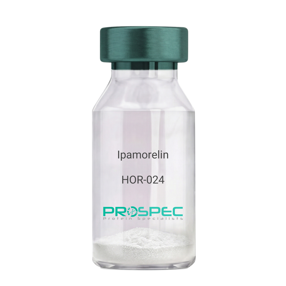
IpamorelinDescription:
Ipamorelin
Ipamorelin
Product # :
HOR-024Price :
Quantity :
Shipping Method :
Shipped at Room temp
More Info
- description
- formulation
- purity
- More Info
Description
Ipamorelin Synthetic is a single, non-glycosylated polypeptide chain containing 4 amino acids, having a molecular mass of 711.85 Dalton and a Molecular formula of C38H49N9O5.
Formulation
The protein was lyophilized with no additives.
Purity
Greater than 97.0% as determined by analysis by RP-HPLC.
More Info
-
Introduction
Ipamorelin is a peptide selective agonist of the ghrelin/growth hormone secretagogue receptor and a growth hormone secretagogue. Ipamorelin is a pentapeptide that was derived from GHRP1. Ipamorelin significantly increases plasma growth hormone levels in both animals and humans. Like pralmorelin and GHRP-6, ipamorelin does not affect prolactin, FSH, LH or TSH levels. However, unlike GHRP2 and GHRP6, but as growth hormone-releasing hormone (GHRH), ipamorelin does not stimulate the secretion of adrenocorticotropic hormone (ACTH) or cortisol, and is highly selective for inducing the secretion only of GH.
-
Synonyms
Ipamorelin
-
Physical Appearance
Sterile Filtered White lyophilized (freeze-dried) powder.
-
Stability
Lyophilized Ipamorelin although stable at room temperature for 3 weeks, should be stored desiccated below -18°C. Upon reconstitution Ipamorelin should be stored at 4°C between 2-7 days and for future use below -18°C. For long term storage it is recommended to add a carrier protein (0.1% HSA or BSA). Please prevent freeze-thaw cycles.
-
Solubility
It is recommended to reconstitute the lyophilized Ipamorelin in sterile 18MΩ-cm H2O not less than 100 µg/ml, which can then be further diluted to other aqueous solutions.
-
Amino Acid Sequence
Aib-His-D-2-Nal-D-Phe-Lys-NH2.
ProSpec's products are furnished for LABORATORY RESEARCH USE ONLY. The product may not be used as drugs, agricultural or pesticidal products, food additives or household chemicals.
- View Data Sheet
Name :
ADRM1 AntibodyDescription:
Adhesion Regulating Molecule 1, Mouse Anti Human
Proteasomal ubiquitin receptor ADRM1, 110 kDa cell membrane glycoprotein, Gp110, Adhesion-regulating molecule 1, ARM-1, Proteasome regulatory particle non-ATPase 13, hRpn13, Rpn13 homolog, ADRM1, ARM1, Rpn13.
Product # :
ANT-409Price :
Quantity :
Shipping Method :
Shipped with Ice Packs
More Info
- formulation
- More Info
Formulation
1mg/ml containing PBS, pH-7.4, 10% Glycerol and 0.02% Sodium Azide.
More Info
-
Introduction
Proteasomal ubiquitin receptor ADRM1 (ADRM1) functions as a proteasomal ubiquitin receptor. ADRM1 is an integral plasma membrane protein which promotes cell adhesion. ADRM1 is assumed to undergo O-linked glycosylation. ADRM1 gene expression is induced by gamma interferon in some cancer cells. ADRM1 employs the deubiquitinating enzyme UCHL5 at the 26S proteasome and promotes its activity.
-
Synonyms
Proteasomal ubiquitin receptor ADRM1, 110 kDa cell membrane glycoprotein, Gp110, Adhesion-regulating molecule 1, ARM-1, Proteasome regulatory particle non-ATPase 13, hRpn13, Rpn13 homolog, ADRM1, ARM1, Rpn13.
-
Physical Appearance
Sterile filtered colorless solution.
-
Immunogen
Anti-human ADRM1 mAb, is derived from hybridization of mouse F0 myeloma cells with spleen cells from BALB/c mice immunized with a recombinant human ADRM1 protein 1-407 amino acids purified from E. coli.
-
Ig Subclass
Mouse IgG2b heavy chain and k light chain.
-
Clone
PAT34C2AT.
-
Applications
The antibody has been tested by ELISA, Western blot analysis, Flow cytometry and ICC/IF to assure specificity and reactivity. Since application varies, however, each investigation should be titrated by the reagent to obtain optimal results.
-
Type
Mouse Anti Human Monoclonal.
-
Storage Procedures
For periods up to 1 month store at 4°C, for longer periods of time, store at -20°C. Prevent freeze thaw cycles.
-
Purification Method
ADRM1 antibody was purified from mouse ascitic fluids by protein-G affinity chromatography.
ProSpec's products are furnished for LABORATORY RESEARCH USE ONLY. The product may not be used as drugs, agricultural or pesticidal products, food additives or household chemicals.
- View Data Sheet
Name :
IL 4 HumanDescription:
Interleukin-4 Human Recombinant
BCGF, BCDF, B cell stimulating factor, BSF-1, Lymphocyte stimulatory factor 1, IL-4, MGC79402, Binetrakin, Pitrakinra.
Product # :
CYT-211Price :
Quantity :
Shipping Method :
Shipped at Room temp
More Info
- description
- source
- formulation
- purity
- biological activity
- More Info
Description
Interleukin-4 Human Recombinant produced in E.Coli is a single, non-glycosylated polypeptide chain containing 130 amino acids and having a molecular mass of 15kDa. The IL-4 is purified by proprietary chromatographic techniques.
Source
Escherichia Coli.
Formulation
IL-4 was lyophilized from a 0.2µm filtered concentrated (1mg/ml) solution in PBS, pH 7.4.
Purity
Greater than 97.0% as determined by:
(a) Analysis by RP-HPLC.
(b) Analysis by SDS-PAGE.Biological Activity
The ED50 as determined by the dose-dependant stimulation of TF-1 cells is < 0.2 ng/ml, corresponding to a Specific Activity of 5,000,000 IU/mg.More Info
-
Synonyms
BCGF, BCDF, B cell stimulating factor, BSF-1, Lymphocyte stimulatory factor 1, IL-4, MGC79402, Binetrakin, Pitrakinra.
-
Physical Appearance
Sterile Filtered White lyophilized (freeze-dried) powder.
-
Stability
Lyophilized Interleukin-4 although stable at room temperature for 3 weeks, should be stored desiccated below -18°C. Upon reconstitution IL-4 should be stored at 4°C between 2-7 days and for future use below -18°C.For long term storage it is recommended to add a carrier protein (0.1% HSA or BSA).Please prevent freeze-thaw cycles.
-
Solubility
It is recommended to reconstitute the lyophilized IL-4 in sterile 18MΩ-cm H2O not less than 100µg/ml, which can then be further diluted to other aqueous solutions.
-
Amino Acid Sequence
MHKCDITLQE IIKTLNSLTE QKTLCTELTV TDIFAASKNT TEKETFCRAA TVLRQFYSHH EKDTRCLGAT AQQFHRHKQL IRFLKRLDRN LWGLAGLNSC PVKEANQSTL ENFLERLKTI MREKYSKCSS.
-
Background
Everything You Should Know About IL-4 Human Recombinant
Interleukin-4 (IL-4) is an important type-2 cytokine with multiple effects on numerous cells and tissues in the body. As such, it's crucial for the immune system's maintenance and function.
Recent advancements have led to the development of interleukin-4 (rhIL-4) human recombinant in laboratories. As a result, experts have started studying its potential uses and therapeutic applications.
If you're interested in learning more about IL-4 human recombinant, check out the information below!
How Does Interleukin-4 (IL-4) Work?
Interleukin-4 (IL-4) acts as a signaling molecule, specifically a protein. It communicates with various immune cells by binding to specific receptors on their surface.
This process triggers responses against common threats, such as allergens, contaminants, and infections.
IL-4 is produced by T helper 2 (Th2) cells (a type of white blood cells), mast cells, eosinophils, and basophils.
What Interleukin-4 (IL-4) Does
As mentioned, this type-2 cytokine has multiple effects on various cells and tissues, so it fulfills several functions. Below are the most important ones:
- Th2 cell differentiation: IL-4 promotes the differentiation of T cells, triggering the response of Th2 cells, which fight parasites and regulate allergic responses.
- B cell activation and antibody production: IL-4 stimulates B cells, helping them proliferate and produce antibodies, particularly immunoglobulin E (IgE), which is involved in allergic reactions.
- Inflammation reduction: IL-4 can promote inflammation to address threats, such as allergens or parasites, and suppress it when necessary, depending on the context.
- Tissue repair and remodeling: IL-4 is key for tissue repair and remodeling processes, such as wound healing and fibrosis.
What Is IL-4 Human Recombinant?
IL-4 human recombinant is a version produced in a laboratory using DNA technology. The cytokine is inserted into a host organism, commonly E. Coli bacteria, for mass production.
This method allows experts to obtain pure and contaminant-free rhIL-4 with uniform biological activity and in large quantities.
What Can IL-4 Human Recombinant Be Used For?
Since this cytokine plays a key role in different processes, it may have multiple therapeutic applications for numerous diseases. These are the most common:
- Allergic diseases, such as asthma, rhinitis, and eczema
- Autoimmune diseases, such as rheumatoid arthritis and multiple sclerosis
- Infectious diseases, including those caused by parasites and viruses
- Cancer, as research suggests IL-4 could enhance the immune system's ability to fight cancer cells
Final Thoughts
The future of IL-4 human recombinant seems promising. This laboratory-produced version of an essential type-2 cytokine may be key to developing innovative therapies. However, more research is needed on potential applications to determine if it's safe and effective.
-
Protein content
Protein quantitation was carried out by two independent methods:1. UV spectroscopy at 280 nm using the absorbency value of 0.594 as the extinction coefficient for a 0.1% (1mg/ml) solution. This value is calculated by the PC GENE computer analysis program of protein sequences (IntelliGenetics). 2. Analysis by RP-HPLC, using a standard solution of IL-4 as a Reference Standard.
ProSpec's products are furnished for LABORATORY RESEARCH USE ONLY. The product may not be used as drugs, agricultural or pesticidal products, food additives or household chemicals.
- View Data Sheet
Name :
CD19 HumanDescription:
CD19 Human Recombinant
CD19 Molecule, B-Lymphocyte Surface Antigen B4, T-Cell Surface Antigen Leu-12, Differentiation Antigen CD19, CD19 Antigen, B-Lymphocyte Antigen CD19, CVID3, B4.
Product # :
PRO-2552Price :
Quantity :
Shipping Method :
Shipped with Ice Packs
More Info
- description
- source
- formulation
- purity
- More Info
Description
CD19 produced in Sf9 Baculovirus cells is a single, glycosylated polypeptide chain containing 510 amino acids (21-291a.a.) and having a molecular mass of 57.0kDa. (Molecular size on SDS-PAGE will appear at approximately 50-70kDa).CD19 is expressed with a 239 amino acid hIgG-His tag at C-Terminus and purified by proprietary chromatographic techniques.
Source
Sf9, Baculovirus cells.
Formulation
CD19 protein solution (0.25mg/ml) contains Phosphate Buffered Saline (pH 7.4) and 10% glycerol.
Purity
Greater than 85.0% as determined by SDS-PAGE.
More Info
-
Introduction
CD19, also known as B-Lymphocyte Surface Antigen B4, belongs to the Ig superfamily which is expressed on the surface of all B-lymphoid cells with the exception of terminally differentiated plasma cells. CD19 is involved as a signal-transducing receptor in the control of differentiation as well as proliferation. CD19 is as an adaptor protein which drafts cytoplasmic signaling proteins to the membrane and operates within the CD19/CD21 complex to decrease the threshold for B cell receptor signaling pathways. CD19 is present on all B cells, and therefore is a biomarker for lymphoma diagnosis, B lymphocyte development and can be used as a target for leukemia immunotherapies.
-
Synonyms
CD19 Molecule, B-Lymphocyte Surface Antigen B4, T-Cell Surface Antigen Leu-12, Differentiation Antigen CD19, CD19 Antigen, B-Lymphocyte Antigen CD19, CVID3, B4.
-
Physical Appearance
Sterile Filtered colorless solution.
-
Stability
Store at 4°C if entire vial will be used within 2-4 weeks. Store, frozen at -20°C for longer periods of time. For long term storage it is recommended to add a carrier protein (0.1% HSA or BSA). Avoid multiple freeze-thaw cycles.
-
Amino Acid Sequence
EEPLVVKVEE GDNAVLQCLK GTSDGPTQQL TWSRESPLKP FLKLSLGLPG LGIHMRPLAI WLFIFNVSQQ MGGFYLCQPG PPSEKAWQPG WTVNVEGSGE LFRWNVSDLG GLGCGLKNRS SEGPSSPSGK LMSPKLYVWA KDRPEIWEGE PPCLPPRDSL NQSLSQDLTM APGSTLWLSC GVPPDSVSRG PLSWTHVHPK GPKSLLSLEL KDDRPARDMW VMETGLLLPR ATAQDAGKYY CHRGNLTMSF HLEITARPVL WHWLLRTGGW KLEPKSCDKT HTCPPCPAPE LLGGPSVFLF PPKPKDTLMI SRTPEVTCVV VDVSHEDPEV KFNWYVDGVE VHNAKTKPRE EQYNSTYRVV SVLTVLHQDW LNGKEYKCKV SNKALPAPIE KTISKAKGQP REPQVYTLPP SRDELTKNQV SLTCLVKGFY PSDIAVEWES NGQPENNYKT TPPVLDSDGS FFLYSKLTVD KSRWQQGNVF SCSVMHEALH NHYTQKSLSL SPGKHHHHHH
ProSpec's products are furnished for LABORATORY RESEARCH USE ONLY. The product may not be used as drugs, agricultural or pesticidal products, food additives or household chemicals.
- View Data Sheet
Name :
IL 2 HumanDescription:
Interleukin-2 Human Recombinant
T-cell growth factor (TCGF), Lymphokine, IL-2.
Product # :
CYT-209Price :
Quantity :
Shipping Method :
Shipped at Room temp
More Info
- description
- source
- formulation
- purity
- biological activity
- More Info
Description
Interleukin-2 Human Recombinant produced in E.Coli is a single, non-glycosylated polypeptide chain containing 133 amino acids and having a molecular mass of 15.4kDa. The IL-2 is purified by proprietary chromatographic techniques.
Source
Escherichia Coli.
Formulation
The protein was lyophilized from a 0.2μm filtered concentrated solution (1.0mg/ml) in PBS, pH 5.0.
Purity
Greater than 97.0% as determined by:
(a) Analysis by RP-HPLC.
(b) Analysis by SDS-PAGE.Biological Activity
The ED50 as determined by the dose-dependent stimulation of murine CTLL-2 cells is less than 0.1 ng/ml, corresponding to a Specific Activity of 1x10⁷IU/mg.
More Info
-
Introduction
IL2 is a secreted cytokine that is important for the proliferation of T and B lymphocytes. The receptor of this cytokine is a heterotrimeric protein complex whose gamma chain is also shared by interleukin 4 (IL4) and interleukin 7 (IL7). The expression of this gene in mature thymocytes is monoallelic, which represents an unusual regulatory mode for controlling the precise expression of a single gene. The targeted disruption of a similar gene in mice leads to ulcerative colitis-like disease, which suggests an essential role of this gene in the immune response to antigenic stimuli.
-
Synonyms
T-cell growth factor (TCGF), Lymphokine, IL-2.
-
Physical Appearance
Sterile Filtered White lyophilized (freeze-dried) powder.
-
Stability
Lyophilized Interleukin-2 although stable at room temperature for 3 weeks, should be stored desiccated below -18°C. Upon reconstitution IL2 should be stored at 4°C between 2-7 days and for future use below -18°C.For long term storage it is recommended to add a carrier protein (0.1% HSA or BSA).Please prevent freeze-thaw cycles.
-
Solubility
It is recommended to reconstitute the lyophilized Interleukin-2 in sterile distilled water not less than 100µg/ml, which can then be further diluted to other aqueous solutions.
-
Amino Acid Sequence
APTSSSTKK TQLQLEHLLL DLQMILNGIN NYKNPKLTRM LTFKFYMPKK ATELKHLQCL EEELKPLEEV LNLAQSKNFH LRPRDLISNI NVIVLELKGS ETTFMCEYAD ETATIVEFLN RWITFCQSII STLT.
-
Background
IL 2 HUMAN: A Vital Tool for Advancing Immunotherapy Research
IL 2 HUMAN refers to the human form of Interleukin-2, a critical cytokine in the immune system. This protein is crucial for the growth and activity of T and B lymphocytes, the cells responsible for the body's immune defense against infections.
By promoting the growth of these cells, IL 2 plays a vital role in maintaining and activating the immune response.
Production and Characteristics
Typically, IL 2 is produced through recombinant DNA technology using E. coli bacteria. This method allows for creating large quantities of pure and functional IL 2 protein.
The product appears as a sterile, white lyophilized powder that is non-glycosylated and consists of 133 amino acids with a molecular mass of approximately 15.4 kDa.
Reconstituting IL-2 Human for Laboratory Use
To dissolve the lyophilized IL-2 effectively, it should be reconstituted with sterile distilled water to achieve a minimum 100µg/ml concentration. This makes the protein readily usable for further experimental procedures.
Moreover, the concentration allows for easy adjustment in solubility and ensures optimal conditions for the protein's activity in various research setups. Proper dissolution and storage are crucial to maintain the protein's functionality and stability.
Storage and Stability
The stability of IL 2 is dependent on its storage conditions. When lyophilized, it can remain stable at room temperature for up to three weeks, but for long-term storage, it should be kept desiccated below -18°C.
Once reconstituted, IL 2 should be used within a week if kept at 4°C, or it can be stored at below -18°C for future use. It's important to avoid freeze-thaw cycles, which can degrade the protein.
Purity and Biological Activity
IL 2 HUMAN's purity is generally greater than 97%, confirmed by reverse-phase high-performance liquid chromatography (RP-HPLC) and SDS-PAGE analyses.
Its biological activity is evaluated by its ability to stimulate the proliferation of murine CTLL-2 cells, with an effective dose (ED50) typically less than 0.1 ng/ml. This indicates a very high specific activity, making it highly potent and effective in research settings.
Applications in Research
IL 2 is used extensively in research, particularly in studies focusing on the immune system, autoimmune diseases, and cancer. Furthermore, its role in T-cell growth and function makes it invaluable for developing new therapeutic strategies and understanding immune response mechanisms.
Important Considerations
It is important to remember that IL 2, like all cytokines used in research, is intended for laboratory use only. This protein is not suitable for drug, food, or cosmetic applications. Proper handling and usage instructions must be followed to ensure safety and efficacy in research environments.
Essentially, IL 2 HUMAN is a vital tool in immunological research, offering insights into the complex workings of the immune system and providing avenues for potential therapeutic developments. The careful production, handling, and storage of IL 2 are crucial for its effective use in laboratory settings.
ProSpec's products are furnished for LABORATORY RESEARCH USE ONLY. The product may not be used as drugs, agricultural or pesticidal products, food additives or household chemicals.
- View Data Sheet
Name :
SCGB1A1 RatDescription:
Uteroglobin Rat Recombinant
Uteroglobin, Clara cell 17 kDa protein, Clara cell phospholipid-binding protein, CCPBP, Clara cells 10 kDa secretory protein, CC10, PCB-binding protein, Secretoglobin family 1A member 1, Scgb1a1, Ugb, Utg, UG, CC16, CCSP, PCB-BP.
Product # :
CYT-820Price :
Quantity :
Shipping Method :
Shipped at Room temp
More Info
- description
- source
- formulation
- purity
- biological activity
- More Info
Description
SCGB1A1 Rat Recombinant produced in E.Coli is a homodimeric non-glycosylated polypeptide chains consisting of two 77 amino acids and having a molecular mass of 17.0kDa.
Source
Escherichia Coli.
Formulation
SCGB1A1 protein was lyophilized from a 0.2 µm filtered concentrated solution in PBS, pH 7.4.
Purity
Greater than 97.0% as determined by:
(a) Analysis by RP-HPLC.
(b) Analysis by SDS-PAGE.Biological Activity
Fully biologically active when compared to standard. The ED50 as determined by the ability of the immobilized protein to support the adhesion of the A549 human lung carcinoma cells is less than 5.0 ?g/ml, corresponding to a specific activity of >200IU/mg.More Info
-
Introduction
Uteroglobin (SCGB1A1) which belongs to the Secretoglobin (SCGBs) superfamily, is a multifunctional protein that exerts anti-inflammatory and anti-tumorigenic effects by binding small hydrophobic molecules such as phospholipids and prostaglandins. Uteroglobin is involved in numerous functions including anti-inflammation, inhibition of phospholipase A2 and the sequestering of hydrophobic ligands. SCGB1A1 is expressed by Clara cells, the non-ciliated, non-mucous secretory cells predominant in lung bronchioles, and by other epithelia which communicate with the external environment. On top of sequestering pro-inflammatory mediators and carcinogens, Uteroglobin is implicated in the inhibition of cell migration and invasion, platelet aggregation, and T cell differentiation. SCGB1A1 gene defects are associated with a susceptibility to asthma.
-
Synonyms
Uteroglobin, Clara cell 17 kDa protein, Clara cell phospholipid-binding protein, CCPBP, Clara cells 10 kDa secretory protein, CC10, PCB-binding protein, Secretoglobin family 1A member 1, Scgb1a1, Ugb, Utg, UG, CC16, CCSP, PCB-BP.
-
Physical Appearance
Sterile Filtered White lyophilized (freeze-dried) powder.
-
Stability
Lyophilized SCGB1A1 although stable at room temperature for 3 weeks, should be stored desiccated below -18°C. Upon reconstitution SCGB1A1 should be stored at 4°C between 2-7 days and for future use below -18°C. For long term storage it is recommended to add a carrier protein (0.1% HSA or BSA). Please prevent freeze-thaw cycles.
-
Solubility
It is recommended to reconstitute the lyophilized SCGB1A1 in sterile 18MΩ-cm H2O not less than 100µg/ml, which can then be further diluted to other aqueous solutions.
-
Amino Acid Sequence
SSDICPGFLQ VLEALLLGSE SNYEAALKPF NPASDLQNAG TQLKRLVDTL PQETRINIVK LTEKILTSPL CEQDLRV
ProSpec's products are furnished for LABORATORY RESEARCH USE ONLY. The product may not be used as drugs, agricultural or pesticidal products, food additives or household chemicals.
- View Data Sheet
Name :
IL1A HumanDescription:
Interleukin-1 alpha Human Recombinant
Hematopoietin-1, Lymphocyte-activating factor (LAF), Endogenous Pyrogen (EP), Leukocyte Endogenous Mediator (LEM), Mononuclear Cell Factor (MCF), IL-1 alpha,IL1, IL-1A, IL1F1.
Product # :
CYT-253Price :
Quantity :
Shipping Method :
Shipped at Room temp
More Info
- description
- source
- formulation
- purity
- biological activity
- More Info
Description
Interleukin-1 alpha Human Recombinant produced in E.Coli is single, a non-glycosylated, Polypeptide chain containing 159 amino acids and having a molecular mass of 18022 Dalton. The IL-1A is purified by proprietary chromatographic techniques.
Source
Escherichia Coli.
Formulation
The protein was lyophilized from a concentrated (1mg/ml) sterile solution containing 25mM Tris-HCl, pH 8.
Purity
Greater than 97.0% as determined by:
(a) Analysis by RP-HPLC.
(b) Analysis by SDS-PAGE.Biological Activity
The ED50 as determined by the dose-dependant stimulation of murine D10S cells is < 0.001 ng/ml, corresponding to a Specific Activity of 1 x 10⁹ IU/mg.More Info
-
Introduction
IL-1 alpha is produced by activated macrophages, stimulates thymocyte proliferation by inducing il-2 release, b-cell maturation and proliferation, and fibroblast growth factor activity. IL1A proteins are involved in the inflammatory response, being identified as endogenous pyrogens, and are reported to stimulate the release of prostaglandin from synovial cells.
-
Synonyms
Hematopoietin-1, Lymphocyte-activating factor (LAF), Endogenous Pyrogen (EP), Leukocyte Endogenous Mediator (LEM), Mononuclear Cell Factor (MCF), IL-1 alpha,IL1, IL-1A, IL1F1.
-
Physical Appearance
Sterile Filtered White lyophilized (freeze-dried) powder.
-
Stability
Lyophilized Interleukin-1 alpha although stable at room temperature for 3 weeks, should be stored desiccated below -18°C. Upon reconstitution IL-1a should be stored at 4°C between 2-7 days and for future use below -18°C.Please prevent freeze-thaw cycles.
-
Solubility
It is recommended to reconstitute the lyophilized Interleukin 1 alpha in sterile 18MΩ-cm H2O not less than 100µg/ml, which can then be further diluted to other aqueous solutions.
-
Amino Acid Sequence
SAPFSFLSNVKYNFMRIIKYEFILNDALNQSIIRANDQYLTAAALHNLDEAV KFDMGAYKSSKDDAKITVILRISKTQLYVTAQDEDQPVLLKEMPEIPKTITG SETNLLFFWETHGTKNYFTSVAHPNLFIATKQDYWVCLAGGPPSITDFQILE NQA.
-
Background
What is the molecular weight/Mw of IL1A HUMAN Protein?
IL1A HUMAN Protein has a total Mw of 18kDa.
What is the source or expression system of IL1A HUMAN Protein?
Escherichia Coli.
What is the Purity of IL1A HUMAN Protein?
IL1A HUMAN Protein is >97% pure as determined by SDS-PAGE.
What is the Biological Activity of IL1A HUMAN Protein?
The ED50 as determined by the dose-dependant stimulation of murine D10S cells is < 0.001 ng/ml, corresponding to a Specific Activity of 1 x 10⁹ IU/mg.
What is the amino acid sequence of IL1A HUMAN Protein?
SAPFSFLSNVKYNFMRIIKYEFILNDALNQSIIRANDQYLTAAALHNLDEAV KFDMGAYKSSKDDAKITVILRISKTQLYVTAQDEDQPVLLKEMPEIPKTITG SETNLLFFWETHGTKNYFTSVAHPNLFIATKQDYWVCLAGGPPSITDFQILE NQA.
What applications can IL1A HUMAN Protein be used in?
IL1A HUMAN Protein can probably be used in western blot, ELISA and Lateral Flow.
What is the endotoxin level for IL1A HUMAN Protein?
The endotoxin level is minimal, IL1A HUMAN Protein was purified using conventional chromatography techniques.
-
Protein content
Protein quantitation was carried out by two independent methods:1. UV spectroscopy at 280 nm using the absorbency value of 1.13 as the extinction coefficient for a 0.1% (1mg/ml) solution. This value is calculated by the PC GENE computer analysis program of protein sequences (IntelliGenetics). 2. Analysis by RP-HPLC, using a standard solution of IL-1 as a Reference Standard.
ProSpec's products are furnished for LABORATORY RESEARCH USE ONLY. The product may not be used as drugs, agricultural or pesticidal products, food additives or household chemicals.
- View Data Sheet
Name :
IL 1 beta EquineDescription:
Interleukin-1 beta Equine Recombinant
Catabolin, Lymphocyte-activating factor (LAF), Endogenous Pyrogen (EP), Leukocyte Endogenous Mediator (LEM), Mononuclear Cell Factor (MCF), IL1F2, IL-1 beta.
Product # :
CYT-411Price :
Quantity :
Shipping Method :
Shipped at Room temp
More Info
- description
- source
- formulation
- purity
- biological activity
- More Info
Description
Recombinant IL 1 beta Equine produced in E.coli cells is a non-glycosylated, homodimeric protein containing 153 amino acid chain and having a molecular mass of 17.3kDa. The IL 1 beta is purified by proprietary chromatographic techniques.
Source
Escherichia Coli.
Formulation
The IL 1 beta was lyophilized from a 0.2µm filtered concentrated solution in PBS pH 7.4, containing 0.1 % Tween-80.
Purity
Greater than 95.0% as determined by:
(a) Analysis by RP-HPLC.
(b) Analysis by SDS-PAGE.Biological Activity
The ED50 as determined by a cell proliferation assay using murine D10S cells is less than 20 pg/ml, corresponding to a specific activity of > 5.0 × 10⁷ IU/mg.
More Info
-
Introduction
Interleukin-1b is produced by activated macrophages, IL-1B stimulates thymocyte proliferation by inducing il-2 release, b-cell maturation and proliferation, and fibroblast growth factor activity. IL1B proteins are involved in the inflammatory response, being identified as endogenous pyrogens, and are reported to stimulate the release of prostaglandin and collagenase from synovial cells.
-
Synonyms
Catabolin, Lymphocyte-activating factor (LAF), Endogenous Pyrogen (EP), Leukocyte Endogenous Mediator (LEM), Mononuclear Cell Factor (MCF), IL1F2, IL-1 beta.
-
Physical Appearance
Sterile Filtered White lyophilized (freeze-dried) powder.
-
Stability
Lyophilized IL 1 beta although stable at room temperature for 3 weeks, should be stored desiccated below -18°C. Upon reconstitution IL 1 beta should be stored at 4°C between 2-7 days and for future use below -18°C.For long term storage it is recommended to add a carrier protein (0.1% HSA or BSA).Please prevent freeze-thaw cycles.
-
Solubility
It is recommended to reconstitute the lyophilized IL 1 beta in sterile distilled H2O not less than 100µg/ml, which can then be further diluted to other aqueous solutions.
-
Amino Acid Sequence
AAMHSVNCRL RDIYHKSLVL SGACELQAVH LNGENTNQQV VFCMSFVQGE EETDKIPVAL GLKEKNLYLS CGMKDGKPTL QLETVDPNTY PKRKMEKRFV FNKMEIKGNV EFESAMYPNW YISTSQAEKS PVFLGNTRGG RDITDFIMEI TSA
ProSpec's products are furnished for LABORATORY RESEARCH USE ONLY. The product may not be used as drugs, agricultural or pesticidal products, food additives or household chemicals.
- View Data Sheet
Name :
IL 4 Human, HisDescription:
Interleukin-4 Human Recombinant, His Tag
BCGF, BCDF, B cell stimulating factor, BSF-1, Lymphocyte stimulatory factor 1, IL-4, MGC79402, Binetrakin, Pitrakinra.
Product # :
CYT-483Price :
Quantity :
Shipping Method :
Shipped with Ice Packs
More Info
- description
- source
- formulation
- purity
- biological activity
- More Info
Description
Interleukin-4 Human Recombinant produced in E.Coli is single, a non-glycosylated, Polypeptide chain containing 150 amino acids fragment (25-153) and having a total molecular mass of 17.2kDa.The IL-4 is fused to a 20 amino acid His-Tag at N-terminus and purified by proprietary chromatographic techniques.
Source
Escherichia Coli.
Formulation
Interleukin-4 His-Tag is supplied in 20mM Tris-HCl and10% glycerol.
Purity
Greater than 95.0% as determined by SDS-PAGE.
Biological Activity
The ED50 for this effect is <0.5ng/ml. Measured in a cell proliferation assay using TF1 human erythroleukemic cells.
More Info
-
Introduction
Interleukin-4 is a pleiotropic cytokine produced primarily by activated T lymphocytes, basophils and mast cells. Multiple immune response-modulating functions are performed by IL-4 on a variety of cell types and it has an important role in the regulator of isotype switching, induction of IgE production in B lymphocytes and differentiation of precursor T helper cells. IL-4 binds to both membrane-bound and secreted soluble IL-4 receptors.
-
Synonyms
BCGF, BCDF, B cell stimulating factor, BSF-1, Lymphocyte stimulatory factor 1, IL-4, MGC79402, Binetrakin, Pitrakinra.
-
Physical Appearance
Sterile Filtered clear solution.
-
Stability
Store at 4°C if entire vial will be used within 2-4 weeks. Store, frozen at -20°C for longer periods of time.For long term storage it is recommended to add a carrier protein (0.1% HSA or BSA).Please avoid freeze thaw cycles.
-
Amino Acid Sequence
MGSSHHHHHH SSGLVPRGSH MHKCDITLQE IIKTLNSLTE QKTLCTELTV TDIFAASKNT TEKETFCRAA TVLRQFYSHH EKDTRCLGAT AQQFHRHKQL IRFLKRLDRN LWGLAGLNSC PVKEANQSTL ENFLERLKTI MREKYSKCSS.
-
Background
Recombinant IL-4 (Interleukin-4) is a bioengineered version of a naturally occurring cytokine, which plays a crucial role in the immune system. IL-4 is primarily produced by activated T cells, mast cells, and basophils, and it is involved in the regulation of immune responses, including the differentiation of T helper cells, B cell activation, and the production of immunoglobulins.Recombinant IL-4 is synthesized using recombinant DNA technology, which involves inserting the gene encoding IL-4 into a suitable expression system, such as bacteria, yeast, or mammalian cells. The host cells are then cultured, allowing them to produce the desired protein, which can be purified and used for various applications.One of the main functions of IL-4 is to promote the differentiation of naïve CD4+ T cells into T helper 2 (Th2) cells. Th2 cells are essential for coordinating immune responses against extracellular pathogens, such as parasites and allergens. They achieve this by secreting cytokines, including IL-4 itself, IL-5, and IL-13, which stimulate B cells to produce specific antibodies, eosinophils to combat parasites, and mast cells to release histamine and other inflammatory mediators.Recombinant IL-4 has been extensively studied for its potential therapeutic applications. It has been shown to have anti-inflammatory properties, making it a potential candidate for the treatment of autoimmune and inflammatory diseases, such as rheumatoid arthritis, multiple sclerosis, and inflammatory bowel disease. Additionally, IL-4 has been found to inhibit the growth of certain cancer cells, suggesting that it may have potential as an anti-cancer agent.However, the use of recombinant IL-4 as a therapeutic agent is not without challenges. One of the main concerns is the potential for adverse effects due to its immunomodulatory properties. For example, excessive IL-4 activity can lead to the development of allergies and asthma, as it promotes the production
ProSpec's products are furnished for LABORATORY RESEARCH USE ONLY. The product may not be used as drugs, agricultural or pesticidal products, food additives or household chemicals.
- View Data Sheet
Name :
NRP1 RatDescription:
Neuropilin 1 Rat Recombinant
Neuropilin-1, Vascular endothelial cell growth factor 165 receptor, CD304, Nrp1.
Product # :
CYT-973Price :
Quantity :
Shipping Method :
Shipped with Ice Packs
More Info
- description
- source
- formulation
- purity
- More Info
Description
NRP1 Rat Recombinant produced in Sf9 Baculovirus cells is a single, glycosylated polypeptide chain containing 842 amino acids (22-855 a.a.) and having a molecular mass of 94.8kDa (Migrates at 100-150kDa on SDS-PAGE under reducing conditions).NRP1 is expressed with an 8 amino acid His tag at C-Terminus and purified by proprietary chromatographic techniques.
Source
Sf9, Baculovirus cells.
Formulation
NRP1 protein solution (0.25mg/ml) containing Phosphate Buffered Saline (pH 7.4) and 10% glycerol.
Purity
Greater than 90.0% as determined by SDS-PAGE.
More Info
-
Introduction
Neuropilin 1 (Nrp1) is a transmembrane glycoprotein which functions as a co-receptor for several extracellular ligands including class III/IV semaphorins, some isoforms of vascular endothelial growth factor and transforming growth factor beta. Nrp1 binds vascular endothelial growth factor (VEGF)-A and is believed to serve as a coreceptor for kinase insert domain-containing receptor (KDR) by connecting with KDR and enhancing VEGF signaling. Nrp1 is a marker of regulatory T cells.
-
Synonyms
Neuropilin-1, Vascular endothelial cell growth factor 165 receptor, CD304, Nrp1.
-
Physical Appearance
Sterile Filtered colorless solution.
-
Stability
Store at 4°C if entire vial will be used within 2-4 weeks. Store, frozen at -20°C for longer periods of time. For long term storage it is recommended to add a carrier protein (0.1% HSA or BSA). Avoid multiple freeze-thaw cycles.
-
Amino Acid Sequence
FRSDKCGGTI KIENPGYLTS PGYPHSYHPS EKCEWLIQAP EPYQRIMINF NPHFDLEDRD CKYDYVEVID GENEGGRLWG KFCGKIAPSP VVSSGPFLFI KFVSDYETHG AGFSIRYEIF KRGPECSQNY TAPTGVIKSP GFPEKYPNSL ECTYIIFAPK MSEIILEFES FDLEQDSNPP GGVFCRYDRL EIWDGFPEVG PHIGRYCGQK TPGRIRSSSG ILSMVFYTDS AIAKEGFSAN YSVLQSSISE DFKCMEALGM ESGEIHSDQI TASSQYGTNW SVERSRLNYP ENGWTPGEDS YREWIQVDLG LLRFVTAVGT QGAISKETKK KYYVKTYRVD ISSNGEDWIT LKEGNKAIIF QGNTNPTDVV FGVFPKPLIT RFVRIKPASW ETGISMRFEV YGCKITDYPC SGMLGMVSGL ISDSQITASN QGDRNWMPEN IRLVTSRTGW ALPPSPHPYI NEWLQVDLGD EKIVRGVIIQ GGKHRENKVF MRKFKIAYSN NGSDWKMIMD DSKRKAKSFE GNNNYDTPEL RAFTPLSTRF IRIYPERATH SGLGLRMELL GCEVEVPTAG PTTPNGNPVD ECDDDQANCH SGTGDDFQLT GGTTVLATEK PTIIDSTIQS EFPTYGFNCE FGWGSHKTFC HWEHDSHAQL RWRVLTSKTG PIQDHTGDGN FIYSQADENQ KGKVARLVSP VVYSQSSAHC MTFWYHMSGS HVGTLRVKLH YQKPEEYDQL VWMVVGHQGD HWKEGRVLLH KSLKLYQVIF EGEIGKGNLG GIAVDDISIN NHIPQEDCAK PTDLDKKNTE IKIDETGSTP GYEEGKGDKN ISRKPGNVLK TLDPLEHHHH HH
ProSpec's products are furnished for LABORATORY RESEARCH USE ONLY. The product may not be used as drugs, agricultural or pesticidal products, food additives or household chemicals.
- View Data Sheet
Name :
Dengue NS1 ST4, InsectDescription:
Dengue Virus NS1 Subtype 4 Recombinant, Insect Cells
Product # :
DEN-032Price :
Quantity :
Shipping Method :
Shipped with Ice Packs
More Info
- description
- source
- formulation
- purity
- More Info
Description
Recombinant Dengue Virus NS1 Subtype 4 produced in Insect Cells is a polypeptide chain containing amino acids 776-1130 and having a molecular weight of approximately 50kDa. Dengue NS1 ST4 is purified by proprietary chromatographic technique.
Source
Insect cells.
Formulation
Dengue NS1 ST4 protein solution in 1xD-PBS, pH7.4, 0.1% Thimerosal, 5mM EDTA, 1?g/ml of Leupeptin, Aprotinin and Pepstatin A.
Purity
Protein is >95% pure as determined by 12.5% SDS-PAGE.
More Info
-
Introduction
Caused by one of four closely related virus serotypes of the genus Flavivirus, family Flaviviridae, each serotype is sufficiently different that there is no cross-protection and epidemics caused by multiple serotypes (hyperendemicity) can occur. In cell culture experiments and mice Morpholino antisense oligos have shown specific activity against Dengue virus.
-
Physical Appearance
Sterile filtered colorless solution.
-
Stability
Store at 4°C if entire vial will be used within 2-4 weeks. Store, frozen at -20°C for longer periods of time. For long term storage it is recommended to add a carrier protein (0.1% HSA or BSA).Avoid multiple freeze-thaw cycles.
ProSpec's products are furnished for LABORATORY RESEARCH USE ONLY. The product may not be used as drugs, agricultural or pesticidal products, food additives or household chemicals.
- View Data Sheet
Name :
HAVCR1 HumanDescription:
Hepatitis A Virus Cellular Receptor 1 Human Recombinant
Hepatitis A Virus Cellular Receptor 1, T-Cell Immunoglobulin Mucin Family Member 1, T-Cell Immunoglobulin Mucin Receptor 1, T-Cell Membrane Protein 1, Kidney Injury Molecule 1, HAVCR-1, TIMD-1, HAVCR, KIM-1, TIM-1, TIMD1, TIM1, KIM1, TIM, T-Cell Immunoglobulin And Mucin Domain-Containing Protein 1, T Cell Immunoglobin Domain And Mucin Domain Protein 1, HAVCR1.
Product # :
HAV-222Price :
Quantity :
Shipping Method :
Shipped with Ice Packs
More Info
- description
- source
- formulation
- purity
- More Info
Description
HAVCR1 Human Recombinant produced in E. coli is a single polypeptide chain containing 298 amino acids (21-295aa) and having a molecular mass of 31.9kDa.HAVCR1 is fused to a 23 amino acid His-tag at N-terminus & purified by proprietary chromatographic techniques.
Source
Escherichia Coli.
Formulation
The HAVCR1 solution (1mg/ml) contains 20mM Tris-HCl (pH 8.0) and 10% glycerol.
Purity
Greater than 85% as determined by SDS-PAGE.
More Info
-
Introduction
Hepatitis A virus cellular receptor 1 (HAVCR1) is a membrane receptor for both human hepatitis A virus (HHAV) and TIMD4. HAVCR1 is a type I trans-membrane structural glycoprotein located in the renal proximal tubule epithelial cells. HAVCR1 protein may be involved in the control of asthma and allergic diseases. The reference genome represents an allele which retains a MTTVP amino acid segment that presents defense against atopy in HHAV seropositive individuals.
-
Synonyms
Hepatitis A Virus Cellular Receptor 1, T-Cell Immunoglobulin Mucin Family Member 1, T-Cell Immunoglobulin Mucin Receptor 1, T-Cell Membrane Protein 1, Kidney Injury Molecule 1, HAVCR-1, TIMD-1, HAVCR, KIM-1, TIM-1, TIMD1, TIM1, KIM1, TIM, T-Cell Immunoglobulin And Mucin Domain-Containing Protein 1, T Cell Immunoglobin Domain And Mucin Domain Protein 1, HAVCR1.
-
Physical Appearance
Sterile filtered colorless solution.
-
Stability
Store at 4°C if entire vial will be used within 2-4 weeks. Store, frozen at -20°C for longer periods of time. For long term storage it is recommended to add a carrier protein (0.1% HSA or BSA).Avoid multiple freeze-thaw cycles.
-
Amino Acid Sequence
MGSSHHHHHH SSGLVPRGSH MGSSVKVGGE AGPSVTLPCH YSGAVTSMCW NRGSCSLFTC QNGIVWTNGT HVTYRKDTRY KLLGDLSRRD VSLTIENTAV SDSGVYCCRV EHRGWFNDMK ITVSLEIVPP KVTTTPIVTT VPTVTTVRTS TTVPTTTTVP MTTVPTTTVP TTMSIPTTTT VLTTMTVSTT TSVPTTTSIP TTTSVPVTTT VSTFVPPMPL PRQNHEPVAT SPSSPQPAET HPTTLQGAIR REPTSSPLYS YTTDGNDTVT ESSDGLWNNN QTQLFLEHSL LTANTTKG
ProSpec's products are furnished for LABORATORY RESEARCH USE ONLY. The product may not be used as drugs, agricultural or pesticidal products, food additives or household chemicals.
- View Data Sheet
Name :
VEGF EquineDescription:
Vascular Endothelial Growth Factor Equine Recombinant
VEGF-A, VPF, glioma-derived endothelial cell mitogen.
Product # :
CYT-1200Price :
Quantity :
Shipping Method :
Shipped at Room temp
More Info
- description
- source
- formulation
- purity
- biological activity
- More Info
Description
VEGF Equine Recombinant produced in E.Coli is a single, non-glycosylated, polypeptide chain containing 2 x 165 amino acids and having a total molecular mass of 38.6 kDa.The VEGF is purified by proprietary chromatographic techniques.
Source
Escherichia Coli.
Formulation
Lyophilized from a sterile (0.2 micron) filtered aqueous solution containing 10 mM Sodium Phosphate, pH 7.5.
Purity
Greater than 95.0% as determined by SDS-PAGE.
Biological Activity
The ED50 as determined by HUVEC Proliferation is ≤ 10 ng/mL, corresponding to a specific activity of ≥ 1.0 x 10^5 units/mg.
More Info
-
Introduction
Vascular endothelial growth factor is an important signaling protein involved in both vasculogenesis and angiogenesis. As its name implies, VEGF activity has been mostly studied on cells of the vascular endothelium, although it does have effects on a number of other cell types (e.g. stimulation monocyte/macrophage migration, neurons, cancer cells, kidney epithelial cells ).VEGF mediates increased vascular permeability, induces angiogenesis, vasculogenesis and endothelial cell growth, promotes cell migration, and inhibits apoptosis. In vitro, VEGF has been shown to stimulate endothelial cell mitogenesis and cell migration. VEGF is also a vasodilator and increases microvascular permeability and was originally referred to as vascular permeability factor. Elevated levels of this protein is linked to POEMS syndrome, also known as Crow-Fukase syndrome. Mutations in this gene have been associated with proliferative and nonproliferative diabetic retinopathy.
-
Synonyms
VEGF-A, VPF, glioma-derived endothelial cell mitogen.
-
Physical Appearance
Sterile Filtered White lyophilized (freeze-dried) powder.
-
Stability
Lyophilized VEGF although stable at room temperature for 3 weeks, should be stored desiccated below -18°C. Upon reconstitution VEGF Human should be stored at 4°C between 2-7 days and for future use below -18°C.For long term storage it is recommended to add a carrier protein (0.1% HSA or BSA).Please prevent freeze-thaw cycles.
-
Solubility
It is recommended to reconstitute the lyophilized VEGF in sterile water at a concentration of 0.1 mg/ml, which can then be further diluted to other aqueous solutions.
-
Amino Acid Sequence
MAPMAEGEHK THEVVKFMDV YQRSYCRPIE TLVDIFQEYP DEIEYIFKPS CVPLMRCGGC CNDEGLECVP TAEFNITMQI MRIKPHQSQH IGEMSFLQHS KCECRPKKDK ARQENPCGPC SERRKHLFVQ DPQTCKCSCK NTDSRCKARQ LELNERTCRC DKPRR
ProSpec's products are furnished for LABORATORY RESEARCH USE ONLY. The product may not be used as drugs, agricultural or pesticidal products, food additives or household chemicals.
- View Data Sheet
Name :
IFN a 2a HumanDescription:
IFN-Alpha 2a Human Recombinant
Leukocyte IFN, B cell IFN, Type I IFN, IFNA2, IFN-a 2a, Interferon-alpha-2a.
Product # :
CYT-204Price :
Quantity :
Shipping Method :
Shipped at Room temp
More Info
- description
- source
- formulation
- purity
- biological activity
- More Info
Description
IFN Alpha Human 2a Recombinant produced in E.Coli is a single, non-glycosylated, polypeptide chain containing 165 amino acids and having a molecular mass of 19241 Dalton. The difference between IFNA2A and IFNA2B is in the amino acid present at position 23. IFN-alpha 2a has a lysine at that position 23 while IFN-alpha 2b has arginine.The Interferon-alpha-2a gene was obtained from human leukocytes.The IFNA2A is purified by proprietary chromatographic techniques.
Source
Escherichia Coli.
Formulation
Lyophilized without additives.
Purity
Greater than 97.0% as determined by both:
(a) Analysis by RP-HPLC.
(b) Analysis by SDS-PAGE.Biological Activity
The specific activity as determined in a viral resistance assay using bovine kidney MDBK cells was found to be 270,000,000 IU/mg.More Info
-
Introduction
IFN-alpha is produced by macrophages and has antiviral activities. IFN stimulates the production of two enzymes: protein kinase and an oligoadenylate synthetase.
-
Synonyms
Leukocyte IFN, B cell IFN, Type I IFN, IFNA2, IFN-a 2a, Interferon-alpha-2a.
-
Physical Appearance
Sterile Filtered White lyophilized (freeze-dried) powder.
-
Stability
Lyophilized IFNA-2A although stable at room temperature for 3 weeks, should be stored desiccated below -18°C. Upon reconstitution IFN-alpha 2a should be stored at 4°C between 2-7 days and for future use below -18°C.For long term storage it is recommended to add a carrier protein (0.1% HSA or BSA).Please prevent freeze-thaw cycles.
-
Solubility
It is recommended to reconstitute the lyophilized Interferon-alpha-2a in sterile 18MΩ-cm H2O not less than 100µg/ml, which can then be further diluted to other aqueous solutions.
-
Amino Acid Sequence
CDLPQTHSLGSRRTLMLLAQMRKISLFSCLKDRHDFGFPQEEFGNQFQKAETIPVLHEM IQQIFNLFSTKDSSAAWDETLLDKFYTELYQQLNDLEACVIQGVGVTETPLMKEDSILAV RKYFQRITLYLKEKKYSPCAWEVVRAEIMRSFSLSTNLQESLRSKE.
N-terminal methionine has been completely removed enzymatically.
-
Background
What is the molecular weight/Mw of IFN A 2A HUMAN Protein?
IFN A 2A HUMAN Protein has a total Mw of 19.2kDa.
What is the source or expression system of IFN A 2A HUMAN Protein?
Escherichia Coli.
What is the Purity of IFN A 2A HUMAN Protein?
IFN A 2A HUMAN Protein is >97% pure as determined by SDS-PAGE.
What is the Biological Activity of IFN A 2A HUMAN Protein?
The specific activity as determined in a viral resistance assay using bovine kidney MDBK cells was found to be 270,000,000 IU/mg.
What is the amino acid sequence of IFN A 2A HUMAN Protein?
CDLPQTHSLGSRRTLMLLAQMRKISLFSCLKDRHDFGFPQEEFGNQFQKAETIPVLHEM IQQIFNLFSTKDSSAAWDETLLDKFYTELYQQLNDLEACVIQGVGVTETPLMKEDSILAV RKYFQRITLYLKEKKYSPCAWEVVRAEIMRSFSLSTNLQESLRSKE.
N-terminal methionine has been completely removed enzymatically.
What applications can IFN A 2A HUMAN Protein be used in?
IFN A 2A HUMAN Protein can probably be used in western blot, ELISA and Lateral Flow.
What is the endotoxin level for IFN A 2A HUMAN Protein?
The endotoxin level is minimal, IFN A 2A HUMAN Protein was purified using conventional chromatography techniques.
-
Protein content
Protein quantitation was carried out by two independent methods:1. UV spectroscopy at 280 nm using the absorbency value of 0.924 as the extinction coefficient for a 0.1% (1mg/ml) solution. This value is calculated by the PC GENE computer analysis program of protein sequences (IntelliGenetics). 2. Analysis by RP-HPLC, using a calibrated solution of IFN-a 2a as a Reference Standard.
ProSpec's products are furnished for LABORATORY RESEARCH USE ONLY. The product may not be used as drugs, agricultural or pesticidal products, food additives or household chemicals.
- View Data Sheet
Name :
SCF Human, Sf9Description:
Stem Cell Factor Human Recombinant, Sf9
Kit ligand Precursor, C-kit ligand, SCF, Mast cell growth factor, MGF, SF, KL-1, Kitl, DKFZp686F2250.
Product # :
CYT-421Price :
Quantity :
Shipping Method :
Shipped at Room temp
More Info
- description
- source
- formulation
- purity
- biological activity
- More Info
Description
Stem Cell Factor Human Recombinant produced in insect cells is a single, glycosylated polypeptide chain containing 165 amino acids and having a molecular mass of 18409 Dalton. The SCF is fused to a C-terminal His-tag (6xHis) and purified by proprietary chromatographic techniques.
Source
Sf9, Insect Cells.
Formulation
The protein is supplied in 1xPBS, pH 7.4.
Purity
Greater than 98.0% as determined by analysis by SDS-PAGE.
Biological Activity
The ED50 as determined by the dose-dependent stimulation of Human TF-1 cells is typically 1-5 ng/ml.More Info
-
Introduction
Stem cell factor / KIT ligand (SCF) is a cytokine which binds CD117(c-Kit). SCF is also known as "steel factor" or "c-kit ligand". SCF exists in two forms, cell surface bound SCF and soluble (or free) SCF. Soluble SCF is produced by the cleavage of surface bound SCF by metalloproteases.
SCF is a growth factor important for the survival, proliferation, and differentiation of hematopoietic stem cells and other hematopoietic progenitor cells. One of its roles is to change the BFU-E (burst-forming unit-erythroid) cells, which are the earliest erythrocyte precursors in the erythrocytic series, into the CFU-E (colony-forming unit-erythroid). -
Synonyms
Kit ligand Precursor, C-kit ligand, SCF, Mast cell growth factor, MGF, SF, KL-1, Kitl, DKFZp686F2250.
-
Physical Appearance
Sterile Filtered White lyophilized (freeze-dried) powder.
-
Stability
Lyophilized KIT ligand although stable at room temperature for 3 weeks, should be stored desiccated below -18°C. Upon reconstitution SCF should be stored at 4°C between 2-7 days and for future use below -18°C. For long term storage it is recommended to add a carrier protein (0.1% HSA or BSA).Please prevent freeze-thaw cycles.
-
Solubility
It is recommended to reconstitute the lyophilized Stem Cell Factor in 10mM acetic acid not less than 100µg/ml, which can then be further diluted to other aqueous solutions.
-
Protein content
Protein quantitation was carried out by two independent methods:1. UV spectroscopy at 280 nm using the absorbency value of 0.52 as the extinction coefficient for a 0.1% (1mg/ml) solution. This value is calculated by the PC GENE computer analysis program of protein sequences (IntelliGenetics). 2. Analysis by RP-HPLC, using a standard solution of Stem Cell Factor as a Reference Standard.
ProSpec's products are furnished for LABORATORY RESEARCH USE ONLY. The product may not be used as drugs, agricultural or pesticidal products, food additives or household chemicals.
- View Data Sheet
Name :
Dengue Envelope-2, InsectDescription:
Dengue Virus Subtype 2 Recombinant, Insect Cells
Product # :
DEN-034Price :
Quantity :
Shipping Method :
Shipped with Ice Packs
More Info
- description
- source
- formulation
- purity
- More Info
Description
Recombinant Dengue Virus Subtype 2 produced in Insect Cells is a polypeptide chain containing amino acids 2-395 & having a molecular weight of approximately 45.3 kDa. Dengue Envelope-2 is purified by proprietary chromatographic technique.
Source
Insect cells.
Formulation
Dengue Envelope-2 protein solution in 1xD-PBS, pH7.4, 1μg/mL Pepstatin A, 0.099% Thimerosal, 1μg/mL Leupeptin, 1μg/mL Aprotinin, & 2.5% D-trehalose.
Purity
Protein is >95% pure as determined by 12.5% SDS-PAGE.
More Info
-
Introduction
Caused by one of four closely related virus serotypes of the genus Flavivirus, family Flaviviridae, each serotype is sufficiently different that there is no cross-protection and epidemics caused by multiple serotypes (hyperendemicity) can occur. In cell culture experiments and mice Morpholino antisense oligos have shown specific activity against Dengue virus.
-
Physical Appearance
Sterile filtered colorless solution.
-
Stability
Store at 4°C if entire vial will be used within 2-4 weeks. Store, frozen at -20°C for longer periods of time. For long term storage it is recommended to add a carrier protein (0.1% HSA or BSA).Avoid multiple freeze-thaw cycles.
ProSpec's products are furnished for LABORATORY RESEARCH USE ONLY. The product may not be used as drugs, agricultural or pesticidal products, food additives or household chemicals.
- View Data Sheet
Name :
Dengue Envelope-3, InsectDescription:
Dengue Virus Subtype 3 Recombinant, Insect Cells
Product # :
DEN-035Price :
Quantity :
Shipping Method :
Shipped with Ice Packs
More Info
- description
- source
- formulation
- purity
- More Info
Description
Recombinant Dengue Virus Subtype 3 produced in Insect Cells is a polypeptide chain containing amino acids 281-673 and having a molecular weight of approximately 50kDa. Dengue Envelope-3 is purified by proprietary chromatographic technique.
Source
Insect cells.
Formulation
Dengue Envelope-3 protein solution in 1xD-PBS, pH7.4, 0.1% Thimerosal, 5mM EDTA, 1µg/ml of Leupeptin, Aprotinin and Pepstatin A.
Purity
Protein is >90% pure as determined by 12.5% SDS-PAGE.
More Info
-
Introduction
Caused by one of four closely related virus serotypes of the genus Flavivirus, family Flaviviridae, each serotype is sufficiently different that there is no cross-protection and epidemics caused by multiple serotypes (hyperendemicity) can occur. In cell culture experiments and mice Morpholino antisense oligos have shown specific activity against Dengue virus.
-
Physical Appearance
Sterile filtered colorless solution.
-
Stability
Store at 4°C if entire vial will be used within 2-4 weeks. Store, frozen at -20°C for longer periods of time. For long term storage it is recommended to add a carrier protein (0.1% HSA or BSA).Avoid multiple freeze-thaw cycles.
ProSpec's products are furnished for LABORATORY RESEARCH USE ONLY. The product may not be used as drugs, agricultural or pesticidal products, food additives or household chemicals.