Search results
1000 results found for “PDGF”
Name
Description
Product #
Price
Quantity
Shipping Method
- View Data Sheet
Name :
GFPDescription:
Glial Filament Protein
Glial Filament Protein, GFP.
Product # :
PRO-522Price :
Quantity :
Shipping Method :
Shipped at Room temp
More Info
- description
- source
- formulation
- purity
- More Info
Description
Ultra Pure Glial Filament Protein having a Molecular mass of 52 kDa.
Source
Bovine Spinal Cord.
Formulation
The protein was lyophilized from a 1mg/ml solution containing 10mM sodium phosphate buffer pH 7.5, 6M urea, 2mM DTT, 1mM EDTA and 10mM methylammonium chloride.
Purity
Greater than 98.0% as determined by SDS-PAGE.
More Info
-
Introduction
GFP is an intermediate filament. GFP and vimentin are linked to the same filament network; they are localized in the same filaments.
mRNAs encoding the glial intermediate filament protein are spatially dispersed in the glial cell cytoplasm close to the location of the glial filaments. -
Synonyms
Glial Filament Protein, GFP.
-
Physical Appearance
Sterile Filtered White lyophilized (freeze-dried) powder.
-
Stability
Lyophilized GFP although stable at room temperature for 3 weeks, should be stored desiccated below -18°C. Upon reconstitution GFP should be stored at 4°C between 2-7 days and for future use below -18°C.For long term storage it is recommended to add a carrier protein (0.1% HSA or BSA).Please prevent freeze-thaw cycles.
-
Solubility
It is recommended to reconstitute the lyophilized GFP in sterile 18MΩ-cm H2O not less than 100µg/ml, which can then be further diluted to other aqueous solutions.
ProSpec's products are furnished for LABORATORY RESEARCH USE ONLY. The product may not be used as drugs, agricultural or pesticidal products, food additives or household chemicals.
- View Data Sheet
Name :
CDK1 Human, Sf9Description:
Cyclin-Dependent Kinase 1 Human Recombinant, Sf9
Cyclin-Dependent Kinase 1, CDC2, Cell Division Cycle 2, G1 To S And G2 To M, Cell Division Control Protein 2 Homolog, Cell Division Protein Kinase 1, P34 Protein Kinase, P34CDC2, CDC28A, Cell Cycle Controller CDC2, EC 2.7.11.22, EC 2.7.11.23, CDKN1, Cyclin-dependent kinase 1.
Product # :
PKA-065Price :
Quantity :
Shipping Method :
Shipped with Ice Packs
More Info
- description
- source
- formulation
- purity
- More Info
Description
CDK1 produced in Sf9 Baculovirus cells is a single, glycosylated polypeptide chain containing 305 amino acids (1-297a.a.) and having a molecular mass of 35.1kDa. CDK1 is expressed with an 8 amino acid His tag at C-Terminus and purified by proprietary chromatographic techniques.
Source
Sf9, Baculovirus cells.
Formulation
CDK1 protein solution (0.5mg/ml) contains Phosphate buffered saline (pH7.4), 30% glycerol, 2mM DTT and 0.1mM PMSF.
Purity
Greater than 95.0% as determined by SDS-PAGE.
More Info
-
Introduction
Cyclin-dependent kinase 1 (CDK1) plays a significant part in the control of the eukaryotic cell cycle through modulating the centrosome cycle in addition to mitotic onset; CDK1 promotes G2-M transition and regulates G1 progress and G1-S transition using association with multiple interphase cyclins. CDK1 is essential in higher cells for the entry into S-phase and mitosis.
-
Synonyms
Cyclin-Dependent Kinase 1, CDC2, Cell Division Cycle 2, G1 To S And G2 To M, Cell Division Control Protein 2 Homolog, Cell Division Protein Kinase 1, P34 Protein Kinase, P34CDC2, CDC28A, Cell Cycle Controller CDC2, EC 2.7.11.22, EC 2.7.11.23, CDKN1, Cyclin-dependent kinase 1.
-
Physical Appearance
Sterile filtered colorless solution.
-
Stability
Store at 4°C if entire vial will be used within 2-4 weeks. Store, frozen at -20°C for longer periods of time. For long term storage it is recommended to add a carrier protein (0.1% HSA or BSA).Avoid multiple freeze-thaw cycles.
-
Amino Acid Sequence
MEDYTKIEKI GEGTYGVVYK GRHKTTGQVV AMKKIRLESE EEGVPSTAIR EISLLKELRH PNIVSLQDVL MQDSRLYLIF EFLSMDLKKY LDSIPPGQYM DSSLVKSYLY QILQGIVFCH SRRVLHRDLK PQNLLIDDKG TIKLADFGLA RAFGIPIRVY THEVVTLWYR SPEVLLGSAR YSTPVDIWSI GTIFAELATK KPLFHGDSEI DQLFRIFRAL GTPNNEVWPE VESLQDYKNT FPKWKPGSLA SHVKNLDENG LDLLSKMLIY DPAKRISGKM ALNHPYFNDL DNQIKKMLEH HHHHH.
ProSpec's products are furnished for LABORATORY RESEARCH USE ONLY. The product may not be used as drugs, agricultural or pesticidal products, food additives or household chemicals.
- View Data Sheet
Name :
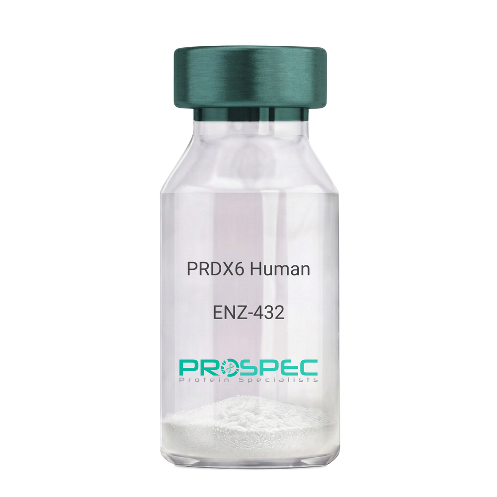
PRDX6 HumanDescription:
Peroxiredoxin-6 Human Recombinant
Peroxiredoxin-6, Antioxidant protein 2, 1-Cys peroxiredoxin, Acidic calcium-independent phospholipase A2, Non-selenium glutathione peroxidase, 24 kDa protein, Liver 2D page spot 40, Red blood cells page spot 12, 1-Cys PRX, aiPLA2, NSGPx, PRDX6, AOP2, KIAA0106, PRX, p29, 1-Cys, MGC46173.
Product # :
ENZ-432Price :
Quantity :
Shipping Method :
Shipped with Ice Packs
More Info
- description
- source
- formulation
- purity
- biological activity
- More Info
Description
Peroxiredoxin- 6 Human Recombinant fused with a 20 amino acid His tag at N-terminus produced in E.Coli is a single, non-glycosylated, polypeptide chain containing 244 amino acids (1-224 a.a.) and having a molecular mass of 27.1kDa.The Peroxiredoxin-6 is purified by proprietary chromatographic techniques.
Source
Escherichia Coli.
Formulation
The Peroxiredoxin-6 solution contains 20mM Tris-HCl buffer (pH8.0) and 20% Glycerol.
Purity
Greater than 95.0% as determined by SDS-PAGE.
Biological Activity
Specific activity is >2,000pmol/min/ug was defined as the amount of hydroperoxide that 1ug of enzyme can reduce at 25C for 1minute.
More Info
-
Introduction
Peroxiredoxin 6 (PRDX6) belongs to the thiol-specific antioxidant protein family. PRDX6 is a bifunctional enzyme with 2 distinct active sites. PRDX6 is involved in redox regulation of the cell and can reduce Hydrogen peroxide and short chain organic, fatty acid, and phospholipid hydroperoxides. PRDX6 may have a role in the regulation of phospholipid turnover as well as in protection against oxidative injury. Furthermore, PRDX6 eases the oxidative stress and TGF-beta-induced abnormalities of human trabecular meshwork cells. In addition, PRDX6 is necessary for blood vessel integrity in injured skin.
At acidic pH, PRDX6 binds to reduced phospholipids, however at cytosolic pH PRDX6 binds only to phospholipids that are oxidized which is compatible with the role for PRDX6 in the repair of peroxidized cell membranes. Hydrogen peroxide-mediated hyperoxidation of PRDX6 induces cell cycle arrest at the G2/M transition via up-regulation of iPLA2 activity. Overexpression of PRDX6 is linked to oligodendroglioma. -
Synonyms
Peroxiredoxin-6, Antioxidant protein 2, 1-Cys peroxiredoxin, Acidic calcium-independent phospholipase A2, Non-selenium glutathione peroxidase, 24 kDa protein, Liver 2D page spot 40, Red blood cells page spot 12, 1-Cys PRX, aiPLA2, NSGPx, PRDX6, AOP2, KIAA0106, PRX, p29, 1-Cys, MGC46173.
-
Physical Appearance
Sterile Filtered colorless solution.
-
Stability
Store at 4°C if entire vial will be used within 2-4 weeks. Store, frozen at -20°C for longer periods of time. For long term storage it is recommended to add a carrier protein (0.1% HSA or BSA).Avoid multiple freeze-thaw cycles.
-
Amino Acid Sequence
MGSSHHHHHH SSGLVPRGSH MPGGLLLGDV APNFEANTTV GRIRFHDFLG DSWGILFSHP RDFTPVCTTE LGRAAKLAPE FAKRNVKLIA LSIDSVEDHL AWSKDINAYN CEEPTEKLPF PIIDDRNREL AILLGMLDPA EKDEKGMPVT ARVVFVFGPD KKLKLSILYP ATTGRNFDEI LRVVISLQLT AEKRVATPVD WKDGDSVMVL PTIPEEEAKK LFPKGVFTKE LPSGKKYLRY TPQP.
ProSpec's products are furnished for LABORATORY RESEARCH USE ONLY. The product may not be used as drugs, agricultural or pesticidal products, food additives or household chemicals.
- View Data Sheet
Name :
CDKN1B HumanDescription:
Cyclin-Dependent Kinase Inhibitor 1B Human Recombinant
Cyclin-dependent kinase inhibitor 1B, Cyclin-dependent kinase inhibitor p27, p27Kip1, CDKN1B, KIP1, MEN4, CDKN4, MEN1B.
Product # :
PKA-012Price :
Quantity :
Shipping Method :
Shipped with Ice Packs
More Info
- description
- source
- formulation
- purity
- More Info
Description
CDKN1B Human Recombinant produced in E.Coli is a single, non-glycosylated polypeptide chain containing 218 amino acids (1-198 a.a.) and having a molecular mass of 24.2kDa (Molecular weight on SDS-PAGE will appear higher).CDKN1B is fused to a 20 amino acid His-tag at N-terminus & purified by proprietary chromatographic techniques.
Source
Escherichia Coli.
Formulation
CDKN1B protein solution (0.25mg/ml) containing 20mM Tris-HCl buffer (pH8.0) and 20% glycerol.
Purity
Greater than 90.0% as determined by SDS-PAGE.
More Info
-
Introduction
Cyclin-dependent kinase inhibitor 1B (CDKN1B) is a member of the Cip/Kip family of cyclin dependent kinase (Cdk) inhibitor proteins. The CDKN1B protein binds to and prevents the activation of cyclin E-CDK2 or cyclin D-CDK4 complexes, and so controls the cell cycle progression at G1. CDKN1B is often referred to as a cell cycle inhibitor protein since its major function is to stop or slow down the cell division cycle. The degradation of the CDKN1B protein, which is triggered by its CDK dependent phosphorylation and ensuing ubiquitination by SCF complexes, is compulsory for the cellular transition from inactive to the proliferative state.
-
Synonyms
Cyclin-dependent kinase inhibitor 1B, Cyclin-dependent kinase inhibitor p27, p27Kip1, CDKN1B, KIP1, MEN4, CDKN4, MEN1B.
-
Physical Appearance
Sterile filtered colorless solution.
-
Stability
Store at 4°C if entire vial will be used within 2-4 weeks. Store, frozen at -20°C for longer periods of time. For long term storage it is recommended to add a carrier protein (0.1% HSA or BSA).Avoid multiple freeze-thaw cycles.
-
Amino Acid Sequence
MGSSHHHHHH SSGLVPRGSH MSNVRVSNGS PSLERMDARQ AEHPKPSACR NLFGPVDHEE LTRDLEKHCR DMEEASQRKW NFDFQNHKPL EGKYEWQEVE KGSLPEFYYR PPRPPKGACK VPAQESQDVS GSRPAAPLIG APANSEDTHL VDPKTDPSDS QTGLAEQCAG IRKRPATDDS STQNKRANRT EENVSDGSPN AGSVEQTPKK PGLRRRQT.
ProSpec's products are furnished for LABORATORY RESEARCH USE ONLY. The product may not be used as drugs, agricultural or pesticidal products, food additives or household chemicals.
- View Data Sheet
Name :
TSFM HumanDescription:
Ts Translation Elongation Factor Mitochondrial Human Recombinant
Elongation factor Ts, mitochondrial, Ts Translation Elongation Factor Mitochondrial, TSFM, EF-Ts, EF-TsMt, COXPD3.
Product # :
PRO-1971Price :
Quantity :
Shipping Method :
Shipped with Ice Packs
More Info
- description
- source
- formulation
- purity
- More Info
Description
TSFM Human Recombinant produced in E.Coli is a single, non-glycosylated polypeptide chain containing 302 amino acids (46-346 a.a) and having a molecular mass of 32.9kDa.TSFM is purified by proprietary chromatographic techniques.
Source
Escherichia Coli.
Formulation
TSFM protein solution (1mg/ml) containing 20mM Tris-HCl buffer (pH 8.0), 10% glycerol and 0.4M Urea.
Purity
Greater than 85.0% as determined by SDS-PAGE.
More Info
-
Introduction
TSFM is a mitochondrial translation elongation factor which is linked with the EF-Tu.GDP complex and induces the exchange of GDP to GTP. TSFM stays bound to the aminoacyl-tRNA.EF-Tu.GTP complex until the GTP hydrolysis stage on the ribosome. Mutations in TSFM are related with combined oxidative phosphorylation deficiency-3 syndrome.
-
Synonyms
Elongation factor Ts, mitochondrial, Ts Translation Elongation Factor Mitochondrial, TSFM, EF-Ts, EF-TsMt, COXPD3.
-
Physical Appearance
Sterile Filtered colorless solution.
-
Stability
Store at 4°C if entire vial will be used within 2-4 weeks. Store, frozen at -20°C for longer periods of time. For long term storage it is recommended to add a carrier protein (0.1% HSA or BSA).Avoid multiple freeze-thaw cycles.
-
Amino Acid Sequence
MSKELLMKLR RKTGYSFVNC KKALETCGGD LKQAEIWLHK EAQKEGWSKA AKLQGRKTKE GLIGLLQEGN TTVLVEVNCE TDFVSRNLKF QLLVQQVALG TMMHCQTLKD QPSAYSKVQW LTPVNLALWE AEAGGSLEGF LNSSELSGLP AGPDREGSLK DQLALAIGKL GENMILKRAA WVKVPSGFYV GSYVHGAMQS PSLHKLVLGK YGALVICETS EQKTNLEDVG RRLGQHVVGM APLSVGSLDD EPGGEAETKM LSQPYLLDPS ITLGQYVQPQ GVSVVDFVRF ECGEGEEAAE TE.
ProSpec's products are furnished for LABORATORY RESEARCH USE ONLY. The product may not be used as drugs, agricultural or pesticidal products, food additives or household chemicals.
- View Data Sheet
Name :
CHGB HumanDescription:
Chromogranin B Human Recombinant
SCG1, Secretogranin 1, secretogranin B, CHGB, Sgl, CgB.
Product # :
PRO-824Price :
Quantity :
Shipping Method :
Shipped with Ice Packs
More Info
- description
- source
- formulation
- purity
- More Info
Description
CHGB Human Recombinant produced in E.Coli is a single, non-glycosylated, polypeptide chain containing 677 amino acids (21-677 a.a.) and having a molecular mass of 78.4kDa. CHGB protein is fused to a 20 amino acid His-Tag at N-terminus and purified by standard chromatography.
Source
Escherichia Coli.
Formulation
The CHGB protein 0.25mg/ml solution containing 20mM Tris-HCl pH-8, 0.15M NaCl & 10% glycerol.
Purity
Greater than 80% as determined by SDS-PAGE.
More Info
-
Introduction
CHGB is a neuroendocrine secretory granule protein, that is the precursor for other biologically active peptides. CHGB is part of the chromogranin/secretogranin protein family and is expressed in the adrenal medulla, and in pheochromocytoma.
-
Synonyms
SCG1, Secretogranin 1, secretogranin B, CHGB, Sgl, CgB.
-
Physical Appearance
Sterile filtered colorless solution.
-
Stability
Store at 4°C if entire vial will be used within 2-4 weeks. Store, frozen at -20°C for longer periods of time. For long term storage it is recommended to add a carrier protein (0.1% HSA or BSA).Avoid multiple freeze-thaw cycles.
-
Amino Acid Sequence
MGSSHHHHHH SSGLVPRGSH MPVDNRNHNE GMVTRCIIEV LSNALSKSSA PPITPECRQV LKTSRKDVKD KETTENENTK FEVRLLRDPA DASEAHESSS RGEAGAPGEE DIQGPTKADT EKWAEGGGHS RERADEPQWS LYPSDSQVSE EVKTRHSEKS QREDEEEEEG ENYQKGERGE DSSEEKHLEE PGETQNAFLN ERKQASAIKK EELVARSETH AAGHSQEKTH SREKSSQESG EEAGSQENHP QESKGQPRSQ EESEEGEEDA TSEVDKRRTR PRHHHGRSRP DRSSQGGSLP SEEKGHPQEE SEESNVSMAS LGEKRDHHST HYRASEEEPE YGEEIKGYPG VQAPEDLEWE RYRGRGSEEY RAPRPQSEES WDEEDKRNYP SLELDKMAHG YGEESEEERG LEPGKGRHHR GRGGEPRAYF MSDTREEKRF LGEGHHRVQE NQMDKARRHP QGAWKELDRN YLNYGEEGAP GKWQQQGDLQ DTKENREEAR FQDKQYSSHH TAEKRKRLGE LFNPYYDPLQ WKSSHFERRD NMNDNFLEGE EENELTLNEK NFFPEYNYDW WEKKPFSEDV NWGYEKRNLA RVPKLDLKRQ YDRVAQLDQL LHYRKKSAEF PDFYDSEEPV STHQEAENEK DRADQTVLTE DEKKELENLA AMDLELQKIA EKFSQRG.
ProSpec's products are furnished for LABORATORY RESEARCH USE ONLY. The product may not be used as drugs, agricultural or pesticidal products, food additives or household chemicals.
- View Data Sheet
Name :
COPS8 HumanDescription:
COP9 Constitutive Photomorphogenic 8 Human Recombinant
COP9 signalosome complex subunit 8, SGN8, Signalosome subunit 8, COP9 homolog, hCOP9, JAB1-containing signalosome subunit 8, COPS8, CSN8, COP9.
Product # :
PRO-983Price :
Quantity :
Shipping Method :
Shipped with Ice Packs
More Info
- description
- source
- formulation
- purity
- More Info
Description
COPS8 Human Recombinant produced in E.coli is a single, non-glycosylated polypeptide chain containing 229 amino acids (1-209) and having a molecular mass of 25.3kDa.COPS8 is fused to a 20 amino acid His-tag at N-terminus & purified by proprietary chromatographic techniques.
Source
Escherichia Coli.
Formulation
The COPS8 solution (1mg/ml) contains 20mM Tris-HCl buffer (pH 8.0), 0.4M Urea and 10% glycerol.
Purity
Greater than 90.0% as determined by SDS-PAGE.
More Info
-
Introduction
COP9 signalosome complex subunit 8 isoform 1 (COPS8) is one of the 8 subunits of COP9 signalosome, which is a much conserved protein complex that functions as an imperative regulator in multiple signaling pathways. The structure and function of COP9 signalosome is analogous to that of the 19S regulatory particle of 26S proteasome. COP9 signalosome interacts with SCF-type E3 ubiquitin ligases and acts as a positive regulator of E3 ubiquitin ligases.
-
Synonyms
COP9 signalosome complex subunit 8, SGN8, Signalosome subunit 8, COP9 homolog, hCOP9, JAB1-containing signalosome subunit 8, COPS8, CSN8, COP9.
-
Physical Appearance
Sterile Filtered colorless solution.
-
Stability
Store at 4°C if entire vial will be used within 2-4 weeks. Store, frozen at -20°C for longer periods of time. For long term storage it is recommended to add a carrier protein (0.1% HSA or BSA).Avoid multiple freeze-thaw cycles.
-
Amino Acid Sequence
MGSSHHHHHH SSGLVPRGSH MPVAVMAESA FSFKKLLDQC ENQELEAPGG IATPPVYGQL LALYLLHNDM NNARYLWKRI PPAIKSANSE LGGIWSVGQR IWQRDFPGIY TTINAHQWSE TVQPIMEALR DATRRRAFAL VSQAYTSIIA DDFAAFVGLP VEEAVKGILE QGWQADSTTR
MVLPRKPVAG ALDVSFNKFI PLSEPAPVPP IPNEQQLARL TDYVAFLEN.
ProSpec's products are furnished for LABORATORY RESEARCH USE ONLY. The product may not be used as drugs, agricultural or pesticidal products, food additives or household chemicals.
- View Data Sheet
Name :
OX40L HumanDescription:
OX40 Ligand Human Recombinant
Tumor necrosis factor ligand superfamily member 4, OX40 ligand, OX40L, OX-40L, CD252, Tnfsf4, Ox40l, Txgp1l, gp3, OX4, Ath-1, Ath1, CD134L, gp34, Tnlg2b, Txgp1l.
Product # :
CYT-1226Price :
Quantity :
Shipping Method :
Shipped with Ice Packs
More Info
- description
- source
- formulation
- purity
- biological activity
- More Info
Description
OX40L Human Recombinant is a single, glycosylated, polypeptide chain (51-183 a.a) containing a total of139 amino acids and having a molecular mass of 16.2 kDa. OX40L is fused to a 6 a.a his-Tag at C-terminus and is purified by proprietary chromatographic techniques.
Source
HEK293 Cells.
Formulation
The OX40L solution (1mg/ml) contains 10% Glycerol and Phosphate-Buffered Saline (pH 7.4).
Purity
Greater than 90.0% as determined by SDS-PAGE.
Biological Activity
Measured by its binding ability in a functional ELISA with Human OX40/TNFRSF4.
More Info
-
Synonyms
Tumor necrosis factor ligand superfamily member 4, OX40 ligand, OX40L, OX-40L, CD252, Tnfsf4, Ox40l, Txgp1l, gp3, OX4, Ath-1, Ath1, CD134L, gp34, Tnlg2b, Txgp1l.
-
Physical Appearance
Sterile Filtered colorless solution.
-
Stability
Store at 4°C if entire vial will be used within 2-4 weeks. Store, frozen at -20°C for longer periods of time. For long term storage it is recommended to add a carrier protein (0.1% HSA or BSA). Avoid multiple freeze-thaw cycles.
-
Amino Acid Sequence
QVSHRYPRIQ SIKVQFTEYK KEKGFILTSQ KEDEIMKVQN NSVIINCDGF YLISLKGYFS QEVNISLHYQ KDEEPLFQLK KVRSVNSLMV ASLTYKDKVY LNVTTDNTSL DDFHVNGGEL ILIHQNPGEF CVLHHHHHH.
-
Background
OX40 Ligand (OX40L), a member of the tumor necrosis factor (TNF) superfamily, plays a pivotal role in regulating immune responses and orchestrating the delicate balance between activation and tolerance. The human recombinant form of OX40L has emerged as a potent tool in immunology, offering insights into its molecular intricacies and potential applications in therapeutic interventions. This research embarks on a journey to unravel the multifaceted role of OX40L Human Recombinant, shedding light on its structural attributes, signaling pathways, and its promising avenues in immunotherapy. By delving into the properties of OX40L, scientists aim to expand our understanding of immune modulation and open new frontiers in the treatment of immune-related disorders.
Structural Insights into OX40L Human Recombinant:
OX40L, as a trimeric transmembrane protein, exhibits a unique structural configuration that governs its interactions with the OX40 receptor on T cells. The human recombinant form, engineered for controlled study, provides a window into the three-dimensional intricacies of the ligand. Understanding its structure is pivotal for deciphering how OX40L engages with its receptor and modulates immune responses.
Immunomodulatory Signaling Pathways:
OX40L binding to its cognate receptor OX40 on T cells triggers intricate signaling cascades that impact immune cell activation, proliferation, and cytokine production. The OX40-OX40L axis is a crucial regulator of T cell function, influencing both effector and regulatory T cell responses. Unraveling the specific pathways activated by OX40L Human Recombinant provides valuable insights into the modulation of immune responses in health and disease.
Applications in Immunotherapy:
The immunomodulatory properties of OX40L make it an attractive candidate for therapeutic interventions. OX40L Human Recombinant, in preclinical and clinical studies, is being explored for its potential in enhancing antitumor immune responses. By harnessing the ligand's ability to stimulate effector T cells and memory T cell formation, researchers aim to develop novel immunotherapies for cancer and other immune-related disorders.
OX40L in Autoimmune Diseases:
Conversely, OX40L's role in autoimmune diseases has spurred investigations into its inhibition as a therapeutic strategy. Blocking the OX40-OX40L interaction has shown promise in mitigating autoimmune responses, presenting a potential avenue for the development of treatments for conditions such as rheumatoid arthritis and inflammatory bowel disease.
While the potential of OX40L Human Recombinant in immunotherapy is promising, challenges persist. Fine-tuning its applications, understanding potential side effects, and optimizing dosages are critical considerations for translational success. Additionally, comprehending the context-dependent nature of OX40L signaling is essential for tailoring therapeutic strategies to specific diseases and patient profiles.
OX40L Human Recombinant stands at the forefront of immunomodulation research, offering a lens through which we can unravel the complexities of immune responses. Its structural insights, signaling pathways, and therapeutic applications position it as a key player in the evolving landscape of immunotherapy. As researchers continue to dissect the molecular nuances of OX40L, they not only expand our understanding of immune regulation but also pave the way for transformative advancements in the treatment of cancer and autoimmune diseases, shaping the future of precision medicine and immunotherapy.
ProSpec's products are furnished for LABORATORY RESEARCH USE ONLY. The product may not be used as drugs, agricultural or pesticidal products, food additives or household chemicals.
- View Data Sheet
Name :
CD116 Human, sf9Description:
GM-CSF Receptor Alpha Sf9 Human Recombinant
Colony Stimulating Factor 2 Receptor Alpha Subunit, Colony Stimulating Factor 2 Receptor, Alpha, Low-Affinity (Granulocyte-Macrophage), Alpha-GM-CSF Receptor, GM-CSF-R-Alpha, CD116 Antigen, GMCSFR-Alpha, GMR-Alpha, CDw116, CSF2RY, CSF2R, Granulocyte-Macrophage Colony-Stimulating Factor Receptor Subunit Alpha, Granulocyte-Macrophage Colony-Stimulating Factor Receptor Alpha Chain, GM-CSF Receptor Alpha Subunit, AlphaGMR, CSF2RAX, CSF2RAY, CSF2RX, GMCSFR, CD116, SMDP4, GMR.
Product # :
CYT-1044Price :
Quantity :
Shipping Method :
Shipped with Ice Packs
More Info
- description
- source
- formulation
- purity
- biological activity
- More Info
Description
CSF2RA produced in Sf9 Baculovirus cells is a single, glycosylated polypeptide chain containing 310 amino acids (20-320a.a.) and having a molecular mass of 35.9kDa. (Molecular size on SDS-PAGE will appear at approximately 40-57kDa). CSF2RA is expressed with a 9 amino acid His tag at C-Terminus and purified by proprietary chromatographic techniques.
Source
Sf9, Baculovirus cells.
Formulation
CSF2RA protein solution (0.5mg/ml) contains Phosphate Buffered Saline (pH 7.4) and 10% glycerol.
Purity
Greater than 90.0% as determined by SDS-PAGE.
Biological Activity
Measured by its ability to inhibit GM-CSF dependent proliferation of TF1 human erythroleukemic cells. The ED50 for this effect is less or equal to 10ug/ml in the presence of 0.5ng/ml GM-CSF.
More Info
-
Introduction
GM-CSF Receptor Alpha (CSF2RA) is the alpha subunit of the heterodimeric receptor for colony stimulating factor 2, a cytokine which controls the production, differentiation, and function of granulocytes and macrophages. CSFR2 is also a member of the cytokine family of receptors. In addition, this gene is found in the pseudoautosomal region (PAR) of the X and Y chromosomes. Multiple transcript variants encoding various isoforms have been found for this gene, while some of the isoforms being membrane-bound and others being soluble. Diseases associated with CSF2RA include surfactant metabolism dysfunction, pulmonary 4, and csf2ra-related pulmonary surfactant metabolism dysfunction.
-
Synonyms
Colony Stimulating Factor 2 Receptor Alpha Subunit, Colony Stimulating Factor 2 Receptor, Alpha, Low-Affinity (Granulocyte-Macrophage), Alpha-GM-CSF Receptor, GM-CSF-R-Alpha, CD116 Antigen, GMCSFR-Alpha, GMR-Alpha, CDw116, CSF2RY, CSF2R, Granulocyte-Macrophage Colony-Stimulating Factor Receptor Subunit Alpha, Granulocyte-Macrophage Colony-Stimulating Factor Receptor Alpha Chain, GM-CSF Receptor Alpha Subunit, AlphaGMR, CSF2RAX, CSF2RAY, CSF2RX, GMCSFR, CD116, SMDP4, GMR.
-
Physical Appearance
Sterile Filtered colorless solution.
-
Stability
Store at 4°C if entire vial will be used within 2-4 weeks. Store, frozen at -20°C for longer periods of time. For long term storage it is recommended to add a carrier protein (0.1% HSA or BSA). Avoid multiple freeze-thaw cycles.
-
Amino Acid Sequence
ADPLIPEKSD LRTVAPASSL NVRFDSRTMN LSWDCQENTT FSKCFLTDKK NRVVEPRLSN NECSCTFREI CLHEGVTFEV HVNTSQRGFQ QKLLYPNSGR EGTAAQNFSC FIYNADLMNC TWARGPTAPR DVQYFLYIRN SKRRREIRCP YYIQDSGTHV GCHLDNLSGL TSRNYFLVNG TSREIGIQFF DSLLDTKKIE RFNPPSNVTV RCNTTHCLVR WKQPRTYQKL SYLDFQYQLD VHRKNTQPGT ENLLINVSGD LENRYNFPSS EPRAKHSVKI RAADVRILNW SSWSEAIEFG SDDGHHHHHH
-
Background
GM-CSF Receptor Alpha Human Recombinant: A Glimpse into Its Potential and Implications
Abstract:
Granulocyte-Macrophage Colony Stimulating Factor (GM-CSF) receptor alpha, a pivotal component in the GM-CSF signaling pathway, has been the focal point of numerous studies pertaining to hematopoiesis and immune responses. This paper provides an overview of the GM-CSF receptor alpha human recombinant, exploring its characteristics, production techniques, and potential therapeutic applications.
Introduction
GM-CSF, a cytokine responsible for the differentiation and proliferation of white blood cells, functions by binding to its receptor, GM-CSF receptor. The alpha subunit (GM-CSFRα) of this receptor plays a crucial role in ligand binding and is essential for initiating cellular responses. Modern biotechnological advancements have led to the successful production of its human recombinant form, offering new avenues in medical research.
Recombinant GM-CSFRα:
Production and Features Recombinant GM-CSFRα is synthesized using cutting-edge recombinant DNA technologies, predominantly in bacterial or mammalian expression systems. This human recombinant form retains its ability to bind to GM-CSF, maintaining its biological functionality and providing myriad research opportunities.
Therapeutic and Clinical Prospects
- Autoimmune Diseases: GM-CSF is often overexpressed in various autoimmune disorders. By utilizing recombinant GM-CSFRα as a potential decoy receptor, it's feasible to mitigate the effects of excessive GM-CSF, offering a new therapeutic strategy.
- Hematopoietic Disorders: Given its integral role in white blood cell development, recombinant GM-CSFRα might hold promise in treatments or as a diagnostic tool for certain hematological conditions.
- Research Paradigm: Beyond therapeutic applications, the recombinant GM-CSFRα can serve as an invaluable research tool to elucidate the nuances of GM-CSF signaling, aiding in the understanding of immune response mechanisms.
Conclusion:
GM-CSF receptor alpha human recombinant stands at the forefront of exciting research and therapeutic potential. While its full capabilities are yet to be realized, current insights underscore its significance in the realms of immunology and medicine.
What is the molecular weight/Mw of CD116 Protein?
CD116 Protein has a total Mw of 35.9kDa.
What is the source or expression system of CD116 Protein?
Sf9, Baculovirus cells.
What is the Purity of CD116 Protein?
CD116 Protein is >90% pure as determined by SDS-PAGE.
What is the Biological Activity of CD116 Protein?
Measured by its ability to inhibit GM-CSF dependent proliferation of TF1 human erythroleukemic cells. The ED50 for this effect is less or equal to 10ug/ml in the presence of 0.5ng/ml GM-CSF.
What is the amino acid sequence of CD116 Protein?
CD116 Protein is composed from 310 amino acids.
What applications can CD116 Protein be used in?
CD116 Protein can probably be used in western blot, ELISA and Lateral Flow.
What is the endotoxin level for CD116 Protein?
The endotoxin level is minimal, CD116 Protein was purified using conventional chromatography techniques
ProSpec's products are furnished for LABORATORY RESEARCH USE ONLY. The product may not be used as drugs, agricultural or pesticidal products, food additives or household chemicals.
- View Data Sheet
Name :
PCSK1N HumanDescription:
Proprotein Convertase Subtilisin/Kexin Type 1 Inhibitor Human Recombinant
ProSAAS precursor, Proprotein convertase subtilisin/kexin type 1 inhibitor, PROSAAS; SAAS, PCSK1N.
Product # :
PRO-1819Price :
Quantity :
Shipping Method :
Shipped with Ice Packs
More Info
- description
- source
- formulation
- purity
- More Info
Description
PCSK1N Human Recombinant produced in E.coli is a single, non-glycosylated polypeptide chain containing 251 amino acids (34-260) and having a molecular mass of 26.6 kDa.PCSK1N is fused to a 23 amino acid His-Tag at N-terminus and purified by proprietary chromatographic techniques.
Source
Escherichia Coli.
Formulation
The PCSK1N solution (0.25mg/ml) contains 20mM Tris-HCl buffer (pH 8.0), 10% glycerol and 0.4M Urea.
Purity
Greater than 85.0% as determined by SDS-PAGE.
More Info
-
Introduction
Proprotein Convertase Subtilisin/Kexin Type 1 Inhibitor (PCSK1N) takes part in the control of the neuroendocrine secretory pathway. PCSK1N inhibits prohormone convertase 1, which regulates the proteolytic cleavage of neuroendocrine peptide precursors. PCSK1Nslows down convertase-mediated processing of proopiomelanocortin and proenkephalin and also monitors the intracellular timing of PCSK1.
-
Synonyms
ProSAAS precursor, Proprotein convertase subtilisin/kexin type 1 inhibitor, PROSAAS; SAAS, PCSK1N.
-
Physical Appearance
Sterile Filtered colorless solution.
-
Stability
Store at 4°C if entire vial will be used within 2-4 weeks. Store, frozen at -20°C for longer periods of time. For long term storage it is recommended to add a carrier protein (0.1% HSA or BSA).Avoid multiple freeze-thaw cycles.
-
Amino Acid Sequence
MGSSHHHHHH SSGLVPRGSH MGSMARPVKE PRGLSAASPP LAETGAPRRF RRSVPRGEAA GAVQELARAL AHLLEAERQE RARAEAQEAE DQQARVLAQL LRVWGAPRNS DPALGLDDDP DAPAAQLARA LLRARLDPAA LAAQLVPAPV PAAALRPRPP VYDDGPAGPD AEEAGDETPD VDPELLRYLL GRILAGSADS EGVAAPRRLR RAADHDVGSE LPPEGVLGAL LRVKRLETPA PQVPARRLLP P.
ProSpec's products are furnished for LABORATORY RESEARCH USE ONLY. The product may not be used as drugs, agricultural or pesticidal products, food additives or household chemicals.
- View Data Sheet
Name :
TRAIL MouseDescription:
TNF-Related Apoptosis Inducing Ligand/Apo2L Mouse Recombinant
Tumor necrosis factor ligand superfamily member 10, TNF-related apoptosis-inducing ligand, Protein TRAIL, Apo-2 ligand, Apo-2L, CD253 antigen, TL2, APO2L, TNFSF10.
Product # :
CYT-806Price :
Quantity :
Shipping Method :
Shipped at Room temp
More Info
- description
- source
- formulation
- purity
- biological activity
- More Info
Description
TRAIL Recombinant Mouse produced in E.coli is a single, non-glycosylated polypeptide chain containing 175 amino acids and having a molecular mass of 20.2kDa.
Source
Escherichia Coli.
Formulation
The protein was lyophilized containing PBS, pH 7.4, and 3mM DTT.
Purity
Greater than 95.0% as determined by:(a) Analysis by RP-HPLC.(b) Analysis by SDS-PAGE.
Biological Activity
Fully biologically active when compared to standard. The ED50 as determined by a cytotoxicity assay using murine L929 cells is less than 0.5 ng/ml, corresponding to a specific activity of > 2,000,000 IU/mg in the presence of actinomycin D.More Info
-
Introduction
TNF-related apoptosis-inducing ligand (TRAIL) is a ligand molecule which induces apoptosis. It is a type II transmembrane protein with homology to other members of the tumor necrosis factor family.In humans, the gene that encodes for TRAIL is located at chromosome 3q26. TRAIL binds to the death receptors, DR4 and DR5. The process of apoptosis is caspase-8-dependent. This protein preferentially induces apoptosis in transformed and tumor cells, but does not appear to kill normal cells although it is expressed at a significant level in most normal tissues.
-
Synonyms
Tumor necrosis factor ligand superfamily member 10, TNF-related apoptosis-inducing ligand, Protein TRAIL, Apo-2 ligand, Apo-2L, CD253 antigen, TL2, APO2L, TNFSF10.
-
Physical Appearance
Sterile Filtered White lyophilized (freeze-dried) powder.
-
Stability
Lyophilized TRAIL although stable at room temperature for 3 weeks, should be stored desiccated below -18C. Upon reconstitution TRAIL recombinant should be stored at 4C between 2-7 days and for future use below -18C.For long term storage it is recommended to add a carrier protein (0.1% HSA or BSA).Please prevent freeze-thaw cycles.
-
Solubility
It is recommended to reconstitute the lyophilized TRAIL Mouse Recombinant in sterile 18MΩ-cm H2O not less than 100 µg/ml, which can then be further diluted to other aqueous solutions.
-
Amino Acid Sequence
MPRGGRPQKV AAHITGITRR SNSALIPISK DGKTLGQKIE SWESSRKGHS FLNHVLFRNG ELVIEQEGLY YIYSQTYFRF QEAEDASKMV SKDKVRTKQL VQYIYKYTSY PDPIVLMKSA RNSCWSRDAE YGLYSIYQGG LFELKKNDRI FVSVTNEHLM DLDQEASFFG AFLIN
ProSpec's products are furnished for LABORATORY RESEARCH USE ONLY. The product may not be used as drugs, agricultural or pesticidal products, food additives or household chemicals.
- View Data Sheet
Name :
STK17B HumanDescription:
Serine/Threonine Kinase 17B Human Recombinant
Serine/threonine-protein kinase 17B, EC 2.7.11.1, DAP kinase-related apoptosis-inducing protein kinase 2, STK17B, DRAK2.
Product # :
PKA-050Price :
Quantity :
Shipping Method :
Shipped with Ice Packs
More Info
- description
- source
- formulation
- purity
- More Info
Description
STK17B Human Recombinant produced in E.Coli is a single, non-glycosylated polypeptide chain containing 395 amino acids (1-372 a.a) and having a molecular mass of 44.7kDa.STK17B is fused to a 23 amino acid His-tag at N-terminus & purified by proprietary chromatographic techniques.
Source
Escherichia Coli.
Formulation
STK17B protein solution (0.5mg/ml) containing 20mM Tris-HCl buffer (pH 8.0), 0.1M NaCl, 10% glycerol and 2mM DTT.
Purity
Greater than 90.0% as determined by SDS-PAGE.
More Info
-
Introduction
Serine/threonine kinase 17b (STK17B) is a member of the protein kinase superfamily. STK17B functions as a positive regulator of apoptosis. STK17B interacts with CHP1; this interaction stimulates CHP1 to translocate from the Golgi to the nucleus. STK17B is highly expressed in the placenta, lung, pancreas, however it has lower levels in the heart, brain, liver, skeletal muscle and kidney.
-
Synonyms
Serine/threonine-protein kinase 17B, EC 2.7.11.1, DAP kinase-related apoptosis-inducing protein kinase 2, STK17B, DRAK2.
-
Physical Appearance
Sterile filtered colorless solution.
-
Stability
Store at 4°C if entire vial will be used within 2-4 weeks. Store, frozen at -20°C for longer periods of time. For long term storage it is recommended to add a carrier protein (0.1% HSA or BSA).Avoid multiple freeze-thaw cycles.
-
Amino Acid Sequence
MGSSHHHHHH SSGLVPRGSH MGSMSRRRFD CRSISGLLTT TPQIPIKMEN FNNFYILTSK ELGRGKFAVV RQCISKSTGQ EYAAKFLKKR RRGQDCRAEI LHEIAVLELA KSCPRVINLH EVYENTSEII LILEYAAGGE IFSLCLPELA EMVSENDVIR LIKQILEGVY YLHQNNIVHL DLKPQNILLS SIYPLGDIKI VDFGMSRKIG HACELREIMG TPEYLAPEIL NYDPITTATD MWNIGIIAYM LLTHTSPFVG EDNQETYLNI SQVNVDYSEE TFSSVSQLAT DFIQSLLVKN PEKRPTAEIC LSHSWLQQWD FENLFHPEET SSSSQTQDHS VRSSEDKTSK SSCNGTCGDR EDKENIPEDS SMVSKRFRFD DSLPNPHELV SDLLC.
ProSpec's products are furnished for LABORATORY RESEARCH USE ONLY. The product may not be used as drugs, agricultural or pesticidal products, food additives or household chemicals.
- View Data Sheet
Name :
GAGE12F HumanDescription:
G Antigen 12F Human Recombinant
G Antigen 12F, GAGE-12F, G Antigen 12F/G/I.
Product # :
PRO-1736Price :
Quantity :
Shipping Method :
Shipped with Ice Packs
More Info
- description
- source
- formulation
- purity
- More Info
Description
GAGE12F Human Recombinant produced in E.Coli is a single, non-glycosylated polypeptide chain containing 140 amino acids (1-117aa) and having a molecular mass of 15.4kDa.GAGE12F is fused to a 23 amino acid His-tag at N-terminus & purified by proprietary chromatographic techniques.
Source
Escherichia Coli.
Formulation
GAGE12F protein solution (0.25mg/ml) containing 20mM Tris-HCl buffer (pH 8.0), 0.15M NaCl and 10% glycerol.
Purity
Greater than 85.0% as determined by SDS-PAGE.
More Info
-
Introduction
GAGE12F is a member of the GAGE multigene family expressed in a large range of tumors whereas in normal tissues, his expression is restricted to germ cells. These genes are organized in clustered repeats, and have a high degree of forecasted sequence identity, however differ by scattered single nucleotide substitution. Their sequences contain either the antigenic peptide YYWPRPRRY or YRPRPRRY which is recognized by cytotoxic T-cells.
-
Synonyms
G Antigen 12F, GAGE-12F, G Antigen 12F/G/I.
-
Physical Appearance
Sterile Filtered clear solution.
-
Stability
Store at 4°C if entire vial will be used within 2-4 weeks. Store, frozen at -20°C for longer periods of time. For long term storage it is recommended to add a carrier protein (0.1% HSA or BSA).Avoid multiple freeze-thaw cycles.
-
Amino Acid Sequence
MGSSHHHHHH SSGLVPRGSH MGSMSWRGRS TYYWPRPRRY VQPPEMIGPM RPEQFSDEVE PATPEEGEPA TQRQDPAAAQ EGEDEGASAG QGPKPEAHSQ EQGHPQTGCE CEDGPDGQEM DPPNPEEVKT PEEGEKQSQC
ProSpec's products are furnished for LABORATORY RESEARCH USE ONLY. The product may not be used as drugs, agricultural or pesticidal products, food additives or household chemicals.
- View Data Sheet
Name :
GPT2 Mouse, ActiveDescription:
Glutamic-Pyruvate Transaminase 2, Active Mouse Recombinant
ALT2, AAT2, Alanine aminotransferase 2, Glutamate pyruvate transaminase 2, Glutamic--alanine transaminase 2, Glutamic--pyruvic transaminase 2.
Product # :
ENZ-1110Price :
Quantity :
Shipping Method :
Shipped with Ice Packs
More Info
- description
- source
- formulation
- purity
- biological activity
- More Info
Description
GPT2 Mouse Recombinant produced in E.Coli is a single, non-glycosylated polypeptide chain containing 542 amino acids (1-522 aa) and having a molecular mass of 60.1kDa.GPT2 is fused to a 20 amino acid His tag at N-terminus and purified by proprietary chromatographic techniques.
Source
Escherichia Coli.
Formulation
The GPT2 solution (1 mg/ml) contains 2mM DTT, 20% Glycerol and 20mM Tris-HCl (pH7.5).
Purity
Greater than 85.0% as determined by SDS-PAGE.
Biological Activity
Specific activity is > 50 units/mg. Measured by the amount of enzyme that cleaves 1umole of L-Alanineto L-Glutamate per minute at pH7.5 at 37C˚.
More Info
-
Introduction
Glutamic-Pyruvate Transaminase 2 (GPT2) catalyzes the reversible transamination among alanine and 2-oxoglutarate to create pyruvate and glutamate. GPT2 expressed mainly in muscle, fat and kidney and participates in the intermediary metabolism of glucose and amino acids.
-
Synonyms
ALT2, AAT2, Alanine aminotransferase 2, Glutamate pyruvate transaminase 2, Glutamic--alanine transaminase 2, Glutamic--pyruvic transaminase 2.
-
Physical Appearance
Sterile Filtered colorless solution.
-
Stability
Store at 4°C if entire vial will be used within 2-4 weeks. Store, frozen at -20°C for longer periods of time. For long term storage it is recommended to add a carrier protein (0.1% HSA or BSA).Avoid multiple freeze-thaw cycles.
-
Amino Acid Sequence
MGSSHHHHHH SSGLVPRGSH MQRAAVLVRR GSCPRASGPW GRSHSSAAAE ASAALKVRPE
RSPRDRILTL ESMNPQVKAV EYAVRGPIVL KAGEIEMELQ RGIKKPFTEV IRANIGDAHA
MGQQPITFLR QVMALCTYPN LLNSPSFPED AKKRARRILQ ACGGNSLGSY SASQGVNCIR
EDVAAFITRR DGVPADPDNI YLTTGASDGI STILKLLVSG GGKSRTGVMI PIPQYPLYSA
VISELDAVQV NYYLDEENCW ALNVDELRRA LRQAKDHCDP KVLCIINPGN PTGQVQSRKC
IEDVIHFAWE EKLFLLADEV YQDNVYSPDC RFHSFKKVLY QMGHEYSSNV ELASFHSTSK
GYMGECGYRG GYMEVINLHP EIKGQLVKLL SVRLCPPVSG QAAMDIVVNP PEPGEESFEQ
FSREKEFVLG NLAKKAKLTE DLFNQVPGIQ CNPLQGAMYA FPRILIPAKA VEAAQSHKMA
PDMFYCMKLL EETGICVVPG SGFGQREGTY HFRMTILPPV DKLKTVLHKV KDFHLKFLEQ YS
ProSpec's products are furnished for LABORATORY RESEARCH USE ONLY. The product may not be used as drugs, agricultural or pesticidal products, food additives or household chemicals.
- View Data Sheet
Name :
MCSF Mouse, Sf9Description:
Macrophage Colony Stimulating Factor Mouse Recombinant, Sf9
M-Csf, Macrophage colony-stimulating factor 1, CSF-1, MCSF, Csf1, C87615, MCSF, op, Processedmacrophage colony-stimulating factor 1.
Product # :
CYT-1141Price :
Quantity :
Shipping Method :
Shipped with Ice Packs
More Info
- description
- source
- formulation
- purity
- biological activity
- More Info
Description
MCSF Mouse produced in Sf9 Baculovirus cells is a single, glycosylated polypeptide chain containing 164 amino acids (33-187 aa) and having a molecular mass of 19.1 kDa.MCSF is fused to a 6 amino acid His tag at C-terminus and purified by proprietary chromatographic techniques.
Source
Sf9, Baculovirus cells.
Formulation
The MCSF solution (1mg/ml) contains 10% Glycerol and Phosphate-Buffered Saline (pH 7.4).
Purity
Greater than 90.0% as determined by SDS-PAGE.
Biological Activity
Determined by cell proliferation assay using M-NFS-60 mouse myelogenous leukemia lymphoblast cells. ED50 range for this effect is ≤ 4ng/ml.
More Info
-
Introduction
Macrophage Colony Stimulating Factor (M CSF) or colony stimulating factor 1, is a cytokine, that promoted differentiation in hematopoietic stem cells to macrophages. Another role of M CSF is to bind to its receptor (colony stimulating factor 1 receptor) and to take part in placenta growth and development.
-
Synonyms
M-Csf, Macrophage colony-stimulating factor 1, CSF-1, MCSF, Csf1, C87615, MCSF, op, Processedmacrophage colony-stimulating factor 1.
-
Physical Appearance
Sterile Filtered colorless solution.
-
Stability
Store at 4°C if entire vial will be used within 2-4 weeks. Store, frozen at -20°C for longer periods of time. For long term storage it is recommended to add a carrier protein (0.1% HSA or BSA).Avoid multiple freeze-thaw cycles.
-
Amino Acid Sequence
ADPKEVSEHC SHMIGNGHLK VLQQLIDSQM ETSCQIAFEF VDQEQLDDPV CYLKKAFFLV
QDIIDETMRF KDNTPNANAT ERLQELSNNL NSCFTKDYEE QNKACVRTFH ETPLQLLEKI
KNFFNETKNL LEKDWNIFTK NCNNSFAKCS SRDVVTKPHH HHHH
ProSpec's products are furnished for LABORATORY RESEARCH USE ONLY. The product may not be used as drugs, agricultural or pesticidal products, food additives or household chemicals.
- View Data Sheet
Name :
ARF4 HumanDescription:
ADP-Ribosylation Factor 4 Human Recombinant
ADP-ribosylation factor 4, ARF4, ARF2.
Product # :
PRO-066Price :
Quantity :
Shipping Method :
Shipped with Ice Packs
More Info
- description
- source
- formulation
- purity
- More Info
Description
ARF4 Human Recombinant fused with a 24 amino acid His tag at N-terminus produced in E.Coli is a single, non-glycosylated, polypeptide chain containing 204 amino acids (1-180 a.a.) and having a molecular mass of 23kDa. The ARF4 is purified by proprietary chromatographic techniques.
Source
Escherichia Coli.
Formulation
The ARF4 solution (0.5 mg/ml) in 20mM Tris-HCl buffer (pH8.0), 0.2M NaCl, 5mM DTT and 20% glycerol.
Purity
Greater than 95.0% as determined by SDS-PAGE.
More Info
-
Introduction
ARF4 belongs to the ARF gene family whose members encode small guanine nucleotide-binding proteins which stimulate the ADP-ribosyltransferase activity of cholera toxin and have a role in vesicular trafficking and as activators of phospholipase D. The ARF proteins include five ARF proteins and eleven ARF-like proteins and constitute one family of the RAS superfamily. They are classified as class I, class II and class III; the ARF4 gene is a class II member. The members of each class share a common gene organization. The ARF4 gene spans approximately 12kb and contains 6 exons and 5 introns. The ARF4 gene is the most divergent member of the human ARFs.
-
Synonyms
ADP-ribosylation factor 4, ARF4, ARF2.
-
Physical Appearance
Sterile Filtered colorless solution.
-
Stability
Store at 4°C if entire vial will be used within 2-4 weeks.Store, frozen at -20°C for longer periods of time. For long term storage it is recommended to add a carrier protein (0.1% HSA or BSA).Avoid multiple freeze-thaw cycles.
-
Amino Acid Sequence
MGSSHHHHHH SSGLVPRGSH MGSHMGLTIS SLFSRLFGKK QMRILMVGLD AAGKTTILYK LKLGEIVTTI PTIGFNVETV EYKNICFTVW DVGGQDRIRP LWKHYFQNTQ GLIFVVDSND RERIQEVADE LQKMLLVDEL RDAVLLLFAN KQDLPNAMAI SEMTDKLGLQ SLRNRTWYVQ ATCATQGTGL YEGLDWLSNE LSKR.
ProSpec's products are furnished for LABORATORY RESEARCH USE ONLY. The product may not be used as drugs, agricultural or pesticidal products, food additives or household chemicals.
- View Data Sheet
Name :
ASRGL1 HumanDescription:
ASRGL1 Human Recombinant
ALP, ALP1, CRASH, ,Beta-aspartyl-peptidase, Isoaspartyl dipeptidase.
Product # :
ENZ-837Price :
Quantity :
Shipping Method :
Shipped with Ice Packs
More Info
- description
- source
- formulation
- purity
- More Info
Description
ASRGL1 Human Recombinant produced in E.Coli is a single, non-glycosylated polypeptide chain containing 331 amino acids (1-308 a.a) and having a molecular mass of 34.4kDa.ASRGL1 is fused to a 23 amino acid His-tag at N-terminus & purified by proprietary chromatographic techniques.
Source
Escherichia Coli.
Formulation
ASRGL1 protein solution (0.5mg/ml) containing Phosphate buffer saline, (pH 7.4) ,10% glycerol and 1mM DTT.
Purity
Greater than 90.0% as determined by SDS-PAGE.
More Info
-
Introduction
ASRGL1 is a 308 amino acid protein which is a member of the Ntn-hydrolase family. ASRGL1 is an autoantigenic protein which is present in the mid-piece of sperm after obstruction of the male reproductive tract. ASRGL1 is expressed highly in the testis, but is also expressed in the brain, kidney and gastrointestinal tissues. High levels of ASRGL1 are also detected in ovarian, uterine and mammary tumors in comparison with normal tissues of the same origin.
-
Synonyms
ALP, ALP1, CRASH, ,Beta-aspartyl-peptidase, Isoaspartyl dipeptidase.
-
Physical Appearance
Sterile Filtered colorless solution.
-
Stability
Store at 4°C if entire vial will be used within 2-4 weeks. Store, frozen at -20°C for longer periods of time. For long term storage it is recommended to add a carrier protein (0.1% HSA or BSA).Avoid multiple freeze-thaw cycles.
-
Amino Acid Sequence
MGSSHHHHHH SSGLVPRGSH MGSMNPIVVV HGGGAGPISK DRKERVHQGM VRAATVGYGI LREGGSAVDA VEGAVVALED DPEFNAGCGS VLNTNGEVEM DASIMDGKDL SAGAVSAVQC IANPIKLARL VMEKTPHCFL TDQGAAQFAA AMGVPEIPGE KLVTERNKKR LEKEKHEKGA QKTDCQKNLG TVGAVALDCK GNVAYATSTG GIVNKMVGRV GDSPCLGAGG YADNDIGAVS TTGHGESILK VNLARLTLFH IEQGKTVEEA ADLSLGYMKS RVKGLGGLIV VSKTGDWVAK WTSTSMPWAA AKDGKLHFGI DPDDTTITDL P.
ProSpec's products are furnished for LABORATORY RESEARCH USE ONLY. The product may not be used as drugs, agricultural or pesticidal products, food additives or household chemicals.
- View Data Sheet
Name :
PARK7 MouseDescription:
Parkinson Disease Protein 7 Mouse Recombinant
Protein deglycase DJ-1, Parkinson disease protein 7 homolog.
Product # :
PRO-2226Price :
Quantity :
Shipping Method :
Shipped with Ice Packs
More Info
- description
- source
- formulation
- purity
- More Info
Description
PARK7 Mouse Recombinant produced in E.Coli is a single, non-glycosylated polypeptide chain containing 212 amino acids (1-189 a.a) and having a molecular mass of 22.4kDa. PARK7 is fused to a 23 amino acid His-tag at N-terminus & purified by proprietary chromatographic techniques.
Source
Escherichia Coli.
Formulation
PARK7 protein solution (0.5mg/ml) containing Phosphate Buffered Saline (pH 7.4), 20% glycerol and 1mM DTT.
Purity
Greater than 90.0% as determined by SDS-PAGE.
More Info
-
Introduction
The PARK7 is a ubiquitously expressed protein involved in various cellular processes including spermatogenesis and fertilization, cancer, RNA-binding, androgen-receptor signaling and oxidative stress. Mutations in the PARK7 are the cause of autosomal recessive early-onset Parkinson’s disease 7 (Park7).
-
Synonyms
Protein deglycase DJ-1, Parkinson disease protein 7 homolog.
-
Physical Appearance
Sterile Filtered colorless solution.
-
Stability
Store at 4°C if entire vial will be used within 2-4 weeks. Store, frozen at -20°C for longer periods of time. For long term storage it is recommended to add a carrier protein (0.1% HSA or BSA).Avoid multiple freeze-thaw cycles.
-
Amino Acid Sequence
MGSSHHHHHH SSGLVPRGSH MGSMASKRAL VILAKGAEEM ETVIPVDVMR RAGIKVTVAG LAGKDPVQCS RDVMICPDTS LEDAKTQGPY DVVVLPGGNL GAQNLSESPM VKEILKEQES RKGLIAAICA GPTALLAHEV GFGCKVTTHP LAKDKMMNGS HYSYSESRVE KDGLILTSRG PGTSFEFALA IVEALVGKDM ANQVKAPLVL KD.
ProSpec's products are furnished for LABORATORY RESEARCH USE ONLY. The product may not be used as drugs, agricultural or pesticidal products, food additives or household chemicals.
- View Data Sheet
Name :
TNFSF12 HumanDescription:
TNF Ligand Superfamily Member 12 Human Recombinant
TWEAK, TNF-related weak inducer of apoptosis, TNFSF12, DR3LG, Apo3-Ligand, APO3L, TNFRSF12A, Tumor necrosis factor ligand superfamily member 12, MGC20669, MGC129581.
Product # :
CYT-699Price :
Quantity :
Shipping Method :
Shipped at Room temp
More Info
- description
- source
- formulation
- purity
- biological activity
- More Info
Description
TNFSF12 Human Recombinant (94-249 a.a.) produced in E.Coli is a single, non-glycosylated, polypeptide chain containing 156 amino acids and having a total molecular mass of 18kDa. The TNFSF12 is fused with an 8 amino acids his tag at N-terminal (M-HHHHHH-R, total 164 a.a.) and purified by proprietary chromatographic techniques.
Source
Escherichia coli.
Formulation
Lyophilized from a 0.2μm filtered solution in PBS.
Purity
Greater than 95% as determined by SDS-PAGE.
Biological Activity
The ED50, as measured in a proliferation assay using HUVECs, is less than 8ng/ml.
More Info
-
Introduction
TNFSF12 is a cytokine that is part of the TNF ligand family. TNFSF12 is a ligand for the FN14/TWEAKR receptor. TNFSF12 has overlapping signaling functions with TNF, but displays a much wider tissue distribution. TNFSF12 induces apoptosis through multiple pathways of cell death in a cell type-specific manner. TNFSF12 promotes proliferation and migration of endothelial cells, and therefore acts as a regulator of angiogenesis. TNFSF12 is expressed in adult heart, pancreas, skeletal muscle, small intestine, spleen and peripheral blood lymphocytes. TWEAK h induces NFkB and chemokine secretion. TNFSF12 exerts an apoptotic activity in HT-29 human adenocarcinoma cells whilst cultured in the presence of IFN-?. TNFSF12 promotes proliferation and migration of endothelial cells.
-
Synonyms
TWEAK, TNF-related weak inducer of apoptosis, TNFSF12, DR3LG, Apo3-Ligand, APO3L, TNFRSF12A, Tumor necrosis factor ligand superfamily member 12, MGC20669, MGC129581.
-
Physical Appearance
Filtered White lyophilized (freeze-dried) powder.
-
Stability
Lyophilized TNFSF12 although stable at room temperature for 3 weeks, should be stored desiccated below -18°C. Upon reconstitution TWEAK should be stored at 4°C between 2-7 days and for future use below -18°C. For long term storage it is recommended to add a carrier protein (0.1% HSA or BSA). Please prevent freeze-thaw cycles.
-
Solubility
It is recommended to reconstitute the lyophilized TNFSF12 in sterile 18MΩ-cm H2O not less than 100µg/ml, which can then be further diluted to other aqueous solutions.
-
Amino Acid Sequence
MHHHHHHRSA PKGRKTRARR AIAAHYEVHP RPGQDGAQAG VDGTVSGWEE ARINSSSPLR YNRQIGEFIV TRAGLYYLYC QVHFDEGKAV YLKLDLLVDG VLALRCLEEF SATAASSLGP QLRLCQVSGL LALRPGSSLR IRTLPWAHLK AAPFLTYFGL FQVH.
ProSpec's products are furnished for LABORATORY RESEARCH USE ONLY. The product may not be used as drugs, agricultural or pesticidal products, food additives or household chemicals.
- View Data Sheet
Name :
PPIL3 HumanDescription:
Cyclophilin-J Human Recombinant
Peptidyl-prolyl cis-trans isomerase-like 3, PPIase, Cyclophilin J, CyPJ, Cyclophilin-like protein PPIL3, Rotamase PPIL3, PPIL3.
Product # :
ENZ-174Price :
Quantity :
Shipping Method :
Shipped with Ice Packs
More Info
- description
- source
- formulation
- purity
- biological activity
- More Info
Description
PPIL3 Human Recombinant produced in E.Coli is a single, non-glycosylated polypeptide chain containing 181 amino acids (1-161 a.a.) and having a molecular mass of 20.3kDa.PPIL3 is fused to a 20 amino acid His-tag at N-terminus & purified by proprietary chromatographic techniques.
Source
Escherichia Coli.
Formulation
PPIL3 protein solution (1mg/ml) containing 20mM Tris-HCl buffer (pH8.0), 20% glycerol, 0.1M NaCl and 1mM DTT.
Purity
Greater than 90.0% as determined by SDS-PAGE.
Biological Activity
Specific activity is > 280 nmoles/min/mg, and is defined as the amount of enzyme that cleaves 1umole of suc-AAFP-pNA per minute at 25C in Tris-Hcl pH8.0 using chymotrypsin.More Info
-
Introduction
Peptidyl-prolyl cis-trans isomerase-like 3 (PPIL3) belongs to the cyclophilin family which catalyzes the cis-trans isomerization of peptidylprolyl imide bonds in oligopeptides. PPIL3 acts either as catalyst or as molecular chaperone in protein-folding events.
-
Synonyms
Peptidyl-prolyl cis-trans isomerase-like 3, PPIase, Cyclophilin J, CyPJ, Cyclophilin-like protein PPIL3, Rotamase PPIL3, PPIL3.
-
Physical Appearance
Sterile Filtered colorless solution.
-
Stability
Store at 4°C if entire vial will be used within 2-4 weeks.Store, frozen at -20°C for longer periods of time. For long term storage it is recommended to add a carrier protein (0.1% HSA or BSA).Avoid multiple freeze-thaw cycles.
-
Amino Acid Sequence
MGSSHHHHHH SSGLVPRGSH MSVTLHTDVG DIKIEVFCER TPKTCENFLA LCASNYYNGC IFHRNIKGFM VQTGDPTGTG RGGNSIWGKK FEDEYSEYLK HNVRGVVSMA NNGPNTNGSQ FFITYGKQPH LDMKYTVFGK VIDGLETLDE LEKLPVNEKT YRPLNDVHIK DITIHANPFA Q.
ProSpec's products are furnished for LABORATORY RESEARCH USE ONLY. The product may not be used as drugs, agricultural or pesticidal products, food additives or household chemicals.
- View Data Sheet
Name :
tPA HumanDescription:
Tissue Plasminogen Activator Human Recombinant
Tissue-type plasminogen activator, EC 3.4.21.68, tPA, t-PA, t-plasminogen activator, TPA, T-PA, DKFZp686I03148.
Product # :
ENZ-263Price :
Quantity :
Shipping Method :
Shipped at Room temp
More Info
- description
- source
- formulation
- purity
- More Info
Description
Tissue Plasminogen Activator Human Recombinant produced in CHO cells is a single, glycosylated polypeptide chain containing 527 amino acids and having a molecular mass of 59008.71 Dalton. tPA is a serine protease enzyme that converts plasminogen to plasmin. The tPA is purified by proprietary chromatographic techniques.
Source
Chinese Hamster Ovary Cells (CHO)
Formulation
Each mg of t-PA contains 1.7 gr L-arginine, 0.5 gr phosphoric acid and 4 mg tween 80.
Purity
Greater than 98.0% as determined by:
(a) Analysis by RP-HPLC.
(b) Analysis by SDS-PAGE.More Info
-
Introduction
Tissue plasminogen activator (abbreviated PLAT or tPA) is a secreted serine proteasewhich converts the proenzymeplasminogento plasmin, a fibrinolyticenzyme. Plasminogen is synthesized as a single chain which is cleaved by PLAT into the two chain disulfide linked plasmin.
This enzyme plays a role in cell migrationand tissue remodeling. Increased enzymatic activity causes hyperfibrinolysis, which manifests as excessive bleeding; decreased activity leads to hypofibrinolysiswhich can result in thrombosisor embolism. -
Synonyms
Tissue-type plasminogen activator, EC 3.4.21.68, tPA, t-PA, t-plasminogen activator, TPA, T-PA, DKFZp686I03148.
-
Physical Appearance
Sterile Filtered White lyophilized (freeze-dried) powder.
-
Stability
Lyophilized t-PA although stable at room temperature for 3 weeks, should be stored desiccated below -18°C. Upon reconstitution tPA should be stored at 4°C between 2-7 days and for future use below -18°C.For long term storage it is recommended to add a carrier protein (0.1% HSA or BSA).Please prevent freeze-thaw cycles.
-
Solubility
It is recommended to reconstitute the lyophilized t-PA in sterile 18MΩ-cm H2O not less than 100 µg/ml, which can then be further diluted to other aqueous solutions.
-
Enzymatic Activity
580,000 IU/mg.
ProSpec's products are furnished for LABORATORY RESEARCH USE ONLY. The product may not be used as drugs, agricultural or pesticidal products, food additives or household chemicals.
- View Data Sheet
Name :
EGFR Human Sf9Description:
Epidermal Growth Factor Receptor Sf9 Human Recombinant
Epidermal growth factor receptor, EC 2.7.10.1, Receptor tyrosine-protein kinase ErbB-1, ERBB, mENA, ERBB1, EGFR.
Product # :
PKA-344Price :
Quantity :
Shipping Method :
Shipped at Room temp
More Info
- description
- source
- formulation
- purity
- More Info
Description
The EGFR contains the extracellular domain of the human EGFR (25-647 a.a.) excluding the signal peptide which is cleaved by the insect cells having an approximate Mw of 85kDa. The EGFR is fused to a C-terminal Strep-tag and purified by proprietary chromatographic techniques.
Source
Sf9 Insect Cells.
Formulation
ErbB1 was lyophilized from a concentrated (1mg/ml) sterile solution containing 1x PBS pH-7.4.
Purity
Greater than 90.0% as determined by SDS-PAGE.
More Info
-
Introduction
The epidermal growth factor receptor (EGF R) subfamily of receptor tyrosine kinases comprises four members: EGF R (also known as HER1, ErbB1 or ErbB), ErbB2 (Neu, HER-2), ErbB3 (HER-3), and ErbB4 (HER-4). All family members are type I transmembrane glycoprotein that has an extracellular domain which contains two cysteine-rich domains separated by a spacer region that is involved in ligand-binding, and a cytoplasmic domain which has a membrane-proximal tyrosine kinase domain and a C-terminal tail with multiple tyrosine autophosphorylation sites. The human EGF R gene encodes a 1210 amino acid (aa) residue precursor with a 24 aa putative signal peptide, a 621 aa extracellular domain, a 23 aa transmembrane domain, and a 542 aa cytoplasmic domain. EGF R has been shown to bind a subset of the EGF family ligands, including EGF, amphiregulin, TGF-a , betacellulin, epiregulin, HBEGF and neuregulin-2 in the absence of a co-receptor. Ligand binding induces EGF R homodimerization as well as heterdimerization with ErbB2, resulting in kinase activation, tyrosine phosphorylation and cell signaling. EGF R can also be recruited to form heterodimers with the ligand-activated ErbB3 or ErbB4. EGF R signaling has been shown to regulate multiple biological functions including cell proliferation, differentiation, motility and apoptosis. In addition, EGF R signaling has also been shown to play a role in carcinogenesis.
-
Synonyms
Epidermal growth factor receptor, EC 2.7.10.1, Receptor tyrosine-protein kinase ErbB-1, ERBB, mENA, ERBB1, EGFR.
-
Physical Appearance
Sterile Filtered White lyophilized (freeze-dried) powder.
-
Stability
Lyophilized EGFR although stable at room temperature for 3 weeks, should be stored desiccated below -18°C. Upon reconstitution EGFR should be stored at 4°C between 2-7 days and for future use below -18°C.For long term storage it is recommended to add a carrier protein (0.1% HSA or BSA).Please prevent freeze-thaw cycles.
-
Solubility
It is recommended to reconstitute the lyophilized EGFR in sterile PBS not less than 100µg/ml, which can then be further diluted to other aqueous solutions.
-
Amino Acid Sequence
LEEKKV CQGTSNKLTQ LGTFEDHFLS LQRMFNNCEV VLGNLEITYV QRNYDLSFLK TIQEVAGYVL IALNTVERIP LENLQIIRGN MYYENSYALA VLSNYDANKT GLKELPMRNL QEILHGAVRF SNNPALCNVE SIQWRDIVSS DFLSNMSMDF QNHLGSCQKC DPSCPNGSCW GAGEENCQKL TKIICAQQCS GRCRGKSPSD CCHNQCAAGC TGPRESDCLV CRKFRDEATC KDTCPPLMLY NPTTYQMDVN PEGKYSFGAT CVKKCPRNYV VTDHGSCVRA CGADSYEMEE DGVRKCKKCE GPCRKVCNGI GIGEFKDSLS INATNIKHFK NCTSISGDLH ILPVAFRGDS FTHTPPLDPQ ELDILKTVKE ITGFLLIQAW PENRTDLHAF ENLEIIRGRT KQHGQFSLAV VSLNITSLGL RSLKEISDGD VIISGNKNLC YANTINWKKL FGTSGQKTKI ISNRGENSCK ATGQVCHALC SPEGCWGPEP RDCVSCRNVS RGRECVDKCK LLEGEPREFV ENSECIQCHP ECLPQAMNIT CTGRGPDNCI QCAHYIDGPH CVKTCPAGVM GENNTLVWKY ADAGHVCHLC HPNCTYGCTG PGLEGCPTNG PKIPSIAASW SHPQFEK.
ProSpec's products are furnished for LABORATORY RESEARCH USE ONLY. The product may not be used as drugs, agricultural or pesticidal products, food additives or household chemicals.
- View Data Sheet
Name :
PTH HumanDescription:
Parathyroid Hormone (1-34) Human Recombinant
Parathyrin, PTH, Parathormone.
Product # :
HOR-247Price :
Quantity :
Shipping Method :
Shipped at Room temp
More Info
- description
- source
- formulation
- purity
- biological activity
- More Info
Description
Parathyroid Hormone Human Recombinant (C181H290N55O51S2) produced in E.Coli is a single, non-glycosylated, polypeptide chain containing 34 amino acids and having a molecular mass of 4117.8 Dalton. The PTH is purified by proprietary chromatographic techniques.
Source
Escherichia Coli.
Formulation
The protein (1 mg/ml) was lyophilized after extensive dialyses against 1.15 mg sodium citrate, sodium chloride 7.31 mg, 0.21 mg citric acid, 0.1117 EDTA-Na2, 0.2 mg Tween 80 and 50 mg Mannitol.
Purity
Greater than 98.0% as determined by:
(a) Analysis by RP-HPLC.
(b) Analysis by SDS-PAGE.Biological Activity
The activity calculated by UMR106 cell/cAMP method corresponding to a specific activity of 10,000 Units/mg.More Info
-
Introduction
Parathyroid hormone (PTH), or parathormone, is secreted by the parathyroid glands as a polypeptide containing 84 amino acids. It acts to increase the concentration of calciumin the blood, whereas calcitonin (a hormone produced by the parafollicular cells of the thyroid gland) acts to decrease calcium concentration. PTH acts to increase the concentration of calcium in the blood by acting upon parathyroid hormone receptorin three parts of the body: In the bones- It enhances the release of calcium from the large reservoir contained in the bones. Bone resorption is the normal destruction of bone by osteoclasts, which are indirectly stimulated by PTH. Stimulation is indirect since osteoclasts do not have a receptor for PTH; rather, PTH binds to osteoblasts, the cells responsible for creating bone. Binding stimulates osteoblasts to increase their expression of RANKL, which can bind to osteoclast precursors containing RANK, a receptor for RANKL. The binding of RANKL to RANK stimulates these precursors to fuse, forming new osteoclasts which ultimately enhances the resorption of bone.
In the kidney- It enhances active reabsorption of calcium from distal tubules and the thick ascending limb.
In the intestine- It enhances the absorption of calcium in the intestine by increasing the production of vitamin D and upregulating the enzyme responsible for 1-alpha hydroxylationof 25-hydroxy vitamin D, converting vitamin D to its active form (1,25-dihydroxy vitamin D) which effects the actual absorption of calcium (as Ca2+ ions) by the intestine via calbindin.
Recombinant Human full length PTH 1-84 has potential as an anti-osteoporotic agent, due to its properties as a bone formation stimulant, it increases bone turnover, stimulating osteoblasts and reducing both vertebral and non vertebral fractures. -
Synonyms
Parathyrin, PTH, Parathormone.
-
Physical Appearance
Sterile Filtered White lyophilized (freeze-dried) powder.
-
Stability
Lyophilized Parathyrin although stable at room temperature for 3 weeks, should be stored desiccated below -18°C. Upon reconstitution PTH should be stored at 4°C between 2-7 days and for future use below -18°C.For long term storage it is recommended to add a carrier protein (0.1% HSA or BSA).Please prevent freeze-thaw cycles.
-
Solubility
It is recommended to reconstitute the lyophilized Parathormone in sterile 18MΩ-cm H2O not less than 100 µg/ml, which can then be further diluted to other aqueous solutions.
-
Amino Acid Sequence
Ser-Val-Ser-Glu-Ile-Gln-Leu-Met-His-Asn-Leu-Gly-Lys-His-Leu-Asn-Ser-Met-Glu-Arg-Val-Glu-Trp-Leu-Arg-Lys-Lys-Leu-Gln-Asp-Val-His-Asn-Phe.
ProSpec's products are furnished for LABORATORY RESEARCH USE ONLY. The product may not be used as drugs, agricultural or pesticidal products, food additives or household chemicals.
- View Data Sheet
Name :
PGLYRP1 Human, HEKDescription:
Peptidoglycan Recognition Protein 1 Human Recombinant, HEK
PGLYRP, PGRP, PGRP-S, PGRPS, TAG7, TNFSF3L, Peptidoglycan recognition protein 1, peptidoglycan recognition protein short.
Product # :
PRO-2726Price :
Quantity :
Shipping Method :
Shipped with Ice Packs
More Info
- description
- source
- formulation
- purity
- biological activity
- More Info
Description
PGLYRP1 Human Recombinant produced in HEK293 Cells is a single, glycosylated polypeptide chain containing 184 amino acids (22-196 a.a) and having a molecular mass of 20.5 kDa.PGLYRP1 is expressed with a 6 amino acid His tag at C-Terminus and purified by proprietary chromatographic techniques.
Source
HEK293 Cells.
Formulation
The PGLYRP1 solution (0.25mg/1ml) contains phosphate buffered saline (pH7.4) and 10% glycerol.
Purity
Greater than 95.0% as determined by SDS-PAGE.
Biological Activity
Measured by its binding ability in a functional ELISA with Peptidoglycan. The ED50 range ≤ 30 ng/ml.
More Info
-
Introduction
Peptidoglycan Recognition Protein 1 (PGLYRP1) is a member of the N-acetylmuramoyl-L-alanine amidase 2 family. PGLYRP1 binds to peptidoglycan of bacteria and affects the peptidoglycan biosynthesis. PGLYRP1 displays bactericidal activity towards Gram-positive bacteria and is bacteriostatic towards Gram-negative bacteria. PGLYRP1 has a role in innate immunity. PGLYRP1 is highly expressed in the bone marrow, and weakly expressed in the kidney, liver, small intestine, spleen, thymus, peripheral leukocyte, lung, fetal spleen and neutrophils.
-
Synonyms
PGLYRP, PGRP, PGRP-S, PGRPS, TAG7, TNFSF3L, Peptidoglycan recognition protein 1, peptidoglycan recognition protein short.
-
Physical Appearance
Sterile filtered colorless solution.
-
Stability
Store at 4°C if entire vial will be used within 2-4 weeks.Store, frozen at -20°C for longer periods of time.For long term storage it is recommended to add a carrier protein (0.1% HSA or BSA).Avoid multiple freeze-thaw cycles.
-
Amino Acid Sequence
DGSQETEDPA CCSPIVPRNE WKALASECAQ HLSLPLRYVV VSHTAGSSCN TPASCQQQAR NVQHYHMKTL GWCDVGYNFL IGEDGLVYEG RGWNFTGAHS GHLWNPMSIG ISFMGNYMDR VPTPQAIRAA QGLLACGVAQ GALRSNYVLK GHRDVQRTLS PGNQLYHLIQ NWPHYRSPHH HHHH
ProSpec's products are furnished for LABORATORY RESEARCH USE ONLY. The product may not be used as drugs, agricultural or pesticidal products, food additives or household chemicals.