Search results
798 results found for “helicobacter pylori”
Name
Description
Product #
Price
Quantity
Shipping Method
- View Data Sheet
Name :
Dengue Envelope-2 & 4Description:
Dengue Virus Subtype 2 & 4 fused Envelope 52kDa Recombinant
Product # :
DEN-026Price :
Quantity :
Shipping Method :
Shipped with Ice Packs
More Info
- description
- source
- formulation
- purity
- More Info
Description
The E.coli derived recombinant 52kDa protein is genetically engineered peptide which is derived from Dengue Type-2 and 4 to be expressed as a fused envelope, each part in this fusion contains 170 a.a (positions 46-217),it is used in ELISA assay. This fusion protein is connected to a 6xHis Tag. Dengue Type-2 and 4 is purified by proprietary chromatographic technique.
Source
Escherichia Coli.
Formulation
The protein solution contains Phosphate buffered saline, pH-7.4 and 0.05% sodium azide.
Purity
Protein is >95% pure as determined by 12% PAGE (coomassie staining).
More Info
-
Introduction
Caused by one of four closely related virus serotypes of the genus Flavivirus, family Flaviviridae, each serotype is sufficiently different that there is no cross-protection and epidemics caused by multiple serotypes (hyperendemicity) can occur. In cell culture experiments and mice Morpholino antisense oligos have shown specific activity against Dengue virus.
-
Physical Appearance
Sterile filtered colorless solution.
-
Stability
Dengue Envelope-2 & 4 Recombinant although stable at 4°C for 1 week, should be stored below -18°C. Please prevent freeze thaw cycles.
ProSpec's products are furnished for LABORATORY RESEARCH USE ONLY. The product may not be used as drugs, agricultural or pesticidal products, food additives or household chemicals.
- View Data Sheet
Name :
Encephalitis Japanese 12kdaDescription:
Japanese Encephalitis Virus 12kda Recombinant
Product # :
TBE-288Price :
Quantity :
Shipping Method :
Shipped with Ice Packs
More Info
- description
- source
- formulation
- purity
- More Info
Description
Recombinant Japanese Encephalitis Virus produced in E. coli contains 110 amino acids and having a Mw of 12kDa. Encephalitis Japanese is fused to a His tag at its N-terminus and purified by proprietary chromatographic technique.
Source
Escherichia Coli.
Formulation
Encephalitis Japanese solution contains PBS & 25mM K2CO3.
Purity
Protein is >95% pure as determined by 10% PAGE (coomassie staining).
More Info
-
Physical Appearance
Sterile Filtered clear solution.
-
Stability
Store at 4°C if entire vial will be used within 2-4 weeks. Store, frozen at -20°C for longer periods of time. For long term storage it is recommended to add a carrier protein (0.1% HSA or BSA). Avoid multiple freeze-thaw cycles.
-
Applications
ELISA.
-
Background
Recombinant JEV is designed to elicit a strong immune response, making it a valuable tool for vaccine development. Studies have shown that rJEV can induce robust neutralizing antibodies and cellular immune responses in animal models, providing protection against wild-type JEV infection. Pathogenicity and Virulence Factors: rJEV is used to dissect the roles of various viral proteins in disease pathogenesis. By mutating specific genes, researchers can identify virulence factors and understand how the virus interacts with host cells and evades the immune system.
ProSpec's products are furnished for LABORATORY RESEARCH USE ONLY. The product may not be used as drugs, agricultural or pesticidal products, food additives or household chemicals.
- View Data Sheet
Name :
sCD23 HumanDescription:
Soluble CD23 Human Recombinant
Low affinity immunoglobulin epsilon Fc receptor, Lymphocyte IgE receptor, Fc-epsilon-RII, BLAST-2, Immunoglobulin E-binding factor, CD23 antigen, FCER2, CD23, FCE2, CD23A, IGEBF, CLEC4J.
Product # :
PRO-1789Price :
Quantity :
Shipping Method :
Shipped at Room temp
More Info
- description
- source
- formulation
- purity
- biological activity
- More Info
Description
sCD23 Human Recombinant produced in E.Coli is a single, non-glycosylated, polypeptide chain containing 172 amino acids and having a molecular mass of 19.2kDa.The sCD23 is purified by proprietary chromatographic techniques.
Source
Escherichia Coli.
Formulation
Lyophilized from a 0.2µm filtered concentrated solution in PBS, pH 7.4.
Purity
Greater than 97.0% as determined by:
(a) Analysis by RP-HPLC.
(b) Analysis by SDS-PAGE.Biological Activity
The ED50 is determined by its ability to induce TNF-alpha production by human PBMCs.More Info
-
Introduction
CD23 is a 45kDa glycoprotein, which is present on a subpopulation of freshly isolated peripheral blood and tonsil B cells and strongly expressed on EBV-transformed B lymphoblasts. The CD23 molecule is identical to the low affinity IgE receptor found on B cells. Expression of CD23 has been detected in neoplastic cells from cases of B cell chronic lymphocyctic leukaemia and some cases of centroblastic/centrocytic lymphoma.
-
Synonyms
Low affinity immunoglobulin epsilon Fc receptor, Lymphocyte IgE receptor, Fc-epsilon-RII, BLAST-2, Immunoglobulin E-binding factor, CD23 antigen, FCER2, CD23, FCE2, CD23A, IGEBF, CLEC4J.
-
Physical Appearance
Sterile Filtered White lyophilized (freeze-dried) powder.
-
Stability
Lyophilized sCD23 although stable at room temperature for 3 weeks, should be stored desiccated below -18°C. Upon reconstitution sCD23 should be stored at 4°C between 2-7 days and for future use below -18°C. For long term storage it is recommended to add a carrier protein (0.1% HSA or BSA). Please prevent freeze-thaw cycles.
-
Solubility
It is recommended to reconstitute the lyophilized sCD23 in sterile 18M-cm H2O not less than 100µg/ml, which can then be further diluted to other aqueous solutions.
-
Amino Acid Sequence
MELQVSSGFV CNTCPEKWIN FQRKCYYFGK GTKQWVHARY ACDDMEGQLV SIHSPEEQDF LTKHASHTGS WIGLRNLDLK GEFIWVDGSH VDYSNWAPGE PTSRSQGEDC VMMRGSGRWN DAFCDRKLGA WVCDRLATCT PPASEGSAES MGPDSRPDPD GRLPTPSAPL HS
ProSpec's products are furnished for LABORATORY RESEARCH USE ONLY. The product may not be used as drugs, agricultural or pesticidal products, food additives or household chemicals.
- View Data Sheet
Name :
FTCD HumanDescription:
Formiminotransferase Cyclodeaminase Human Recombinant
Formiminotransferase-cyclodeaminase, FTCD, LCHC1, LC-1.
Product # :
ENZ-302Price :
Quantity :
Shipping Method :
Shipped with Ice Packs
More Info
- description
- source
- formulation
- purity
- More Info
Description
Formiminotransferase Cyclodeaminase Human Recombinant (also called liver cytosol type 1) produced in SF9, is a glycosylated, polypeptide chain having a molecular mass of 59,749 Dalton.The FTCD is expressed with a -6xHis tag and purified by proprietary chromatographic techniques.
Source
Sf9 insect cells.
Formulation
FTCD is supplied in 16mM HEPES buffer pH-7.6, 240mM sodium chloride, and 20% glycerol.
Purity
Greater than 95% as determined by SDS-PAGE.
More Info
-
Introduction
Formiminotransferase cyclodeaminase is an enzyme which catalyzes the conversion of formiminoglutamateand tetrahydrofolateinto formiminotetrahydrofolateand glutamate.
Formiminotransferase cyclodeaminase, a bifunctional enzyme of tetrahydrofolate synthesis, is the target antigen of anti-LC1 (liver cytosol antigen type 1) autoantibodies. Presence of LC1 autoantibodies is a marker for type 2 autoimmune hepatitis (for which anti-LKM 1/cytochrome P450 2D6 autoantibodies are a further marker).
Serum LC1 autoantibody concentrations appear to fluctuate in parallel with aminotransferase levels, a particularly intriguing observation that suggests a possible role of LC1 autoreactivity in the pathogenic mechanism leading to hepatocyte injury. -
Synonyms
Formiminotransferase-cyclodeaminase, FTCD, LCHC1, LC-1.
-
Stability
Store at 4°C if entire vial will be used within 2-4 weeks. Store, frozen at -20°Cfor longer periods of time. Avoid multiple freeze-thaw cycles.
-
Immunological Functions
1. Binds IgG-type human auto-antibodies.2. Standard ELISA test (checker-board analysis of positive/negative sera panels, immuno-dot test).
-
coating concentration
0.35-0.7 µg/ml (depending on the type of ELISA plate and coating buffer).Suitable for biotinylation and iodination.
ProSpec's products are furnished for LABORATORY RESEARCH USE ONLY. The product may not be used as drugs, agricultural or pesticidal products, food additives or household chemicals.
- View Data Sheet
Name :
HIV-1 gp120 AntibodyDescription:
HIV-1 gp120, Rabbit Antibody
HIV-1 gp120, HIV1gp120, HIV-I gp120, HIVIgp120.
Product # :
ANT-236Price :
Quantity :
Shipping Method :
Shipped at Room temp
More Info
- description
- formulation
- purity
- More Info
Description
Purified Rabbit polyclonal antibody to HIV-1 gp120.
Formulation
Lyophilized in PBS.
Purity
>95% as estimated by SDS-PAGE.
More Info
-
Introduction
Human immunodeficiency virus (HIV) is a retrovirus that can lead to a condition in which the immune system begins to fail, leading to opportunistic infections. HIV primarily infects vital cells in the human immune system such as helper T cells (specifically CD4+ T cells), macrophages and dendritic cells. HIV infection leads to low levels of CD4+ T cells through three main mechanisms: firstly, direct viral killing of infected cells; secondly, increased rates of apoptosis in infected cells; and thirdly, killing of infected CD4+ T cells by CD8 cytotoxic lymphocytes that recognize infected cells. When CD4+ T cell numbers decline below a critical level, cell-mediated immunity is lost, and the body becomes progressively more susceptible to opportunistic infections. HIV was classified as a member of the genus Lentivirus, part of the family of Retroviridae. Lentiviruses have many common morphologies and biological properties. Many species are infected by lentiviruses, which are characteristically responsible for long-duration illnesses with a long incubation period. Lentiviruses are transmitted as single-stranded, positive-sense, enveloped RNA viruses. Upon entry of the target cell, the viral RNA genome is converted to double-stranded DNA by a virally encoded reverse transcriptase that is present in the virus particle. This viral DNA is then integrated into the cellular DNA by a virally encoded integrase so that the genome can be transcribed. Once the virus has infected the cell, two pathways are possible: either the virus becomes latent and the infected cell continues to function, or the virus becomes active and replicates, and a large number of virus particles are liberated that can then infect other cells.
-
Synonyms
HIV-1 gp120, HIV1gp120, HIV-I gp120, HIVIgp120.
-
Physical Appearance
Sterile Filtered White lyophilized (freeze-dried) powder.
-
Stability
Store at -200C. Avoid freeze/thaw cycles.
-
Solubility
The lyophilized antibody should be reconstituted in sterile dH2O. The amount of sterilized dH2O can vary depending on the final concentration desired.
-
Immunogen
Recombinant HIV-1 gp120 produced in baculovirus expression system.
-
Applications
Binds to recombinant HIV-1 gp120 in ELISA and Western Blot.
Optimal dilutions should be determined by the customer, based on titration. Suggested starting dilutions if the protein concentration is
1mg/mL are listed below:
• Western Blot – 1:500 to 1:1000
• ELISA – 1:500 to 1:1000. -
Type
Polyclonal Rabbit Antibody.
ProSpec's products are furnished for LABORATORY RESEARCH USE ONLY. The product may not be used as drugs, agricultural or pesticidal products, food additives or household chemicals.
- View Data Sheet
Name :
CCN1 HumanDescription:
Cysteine-Rich Angiogenic Inducer 61 Human Recombinant
CYR61, Protein CYR61, Cysteine-rich angiogenic inducer 61, IGF-binding protein 10, IGFBP-10, IBP-10, Protein GIG1, CCN family member 1, CCN1, GIG1, IGFBP10.
Product # :
CYT-164Price :
Quantity :
Shipping Method :
Shipped at Room temp
More Info
- description
- source
- formulation
- purity
- biological activity
- More Info
Description
CYR61 Human Recombinant produced in E.Coli is a single, non-glycosylated, polypeptide chain containing 357 amino acids and having a molecular mass of 39.5kDa.The CYR61 is purified by proprietary chromatographic techniques.
Source
Escherichia Coli.
Formulation
Lyophilized from a 0.2m filtered concentrated solution in PBS, pH 7.4.
Purity
Greater than 95.0% as determined by SDS-PAGE.
Biological Activity
The ED50 was determined by the proliferation of mouse 3T3 cells is < 2.0 ug/ml, corresponding to a specific activity of > 500 units/mg.More Info
-
Introduction
CYR61 is a growth factor-inducible, immediate-early gene that has multifaceted activities in various cancers. CYR61 is a secreted, cysteine-rich, binding protein which is encoded by a growth factor-inducible immediate-early gene. Acting as an extracellular, matrix-associated signaling molecule, CYR61 promotes the adhesion of endothelial cells through interaction with integrin and enhances growth factor-induced DNA synthesis in the same cell type.
-
Synonyms
CYR61, Protein CYR61, Cysteine-rich angiogenic inducer 61, IGF-binding protein 10, IGFBP-10, IBP-10, Protein GIG1, CCN family member 1, CCN1, GIG1, IGFBP10.
-
Physical Appearance
Sterile Filtered White lyophilized (freeze-dried) powder.
-
Stability
Lyophilized CYR61 Human although stable at room temperature for 3 weeks, should be stored desiccated below -18°C. Upon reconstitution CYR61 should be stored at 4°C between 2-7 days and for future use below -18°C.Please prevent freeze-thaw cycles.
-
Solubility
It is recommended to reconstitute the lyophilized CYR61 in sterile 18M-cm H2O not less than 100µg/ml, which can then be further diluted to other aqueous solutions.
-
Amino Acid Sequence
TCPAACHCPL EAPKCAPGVG LVRDGCGCCK VCAKQLNEDC SKTQPCDHTK GLECNFGASS TALKGICRAQ SEGRPCEYNS RIYQNGESFQ PNCKHQCTCI DGAVGCIPLC PQELSLPNLG CPNPRLVKVT GQCCEEWVCD EDSIKDPMED QDGLLGKELG FDASEVELTR NNELIAVGKG SSLKRLPVFG MEPRILYNPL QGQKCIVQTT SWSQCSKTCG TGISTRVTND NPECRLVKET RICEVRPCGQ PVYSSLKKGK KCSKTKKSPE PVRFTYAGCL SVKKYRPKYC GSCVDGRCCT
PQLTRTVKMR FRCEDGETFS KNVMMIQSCK CNYNCPHANE AAFPFYRLFN DIHKFRD -
Background
Title: Cysteine-Rich Angiogenic Inducer 61 Human Recombinant: A Potential Regulator of Angiogenesis
Abstract:
Cysteine-rich angiogenic inducer 61 (CYR61) is an important extracellular matrix-associated protein that plays a significant role in angiogenesis and cell adhesion. This research paper provides a comprehensive analysis of human recombinant CYR61, focusing on its production, characterization, and potential applications in regulating angiogenesis. The paper discusses the significance of CYR61 in physiological and pathological angiogenesis, including wound healing, tumor development, and cardiovascular diseases. Furthermore, it explores ongoing research and clinical trials investigating the therapeutic potential of recombinant CYR61 in various pathological conditions. The information presented in this paper aims to enhance our understanding of human recombinant CYR61 and its utility as a research tool and a potential regulator of angiogenesis.Introduction:
Cysteine-rich angiogenic inducer 61 (CYR61) is an extracellular matrix-associated protein that plays a crucial role in angiogenesis, the formation of new blood vessels from pre-existing ones. Human recombinant CYR61, produced through genetic engineering techniques, provides researchers with a valuable tool to study its biological functions and explore its therapeutic potential.Production and Characterization:
Recombinant CYR61 is typically generated using expression systems such as mammalian cells or bacteria. The protein is then purified and characterized to ensure its structural integrity and functional activity. Quality control measures are implemented to confirm the specificity and bioactivity of the recombinant CYR61.Role in Angiogenesis:
CYR61 is involved in various aspects of angiogenesis, including endothelial cell proliferation, migration, and tube formation. It interacts with integrins and other cell surface receptors to modulate signaling pathways involved in angiogenic processes. Recombinant CYR61 serves as a valuable tool for studying the mechanisms underlying angiogenesis and exploring its potential as a therapeutic target.Therapeutic Implications:
The dysregulation of angiogenesis is associated with several pathological conditions, including cancer, cardiovascular diseases, and chronic wounds. Recombinant CYR61 has shown promise as a potential regulator of angiogenesis and a therapeutic agent. It can be used to promote or inhibit angiogenesis, depending on the specific context. Ongoing research and clinical trials are investigating the therapeutic applications of recombinant CYR61 in various diseases, including cancer and ischemic disorders.Conclusion:
Human recombinant CYR61 is a valuable research tool and a potential regulator of angiogenesis. Its production, characterization, and applications in modulating angiogenic processes contribute to our understanding of angiogenesis and the development of novel therapeutic interventions. Continued research and clinical trials exploring the therapeutic potential of recombinant CYR61 offer promising prospects for improving outcomes in cancer, cardiovascular diseases, and wound healing.What is the molecular weight/Mw of CCN1 Protein?
CCN1 Protein has a total Mw of 39.5kDa.
What is the source or expression system of CCN1 Protein?
Escherichia Coli.
What is the Purity of CCN1 Protein?
CCN1 Protein is >95% pure as determined by SDS-PAGE.
What is the Biological Activity of CCN1 Protein?
The ED50 was determined by the proliferation of mouse 3T3 cells is < 2.0 ug/ml, corresponding to a specific activity of > 500 units/mg.
What is the amino acid sequence of CCN1 Protein?
TCPAACHCPL EAPKCAPGVG LVRDGCGCCK VCAKQLNEDC SKTQPCDHTK GLECNFGASS TALKGICRAQ SEGRPCEYNS RIYQNGESFQ PNCKHQCTCI DGAVGCIPLC PQELSLPNLG CPNPRLVKVT GQCCEEWVCD EDSIKDPMED QDGLLGKELG FDASEVELTR NNELIAVGKG SSLKRLPVFG MEPRILYNPL QGQKCIVQTT SWSQCSKTCG TGISTRVTND NPECRLVKET RICEVRPCGQ PVYSSLKKGK KCSKTKKSPE PVRFTYAGCL SVKKYRPKYC GSCVDGRCCT
PQLTRTVKMR FRCEDGETFS KNVMMIQSCK CNYNCPHANE AAFPFYRLFN DIHKFRD
What applications can CCN1 Protein be used in?
CCN1 Protein can probably be used in western blot, ELISA and Lateral Flow.
What is the endotoxin level for CCN1 Protein?
The endotoxin level is minimal, CCN1 Protein was purified using conventional chromatography techniques.
ProSpec's products are furnished for LABORATORY RESEARCH USE ONLY. The product may not be used as drugs, agricultural or pesticidal products, food additives or household chemicals.
- View Data Sheet
Name :
AMTN HumanDescription:
Amelotin Human Recombinant
UNQ689, Amelotin, PRO1329.
Product # :
PRO-1301Price :
Quantity :
Shipping Method :
Shipped with Ice Packs
More Info
- description
- source
- formulation
- purity
- More Info
Description
AMTN Human Recombinant produced in E.Coli is a single, non-glycosylated polypeptide chain containing 216 amino acids (17-209 a.a.) and having a molecular mass of 22.2kDa.AMTN is fused to a 23 amino acid His-tag at N-terminus & purified by proprietary chromatographic techniques.
Source
Escherichia Coli.
Formulation
AMTN protein solution (0.5mg/ml) contains mM Tris-HCl buffer (pH 8.0), 0.2M NaCl, 20% glycerol and 2mM DTT.
Purity
Greater than 90.0% as determined by SDS-PAGE.
More Info
-
Introduction
AMTN is a member of the amelotin family. AMTN is a lately discovered secreted enamel protein. AMTN mainly expressed during the maturation stage of enamel formation. AMTN gathers in a basal lamina-like structure at the interface between ameloblasts and enamel mineral and it co-localizes with one more lately described enamel protein, odontogenic ameloblast- related protein (ODAM(.
-
Synonyms
UNQ689, Amelotin, PRO1329.
-
Physical Appearance
Sterile Filtered clear solution.
-
Stability
Store at 4°C if entire vial will be used within 2-4 weeks. Store, frozen at -20°C for longer periods of time. For long term storage it is recommended to add a carrier protein (0.1% HSA or BSA).Avoid multiple freeze-thaw cycles.
-
Amino Acid Sequence
MGSSHHHHHH SSGLVPRGSH MGSLPQLKPA LGLPPTKLAP DQGTLPNQQQ SNQVFPSLSL IPLTQMLTLG PDLHLLNPAA GMTPGTQTHP LTLGGLNVQQ QLHPHVLPIF VTQLGAQGTI LSSEELPQIF TSLIIHSLFP GGILPTSQAG ANPDVQDGSL PAGGAGVNPA TQGTPAGRLP TPSGTDDDFA VTTPAGIQRS THAIEEATTE SANGIQ.
ProSpec's products are furnished for LABORATORY RESEARCH USE ONLY. The product may not be used as drugs, agricultural or pesticidal products, food additives or household chemicals.
- View Data Sheet
Name :
NAPA HumanDescription:
N-Ethylmaleimide-Sensitive Factor Attachment Protein, Alpha Human Recombinant
SNAPA, SNAP-alpha.
Product # :
PRO-250Price :
Quantity :
Shipping Method :
Shipped with Ice Packs
More Info
- description
- source
- formulation
- purity
- More Info
Description
NAPA Human Recombinant produced in E.Coli is a single, non-glycosylated polypeptide chain containing 315 amino acids (1-295) and having a molecular mass of 35.3 kDa. NAPA is fused to 20 amino acid His Tag at N-terminus and purified by standard chromatography techniques.
Source
Escherichia Coli.
Formulation
NAPA protein solution (1mg/ml) contains 20mM Tris-HCl pH-7.5 and 10% glycerol.
Purity
Greater than 95.0% as determined by SDS-PAGE.
More Info
-
Introduction
NAPA is part of the SNAP (Soluble NSF Attachment Protein) family. SNAPs, acting together with SNAREs (SNAP receptors) and the N-ethylmaleimide-sensitive fusion protein (NSF), are necessary for the fusion of transport vesicles to their objective membranes in synaptic transmission, intra-Golgi transport, endosome-to-endosome fusion and transcytotic vesicles-to-plasma membrane transport. NAPA is in charge of the binding of NSF and therefore the formation of a 20S fusion particle.
-
Synonyms
SNAPA, SNAP-alpha.
-
Physical Appearance
Sterile Filtered colorless solution.
-
Stability
Store at 4°C if entire vial will be used within 2-4 weeks. Store, frozen at -20°C for longer periods of time. For long term storage it is recommended to add a carrier protein (0.1% HSA or BSA).Avoid multiple freeze-thaw cycles.
-
Amino Acid Sequence
MGSSHHHHHH SSGLVPRGSH MDNSGKEAEA MALLAEAERK VKNSQSFFSG LFGGSSKIEE ACEIYARAAN MFKMAKNWSA AGNAFCQAAQ LHLQLQSKHD AATCFVDAGN AFKKADPQEA INCLMRAIEI YTDMGRFTIA AKHHISIAEI YETELVDIEK AIAHYEQSAD YYKGEESNSS ANKCLLKVAG YAALLEQYQK AIDIYEQVGT NAMDSPLLKY SAKDYFFKAA LCHFCIDMLN AKLAVQKYEE LFPAFSDSRE CKLMKKLLEA
HEEQNVDSYT ESVKEYDSIS RLDQWLTTML LRIKKTIQGD EEDLR.
ProSpec's products are furnished for LABORATORY RESEARCH USE ONLY. The product may not be used as drugs, agricultural or pesticidal products, food additives or household chemicals.
- View Data Sheet
Name :
VEGF E (Orf Virus)Description:
Vascular Endothelial Growth Factor-E Recombinant (Orf Virus)
Product # :
CYT-263Price :
Quantity :
Shipping Method :
Shipped at Room temp
More Info
- description
- source
- formulation
- purity
- biological activity
- More Info
Description
A DNA sequence encoding the mature variant of ovVEGF-E isolate D1701 (Dehio et al., 1999; GenBank accession No. AF106020) was expressed in E. coli as a 132 amino acid residue fusion protein with an N-terminal His-tag sequence and a thrombin cleavage site. Recombinant VEGF-E homodimer was dimerized in vitro and has a predicted mass of approximately 35 kDa.
Source
Escherichia Coli.
Formulation
The protein was lyophilized from a concentrated (1mg/ml) solution containing PBS.
Purity
Greater than 90.0% as determined by SDS-PAGE.
Biological Activity
The biological activity was determined (1) by the ability to induce VEGFR-2/KDR receptor phosphorylation in PAE/KDR cells and (2) in a cell proliferation assay using primary HUVECs. The ED50 for this effect is typically 1-5ng/ml.More Info
-
Introduction
Based on sequence similarity to VEGF-A, a gene encoding a VEGF homologue has recently been discovered in the genome of Orf virus (OV) (Lyttle et al., 1994). Different isolates of Orf virus show significant amino acid sequence similarity to VEGF-A and described as a viral virulence factor that appears to be derived from captured host genes. All eight cysteine residues of the central cysteine knot motif characteristic of members of the VEGF family are conserved among other residues in the VEGF-E proteins (Dehio et al., 1999; Wise et al., 1999). Alignment of all mammalian VEGF sequences indicated that VEGF-E is distinct from the previously described VEGFs but most closely related to VEGF-A. Like VEGF-A, VEGF-E was found to bind with high affinity to VEGF receptor-2 (KDR) resulting in receptor autophosphorylation, whilst in contrast to VEGF-A, VEGF-E can not bind to VEGF receptor-1 (Flt-1). Furthermore VEGF-E can also not bind to VEGF receptor-3 (FLT-4). Therefore VEGF-E is a potent angiog
-
Physical Appearance
Sterile Filtered White lyophilized (freeze-dried) powder.
-
Stability
Lyophilized Vascular Endothelial Growth Factor-E Orf Virus although stable at room temperature for 3 weeks, should be stored desiccated below -18°C. Upon reconstitution VEGF E -OV should be stored at 4°C between 2-7 days and for future use below -18°C.Please prevent freeze-thaw cycles.
-
Solubility
The lyophilized oVEGF-E Orf Virus should be reconstituted in water or medium to a concentration not lower than 50µg/ml. For long term storage we would recommend to add at least 0.1% human or bovine serum albumin.
-
Amino Acid Sequence
MGSSHHHHHH SSGLVPRGSH DSTKTWSEVF ENSGCKPRPM VFRVHDEHPE LTSQRFNPPC VTLMRCGGCC NDESLECVPT EEANVTMQLM GASVSGGNGM QHLSFVEHKK CDCKPPLTTT PPTTTRPPRR RR
ProSpec's products are furnished for LABORATORY RESEARCH USE ONLY. The product may not be used as drugs, agricultural or pesticidal products, food additives or household chemicals.
- View Data Sheet
Name :
Dengue PolyvalentDescription:
Polyvalent Dengue Antigen Recombinant
Product # :
DEN-010Price :
Quantity :
Shipping Method :
Shipped with Ice Packs
More Info
- description
- source
- formulation
- purity
- More Info
Description
Polyvalent dengue antigen contains 4 dengue antigen subtypes, produced in E.coli and fused to a 6xHis Tag.
Source
Escherichia Coli.
Formulation
Phosphate buffered saline, pH-7.4.
Purity
Protein is >95% pure as determined by 12% PAGE (coomassie staining).
More Info
-
Introduction
Caused by one of four closely related virus serotypes of the genus Flavivirus, family Flaviviridae, each serotype is sufficiently different that there is no cross-protection and epidemics caused by multiple serotypes (hyperendemicity) can occur. In cell culture experiments and mice Morpholino antisense oligos have shown specific activity against Dengue virus.
-
Stability
Polyvalent Dengue although stable at 4°C for 1 week, should be stored below -18°C. Please prevent freeze thaw cycles.
-
Purification Method
Purified by proprietary chromatographic technique.
ProSpec's products are furnished for LABORATORY RESEARCH USE ONLY. The product may not be used as drugs, agricultural or pesticidal products, food additives or household chemicals.
- View Data Sheet
Name :
Dengue PremembraneDescription:
Dengue Virus Subtype 2, Premembrane Recombinant
Product # :
DEN-028Price :
Quantity :
Shipping Method :
Shipped with Ice Packs
More Info
- description
- source
- formulation
- purity
- More Info
Description
Recombinant dengue 2 premembrane is a whole length peptide expressed from E. coil with 18kDa in size. PreM is considered as a signal peptide responsible for dengue envelope translocation in cells. Experiment showed it can elicit protective immune response, it is considered as a potentialtarget for vaccine development. Recombinant dengue 2 PreM is fused with a 6xHis tag.
Source
Escherichia Coli.
Formulation
phosphate buffer saline and 0.25mM k2co3.
Purity
Protein is >95% pure as determined by 10% PAGE (coomassie staining).
More Info
-
Introduction
Caused by one of four closely related virus serotypes of the genus Flavivirus, family Flaviviridae, each serotype is sufficiently different that there is no cross-protection and epidemics caused by multiple serotypes (hyperendemicity) can occur. In cell culture experiments and mice Morpholino antisense oligos have shown specific activity against Dengue virus.
-
Physical Appearance
Sterile filtered colorless solution.
-
Stability
Recombinant dengue 2 premembrane although stable at 4°C for 1 week, should be stored below -18°C. Please prevent freeze thaw cycles.
-
Amino Acid Sequence
FHLTTRNGEPHMIVSRQEKGKSLLFKTEDGVNMCTLMAMDLGELCEDTI
TYNCPLLRQNEPEDIDCWCNSTSTWVTYGTCTTTGEHRREKRSVALVP
HVGMGLETRTETWMSSEGAWKHAQRIETWILRHPGFTIMAAILAYTIGT
TYFQRVLIFILLTAVAPSMT
ProSpec's products are furnished for LABORATORY RESEARCH USE ONLY. The product may not be used as drugs, agricultural or pesticidal products, food additives or household chemicals.
- View Data Sheet
Name :
HIV-1 gag p17, p24Description:
HIV-1 gag p17, p24 Recombinant
Product # :
HIV-121Price :
Quantity :
Shipping Method :
Shipped with Ice Packs
More Info
- description
- source
- formulation
- purity
- More Info
Description
HIV-1 p17, p24 is a non-glycosylated polypeptide chain, containing sequence of HIV-1 immunodominant regions p17-p24. The protein is fused to a GST tag at N-terminus.
Source
Escherichia Coli.
Formulation
1.5M urea, 25mM Tris-HCl pH 8.0, 0.2% Tween-20 & 50% Glycerol.
Purity
Greater than 95.0% as determined by SDS-PAGE.
More Info
-
Introduction
Human immunodeficiency virus (HIV) is a retrovirusthat can lead to a condition in which the immune systembegins to fail, leading to opportunistic infections. HIV primarily infects vital cells in the humanimmune systemsuch as helper T cells(specifically CD4+ T cells), macrophagesand dendritic cells. HIV infection leads to low levels of CD4+ T cells through three main mechanisms: firstly, direct viral killing of infected cells; secondly, increased rates of apoptosisin infected cells; and thirdly, killing of infected CD4+ T cells by CD8 cytotoxic lymphocytesthat recognize infected cells. When CD4+ T cell numbers decline below a critical level, cell-mediated immunityis lost, and the body becomes progressively more susceptible to opportunistic infections. HIV was classified as a member of the genus Lentivirus, part of the family of Retroviridae. Lentiviruses have many common morphologies and biological properties. Many species are infected by lentiviruses, which are characteristically responsible for long-duration illnesses with a long incubation period. Lentiviruses are transmitted as single-stranded, positive-sense, enveloped RNA viruses. Upon entry of the target cell, the viral RNA genomeis converted to double-stranded DNAby a virally encoded reverse transcriptasethat is present in the virus particle. This viral DNA is then integrated into the cellular DNA by a virally encoded integraseso that the genome can be transcribed. Once the virus has infected the cell, two pathways are possible: either the virus becomes latentand the infected cell continues to function, or the virus becomes active and replicates, and a large number of virus particles are liberated that can then infect other cells.
-
Physical Appearance
Sterile filtered colorless clear solution.
-
Stability
HIV-1 gag p17, p24 although stable at 4°C for 1 week, should be stored below -18°C. Please prevent freeze thaw cycles.
-
Applications
HIV-1 p17, p24 antigen is suitable for ELISA and Western blots, excellent antigen for early detection of HIV seroconvertors with minimal specificity problems.
-
Specificity
Immunoreactive with all sera of HIV-1 infected individuals.
ProSpec's products are furnished for LABORATORY RESEARCH USE ONLY. The product may not be used as drugs, agricultural or pesticidal products, food additives or household chemicals.
- View Data Sheet
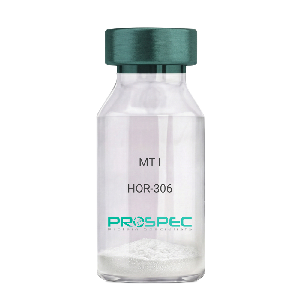
Name :
MT IDescription:
Melanotan-I
Melanotan-I, MT-I, Melanotan-1.
Product # :
HOR-306Price :
Quantity :
Shipping Method :
Shipped at Room temp
More Info
- description
- formulation
- purity
- More Info
Description
Melanotan-I has the amino acid sequence of Ser-Tyr-Ser-Nle-Glu-His-D-Phe-Arg-Trp-Gly-Lys-Pro-Val and a molecular weight of 1647.4 Dalton.
Formulation
The protein (1mg/ml) was lyophilized with no additives.
Purity
Greater than 98.0% as determined by RP-HPLC.
More Info
-
Synonyms
Melanotan-I, MT-I, Melanotan-1.
-
Physical Appearance
Sterile Filtered off-White lyophilized (freeze-dried) powder.
-
Stability
Lyophilized Melanotan-I although stable at room temperature for 3 weeks, should be stored desiccated below -18°C. Upon reconstitution MT-I should be stored at 4°C between 2-7 days and for future use below -18°C.For long term storage it is recommended to add a carrier protein (0.1% HSA or BSA).Please prevent freeze-thaw cycles.
-
Solubility
It is recommended to reconstitute the lyophilized Melanotan-I in sterile 1% acetic acid not less than 100 µg/ml, which can then be further diluted to other aqueous solutions.
ProSpec's products are furnished for LABORATORY RESEARCH USE ONLY. The product may not be used as drugs, agricultural or pesticidal products, food additives or household chemicals.
- View Data Sheet
Name :
Dengue NS1, ST4Description:
Dengue Virus NS1 Recombinant, Subtype-4
Product # :
DEN-017Price :
Quantity :
Shipping Method :
Shipped with Ice Packs
More Info
- description
- formulation
- purity
- More Info
Description
The E.Coli derived recombinant protein contains the NS1 Dengue Virus full length Type-4 immunodominant regions, migrates as 45kDa on 12% SDS-PAGE gel.The dengue protein is fused to a 6xHis tag at C-terminus.
Formulation
Phosphate buffered saline and 50mM arginine.
Purity
Protein is >95% pure as determined by 12% PAGE (coomassie staining).
More Info
-
Introduction
Caused by one of four closely related virus serotypes of the genus Flavivirus, family Flaviviridae, each serotype is sufficiently different that there is no cross-protection and epidemics caused by multiple serotypes (hyperendemicity) can occur. In cell culture experiments and mice Morpholino antisense oligos have shown specific activity against Dengue virus.
-
Stability
Dengue NS1, ST4 although stable at 4°C for 1 week, should be stored below -18°C. Please prevent freeze thaw cycles.
-
Applications
Each laboratory should determine an optimum working titer for use in its particular application.
-
Purification Method
Purified by proprietary chromatographic technique.
ProSpec's products are furnished for LABORATORY RESEARCH USE ONLY. The product may not be used as drugs, agricultural or pesticidal products, food additives or household chemicals.
- View Data Sheet
Name :
SPP1 HumanDescription:
Osteopontin Human Recombinant
Secreted Phosphoprotein-1, OPN, BNSP, BSPI, ETA-1, MGC110940, SPP-1, Osteopontin, Bone sialoprotein 1, Urinary stone protein, Nephropontin, Uropontin, SPP1.
Product # :
CYT-635Price :
Quantity :
Shipping Method :
Shipped with Ice Packs
More Info
- description
- source
- formulation
- purity
- More Info
Description
Secreted Phosphoprotein-1 Human Recombinant produced in E.Coli is single, a non-glycosylated, Polypeptide chain containing 321 amino acids fragment (17-314) and having a total molecular mass of 36.2 kDa(molecular weight on SDS-PAGE will shift up). The SPP1 protein is fused to a 20 amino acid His-Tag at N-terminus and purified by proprietary chromatographic techniques.
Source
Escherichia Coli.
Formulation
Osteopontin is supplied in 20mM Tris-HCl buffer pH-7.5, 1mM DTT, 2mM EDTA and 10% glycerol.
Purity
Greater than 90.0% as determined by SDS-PAGE.
More Info
-
Introduction
Osteopontin is a glycoprotein that was first identified in osteoblasts and is involved in bone remodeling, immune functions in fibroblasts, macrophages, and lymphocytes during inflammation and wound healing. SPP1 binds tightly to hydroxyapatite. SPP1 forms an integral part of the mineralized matrix. SPP1 is vital to cell-matrix interaction.
Secreted Phosphoprotein-1 protects against cardiac ischemia-reperfusion injury via late preconditioning. Expression of both Ostepontin and CD44 in hepatocellular carcinoma is linked with advanced tumor stage and contributes to prognosis information. SPP1 is the most over-expressed gene in intrahepatic cholangiocarcinoma. Secreted Phosphoprotein-1 overexpression is related with interstitial lung diseases. -
Synonyms
Secreted Phosphoprotein-1, OPN, BNSP, BSPI, ETA-1, MGC110940, SPP-1, Osteopontin, Bone sialoprotein 1, Urinary stone protein, Nephropontin, Uropontin, SPP1.
-
Physical Appearance
Sterile Filtered clear colorless solution.
-
Stability
Store at 4°C if entire vial will be used within 2-4 weeks. Store, frozen at -20°C for longer periods of time. For long term storage it is recommended to add a carrier protein (0.1% HSA or BSA).Avoid multiple freeze-thaw cycles.
-
Amino Acid Sequence
MGSSHHHHHH SSGLVPRGSH RSMIPVKQAD SGSSEEKQLY NKYPDAVATW LNPDPSQKQN LLAPQNAVSS EETNDFKQET LPSKSNESHD HMDDMDDEDD DDHVDSQDSI DSNDSDDVDD TDDSHQSDES HHSDESDELV TDFPTDLPAT EVFTPVVPTV DTYDGRGDSV VYGLRSKSKK FRRPDIQYPD ATDEDITSHM ESEELNGAYK AIPVAQDLNA PSDWDSRGKD SYETSQLDDQ SAETHSHKQS RLYKRKANDE SNEHSDVIDS QELSKVSREF HSHEFHSHED MLVVDPKSKE EDKHLKFRIS HELDSASSEV N.
ProSpec's products are furnished for LABORATORY RESEARCH USE ONLY. The product may not be used as drugs, agricultural or pesticidal products, food additives or household chemicals.
- View Data Sheet
Name :
CD20 antibodyDescription:
CD20 Recombinant monoclonal Anti-Human
B1, S7, Bp35, MS4A2, LEU-16, MGC3969.
Product # :
ANT-191Price :
Quantity :
Shipping Method :
Shipped at Room temp
More Info
- description
- source
- formulation
- purity
- biological activity
- More Info
Description
Recombinant Anti Human CD20 monoclonal Antibody produced in CHO is a glycosylated polypeptide chain having a molecular mass of 80 kDa.CD-20 is purified by proprietary chromatographic techniques.
Source
CHO.
Formulation
Lyophilized from a 0.2μm filtered concentrated (1mg/ml) solution in PBS, pH 7.4.
Purity
Greater than 95.0% as determined by:
Analysis by RP-HPLC.
Analysis by SDS-PAGE gel.Biological Activity
Fully biologically active when compared to standard determined by the apoptosis assay of Raji cell in vitro.More Info
-
Introduction
CD20 is a member of the membrane-spanning 4A gene family. Members of this nascent protein family are characterized by common structural features and similar intron/exon splice boundaries and display unique expression patterns among hematopoietic cells and nonlymphoid tissues. CD20 plays a role in the development and differentiation of B-cells into plasma cells. CD20 is localized to 11q12, among a cluster of family members. Alternative splicing of this gene results in two transcript variants which encode the same protein.
-
Synonyms
B1, S7, Bp35, MS4A2, LEU-16, MGC3969.
-
Physical Appearance
Sterile Filtered White lyophilized (freeze-dried) powder.
-
Stability
Lyophilized CD-20 Recombinant Antibody although stable at room temperature for 3 weeks, should be stored desiccated below -18°C. Upon reconstitution CD20 Antibody should be stored at 4°C between 2-7 days and for future use below -18°C.For long term storage it is recommended to add a carrier protein (0.1% HSA or BSA).Please prevent freeze-thaw cycles.
-
Solubility
It is recommended to reconstitute the lyophilized rAHuCD-20 in sterile 18MΩ-cm H2O not less than 100µg/ml, which can then be further diluted to other aqueous solutions.
-
Type
Anti Human Recombinant.
ProSpec's products are furnished for LABORATORY RESEARCH USE ONLY. The product may not be used as drugs, agricultural or pesticidal products, food additives or household chemicals.
- View Data Sheet
Name :
JMJD7 HumanDescription:
Jumonji Domain Containing 7 Human Recombinant
Jumonji Domain Containing 7, Jumonji Domain-Containing Protein 7, JmjC Domain-Containing Protein 7.
Product # :
PRO-1762Price :
Quantity :
Shipping Method :
Shipped with Ice Packs
More Info
- description
- source
- formulation
- purity
- More Info
Description
JMJD7 Human Recombinant produced in E.Coli is a single, non-glycosylated polypeptide chain containing 339 amino acids (1-316 a.a) and having a molecular mass of 38.3kDa.JMJD7 is fused to a 23 amino acid His-tag at N-terminus & purified by proprietary chromatographic techniques.
Source
Escherichia Coli.
Formulation
JMJD7 protein solution (1mg/ml) containing 20mM Tris-HCl buffer (pH8.0), 10% glycerol and 0.1M NaCl.
Purity
Greater than 90.0% as determined by SDS-PAGE.
More Info
-
Introduction
JmjC domain-containing protein 7 (JMJD7) is a member of the JMJD family. JMJD7 encodes highly conserved proteins with a JmjC domain, which are part of the cupin metalloenzyme superfamily. JMJD7 was first thought to be an alternatively spliced isoform of PLA2G4B. It is derived from JMJD7 which is located upstream of PLA2G4B. Most tissues also express read-through transcripts from JMJD7 into the downstream PLA2G4B gene, some of which may encode fusion proteins combining the N-terminus of JMJD7 with PLA2G4B protein.
-
Synonyms
Jumonji Domain Containing 7, Jumonji Domain-Containing Protein 7, JmjC Domain-Containing Protein 7.
-
Physical Appearance
Sterile Filtered clear solution.
-
Stability
Store at 4°C if entire vial will be used within 2-4 weeks. Store, frozen at -20°C for longer periods of time.For long term storage it is recommended to add a carrier protein (0.1% HSA or BSA).Please avoid freeze thaw cycles.
-
Amino Acid Sequence
MGSSHHHHHH SSGLVPRGSH MGSMAEAALE AVRSELREFP AAARELCVPL AVPYLDKPPT PLHFYRDWVC PNRPCIIRNA LQHWPALQKW SLPYFRATVG STEVSVAVTP DGYADAVRGD RFMMPAERRL PLSFVLDVLE GRAQHPGVLY VQKQCSNLPS ELPQLLPDLE SHVPWASEAL GKMPDAVNFW LGEAAAVTSL HKDHYENLYC VVSGEKHFLF HPPSDRPFIP YELYTPATYQ LTEEGTFKVV DEEAMEKVPW IPLDPLAPDL ARYPSYSQAQ ALRCTVRAGE MLYLPALWFH HVQQSQGCIA VNFWYDMEYD LKYSYFQLLD SLTKASGLD
ProSpec's products are furnished for LABORATORY RESEARCH USE ONLY. The product may not be used as drugs, agricultural or pesticidal products, food additives or household chemicals.
- View Data Sheet
Name :
SPP1 MouseDescription:
Osteopontin 1 Mouse Recombinant
Secreted Phosphoprotein-1, OPN, BNSP, BSPI, ETA-1, MGC110940, SPP-1, Osteopontin, Bone sialoprotein 1, Urinary stone protein, Nephropontin, Uropontin, SPP1
Product # :
CYT-1201Price :
Quantity :
Shipping Method :
Shipped with Ice Packs
More Info
- description
- source
- formulation
- purity
- biological activity
- More Info
Description
SPP1 Mouse Recombinant produced in HEK293 Cells is a single, glycosylated polypeptide chain containing 287 amino acids (17-294 a.a) and having a molecular mass of 31.8kDa.SPP1 is expressed with a 6 amino acid His tag at C-Terminus and purified by proprietary chromatographic techniques.
Source
HEK293 Cells.
Formulation
The SPP1 solution (0.25mg/1ml) contains phosphate buffered saline (pH7.4) and 10% glycerol.
Purity
Greater than 90.0% as determined by SDS-PAGE.
Biological Activity
Measured by the ability of the immobilized protein to support the adhesion of HEK293 HEK cells when the cells are added to mouse SPP1 coated plates. The ED50 range is ≤ 1.5 ug/ml.
More Info
-
Introduction
Osteopontin is a glycoprotein that was first identified in osteoblasts and is involved in bone remodeling, immune functions in fibroblasts, macrophages, and lymphocytes during inflammation and wound healing. SPP1 binds tightly to hydroxyapatite. SPP1 forms an integral part of the mineralized matrix. SPP1 is vital to cell-matrix interaction. Secreted Phosphoprotein-1 protects against cardiac ischemia-reperfusion injury via late preconditioning. Expression of both Ostepontin and CD44 in hepatocellular carcinoma is linked with advanced tumor stage and contributes to prognosis information. SPP1 is the most over-expressed gene in intrahepatic cholangiocarcinoma. Secreted Phosphoprotein-1 overexpression is related with interstitial lung diseases.
-
Synonyms
Secreted Phosphoprotein-1, OPN, BNSP, BSPI, ETA-1, MGC110940, SPP-1, Osteopontin, Bone sialoprotein 1, Urinary stone protein, Nephropontin, Uropontin, SPP1
-
Physical Appearance
Sterile Filtered colorless solution.
-
Stability
Store at 4°C if entire vial will be used within 2-4 weeks.Store, frozen at -20°C for longer periods of time.For long term storage it is recommended to add a carrier protein (0.1% HSA or BSA).Avoid multiple freeze-thaw cycles.
-
Amino Acid Sequence
DGSLPVKVTD SGSSEEKLYS LHPDPIATWL VPDPSQKQNL LAPQNAVSSE EKDDFKQETL PSNSNESHDH MDDDDDDDDD DGDHAESEDS VDSDESDESH HSDESDETVT ASTQADTFTP IVPTVDVPNG RGDSLAYGLR SKSRSFQVSD EQYPDATDED LTSHMKSGES KESLDVIPVA QLLSMPSDQD NNGKGSHESS QLDEPSLETH RLEHSKESQE SADQSDVIDS QASSKASLEH QSHKFHSHKD KLVLDPKSKE DDRYLKFRIS HELESSSSEV NHHHHHH
ProSpec's products are furnished for LABORATORY RESEARCH USE ONLY. The product may not be used as drugs, agricultural or pesticidal products, food additives or household chemicals.
- View Data Sheet
Name :
RSV Paired AntibodyDescription:
Mouse Anti Human Respiratory Syncytial Virus
Product # :
ANT-653Price :
Quantity :
Shipping Method :
Shipped with Ice Packs
More Info
- description
- formulation
- purity
- More Info
Description
RSV gold conjugation antibody and RSV capture antibody are used to develop rapid test for RSV rapid test. Please note that when ordering for example: 100µg antibody we ship 50µg from each of the antibodies (100µg in total).
Formulation
* RSV gold conjugation antibody in PBS, PH 7.4.
* RSV capture antibody in PBS, PH 7.4.Purity
Greater than 95%.
More Info
-
Physical Appearance
2 vials of sterile Filtered clear colorless solution.
-
Stability
RSV Paired Antibody although stable at 4°C for 1 week, should be stored below -18°C. Please prevent freeze thaw cycles.
-
Applications
Lateral flow rapid test.
-
Type
Mouse antibody Monoclonal.
-
Purification Method
Purified monoclonal IgG2a by protein A chromatography.
ProSpec's products are furnished for LABORATORY RESEARCH USE ONLY. The product may not be used as drugs, agricultural or pesticidal products, food additives or household chemicals.
- View Data Sheet
Name :
LY6D HumanDescription:
Lymphocyte Antigen 6 Complex, Locus D Human Recombinant
Lymphocyte Antigen 6 Complex Locus D, E48 Antigen, Ly-6D, Lymphocyte Antigen 6D.
Product # :
PRO-1838Price :
Quantity :
Shipping Method :
Shipped with Ice Packs
More Info
- description
- source
- formulation
- purity
- More Info
Description
LY6D Human Recombinant produced in E.coli is a single, non-glycosylated polypeptide chain containing 101 amino acids (21-98) and having a molecular mass of 10.8 kDa. LY6D is fused to a 23 amino acid His-tag at N-terminus.
Source
Escherichia Coli.
Formulation
The LY6D solution (0.5mg/ml) contains 20mM Tris-HCl buffer (pH 8.0), 0.15M NaCl and 50% glycerol.
Purity
Greater than 90% as determined by SDS-PAGE.
More Info
-
Introduction
LY6D encloses 1 UPAR/Ly6 domain and is only expressed at the keratinocyte of stratified squamous epithelia and the outer cell surface of transitional epithelia. LY6D performs as a specification marker at earliest stage specification of lymphocytes between B- and T-cell developments.
-
Synonyms
Lymphocyte Antigen 6 Complex Locus D, E48 Antigen, Ly-6D, Lymphocyte Antigen 6D.
-
Physical Appearance
Sterile Filtered clear solution.
-
Stability
Store at 4°C if entire vial will be used within 2-4 weeks. Store, frozen at -20°C for longer periods of time. For long term storage it is recommended to add a carrier protein (0.1% HSA or BSA).Avoid multiple freeze-thaw cycles.
-
Amino Acid Sequence
MGSSHHHHHH SSGLVPRGSH MGSLRCHVCT SSSNCKHSVV CPASSRFCKT TNTVEPLRGN LVKKDCAESC TPSYTLQGQV SSGTSSTQCC QEDLCNEKLH N
ProSpec's products are furnished for LABORATORY RESEARCH USE ONLY. The product may not be used as drugs, agricultural or pesticidal products, food additives or household chemicals.
- View Data Sheet
Name :
HIV 1 gp41 AntibodyDescription:
HIV-1 gp41, Rabbit Polyclonal Antibody
Product # :
ANT-160Price :
Quantity :
Shipping Method :
Shipped with Ice Packs
More Info
- description
- More Info
Description
Rabbit serum against the E. coli derived recombinant HIV-I protein, DEV-I (contains the C- terminus of gp120 and most of gp41).
More Info
-
Introduction
Human immunodeficiency virus (HIV) is a retrovirusthat can lead to a condition in which the immune systembegins to fail, leading to opportunistic infections. HIV primarily infects vital cells in the humanimmune systemsuch as helper T cells(specifically CD4+ T cells), macrophagesand dendritic cells. HIV infection leads to low levels of CD4+ T cells through three main mechanisms: firstly, direct viral killing of infected cells; secondly, increased rates of apoptosisin infected cells; and thirdly, killing of infected CD4+ T cells by CD8 cytotoxic lymphocytesthat recognize infected cells. When CD4+ T cell numbers decline below a critical level, cell-mediated immunityis lost, and the body becomes progressively more susceptible to opportunistic infections. HIV was classified as a member of the genus Lentivirus, part of the family of Retroviridae. Lentiviruses have many common morphologies and biological properties. Many species are infected by lentiviruses, which are characteristically responsible for long-duration illnesses with a long incubation period. Lentiviruses are transmitted as single-stranded, positive-sense, enveloped RNA viruses. Upon entry of the target cell, the viral RNA genomeis converted to double-stranded DNAby a virally encoded reverse transcriptasethat is present in the virus particle. This viral DNA is then integrated into the cellular DNA by a virally encoded integraseso that the genome can be transcribed. Once the virus has infected the cell, two pathways are possible: either the virus becomes latentand the infected cell continues to function, or the virus becomes active and replicates, and a large number of virus particles are liberated that can then infect other cells.
-
Physical Appearance
Sterile Filtered solution.
-
Specificity
Immunoreactive with HIV-I gp41. Generates a Strong positive control spot on Immuno-blot tests. Generates 1 OD (410nm) at a dilution of 1: 250 on AS 1001 (DEV-1) ELISA.
-
Type
Polyclonal Rabbit Antibody.
-
Storage Procedures
Store at -20°C.
ProSpec's products are furnished for LABORATORY RESEARCH USE ONLY. The product may not be used as drugs, agricultural or pesticidal products, food additives or household chemicals.
- View Data Sheet
Name :
HIV-1 TAT Clade-BDescription:
HIV-1 TAT Clade-B Recombinant
Product # :
HIV-129Price :
Quantity :
Shipping Method :
Shipped at Room temp
More Info
- description
- source
- formulation
- purity
- More Info
Description
HIV-1 TAT Recombinant- produced in E.coli is a single, non-glycosylated, polypeptide chain containing 86 amino acids encoded by two exons and having a molecular mass of 14kDa.
Source
Escherichia Coli.
Formulation
Lyophilized with 0.1% glycerol.
Purity
Greater than 90.0% as determined by SDS-PAGE.
More Info
-
Introduction
Human immunodeficiency virus type-1 (HIV-1) regulatory Tat protein plays an essential role in viral replication (Jones KA, 1994) and infectivity (Arya SK, 1985; Fisher AG, 1986). In addition, during acute infection, Tat is released extracellularly by infected cells (Chang HC, 1997; Ensoli B, 1990) and is taken up by neighboring cells where it transactivates viral replication (Ensoli B, 1993) and increases virus infectivity.
HIV-1 Tat activates transcription of HIV-1 viral genes by inducing phosphorylation of the C-terminal domain (CTD) of RNA polymerase II (RNAPII). Tat can also disturb cellular metabolism by inhibiting proliferation of antigen-specific T lymphocytes and by inducing cellular apoptosis. Tat-induced apoptosis of T-cells is attributed, in part, to the distortion of microtubules polymerization. LIS1 is a microtubule-associated protein that facilitates microtubule polymerization. -
Physical Appearance
Sterile Filtered and lyophilized, though might appear as a solution as a result of the glycerol content.
-
Stability
Lyophilized HIV-1 TAT although stable at room temperature for 1 week, should be stored desiccated below -18°C. Upon reconstitution HIV-1 TAT should be stored at 4°C between 2-7 days and for future use below -18°C.For long-term storage it is recommended to add a carrier protein (0.1% HSA or BSA).Please prevent freeze-thaw cycles.
-
Solubility
It is recommended to reconstitute the lyophilized HIV-1 TAT in sterile 18MΩ-cm H2O not less than 100µg/ml, which can then be further diluted to other aqueous solutions.
-
Amino Acid Sequence
MEPVDPRLEP WKHPGSQPKT ACTNCYCKKC CFHCQVCFIT KALGISYGRK KRRQRRRPPQ GSQTHQVSLS KQPTSQSRGD PTGPKE.
-
Specificity
Immunoreactive with all sera of HIV-1 infected individuals.
ProSpec's products are furnished for LABORATORY RESEARCH USE ONLY. The product may not be used as drugs, agricultural or pesticidal products, food additives or household chemicals.
- View Data Sheet
Name :
HCV NS3 Genotype-5aDescription:
Hepatitis C Virus NS3 Genotype-5a Recombinant
Product # :
HCV-212Price :
Quantity :
Shipping Method :
Shipped with Ice Packs
More Info
- description
- formulation
- purity
- More Info
Description
E.coli derived recombinant from HCV subtype 5a fused to a GST tag. The protein contains the full-length HCV NS3 (c33c) immunodominant region.
Formulation
25mM Tris-HCl, 1mM EDTA, 1.5M urea & 50% glycerol.
Purity
HCV NS3 Genotype-5a protein is >95% pure as determined by 10% PAGE (coomassie staining).
More Info
-
Introduction
HCV is a small 50nm, enveloped, single-stranded, positive sense RNAvirus in the family Flaviviridae.
HCV has a high rate of replication with approximately one trillion particles produced each day in an infected individual. Due to lack of proofreading by the HCV RNA polymerase, the HCV has an exceptionally high mutation rate, a factor that may help it elude the host's immune response. Hepatitis C virus is classified into six genotypes(1-6) with several subtypes within each genotype. The preponderance and distribution of HCV genotypes varies globally. Genotype is clinically important in determining potential response to interferon-based therapy and the required duration of such therapy. Genotypes 1 and 4 are less responsive to interferon-based treatment than are the other genotypes (2, 3, 5 and 6). -
Stability
HCV NS3 Genotype-5a although stable at 4°C for 1 week, should be stored below -18°C. Please prevent freeze thaw cycles.
-
Applications
HCV NS3 Genotype-5a antigen is suitable for ELISA and Western blots, excellent antigen for detection of HCV with minimal specificity problems.
-
Specificity
Immunoreactive with sera of HCV-infected individuals.
-
Purification Method
HCV NS3 Genotype-5a protein was purified by proprietary chromatographic technique.
ProSpec's products are furnished for LABORATORY RESEARCH USE ONLY. The product may not be used as drugs, agricultural or pesticidal products, food additives or household chemicals.
- View Data Sheet
Name :
ANGPTL2 HumanDescription:
Angiopoietin-like Protein 2 Human Recombinant
Angiopoietin-related protein 2, Angiopoietin-like protein 2, ANGPTL2, ARP2, HARP.
Product # :
CYT-765Price :
Quantity :
Shipping Method :
Shipped with Ice Packs
More Info
- description
- source
- formulation
- purity
- More Info
- sds-page
Description
ANGPTL2 Human Recombinant produced in E.Coli is a single, non-glycosylated, polypeptide chain containing 493 amino acids ( 22-493 a.a.) including a 20 a.a N-terminal His tag. The total molecular mass is 57.1kDa (calculated).
Source
Escherichia Coli.
Formulation
ANGPTL2 protein solution (0.5mg/ml) contains 20mM Tris HCL (pH7-8) and 10% glycerol.
Purity
Greater than 85.0% as determined by SDS-PAGE.
sds-page
More Info
-
Introduction
Angiopoietins belong to the vascular endothelial growth factor family and the only known growth factors largely specific for vascular endothelium. Angiopoietins-1, 2 and 4 partake in the formation of blood vessels. ANGPTL2 displays angiogenic effects. Angiopoietin-like Protein 2 (ANGPTL2) is an anti-diabetic factor. ANGPTL2 induces sprouting in endothelial cells through an autocrine and paracrine action. ANGPTL2 increases insulin sensitivity in adipocytes. In addition, ANGPTL2 is a mediator of chronic adipose tissue inflammation. ANGPTL2 is widely expressed in the heart, small intestine, spleen and stomach. ANGPTL2 is also found in lower levels in the colon, ovary, adrenal gland, skeletal muscle and in prostate.
-
Synonyms
Angiopoietin-related protein 2, Angiopoietin-like protein 2, ANGPTL2, ARP2, HARP.
-
Physical Appearance
Sterile filtered colorless solution.
-
Stability
Store at 4°C if entire vial will be used within 2-4 weeks.Store, frozen at -20°C for longer periods of time.For long term storage it is recommended to add a carrier protein (0.1% HSA or BSA).Avoid multiple freeze-thaw cycles.
-
Amino Acid Sequence
MGSSHHHHHH SSGLVPRGSH MGQEDGFEGT EEGSPREFIY LNRYKRAGES QDKCTYTFIV PQQRVTGAIC VNSKEPEVLL ENRVHKQELE LLNNELLKQK RQIETLQQLV EVDGGIVSEV KLLRKESRNM NSRVTQLYMQ LLHEIIRKRD NALELSQLEN RILNQTADML QLASKYKDLE HKYQHLATLA HNQSEIIAQL EEHCQRVPSA RPVPQPPPAA PPRVYQPPTY NRIINQISTN EIQSDQNLKV LPPPLPTMPT LTSLPSSTDK PSGPWRDCLQ ALEDGHDTSS IYLVKPENTN RLMQVWCDQR HDPGGWTVIQ RRLDGSVNFF RNWETYKQGF GNIDGEYWLG LENIYWLTNQ GNYKLLVTME DWSGRKVFAE YASFRLEPES EYYKLRLGRY HGNAGDSFTW HNGKQFTTLD RDHDVYTGNC AHYQKGGWWY NACAHSNLNG VWYRGGHYRS RYQDGVYWAE FRGGSYSLKK VVMMIRPNPN TFH
-
Background
Angiopoietin-like Protein 2 Human Recombinant: A Potential Therapeutic Target for Metabolic and Cardiovascular Disorders
Abstract:
Angiopoietin-like protein 2 (ANGPTL2) has emerged as a crucial regulator in metabolic and cardiovascular disorders. This multifunctional protein is involved in various biological processes, including angiogenesis, adipose tissue function, and inflammation. The availability of human recombinant ANGPTL2 protein has provided researchers with a valuable tool to explore its therapeutic potential. This concise review provides an overview of the role of ANGPTL2 in metabolic and cardiovascular health and discusses the potential of ANGPTL2 human recombinant protein as a therapeutic target.
Introduction:
Metabolic disorders, such as obesity and type 2 diabetes, are closely associated with cardiovascular diseases and pose significant global health challenges. ANGPTL2, a member of the angiopoietin-like protein family, has gained attention for its involvement in metabolic regulation and cardiovascular homeostasis. Through interactions with various receptors and signaling pathways, ANGPTL2 influences lipid metabolism, insulin sensitivity, inflammation, and vascular integrity.
Mechanisms of ANGPTL2 Action:
ANGPTL2 acts through binding to integrins, toll-like receptors (TLRs), and other receptors on different cell types, including adipocytes, endothelial cells, and immune cells. By influencing angiogenesis, inflammation, and extracellular matrix remodeling, ANGPTL2 affects adipose tissue function, lipid metabolism, and insulin signaling.
Role of ANGPTL2 in Metabolic Regulation:
ANGPTL2 plays a critical role in metabolic regulation and the development of metabolic disorders. It promotes adipose tissue inflammation, impairs adipogenesis, and alters adipokine secretion, contributing to metabolic dysfunction. Furthermore, ANGPTL2 modulates lipid metabolism by regulating lipoprotein lipase activity, affecting triglyceride clearance, and promoting hepatic lipid accumulation.
ANGPTL2 in Cardiovascular Health and Disease:
Increasing evidence suggests that ANGPTL2 is implicated in cardiovascular diseases, including atherosclerosis and heart failure. ANGPTL2 promotes vascular inflammation, endothelial dysfunction, and smooth muscle cell proliferation, which contribute to atherosclerotic plaque progression and vascular remodeling. Additionally, ANGPTL2 influences cardiac remodeling and fibrosis, impacting heart failure development.
Therapeutic Potential of ANGPTL2 Human Recombinant Protein:
The availability of ANGPTL2 human recombinant protein opens avenues for therapeutic interventions targeting metabolic and cardiovascular disorders. Preclinical studies employing ANGPTL2 blockade or supplementation have shown promising results in improving metabolic parameters, reducing atherosclerosis, and preserving cardiac function. However, further research is needed to optimize the clinical application of ANGPTL2 human recombinant protein, including dosage, timing, and delivery methods.
Conclusion:
ANGPTL2 holds promise as a therapeutic target for metabolic and cardiovascular disorders. Its involvement in key biological processes makes it an attractive candidate for interventions aiming to improve metabolic health and prevent cardiovascular complications. The development of ANGPTL2 human recombinant protein provides a valuable tool for investigating its therapeutic potential further.
What is the molecular weight/Mw of ANGPTL2 Protein?
ANGPTL2 Protein has a total Mw of 57.1kDa.
What is the source or expression system of ANGPTL2 Protein?
Escherichia Coli.
What is the Purity of ANGPTL2 Protein?
ANGPTL2 Protein is >85% pure as determined by SDS-PAGE.
What is the Biological Activity of ANGPTL2 Protein?
The biological functionality of ANGPTL2 Protein will be determined in the future.
What is the amino acid sequence of ANGPTL2 Protein?
MGSSHHHHHH SSGLVPRGSH MGQEDGFEGT EEGSPREFIY LNRYKRAGES QDKCTYTFIV PQQRVTGAIC VNSKEPEVLL ENRVHKQELE LLNNELLKQK RQIETLQQLV EVDGGIVSEV KLLRKESRNM NSRVTQLYMQ LLHEIIRKRD NALELSQLEN RILNQTADML QLASKYKDLE HKYQHLATLA HNQSEIIAQL EEHCQRVPSA RPVPQPPPAA PPRVYQPPTY NRIINQISTN EIQSDQNLKV LPPPLPTMPT LTSLPSSTDK PSGPWRDCLQ ALEDGHDTSS IYLVKPENTN RLMQVWCDQR HDPGGWTVIQ RRLDGSVNFF RNWETYKQGF GNIDGEYWLG LENIYWLTNQ GNYKLLVTME DWSGRKVFAE YASFRLEPES EYYKLRLGRY HGNAGDSFTW HNGKQFTTLD RDHDVYTGNC AHYQKGGWWY NACAHSNLNG VWYRGGHYRS RYQDGVYWAE FRGGSYSLKK VVMMIRPNPN TFH
What applications can ANGPTL2 Protein be used in?
ANGPTL2 Protein can probably be used in western blot, ELISA and Lateral Flow.
What is the endotoxin level for ANGPTL2 Protein?
The endotoxin level is minimal, ANGPTL2 Protein was purified using conventional chromatography techniques.
ProSpec's products are furnished for LABORATORY RESEARCH USE ONLY. The product may not be used as drugs, agricultural or pesticidal products, food additives or household chemicals.