Skip to content

High-quality research proteins.

  • 1-800-805-6531

  • Mon - Sat 8.00-18.00, Sun - Closed

  • info@prospecbio.com

  • PO Box 6591 East Brunswick NJ 08816

  • Products
    • Cytokines
    • Growth factors
    • Chemokines
    • Cd antigens
    • Neurotrophins
    • Hormones
    • Enzymes
    • Viral antigens
    • Recombinant proteins
    • Natural proteins
    • Monoclonal antibodies
    • Polyclonal antibodies
    • Thermal Packaging
  • About
    • Company
    • Founder
    • Contribution
  • Customers
    • Eastern Asia
    • Eastern Europe
    • North America
    • South America
    • South East Asia
    • Southern Asia
    • Western Europe
  • Ordering
    • Ordering Method
    • Ordering - Credit Card
    • Ordering - PO
    • Shipping Fees
    • FAQs
  • Services
    • DNA Cloning
    • Protein Expression
    • Monoclonal Antibody
    • Fermentation
    • Technology License
  • Contact us
Log in

Country/region

  • USD $ | Albania
  • USD $ | Andorra
  • USD $ | Argentina
  • USD $ | Armenia
  • USD $ | Australia
  • USD $ | Austria
  • USD $ | Azerbaijan
  • USD $ | Belarus
  • USD $ | Belgium
  • USD $ | Bolivia
  • USD $ | Brazil
  • USD $ | Bulgaria
  • USD $ | Cambodia
  • USD $ | Canada
  • USD $ | Chile
  • USD $ | China
  • USD $ | Colombia
  • USD $ | Costa Rica
  • USD $ | Croatia
  • USD $ | Cyprus
  • USD $ | Czechia
  • USD $ | Denmark
  • USD $ | Estonia
  • USD $ | Finland
  • USD $ | France
  • USD $ | Georgia
  • USD $ | Germany
  • USD $ | Greece
  • USD $ | Hong Kong SAR
  • USD $ | Hungary
  • USD $ | Iceland
  • USD $ | India
  • USD $ | Ireland
  • USD $ | Israel
  • USD $ | Italy
  • USD $ | Japan
  • USD $ | Kazakhstan
  • USD $ | Latvia
  • USD $ | Liechtenstein
  • USD $ | Lithuania
  • USD $ | Madagascar
  • USD $ | Malta
  • USD $ | Mexico
  • USD $ | Moldova
  • USD $ | Monaco
  • USD $ | Netherlands
  • USD $ | New Zealand
  • USD $ | North Macedonia
  • USD $ | Norway
  • USD $ | Panama
  • USD $ | Paraguay
  • USD $ | Peru
  • USD $ | Philippines
  • USD $ | Poland
  • USD $ | Portugal
  • USD $ | Romania
  • USD $ | Singapore
  • USD $ | Slovakia
  • USD $ | Slovenia
  • USD $ | South Africa
  • USD $ | South Korea
  • USD $ | Spain
  • USD $ | Sri Lanka
  • USD $ | Sweden
  • USD $ | Switzerland
  • USD $ | Taiwan
  • USD $ | Thailand
  • USD $ | Türkiye
  • USD $ | Ukraine
  • USD $ | United Kingdom
  • USD $ | United States
  • USD $ | Uruguay
  • USD $ | Vatican City
  • USD $ | Venezuela
  • USD $ | Vietnam
  • Facebook
  • Instagram
logoProSpec - Recombinant Protein Manufacturer for academic and R&D industry
Become a distributor
Account Log in
Wishlist
Cart Cart(0)
  • Products
    +
    • Cytokines
    • Growth factors
    • Chemokines
    • Cd antigens
    • Neurotrophins
    • Hormones
    • Enzymes
    • Viral antigens
    • Recombinant proteins
    • Natural proteins
    • Monoclonal antibodies
    • Polyclonal antibodies
    • Thermal Packaging
  • About
    +
    • Company
    • Founder
    • Contribution
  • Customers
    +
    • Eastern Asia
    • Eastern Europe
    • North America
    • South America
    • South East Asia
    • Southern Asia
    • Western Europe
  • Ordering
    +
    • Ordering Method
    • Ordering - Credit Card
    • Ordering - PO
    • Shipping Fees
    • FAQs
  • Services
    +
    • DNA Cloning
    • Protein Expression
    • Monoclonal Antibody
    • Fermentation
    • Technology License
  • Contact us

Item added to your cart

View cart
View All
  • Cytokines
  • Growth factors
  • Chemokines
  • Cd antigens
  • Neurotrophins
  • Hormones
  • Enzymes
  • Viral antigens
  • Recombinant proteins
  • Natural proteins
  • Monoclonal antibodies
  • Polyclonal antibodies
  • Thermal Packaging
  • Tumor Necrosis Factor

    Tumor Necrosis Factor

    View All
  • 4-1BB

    4-1BB

    View All
  • Adiponectin

    Adiponectin

    View All
  • AIF1

    AIF1

    View All
  • Other Cytokines

    Other Cytokines

    View All
  • AITRL

    AITRL

    View All
  • Angiopoietin

    Angiopoietin

    View All
  • Apolipoprotein

    Apolipoprotein

    View All
  • B-Cell Activating Factor

    B-Cell Activating Factor

    View All
  • Beta Defensin

    Beta Defensin

    View All
  • Bone Morphogenetic Protein

    Bone Morphogenetic Protein

    View All
  • B type Natriuretic Peptide

    B type Natriuretic Peptide

    View All
  • BST

    BST

    View All
  • Betacellulin

    Betacellulin

    View All
  • Cardiotrophin

    Cardiotrophin

    View All
  • Activin

    Activin

    View All
  • Fibroblast Growth Factor

    Fibroblast Growth Factor

    View All
  • Other Growth Factors

    Other Growth Factors

    View All
  • CSF

    CSF

    View All
  • CTGF

    CTGF

    View All
  • IGFBP

    IGFBP

    View All
  • VEGF Protein

    VEGF Protein

    View All
  • EGF

    EGF

    View All
  • Epigen

    Epigen

    View All
  • Erythropoietin

    Erythropoietin

    View All
  • Insulin-Like Growth Factor

    Insulin-Like Growth Factor

    View All
  • Growth Hormone

    Growth Hormone

    View All
  • HDGF

    HDGF

    View All
  • Hepatocyte Growth Factor

    Hepatocyte Growth Factor

    View All
  • Insulin

    Insulin

    View All
  • BCA-1/ BLC (CXCL13)

    BCA-1/ BLC (CXCL13)

    View All
  • BRAK (CXCL14)

    BRAK (CXCL14)

    View All
  • C-10 (CCL6)

    C-10 (CCL6)

    View All
  • Other Chemokines

    Other Chemokines

    View All
  • MEC (CCL28)

    MEC (CCL28)

    View All
  • LD78-beta (CCL3L1)

    LD78-beta (CCL3L1)

    View All
  • CTACK (CCL27)

    CTACK (CCL27)

    View All
  • CXCL16

    CXCL16

    View All
  • CXCL17

    CXCL17

    View All
  • Platelet Factor-4 (CXCL4)

    Platelet Factor-4 (CXCL4)

    View All
  • ENA-78 (CXCL5)

    ENA-78 (CXCL5)

    View All
  • Eotaxin (CCL11,24,26)

    Eotaxin (CCL11,24,26)

    View All
  • Exodus-2 (CCL21)

    Exodus-2 (CCL21)

    View All
  • Fractalkine (CX3CL1)

    Fractalkine (CX3CL1)

    View All
  • CXCL6

    CXCL6

    View All
  • Other CD Antigens

    Other CD Antigens

    View All
  • CD14

    CD14

    View All
  • CD164

    CD164

    View All
  • CD1

    CD1

    View All
  • CD2

    CD2

    View All
  • CD200

    CD200

    View All
  • CD207

    CD207

    View All
  • CD226

    CD226

    View All
  • CD23

    CD23

    View All
  • CD244

    CD244

    View All
  • CD247

    CD247

    View All
  • CD27

    CD27

    View All
  • CD274

    CD274

    View All
  • CD300

    CD300

    View All
  • CD33

    CD33

    View All
  • Other Neurotrophins

    Other Neurotrophins

    View All
  • Beta-NGF

    Beta-NGF

    View All
  • BDNF

    BDNF

    View All
  • CDNF

    CDNF

    View All
  • CNTF

    CNTF

    View All
  • GDNF

    GDNF

    View All
  • Glia Maturation Factor

    Glia Maturation Factor

    View All
  • MANF

    MANF

    View All
  • Midkine

    Midkine

    View All
  • Neuroglobin

    Neuroglobin

    View All
  • Neurotrophic factors

    Neurotrophic factors

    View All
  • Neuregulin

    Neuregulin

    View All
  • Neuropilin

    Neuropilin

    View All
  • Pigment Epithelium-Derived Factor

    Pigment Epithelium-Derived Factor

    View All
  • Pleiotrophin

    Pleiotrophin

    View All
  • Peptide Hormones

    Peptide Hormones

    View All
  • Other Hormones

    Other Hormones

    View All
  • HCG

    HCG

    View All
  • Endothelin

    Endothelin

    View All
  • Exendin

    Exendin

    View All
  • FSH

    FSH

    View All
  • Glucagon

    Glucagon

    View All
  • GHRP

    GHRP

    View All
  • GLP

    GLP

    View All
  • Thyrostimulin

    Thyrostimulin

    View All
  • Inhibin A

    Inhibin A

    View All
  • LHRH

    LHRH

    View All
  • Melanotan

    Melanotan

    View All
  • Procalcitonin

    Procalcitonin

    View All
  • PTH

    PTH

    View All
  • Peroxiredoxin

    Peroxiredoxin

    View All
  • Peptidase

    Peptidase

    View All
  • Synthetase

    Synthetase

    View All
  • Transferase

    Transferase

    View All
  • Hydrolase

    Hydrolase

    View All
  • Dehydrogenase

    Dehydrogenase

    View All
  • ACE2

    ACE2

    View All
  • Esterase

    Esterase

    View All
  • Protein Kinases

    Protein Kinases

    View All
  • Other Enzymes

    Other Enzymes

    View All
  • Phosphatase

    Phosphatase

    View All
  • Deaminase

    Deaminase

    View All
  • Oxygenase

    Oxygenase

    View All
  • Adenylate Kinase

    Adenylate Kinase

    View All
  • Lyase

    Lyase

    View All
  • Chagas

    Chagas

    View All
  • SARS

    SARS

    View All
  • Influenza

    Influenza

    View All
  • ASFV

    ASFV

    View All
  • Astrovirus

    Astrovirus

    View All
  • Borrelia

    Borrelia

    View All
  • Helicobacter Pylori

    Helicobacter Pylori

    View All
  • Chikungunya

    Chikungunya

    View All
  • Chlamydia

    Chlamydia

    View All
  • Cytomegalo

    Cytomegalo

    View All
  • Dengue

    Dengue

    View All
  • EBV

    EBV

    View All
  • Ebola

    Ebola

    View All
  • Feline Leukemia Virus

    Feline Leukemia Virus

    View All
  • Hepatitis A

    Hepatitis A

    View All
  • Other

    Other

    View All
  • Actin

    Actin

    View All
  • ADAM

    ADAM

    View All
  • Complement Component

    Complement Component

    View All
  • Ag85

    Ag85

    View All
  • Eukaryotic Translation Initiation Factor

    Eukaryotic Translation Initiation Factor

    View All
  • Anterior Gradient Protein

    Anterior Gradient Protein

    View All
  • Heat Shock Protein

    Heat Shock Protein

    View All
  • Alpha-2-HS-Glycoprotein

    Alpha-2-HS-Glycoprotein

    View All
  • Hemoglobin

    Hemoglobin

    View All
  • Allergy

    Allergy

    View All
  • Anaplasma

    Anaplasma

    View All
  • Angiogenin

    Angiogenin

    View All
  • Ankyrin Repeat Domain

    Ankyrin Repeat Domain

    View All
  • Annexin

    Annexin

    View All
  • Other Natural Proteins

    Other Natural Proteins

    View All
  • Aprotinin

    Aprotinin

    View All
  • Transferrin

    Transferrin

    View All
  • Avidin

    Avidin

    View All
  • Anti Coagulation Factors

    Anti Coagulation Factors

    View All
  • Natural Albumin

    Natural Albumin

    View All
  • Natural Coagulation Factors

    Natural Coagulation Factors

    View All
  • Fibronectin

    Fibronectin

    View All
  • Thrombin

    Thrombin

    View All
  • Anti Human Enzyme

    Anti Human Enzyme

    View All
  • Other Monoclonal Antibodies

    Other Monoclonal Antibodies

    View All
  • Anti Human Cytokine

    Anti Human Cytokine

    View All
  • Anti Human Heat Shock Protein

    Anti Human Heat Shock Protein

    View All
  • Anti Mouse Lymphocyte

    Anti Mouse Lymphocyte

    View All
  • Anti Human Chemokine

    Anti Human Chemokine

    View All
  • Anti Human Lymphocyte

    Anti Human Lymphocyte

    View All
  • Anti Viral Monoclonal

    Anti Viral Monoclonal

    View All
  • Anti Mouse Cytokine

    Anti Mouse Cytokine

    View All
  • Anti-GST Monoclonal

    Anti-GST Monoclonal

    View All
  • Other Polyclonal Antibodies

    Other Polyclonal Antibodies

    View All
  • Anti Viral

    Anti Viral

    View All
  • Anti-GST Monoclonal

    Anti-GST Monoclonal

    View All
Back

Search results

1000 results found for “anti mouse cytokine”

Name

Description

Product #

Price

Quantity

Shipping Method

  • View Data Sheet

    Name :

    FGF 9 Mouse

    Description:

    Fibroblast Growth Factor-9 Mouse Recombinant

    GAF (Glia-activating factor), HBGF-9, MGC119914, MGC119915, FGF-9.

    Product # :

    CYT-349

    Price :

    Quantity :

    Shipping Method :

    Room Temp Icon

    Shipped at Room temp

    Add To Cart

    More Info

    • description
    • source
    • formulation
    • purity
    • biological activity
    • More Info

    Description

    Fibroblast Growth Factor-9 Mouse Recombinant produced in E.Coli is a single, non-glycosylated, polypeptide chain containing 205 amino acids and having a molecular mass of 23308 Dalton.The FGF-9 Mouse Recombinant is purified by proprietary chromatographic techniques.

    Source

    Escherichia Coli.

    Formulation

    The protein was lyophilized from 10mM Tris, pH 8.0, 0.15M Amonium Sulfate.

    Purity

    Greater than 95.0% as determined by:
    (a) Analysis by RP-HPLC.
    (b) Analysis by SDS-PAGE.

    Biological Activity

    The ED50, calculated by the dose-dependant proliferation of BAF3 cells expressing FGF receptors (measured by 3H-thymidine uptake) is <0.5 ng/ml, corresponding to a specific activity of 2MU/mg.

    More Info

    • Introduction

      Rat and mouse FGF-9 show a very high homology to human FGF-9. The transcripts for FGF-9 have been found in brain and in kidney tissue. Fibroblast Growth Factor-9 is a member of the fibroblast growth factor (FGF) family. FGF family members possess broad mitogenic and cell survival activities, and are involved in a variety of biological processes, including embryonic development, cell growth, morphogenesis, tissue repair, tumor growth and invasion. FGF9 was isolated as a secreted factor that exhibits a growth-stimulating effect on cultured glial cells. In nervous system, this protein is produced mainly by neurons and may be important for glial cell development. Expression of the mouse homolog of this gene was found to be dependent on Sonic hedgehog (Shh) signaling. Mice lacking the homolog gene displayed a male-to-female sex reversal phenotype, which suggested a role in testicular embryogenesis Fibroblast Growth Factor 9 may have a role in glial cell growth and differentiation during development, gliosis during repair and regeneration of brain tissue after damage, differentiation and survival of neuronal cells, and growth stimulation of glial tumors.

    • Synonyms

      GAF (Glia-activating factor), HBGF-9, MGC119914, MGC119915, FGF-9.

    • Physical Appearance

      Sterile Filtered White lyophilized (freeze-dried) powder.

    • Stability

      Lyophilized Fibroblast Growth Factor-9 although stable at room temperature for 3 weeks, should be stored desiccated below -18°C. Upon reconstitution FGF9 Mouse Recombinant should be stored at 4°C between 2-7 days and for future use below -18°C.For long term storage it is recommended to add a carrier protein (0.1% HSA or BSA).Please prevent freeze-thaw cycles.

    • Solubility

      It is recommended to reconstitute the lyophilized Fibroblast Growth Factor 9 in sterile 18MΩ-cm H2O not less than 100µg/ml, which can then be further diluted to other aqueous solutions.

    • Amino Acid Sequence

      The sequence of the first five N-terminal amino acids was determined and was found to be Pro-Leu-Gly-Glu-Val.

    • Background

      What is the molecular weight/Mw of FGF9 Protein?
      FGF9 Protein has a total Mw of 23.3kDa.

      What is the source or expression system of FGF9 Protein?
      Escherichia Coli.

      What is the Purity of FGF9 Protein?
      FGF9 Protein is >95% pure as determined by SDS-PAGE.

      What is the Biological Activity of FGF9 Protein?
      The ED50, calculated by the dose-dependant proliferation of BAF3 cells expressing FGF receptors (measured by 3H-thymidine uptake) is <0.5 ng/ml, corresponding to a specific activity of 2MU/mg.

      What is the amino acid sequence of FGF9 Protein?
      FGF9 Protein is composed from 205 amino acids.

      What applications can FGF9 Protein be used in?
      FGF9 Protein can probably be used in western blot, ELISA and Lateral Flow.

      What is the endotoxin level for FGF9 Protein?
      The endotoxin level is minimal, FGF9 Protein was purified using conventional chromatography techniques.

    ProSpec's products are furnished for LABORATORY RESEARCH USE ONLY. The product may not be used as drugs, agricultural or pesticidal products, food additives or household chemicals.

    Fgf9 Mouse
  • View Data Sheet

    Name :

    AICDA Antibody

    Description:

    Activation-induced cytidine deaminase, Mouse Anti Human

    Activation-induced cytidine deaminase, Cytidine aminohydrolase, AICDA, AID, ARP2, CDA2, HIGM2.

    Product # :

    ANT-411

    Price :

    Quantity :

    Shipping Method :

    Ice Icon

    Shipped with Ice Packs

    Add To Cart

    More Info

    • formulation
    • More Info

    Formulation

    1mg/ml containing PBS, pH-7.4, & 0.1% Sodium Azide.

    More Info

    • Introduction

      AICDA is an RNA-editing deaminase which belongs to the cytidine deaminase family. AICDA is involved in somatic hypermutation, gene conversion, and class-switch recombination of immunoglobulin genes. Defects in the AICDA gene are the cause of autosomal recessive hyper-IgM immunodeficiency syndrome type 2 (HIGM2).

    • Synonyms

      Activation-induced cytidine deaminase, Cytidine aminohydrolase, AICDA, AID, ARP2, CDA2, HIGM2.

    • Immunogen

      Anti-human AICDA mAb, is derived from hybridization of mouse SP2/0 myeloma cells with spleen cells from BALB/c mice immunized with recombinant human AICDA amino acids 1-54 purified from E. coli.

    • Ig Subclass

      Mouse IgG1 heavy chain and κ light chain.

    • Clone

      P2D3AT.

    • Applications

      AICDA antibody has been tested by ELISA and Western blot analysis to assure specificity and reactivity. Since application varies, however, each investigation should be titrated by the reagent to obtain optimal results. Recommended dilution range for Western blot analysis is 1:500 ~ 1,000. Recommended starting dilution is 1:500.

    • Type

      Mouse Anti Human Monoclonal.

    • Storage Procedures

      For periods up to 1 month store at 4°C, for longer periods of time, store at -20°C. Prevent freeze thaw cycles.

    • Purification Method

      AICDA antibody was purified from mouse ascitic fluids by protein-G affinity chromatography.

    ProSpec's products are furnished for LABORATORY RESEARCH USE ONLY. The product may not be used as drugs, agricultural or pesticidal products, food additives or household chemicals.

    Aicda Antibody
  • View Data Sheet

    Name :

    S100A9 Mouse

    Description:

    S100 Calcium Binding Protein A9 Mouse Recombinant

    Calgranulin B, 60B8AG, CAGB, CFAG, CGLB, L1AG, LIAG, MAC387, MIF, MRP14, NIF, P14, Protein S100-A9, S100 calcium-binding protein A9.

    Product # :

    PRO-878

    Price :

    Quantity :

    Shipping Method :

    Ice Icon

    Shipped with Ice Packs

    Add To Cart

    More Info

    • description
    • source
    • formulation
    • purity
    • More Info

    Description

    S100A9 Mouse Recombinant produced in E.coli is a single, non-glycosylated polypeptide chain containing 133 amino acids (1-113) and having a molecular mass of 15.2 kDa.The S100A9 is fused to a 20 amino acid His-Tag at N-terminus and purified by proprietary chromatographic techniques.

    Source

    Escherichia Coli.

    Formulation

    S100A9 protein (0.5mg/ml) is supplied in 20mM Tris-HCL, pH-8, 1mM DTT and 20% Glycerol.

    Purity

    Greater than 90.0% as determined by SDS-PAGE.

    More Info

    • Introduction

      S100A9 is part of the S100 family of proteins containing 2 EF-hand calcium-binding motifs. S100A9 protein is localized in the cytoplasm and/or nucleus of a wide range of cells, and participates in the regulation of several cellular processes such as cell cycle progression and differentiation. S100 genes include no less than 13 proteins which are localized as a cluster on chromosome 1q21. S100A9 is involved in the inhibition of casein kinase and altered expression of this protein is associated with the disease cystic fibrosis.

    • Synonyms

      Calgranulin B, 60B8AG, CAGB, CFAG, CGLB, L1AG, LIAG, MAC387, MIF, MRP14, NIF, P14, Protein S100-A9, S100 calcium-binding protein A9.

    • Physical Appearance

      S100A9 is supplied as a sterile filtered yellowish solution.

    • Stability

      Store at 4°C if entire vial will be used within 2-4 weeks. Store, frozen at -20°C for longer periods of time. For long term storage it is recommended to add a carrier protein (0.1% HSA or BSA).Avoid multiple freeze-thaw cycles.

    • Amino Acid Sequence

      MGSSHHHHHH SSGLVPRGSH MANKAPSQME RSITTIIDTF HQYSRKEGHP DTLSKKEFRQ MVEAQLATFM KKEKRNEALI NDIMEDLDTN QDNQLSFEEC MMLMAKLIFA CHEKLHENNP RGHGHSHGKG CGK

    ProSpec's products are furnished for LABORATORY RESEARCH USE ONLY. The product may not be used as drugs, agricultural or pesticidal products, food additives or household chemicals.

    S100A9 Mouse
  • View Data Sheet

    Name :

    FGF 18 Mouse

    Description:

    Fibroblast Growth Factor-18 Mouse Recombinant

    Fibroblast growth factor 18, FGF-18, zFGF5, Fgf18, D130055P09Rik.

    Product # :

    CYT-064

    Price :

    Quantity :

    Shipping Method :

    Room Temp Icon

    Shipped at Room temp

    Add To Cart

    More Info

    • description
    • source
    • formulation
    • purity
    • biological activity
    • More Info
    • sds-page

    Description

    FGF-18 Mouse Recombinant produced in E.coli is a single, non-glycosylated polypeptide chain containing 180 amino acids and having a molecular mass of 21kDa.The FGF-18 is purified by proprietary chromatographic techniques.

    Source

    Escherichia Coli.

    Formulation

    FGF-18 protein was lyophilized from a 0.2µm filtered concentrated solution in PBS, pH 7.4.

    Purity

    Greater than 95.0% as determined by:
    (a) Analysis by RP-HPLC.
    (b) Analysis by SDS-PAGE.

    Biological Activity

    The ED50 as determined by the dose-dependent stimulation of thymidine uptake by BaF3 cells expressing FGF-receptors is < 0.5ng/ml, corresponding to a specific activity of > 2.0×10⁶ units/mg.

    sds-page

    fgf18 mouse sds-page - Product image 1

    More Info

    • Introduction

      Fibroblast growth factor 18 (FGF18) is a member of the large FGF family which has at least 23 members. FGF18 is a heparin binding growth factor with a core 120 amino acid FGF domain which allows for a common tertiary structure. FGFs are expressed in the course of the embryonic development and in restricted adult tissues. FGF-18 is an indispensable regulator of long bone and calvarial development. FGF-18 signals via FGFR 1c, 2c, 3c, and 4.

    • Synonyms

      Fibroblast growth factor 18, FGF-18, zFGF5, Fgf18, D130055P09Rik.

    • Physical Appearance

      Sterile Filtered White lyophilized (freeze-dried) powder.

    • Stability

      Lyophilized FGF-18 although stable at room temperature for 3 weeks, should be stored desiccated below -18°C. Upon reconstitution FGF-18 should be stored at 4°C between 2-7 days and for future use below -18°C. For long term storage it is recommended to add a carrier protein (0.1% HSA or BSA). Please prevent freeze-thaw cycles.

    • Solubility

      It is recommended to reconstitute the lyophilized FGF-18 in sterile 18M-cm H2O not less than 100µg/ml, which can then be further diluted to other aqueous solutions.

    • Amino Acid Sequence

      EENVDFRIHV ENQTRARDDV SRKQLRLYQL YSRTSGKHIQ VLGRRISARG EDGDKYAQLL VETDTFGSQV RIKGKETEFY LCMNRKGKLV GKPDGTSKEC VFIEKVLENN YTALMSAKYS GWYVGFTKKG RPRKGPKTRE NQQDVHFMKR YPKGQAELQK PFKYTTVTKR SRRIRPTHPG.

    • Background

      What is the molecular weight/Mw of FGF18 Protein?
      FGF18 Protein has a total Mw of 21kDa.

      What is the source or expression system of FGF18 Protein?
      Escherichia Coli.

      What is the Purity of FGF18 Protein?
      FGF18 Protein is >95% pure as determined by SDS-PAGE.

      What is the Biological Activity of FGF18 Protein?
      The ED50 as determined by the dose-dependent stimulation of thymidine uptake by BaF3 cells expressing FGF-receptors is < 0.5ng/ml, corresponding to a specific activity of > 2.0×10⁶ units/mg.

      What is the amino acid sequence of FGF18 Protein?
      EENVDFRIHV ENQTRARDDV SRKQLRLYQL YSRTSGKHIQ VLGRRISARG EDGDKYAQLL VETDTFGSQV RIKGKETEFY LCMNRKGKLV GKPDGTSKEC VFIEKVLENN YTALMSAKYS GWYVGFTKKG RPRKGPKTRE NQQDVHFMKR YPKGQAELQK PFKYTTVTKR SRRIRPTHPG.

      What applications can FGF18 Protein be used in?
      FGF18 Protein can probably be used in western blot, ELISA and Lateral Flow.

      What is the endotoxin level for FGF18 Protein?
      The endotoxin level is minimal, FGF18 Protein was purified using conventional chromatography techniques.

    ProSpec's products are furnished for LABORATORY RESEARCH USE ONLY. The product may not be used as drugs, agricultural or pesticidal products, food additives or household chemicals.

    Fgf 18 Mouse
  • View Data Sheet

    Name :

    SAA1 Monkey

    Description:

    Serum Amyloid A (APO-SAA1) Rhesus Macaque Recombinant

    Serum amyloid A protein, SAA, Amyloid protein A, Amyloid fibril protein AA, SAA1, SAA2, PIG4, TP53I4, MGC111216.

    Product # :

    CYT-719

    Price :

    Quantity :

    Shipping Method :

    Room Temp Icon

    Shipped at Room temp

    Add To Cart

    More Info

    • description
    • source
    • formulation
    • purity
    • biological activity
    • More Info

    Description

    SAA1 monkey Recombinant produced in E.Coli is a single, non-glycosylated, polypeptide chain containing 104 amino acids and having a total molecular mass of 11.8 kDa. SAA1 is purified by proprietary chromatographic techniques.

    Source

    Escherichia Coli.

    Formulation

    The protein was lyophilized with 1x PBS pH-7.4

    Purity

    Greater than 97.0% as determined by: (a) Analysis by RP-HPLC. (b) Analysis by SDS-PAGE.

    Biological Activity

    Determined by its ability to chemoattract human monocytes using a concentration range of 1.0-10.0 ng/ml.

    More Info

    • Introduction

      SAA1 protein is an acute phase apolipoprotein reactant which is produced mostly by hepatocytes and under regulation of inflammatory cytokines. SAA1 (Serum amyloid A1) protein is produced mainly in the liver and circulates in low levels in the blood. The SAA1 seems to have a role in the immune system. SAA1 protein levels increase in the blood and other tissues under conditions of inflammation. SAA1 may facilitate the repair of injured tissues; it also acts as an antibacterial agent, and signals the migration of germ-fighting cells to sites of infection. SAA1 also functions as an apolipoprotein of the HDL complex.
      Elevated levels of SAA1 ultimately affect secondary amyloidosis, extracellular amassing of amyloid fibrils, resulting from a circulating precursor, in a variety of tissues and organs. The most widespread type of amyloidosis appears secondary to chronic inflammatory disease, mainly rheumatoid arthritis. The SAA1 cleavage product a designated amyloid protein A is deposited systemically as amyloid in vital organs such as the liver, spleen, and kidneys in chronic inflammatory diseases patients. These deposits are extremely insoluble and resistant to proteolysis; they disrupt tissue structure and compromise performance.

    • Synonyms

      Serum amyloid A protein, SAA, Amyloid protein A, Amyloid fibril protein AA, SAA1, SAA2, PIG4, TP53I4, MGC111216.

    • Physical Appearance

      Sterile Filtered White lyophilized (freeze-dried) powder.

    • Stability

      Lyophilized SAA1 although stable at room temperature for 3 weeks, should be stored desiccated below -18°C. Upon reconstitution SAA1 should be stored at 4°C between 2-7 days and for future use below -18°C. For long term storage it is recommended to add a carrier protein (0.1% HSA or BSA). Please prevent freeze-thaw cycles.

    • Solubility

      It is recommended to reconstitute the lyophilized SAA1 in sterile 18MΩ-cm H2O not less than 100µg/ml, which can then be further diluted to other aqueous solutions.

    • Amino Acid Sequence

      RSWFSFLGEA YDGARDMWRA YSDMKEANYK NSDKYFHARG NYDAAQRGPG GVWAAEVISD ARENIQKLLG RGAEDTLADQ AANEWGRSGK DPNHFRPAGL PEKY.

    ProSpec's products are furnished for LABORATORY RESEARCH USE ONLY. The product may not be used as drugs, agricultural or pesticidal products, food additives or household chemicals.

    Saa1 Monkey
  • View Data Sheet

    Name :

    Dengue NS1 Paired Antibody

    Description:

    Mouse Anti Dengue NS1 Paired

    Product # :

    ANT-531

    Price :

    Quantity :

    Shipping Method :

    Ice Icon

    Shipped with Ice Packs

    Add To Cart

    More Info

    • description
    • formulation
    • purity
    • More Info

    Description

    A paired monoclonal antibody has been developed for dengue NS1 antigen lateral flow immunoassay product. It is suggested to use 10µg/ml gold concentration (OD 1.2-1.5) to conjugate with clone 24/c, and 1.2µg/cm coating concentration for clone 2/b. The sensitivity is directly proportional to conjugated gold OD. The specimen used for the test is 80-90 µl. The rapid test developed by these 2 paired antibodies has 90% sensitivity for the samples with OD over 5 and 60% sensitivity for the sample with OD below 5 tested by ELISA. Please note that when ordering for example: 100µg antibody we ship 50µg from each of the antibodies (100µg in total).

    Formulation

    * Dengue NS1 Conjugation antibody (clone 24/c): (1.2mg/ml) in PBS and Sodium nitrate.

    * Dengue NS1 Coating antibody (clone 2/b): (3.4mg/ml) in PBS and Sodium nitrate.

    Purity

    Greater than 95%.

    More Info

    • Introduction

      Dengue NS1 is an early marker for dengue infection from the first day of fever and lasting about one week. Dengue IgM is usually detected early on the first day to 5 days of infection. Dengue NS1 could be detected from both primary and secondary infection for all four subtypes.

    • Physical Appearance

      2 vials of sterile Filtered clear colorless solution.

    • Stability

      Dengue NS1 Paired Antibody should be stored at 4°C.

    • Applications

      Lateral flow immunoassay.

    • Assay Conditions

      1. Fresh gold particle prepared within 1 to 2 days with maximal homogeneity in particle size, λ 525 to 5529, OD 1.2- 1.4. 2. Conjugation pH 8.5, conjugation time should be 6-8 hours. 3. Blocking condition: adjust OD to 8.0-8.2 for re-suspended ant-dengue NS1 conjugated colloid gold, add casein to the final 0.3-0.4%.

    • Type

      Mouse antibody Monoclonal.

    • Purification Method

      Purified monoclonal IgG by protein A chromatography.

    ProSpec's products are furnished for LABORATORY RESEARCH USE ONLY. The product may not be used as drugs, agricultural or pesticidal products, food additives or household chemicals.

    Dengue Ns1 Paired Antibody
  • View Data Sheet

    Name :

    BD 1 Human

    Description:

    Beta Defensin-1 Human Recombinant

    Beta-defensin 1, BD-1, Defensin beta 1, hBD-1, HBD1, HBP1, DEFB1, HBD-1, HBP-1, DEFB101, DEFB-1, MGC51822.

    Product # :

    CYT-564

    Price :

    Quantity :

    Shipping Method :

    Room Temp Icon

    Shipped at Room temp

    Add To Cart

    More Info

    • description
    • source
    • formulation
    • purity
    • biological activity
    • More Info

    Description

    Beta Defensin-1 Human Recombinant produced in E.Coli is a single, non-glycosylated polypeptide chain containing 47 amino acids and having a molecular mass of 5 kDa.The BD-1 is purified by proprietary chromatographic techniques.

    Source

    Escherichia Coli.

    Formulation

    The Human BD-1 was lyophilized from a concentrated (1mg/ml) solution containing 20mM PBS pH-7.4 and 130mM sodium chloride.

    Purity

    Greater than 98.0% as determined by(a) Analysis by RP-HPLC.
    (b) Analysis by SDS-PAGE.

    Biological Activity

    Determined by its ability to chemoattract CD34+ dendritic cells using a concentration range of 100-1000ng/ml corresponding to a specific activity of 1,000-10,000IU/mg.

    More Info

    • Synonyms

      Beta-defensin 1, BD-1, Defensin beta 1, hBD-1, HBD1, HBP1, DEFB1, HBD-1, HBP-1, DEFB101, DEFB-1, MGC51822.

    • Physical Appearance

      Sterile Filtered White lyophilized (freeze-dried) powder.

    • Stability

      Lyophilized Beta Defensin-1 Recombinant although stable at room temperature for 3 weeks, should be stored desiccated below -18°C. Upon reconstitution BD-1 should be stored at 4°C between 2-7 days and for future use below -18°C.Please prevent freeze-thaw cycles.

    • Solubility

      It is recommended to reconstitute the lyophilized Beta Defensin-1 in sterile 18MΩ-cm H2O not less than 100µg/ml, which can then be further diluted to other aqueous solutions.

    • Amino Acid Sequence

      GNFLTGLGHR SDHYNCVSSG GQCLYSACPI FTKIQGTCYR GKAKCCK.

    • Background

      Beta Defensin-1 Human Recombinant: Unveiling its Potential in Innate Immunity and Therapeutic Applications

      Abstract:


      Beta Defensin-1 (BD-1), a member of the defensin family, plays a crucial role in innate immunity and host defense. This research paper provides an overview of BD-1 human recombinant, exploring its molecular characteristics, antimicrobial properties, and therapeutic applications. Understanding the multifaceted role of BD-1 offers new avenues for developing innovative immunotherapies. This article offers a concise analysis of BD-1, highlighting its impact on innate immunity and its therapeutic potential.

      Introduction:


      Innate immunity serves as the first line of defense against invading pathogens. BD-1, a key peptide within the defensin family, exhibits broad-spectrum antimicrobial activity and plays a pivotal role in host defense mechanisms. This paper provides an overview of BD-1, shedding light on its structure, function, and therapeutic potential.

      BD-1 Structure and Function:


      BD-1 is a cationic peptide with a conserved cysteine motif that confers its antimicrobial properties. It acts by disrupting the integrity of microbial cell membranes, leading to microbial death. Additionally, BD-1 exhibits immunomodulatory effects by stimulating immune cell recruitment and cytokine production.

      Antimicrobial Properties and Therapeutic Applications:


      BD-1 demonstrates antimicrobial activity against a wide range of pathogens, including bacteria, fungi, and viruses. Its ability to combat multidrug-resistant strains makes it an attractive candidate for the development of novel antimicrobial therapies. Furthermore, BD-1's immunomodulatory effects contribute to its potential in treating inflammatory and infectious diseases.

      Therapeutic Potential of BD-1 Human Recombinant:


      BD-1 human recombinant holds significant promise in the field of immunotherapy. Strategies aimed at enhancing BD-1 expression or delivering exogenous BD-1 may help boost innate immune responses in patients with compromised immune systems or chronic infections. Furthermore, BD-1-based therapeutics could be developed to combat antibiotic-resistant infections and prevent biofilm formation.

      Challenges and Future Directions:


      While BD-1 shows immense therapeutic potential, challenges must be addressed. Further research is necessary to optimize the delivery methods of BD-1 and evaluate its long-term safety and efficacy. Additionally, understanding the interplay between BD-1 and other immune factors will aid in developing combinatorial approaches for enhanced therapeutic outcomes.

      Conclusion:


      BD-1 human recombinant represents a promising avenue for developing novel immunotherapies and combating antimicrobial resistance. Understanding the molecular mechanisms and functional implications of BD-1 in innate immunity opens new horizons for innovative treatments. Continued research in this field has the potential to revolutionize the field of immunotherapy and improve patient outcomes.

      What is the molecular weight/Mw of BD1 Protein?
      BD1 Protein has a total Mw of 5kDa.

      What is the source or expression system of BD1 Protein?
      Escherichia Coli.

      What is the Purity of BD1 Protein?
      BD1 Protein is >98% pure as determined by SDS-PAGE.

      What is the Biological Activity of BD1 Protein?
      Determined by its ability to chemoattract CD34+ dendritic cells using a concentration range of 100-1000ng/ml corresponding to a specific activity of 1,000-10,000IU/mg.

      What is the amino acid sequence of BD1 Protein?
      GNFLTGLGHR SDHYNCVSSG GQCLYSACPI FTKIQGTCYR GKAKCCK.

      What applications can BD1 Protein be used in?
      BD1 Protein can probably be used in western blot, ELISA and Lateral Flow.

      What is the endotoxin level for BD1 Protein?
      The endotoxin level is minimal, BD1 Protein was purified using conventional chromatography techniques.

    ProSpec's products are furnished for LABORATORY RESEARCH USE ONLY. The product may not be used as drugs, agricultural or pesticidal products, food additives or household chemicals.

    Beta Defensin 1 Human
  • View Data Sheet

    Name :

    ENTPD6 Mouse

    Description:

    Ectonucleoside Triphosphate Diphosphohydrolase 6 Mouse Recombinant

    ectonucleoside triphosphate diphosphohydrolase 6 isoform1, 2700026H11Rik, Cd39l, Cd39l2, dJ738P15.3, NTPDa, NTPDase-6, Entpd6.

    Product # :

    PRO-2672

    Price :

    Quantity :

    Shipping Method :

    Ice Icon

    Shipped with Ice Packs

    Add To Cart

    More Info

    • description
    • source
    • formulation
    • purity
    • biological activity
    • More Info

    Description

    ENTPD6 Mouse Recombinant produced in HEK293 cells is a single, glycosylated polypeptide chain (33-455 a.a) containing 433 amino acids and having a molecular mass of 47.3 kDa.ENTPD6 is fused to a 6 amino acid His-tag at C-terminus & purified by proprietary chromatographic techniques.

    Source

    HEK293 cells.

    Formulation

    ENTPD6 protein (0.25mg/ml) contains 20% glycerol and Phosphate-Buffered Saline (pH 7.4).

    Purity

    Greater than 95.0% as determined by SDS-PAGE.

    Biological Activity

    Specific activity is > 80,000 pmol/min/ug, and is defined as the amount of enzyme that hydrolyze GDP per minute at pH 7.5 at 25C.

    More Info

    • Introduction

      Ectonucleoside Triphosphate Diphosphohydrolase 6 or ENTPD6 is an enzyme, akin to E-type nucleotidases. E-type nucleotidases, for instance CD39, are responsible for extracellular nucleotides catabolism. ENTPD6 has four apyrase-conserved areas like all to E-type nucleotidases. Rather spliced transcript agents that codes various isoforms were discovered for the DNA segment.

    • Synonyms

      ectonucleoside triphosphate diphosphohydrolase 6 isoform1, 2700026H11Rik, Cd39l, Cd39l2, dJ738P15.3, NTPDa, NTPDase-6, Entpd6.

    • Physical Appearance

      Sterile Filtered colorless solution.

    • Stability

      Store at 4°C if entire vial will be used within 2-4 weeks. Store, frozen at -20°C for longer periods of time. For long term storage it is recommended to add a carrier protein (0.1% HSA or BSA).Avoid multiple freeze-thaw cycles.

    • Amino Acid Sequence

      DGSMKWHRAS AAQAFFTIAG AASGARWTQQ AFSSPGSAAR GHEVFYGIMF DAGSTGTRIH VFQFARPPGE TPTLTHETFK ALKPGLSAYA DDVEKSAQGI QELLNVAKQH IPYDFWKATP LVLKATAGLR LLPGEKAQKL LQKVKEVFKA SPFLVGDDCV SIMNGTDEGV SAWITVNFLT GSLKTPGSSS VGMLDLGGGS TQITFLPRVE GTLQASPPGH LTALQMFNRT
      YKLYSYSYLG LGLMSARLAI LGGVEGKPAE NDKELVSPCL SPRFRGEWEH AEVTYRISGQ KAVGLYELCA SRVSEVLRNK VHRTEEAQHV DFYAFSYYYD LAASFGLIDA EKGGSLVVGD FEIAAKYVCR TLETQPPSSP FACMDLTYIS LLLHEFGFPG DKVLKLARKI DNVETSWALG AIFHYIDSLK RQKVPALHHH HHH

    ProSpec's products are furnished for LABORATORY RESEARCH USE ONLY. The product may not be used as drugs, agricultural or pesticidal products, food additives or household chemicals.

    Entpd6 Mouse
  • View Data Sheet

    Name :

    HK2 Antibody

    Description:

    Hexokinase-2, Mouse Anti Human

    Hexokinase-2, EC 2.7.1.1, HK2, Hexokinase type II, HK II, Muscle form hexokinase, HXK2, DKFZp686M1669.

    Product # :

    ANT-378

    Price :

    Quantity :

    Shipping Method :

    Ice Icon

    Shipped with Ice Packs

    Add To Cart

    More Info

    • formulation
    • More Info

    Formulation

    1mg/ml containing PBS, pH-7.4, & 0.1% Sodium Azide.

    More Info

    • Introduction

      Hexokinases phosphorylate glucose to produce glucose-6-phosphate, thus committing glucose to the glycolytic pathway. Hexokinase 2 is the predominant form found in skeletal muscle. It localizes to the outer membrane of mitochondria. Expression of this gene is insulin-responsive, and studies in rat suggest that it is involved in the increased rate of glycolysis seen in rapidly growing cancer cells.

    • Synonyms

      Hexokinase-2, EC 2.7.1.1, HK2, Hexokinase type II, HK II, Muscle form hexokinase, HXK2, DKFZp686M1669.

    • Immunogen

      Anti-human Hexokinase-2 mAb is derived from hybridization of mouse F0 myeloma cells with spleen cells from BALB/c mice immunized with recombinant human Hexokinase-2 amino acids 1-917 purified from E. coli.

    • Ig Subclass

      Mouse IgG1 heavy chain and κ light chain.

    • Clone

      P1A7AT.

    • Applications

      Hexokinase-2 antibody has been tested by ELISA and Western blot analysis to assure specificity and reactivity. Since application varies, however, each investigation should be titrated by the reagent to obtain optimal results. Recommended dilution range for Western blot analysis is 1:1,000 ~ 3,000. Recommended starting dilution is 1:2,000.

    • Type

      Mouse Anti Human Monoclonal.

    • Storage Procedures

      For periods up to 1 month store at 4°C, for longer periods of time, store at -20°C. Prevent freeze thaw cycles.

    • Purification Method

      Hexokinase-2 antibody was purified from mouse ascitic fluids by protein-G affinity chromatography.

    ProSpec's products are furnished for LABORATORY RESEARCH USE ONLY. The product may not be used as drugs, agricultural or pesticidal products, food additives or household chemicals.

    Hk2 Antibody
  • View Data Sheet

    Name :

    GMCSF Porcine

    Description:

    Granulocyte Macrophage-Colony Stimulating Factor Porcine Recombinant

    CSF-2, MGI-1GM, GM-CSF, Pluripoietin-alpha, Molgramostin, Sargramostim.

    Product # :

    CYT-1095

    Price :

    Quantity :

    Shipping Method :

    Room Temp Icon

    Shipped at Room temp

    Add To Cart

    More Info

    • description
    • source
    • formulation
    • purity
    • biological activity
    • More Info

    Description

    Granulocyte Macrophage-Colony Stimulating Factor Porcine Recombinant produced in E. coli is a non-glycosylated monomer chain containing 128 amino acids and having a molecular mass of 14.5kDa. GMCSF is purified by proprietary chromatographic techniques.

    Source

    Escherichia Coli.

    Formulation

    The protein was lyophilized from a sterile (0.2µm) filtered solution containing 10mM sodium phosphate, pH 7.5.

    Purity

    Greater than 95.0% as determined by SDS-PAGE.

    Biological Activity

    The ED50, as determined by TF-1 cell proliferation is 4.52ng/ml corresponding to a specific activity which is 2.2 x 10^5 units/mg.

    More Info

    • Introduction

      GMCSF is a cytokine that controls the production, differentiation, and function of granulocytes and macrophages. The active form of GMCSF is found extracellularly as a homodimer. GMCSF has been localized to a cluster of related genes at chromosome region 5q31, which is known to be associated with interstitial deletions in the 5q- syndrome and acute myelogenous leukemia. Other genes in the cluster include those encoding interleukins 4, 5, and 13.
      GM-CSF stimulates the growth and differentiation of hematopoietic precursor cells from various lineages, including granulocytes, macrophages, eosinophils and erythrocytes.

    • Synonyms

      CSF-2, MGI-1GM, GM-CSF, Pluripoietin-alpha, Molgramostin, Sargramostim.

    • Physical Appearance

      Sterile Filtered White lyophilized (freeze-dried) powder.

    • Stability

      Lyophilized GMCSF although stable at room temperature for 3 weeks, should be stored desiccated below -18°C. Upon reconstitution GMCSF should be stored at 4°C between 2-7 days and for future use below -18°C. Please prevent freeze-thaw cycles.

    • Solubility

      It is recommended to reconstitute the lyophilized GMCSF in sterile 18MΩ-cm H2O not less than 100µg/ml, which can then be further diluted to other aqueous solutions.

    • Amino Acid Sequence

      MAPTRPPSPV TRPWQHVDAI KEALSLLNNS NDTAAVMNET VDVVCEMFDP QEPTCVQTRL NLYKQGLRGS LTRLKSPLTL LAKHYEQHCP LTEETSCETQ SITFKSFKDS LNKFLFTIPF DCWGPVKK.

    ProSpec's products are furnished for LABORATORY RESEARCH USE ONLY. The product may not be used as drugs, agricultural or pesticidal products, food additives or household chemicals.

    Gmcsf Porcine
  • View Data Sheet

    Name :

    PECAM1 Human

    Description:

    Platelet Endothelial Cell Adhesion Molecule 1 Human Recombinant

    Platelet endothelial cell adhesion molecule, PECAM1, CD31, CD31/EndoCAM, endoCAM, GPIIA', PECA1, PECAM-1, Platelet Endothelial Cell Adhesion Molecule 1.

    Product # :

    CYT-924

    Price :

    Quantity :

    Shipping Method :

    Ice Icon

    Shipped with Ice Packs

    Add To Cart

    More Info

    • description
    • source
    • formulation
    • purity
    • More Info

    Description

    PECAM1 Human Recombinant produced in Sf9 Baculovirus is a single, non-glycosylated polypeptide chain containing 582 amino acids (28-601a.a.) and having a molecular mass of 65.5kDa (Migrates at 70-100kDa on SDS-PAGE under reducing conditions).PECAM1 is fused to an 8 amino acid His-tag at C-terminus & purified by proprietary chromatographic techniques.

    Source

    Sf9, Baculovirus cells.

    Formulation

    PECAM1 protein solution (0.25mg/ml) containing Phosphate Buffered Saline (pH 7.4) and 10% glycerol.

    Purity

    Greater than 95.0% as determined by SDS-PAGE.

    More Info

    • Introduction

      Platelet endothelial cell adhesion molecule (PECAM1) induces susceptibility to atherosclerosis. PECAM1 averts phagocyte ingestion of closely apposed viable cells by transmitting detachment signals, and transforms function upon apoptosis, thus promoting tethering of dying cells to phagocytes. The encounter of a viable cell with a phagocyte via the homophilic interaction of PECAM1 on both cell surfaces as a result causing the viable cell's active repulsion from the phagocyte.

    • Synonyms

      Platelet endothelial cell adhesion molecule, PECAM1, CD31, CD31/EndoCAM, endoCAM, GPIIA', PECA1, PECAM-1, Platelet Endothelial Cell Adhesion Molecule 1.

    • Physical Appearance

      Sterile filtered colorless solution.

    • Stability

      Store at 4°C if entire vial will be used within 2-4 weeks. Store, frozen at -20°C for longer periods of time. For long term storage it is recommended to add a carrier protein (0.1% HSA or BSA).Avoid multiple freeze-thaw cycles.

    • Amino Acid Sequence

      QENSFTINSV DMKSLPDWTV QNGKNLTLQC FADVSTTSHV KPQHQMLFYK DDVLFYNISS MKSTESYFIP EVRIYDSGTY KCTVIVNNKE KTTAEYQVLV EGVPSPRVTL DKKEAIQGGI VRVNCSVPEE KAPIHFTIEK LELNEKMVKL KREKNSRDQN FVILEFPVEE QDRVLSFRCQ ARIISGIHMQ TSESTKSELV TVTESFSTPK FHISPTGMIM EGAQLHIKCT IQVTHLAQEF PEIIIQKDKA IVAHNRHGNK AVYSVMAMVE HSGNYTCKVE SSRISKVSSI VVNITELFSK PELESSFTHL DQGERLNLSC SIPGAPPANF TIQKEDTIVS QTQDFTKIAS KSDSGTYICT AGIDKVVKKS NTVQIVVCEM LSQPRISYDA QFEVIKGQTI EVRCESISGT LPISYQLLKT SKVLENSTKN SNDPAVFKDN PTEDVEYQCV ADNCHSHAKM LSEVLRVKVI APVDEVQISI LSSKVVESGE DIVLQCAVNE GSGPITYKFY REKEGKPFYQ MTSNATQAFW TKQKASKEQE GEYYCTAFNR ANHASSVPRS KILTVRVILA PWKKVEHHHH HH.

    ProSpec's products are furnished for LABORATORY RESEARCH USE ONLY. The product may not be used as drugs, agricultural or pesticidal products, food additives or household chemicals.

    Pecam1 Human
  • View Data Sheet

    Name :

    LY6C/G Antibody, Biotin

    Description:

    LY6C/G, Rat Anti Mouse Antibody, Biotin

    Product # :

    ANT-077

    Price :

    Quantity :

    Shipping Method :

    Room Temp Icon

    Shipped at Room temp

    Add To Cart

    More Info

    • formulation
    • More Info

    Formulation

    1mg/ml in PBS (after reconstitution).

    More Info

    • Solubility

      Reconstitute with H20. Mix gently, wash the sides of the vial and wait 30-60 seconds before use.

    • Immunogen

      BM-enriched myeloid cells.

    • Ig Subclass

      Rat IgG2b.

    • Clone

      YRmLy-6C/G.

    • Applications

      Flow cytometry, immunohistochemistry.
      This antibody recognizes both Ly-6G and Ly-6C (granulocyte receptor 1- Gr-1). Ly-6G is expressed on most myeloid cells in the bone marrow and all peripheral granulocytes. Ly-6C is also expressed on subsets of lymphocytes and monocytes. For flow cytometry, use 10 µl/106 cells. Exact titer for IHC should be determined by the investigator.

    • Shipping Conditions

      Antibody is shipped lyophilized at ambient temperature.

    • Available Conjugates

      This antibody is also available unconjugated and conjugated to FITC.

    • Type

      Rat Anti Mouse Monoclonal.

    • Storage Procedures

      In lyophilized form, for long periods, store at 4°C in a dry environment. After reconstitution, if not intended for use within a month, aliquot and store at -20°C.

    • Purification Method

      Protein A column.

    ProSpec's products are furnished for LABORATORY RESEARCH USE ONLY. The product may not be used as drugs, agricultural or pesticidal products, food additives or household chemicals.

    Ly6C G Antibody Biotin
  • View Data Sheet

    Name :

    IGHG2A Mouse

    Description:

    Ig gamma-2A chain C region Mouse Recombinant

    Immunoglobulin heavy chain constant region gamma 2a, Immunoglobulin G, Ig gamma-2A chain C region, A allele, Ighg.

    Product # :

    PRO-2842

    Price :

    Quantity :

    Shipping Method :

    Ice Icon

    Shipped with Ice Packs

    Add To Cart

    More Info

    • description
    • source
    • formulation
    • purity
    • More Info

    Description

    IGHG2A Mouse Recombinant produced inSf9 Baculovirus cells is a single glycosylated polypeptide chain containing 242 amino acids (98-330 a.a.) and having a molecular mass of 27.5 kDa. IGHG2A Mouse is fused to a 6 amino acid His-tag at C-terminus & purified by proprietary chromatographic techniques.

    Source

    Sf9, Baculovirus cells.

    Formulation

    IGHG2A protein solution (1mg/ml) containing 20mM Tris-HCl buffer (pH 8.0) and 10% glycerol.

    Purity

    Greater than 90.0% as determined by SDS-PAGE.

    More Info

    • Synonyms

      Immunoglobulin heavy chain constant region gamma 2a, Immunoglobulin G, Ig gamma-2A chain C region, A allele, Ighg.

    • Physical Appearance

      Sterile filtered colorless solution.

    • Stability

      Store at 4°C if entire vial will be used within 2-4 weeks. Store, frozen at -20°C for longer periods of time. For long term storage it is recommended to add a carrier protein (0.1% HSA or BSA). Avoid multiple freeze-thaw cycles.

    • Amino Acid Sequence

      ADPEPRGPTI KPCPPCKCPA PNLLGGPSVF IFPPKIKDVL MISLSPIVTC VVVDVSEDDP DVQISWFVNN VEVHTAQTQT HREDYNSTLR VVSALPIQHQ DWMSGKEFKC KVNNKDLPAP IERTISKPKG SVRAPQVYVL PPPEEEMTKK QVTLTCMVTD FMPEDIYVEW TNNGKTELNY KNTEPVLDSD GSYFMYSKLR VEKKNWVERN SYSCSVVHEG LHNHHTTKSF SRTPGKHHHH HH.

    • Background

      The Ig gamma-2A chain C region is the constant region of the gamma-2A heavy chain in immunoglobulin molecules, particularly in mice. This region is responsible for various effector functions of IgG2a antibodies, such as antibody-dependent cellular cytotoxicity and phagocytosis and is encoded by the Ighg2a gene. The constant region of the gamma-2A chain takes part in various immune response such as regulation of the immune response, antibody-dependent cellular cytotoxicity and phagocytosis.

    ProSpec's products are furnished for LABORATORY RESEARCH USE ONLY. The product may not be used as drugs, agricultural or pesticidal products, food additives or household chemicals.

    Ighg2A Mouse
  • View Data Sheet

    Name :

    BID Antibody

    Description:

    BH3 Interacting Domain Death Agonist , Mouse Anti Human

    BH3-interacting domain death agonist, p22 BID, BID, FP497, MGC15319, MGC42355.

    Product # :

    ANT-353

    Price :

    Quantity :

    Shipping Method :

    Ice Icon

    Shipped with Ice Packs

    Add To Cart

    More Info

    • formulation
    • More Info

    Formulation

    1mg/ml containing PBS, pH-7.4, & 0.1% Sodium Azide.

    More Info

    • Introduction

      BID accession number NP_001187 is a pro-apoptotic Bcl-2 protein having only the BH3 domain. In reaction to apoptotic signaling, BID interacts with another Bcl-2 family of cell death regulators, called Bax, they form a heterodimer resulting to the insertion of Bax into the outer mitochondrial membrane. Bax induces the opening of the mitochondrial voltage-dependent anion channel which leads to the release of cytochrome c and other pro-apoptotic factors from the mitochondria resulting in activation of caspases. BID is a mediator of mitochondrial damage induced by caspase-8 (CASP8). CASP8 cleaves BID, and the COOH-terminal part translocates to mitochondria where it triggers cytochrome c release. The major proteolytic product p15 BID releasea cytochrome c. Isoform 1, Isoform 2 and Isoform 4 induce ice-like proteases and apoptosis while Isoform 3 does not induce apoptosis.

    • Synonyms

      BH3-interacting domain death agonist, p22 BID, BID, FP497, MGC15319, MGC42355.

    • Immunogen

      Anti-human BID mAb is derived from hybridization of mouse F0 myeloma cells with spleen cells from BALB/c mice immunized with recombinant human BID amino acids 1-195 purified from E. coli.

    • Ig Subclass

      Mouse IgG1 heavy chain and κ light chain.

    • Clone

      P4D3AT.

    • Applications

      BID antibody has been tested by ELISA and Western blot analysis to assure specificity and reactivity. Since application varies, however, each investigation should be titrated by the reagent to obtain optimal results. Recommended dilution range for Western blot analysis is 1:1,000 ~ 2,000.Recommended starting dilution is 1:1,000.

    • Type

      Mouse Anti Human Monoclonal.

    • Storage Procedures

      For periods up to 1 month store at 4°C, for longer periods of time, store at -20°C. Prevent freeze thaw cycles.

    • Purification Method

      BID antibody was purified from mouse ascitic fluids by protein-G affinity chromatography.

    ProSpec's products are furnished for LABORATORY RESEARCH USE ONLY. The product may not be used as drugs, agricultural or pesticidal products, food additives or household chemicals.

    Bid Antibody
  • View Data Sheet

    Name :

    AK1 Mouse

    Description:

    Adenylate Kinase 1 Mouse Recombinant

    Adenylate kinase Isoenzyme 1 isoform 1, Ak-1, B430205N08Rik, ATP-AMP transphosphorylase 1, ATP:AMP phosphotransferase, Adenylate monophosphate kinase, Myokinase.

    Product # :

    PKA-106

    Price :

    Quantity :

    Shipping Method :

    Ice Icon

    Shipped with Ice Packs

    Add To Cart

    More Info

    • description
    • source
    • formulation
    • purity
    • biological activity
    • More Info

    Description

    AK1 Mouse Recombinant produced in E.Coli is a single, non-glycosylated polypeptide chain containing 233 amino acids (1-210 a.a) and having a molecular mass of 25.5kDa.AK1 is fused to a 23 amino acid His-tag at N-terminus & purified by proprietary chromatographic techniques.

    Source

    Escherichia Coli.

    Formulation

    AK1 protein solution (0.5mg/ml) containing 20mM Tris-Hcl buffer (pH8.0), 10% glycerol and 1mM DTT.

    Purity

    Greater than 90.0% as determined by SDS-PAGE.

    Biological Activity

    Specific activity is > 150 units/mg. One unit will convert 2.0 umoles of ADP to ATP + AMP per minute at pH 7.5 at 37C.

    More Info

    • Introduction

      AK1 is a small ubiquitous enzyme which is essential for maintenance and cell growth. It is involved in the regulation of adenine nucleotide composition within a cell by catalyzing the reversible transfer of the terminal phosphate group between ATP and AMP. The AK1 protein is found in the cytosol of skeletal muscle, brain and erythrocytes. Defects in the AK1 gene are the cause of a form of hemolytic anemia.

    • Synonyms

      Adenylate kinase Isoenzyme 1 isoform 1, Ak-1, B430205N08Rik, ATP-AMP transphosphorylase 1, ATP:AMP phosphotransferase, Adenylate monophosphate kinase, Myokinase.

    • Physical Appearance

      Sterile Filtered colorless solution.

    • Stability

      Store at 4°C if entire vial will be used within 2-4 weeks.Store, frozen at -20°C for longer periods of time. For long term storage it is recommended to add a carrier protein (0.1% HSA or BSA).Avoid multiple freeze-thaw cycles.

    • Amino Acid Sequence

      MGSSHHHHHH SSGLVPRGSH MGSMGCCVSS EPQEEGGRKT GEKLKKAKII FVVGGPGSGK GTQCEKIVQK YGYTHLSTGD LLRAEVSSGS ERGKKLSAIM EKGELVPLDT VLDMLRDAML AKVDSSNGFL IDGYPREVKQ GEEFEQKIGQ PTLLLYVDAG AETMTQRLLK RGETSGRVDD
      NEETIKKRLE TYYNATEPVI SFYDKRGIVR KVNAEGTVDT VFSEVCTYLD SLK.

    ProSpec's products are furnished for LABORATORY RESEARCH USE ONLY. The product may not be used as drugs, agricultural or pesticidal products, food additives or household chemicals.

    Ak1 Mouse
  • View Data Sheet

    Name :

    HBV AG-1 antibody

    Description:

    Hepatitis B Virus (AD & AY Antigens) AG-1 for Capture ELISA, Mouse antibody

    Product # :

    ANT-038

    Price :

    Quantity :

    Shipping Method :

    Room Temp Icon

    Shipped at Room temp

    Add To Cart

    More Info

    • formulation
    • More Info

    Formulation

    1mg/ml in PBS (after reconstitution).

    More Info

    • Introduction

      Hepatitis B is one of a few known non-retroviralviruses which employ reverse transcriptionas a part of its replication process. (HIV, a completely unrelated virus, also uses reverse transcription, but it is a retrovirus.) HBV invades the cell by binding to surface receptor and become internalized. The viral core particles then migrate to the hepatocyte nucleus and the partially double-stranded, relaxed circular genomes (RC-DNA) are repaired to form a covalently closed circular DNA (cccDNA), which is the template for viral genomic and sub-genomic RNAs by cellular RNA polymerase II. Of these, the pregenomic RNA (pgRNA is selectively packaged into progeny capsids and is then reverse-transcribed into new RC-DNA. The core can either bud into the endoplasmic reticulum to be enveloped or exported from the cell or recycled back into the genome for conversion to cccDNA.

    • Solubility

      Reconstitute with H20. Mix gently, wash the sides of the vial and wait 30-60 seconds before use.

    • Immunogen

      r.Hep B vaccine (BTG).

    • Ig Subclass

      mouse IgG2b.

    • Clone

      NYRHepB-1.

    • Applications

      Direct ELISA, Western Blot, Immuneprecipitation, immunohistochemistry.

    • Note

      This antibody recognizes both the AD and the AY antigens in direct ELISA and works in capture ELISA with anti-hepatitis Ag-2.

    • Titer

      By direct ELISA (against r.hepatitis vaccine), 1:10,000 dilution will yield 0.5 O.D using alkaline phosphatase conjugated rabbit anti-mouse Ig (Jackson Laboratories).

    • Shipping Conditions

      Antibody is shipped lyophilized at ambient temperature.

    • Type

      Mouse antibody Monoclonal.

    • Storage Procedures

      In lyophilized form, for long periods, store at 4°C in a dry environment. After reconstitution, if not intended for use within a month, aliquot and store at -20°C.

    • Purification Method

      Ion exchange column.

    ProSpec's products are furnished for LABORATORY RESEARCH USE ONLY. The product may not be used as drugs, agricultural or pesticidal products, food additives or household chemicals.

    HBV AG-1 antibody
  • View Data Sheet

    Name :

    GSTM1 Mouse

    Description:

    Glutathione S-Transferase M1 Mouse Recombinant

    GST1, GTH4, GTM1, GSTM1-1, MGC26563, GSTM1a-1a, GSTM1b-1b, GSTM1, Glutathione S-transferase Mu 1, GST class-mu 1, Glutathione S-transferase GT8.7, pmGT10, GST 1-1.

    Product # :

    ENZ-397

    Price :

    Quantity :

    Shipping Method :

    Ice Icon

    Shipped with Ice Packs

    Add To Cart

    More Info

    • description
    • source
    • formulation
    • purity
    • biological activity
    • More Info

    Description

    GSTM1 Mouse Recombinant produced in E.Coli is a single, non-glycosylated, polypeptide chain containing 218 amino acids and having a molecular mass of 25.9 kDa.The GTM1 is purified by proprietary chromatographic techniques.

    Source

    Escherichia Coli.

    Formulation

    The GSTM1 solution contains PBS pH-7.4 & 5mM glutathione.

    Purity

    Greater than 95.0% as determined by SDS-PAGE.

    Biological Activity

    Specific activity is 3 units/mg, and is defined as the amount of enzyme that conjugate 1.0 u mole of 1-chloro-2,4-dinitrobenzene (CDNB) with reduced glutathione per minute at pH 6.5 at 25°C.

    More Info

    • Introduction

      Cytosolic and membrane-bound types of GST are encoded by 2 different supergene families. There are 8 classes of the soluble cytoplasmic mammalian GST: alpha, kappa, mu, omega, pi, sigma, theta and zeta. The mu class of enzymes functions in the detoxification of electrophilic compounds, including carcinogens, therapeutic drugs, environmental toxins and products of oxidative stress, by conjugation with glutathione. The genes encoding the mu class of enzymes are arranged in a gene cluster on chromosome 1p13.3 and aare highly polymorphic. These genetic differences can change an individual's resistance to carcinogens and toxins as well as affect the toxicity and efficacy of certain drugs. Null mutations of this class mu gene have been linked with the rise in a number of cancers.

    • Synonyms

      GST1, GTH4, GTM1, GSTM1-1, MGC26563, GSTM1a-1a, GSTM1b-1b, GSTM1, Glutathione S-transferase Mu 1, GST class-mu 1, Glutathione S-transferase GT8.7, pmGT10, GST 1-1.

    • Physical Appearance

      Sterile Filtered colorless solution.

    • Stability

      Store at 4°C if entire vial will be used within 2-4 weeks. Store, frozen at -20°C for longer periods of time. For long term storage it is recommended to add a carrier protein (0.1% HSA or BSA).Avoid multiple freeze-thaw cycles.

    • Amino Acid Sequence

      MPMILGYWNV RGLTHPIRML LEYTDSSYDE KRYTMGDAPD FDRSQWLNEK FKLGLDFPNL PYLIDGSHKI TQSNAILRYL ARKHHLDGET EEERIRADIV ENQVMDTRMQ LIMLCYNPDF EKQKPEFLKT IPEKMKLYSE FLGKRPWFAG DKVTYVDFLA YDILDQYRMF EPKCLDAFPN LRDFLARFEG LKKISAYMKS SRYIATPIFS KMAHWSNK.

    ProSpec's products are furnished for LABORATORY RESEARCH USE ONLY. The product may not be used as drugs, agricultural or pesticidal products, food additives or household chemicals.

    Gstm1 Mouse
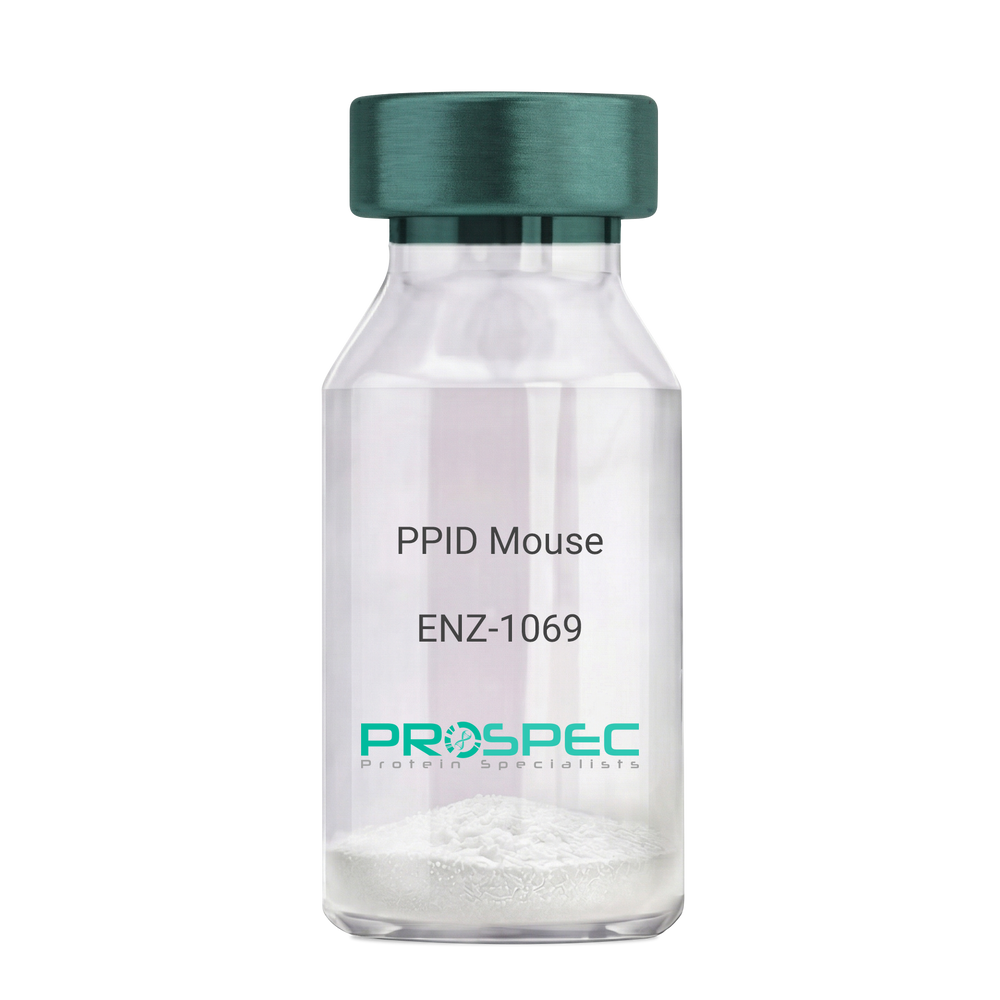
  • View Data Sheet

    Name :

    PPID Mouse

    Description:

    Peptidylprolyl Isomerase D Mouse Recombinant

    Peptidyl-prolyl cis-trans isomerase D, PPIase D, 40 kDa peptidyl-prolyl cis-trans isomerase, Cyclophilin-40, CYP-40, Cyclophilin-related protein, CYP40, CYPD, PPID, Peptidylprolyl Isomerase D.

    Product # :

    ENZ-1069

    Price :

    Quantity :

    Shipping Method :

    Ice Icon

    Shipped with Ice Packs

    Add To Cart

    More Info

    • description
    • source
    • formulation
    • purity
    • biological activity
    • More Info

    Description

    PPID Mouse Recombinant produced in E.Coli is a single, non-glycosylated polypeptide chain containing 395 amino acids (1-370a.a.) and having a molecular mass of 43.4kDa. PPID is fused to a 25 amino acid His-tag at N-terminus & purified by proprietary chromatographic techniques.

    Source

    Escherichia Coli.

    Formulation

    PPID protein solution (1mg/ml) containing 20mM Tris-Hcl buffer (pH8.0), 10% glycerol and 1mM DTT.

    Purity

    Greater than 90.0% as determined by SDS-PAGE.

    Biological Activity

    Specific activity is > 700nmol/min/mg, and is defined as the amount of enzyme that cleaves 1umole of suc-AAFP-PNA per minute at 37°C in Tris–HCl pH 8.0 using chymotrypsin.

    More Info

    • Introduction

      Cyclophilin-D is a member of the peptidyl-prolyl cis-trans isomerase (PPIase) family. PPIases catalyze the cis-trans isomerization of proline imidic peptide bonds in oligopeptides and speeds up the protein folding. Cyclophilin-D possess PPIase activity and binds to the immunosuppressant cyclosporin-A. Cyclophilin-D is very well known that its overexpression suppresses the apoptosis in cancer cell. Cyclophilin-D suppresses apoptotic cell death by the use of mitochondrial hexokinase-2 dependent mechanism in cancer cells.

    • Synonyms

      Peptidyl-prolyl cis-trans isomerase D, PPIase D, 40 kDa peptidyl-prolyl cis-trans isomerase, Cyclophilin-40, CYP-40, Cyclophilin-related protein, CYP40, CYPD, PPID, Peptidylprolyl Isomerase D.

    • Physical Appearance

      Sterile Filtered colorless solution.

    • Stability

      Store at 4°C if entire vial will be used within 2-4 weeks. Store, frozen at -20°C for longer periods of time. For long term storage it is recommended to add a carrier protein (0.1% HSA or BSA). Please prevent freeze-thaw cycles.

    • Amino Acid Sequence

      MGSSHHHHHH SSGLVPRGSH MGSEFMSHAS PAAKPSNSKN PRVFFDVDIG GERVGRIVLE LFADIVPKTA ENFRALCTGE KGTGSTTGKP LHFKGCPFHR IIKKFMIQGG DFSNQNGTGG ESIYGEKFED ENFHYKHDRE GLLSMANAGP NTNGSQFFIT TVPTPHLDGK HVVFGQVIKG LGVARTLENV EVNGEKPAKL CVIAECGELK EGDDWGIFPK DGSGDSHPDF PEDADIDLKD VDKILLISED LKNIGNTFFK SQNWEMAIKK YAKVLRYVDS SKAVIEKADR SRLQPIALSC VLNIGACKLK MSNWQGAIDS CLEALEMDPS NTKALYRKAQ GWQGLKEYDQ ALADLKKAQE IAPGDKAIQA ELLKVKQMIK AQKDKEKAVY AKMFA.

    ProSpec's products are furnished for LABORATORY RESEARCH USE ONLY. The product may not be used as drugs, agricultural or pesticidal products, food additives or household chemicals.

    Ppid Mouse
  • View Data Sheet

    Name :

    HGFR Mouse

    Description:

    Hepatocyte Growth Factor Receptor Mouse Recombinant

    hepatocyte growth factor receptor, HGF R/c-MET, Met, AI838057, c-Met, HGF, HGFR, Par4, HGF receptor, HGF/SF receptor, Proto-oncogene c-Met, Scatter factor receptor, SF receptor, Tyrosine-protein kinase Met.

    Product # :

    CYT-1139

    Price :

    Quantity :

    Shipping Method :

    Ice Icon

    Shipped with Ice Packs

    Add To Cart

    More Info

    • description
    • source
    • formulation
    • purity
    • More Info

    Description

    HGF Receptor Mouse Recombinant produced in Baculovirus is a single glycosylated polypeptide chain containing 1146 amino acids (25-931aa) and having a molecular mass of 127.8kDa. HGF is fused to a 239 amino acid hIgG-His-Tag at C-terminus and purified by proprietary chromatographic techniques.

    Source

    Sf9, Baculovirus cells.

    Formulation

    HGF Receptor protein (0.25mg/ml) contains Phosphate Buffered Saline (pH 7.4) and 10% glycerol.

    Purity

    Greater than 85.0% as determined by SDS-PAGE.

    More Info

    • Introduction

      HGF, also known as scatter factor (SF) and Hepatocyte growth factor, is secreted from mesenchymal cells, targets and acts mainly on epithelial and endothelial cells. The protein is a paracrine cellular growth, motility and morphogenic factor. HGF can also be part of haemopoietic progenitor cells and T cells. HGF has a crucial role in embryonic organ development, mainly in myogenesis. In adults HGF takes part in organ regeneration and wound healing.

    • Synonyms

      hepatocyte growth factor receptor, HGF R/c-MET, Met, AI838057, c-Met, HGF, HGFR, Par4, HGF receptor, HGF/SF receptor, Proto-oncogene c-Met, Scatter factor receptor, SF receptor, Tyrosine-protein kinase Met.

    • Physical Appearance

      Sterile Filtered colorless solution.

    • Stability

      Store at 4°C if entire vial will be used within 2-4 weeks. Store, frozen at -20°C for longer periods of time. For long term storage it is recommended to add a carrier protein (0.1% HSA or BSA).Avoid multiple freeze-thaw cycles.

    • Amino Acid Sequence

      ECKEALVKSE MNVNMKYQLP NFTAETPIQN VVLHGHHIYL GATNYIYVLN DKDLQKVSEF KTGPVLEHPD CLPCRDCSSK ANSSGGVWKD NINMALLVDT YYDDQLISCG SVNRGTCQRH VLPPDNSADI QSEVHCMFSP EEESGQCPDC VVSALGAKVL LSEKDRFINF FVGNTINSSY PPGYSLHSIS VRRLKETQDG FKFLTDQSYI DVLPEFQDSY PIKYIHAFES NHFIYFLTVQ KETLDAQTFH TRIIRFCSVD SGLHSYMEMP LECILTEKRR KRSTREEVFN ILQAAYVSKP GANLAKQIGA SPSDDILFGV FAQSKPDSAE PVNRSAVCAF PIKYVNDFFN KIVNKNNVRC LQHFYGPNHE HCFNRTLLRN SSGCEARSDE YRTEFTTALQ RVDLFMGRLN QVLLTSISTF IKGDLTIANL GTSEGRFMQV VLSRTAHLTP HVNFLLDSHP VSPEVIVEHP SNQNGYTLVV TGKKITKIPL NGLGCGHFQS CSQCLSAPYF IQCGWCHNQC VRFDECPSGT WTQEICLPAV YKVFPTSAPL EGGTVLTICG WDFGFRKNNK FDLRKTKVLL GNESCTLTLS ESTTNTLKCT VGPAMSEHFN VSVIISNSRE TTQYSAFSYV DPVITSISPR YGPQAGGTLL TLTGKYLNSG NSRHISIGGK TCTLKSVSDS ILECYTPAQT TSDEFPVKLK IDLANRETSS FSYREDPVVY EIHPTKSFIS GGSTITGIGK TLNSVSLPKL VIDVHEVGVN YTVACQHRSN SEIICCTTPS LKQLGLQLPL KTKAFFLLDG ILSKHFDLTY VHNPVFEPFE KPVMISIGNE NVVEIKGNNI DPEAVKGEVL KVGNQSCESL HWHSGAVLCT VPSDLLKLNS ELNIEWKQAV SSTVLGKVIV QPDQNFALEP KSCDKTHTCP PCPAPELLGG PSVFLFPPKP KDTLMISRTP EVTCVVVDVS HEDPEVKFNW YVDGVEVHNA KTKPREEQYN STYRVVSVLT VLHQDWLNGK EYKCKVSNKA LPAPIEKTIS KAKGQPREPQ VYTLPPSRDE LTKNQVSLTC LVKGFYPSDI AVEWESNGQP ENNYKTTPPV LDSDGSFFLY SKLTVDKSRW QQGNVFSCSV MHEALHNHYT QKSLSLSPGK HHHHHH.

    ProSpec's products are furnished for LABORATORY RESEARCH USE ONLY. The product may not be used as drugs, agricultural or pesticidal products, food additives or household chemicals.

    Hgf Mouse
  • View Data Sheet

    Name :

    GSTP1 Mouse

    Description:

    Glutathione S-Transferase pi 1 Mouse Recombinant

    Glutathione S-transferase P 1, Gst P1, GST YF-YF, GST class-pi, GST-piB, Preadipocyte growth factor.

    Product # :

    ENZ-909

    Price :

    Quantity :

    Shipping Method :

    Ice Icon

    Shipped with Ice Packs

    Add To Cart

    More Info

    • description
    • source
    • formulation
    • purity
    • biological activity
    • More Info

    Description

    GSTP1 Mouse Recombinant produced in E.Coli is a single, non-glycosylated polypeptide chain containing 233 amino acids (1-210 a.a) and having a molecular mass of 26kDa. GSTP1 is fused to a 23 amino acid His-tag at N-terminus & purified by proprietary chromatographic techniques.

    Source

    Escherichia Coli.

    Formulation

    GSTP1 protein solution (1mg/ml) containing Phosphate Buffered Saline (pH 7.4) and 10% glycerol.

    Purity

    Greater than 90.0% as determined by SDS-PAGE.

    Biological Activity

    Specific activity is > 40 units/mg, and is defined as the amount of enzyme that conjugate 1.0 umole of 1-chloro-2,4-dinitrobenzene (CDNB) with reduced glutathione per minute at pH 6.5 at 25C.

    More Info

    • Introduction

      GSTP1 is a polymorphic gene encoding active, functionally different GSTP1 variant proteins that are believed to function in xenobiotic metabolism and have a part in susceptibility to cancer, and other diseases. GSTP1 is a glutathione S-transferase belonging to the pi class. GST family enzymes play a significant role in detoxification by catalyzing the conjugation of many hydrophobic and electrophilic compounds with reduced glutathione. Based upon the biochemical, immunologic and structural properties of the soluble GSTs they are grouped into 4 main classes: alpha, mu, pi, and theta. The GSTP1 enzyme acts by catalyzing the reaction of glutathione with an acceptor molecule to form a Sulfur-substituted glutathione. The reactions employing glutathione contribute the transformation of a broad range of electrophiles, including reactive products of lipid, protein, carcinogens, therapeutic drugs, environmental toxins, and products of oxidative stress.
      The GSTP1 inactivation through CpG hypermethylation is frequent in pituitary adenomas and may be a factor in aggressive pituitary tumor behavior. GSTP1 is may be a transcriptional target of the p53 tumor suppressor gene. Single-nucleotide polymorphism in GSTP1 is linked to modified protein binding, which influence GSTP1's contribution to carcinogen and drug metabolism, and possibly disease pathogenesis and/or drug response. GST-pi might have central roles in proliferation of androgen-independent human prostate cancer cells.

    • Synonyms

      Glutathione S-transferase P 1, Gst P1, GST YF-YF, GST class-pi, GST-piB, Preadipocyte growth factor.

    • Physical Appearance

      Sterile Filtered colorless solution.

    • Stability

      Store at 4°C if entire vial will be used within 2-4 weeks. Store, frozen at -20°C for longer periods of time. For long term storage it is recommended to add a carrier protein (0.1% HSA or BSA).Avoid multiple freeze-thaw cycles.

    • Amino Acid Sequence

      MGSSHHHHHH SSGLVPRGSH MGSMPPYTIV YFPVRGRCEA MRMLLADQGQ SWKEEVVTID TWMQGLLKPT CLYGQLPKFE DGDLTLYQSN AILRHLGRSL GLYGKNQREA AQMDMVNDGV EDLRGKYVTL IYTNYENGKN DYVKALPGHL KPFETLLSQN QGGKAFIVGD QISFADYNLL DLLLIHQVLA PGCLDNFPLL SAYVARLSAR PKIKAFLSSP EHVNRPINGN GKQ.

    ProSpec's products are furnished for LABORATORY RESEARCH USE ONLY. The product may not be used as drugs, agricultural or pesticidal products, food additives or household chemicals.

    Gstp1 Mouse
  • View Data Sheet

    Name :

    TNFRSF12A Human, sf9

    Description:

    TNF Ligand Receptor Superfamily Member 12A Human Recombinant, Sf9

    Tumor necrosis factor receptor superfamily member 12A, FN14, CD266 antigen, TweakR, tweak-receptor, Fibroblast growth factor-inducible immediate-early response protein 14, FGF-inducible 14, type I transmembrane protein Fn14.

    Product # :

    CYT-1021

    Price :

    Quantity :

    Shipping Method :

    Ice Icon

    Shipped with Ice Packs

    Add To Cart

    More Info

    • description
    • source
    • formulation
    • purity
    • More Info

    Description

    TNFRSF12A Human Recombinant produced in Sf9 Baculovirus cells is a single, glycosylated polypeptide chain containing 292 amino acids (28-80a.a.) and having a molecular mass of 32.6kDa (Molecular size on SDS-PAGE will appear at approximately 28-40kDa). TNFRSF12A is expressed with a 239 amino acids hIgG- His tag at C-Terminus and purified by proprietary chromatographic techniques.

    Source

    Sf9, Baculovirus cells.

    Formulation

    TNFRSF12A protein solution (1mg/ml) contains Phosphate Buffered Saline (pH 7.4), 1mM DTT and 20% glycerol.

    Purity

    Greater than 90% as determined by SDS-PAGE.

    More Info

    • Introduction

      The gene for TNFRSF12A was initially recognized as a fibroblast growth factor inducible immediate early response gene Fn14 in mouse NIH 3T3 fibroblasts. Human TNFRSF12A cDNA encodes a 129 amino acid residue type I transmembrane protein with a 27 aa signal peptide, a 53 aa extracellular domain, a 21 aa transmembrane domain and a 28 aa cytoplasmic domain. Human and mouse TNFRSF12A hold 82% aa sequence identity. TNFRSF12 is the tiniest member of the TNF receptor superfamily and has only one cysteine rich region in its extracellular domain. The TNFRSF12A cytoplasmic domain holds one TRAF binding motif which binds TRAFs 1, 2, and 3. TNFRSF12A binds its ligand TWEAK/TNFSF12A with high affinity to initiate a signal transduction cascade which subject to the cell type, causes different cellular responses such as cell death, cell proliferation, and angiogenesis.

    • Synonyms

      Tumor necrosis factor receptor superfamily member 12A, FN14, CD266 antigen, TweakR, tweak-receptor, Fibroblast growth factor-inducible immediate-early response protein 14, FGF-inducible 14, type I transmembrane protein Fn14.

    • Physical Appearance

      Sterile Filtered colorless solution.

    • Stability

      Store at 4°C if entire vial will be used within 2-4 weeks. Store, frozen at -20°C for longer periods of time. For long term storage it is recommended to add a carrier protein (0.1% HSA or BSA).Avoid multiple freeze-thaw cycles.

    • Amino Acid Sequence

      EQAPGTAPCS RGSSWSADLD KCMDCASCRA RPHSDFCLGC AAAPPAPFRL LWPLEPKSCD KTHTCPPCPA PELLGGPSVF LFPPKPKDTL MISRTPEVTC VVVDVSHEDP EVKFNWYVDG VEVHNAKTKP REEQYNSTYR VVSVLTVLHQ DWLNGKEYKC KVSNKALPAP IEKTISKAKG QPREPQVYTL PPSRDELTKN QVSLTCLVKG FYPSDIAVEW ESNGQPENNY KTTPPVLDSD GSFFLYSKLT VDKSRWQQGN VFSCSVMHEA LHNHYTQKSL SLSPGKHHHH HH.

    ProSpec's products are furnished for LABORATORY RESEARCH USE ONLY. The product may not be used as drugs, agricultural or pesticidal products, food additives or household chemicals.

    Tnfrsf12A Protein
  • View Data Sheet

    Name :

    MHC CLASS II, I-E Antibody, Biotin

    Description:

    MHC CLASS II, (I-E) Mouse Antibody, Biotin

    Product # :

    ANT-298

    Price :

    Quantity :

    Shipping Method :

    Room Temp Icon

    Shipped at Room temp

    Add To Cart

    More Info

    • formulation
    • More Info

    Formulation

    1mg/ml in PBS (after reconstitution).

    More Info

    • Introduction

      HC Class II is consists of 2 membrane spanning proteins. Each of these proteins is roughly 30 kDa, and has of two Globular domains, Alpha-1, Alpha-2, Beta-1 and Beta-2. The two regions farthest away from the membrane are alpha-1 and beta-1. The two proteins link without Covalent Bonds. The MHC Class II protein mainly presents peptides, which have been digested from external sources. MHC Class II is normally only expressed by Antigen Presenting Cells (APC) which digest foreign protein. Within the RER the alpha and beta proteins of the molecule, associate with each other, while a third protein called the "invariant chain," is necessary for stabilizing the complex, without it, they will not associate. The MHC-invariant complex is then passed from the RER, into and out of, the Golgi body. It fuses with an Endocytic compartment, where an external protein has been sampled and degraded.

    • Solubility

      Reconstitute with of H20. Mix gently, wash the sides of the vial and wait 30-60 seconds before use.

    • Immunogen

      Purified mouse LN B cells (C3H anti-C57Bl6).

    • Ig Subclass

      Mouse IgG2a.

    • Clone

      NYRmI-E.

    • Applications

      Cytotoxic and staining antibody. For staining, use 10ml/1,000,000 cells. Titer for cytotoxicity should be determined by the investigator.

    • Specificity

      Recognizes most MHC I-E class II haplotypes Reacts with H-2k, H-2d, H-2p and H-2r.

    • Available Conjugates

      This antibody is also available as unconjugated antibody and conjugated to FITC.

    • Type

      Mouse Antibody Monoclonal.

    • Storage Procedures

      Lyophilized: store at 4°C. After reconstitution, if not intended for use within a month, aliquot and store at -20°C.

    • Purification Method

      Ion exchange column.

    ProSpec's products are furnished for LABORATORY RESEARCH USE ONLY. The product may not be used as drugs, agricultural or pesticidal products, food additives or household chemicals.

    Mhc Class Ii I E Antibody Biotin
  • View Data Sheet

    Name :

    CSK Antibody

    Description:

    C-Src Tyrosine Kinase, Mouse Anti Human

    Tyrosine-protein kinase CSK, C-Src kinase, Protein-tyrosine kinase CYL, CSK, C-Src Tyrosine Kinase.

    Product # :

    ANT-693

    Price :

    Quantity :

    Shipping Method :

    Ice Icon

    Shipped with Ice Packs

    Add To Cart

    More Info

    • formulation
    • More Info

    Formulation

    1mg/ml containing PBS, pH-7.4, 10% Glycerol and 0.02% Sodium Azide.

    More Info

    • Introduction

      C-Src Tyrosine Kinase, also known as CSK, regulates cell growth, migration, differentiation, and immune response. CSK Phosphorylates tyrosine residues located in the C-terminal tails of Src-family kinases. CSK is a protein coding gene which suppresses signaling by several surface receptors. Among CSK's related pathways are PI-3K cascade and Signaling by FGFR.

    • Synonyms

      Tyrosine-protein kinase CSK, C-Src kinase, Protein-tyrosine kinase CYL, CSK, C-Src Tyrosine Kinase.

    • Physical Appearance

      Sterile filtered colorless solution.

    • Immunogen

      Anti-human CSK mAb, is derived from hybridization of mouse F0 myeloma cells with spleen cells from BALB/c mice immunized with recombinant human CSK amino acids 1-450 purified from E. coli.

    • Ig Subclass

      Mouse IgG2b heavy chain and κ light chain.

    • Clone

      PAT7C6AT.

    • Applications

      CSK antibody has been tested by ELISA, Western blot analysis and ICC/IF to assure specificity and reactivity. Since application varies, however, each investigation should be titrated by the reagent to obtain optimal results.

    • Type

      Mouse Anti Human Monoclonal.

    • Storage Procedures

      For periods up to 1 month store at 4°C, for longer periods of time, store at -20°C. Prevent freeze thaw cycles.

    • Purification Method

      CSK antibody was purified from mouse ascitic fluids by protein-A affinity chromatography.

    ProSpec's products are furnished for LABORATORY RESEARCH USE ONLY. The product may not be used as drugs, agricultural or pesticidal products, food additives or household chemicals.

    Csk Antibody
  • View Data Sheet

    Name :

    JAK2 Antibody

    Description:

    Janus Kinase 2, Mouse Anti Human

    Tyrosine-protein kinase JAK2, Janus kinase 2, JAK-2, JAK2, Janus kinase 2 (a protein tyrosine kinase), JTK10.

    Product # :

    ANT-644

    Price :

    Quantity :

    Shipping Method :

    Ice Icon

    Shipped with Ice Packs

    Add To Cart

    More Info

    • formulation
    • More Info

    Formulation

    1mg/ml containing PBS, pH-7.4, 10% Glycerol and 0.02% Sodium Azide.

    More Info

    • Introduction

      Janus Kinase 2 (JAK2) is a protein tyrosine kinase which takes part in a specific subset of cytokine receptor signaling pathways such as cell growth, development, differentiation or histone modifications. JAK2 is linked with the prolactin receptor and is necessary for responses to gamma interferon. Mice which do not express an active protein for JAK2 show embryonic lethality associated with the absence of definitive erythropoiesis.

    • Synonyms

      Tyrosine-protein kinase JAK2, Janus kinase 2, JAK-2, JAK2, Janus kinase 2 (a protein tyrosine kinase), JTK10.

    • Physical Appearance

      Sterile filtered colorless solution.

    • Immunogen

      Anti-human JAK2 mAb, is derived from hybridization of mouse F0 myeloma cells with spleen cells from BALB/c mice immunized with recombinant human JAK2 amino acids 1014-1132 purified from E. coli.

    • Ig Subclass

      Mouse IgG2b heavy chain and k light chain.

    • Clone

      PAT53B7AT.

    • Applications

      JAK2 antibody has been tested by ELISA, Western blot analysis, Flow cytometry and ICC/IF to assure specificity and reactivity. Since application varies, however, each investigation should be titrated by the reagent to obtain optimal results.

    • Type

      Mouse Anti Human Monoclonal.

    • Storage Procedures

      For periods up to 1 month store at 4°C, for longer periods of time, store at -20°C. Prevent freeze thaw cycles.

    • Purification Method

      JAK2 antibody was purified from mouse ascitic fluids by protein-A affinity chromatography.

    ProSpec's products are furnished for LABORATORY RESEARCH USE ONLY. The product may not be used as drugs, agricultural or pesticidal products, food additives or household chemicals.

    Jak2 Antibody
  • 1
  • …
  • 31
  • 32
  • 33
  • 34
  • 35
  • …
  • 42
Your browser does not support the video tag.

Products

  • Cytokines
  • Growth Factors
  • Chemokines
  • CD Antigens
  • Neurotrophins
  • Hormones
  • Enzymes
  • Viral Antigens
  • Recombinant Proteins
  • Natural Proteins
  • Monoclonal Antibodies
  • Polyclonal Antibodies
  • Thermal Packaging

About

  • Company
  • Founder
  • Contribution
  • Contact Us
  • My Account

Ordering

  • Ordering Method
  • Ordering - Credit Card
  • Ordering PO
  • Shipping Fees
  • FAQ's

Legal

  • Terms & Conditions
  • Privacy
  • Site Map
  • info@prospecbio.com
  • Facebook
  • Instagram
  • Linkedin Profile

© 2026 ProSpec-Tany TechnoGene Ltd. All Rights Reserved.

  • Choosing a selection results in a full page refresh.
  • Opens in a new window.