Search results
1000 results found for “Myelin Oligodendrocyte Glycoprotein”
Name
Description
Product #
Price
Quantity
Shipping Method
- View Data Sheet
Name :
MYD88 HumanDescription:
Myeloid Differentiation Primary Response 88 Human Recombinant
Myeloid differentiation primary response protein MyD88, MYD88, MYD88D.
Product # :
PRO-1377Price :
Quantity :
Shipping Method :
Shipped with Ice Packs
More Info
- description
- source
- formulation
- purity
- More Info
Description
MYD88 Human Recombinant produced in E.Coli is a single, non-glycosylated polypeptide chain containing 345 amino acids (1-309 a.a) and having a molecular mass of 38.7kDa.MYD88 is fused to a 36 amino acid His-tag at N-terminus & purified by proprietary chromatographic techniques.
Source
Escherichia Coli.
Formulation
MYD88 protein solution (1mg/ml) containing 20mM Tris-HCl buffer (pH 8.0), 0.4M urea and 10% glycerol.
Purity
Greater than 90.0% as determined by SDS-PAGE.
More Info
-
Introduction
Myeloid differentiation primary response gene 88 (MYD88) is a cytosolic adapter protein, which has a central role in the innate and adaptive immune response. MYD88 functions as a vital signal transducer in the interleukin-1 and Toll-like receptor signaling pathways. MYD88 acts via IRAK1, IRAK2, IRF7 and TRAF6, leading to NF-kappa-B activation, cytokine secretion and the inflammatory response. The MYD88 protein increases IL-8 transcription. MYD88 is involved in IL-18-mediated signaling pathway. MYD88 activates IRF1, resulting in its rapid migration into the nucleus to mediate an efficient induction of IFN-beta, NOS2/INOS, and IL12A genes. MYD88 is comprised of an N-terminal death domain and a C-terminal Toll-interleukin1 receptor domain. Patients with defects in the MYD88 gene have an increased susceptibility to pyogenic bacterial infections.
-
Synonyms
Myeloid differentiation primary response protein MyD88, MYD88, MYD88D.
-
Physical Appearance
Sterile Filtered colorless solution.
-
Stability
Store at 4°C if entire vial will be used within 2-4 weeks. Store, frozen at -20°C for longer periods of time. For long term storage it is recommended to add a carrier protein (0.1% HSA or BSA).Avoid multiple freeze-thaw cycles.
-
Amino Acid Sequence
MRGSHHHHHH GMASMTGGQQ MGRDLYDDDD KDRWGSMRPD RAEAPGPPAM AAGGPGAGSA APVSSTSSLP LAALNMRVRR RLSLFLNVRT QVAADWTALA EEMDFEYLEI RQLETQADPT GRLLDAWQGR PGASVGRLLE LLTKLGRDDV LLELGPSIEE DCQKYILKQQ QEEAEKPLQV AAVDSSVPRT AELAGITTLD DPLGHMPERF DAFICYCPSD IQFVQEMIRQ LEQTNYRLKL CVSDRDVLPG TCVWSIASEL IEKRCRRMVV VVSDDYLQSK ECDFQTKFAL SLSPGAHQKR LIPIKYKAMK KEFPSILRFI TVCDYTNPCT KSWFWTRLAK ALSLP.
ProSpec's products are furnished for LABORATORY RESEARCH USE ONLY. The product may not be used as drugs, agricultural or pesticidal products, food additives or household chemicals.
- View Data Sheet
Name :
C6ORF108 HumanDescription:
Chromosome 6 Open Reading Frame 108 Human Recombinant
c-Myc-responsive protein Rcl, RCL,putative c-Myc-responsive.
Product # :
PRO-249Price :
Quantity :
Shipping Method :
Shipped with Ice Packs
More Info
- description
- source
- formulation
- purity
- More Info
Description
C6ORF108 produced in E.Coli is a single, non-glycosylated polypeptide chain containing 194 amino acids (1-174 a.a.) and having a molecular mass of 21.2kDa.C6ORF108 is fused to a 20 amino acid His-tag at N-terminus & purified by proprietary chromatographic techniques.
Source
Escherichia Coli.
Formulation
The C6ORF108 protein solution (1mg/ml) contains 20mM Tris-HCl buffer (pH-8), 1mM DTT, and 10% glycerol.
Purity
Greater than 95.0% as determined by SDS-PAGE.
More Info
-
Introduction
C6ORF108 is stimulated by c-Myc protein that is a transcription factor that is involved in the regulation of cell proliferation, differentiation, and apoptosis. C6ORF108 functions in rat in cellular proliferation and c-Myc-mediated transformation. C6ORF108 catalyzes the cleavage of the N-glycosidic bond of deoxyribonucleoside 5''-monophosphates to yield deoxyribose 5-phosphate and a purine or pyrimidine base. Deoxyribonucleoside 5''-monophosphates comprising purine bases are favored to those comprising of pyrimidine bases.
-
Synonyms
c-Myc-responsive protein Rcl, RCL,putative c-Myc-responsive.
-
Physical Appearance
Sterile filtered colorless solution.
-
Stability
Store at 4°C if entire vial will be used within 2-4 weeks. Store, frozen at -20°C for longer periods of time. For long term storage it is recommended to add a carrier protein (0.1% HSA or BSA).Avoid multiple freeze-thaw cycles.
-
Amino Acid Sequence
MGSSHHHHHH SSGLVPRGSH MAAAMVPGRS ESWERGEPGR PALYFCGSIR GGREDRTLYE RIVSRLRRFG TVLTEHVAAA ELGARGEEAA
GGDRLIHEQD LEWLQQADVV VAEVTQPSLG VGYELGRAVA FNKRILCLFR PQSGRVLSAM IRGAADGSRF QVWDYEEGEV EALLDRYFEA
DPPGQVAASP DPTT.
ProSpec's products are furnished for LABORATORY RESEARCH USE ONLY. The product may not be used as drugs, agricultural or pesticidal products, food additives or household chemicals.
- View Data Sheet
Name :
TGM2 Human, Sf9Description:
Tissue Transglutaminase Human Recombinant, Sf9
Protein-glutamine gamma-glutamyltransferase 2, EC 2.3.2.13, Tissue transglutaminase, TGase C, TGC, TG(C), Transglutaminase-2, TGase-H, TG2, TGM2.
Product # :
ENZ-303Price :
Quantity :
Shipping Method :
Shipped with Ice Packs
More Info
- description
- source
- formulation
- purity
- More Info
Description
Tissue Transglutaminase Human Recombinant produced in SF9 is a glycosylated, polypeptide chain having a molecular mass of 83 kDa. tTG is expressed with a -6xHis tag and purified by proprietary chromatographic techniques. By point mutation of the active center the catalytic transglutaminase activity has been eliminated, resulting in increased stability during storage and coating.
Source
Sf9 insect cells.
Formulation
TGM2 is supplied in 16mM HEPES buffer pH-8.0, 320mM NaCl and 20% glycerol.
Purity
Greater than 95% as determined by SDS-PAGE.
More Info
-
Introduction
Celiac disease is an enteropathy that is characterized by intestinal lesions of variable severity. Tissue-type transglutaminase (tTG) is believed to be the predominant autoantigen for celiac disease and the corresponding autoantibodies show higher sensitivity and specificity than anti-gliadin antibodies. Highly pure recombinant human tTG is now available to replace the traditionally used tTG fraction from guinea pig.
Tissue-type transglutaminase antigens have been specifically modified for improved handling: exchange of an active site amino acid eliminates the protein cross-linking activity of the enzyme, while maintaining the native three-dimensional structure and the enzyme's secondary GTPase activity. This engineering assures reproducible properties of the antigen preparations through the absence of variable and ill-defined covalent aggregates of tTG antigen and host cell proteins. -
Synonyms
Protein-glutamine gamma-glutamyltransferase 2, EC 2.3.2.13, Tissue transglutaminase, TGase C, TGC, TG(C), Transglutaminase-2, TGase-H, TG2, TGM2.
-
Stability
Store at 4°C if entire vial will be used within 2-4weeks. Store, frozen at -20°C for longer periods of time. Avoid multiple freeze-thaw cycles.
ProSpec's products are furnished for LABORATORY RESEARCH USE ONLY. The product may not be used as drugs, agricultural or pesticidal products, food additives or household chemicals.
- View Data Sheet
Name :
OSM HumanDescription:
Oncostatin-M Human Recombinant
OSM, MGC20461.
Product # :
CYT-231Price :
Quantity :
Shipping Method :
Shipped at Room temp
More Info
- description
- source
- formulation
- purity
- biological activity
- More Info
Description
Oncostatin-M Human Recombinant produced in E.Coli is a single, non-glycosylated polypeptide chain containing 227 amino acids and having a molecular mass of 26kDa. The OSM is purified by proprietary chromatographic techniques.
Source
Escherichia Coli.
Formulation
Lyophilized from a concentrated (1mg/ml) solution containing PBS pH-7.4.
Purity
Greater than 95.0% as determined by
(a) Analysis by RP-HPLC.
(b) Analysis by SDS-PAGE.Biological Activity
The ED50 as determined by the dose-dependant stimulation of Human TF-1 cells is < 2 ng/ml, corresponding to a Specific Activity of 500,000IU/mg.More Info
-
Introduction
Oncostatin M is a member of a cytokine family that includes leukemia-inhibitory factor, granulocyte colony-stimulating factor, and interleukin 6. This gene encodes a growth regulator which inhibits the proliferation of a number of tumor cell lines. It regulates cytokine production, including IL-6, G-CSF and GM-CSF from endothelial cells.
-
Synonyms
OSM, MGC20461.
-
Physical Appearance
Sterile Filtered White lyophilized (freeze-dried) powder.
-
Stability
Lyophilized Oncostatin M although stable at room temperature for 3 weeks, should be stored desiccated below -18°C. Upon reconstitution Oncostatin should be stored at 4°C between 2-7 days and for future use below -18°C.For long term storage it is recommended to add a carrier protein (0.1% HSA or BSA).Please prevent freeze-thaw cycles.
-
Solubility
It is recommended to reconstitute the lyophilized Oncostatin M in sterile 18MΩ-cm H2O not less than 100µg/ml, which can then be further diluted to other aqueous solutions.
-
Amino Acid Sequence
AAIGSCSKEY RVLLGQLQKQ TDLMQDTSRL LDPYIRIQGL DVPKLREHCR
ERPGAFPSEE TLRGLGRRGF LQTLNATLGC VLHRLADLEQ RLPKAQDLER
SGLNIEDLEK LQMARPNILG LRNNIYCMAQ LLDNSDTAEP TKAGRGASQP
PTPTPASDAF QRKLEGCRFL HGYHRFMHSV GRVFSKWGES PNRSRRHSPH
QALRKGVRRT RPSRKGKRLM TRGQLPR. -
Protein content
Protein quantitation was carried out by two independent methods1. UV spectroscopy at 280 nm using the absorbency value of 0.45 as the extinction coefficient for a 0.1% (1mg/ml) solution. This value is calculated by the PC GENE computer analysis program of protein sequences (IntelliGenetics). 2. Analysis by RP-HPLC, using standard solution of Oncostatin as Reference.
ProSpec's products are furnished for LABORATORY RESEARCH USE ONLY. The product may not be used as drugs, agricultural or pesticidal products, food additives or household chemicals.
- View Data Sheet
Name :
GDF10 HumanDescription:
Growth differentiation factor 10 Human Recombinant
Bone morphogenetic protein 3b, BMP-3b, Growth/differentiation factor 10, GDF-10, Bone-inducing protein, BIP, GDF10, BMP3B.
Product # :
CYT-659Price :
Quantity :
Shipping Method :
Shipped with Ice Packs
More Info
- description
- source
- formulation
- purity
- More Info
- sds-page
Description
GDF10 Human Recombinant produced in E.Coli is a single, non-glycosylated, polypeptide chain containing 111 amino acids (369-478 a.a.) and having a total molecular mass of 12.5 kDa. GDF10 is purified by proprietary chromatographic techniques.
Source
Escherichia Coli.
Formulation
The GDF10 solution (1mg/ml) contains 10mM Sodium citrate (pH 3.5), 1mM DTT, 40% glycerol and 0.1M NaCl.
Purity
Greater than 95.0% as determined by SDS-PAGE.
sds-page
More Info
-
Introduction
GDF10 is a member of the BMP family and the TGF-beta superfamily. GDF10 is expressed in femur, brain, lung, skeletal, muscle, pancreas and testis, and has a role in head formation and possibly multiple roles in skeletal morphogenesis. In humans, GDF10 mRNA is found in the cochlea and lung of fetuses, and in testis, retina, pineal gland, and other neural tissues of adults. The BMP family members are regulators of cell growth and differentiation in both embryonic and adult tissues. These proteins are characterized by a polybasic proteolytic processing site which is cleaved to produce a mature protein containing 7 conserved cysteine residues.
-
Synonyms
Bone morphogenetic protein 3b, BMP-3b, Growth/differentiation factor 10, GDF-10, Bone-inducing protein, BIP, GDF10, BMP3B.
-
Physical Appearance
Sterile Filtered colorless solution.
-
Stability
Store at 4°C if entire vial will be used within 2-4 weeks. Store, frozen at -20°C for longer periods of time. For long term storage it is recommended to add a carrier protein (0.1% HSA or BSA).Avoid multiple freeze-thaw cycles.
-
Amino Acid Sequence
MQWDEPRVCS RRYLKVDFAD IGWNEWIISP KSFDAYYCAG ACEFPMPKIV RPSNHATIQS IVRAVGIIPG IPEPCCVPDK MNSLGVLFLD ENRNVVLKVY PNMSVDTCAC R.
-
Background
What is the molecular weight/Mw of GDF10 HUMAN Protein?
GDF10 HUMAN Protein has a total Mw of 12.5kDa.
What is the source or expression system of GDF10 HUMAN Protein?
Escherichia Coli.
What is the Purity of GDF10 HUMAN Protein?
GDF10 HUMAN Protein is >95% pure as determined by SDS-PAGE.
What is the Biological Activity of GDF10 HUMAN Protein?
The biological functionality of GDF10 HUMAN Protein will be determined in the future.
What is the amino acid sequence of GDF10 HUMAN Protein?
MQWDEPRVCS RRYLKVDFAD IGWNEWIISP KSFDAYYCAG ACEFPMPKIV RPSNHATIQS IVRAVGIIPG IPEPCCVPDK MNSLGVLFLD ENRNVVLKVY PNMSVDTCAC R.
What applications can GDF10 HUMAN Protein be used in?
GDF10 HUMAN Protein can probably be used in western blot, ELISA and Lateral Flow.
What is the endotoxin level for GDF10 HUMAN Protein?
The endotoxin level is minimal, GDF10 HUMAN Protein was purified using conventional chromatography techniques.
ProSpec's products are furnished for LABORATORY RESEARCH USE ONLY. The product may not be used as drugs, agricultural or pesticidal products, food additives or household chemicals.
- View Data Sheet
Name :
NAPA HumanDescription:
N-Ethylmaleimide-Sensitive Factor Attachment Protein, Alpha Human Recombinant
SNAPA, SNAP-alpha.
Product # :
PRO-250Price :
Quantity :
Shipping Method :
Shipped with Ice Packs
More Info
- description
- source
- formulation
- purity
- More Info
Description
NAPA Human Recombinant produced in E.Coli is a single, non-glycosylated polypeptide chain containing 315 amino acids (1-295) and having a molecular mass of 35.3 kDa. NAPA is fused to 20 amino acid His Tag at N-terminus and purified by standard chromatography techniques.
Source
Escherichia Coli.
Formulation
NAPA protein solution (1mg/ml) contains 20mM Tris-HCl pH-7.5 and 10% glycerol.
Purity
Greater than 95.0% as determined by SDS-PAGE.
More Info
-
Introduction
NAPA is part of the SNAP (Soluble NSF Attachment Protein) family. SNAPs, acting together with SNAREs (SNAP receptors) and the N-ethylmaleimide-sensitive fusion protein (NSF), are necessary for the fusion of transport vesicles to their objective membranes in synaptic transmission, intra-Golgi transport, endosome-to-endosome fusion and transcytotic vesicles-to-plasma membrane transport. NAPA is in charge of the binding of NSF and therefore the formation of a 20S fusion particle.
-
Synonyms
SNAPA, SNAP-alpha.
-
Physical Appearance
Sterile Filtered colorless solution.
-
Stability
Store at 4°C if entire vial will be used within 2-4 weeks. Store, frozen at -20°C for longer periods of time. For long term storage it is recommended to add a carrier protein (0.1% HSA or BSA).Avoid multiple freeze-thaw cycles.
-
Amino Acid Sequence
MGSSHHHHHH SSGLVPRGSH MDNSGKEAEA MALLAEAERK VKNSQSFFSG LFGGSSKIEE ACEIYARAAN MFKMAKNWSA AGNAFCQAAQ LHLQLQSKHD AATCFVDAGN AFKKADPQEA INCLMRAIEI YTDMGRFTIA AKHHISIAEI YETELVDIEK AIAHYEQSAD YYKGEESNSS ANKCLLKVAG YAALLEQYQK AIDIYEQVGT NAMDSPLLKY SAKDYFFKAA LCHFCIDMLN AKLAVQKYEE LFPAFSDSRE CKLMKKLLEA
HEEQNVDSYT ESVKEYDSIS RLDQWLTTML LRIKKTIQGD EEDLR.
ProSpec's products are furnished for LABORATORY RESEARCH USE ONLY. The product may not be used as drugs, agricultural or pesticidal products, food additives or household chemicals.
- View Data Sheet
Name :
AFP ProteinDescription:
Alpha Fetoprotein Human Recombinant
Alpha-Fetoprotein, Alpha-1-Fetoprotein, Alpha-Fetoglobulin, HPAFP, AFPD, FETA, HP, Alpha-fetoprotein.
Product # :
PRO-2229Price :
Quantity :
Shipping Method :
Shipped with Ice Packs
More Info
- description
- source
- formulation
- purity
- More Info
Description
AFP produced in Sf9 Baculovirus cells is a single, glycosylated polypeptide chain containing 600 amino acids (19-609a.a.) and having a molecular mass of 67.5kDa (Molecular size on SDS-PAGE will appear at approximately 57-70kDa). AFP is expressed with a ADP at N-terminus and 6 amino acid His tag at C-Terminus and purified by proprietary chromatographic techniques.
Source
Sf9, Baculovirus cells.
Formulation
AFP protein solution (0.5mg/ml) contains Phosphate Buffered Saline (pH 7.4) and 10% glycerol.
Purity
Greater than 95.0% as determined by SDS-PAGE.
More Info
-
Introduction
AFP is normally synthesized in the liver, intestinal tract, and yolk sac of the fetus. Antibody to AFP has been shown to be useful in detecting hepatocellular carcinomas (HCC) and germ cell neoplasms, especially yolk sac tumors.
-
Synonyms
Alpha-Fetoprotein, Alpha-1-Fetoprotein, Alpha-Fetoglobulin, HPAFP, AFPD, FETA, HP, Alpha-fetoprotein.
-
Physical Appearance
Sterile Filtered colorless solution.
-
Stability
Store at 4°C if entire vial will be used within 2-4 weeks. Store, frozen at -20°C for longer periods of time. For long term storage it is recommended to add a carrier protein (0.1% HSA or BSA).Avoid multiple freeze-thaw cycles.
-
Amino Acid Sequence
ADPRTLHRNEYGI ASILDSYQCT AEISLADLAT IFFAQFVQEA TYKEVSKMVK DALTAIEKPT GDEQSSGCLE NQLPAFLEEL CHEKEILEKY GHSDCCSQSE EGRHNCFLAH KKPTPASIPL FQVPEPVTSC EAYEEDRETF MNKFIYEIAR RHPFLYAPTI LLWAARYDKI IPSCCKAENA VECFQTKAAT VTKELRESSL LNQHACAVMK NFGTRTFQAI TVTKLSQKFT KVNFTEIQKL VLDVAHVHEH CCRGDVLDCL QDGEKIMSYI CSQQDTLSNK ITECCKLTTL ERGQCIIHAE NDEKPEGLSP NLNRFLGDRD FNQFSSGEKN IFLASFVHEY SRRHPQLAVS VILRVAKGYQ ELLEKCFQTE NPLECQDKGE EELQKYIQES QALAKRSCGL FQKLGEYYLQ NAFLVAYTKK APQLTSSELM AITRKMAATA ATCCQLSEDK LLACGEGAAD IIIGHLCIRH EMTPVNPGVG QCCTSSYANR RPCFSSLVVD ETYVPPAFSD DKFIFHKDLC QAQGVALQTM KQEFLINLVK QKPQITEEQL EAVIADFSGL LEKCCQGQEQ EVCFAEEGQK LISKTRAALG VHHHHHH.
ProSpec's products are furnished for LABORATORY RESEARCH USE ONLY. The product may not be used as drugs, agricultural or pesticidal products, food additives or household chemicals.
- View Data Sheet
Name :
MESDC2 MouseDescription:
Mesoderm Development Candidate 2 Mouse Recombinant
LDLR chaperone MESD, Mesoderm development candidate 2, Mesoderm development protein, Mesdc2, Mesd.
Product # :
PRO-2260Price :
Quantity :
Shipping Method :
Shipped with Ice Packs
More Info
- description
- source
- formulation
- purity
- More Info
Description
MESDC2 Mouse Recombinant produced in E.Coli is a single, non-glycosylated polypeptide chain containing 218 amino acids (30-224 a.a) and having a molecular mass of 24.4kDa. MESDC2 is fused to a 23 amino acid His-tag at N-terminus & purified by proprietary chromatographic techniques.
Source
Escherichia Coli.
Formulation
MESDC2 protein solution (1mg/ml) containing Phosphate Buffered Saline (pH 7.4) and 10% glycerol.
Purity
Greater than 90.0% as determined by SDS-PAGE.
More Info
-
Introduction
Mesoderm development candidate 2 (MESDC2) is required for formation of the primitive streak and mesoderm during embryogenesis. MESDC2 binds to the Wnt co-receptors LRP5 and LRP6 in the endoplasmic reticulum and is required for their proper folding and cell surface expression.
-
Synonyms
LDLR chaperone MESD, Mesoderm development candidate 2, Mesoderm development protein, Mesdc2, Mesd.
-
Physical Appearance
Sterile Filtered colorless solution.
-
Stability
Store at 4°C if entire vial will be used within 2-4 weeks. Store, frozen at -20°C for longer periods of time. For long term storage it is recommended to add a carrier protein (0.1% HSA or BSA).Avoid multiple freeze-thaw cycles.
-
Amino Acid Sequence
MGSSHHHHHH SSGLVPRGSH MGSADTPGEA TPPPRKKKDI RDYNDADMAR LLEQWEKDDD IEEGDLPEHK RPSAPIDFSK LDPGKPESIL KMTKKGKTLM MFVTVSGNPT EKETEEITSL WQGSLFNANY DVQRFIVGSD RAIFMLRDGS YAWEIKDFLV SQDRCAEVTL EGQMYPGKGG GSKEKNKTKP EKAKKKEGDP KPRASKEDNR AGSRREDL.
ProSpec's products are furnished for LABORATORY RESEARCH USE ONLY. The product may not be used as drugs, agricultural or pesticidal products, food additives or household chemicals.
- View Data Sheet
Name :
LA/SS-B Human, BiotinDescription:
LA / SS-B Human Recombinant, Biotinylated
Lupus La protein, Sjoegren syndrome type B antigen, SS-B, La ribonucleoprotein, La autoantigen, SSB, La, LARP3, LA/SS-B, La(SS-B).
Product # :
PRO-2562Price :
Quantity :
Shipping Method :
Shipped with Ice Packs
More Info
- description
- source
- formulation
- purity
- More Info
Description
LA/SS-B Human Recombinant produced in SF9 is a single, glycosylated, polypeptide chain having a calculated molecular mass of 48 kDa. The LA/SS-B is expressed with a -6x His tag at N-terminus and purified by proprietary chromatographic techniques.
Source
Sf9 insect cells.
Formulation
The protein solution contains 20mM HEPES, pH 7.5, 400mM NaCl, 20% Glycerol.
Purity
Greater than 80.0% as determined by SDS-PAGE.
More Info
-
Introduction
The La protein is a 47 kDa polypeptide that frequently acts as an autoantigen in systemic lupus erythematosus and Sjogren's syndrome patients. La is involved in various aspects of RNA metabolism, including binding and protecting 3-prime UUU(OH) elements of newly RNA polymerase III - transcribed RNA, processing 5-prime and 3-prime ends of pre-tRNA precursors, acting as an RNA chaperone, and binding viral RNAs linked to hepatitis C virus. It occurs in both the nucleus and the cytoplasm, where it assumes different roles. In the nucleus, La protein facilitates the production of tRNAs, acting as an RNA polymerase III (RNAP III) transcription factor by attaching to the U-rich 3'UTR of nascent transcripts, aiding in their folding and maturation. In the cytoplasm, La protein facilitates the translation of specific mRNAs, acting as a translation factor. As an RNA binding protein (RBP), La protein associates with subsets of mRNAs which contain a 5'-terminal oligopyrimidine (5'TOP) motif known to direct protein synthesis. The binding of La protein to particular classes of RNA molecules regulates their downstream processing, guards them from endonuclease digestion, and organizes their export from the nucleus. La/SS-B appears to be readily disposed to proteolysis, which results in many smaller (42kD, 320, and 270) nevertheless still immunoreactive polypeptides. La/SS-B antigen is strongly conserved across species. Anti-La/SS-B autoantibodies were originally found as precipitating autoantibodies in sera of Sjogren's Syndrome patients and referred to as SjT. Anti-La/SS-B precipitins are most frequently found in Sjogren's Syndrome, Systemic Lupus Erythematosus (SLE) and Subacute Cutaneous Lupus. Also, there seems to be a correlation between anti-La/SS-B and the absence of nephritis in SLE patients.
-
Synonyms
Lupus La protein, Sjoegren syndrome type B antigen, SS-B, La ribonucleoprotein, La autoantigen, SSB, La, LARP3, LA/SS-B, La(SS-B).
-
Physical Appearance
Sterile Filtered clear solution.
-
Stability
Store at 4°C if entire vial will be used within 2-4 weeks. Store, frozen at -20°C for longer periods of time. Avoid multiple freeze-thaw cycles.
-
Immunological Functions
1. Binds IgG type human auto antibodies.2. Functional Streptavidin based ELISA test (analysis of positive/negative samples.)
ProSpec's products are furnished for LABORATORY RESEARCH USE ONLY. The product may not be used as drugs, agricultural or pesticidal products, food additives or household chemicals.
- View Data Sheet
Name :
MUC1 HumanDescription:
Mucin-1 (CA15-3) Human
Product # :
PRO-2747Price :
Quantity :
Shipping Method :
Shipped with Ice Packs
More Info
- description
- source
- formulation
- purity
- More Info
Description
The Human Mucin-1 (CA15-3) is having a molecular mass of approximately 400kDa, and was purified from human carcinoma cell line.
Source
Human carcinoma cell line.
Formulation
MUC1 is supplied in a 0.05M sodium phosphate buffer, pH 7.5, 0.09% NaN3 and 0.15M NaCl.
Purity
Greater than 60%.
More Info
-
Introduction
Human Mucin-1 (CA15-3), aka MUC1, is a glycoprotein with extensive O-linked glycosylation of its extracellular domain. This protein has alpha and beta subunits that form a heterodimeric complex. The N-terminal alpha subunit roles in cell-adhesion and the C-terminal beta subunit is involved in cell signaling. Mucins line the apical surface of epithelial cells in the stomach, lungs, intestines, eyes and other tissues. Mucins protect the body from infection by pathogen binding to oligosaccharides in the extracellular domain, preventing the pathogen from reaching the cell surface. Overexpression of CA15-3 is often associated with colon, breast, ovarian, lung and pancreatic cancers.
-
Physical Appearance
Clear to opalescent colorless frozen solution.
-
Stability
Human MUC1 although stable at 4°C for 1 week, should be stored at -20°C.
-
Human Virus Test
Tissue sample tested and found negative for HIV-1 & 2 antibodies, HBsAg, and Hepatitis-C antibodies, Syphilis and HIV/HBV/HCV NAT.
ProSpec's products are furnished for LABORATORY RESEARCH USE ONLY. The product may not be used as drugs, agricultural or pesticidal products, food additives or household chemicals.
- View Data Sheet
Name :
SPP1 Human, HEKDescription:
Osteopontin Human Recombinant, HEK
Secreted Phosphoprotein-1, OPN, BNSP, BSPI, ETA-1, MGC110940, SPP-1, Osteopontin, Bone sialoprotein 1, Urinary stone protein, Nephropontin, Uropontin, SPP1.
Product # :
CYT-047Price :
Quantity :
Shipping Method :
Shipped at Room temp
More Info
- description
- source
- formulation
- purity
- More Info
Description
Osteopontin Human Recombinant is a single, glycosylated, polypeptide chain produced in HEK293 cells, is a full length protein (amino acids 17-314) fused with a polyhistidine tag at the C-terminus, having a total calculated molecular mass of 34.5kDa (The actual molecular mass may be approximately 60-65kDa in SDS-PAGE under reducing conditions due to glycosylation).Osteopontin is purified by proprietary chromatographic techniques.
Source
HEK293 cells.
Formulation
Osteopontin was lyophilized from a 0.2µM filtered solution of 20mM PBS and 150mM NaCl, pH 7.2.
Purity
Greater than 95.0% as determined by SDS-PAGE.
More Info
-
Introduction
Osteopontin is a glycoprotein that was first identified in osteoblasts and is involved in bone remodeling, immune functions in fibroblasts, macrophages, and lymphocytes during inflammation and wound healing. SPP1 binds tightly to hydroxyapatite. SPP1 forms an integral part of the mineralized matrix. SPP1 is vital to cell-matrix interaction.
Secreted Phosphoprotein-1 protects against cardiac ischemia-reperfusion injury via late preconditioning. Expression of both Ostepontin and CD44 in hepatocellular carcinoma is linked with advanced tumor stage and contributes to prognosis information. SPP1 is the most over-expressed gene in intrahepatic cholangiocarcinoma. Secreted Phosphoprotein-1 overexpression is related with interstitial lung diseases. -
Synonyms
Secreted Phosphoprotein-1, OPN, BNSP, BSPI, ETA-1, MGC110940, SPP-1, Osteopontin, Bone sialoprotein 1, Urinary stone protein, Nephropontin, Uropontin, SPP1.
-
Physical Appearance
Sterile Filtered White lyophilized (freeze-dried) powder.
-
Stability
Store at 4°C if entire vial will be used within 2-4 weeks. Store, frozen at -20°C for longer periods of time.Please avoid freeze thaw cycles.
-
Solubility
It is recommended to reconstitute the lyophilized SPP1 in 1xPBS to a concentration no less than 100 µg/ml, which can then be further diluted to other aqueous solutions.
-
Amino Acid Sequence
IPVKQADSGSSEEKQLYNKYPDAVATWLNPDPSQKQNLLAPQNAVSSEETNDFKQETL
PSKSNESHDHMDDMDDEDDDDHVDSQDSIDSNDSDDVDDTDDSHQSDESHHSDESDEL
VTDFPTDLPATEVFTPVVPTVDTYDGRGDSVVYGLRSKSKKFRRPDIQYPDATDEDIT
SHMESEELNGAYKAIPVAQDLNAPSDWDSRGKDSYETSQLDDQSAETHSHKQSRLYKRK
ANDESNEHSDVIDSQELSKVSREFHSHEFHSHEDMLVVDPKSKEEDKHLKFRISHELDS
ASSEVNVDHHHHHH.
ProSpec's products are furnished for LABORATORY RESEARCH USE ONLY. The product may not be used as drugs, agricultural or pesticidal products, food additives or household chemicals.
- View Data Sheet
Name :
TRIM21 Human BiotinDescription:
Tripartite Motif Containing 21 (RO52) Human Recombinant, Biotinylated
52 kDa Ro protein, Sjoegren syndrome type A antigen, SS-A, Ro(SS-A), 52 kDa ribonucleoprotein autoantigen Ro/SS-A, Tripartite motif-containing protein 21, RING finger protein 81, TRIM21, RNF81, RO52, SSA1, SSA, RO-52.
Product # :
PRO-2559Price :
Quantity :
Shipping Method :
Shipped with Ice Packs
More Info
- description
- source
- formulation
- purity
- More Info
Description
TRIM21 Human Recombinant, Biotin produced in SF9 is a glycosylated, polypeptide chain having a calculated molecular mass of 52kDa. TRIM21 is expressed with a -6x His tag at N-terminus and purified by proprietary chromatographic techniques.
Source
Sf9 insect cells.
Formulation
TRIM21 solution is supplied in 20mM HEPES pH-7.6, 0.01mM EDTA and 0.02% SDS.
Purity
Greater than 80% as determined by SDS-PAGE.
More Info
-
Introduction
TRIM21 is a member of the tripartite motif (TRIM) family. The TRIM motif includes three zinc-binding domains, a RING, a B-box type 1 and a B-box type 2, and a coiled-coil region. The 52 kDa Ro protein is part of the RoSSA ribonucleoprotein, which includes a single polypeptide and one of four small RNA molecules. The RoSSA particle localizes to both the cytoplasm and the nucleus. Ro/SSA interacts with autoantigens in patients with Sjogren syndrome and systemic lupus erythematosus. Ribonucleoprotein particle is composed of a single polypeptide and one of four small RNA molecules. The RoSSA is present in all mammalian cells studied but has no known function. At least 2 isoforms are present in nucleated and red blood cells, and tissue specific differences in Ro/SSA proteins were identified.
-
Synonyms
52 kDa Ro protein, Sjoegren syndrome type A antigen, SS-A, Ro(SS-A), 52 kDa ribonucleoprotein autoantigen Ro/SS-A, Tripartite motif-containing protein 21, RING finger protein 81, TRIM21, RNF81, RO52, SSA1, SSA, RO-52.
-
Physical Appearance
Sterile Filtered clear solution.
-
Stability
Store at 4°C if entire vial will be used within 2-4 weeks. Store, frozen at -20°C for longer periods of time. Avoid multiple freeze-thaw cycles.
-
Immunological Functions
1. Binds IgG type human auto antibodies.2. Functional Streptavidin based ELISA test (analysis of positive/negative samples.)
ProSpec's products are furnished for LABORATORY RESEARCH USE ONLY. The product may not be used as drugs, agricultural or pesticidal products, food additives or household chemicals.
- View Data Sheet
Name :
VHL PAT82B10AT AntibodyDescription:
Von Hippel-Lindau Protein, Clone PAT82B10AT, Mouse Anti Human
Von Hippel-Lindau disease tumor suppressor, pVHL, Protein G7, VHL, RCA1, VHL1, HRCA1.
Product # :
ANT-722Price :
Quantity :
Shipping Method :
Shipped with Ice Packs
More Info
- formulation
- More Info
Formulation
1mg/ml containing PBS, pH-7.4, & 0.1% Sodium Azide.
More Info
-
Introduction
Von Hippel-Lindau disease is a dominant inherited syndrome characterized by the predisposition to develop various kinds of benign and malignant tumors, including clear cell renal carcinomas, pheochromocytomas and hemangioblastomas of the central nervous system and retina. VHL syndrome is caused by germline mutation in the VHL tumor suppressor, and VHL tumors are associated with loss or mutation of the remaining wild-type allele. VHL has two domains: a roughly 100-residue NH2-terminal domain rich in b sheet (b-domain) and a smaller a-helical domain (a-domain), held together by two linkers and a polar interface. VHL protein is also involved in the degradation of hypoxia-inducible factor (HIF).
-
Synonyms
Von Hippel-Lindau disease tumor suppressor, pVHL, Protein G7, VHL, RCA1, VHL1, HRCA1.
-
Immunogen
Anti-human VHL mAb, is derived from hybridization of mouse F0 myeloma cells with spleen cells from BALB/c mice immunized with a recombinant human VHL protein 1-154 amino acids purified from E. coli.
-
Ig Subclass
Mouse IgG2b heavy chain and k light chain.
-
Clone
PAT82B10AT.
-
Applications
VHL antibody has been tested by ELISA and Western blot analysis to assure specificity and reactivity. Since application varies, however, each investigation should be titrated by the reagent to obtain optimal results.
-
Type
Mouse Anti Human Monoclonal.
-
Storage Procedures
For periods up to 1 month store at 4°C, for longer periods of time, store at -20°C. Prevent freeze thaw cycles.
-
Purification Method
VHL antibody was purified from mouse ascitic fluids by protein-G affinity chromatography.
ProSpec's products are furnished for LABORATORY RESEARCH USE ONLY. The product may not be used as drugs, agricultural or pesticidal products, food additives or household chemicals.
- View Data Sheet
Name :
NEFL BovineDescription:
Neurofilament Light Bovine
Neurofilament light polypeptide, NF-L, NEFL, NF68, NFL, 68 kDa neurofilament protein.
Product # :
PRO-2786Price :
Quantity :
Shipping Method :
Shipped at Room temp
More Info
- description
- source
- formulation
- purity
- More Info
Description
NEFL Bovine having a calculated molecular mass of 68 kDa, pI-5.0.
Source
Bovine spinal cord.
Formulation
NEFL was lyophilized from a 1mg/ml solution containing 10mM sodium phosphate buffer pH 7.5, 6M urea, 1mM EDTA, 2mM DTT and 10mM methylammonium chloride.
Purity
Greater than 95.0% as determined by SDS-PAGE.
More Info
-
Synonyms
Neurofilament light polypeptide, NF-L, NEFL, NF68, NFL, 68 kDa neurofilament protein.
-
Physical Appearance
Filtered White lyophilized (freeze-dried) powder.
-
Stability
Store the lyophilized NEFL between 2-8°C, do not freeze. Upon reconstitution NEFL should be stored at -20°C.Please prevent freeze-thaw cycles.
-
Solubility
It is recommended to reconstitute the lyophilized NEFL in sterile 18MΩ-cm H2O not less than 100µg/ml, which can then be further diluted to other aqueous solutions.
-
Background
Neurofilament light chain (NEFL) is a critical component of the neuronal cytoskeleton, primarily found in neurons of the central and peripheral nervous systems. While extensive research has been conducted on NEFL in human and rodent models, the study of NEFL in bovine nervous tissues is an emerging area with potential for advancing our understanding of neuronal biology in larger mammals and its applications in veterinary medicine and neurobiology.
Bovine nervous tissues, such as the brain and spinal cord, are of particular interest due to their relevance in cattle health and the food industry. This research aims to provide a comprehensive exploration of NEFL in bovine nervous tissues, elucidating its functions, structural significance, and potential applications.
The primary objective of this research is to elucidate the role of NEFL in bovine nervous tissues, particularly in maintaining the structural integrity of neurons and axons. In vitro and ex vivo experiments, utilizing bovine neuronal cell cultures and tissue specimens, will be conducted to investigate how NEFL contributes to neuronal morphology, axonal transport, and neuronal resilience. Understanding these mechanisms is fundamental for deciphering the complexities of neuronal biology in bovine species.
The second objective is to assess the relevance of bovine NEFL in veterinary medicine. Studies involving bovine models will be conducted to evaluate the impact of NEFL mutations or variations on neuronal health, disease susceptibility, and neurodegenerative conditions. These investigations may provide valuable insights into potential applications in cattle health and the development of diagnostic tools for neurological disorders.
The third objective is to explore the potential applications of bovine NEFL in neurobiology and biotechnology. Research will investigate the use of bovine NEFL-expressing cells as models for studying neuronal-related diseases and for developing tissue engineering approaches for veterinary medicine and biotechnology.
By delving into the functions and roles of NEFL in bovine nervous tissues, this research aims to expand our knowledge of neuronal biology, its implications for veterinary medicine, and its potential applications in neurobiology and cattle health
ProSpec's products are furnished for LABORATORY RESEARCH USE ONLY. The product may not be used as drugs, agricultural or pesticidal products, food additives or household chemicals.
- View Data Sheet
Name :
CCL7 HumanDescription:
Monocyte Chemotactic Protein-3 Human Recombinant (CCL7)
Small inducible cytokine A7, CCL7, Monocyte chemotactic protein 3, MCP-3, Monocyte chemoattractant protein 3, NC28, chemokine (C-C motif) ligand 7, FIC, MARC, MCP3, SCYA6, SCYA7, MGC138463, MGC138465.
Product # :
CHM-317Price :
Quantity :
Shipping Method :
Shipped at Room temp
More Info
- description
- source
- formulation
- purity
- biological activity
- More Info
Description
Monocyte Chemotactic Protein-3 Human Recombinant produced in E.Coli is a non-glycosylated, Polypeptide chain containing 76 amino acids and having a molecular mass of 9011 Dalton. The MCP-3 is purified by proprietary chromatographic techniques.
Source
Escherichia Coli.
Formulation
The protein was lyophilized from a concentrated (1mg/ml) sterile solution containing no additives.
Purity
Greater than 98.0% as determined by:
(a) Analysis by RP-HPLC.
(b) Analysis by SDS-PAGE.Biological Activity
The specific activity as determined by the ability of MCP-3 to chemoattract human peripheral blood at 8 - 80ng/ml corresponding to a Specific Activity of 12,500-125,000IU/mg.More Info
-
Introduction
Chemokine (C-C motif) ligand 7 (CCL7) is a small cytokine known as a chemokine that was previously called monocyte-specific chemokine 3 (MCP3). Due to CCL7 possessing two adjacent N-terminal cysteine residues in its mature protein, it is classified among the subfamily of chemokines known as CC chemokines. CCL7 specifically attracts monocytes, and regulates macrophage function. It is produced by certain tumor cell lines and by macrophages. This chemokine is located on chromosome 17 in humans, in a large cluster containing many other CC chemokines and is most closely related to CCL2(previously called MCP1).
-
Synonyms
Small inducible cytokine A7, CCL7, Monocyte chemotactic protein 3, MCP-3, Monocyte chemoattractant protein 3, NC28, chemokine (C-C motif) ligand 7, FIC, MARC, MCP3, SCYA6, SCYA7, MGC138463, MGC138465.
-
Physical Appearance
Sterile Filtered White lyophilized (freeze-dried) powder.
-
Stability
Lyophilized MCP-3 although stable at room temperature for 3 weeks, should be stored desiccated below -18°C. Upon reconstitution CCL7 should be stored at 4°C between 2-7 days and for future use below -18°C. For long term storage it is recommended to add a carrier protein (0.1% HSA or BSA).Please prevent freeze-thaw cycles.
-
Solubility
It is recommended to reconstitute the lyophilized Monocyte Chemotactic Protein-3in sterile 18MΩ-cm H2O not less than 100µg/ml, which can then be further diluted to other aqueous solutions.
-
Amino Acid Sequence
The sequence of the first five N-terminal amino acids was determined and was found to be Gln-Pro-Val-Gly-Ile.
-
Background
What is the molecular weight/Mw of CCL7 HUMAN Protein?
CCL7 HUMAN Protein has a total Mw of 9.01kDa.
What is the source or expression system of CCL7 HUMAN Protein?
Escherichia Coli.
What is the Purity of CCL7 HUMAN Protein?
CCL7 HUMAN Protein is >98% pure as determined by SDS-PAGE.
What is the Biological Activity of CCL7 HUMAN Protein?
The specific activity as determined by the ability of MCP-3 to chemoattract human peripheral blood at 8 - 80ng/ml corresponding to a Specific Activity of 12,500-125,000IU/mg.
What is the amino acid sequence of CCL7 HUMAN Protein?
The sequence of the first five N-terminal amino acids was determined and was found to be Gln-Pro-Val-Gly-Ile.
What applications can CCL7 HUMAN Protein be used in?
CCL7 HUMAN Protein can probably be used in western blot, ELISA and Lateral Flow.
What is the endotoxin level for CCL7 HUMAN Protein?
The endotoxin level is minimal, CCL7 HUMAN Protein was purified using conventional chromatography techniques.
ProSpec's products are furnished for LABORATORY RESEARCH USE ONLY. The product may not be used as drugs, agricultural or pesticidal products, food additives or household chemicals.
- View Data Sheet
Name :
C1QTNF6 HumanDescription:
Complement C1q Tumor Necrosis Factor-Related Protein 6 Human Recombinant
CTRP6, C1QTNF6, ZACRP6, Complement C1q tumor necrosis factor-related protein 6.
Product # :
PRO-655Price :
Quantity :
Shipping Method :
Shipped with Ice Packs
More Info
- description
- source
- formulation
- purity
- More Info
Description
C1QTNF6 Human Recombinant produced in E.Coli is a single, non-glycosylated, Polypeptide chain containing 242 amino acids and having a molecular mass of 27 kDa. The protein contains an extra His tag at N-terminus. The C1QTNF6 amino acid sequence is identical to UniProtKB/Swiss-Prot entry Q9BXI9 amino acids 29–259.The C1QTNF6 is purified by proprietary chromatographic techniques.
Source
Escherichia Coli.
Formulation
0.23mg/ml in 30mM Acetate Buffer pH-4.
Purity
Greater than 95% as determined by SDS PAGE.
More Info
-
Introduction
C1QTNF6 is a collagen type-IV specific for basement membranes. C1QTNF6 is vital for embryonic development.
-
Synonyms
CTRP6, C1QTNF6, ZACRP6, Complement C1q tumor necrosis factor-related protein 6.
-
Physical Appearance
Sterile Filtered clear solution.
-
Stability
Store at 4°C if entire vial will be used within 2-4 weeks. Store, frozen at -20°C for longer periods of time. Avoid multiple freeze-thaw cycles.
-
Amino Acid Sequence
MKHHHHHHAS TFDRAVASGC QRCCDSEDPL DPAHVSSASS SGRPHALPEI RPYINITILK GDKGDPGPMG LPGYMGREGP QGEPGPQGSK GDKGEMGSPG APCQKRFFAF SVGRKTALHS GEDFQTLLFE RVFVNLDGCF DMATGQFAAP LRGIYFFSLN VHSWNYKETY VHIMHNQKEA VILYAQPSER SIMQSQSVML DLAYGDRVWV RLFKRQRENA IYSNDFDTYI TFSGHLIKAEDD.
ProSpec's products are furnished for LABORATORY RESEARCH USE ONLY. The product may not be used as drugs, agricultural or pesticidal products, food additives or household chemicals.
- View Data Sheet
Name :
GIMAP5 HumanDescription:
GTPase, IMAP Family Member 5 Human Recombinant
GTPase IMAP family member 5, Immunity-associated nucleotide 4-like 1 protein, Immunity-associated nucleotide 5 protein, IAN-5, hIAN5, Immunity-associated protein 3, GIMAP5, IAN4L1, IAN5, IMAP3, HIMAP3, IAN4, IROD.
Product # :
PRO-1695Price :
Quantity :
Shipping Method :
Shipped with Ice Packs
More Info
- description
- source
- formulation
- purity
- More Info
Description
GIMAP5 Human Recombinant produced in E.coli is a single, non-glycosylated polypeptide chain containing 307 amino acids (1-284) and having a molecular mass of 34.4 kDa.GIMAP5 is fused to a 23 amino acid His-tag at N-terminus & purified by proprietary chromatographic techniques.
Source
Escherichia Coli.
Formulation
The GIMAP5 solution (0.25mg/ml) contains 20mM Tris-HCl buffer (pH 8.0), 0.2M NaCl, 40% glycerol and 2mM DTT.
Purity
Greater than 85.0% as determined by SDS-PAGE.
More Info
-
Introduction
GTPase, IMAP Family Member 5 (GIMAP5) is a part of the GTP-binding superfamily and the immuno-associated nucleotide subfamily of nucleotide-binding proteins. GIMAP5 is an antiapoptotic protein which is required for mitochondrial integrity and T-cell survival. Polymorphisms in GIMAP5 are related with systemic lupus erythematosus. Read-through transcription can be found between GIMAP5 and the neighboring upstream GIMAP1 (GTPase, IMAP family member 1) gene. GIMAP5 also contributes to T-cell quiescence.
-
Synonyms
GTPase IMAP family member 5, Immunity-associated nucleotide 4-like 1 protein, Immunity-associated nucleotide 5 protein, IAN-5, hIAN5, Immunity-associated protein 3, GIMAP5, IAN4L1, IAN5, IMAP3, HIMAP3, IAN4, IROD.
-
Physical Appearance
Sterile Filtered colorless solution.
-
Stability
Store at 4°C if entire vial will be used within 2-4 weeks. Store, frozen at -20°C for longer periods of time. For long term storage it is recommended to add a carrier protein (0.1% HSA or BSA).Avoid multiple freeze-thaw cycles.
-
Amino Acid Sequence
MGSSHHHHHH SSGLVPRGSH MGSMGGFQRG KYGTMAEGRS EDNLSATPPA LRIILVGKTG CGKSATGNSI LGQPVFESKL RAQSVTRTCQ VKTGTWNGRK VLVVDTPSIF ESQADTQELY KNIGDCYLLS APGPHVLLLV IQLGRFTAQD TVAIRKVKEV FGTGAMRHVV ILFTHKEDLG GQALDDYVAN TDNCSLKDLV RECERRYCAF NNWGSVEEQR QQQAELLAVI ERLGREREGS FHSNDLFLDA QLLQRTGAGA CQEDYRQYQA KVEWQVEKHK QELRENESNW AYKALLRVKH LMLLHYE.
ProSpec's products are furnished for LABORATORY RESEARCH USE ONLY. The product may not be used as drugs, agricultural or pesticidal products, food additives or household chemicals.
- View Data Sheet
Name :
SPP1 Human, ActiveDescription:
Osteopontin Human Recombinant, BioActive
OPN, SPP-1, BNSP, BSPI, ETA-1, Bone sialoprotein 1, BSP I.Early T lymphocyte activation 1, ETA 1, ETA1, MGC110940, Nephropontin, Secreted phosphoprotein 1, SPP 1, SPP1, urinary stone protein, uropontin.
Product # :
CYT-1166Price :
Quantity :
Shipping Method :
Shipped with Ice Packs
More Info
- description
- source
- formulation
- purity
- biological activity
- More Info
Description
SPP1 Human Recombinant produced in E.Coli is a single, non-glycosylated polypeptide chain containing 321 amino acids (17-314 a.a.) and having a molecular mass of 36.2kDa. SPP1 is fused to a 23 amino acid His tag at N-Terminus and purified by proprietary chromatographic techniques.
Source
Escherichia Coli.
Formulation
The SPP1 solution (1mg/ml) contains 20mM Tris-HCl buffer (pH 7.5), 1mM DTT, 10% glycerol and 2mM EDTA.
Purity
Greater than 90.0% as determined by SDS-PAGE.
Biological Activity
Measured by the ability of the immobilized protein to support the adhesion of HEK293 human embryonic kidney cells. When cells are added to OPN coated plates 10ug/ml. This effect is more to 40%.
More Info
-
Introduction
Osteopontin is a glycoprotein that was primarilyfound in osteoblasts and takes part in bone remodeling, immune functions in fibroblasts, macrophages, & lymphocytes during inflammation and wound healing. SPP1 highlybinds to hydroxyapatite. SPP1 forms an integral part of the mineralized matrix. SPP1 is vital to cell-matrix interaction.
Secreted Phosphoprotein-1 protects against cardiac ischemia-reperfusion injury through late preconditioning. Expression of Ostepontin and CD44 in hepatocellular carcinoma is linked to advanced tumor stage &leads to prognosis information. SPP1 is the most over-expressed gene in intrahepatic cholangiocarcinoma. Secreted Phosphoprotein-1 overexpression is related with interstitial lung diseases. -
Synonyms
OPN, SPP-1, BNSP, BSPI, ETA-1, Bone sialoprotein 1, BSP I.Early T lymphocyte activation 1, ETA 1, ETA1, MGC110940, Nephropontin, Secreted phosphoprotein 1, SPP 1, SPP1, urinary stone protein, uropontin.
-
Physical Appearance
Sterile Filtered clear colorless solution.
-
Stability
Store at 4°C if entire vial will be used within 2-4 weeks. Store, frozen at -20°C for longer periods of time. For long term storage it is recommended to add a carrier protein (0.1% HSA or BSA).Avoid multiple freeze-thaw cycles.
-
Amino Acid Sequence
MGSSHHHHHH SSGLVPRGSH RSMIPVKQAD SGSSEEKQLY NKYPDAVATW LNPDPSQKQN LLAPQNAVSS EETNDFKQET LPSKSNESHD HMDDMDDEDD DDHVDSQDSI DSNDSDDVDD TDDSHQSDES HHSDESDELV TDFPTDLPAT EVFTPVVPTV DTYDGRGDSV VYGLRSKSKK FRRPDIQYPD ATDEDITSHM ESEELNGAYK AIPVAQDLNA PSDWDSRGKD SYETSQLDDQ SAETHSHKQS RLYKRKANDE SNEHSDVIDS QELSKVSREF HSHEFHSHED MLVVDPKSKE EDKHLKFRIS HELDSASSEV N.
ProSpec's products are furnished for LABORATORY RESEARCH USE ONLY. The product may not be used as drugs, agricultural or pesticidal products, food additives or household chemicals.
- View Data Sheet
Name :
ATXN3 HumanDescription:
Ataxin-3 Human Recombinant
Ataxin-3, Machado-Joseph disease protein 1, Spinocerebellar ataxia type 3 protein, ATXN3, ATX3, MJD, MJD1, SCA3, AT3, JOS.
Product # :
PRO-708Price :
Quantity :
Shipping Method :
Shipped with Ice Packs
More Info
- description
- source
- formulation
- purity
- More Info
Description
ATXN3 produced in E.Coli is a single, non-glycosylated polypeptide chain containing 370 amino acids (1-370 a.a.) and having a molecular mass of 42.4kDa.ATXN3 is purified by proprietary chromatographic techniques.
Source
Escherichia Coli.
Formulation
The ATXN3 protein solution contains 20mM Tris-HCl buffer (pH 7.5), 2mM DTT, 50mM NaCl and 10% glycerol.
Purity
Greater than 90.0% as determined by SDS-PAGE.
More Info
-
Introduction
Ataxin 3 is otherwise known as Machado-Joseph disease protein 1. Machado–Joseph disease is a hereditary autosomal dominant neurodegenerative disorder. ATXN3 contains trinucleotide CAG repeats in the coding region, and the expansion of these repeats from the normal 13-36 to 68-79 causes the Machado-Joseph disease. ATXN3 is a poly-ubiquitin-binding protein whose cellular turnover is regulated by its catalytic activity.
In addition, ATXN3 is a proteasome-associated factor which mediates the degradation of ubiquitinated proteins. ATXN3 folds reversibly using a single intermediate; partial destabilization of ATXN3 by chemical denaturation causes the formation of fibrillar aggregates by the non-pathological variant.
Ataxin-3 interacts with the major histone acetyltransferases cAMP-response-element binding protein (CREB)-binding protein, p300, and p300/CREB-binding protein-associated factor and hinders transcription by these coactivators. -
Synonyms
Ataxin-3, Machado-Joseph disease protein 1, Spinocerebellar ataxia type 3 protein, ATXN3, ATX3, MJD, MJD1, SCA3, AT3, JOS.
-
Physical Appearance
Sterile filtered colorless solution.
-
Stability
Store at 4°C if entire vial will be used within 2-4 weeks.Store, frozen at -20°C for longer periods of time. For long term storage it is recommended to add a carrier protein (0.1% HSA or BSA).Avoid multiple freeze-thaw cycles.
-
Amino Acid Sequence
MESIFHEKQE GSLCAQHCLN NLLQGEYFSP VELSSIAHQL DEEERMRMAE GGVTSEDYRT FLQQPSGNMD DSGFFSIQVI SNALKVWGLELILFNSPEYQ RLRIDPINER SFICNYKEHW FTVRKLGKQW FNLNSLLTGP ELISDTYLAL FLAQLQQEGY SIFVVKGDLP DCEADQLLQM IRVQQMHRPK LIGEELAQLK EQRVHKTDLE RVLEANDGSG MLDEDEEDLQ RALALSRQEI DMEDEEADLR RAIQLSMQGS SRNISQDMTQ TSGTNLTSEE LRKRREAYFE KQQQKQQQQQ QQQQQQQQQQ QQQQGDLSGQ SSHPCERPAT SSGALGSDLG DAMSEEDMLQ AAVTMSLETV RNDLKTEGKK.
ProSpec's products are furnished for LABORATORY RESEARCH USE ONLY. The product may not be used as drugs, agricultural or pesticidal products, food additives or household chemicals.
- View Data Sheet
Name :
PLAUR HumanDescription:
PLAUR Human Recombinant
PLAUR, Monocyte Activation Antigen Mo3, U-PAR, UPAR, U-Plasminogen Activator Receptor Form 2, CD87 Antigen , CD87, URKR, MO3.
Product # :
PRO-2340Price :
Quantity :
Shipping Method :
Shipped with Ice Packs
More Info
- description
- source
- formulation
- purity
- More Info
Description
PLAUR produced in Sf9 Baculovirus cells is a single, glycosylated polypeptide chain containing 291 amino acids (23-305 a.a.) and having a molecular mass of 32.5kDa (Molecular size on SDS-PAGE will appear at approximately 40-57 kDa).PLAUR is fused to a 8 amino acid His-tag at C-terminus & purified by proprietary chromatographic techniques.
Source
Sf9, Baculovirus cells.
Formulation
PLAUR protein solution (0.5mg/ml) contains Phosphate Buffered Saline (pH 7.4) and 10% glycerol.
Purity
Greater than 95.0% as determined by SDS-PAGE.
More Info
-
Introduction
PLAUR is one of 2 activators which convert the extracellular zymogen plasminogen to plasmin, a serine protease involved in a various normal and pathological processes that require cell migration and/or tissue destruction. PLAUR protein is synthesized and released from cells as a single-chain proenzyme with narrow enzymatic activity and is converted to an active two-chain disulfide-linked active enzyme by plasmin and other specific proteinases.
-
Synonyms
PLAUR, Monocyte Activation Antigen Mo3, U-PAR, UPAR, U-Plasminogen Activator Receptor Form 2, CD87 Antigen , CD87, URKR, MO3.
-
Physical Appearance
Sterile Filtered colorless solution.
-
Stability
Store at 4°C if entire vial will be used within 2-4 weeks. Store, frozen at -20°C for longer periods of time. For long term storage it is recommended to add a carrier protein (0.1% HSA or BSA). Avoid multiple freeze-thaw cycles.
-
Amino Acid Sequence
LRCMQCKTNG DCRVEECALG QDLCRTTIVR LWEEGEELEL VEKSCTHSEK TNRTLSYRTG LKITSLTEVV CGLDLCNQGN SGRAVTYSRS RYLECISCGS SDMSCERGRH QSLQCRSPEE QCLDVVTHWI QEGEEGRPKD DRHLRGCGYL PGCPGSNGFH NNDTFHFLKC CNTTKCNEGP ILELENLPQN GRQCYSCKGN STHGCSSEET FLIDCRGPMN QCLVATGTHE PKNQSYMVRG CATASMCQHA HLGDAFSMNH IDVSCCTKSG CNHPDLDVQY RSGLEHHHHH H.
ProSpec's products are furnished for LABORATORY RESEARCH USE ONLY. The product may not be used as drugs, agricultural or pesticidal products, food additives or household chemicals.
- View Data Sheet
Name :
C3 RatDescription:
Complement C3 Rat
Complement C3, C3 and PZP-like alpha-2-macroglobulin domain-containing protein 1, C3, CPAMD1.
Product # :
PRO-2706Price :
Quantity :
Shipping Method :
Shipped with Ice Packs
More Info
- description
- source
- formulation
- purity
- More Info
Description
Rat Complement C3 produced in Rat plasma having a molecular weight of 187kDa.
Source
Rat Plasma.
Formulation
C3 solution contains phosphate buffer saline.
Purity
Greater than 95.0% as determined by SDS-PAGE.
More Info
-
Introduction
C3 is central to the activation of all 3 pathways of complement activation. Initiation of each pathway generates proteolytic enzyme complexes which binds the target surface. These enzymes cleave a peptide bond in C3 releasing the anaphylatoxin C3a and activating C3b. Most of the C3 activated during complement activation never attaches to the surface due to its thioester reaction with water forming fluid phase C3b which is rapidly inactivated by factors H and I forming iC3b. Surface-bound C3b is necessary in all 3 pathways for efficient activation of C5 and formation of C5b-9 complexes that lyse the target cell membrane.
-
Synonyms
Complement C3, C3 and PZP-like alpha-2-macroglobulin domain-containing protein 1, C3, CPAMD1.
-
Physical Appearance
Sterile filtered solution.
-
Stability
C3 Mouse is stable at 4°C if entire vial will be used within 2-4 weeks.Store, frozen below -20°C for longer periods of time.For long term storage it is recommended to add a carrier protein (0.1% HSA or BSA).Avoid multiple freeze-thaw cycles.
ProSpec's products are furnished for LABORATORY RESEARCH USE ONLY. The product may not be used as drugs, agricultural or pesticidal products, food additives or household chemicals.
- View Data Sheet
Name :
APIP HumanDescription:
APAF1 interacting protein Human Recombinant
APAF1 interacting protein, CGI-29, APIP2, Mmrp19, APAF1-interacting protein, MTRu-1-P dehydratase, probable methylthioribulose-1-phosphate dehydratase, CGI29, EC 4.2.1.109, dJ179L10.2, MMRP19.
Product # :
PRO-1186Price :
Quantity :
Shipping Method :
Shipped with Ice Packs
More Info
- description
- source
- formulation
- purity
- More Info
Description
APIP Human Recombinant produced E. coli is a single polypeptide chain containing 266 amino acids (1-242) and having a molecular mass of 29.7kDa.APIP is fused to a 24 amino acid His-tag at N-terminus & purified by proprietary chromatographic techniques.
Source
E.coli.
Formulation
The APIP solution (0.5mg/1ml) contains 20mM Tris-HCl buffer (pH 8.0), 1mM DTT and 20% glycerol.
Purity
Greater than 85% as determined by SDS-PAGE.
More Info
-
Introduction
APIP catalyzes the dehydration of methylthioribulose-1-phosphate (MTRu-1-P) into 2,3-diketo-5-methylthiopentyl-1-phosphate (DK-MTP-1-P). APIP has an anti-apoptotic function and inhibits muscle ischemic impairment. APIP inhibits the cytochrome c-dependent and APAF1-mediated cell death.
-
Synonyms
APAF1 interacting protein, CGI-29, APIP2, Mmrp19, APAF1-interacting protein, MTRu-1-P dehydratase, probable methylthioribulose-1-phosphate dehydratase, CGI29, EC 4.2.1.109, dJ179L10.2, MMRP19.
-
Physical Appearance
Sterile Filtered colorless solution.
-
Stability
Store at 4°C if entire vial will be used within 2-4 weeks. Store, frozen at -20°C for longer periods of time. For long term storage it is recommended to add a carrier protein (0.1% HSA or BSA).Avoid multiple freeze-thaw cycles.
-
Amino Acid Sequence
MGSSHHHHHH SSGLVPRGSH MGSHMSGCDA REGDCCSRRC GAQDKEHPRY LIPELCKQFY HLGWVTGTGG GISLKHGDEI YIAPSGVQKE RIQPEDMFVC DINEKDISGP SPSKKLKKSQ CTPLFMNAYT MRGAGAVIHT HSKAAVMATL LFPGREFKIT HQEMIKGIKK CTSGGYYRYD DMLVVPIIEN TPEEKDLKDR MAHAMNEYPD SCAVLVRRHG VYVWGETWEK AKTMCECYDY LFDIAVSMKK VGLDPSQLPV GENGIV
ProSpec's products are furnished for LABORATORY RESEARCH USE ONLY. The product may not be used as drugs, agricultural or pesticidal products, food additives or household chemicals.
- View Data Sheet
Name :
LY6C/G Antibody, BiotinDescription:
LY6C/G, Rat Anti Mouse Antibody, Biotin
Product # :
ANT-077Price :
Quantity :
Shipping Method :
Shipped at Room temp
More Info
- formulation
- More Info
Formulation
1mg/ml in PBS (after reconstitution).
More Info
-
Solubility
Reconstitute with H20. Mix gently, wash the sides of the vial and wait 30-60 seconds before use.
-
Immunogen
BM-enriched myeloid cells.
-
Ig Subclass
Rat IgG2b.
-
Clone
YRmLy-6C/G.
-
Applications
Flow cytometry, immunohistochemistry.
This antibody recognizes both Ly-6G and Ly-6C (granulocyte receptor 1- Gr-1). Ly-6G is expressed on most myeloid cells in the bone marrow and all peripheral granulocytes. Ly-6C is also expressed on subsets of lymphocytes and monocytes. For flow cytometry, use 10 µl/106 cells. Exact titer for IHC should be determined by the investigator. -
Shipping Conditions
Antibody is shipped lyophilized at ambient temperature.
-
Available Conjugates
This antibody is also available unconjugated and conjugated to FITC.
-
Type
Rat Anti Mouse Monoclonal.
-
Storage Procedures
In lyophilized form, for long periods, store at 4°C in a dry environment. After reconstitution, if not intended for use within a month, aliquot and store at -20°C.
-
Purification Method
Protein A column.
ProSpec's products are furnished for LABORATORY RESEARCH USE ONLY. The product may not be used as drugs, agricultural or pesticidal products, food additives or household chemicals.
- View Data Sheet
Name :
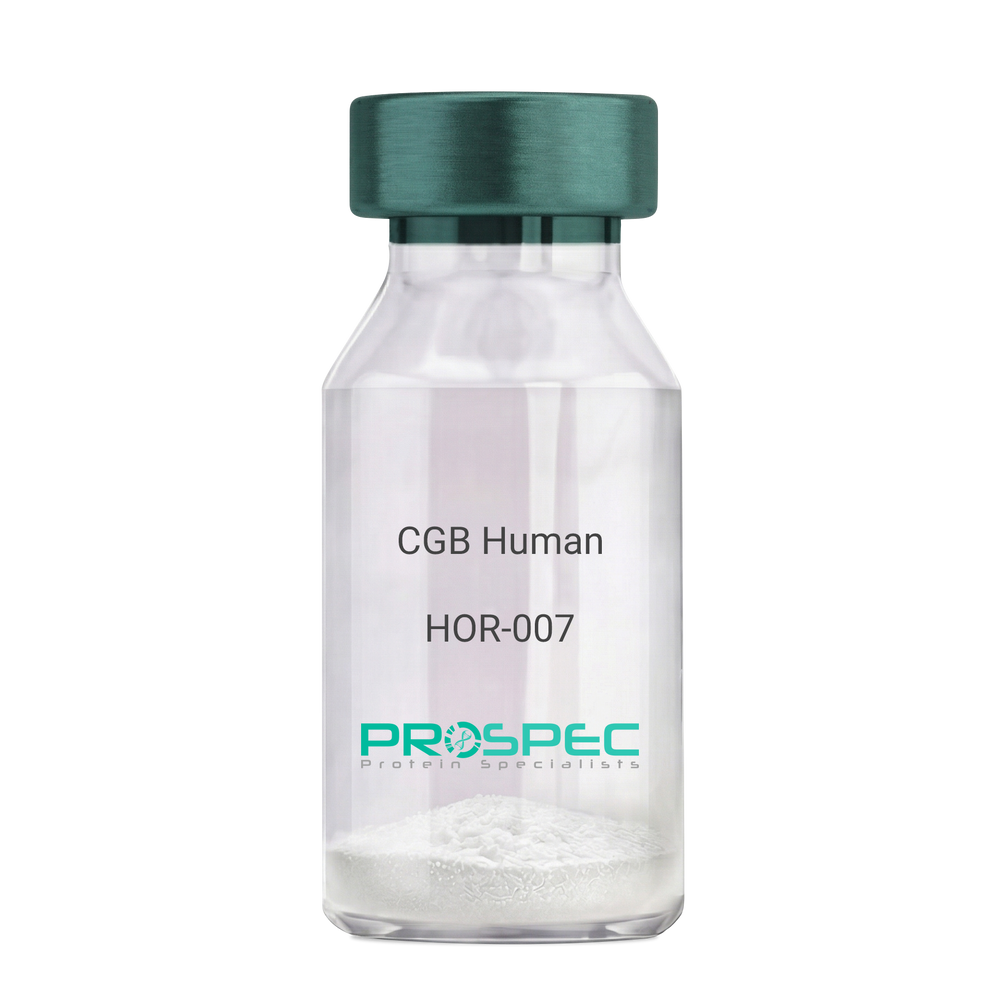
CGB HumanDescription:
CGB Human Recombinant
CGB3, CGB5, CGB7, CGB8, hCGB, CG-beta, CGB.
Product # :
HOR-007Price :
Quantity :
Shipping Method :
Shipped with Ice Packs
More Info
- description
- source
- formulation
- purity
- More Info
Description
CGB Human Recombinant produced in E.Coli is a single, non-glycosylated polypeptide chain containing 168 amino acids (21-165 a.a.) and having a molecular mass of 17.9kDa. CGB is fused to a 23 amino acid His-tag at N-terminus & purified by proprietary chromatographic techniques.
Source
Escherichia Coli.
Formulation
CGB protein solution (0.5mg/ml) containing 20mM Tris-HCl buffer (pH 8.0), 0.4M Urea and 10% glycerol.
Purity
Greater than 85.0% as determined by SDS-PAGE.
More Info
-
Introduction
Glycoprotein hormones are heterodimers consisting of a common alpha subunit and a unique beta subunit which confers biological specificity. CGB is a member of the glycoprotein hormone beta chain family and is the beta 3 subunit of (CG). CG is produced by the trophoblastic cells of the placenta and stimulates the ovaries. The beta subunit of CG is encoded by 6 genes which are arranged in tandem and inverted pairs on chromosome 19q13.3 and contiguous with the luteinizing hormone beta subunit gene.
-
Synonyms
CGB3, CGB5, CGB7, CGB8, hCGB, CG-beta, CGB.
-
Physical Appearance
Sterile filtered colorless solution.
-
Stability
Store at 4°C if entire vial will be used within 2-4 weeks. Store, frozen at -20°C for longer periods of time. For long term storage it is recommended to add a carrier protein (0.1% HSA or BSA).Avoid multiple freeze-thaw cycles.
-
Amino Acid Sequence
MGSSHHHHHH SSGLVPRGSH MGSSKEPLRP RCRPINATLA VEKEGCPVCI TVNTTICAGY CPTMTRVLQG VLPALPQVVC NYRDVRFESI RLPGCPRGVN PVVSYAVALS CQCALCRRST TDCGGPKDHP LTCDDPRFQD SSSSKAPPPS LPSPSRLPGP SDTPILPQ.
ProSpec's products are furnished for LABORATORY RESEARCH USE ONLY. The product may not be used as drugs, agricultural or pesticidal products, food additives or household chemicals.