Search results
1000 results found for “Cyclophilin”
Name
Description
Product #
Price
Quantity
Shipping Method
- View Data Sheet
Name :
ARFIP1 HumanDescription:
ADP-Ribosylation Factor Interacting Protein 1 Human Recombinant
HSU52521, Arfaptin-1, ADP-ribosylation factor-interacting protein 1.
Product # :
PRO-2088Price :
Quantity :
Shipping Method :
Shipped with Ice Packs
More Info
- description
- source
- formulation
- purity
- More Info
Description
ARFIP1 Human Recombinant produced in E.Coli is a single, non-glycosylated polypeptide chain containing 396 amino acids (1-373 a.a) and having a molecular mass of 44.1kDa.ARFIP1 is fused to a 23 amino acid His-tag at N-terminus & purified by proprietary chromatographic techniques.
Source
Escherichia Coli.
Formulation
ARFIP1 protein solution (0.25mg/ml) containing Phosphate buffered saline (pH7.4), 20% glycerol and 1mM DTT.
Purity
Greater than 90.0% as determined by SDS-PAGE.
More Info
-
Introduction
ADP-Ribosylation Factor Interacting Protein 1 (ARFIP1) contains 1 AH domain and is a putative target protein of ADP-ribosylation factor.
-
Synonyms
HSU52521, Arfaptin-1, ADP-ribosylation factor-interacting protein 1.
-
Physical Appearance
Sterile Filtered clear solution.
-
Stability
Store at 4°C if entire vial will be used within 2-4 weeks. Store, frozen at -20°C for longer periods of time. For long term storage it is recommended to add a carrier protein (0.1% HSA or BSA).Avoid multiple freeze-thaw cycles.
-
Amino Acid Sequence
MGSSHHHHHH SSGLVPRGSH MGSMAQESPK NSAAEIPVTS NGEVDDSREH SFNRDLKHSL PSGLGLSETQ ITSHGFDNTK EGVIEAGAFQ GSPAPPLPSV MSPSRVAASR LAQQGSDLIV PAGGQRTQTK SGPVILADEI KNPAMEKLEL VRKWSLNTYK CTRQIISEKL GRGSRTVDLE LEAQIDILRD NKKKYENILK LAQTLSTQLF QMVHTQRQLG DAFADLSLKS LELHEEFGYN ADTQKLLAKN GETLLGAINF FIASVNTLVN KTIEDTLMTV KQYESARIEY DAYRTDLEEL NLGPRDANTL PKIEQSQHLF QAHKEKYDKM RNDVSVKLKF LEENKVKVLH NQLVLFHNAI AAYFAGNQKQ LEQTLKQFHI KLKTPGVDAP SWLEEQ.
ProSpec's products are furnished for LABORATORY RESEARCH USE ONLY. The product may not be used as drugs, agricultural or pesticidal products, food additives or household chemicals.
- View Data Sheet
Name :
STX12 HumanDescription:
Syntaxin-12 Human Recombinant
Syntaxin 12, STX13, STX14.
Product # :
PRO-1099Price :
Quantity :
Shipping Method :
Shipped with Ice Packs
More Info
- description
- source
- formulation
- purity
- More Info
Description
STX12 Human Recombinant produced in E. coli is a single polypeptide chain containing 272 amino acids (1-248) and having a molecular mass of 31.0kDa.STX12 is fused to a 24 amino acid His-tag at N-terminus & purified by proprietary chromatographic techniques.
Source
E.coli.
Formulation
The STX12 solution (0.5mg/1ml) contains 20mM Tris-HCl buffer (pH 8.0), 200mM NaCl, 1mM DTT and 20% glycerol.
Purity
Greater than 85% as determined by SDS-PAGE.
More Info
-
Introduction
STX12 is a member of the syntaxin family which are cellular receptors for transport of vesicles that take part in exocytosis in neutrophils. STX12 regulates protein transport from late endosomes and the trans-Golgi network and back. Additionally, STX12 cooperates with ABC1 (ATP-binding cassette transporter A1), which enables cellular release of cholesterol and choline-phospholipids to apoA-I (apolipoprotein A-I).
-
Synonyms
Syntaxin 12, STX13, STX14.
-
Physical Appearance
Sterile Filtered colorless solution.
-
Stability
Store at 4°C if entire vial will be used within 2-4 weeks. Store, frozen at -20°C for longer periods of time. For long term storage it is recommended to add a carrier protein (0.1% HSA or BSA).Avoid multiple freeze-thaw cycles.
-
Amino Acid Sequence
MGSSHHHHHH SSGLVPRGSH MGSHMSYGPL DMYRNPGPSG PQLRDFSSII QTCSGNIQRI SQATAQIKNL MSQLGTKQDS SKLQENLQQL QHSTNQLAKE TNELLKELGS LPLPLSTSEQ RQQRLQKERL MNDFSAALNN FQAVQRRVSE KEKESIARAR AGSRLSAEER QREEQLVSFD SHEEWNQMQS QEDEVAITEQ DLELIKERET AIRQLEADIL DVNQIFKDLA MMIHDQGDLI DSIEANVESS EVHVERATEQ LQRAAYYQKK SR
ProSpec's products are furnished for LABORATORY RESEARCH USE ONLY. The product may not be used as drugs, agricultural or pesticidal products, food additives or household chemicals.
- View Data Sheet
Name :
KLK15 Human, sf9Description:
Kallikrein-15 Human Recombinant, sf9
Kallikrein Related Peptidase 15, ACO Protease, Kallikrein-Like Serine Protease, Kallikrein 15, Kallikrein-15, Prostinogen, EC 3.4.21.4, EC 3.4.21.-, EC 3.4.21, HSRNASPH, ACO.
Product # :
ENZ-1047Price :
Quantity :
Shipping Method :
Shipped with Ice Packs
More Info
- description
- source
- formulation
- purity
- More Info
Description
KLK15 Human Recombinant produced in Sf9 Baculovirus cells is a single, glycosylated polypeptide chain containing 249 amino acids (17-256a.a.) and having a molecular mass of 27.4kDa (Molecular size on SDS-PAGE will appear at approximately 28-40kDa).KLK15 is expressed with a 6 amino acids His tag at C-Terminus and purified by proprietary chromatographic techniques.
Source
Sf9, Baculovirus cells.
Formulation
KLK15 protein solution (0.25mg/ml) contains Phosphate Buffered Saline (pH 7.4) and 10% glycerol.
Purity
Greater than 80.0% as determined by SDS-PAGE.
More Info
-
Introduction
Kallikrein-15 (KLK15) is one of the 15 kallikrein subfamily members located in a cluster on chromosome 19. KLK15 contains numerous polyadenylation sites and alternative splicing results in multiple transcript variants encoding different isoforms. KLK15 is over expressed in prostate cancer and therefore uses as a diagnostic or prognostic marker for prostate cancer.
-
Synonyms
Kallikrein Related Peptidase 15, ACO Protease, Kallikrein-Like Serine Protease, Kallikrein 15, Kallikrein-15, Prostinogen, EC 3.4.21.4, EC 3.4.21.-, EC 3.4.21, HSRNASPH, ACO.
-
Physical Appearance
Sterile Filtered colorless solution.
-
Stability
Store at 4°C if entire vial will be used within 2-4 weeks. Store, frozen at -20°C for longer periods of time. For long term storage it is recommended to add a carrier protein (0.1% HSA or BSA). Avoid multiple freeze-thaw cycles.
-
Amino Acid Sequence
ADPQDGDKLL EGDECAPHSQ PWQVALYERG RFNCGASLIS PHWVLSAAHC QSRFMRVRLG EHNLRKRDGP EQLRTTSRVI PHPRYEARSH RNDIMLLRLV QPARLNPQVR PAVLPTRCPH PGEACVVSGW GLVSHNEPGT AGSPRSQVSL PDTLHCANIS IISDTSCDKS YPGRLTNTMV CAGAEGRGAE SCEGDSGGPL VCGGILQGIV SWGDVPCDNT TKPGVYTKVC HYLEWIRETM KRNHHHHHH.
ProSpec's products are furnished for LABORATORY RESEARCH USE ONLY. The product may not be used as drugs, agricultural or pesticidal products, food additives or household chemicals.
- View Data Sheet
Name :
PhI p 1Description:
Pollen allergen Phl p 1 Recombinant
Pollen allergen Phl p 1, Allergen Phl p I, Phl p 1, PHLPI.
Product # :
PRO-2305Price :
Quantity :
Shipping Method :
Shipped with Ice Packs
More Info
- description
- source
- formulation
- purity
- More Info
Description
Recombinant Pollen allergen Phl p 1 produced in SF9 is a glycosylated, polypeptide chain having a calculated molecular mass of 29,516 Dalton. PhI p 1 is expressed with a 10xHis tag at N-terminus and purified by proprietary chromatographic techniques.
Source
Sf9 insect cells.
Formulation
PhI p 1 protein solution is supplied in 20mM HEPES pH-8.0, 200mM NaCl and 20% glycerol.
Purity
Greater than 80.0% as determined by SDS-PAGE.
More Info
-
Introduction
Pollen allergen Phl p 1 (PhI p 1) causes an allergic reaction in humans.
-
Synonyms
Pollen allergen Phl p 1, Allergen Phl p I, Phl p 1, PHLPI.
-
Physical Appearance
Sterile Filtered clear solution.
-
Stability
Store at 4°C if entire vial will be used within 2-4 weeks. Store, frozen at -20°C for longer periods of time. Avoid multiple freeze-thaw cycles.
-
Immunological Functions
1. Binds IgE type human antibodies. 2. Immunodot test with positive/negative sera panels.
-
Molar extinction coefficient
46785; A280(1mg/ml)=1.699
ProSpec's products are furnished for LABORATORY RESEARCH USE ONLY. The product may not be used as drugs, agricultural or pesticidal products, food additives or household chemicals.
- View Data Sheet
Name :
KLRC2 HumanDescription:
Killer Cell Lectin-Like Receptor Subfamily C, Member 2 Human Recombinant
Killer cell lectin-like receptor subfamily C member 2, NKG2-C type II integral membrane protein, NKG2-C-activating NK receptor, CD159 antigen-like family member C, NK cell receptor C, NKG2C, CD159c.
Product # :
PRO-1190Price :
Quantity :
Shipping Method :
Shipped with Ice Packs
More Info
- description
- source
- formulation
- purity
- More Info
Description
KLRC2 Human Recombinant produced in E. coli is a single polypeptide chain containing 162 amino acids (94-231) and having a molecular mass of 18.4 kDa.KLRC2 is fused to a 24 amino acid His-tag at N-terminus & purified by proprietary chromatographic techniques.
Source
E.coli.
Formulation
The KLRC2 solution (0.5mg/1ml) contains 20mM Tris-HCl buffer (pH 8.0), 0.4M urea and 10% glycerol.
Purity
Greater than 85% as determined by SDS-PAGE.
More Info
-
Introduction
KLRC2 has a part as a receptor for the recognition of MHC class I HLA-E molecules by NK cells and some cytotoxic T-cells. The group, designated KLRC (NKG2) are expressed mainly in natural killer (NK) cells and encodes a family of transmembrane proteins categorized by a type II membrane orientation (extracellular C terminus) and the presence of a C-type lectin domain. The KLRC (NKG2) gene family is situated inside the NK complex, a region which holds a few C-type lectin genes specially expressed on NK cells. KLRC2 alternative splice variants are known but their full-length nature is yet to be determined.
-
Synonyms
Killer cell lectin-like receptor subfamily C member 2, NKG2-C type II integral membrane protein, NKG2-C-activating NK receptor, CD159 antigen-like family member C, NK cell receptor C, NKG2C, CD159c.
-
Physical Appearance
Sterile Filtered colorless solution.
-
Stability
Store at 4°C if entire vial will be used within 2-4 weeks. Store, frozen at -20°C for longer periods of time. For long term storage it is recommended to add a carrier protein (0.1% HSA or BSA).Avoid multiple freeze-thaw cycles.
-
Amino Acid Sequence
MGSSHHHHHH SSGLVPRGSH MGSMIPFLEQ NNFSPNTRTQ KARHCGHCPE EWITYSNSCY YIGKERRTWE ESLLACTSKN SSLLSIDNEE EMKFLASILP SSWIGVFRNS SHHPWVTING LAFKHKIKDS DNAELNCAVL QVNRLKSAQC GSSMIYHCKH KL
ProSpec's products are furnished for LABORATORY RESEARCH USE ONLY. The product may not be used as drugs, agricultural or pesticidal products, food additives or household chemicals.
- View Data Sheet
Name :
Clusterin HumanDescription:
Clusterin Human Recombinant
CLI, AAG4, KUB1, SGP2, SGP-2, SP-40, TRPM2, MGC24903, Clusterin, Apolipoprotein J, Apo-J.
Product # :
CYT-278Price :
Quantity :
Shipping Method :
Shipped at Room temp
More Info
- description
- source
- formulation
- purity
- More Info
Description
Clusterin Human Recombinant produced in HEK is a glycosylated, polypeptide chain containing 438 amino acids and having a molecular mass of 51.27 kDa. Clusterin (1-427 a.a.) is fused to 11 a.a. flag tag at c-terminal and purified by proprietary chromatographic techniques.
Source
293 cell line (Human embryonic kidney).
Formulation
Filtered (0.4 micron) and lyophilized PBS, pH 7.5.
Purity
Greater than 95% as determined by SDS PAGE.
More Info
-
Introduction
Clusterin also named Apolipoprotein J (APO-J) is a 75-80 kD disulfide-linked heterodimeric protein containing about 30% of N-linked carbohydrate rich in sialic acid but truncated forms targeted to the nucleus have also been identified.
The precursor polypeptide chain is cleaved proteolytically to remove the 22-mer secretory signal peptide and subsequently between residues 227/228 to generate the a and b chains. These are assembled in anti-parallel to give a heterodimeric molecule in which the cysteine-rich centers are linked by five disulfide bridges and are flanked by two predicted coiled-coil a-helices and three predicted amphipathic a-helices.
Across a broad range of species clusterin shows a high degree of sequence homology ranging from 70% to 80%. It is nearly ubiquitously expressed in most mammalian tissues and can be found in plasma, milk, urine, cerebrospinal fluid and semen.
It is able to bind and form complexes with numerous partners such as immunoglobulins, lipids, bacteria, complement components, paraoxonase, beta amyloid, leptin and others. Clusterin has been ascribed a plethora of functions such as phagocyte recruitment, aggregation induction, complement attack prevention, apoptosis inhibition, membrane remodeling, lipid transport, hormone transport and/or scavenging, matrix metalloproteinase inhibition.
A genuine function of clusterin has not been defined. One tempting hypothesis says that clusterin is an extracellular chaperone protecting cells from stress induced insults caused by degraded and misfolded protein precipitates.
Clusterin is up- or down regulated on the mRNA or protein level in many pathological and clinically relevant situations including cancer, organ regeneration, infection, Alzheimer disease, retinitis pigmentosa, myocardial infarction, renal tubular damage, autoimmunity and others. -
Synonyms
CLI, AAG4, KUB1, SGP2, SGP-2, SP-40, TRPM2, MGC24903, Clusterin, Apolipoprotein J, Apo-J.
-
Physical Appearance
Filtered, White, Lyophilized powder.
-
Stability
Store lyophilized protein at -20°C. Aliquot the product after reconstitution to avoid repeated freezing/thawing cycles. Reconstituted protein can be stored at 4°C for a limited period of time; it does not show any change after two weeks at 4°C.
-
Solubility
Add deionized water to prepare a working stock solution of approximately 0.5mg/ml and let the lyophilized pellet dissolve completely. Product not sterile! Please filter the product by an appropriate sterile filter before using it in cell culture.
-
Amino Acid Sequence
DQTVSDNELQ EMSNQGSKYV NKEIQNAVNG VKQIKTLIEK TNEERKTLLS NLEEAKKKKE DALNETRESE TKLKELPGVC NETMMALWEE CKPCLKQTCM KFYARVCRSGS GLVGRQLEE FLNQSSPFYF WMNGDRIDSL LENDRQQTHM LDVMQDHFSRA SSIIDELFQ DRFFTREPQD TYHYLPFSLP HRRPHFFFPK SRIVRSLMPF SPYEPLNFHA MFQPFLEMIH EAQQAMDIHF HSPAFQHPPT EFIREGDDDR TVCREIRHNS TGCLRMKDQC DKCREILSVD CSTNNPSQAKLRRELDESLQ VAERLTRKYN ELLKSYQWKM LNTSSLLEQL NEQFNWVSRL ANLTQGEDQYYLRVTTVASH TSDSDVPSGV TEVVVKLFDS DPITVTVPVE VSRKNPKFME TVAEKALQEY RKKHREEAAA DYKDDDDK.
-
Background
What is the molecular weight/Mw of CLUSTERIN Protein?
CLUSTERIN Protein has a total Mw of 51.27kDa.
What is the source or expression system of CLUSTERIN Protein?
293 cell line
What is the Purity of CLUSTERIN Protein?
CLUSTERIN Protein is >95% pure as determined by SDS-PAGE.
What is the Biological Activity of CLUSTERIN Protein?
The biological functionality of CLUSTERIN Protein will be determined in the future.
What is the amino acid sequence of CLUSTERIN Protein?
DQTVSDNELQ EMSNQGSKYV NKEIQNAVNG VKQIKTLIEK TNEERKTLLS NLEEAKKKKE DALNETRESE TKLKELPGVC NETMMALWEE CKPCLKQTCM KFYARVCRSGS GLVGRQLEE FLNQSSPFYF WMNGDRIDSL LENDRQQTHM LDVMQDHFSRA SSIIDELFQ DRFFTREPQD TYHYLPFSLP HRRPHFFFPK SRIVRSLMPF SPYEPLNFHA MFQPFLEMIH EAQQAMDIHF HSPAFQHPPT EFIREGDDDR TVCREIRHNS TGCLRMKDQC DKCREILSVD CSTNNPSQAKLRRELDESLQ VAERLTRKYN ELLKSYQWKM LNTSSLLEQL NEQFNWVSRL ANLTQGEDQYYLRVTTVASH TSDSDVPSGV TEVVVKLFDS DPITVTVPVE VSRKNPKFME TVAEKALQEY RKKHREEAAA DYKDDDDK.
What applications can CLUSTERIN Protein be used in?
CLUSTERIN Protein can probably be used in western blot, ELISA and Lateral Flow.
What is the endotoxin level for CLUSTERIN Protein?
The endotoxin level is minimal, CLUSTERIN Protein was purified using conventional chromatography techniques.
ProSpec's products are furnished for LABORATORY RESEARCH USE ONLY. The product may not be used as drugs, agricultural or pesticidal products, food additives or household chemicals.
- View Data Sheet
Name :
GDF3 HumanDescription:
Growth Differentiation Factor-3 Human Recombinant
Growth Differentiation Factor 3, Growth/Differentiation Factor 3 , MCOPCB6, MCOP7, GDF-3, KFS3.
Product # :
CYT-694Price :
Quantity :
Shipping Method :
Shipped at Room temp
More Info
- description
- source
- formulation
- purity
- More Info
Description
GDF3 Human Recombinant produced in E.Coli is a single, non-glycosylated, polypeptide chain containing 124 amino acids and having a total molecular mass of 14.15 kDa.GDF3 is fused to a 10 amino acid His Tag at N-terminus and purified by proprietary chromatographic techniques.
Source
Escherichia Coli.
Formulation
The protein was lyophilized from a concentrated solution (0.5mg/ml) containing 30mM Acetate buffer pH-4.
Purity
Greater than 90.0% as determined by SDS-PAGE.
More Info
-
Introduction
GDF3 is a member of the TGF-beta superfamily though it does not show similarity pattern of conserved cysteine residues. GDF3 is linked to Vg-1 and human BMP-4. GDF3 transcripts are identified mainly in adult bone marrow, spleen, thymus, and adipose tissue. GDF3 expression is upregulated strongly in high-fat-fed C57Bl/6J FABP4/aP2 null mice, which develop obesity but not the related hyperglycemia or hyperinsulinemia characteristic of type II diabetes. GDF3 expression therefore bonds fatty acid metabolism in adipocytes and the expression of a differentiation regulator belonging to the bone morphogenetic proteins.
-
Synonyms
Growth Differentiation Factor 3, Growth/Differentiation Factor 3 , MCOPCB6, MCOP7, GDF-3, KFS3.
-
Physical Appearance
Filtered White lyophilized (freeze-dried) powder.
-
Stability
Lyophilized GDF3 although stable at room temperature for 3 weeks, should be stored desiccated below -18°C. Upon reconstitution GDF3 should be stored at 4°C between 2-7 days and for future use below -18°C.For long term storage it is recommended to add a carrier protein (0.1% HSA or BSA).Please prevent freeze-thaw cycles.
-
Solubility
It is recommended to reconstitute the lyophilized GDF3 in sterile 100mM Acetate buffer pH-4 at a concentration of 0.5mg/ml. For the dilution into higher pH values, it is recommended to dilute the protein to a concentration of 10μg/ml. Please note that in higher concentrations the solubility of GDF3 is limited. The protein is not sterile! Please sterile filter before using it in the cell culture.
-
Amino Acid Sequence
MKHHHHHHAS AAIPVPKLSC KNLCHRHQLF INFRDLGWHK WIIAPKGFMA NYCHGECPFS LTISLNSSNY AFMQALMHAV DPEIPQAVCI PTKLSPISML YQDNNDNVIL RHYEDMVVDECGCG.
-
Background
What is the molecular weight/Mw of GDF3 HUMAN Protein?
GDF3 HUMAN Protein has a total Mw of 14.15XkDa.
What is the source or expression system of GDF3 HUMAN Protein?
Escherichia Coli.
What is the Purity of GDF3 HUMAN Protein?
GDF3 HUMAN Protein is >90% pure as determined by SDS-PAGE.
What is the Biological Activity of GDF3 HUMAN Protein?
The biological functionality of GDF3 HUMAN Protein will be determined in the future.
What is the amino acid sequence of GDF3 HUMAN Protein?
MKHHHHHHAS AAIPVPKLSC KNLCHRHQLF INFRDLGWHK WIIAPKGFMA NYCHGECPFS LTISLNSSNY AFMQALMHAV DPEIPQAVCI PTKLSPISML YQDNNDNVIL RHYEDMVVDECGCG.
What applications can GDF3 HUMAN Protein be used in?
GDF3 HUMAN Protein can probably be used in western blot, ELISA and Lateral Flow.
What is the endotoxin level for GDF3 HUMAN Protein?
The endotoxin level is minimal, GDF3 HUMAN Protein was purified using conventional chromatography techniques.
ProSpec's products are furnished for LABORATORY RESEARCH USE ONLY. The product may not be used as drugs, agricultural or pesticidal products, food additives or household chemicals.
- View Data Sheet
Name :
SCF RatDescription:
Stem Cell Factor Rat Recombinant
Kit ligand Precursor, C-kit ligand, SCF, Mast cell growth factor, MGF, SF, KL-1, Kitl, DKFZp686F2250, Hematopoietic growth factor KL.
Product # :
CYT-323Price :
Quantity :
Shipping Method :
Shipped at Room temp
More Info
- description
- source
- formulation
- purity
- biological activity
- More Info
Description
Stem cell factor Rat Recombinant produced in E.Coli is a single, non-glycosylated polypeptide chain containing 164 amino acids (26-189) and having a molecular mass of 18.4 kDa.The Rat SCF is purified by proprietary chromatographic techniques.
Source
Escherichia Coli.
Formulation
Lyophilized from a concentrated (1mg/ml) solution in water containing 0.02% NaHCO3.
Purity
Greater than 98.0% as determined by:
(a) Analysis by SEC-HPLC.
(b) Analysis by SDS-PAGE.Biological Activity
The ED50 is determined by the dose-dependant stimulation of the proliferation of human TF-1 cells which is < 10 ng/ml, corresponding to a specific activity of 100,000units/mg.More Info
-
Introduction
Stem cell factor / KIT ligand (SCF) is a cytokine which binds CD117 (c-Kit). SCF is also known as "steel factor" or "c-kit ligand". SCF exists in two forms, cell surface bound SCF and soluble (or free) SCF. Soluble SCF is produced by the cleavage of surface bound SCF by metalloproteases.
SCF is a growth factor important for the survival, proliferation, and differentiation of hematopoietic stem cells and other hematopoietic progenitor cells. One of its roles is to change the BFU-E (burst-forming unit-erythroid) cells, which are the earliest erythrocyte precursors in the erythrocytic series, into the CFU-E (colony-forming unit-erythroid). -
Synonyms
Kit ligand Precursor, C-kit ligand, SCF, Mast cell growth factor, MGF, SF, KL-1, Kitl, DKFZp686F2250, Hematopoietic growth factor KL.
-
Physical Appearance
Sterile Filtered White lyophilized (freeze-dried) powder.
-
Stability
Lyophilized rat SCF although stable at room temperature for 3 weeks, should be stored desiccated below -18°C. Upon reconstitution SCF should be stored at 4°C between 2-7 days and for future use below -18°C.For long term storage it is recommended to add a carrier protein (0.1% HSA or BSA).Please prevent freeze-thaw cycles.
-
Solubility
It is recommended to reconstitute the lyophilized SCF in sterile 18MΩ-cm H2O not less than 100µg/ml, which can then be further diluted to other aqueous solutions.
-
Amino Acid Sequence
MQEICRNPVT DNVKDITKLV ANLPNDYMIT LNYVAGMDVL PSHCWLRDMV THLSVSLTTL LDKFSNISEG LSNYSIIDKL GKIVDDLVAC MEENAPKNVK ESLKKPETRN FTPEEFFSIF NRSIDAFKDF MVASDTSDCV LSSTLGPEKD SRVSVTKPFM LPPVA.
ProSpec's products are furnished for LABORATORY RESEARCH USE ONLY. The product may not be used as drugs, agricultural or pesticidal products, food additives or household chemicals.
- View Data Sheet
Name :
ARF4 HumanDescription:
ADP-Ribosylation Factor 4 Human Recombinant
ADP-ribosylation factor 4, ARF4, ARF2.
Product # :
PRO-066Price :
Quantity :
Shipping Method :
Shipped with Ice Packs
More Info
- description
- source
- formulation
- purity
- More Info
Description
ARF4 Human Recombinant fused with a 24 amino acid His tag at N-terminus produced in E.Coli is a single, non-glycosylated, polypeptide chain containing 204 amino acids (1-180 a.a.) and having a molecular mass of 23kDa. The ARF4 is purified by proprietary chromatographic techniques.
Source
Escherichia Coli.
Formulation
The ARF4 solution (0.5 mg/ml) in 20mM Tris-HCl buffer (pH8.0), 0.2M NaCl, 5mM DTT and 20% glycerol.
Purity
Greater than 95.0% as determined by SDS-PAGE.
More Info
-
Introduction
ARF4 belongs to the ARF gene family whose members encode small guanine nucleotide-binding proteins which stimulate the ADP-ribosyltransferase activity of cholera toxin and have a role in vesicular trafficking and as activators of phospholipase D. The ARF proteins include five ARF proteins and eleven ARF-like proteins and constitute one family of the RAS superfamily. They are classified as class I, class II and class III; the ARF4 gene is a class II member. The members of each class share a common gene organization. The ARF4 gene spans approximately 12kb and contains 6 exons and 5 introns. The ARF4 gene is the most divergent member of the human ARFs.
-
Synonyms
ADP-ribosylation factor 4, ARF4, ARF2.
-
Physical Appearance
Sterile Filtered colorless solution.
-
Stability
Store at 4°C if entire vial will be used within 2-4 weeks.Store, frozen at -20°C for longer periods of time. For long term storage it is recommended to add a carrier protein (0.1% HSA or BSA).Avoid multiple freeze-thaw cycles.
-
Amino Acid Sequence
MGSSHHHHHH SSGLVPRGSH MGSHMGLTIS SLFSRLFGKK QMRILMVGLD AAGKTTILYK LKLGEIVTTI PTIGFNVETV EYKNICFTVW DVGGQDRIRP LWKHYFQNTQ GLIFVVDSND RERIQEVADE LQKMLLVDEL RDAVLLLFAN KQDLPNAMAI SEMTDKLGLQ SLRNRTWYVQ ATCATQGTGL YEGLDWLSNE LSKR.
ProSpec's products are furnished for LABORATORY RESEARCH USE ONLY. The product may not be used as drugs, agricultural or pesticidal products, food additives or household chemicals.
- View Data Sheet
Name :
IP 10 Rhesus macaqueDescription:
IP-10 Rhesus macaque Recombinant (CXCL10)
C-X-C motif chemokine 10, 10 kDa, Gamma-IP10, IP-10, Small-inducible cytokine B10, CXCL10, SCYB10.
Product # :
CHM-010Price :
Quantity :
Shipping Method :
Shipped at Room temp
More Info
- description
- source
- formulation
- purity
- biological activity
- More Info
Description
IP-10 Rhesus macaque Recombinant produced in E.Coli is a single, non-glycosylated, polypeptide chain containing 77 amino acids and having a molecular mass of 8.7kDa. The IP-10 is purified by proprietary chromatographic techniques.
Source
Escherichia Coli.
Formulation
Lyophilized from a 0.2µm filtered concentrated solution in 1×PBS, pH 7.4.
Purity
Greater than 97.0% as determined by:
(a) Analysis by RP-HPLC.
(b) Analysis by SDS-PAGE.Biological Activity
Determined by its ability to chemoattract human peripheral blood T-Lymphocytes using a concentration range of 10.0-100.0 ng/ml.More Info
-
Introduction
Chemokine (C-X-C motif) ligand 10 (CXCL10) is a small cytokine belonging to the CXC chemokine family that is also known as 10 kDa IP-10. CXCL10 is secreted by several cell types in response to IFN-?. These cell types include monocytes, endothelial cells and fibroblasts. CXCL10 has been attributed to several roles, such as chemoattraction for monocytes and T cells, promotion of T cell adhesion to endothelial cells, antitumor activity, and inhibition of bone marrow colony formation and angiogenesis. The gene for CXCL10 is located on human chromosome 4 in a cluster among several other CXC chemokines. This chemokine elicits its effects by binding to the cell surface chemokine receptor CXCR3. The three-dimensional crystal structure of this chemokine has been determined under 3 different conditions to a resolution of up to 1.92A.
-
Synonyms
C-X-C motif chemokine 10, 10 kDa, Gamma-IP10, IP-10, Small-inducible cytokine B10, CXCL10, SCYB10.
-
Physical Appearance
Sterile Filtered White lyophilized (freeze-dried) powder.
-
Stability
Lyophilized IP-10 although stable at room temperature for 3 weeks, should be stored desiccated below -18°C. Upon reconstitution CXCL10 should be stored at 4°C between 2-7 days and for future use below -18°C. For long term storage it is recommended to add a carrier protein (0.1% HSA or BSA).Please prevent freeze-thaw cycles.
-
Solubility
It is recommended to reconstitute the lyophilized IP-10 in sterile 18MΩ-cm H2O not less than 100µg/ml, which can then be further diluted to other aqueous solutions.
-
Amino Acid Sequence
IPLSRTVRCT CISISNQPVN PRSLEKLEII PPSQFCPHVE IIATMKKKGE KRCLNPESKA IKNLLKAVSK ERSKRSP.
ProSpec's products are furnished for LABORATORY RESEARCH USE ONLY. The product may not be used as drugs, agricultural or pesticidal products, food additives or household chemicals.
- View Data Sheet
Name :
KLK3, HisDescription:
Kallikrein-3 Human Recombinant, His Tag
Prostate-specific antigen, PSA, Gamma-seminoprotein, Seminin, Kallikrein-3, P-30 antigen, Semenogelase, KLK3, APS, hK3, KLK2A1.
Product # :
ENZ-620Price :
Quantity :
Shipping Method :
Shipped with Ice Packs
More Info
- description
- source
- formulation
- purity
- More Info
Description
KLK3 Human Recombinant produced in E.coli is a single, non-glycosylated polypeptide chain containing 262 amino acids (25-261) and having a molecular mass of 28.8kDa.The KLK3 is fused to a 25 amino acid His-Tag at N-terminus and purified by proprietary chromatographic techniques.
Source
Escherichia Coli.
Formulation
KLK3 protein solution (1mg/ml) is supplied in 20mM Tris-HCl buffer (pH8.0) and 0.4M Urea.
Purity
Greater than 85.0% as determined by SDS-PAGE.
More Info
-
Introduction
Kallikrein-3 (KLK3) belongs to the kallikrein-related peptidase family. Kallikreins are a subgroup of serine proteases having various physiological functions. Numerous kallikreins are involved in carcinogenesis and some may be prospective cancer and other disease biomarkers. Kallikrein-3 is 1 of the 15 kallikrein subfamily members located in a cluster on chromosome 19 and is a protease present in seminal plasma. KLK3 hydrolyzes semenogelin-1 consequently leading to the liquefaction of the seminal coagulum. KLK3 is assumed to act normally in the liquefaction of seminal coagulum, probably by hydrolysis of the high molecular mass seminal vesicle protein. Serum level of the KLK3 protein, called PSA in the clinical setting, is beneficial in the diagnosis and monitoring of prostatic carcinoma.
-
Synonyms
Prostate-specific antigen, PSA, Gamma-seminoprotein, Seminin, Kallikrein-3, P-30 antigen, Semenogelase, KLK3, APS, hK3, KLK2A1.
-
Physical Appearance
Sterile Filtered clear solution.
-
Stability
Store at 4°C if entire vial will be used within 2-4 weeks. Store, frozen at -20°C for longer periods of time. For long term storage it is recommended to add a carrier protein (0.1% HSA or BSA).Avoid multiple freeze-thaw cycles.
-
Amino Acid Sequence
MGSSHHHHHH SSGLVPRGSH MGSHMIVGGW ECEKHSQPWQ VLVASRGRAV CGGVLVHPQW VLTAAHCIRN KSVILLGRHS LFHPEDTGQV FQVSHSFPHP LYDMSLLKNR FLRPGDDSSH DLMLLRLSEP AELTDAVKVM DLPTQEPALG TTCYASGWGS IEPEEFLTPK KLQCVDLHVI SNDVCAQVHP QKVTKFMLCA GRWTGGKSTC SGDSGGPLVC NGVLQGITSW GSEPCALPER PSLYTKVVHY RKWIKDTIVA NP.
ProSpec's products are furnished for LABORATORY RESEARCH USE ONLY. The product may not be used as drugs, agricultural or pesticidal products, food additives or household chemicals.
- View Data Sheet
Name :
TRAIL Human (114-281 a.a.)Description:
TRAIL/APO 2 Ligand (114-281 a.a.) Human Recombinant
Tumor necrosis factor ligand superfamily member 10, TNF-related apoptosis-inducing ligand, Protein TRAIL, Apo-2 ligand, Apo-2L, CD253 antigen, TL2, APO2L, TNFSF10.
Product # :
CYT-546Price :
Quantity :
Shipping Method :
Shipped with Ice Packs
More Info
- description
- source
- formulation
- purity
- More Info
Description
Soluble TNF-related apoptosis-inducing ligand Human Recombinant produced in E.Coli is a single, non-glycosylated polypeptide chain containing 169 amino acids (114-281) and having a molecular mass of 19.6 kDa. The sTRAIL is purified by proprietary chromatographic techniques.
Source
Escherichia Coli.
Formulation
1mg/ml in 20mM Tris-HCl pH-7.5, 300mM NaCl, 0.1mM DTT & 10% glycerol.
Purity
Greater than 95.0% as determined by SDS-PAGE.
More Info
-
Introduction
TNF-related apoptosis-inducing ligand (TRAIL) is a ligand molecule which induces apoptosis. It is a type II transmembrane protein with homology to other members of the tumor necrosis factor family.
In humans, the gene that encodes for TRAIL is located at chromosome 3q26.
TRAIL binds to the death receptors, DR4 and DR5. The process of apoptosis is caspase-8-dependent. This protein preferentially induces apoptosis in transformed and tumor cells, but does not appear to kill normal cells although it is expressed at a significant level in most normal tissues. -
Synonyms
Tumor necrosis factor ligand superfamily member 10, TNF-related apoptosis-inducing ligand, Protein TRAIL, Apo-2 ligand, Apo-2L, CD253 antigen, TL2, APO2L, TNFSF10.
-
Physical Appearance
Sterile Filtered colorless liquid.
-
Stability
Store at 4°C if entire vial will be used within 2-4 weeks. Store, frozen at -20°C for longer periods of time. For long term storage it is recommended to add a carrier protein (0.1% HSA or BSA).Avoid multiple freeze-thaw cycles.
-
Amino Acid Sequence
MVRERGPQRV AAHITGTRGR SNTLSSPNSK NEKALGRKIN SWESSRSGHS FLSNLHLRNGELVIHEKGFY YIYSQTYFRF QEEIKENTKN DKQMVQYIYK YTSYPDPILL MKSARNSCWSKDAEYGLYSI YQGGIFELKE NDRIFVSVTN EHLIDMDHEA SFFGAFLVG.
ProSpec's products are furnished for LABORATORY RESEARCH USE ONLY. The product may not be used as drugs, agricultural or pesticidal products, food additives or household chemicals.
- View Data Sheet
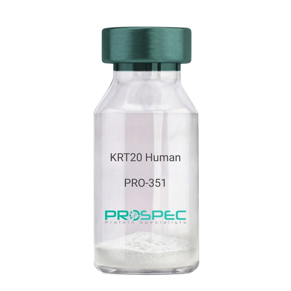
Name :
KRT20 HumanDescription:
Cytokeratin 20 Human Recombinant
Keratin type I cytoskeletal 20, Cytokeratin-20, CK-20, Keratin-20, K20, Protein IT, KRT20, CD20, CK20, KRT21, MGC35423.
Product # :
PRO-351Price :
Quantity :
Shipping Method :
Shipped at Room temp
More Info
- description
- source
- formulation
- purity
- More Info
Description
Cytokeratin 20 Human Recombinant produced in E.Coli is a single,non-glycosylated polypeptide chain having a molecular mass of 48,553 Dalton. The KRT20 is purified by proprietary chromatographic techniques.
Source
Escherichia Coli.
Formulation
The protein (1mg/1ml) was lyophilized after from a sterile solution containing 30mM Tris-HCL pH-8, 9.5M urea, 2mM DDT, 2mM EDTA and 10mM methylammonium chloride.
Purity
Greater than 95.0% as determined by SDS-PAGE.
More Info
-
Introduction
KRT20 is a member of the keratin family. The keratins are intermediate filament proteins responsible for the structural integrity of epithelial cells and are subdivided into cytokeratins and hair keratins. The type I cytokeratins consist of acidic proteins which are arranged in pairs of heterotypic keratin chains. This cytokeratin is a major cellular protein of mature enterocytes and goblet cells and is specifically expressed in the gastric and intestinal mucosa. The type I cytokeratin genes are clustered in a region of chromosome 17q12-q21.
-
Synonyms
Keratin type I cytoskeletal 20, Cytokeratin-20, CK-20, Keratin-20, K20, Protein IT, KRT20, CD20, CK20, KRT21, MGC35423.
-
Physical Appearance
Sterile Filtered White lyophilized (freeze-dried) powder.
-
Stability
Lyophilized KRT20 although stable at room temperature for 3 weeks, should be stored desiccated below -18°C. Upon reconstitution KRT20 should be stored at 4°C between 2-7 days and for future use below -18°C.For long term storage it is recommended to add a carrier protein (0.1% HSA or BSA).Please prevent freeze-thaw cycles.
-
Solubility
It is recommended to reconstitute the lyophilized KRT20 in sterile 18MΩ-cm H2O not less than 100µg/ml, which can then be further diluted to other aqueous solutions.
-
Reconstitution to filaments
Performed by mixing equimolar amounts of cytokeratins of type I and type II at concentrations of approx. 0.5 mg/ml, both dissolved in 9.5 M urea buffer (see above). Protofilaments and filament complexes are obtained by dialyzing the resulting polypeptide solution stepwise to a concentration of 4 M urea and then to low salt condition (50 mM NaCI, 2 mM dithiothreitol, 10 mM Tris-HCI, pH 7.4). For immunization purposes, the solution can be further dialyzed against PBS (phosphate buffered saline, e.g. Dulbecco's PBS).
ProSpec's products are furnished for LABORATORY RESEARCH USE ONLY. The product may not be used as drugs, agricultural or pesticidal products, food additives or household chemicals.
- View Data Sheet
Name :
Flt3 Ligand Human, HEKDescription:
Flt3-Ligand Human Recombinant, HEK derived
Fms-related tyrosine kinase 3 ligand, FLK2, STK1, CD135, Stem Cell Tyrosine Kinase 1, FLT3LG, Flt3.
Product # :
CYT-706Price :
Quantity :
Shipping Method :
Shipped at Room temp
More Info
- description
- source
- formulation
- purity
- biological activity
- More Info
Description
Flt3-Ligand Human Recombinant produced in HEK cells is a glycosylated monomer, having a molecular weight range of 24-30kDa due to glycosylation. The Flt3-Ligand is purified by proprietary chromatographic techniques.
Source
HEK293 (Human Embryonic Kidney cell line).
Formulation
Flt3-Ligand was lyophilized from a 0.2µm filtered solution containing 1xPBS.
Purity
Greater than 95.0% as determined by: (a) Analysis by RP-HPLC. (b) Analysis by SDS-PAGE.
Biological Activity
The activity was determined by the dose- dependent stimulation of the proliferation of the human acute myeloid leukemia cell line OCI-AML5. The ED50 is 0.56ng/ml.
More Info
-
Introduction
FLT3 ligand is a receptor for the fl cytokine has a tyrosine-protein kinase activity & a growth factor that regulates proliferation of early hematopoietic cells. Flt3-Ligand synergizes with other CSFs and interleukins to induce growth and differentiation.
-
Synonyms
Fms-related tyrosine kinase 3 ligand, FLK2, STK1, CD135, Stem Cell Tyrosine Kinase 1, FLT3LG, Flt3.
-
Physical Appearance
Sterile Filtered White lyophilized (freeze-dried) powder.
-
Stability
Lyophilized Flt3-Ligand although stable at room temperature for 3 weeks, should be stored desiccated below -18°C. Upon reconstitution Flt3-Ligand should be stored at 4°C between 2-7 days and for future use below -18°C. For long term storage it is recommended to add a carrier protein (0.1% HSA or BSA). Please prevent freeze-thaw cycles.
-
Solubility
It is recommended to reconstitute the lyophilized Flt3-Ligand in sterile 18MΩ-cm H2O not less than 100µg/ml, which can then be further diluted to other aqueous solutions.
-
Amino Acid Sequence
TQDCSFQHSP ISSDFAVKIR ELSDYLLQDY PVTVASNLQD EELCGGLWRL VLAQRWMERL KTVAGSKMQG LLERVNTEIH FVTKCAFQPP PSCLRFVQTN ISRLLQETSE QLVALKPWIT RQNFSRCLEL QCQPDSSTLP PPWSPRPLEA TAPTAPQP.
-
Background
What is the molecular weight/Mw of FLT3 LIGAND HUMAN, HEK Protein?
FLT3 LIGAND HUMAN, HEK Protein has a total Mw of 27kDa.
What is the source or expression system of FLT3 LIGAND HUMAN, HEK Protein?
HEK293 (Human Embryonic Kidney cell line).
What is the Purity of FLT3 LIGAND HUMAN, HEK Protein?
FLT3 LIGAND HUMAN, HEK Protein is >95% pure as determined by SDS-PAGE.
What is the Biological Activity of FLT3 LIGAND HUMAN, HEK Protein?
The activity was determined by the dose- dependent stimulation of the proliferation of the human acute myeloid leukemia cell line OCI-AML5. The ED50 is 0.56ng/ml.
What is the amino acid sequence of FLT3 LIGAND HUMAN, HEK Protein?
TQDCSFQHSP ISSDFAVKIR ELSDYLLQDY PVTVASNLQD EELCGGLWRL VLAQRWMERL KTVAGSKMQG LLERVNTEIH FVTKCAFQPP PSCLRFVQTN ISRLLQETSE QLVALKPWIT RQNFSRCLEL QCQPDSSTLP PPWSPRPLEA TAPTAPQP.
What applications can FLT3 LIGAND HUMAN, HEK Protein be used in?
FLT3 LIGAND HUMAN, HEK Protein can probably be used in western blot, ELISA and Lateral Flow.
What is the endotoxin level for FLT3 LIGAND HUMAN, HEK Protein?
The endotoxin level is minimal, FLT3 LIGAND HUMAN, HEK Protein was purified using conventional chromatography techniques.
ProSpec's products are furnished for LABORATORY RESEARCH USE ONLY. The product may not be used as drugs, agricultural or pesticidal products, food additives or household chemicals.
- View Data Sheet
Name :
HB-EGF HumanDescription:
HB-EGF Human Recombinant
HBEGF, DTR, DTS, HEGFL, HB-EGF, Diphtheria toxin receptor, DT-R, DTSF.
Product # :
CYT-119Price :
Quantity :
Shipping Method :
Shipped at Room temp
More Info
- description
- source
- formulation
- purity
- biological activity
- More Info
Description
HB-EGF Human Recombinant produced in E.Coli is a single, non-glycosylated, polypeptide chain containing 87 amino acids and having a molecular mass of 9.9kDa. The HB-EGF is purified by proprietary chromatographic techniques.
Source
Escherichia Coli.
Formulation
The protein was lyophilized from a concentrated (1mg/ml) solution containing 10mM sodium phosphate pH-7.5.
Purity
Greater than 95.0% as determined by SDS-PAGE.
Biological Activity
The ED50 was determined by the ability to induce proliferation of 3T3 cells and is 0.13-0.2ng/ml. This corresponds to an expected specific activity of 7.7 x 10⁶units/mg.
More Info
-
Introduction
HB-EGF is an EGF related growth factor which signals via the EGF receptor, and stimulates the proliferation of SMC (smooth muscle cells), fibroblasts, epithelial cells and keratinocytes. HB-EGF is expressed in various cell types and tissues, including vascular endothelial cells and SMC, macrophages, skeletal muscle, keratinocytes and particular tumor cells. HB-EGF’s ability to explicitly bind HPR sulfate proteoglycans is dissimilar from other EGF-like molecules, and might be related to the enhanced mitogenic activity, relative to EGF, that HB-EGF exerts on smooth muscle cells.
-
Synonyms
HBEGF, DTR, DTS, HEGFL, HB-EGF, Diphtheria toxin receptor, DT-R, DTSF.
-
Physical Appearance
Sterile Filtered White lyophilized (freeze-dried) powder.
-
Stability
Lyophilized Human HB-EGF Recombinant although stable at room temperature for 3 weeks, should be stored desiccated below -18°C. Upon reconstitution HB-EGF should be stored at 4°C between 2-7 days and for future use below -18°C.For long term storage it is recommended to add a carrier protein (0.1% HSA or BSA).Please prevent freeze-thaw cycles.
-
Solubility
It is recommended to reconstitute the lyophilized Human HB-EGF in sterile 18M-cm H2O not less than 100 µg/ml, which can then be further diluted to other aqueous solutions.
-
Amino Acid Sequence
MDLQEADLDL LRVTLSSKPQ ALATPNKEEH GKRKKKGKGL GKKRDPCLRK YKDFCIHGEC
KYVKELRAPS CICHPGYHGE RCHGLSL.
-
Background
What is the molecular weight/Mw of HB-EGF Protein?
HB-EGF Protein has a total Mw of 9.9kDa.
What is the source or expression system of HB-EGF Protein?
Escherichia Coli.
What is the Purity of HB-EGF Protein?
HB-EGF Protein is >95% pure as determined by SDS-PAGE.
What is the Biological Activity of HB-EGF Protein?
The ED50 was determined by the ability to induce proliferation of 3T3 cells and is 0.13-0.2ng/ml. This corresponds to an expected specific activity of 7.7 x 10⁶units/mg.
What is the amino acid sequence of HB-EGF Protein?
MDLQEADLDL LRVTLSSKPQ ALATPNKEEH GKRKKKGKGL GKKRDPCLRK YKDFCIHGEC
KYVKELRAPS CICHPGYHGE RCHGLSL.
What applications can HB-EGF Protein be used in?
HB-EGF Protein can probably be used in western blot, ELISA and Lateral Flow.
What is the endotoxin level for HB-EGF Protein?
The endotoxin level is minimal, HB-EGF Protein was purified using conventional chromatography techniques.
ProSpec's products are furnished for LABORATORY RESEARCH USE ONLY. The product may not be used as drugs, agricultural or pesticidal products, food additives or household chemicals.
- View Data Sheet
Name :
Tamm HorsfallDescription:
Recombinant Human Tamm Horsfall Glycoprotein
Tamm-Horsfall urinary glycoprotein, THP, FJHN, HNFJ, THGP, MCKD2, ADMCKD2, UMOD, Uromodulin.
Product # :
ENZ-1206Price :
Quantity :
Shipping Method :
Shipped at Room temp
More Info
- description
- source
- formulation
- purity
- More Info
- SDS-PAGE
Description
Uromodulin Human Recombinant protein produced from HEK Cells, is a polypeptide chain containing 595 amino acids ( 25-613 a.a. ) and having a total Mw of 65 kDa.
Source
HEK293
Formulation
The UMOD protein was lyophilized from 0.4μm filtered solution containing 50mM NaCl, 0.02M TRIS, pH 7.5.
Purity
Greater than 90.0% as determined by SDS-PAGE analysis.
SDS-PAGE
More Info
-
Introduction
Uromodulin (Tamm–Horsfall protein) is produced mainly by cells in the kidney’s thick ascending limb and is the most abundant protein in normal urine.
Uromodulin takes part in salt and water regulation, helps prevent urinary tract infections and kidney stones, and influences inflammation and immune activity in the kidney.
Reduced Uromodulin levels is associated with chronic kidney disease.
UMOD mutations causes unproper protein folding and thus transported incorrectly, resulting in its accumulation inside kidney tubular cells that damages the tubules and cause kidney disease (ADTKD-UMOD), associated with high uric acid, gout, and progressive kidney failure.
-
Synonyms
Tamm-Horsfall urinary glycoprotein, THP, FJHN, HNFJ, THGP, MCKD2, ADMCKD2, UMOD, Uromodulin.
-
Physical Appearance
Filtered White lyophilized (freeze-dried) powder.
-
Stability
Lyophilized UMOD although stable at room temperature for 3 weeks, should be stored at -18C. Upon reconstitution UMOD should be stored at 4C between 2-7 days and for future use below -18C.
For long term storage it is recommended to add a carrier protein (0.1% HSA or BSA).
Please prevent freeze-thaw cycles.
-
Solubility
Add deionized water to prepare a working stock solution of approximately 0.5mg/ml and let the lyophilized pellet dissolve completely. Product is not sterile! Please filter the product by an appropriate sterile filter before using it in the cell culture.
-
Amino Acid Sequence
DTSEARWCSE CHSNATCTED EAVTTCTCQE GFTGDGLTCV DLDECAIPGA HNCSANSSCV NTPGSFSCVC PEGFRLSPGL GCTDVDECAE PGLSHCHALA TCVNVVGSYL CVCPAGYRGD GWHCECSPGS CGPGLDCVPE GDALVCADPC QAHRTLDEYW RSTEYGEGYA CDTDLRGWYR FVGQGGARMA ETCVPVLRCN TAAPMWLNGT HPSSDEGIVS RKACAHWSGH CCLWDASVQV KACAGGYYVY NLTAPPECHL AYCTDPSSVE GTCEECSIDE DCKSNNGRWH CQCKQDFNIT DISLLEHRLE CGANDMKVSL GKCQLKSLGF DKVFMYLSDS RCSGFNDRDN RDWVSVVTPA RDGPCGTVLT RNETHATYSN TLYLADEIII RDLNIKINFA CSYPLDMKVS LKTALQPMVS ALNIRVGGTG MFTVRMALFQ TPSYTQPYQG SSVTLSTEAF LYVGTMLDGG DLSRFALLMT NCYATPSSNA TDPLKYFIIQ DRCPHTRDST IQVVENGESS QGRFSVQMFR FAGNYDLVYL HCEVYLCDTM NEKCKPTCSG TRFRSGSVID QSRVLNLGPI TRKGVQATVH HHHHH
-
Background
Uromodulin is the most abundant protein in normal urine. Its secretion in urine follows proteolytic cleavage of the ectodomain of its glycosyl phosphatidylinosital-anchored counterpart that is situated on the luminal cell surface of the loop of Henle. Uromodulin plays a role as a constitutive inhibitor of calcium crystallization in renal fluids. Secretion of uromodulin in urine provides protection against urinary tract infections caused by uropathogenic bacteria. Defects in Uromodulin expression are associated with the autosomal dominant renal disorders medullary cystic kidney disease-2 (MCKD2) and familial juvenile hyperuricemic nephropathy (FJHN). These disorders are characterized by juvenile onset of hyperuricemia, gout, and progressive renal failure. While several transcript variants may exist for this gene, the full-length natures of only two have been described to date. UMOD is involved in regulating the circulating activity of cytokines as it binds to il-1, il-2 and tnf with high affinity.
What is the molecular weight/Mw of UMOD Protein?
UMOD Protein has a total Mw of 65kDa.
What is the source or expression system of UMOD Protein?
HEK293.
What is the Purity of UMOD Protein?
UMOD Protein is >90% pure as determined by SDS-PAGE.
What is the Biological Activity of UMOD Protein?
The biological functionality of UMOD Protein will be determined in the future.
What is the amino acid sequence of UMOD Protein?
UMOD Protein is composed from 595 amino acids.
What applications can UMOD Protein be used in?
UMOD Protein can probably be used in western blot, ELISA and Lateral Flow.
What is the endotoxin level for UMOD Protein?
The endotoxin level is minimal, UMOD Protein was purified using conventional chromatography techniques.
ProSpec's products are furnished for LABORATORY RESEARCH USE ONLY. The product may not be used as drugs, agricultural or pesticidal products, food additives or household chemicals.
- View Data Sheet
Name :
EGF (1-51), HumanDescription:
Epidermal Growth Factor (1-51 a.a.)Human Recombinant
Urogastrone, URG, EGF.
Product # :
CYT-1115Price :
Quantity :
Shipping Method :
Shipped at Room temp
More Info
- description
- source
- formulation
- purity
- biological activity
- More Info
Description
Epidermal Growth Factor (1-51 a.a.) Human Recombinant produced in yeast is a single, glycosylated polypeptide chain containing 51 amino acids and having a molecular mass of 6.0kDa. The EGF is purified by proprietary chromatographic techniques.
Source
Saccharomyces cerevisiae
Formulation
Lyophilized from a 0.2μm filtered concentrated solution in PBS, pH 7.4.
Purity
Greater than 98.0% as determined by:
(a) Analysis by RP-HPLC.
(b) Analysis by SDS-PAGE.Biological Activity
The ED50 is determined by a cell proliferation assay using murine Balb/c 3T3 cells and is < than 0.1 ng/ml, corresponding to a specific activity of > 1.0 × 10⁷ IU/mg.
More Info
-
Introduction
Epidermal growth factor has a profound effect on the differentiation of specific cells in vivo and is a potent mitogenic factor for a variety of cultured cells of both ectodermal and mesodermal origin. The EGF precursor is believed to exist as a membrane-bound molecule which is proteolytically cleaved to generate the 53-amino acid peptide hormone that stimulates cells to divide. EGF stimulates the growth of several epidermal and epithelial tissues in vivo and in vitro and of some fibroblasts in cell culture.
-
Synonyms
Urogastrone, URG, EGF.
-
Physical Appearance
Sterile Filtered White lyophilized (freeze-dried) powder.
-
Stability
Lyophilized EGF although stable at room temperature for 3 weeks, should be stored desiccated below -18°C. Upon reconstitution Epidermal Growth Factor should be stored at 4°C between 2-7 days and for future use below -18°C.Please prevent freeze-thaw cycles.
-
Solubility
It is recommended to reconstitute the lyophilized Epidermal Growth Factor in sterile 18MΩ-cm H2O not less than 100µg/ml, which can then be further diluted to other aqueous solutions.
-
Amino Acid Sequence
NSDSECPLSH DGYCLHDGVC MYIEALDKYA CNCVVGYIGE RCQYRDLKWW E.
-
Background
Exploring the Potential of Epidermal Growth Factor (1-51 a.a.) Human Recombinant: Novel Insights and Therapeutic Prospects
Abstract:
Epidermal Growth Factor (EGF) stands as a pivotal cytokine orchestrating essential cellular processes. This concise research paper delves into the unique realm of Epidermal Growth Factor (1-51 a.a.) Human Recombinant, unveiling its intricate molecular dynamics, signaling cascades, and therapeutic promise. Employing cutting-edge methodologies encompassing in vitro assays and animal models, this study elucidates the multifaceted cellular responses sparked by this truncated EGF variant, paving the way for potential clinical applications.
Introduction:
The truncated form of EGF, spanning amino acids 1 to 51 (a.a.), carries distinct attributes that set it apart from the full-length counterpart. This paper centers on exploring the intriguing dimensions of Epidermal Growth Factor (1-51 a.a.) Human Recombinant, offering new insights into its interactions and potential utility.
Molecular Insights and Signaling Dynamics:
At the heart of its function lies the interplay between EGF (1-51 a.a.) and the epidermal growth factor receptor (EGFR). High-resolution structural analyses unveil the nuances of their binding interface, initiating a cascade of phosphorylation events that trigger canonical and non-canonical signaling pathways. The MAPK pathway and the PI3K/Akt pathway, intricately modulated by EGF (1-51 a.a.), propel cellular processes like proliferation, migration, and evasion of apoptosis.
In Vitro Profiling and Cellular Responses:
In dissecting the cellular responses, diverse in vitro assays have been employed. These encompass cell viability assays, wound healing assays, and intricate fluorescence resonance energy transfer (FRET) studies. These assays converge to illuminate the dynamic orchestration of EGF-induced cellular behaviors, showcasing its role in promoting cellular migration, division, and wound closure.
In Vivo Implications and Therapeutic Horizons:
Translating these insights into tangible therapeutic possibilities, in vivo studies present a compelling narrative. In animal models, EGF (1-51 a.a.) emerges as a potent player in cutaneous wound healing, fostering accelerated tissue regeneration. Moreover, its potential extends to oncology, as it not only influences tumor microenvironments but also demonstrates anti-apoptotic effects, hinting at its role in tailored cancer interventions.
Future Prospects and Challenges:
While these discoveries hold immense promise, challenges persist. The intricate network of signaling events demands further scrutiny, considering potential cross-talk and off-target effects. Refining delivery mechanisms and dosing regimens is essential for realizing the clinical potential of EGF (1-51 a.a.).
Conclusion:
In a synthesis of complex molecular insights and tangible therapeutic potential, Epidermal Growth Factor (1-51 a.a.) Human Recombinant emerges as a captivating subject. Its truncated structure and distinctive signaling cascades paint a canvas of cellular orchestration. As research advances, harnessing its therapeutic benefits could usher in novel interventions for wound healing and cancer therapy.
What is the molecular weight/Mw of EGF Protein?
EGF Protein has a total Mw of 6kDa.
What is the source or expression system of EGF Protein?
Saccharomyces cerevisiae
What is the Purity of EGF Protein?
EGF Protein is >98% pure as determined by SDS-PAGE.
What is the Biological Activity of EGF Protein?
The ED50 is determined by a cell proliferation assay using murine Balb/c 3T3 cells and is < than 0.1 ng/ml, corresponding to a specific activity of > 1.0 × 10⁷ IU/mg.
What is the amino acid sequence of EGF Protein?
NSDSECPLSH DGYCLHDGVC MYIEALDKYA CNCVVGYIGE RCQYRDLKWW E.
What applications can EGF Protein be used in?
EGF Protein can probably be used in western blot, ELISA and Lateral Flow.
What is the endotoxin level for EGF Protein?
The endotoxin level is minimal, EGF Protein was purified using conventional chromatography techniques.
ProSpec's products are furnished for LABORATORY RESEARCH USE ONLY. The product may not be used as drugs, agricultural or pesticidal products, food additives or household chemicals.
- View Data Sheet
Name :
CST3 RatDescription:
Cystatin C Rat Recombinant
Cystatin-C, Cystatin-3, CYSC, MGC105556.
Product # :
PRO-438Price :
Quantity :
Shipping Method :
Shipped at Room temp
More Info
- description
- source
- formulation
- purity
- More Info
Description
Total 134 AA, Mw: 14.93 kDa (calculated). N-terminal His-tag (14AA).
Source
Escherichia Coli.
Formulation
Filtered (0.4 micron) and lyophilized from 0.5 mg/ml in 0.03M Acetate buffer, pH 4.
Purity
Greater than 96% as determined by SDS-PAGE.
More Info
-
Introduction
Cystatins are a superfamily of cysteine proteinase inhibitors found in both plants and animals. They comprise a group of proteinase inhibitors, widely distributed in tissues and body fluids, and form tight complexes with cysteine proteases such as cathepsin B, H, L and S. Cystatin C, a secreted molecule of this family, is of interest from biochemical, medicine and evolutionary points of view. Cystatin C, with molecular weight of 13260 Da, is composed of 120 amino acids, lacks carbohydrate and has two disulfide bridges located near the carboxyl terminus. Cystatin C is increased in patients with malignant diseases, and is related to the insufficiency of renal function and appears to be a better marker than creatinine. On the other hand, low levels of cystatin C involve cause the breakdown of the elastic laminae and, subsequently, the atherosclerosis and abdominal aortic aneurysm.
-
Synonyms
Cystatin-C, Cystatin-3, CYSC, MGC105556.
-
Physical Appearance
Sterile Filtered White lyophilized (freeze-dried) powder.
-
Stability
Store lyophilized protein at -20°C. Aliquot the product after reconstitution to avoid repeated freezing/ thawing cycles. Reconstituted protein can be stored at 4°C for a limited period of time; it does not show any change after two weeks at 4°C.
-
Solubility
Add 0.2 ml of 0.1M Acetate buffer pH4 and let the lyophilized pellet dissolve completely. For conversion into higher pH value, we recommend intensive dilution by relevant buffer to a concentration of 0.1mg/ml. In higher concentrations the solubility of this antigen is limited.
-
Amino Acid Sequence
MRGSHHHHHH GMASGTSRPP PRLLGAPQEA DASEEGVQRA LDFAVSEYNK GSNDAYHSRA IQVVRARKQLVAGINYYLDV EMGRTTCTKS QTNLTNCPFH DQPHLMRKAL CSFQIYSVPW KGTHTLTKSS.
ProSpec's products are furnished for LABORATORY RESEARCH USE ONLY. The product may not be used as drugs, agricultural or pesticidal products, food additives or household chemicals.
- View Data Sheet
Name :
CST3 Rat, sf9Description:
Cystatin C Rat Recombinant, sf9
Cystatin-C, Cystatin-3.
Product # :
PRO-2311Price :
Quantity :
Shipping Method :
Shipped with Ice Packs
More Info
- description
- source
- formulation
- purity
- More Info
Description
CST3 Rat Recombinant produced in Sf9 Baculovirus cells is a single, glycosylated polypeptide chain containing 128 amino acids (21-140a.a.) and having a molecular mass of 14.3kDa (Molecular size on SDS-PAGE will appear at approximately 13.5-18kDa). CST3 is expressed with an 8 amino acid His tag at C-Terminus and purified by proprietary chromatographic techniques
Source
Sf9, Baculovirus cells.
Formulation
CST3 protein solution (0.5mg/ml) containing Phosphate Buffered Saline (pH 7.4) and 10% glycerol.
Purity
Greater than 95.0% as determined by SDS-PAGE.
More Info
-
Introduction
Cystatins are a superfamily of cysteine proteinase inhibitors found in both plants and animals. They comprise a group of proteinase inhibitors, widely distributed in tissues and body fluids, and form tight complexes with cysteine proteases such as cathepsin B, H, L and S. Cystatin C, a secreted molecule of this family, is of interest from biochemical, medicine and evolutionary points of view. Cystatin C, with molecular weight of 13260 Da, is composed of 120 amino acids, lacks carbohydrate and has two disulfide bridges located near the carboxyl terminus. Cystatin C is increased in patients with malignant diseases, and is related to the insufficiency of renal function and appears to be a better marker than creatinine. On the other hand, low levels of cystatin C involve cause the breakdown of the elastic laminae and, subsequently, the atherosclerosis and abdominal aortic aneurysm.
-
Synonyms
Cystatin-C, Cystatin-3.
-
Physical Appearance
Sterile filtered colorless solution.
-
Stability
Store at 4°C if entire vial will be used within 2-4 weeks.Store, frozen at -20°C for longer periods of time. For long term storage it is recommended to add a carrier protein (0.1% HSA or BSA).Avoid multiple freeze-thaw cycles.
-
Amino Acid Sequence
GTSRPPPRLL GAPQEADASE EGVQRALDFA VSEYNKGSND AYHSRAIQVV RARKQLVAGI NYYLDVEMGR TTCTKSQTNL TNCPFHDQPH LMRKALCSFQ IYSVPWKGTH TLTKSSCKNA LEHHHHHH.
ProSpec's products are furnished for LABORATORY RESEARCH USE ONLY. The product may not be used as drugs, agricultural or pesticidal products, food additives or household chemicals.
- View Data Sheet
Name :
CST5 HumanDescription:
Cystatin 5 Human Recombinant
Cystatin-D, CST5, Cystatin 5, Cystatin-5.
Product # :
PRO-2221Price :
Quantity :
Shipping Method :
Shipped with Ice Packs
More Info
- description
- source
- formulation
- purity
- biological activity
- More Info
Description
CST5 Human Recombinant produced in Sf9 Baculovirus cells is a single, glycosylated polypeptide chain containing 123 amino acids (26-142 a.a) and having a molecular mass of 14.36kDa. CST5 is fused to a 6 amino acid His-tag at C-terminus & purified by proprietary chromatographic techniques.
Source
Sf9, Baculovirus cells.
Formulation
The CST5 solution (0.5mg/1ml) contains phosphate buffered saline (pH7.4) and 10% glycerol.
Purity
Greater than 90.0% as determined by SDS-PAGE.
Biological Activity
The IC50 value is < 2.0nM. The inhibitory function of Cystatin 5 on protease activity of papain was measured by a fluorometric assay using Z-FR-AMC at pH 7.5 at 25C.More Info
-
Introduction
Cystatin 5 (CST5) is a novel member of the cystatin superfamily, which encompasses proteins that contain multiple cystatin-like sequences. Some members are active cysteine protease inhibitors, while others have lost or possibly never had this inhibitory activity. The superfamily is comprised of 3 inhibitory families: the type 1 cystatins (stefins), type 2 cystatins and the kininogens. Type 2 cystatin proteins are a class of cysteine proteinase inhibitors found in various human fluids and secretions.
-
Synonyms
Cystatin-D, CST5, Cystatin 5, Cystatin-5.
-
Physical Appearance
Sterile Filtered clear solution.
-
Stability
Store at 4°C if entire vial will be used within 2-4 weeks. Store, frozen at -20°C for longer periods of time. For long term storage it is recommended to add a carrier protein (0.1% HSA or BSA).Avoid multiple freeze-thaw cycles.
-
Amino Acid Sequence
QSRTLAGGIH ATDLNDKSVQ RALDFAISEY NKVINKDEYY SRPLQVMAAY QQIVGGVNYY FNVKFGRTTC TKSQPNLDNC PFNDQPKLKE EEFCSFQINE VPWEDKISIL NYKCRKVHHH HHH.
ProSpec's products are furnished for LABORATORY RESEARCH USE ONLY. The product may not be used as drugs, agricultural or pesticidal products, food additives or household chemicals.
- View Data Sheet
Name :
MT3 HumanDescription:
Metallothionein 3 Human Recombinant
GIF, GIFB, GRIF, ZnMT3, Metallothionein-3, MT-3, Growth inhibitory factor, Metallothionein-III, MT-III, MT3.
Product # :
PRO-1856Price :
Quantity :
Shipping Method :
Shipped with Ice Packs
More Info
- description
- source
- formulation
- purity
- More Info
Description
MT3 Human Recombinant produced in E. coli is a single polypeptide chain containing 91 amino acids (1-68) and having a molecular mass of 9.3kDa (molecular size on SDS-PAGE will appear higher).MT3 is fused to a 23 amino acid His-tag at N-terminus & purified by proprietary chromatographic techniques.
Source
Escherichia Coli.
Formulation
The MT3 solution (0.5mg/ml) contains 20mM Tris-HCl buffer (pH 8.0), 0.15M NaCl and 10% glycerol.
Purity
Greater than 85% as determined by SDS-PAGE.
More Info
-
Introduction
Metallothionein 3 (MT3) contains 3 zinc and 3 copper atoms per polypeptide chain and a minor amount of cadmium. MT3 inhibits survival and neurite formation of cortical neurons in vitro. MT3 also binds heavy metals.
-
Synonyms
GIF, GIFB, GRIF, ZnMT3, Metallothionein-3, MT-3, Growth inhibitory factor, Metallothionein-III, MT-III, MT3.
-
Physical Appearance
Sterile Filtered clear solution.
-
Stability
Store at 4°C if entire vial will be used within 2-4 weeks. Store frozen at -20°C for longer periods of time. For long term storage it is recommended to add a carrier protein (0.1% HSA or BSA).Avoid multiple freeze-thaw cycles.
-
Amino Acid Sequence
MGSSHHHHHH SSGLVPRGSH MGSMDPETCP CPSGGSCTCA DSCKCEGCKC TSCKKSCCSC CPAECEKCAK DCVCKGGEAA EAEAEKCSCC Q.
ProSpec's products are furnished for LABORATORY RESEARCH USE ONLY. The product may not be used as drugs, agricultural or pesticidal products, food additives or household chemicals.
- View Data Sheet
Name :
SDCBP2 HumanDescription:
Syndecan Binding Protein 2 Human Recombinant
SITAC, SITAC18, ST-2, Syntenin-2, Syndecan-binding protein 2.
Product # :
PRO-1297Price :
Quantity :
Shipping Method :
Shipped with Ice Packs
More Info
- description
- source
- formulation
- purity
- More Info
Description
SDCBP2 Human Recombinant produced in E.Coli is a single, non-glycosylated polypeptide chain containing 315 amino acids (1-292 a.a.) and having a molecular mass of 34.0kDa. SDCBP2 is fused to a 23 amino acid His-tag at N-terminus & purified by proprietary chromatographic techniques.
Source
Escherichia Coli.
Formulation
SDCBP2 protein solution (0.5mg/ml) contains 20mM Tris-HCl buffer (pH 8.0), 0.15M NaCl, 10% glycerol and 1mM DTT.
Purity
Greater than 95.0% as determined by SDS-PAGE.
More Info
-
Introduction
SDCBP2 has two class II PDZ domains. PDZ domains assist protein-protein interactions by attaching to the cytoplasmic C-terminus of transmembrane proteins, and PDZ-containing proteins mediate cell signaling and the organization of protein complexes. SDCBP2 attaches to phosphatidylinositol 4, 5-bisphosphate (PIP2) and take part in nuclear PIP2 arrangement and cell division.
-
Synonyms
SITAC, SITAC18, ST-2, Syntenin-2, Syndecan-binding protein 2.
-
Physical Appearance
Sterile Filtered clear solution.
-
Stability
Store at 4°C if entire vial will be used within 2-4 weeks. Store, frozen at -20°C for longer periods of time. For long term storage it is recommended to add a carrier protein (0.1% HSA or BSA).Avoid multiple freeze-thaw cycles.
-
Amino Acid Sequence
MGSSHHHHHH SSGLVPRGSH MGSMSSLYPS LEDLKVDQAI QAQVRASPKM PALPVQATAI SPPPVLYPNL AELENYMGLS LSSQEVQESL LQIPEGDSTA VSGPGPGQMV APVTGYSLGV RRAEIKPGVR EIHLCKDERG KTGLRLRKVD QGLFVQLVQA NTPASLVGLR FGDQLLQIDG RDCAGWSSHK AHQVVKKASG DKIVVVVRDR PFQRTVTMHK DSMGHVGFVI KKGKIVSLVK GSSAARNGLL TNHYVCEVDG QNVIGLKDKK IMEILATAGN VVTLTIIPSV IYEHMVKKLP PVLLHHTMDH SIPDA.
ProSpec's products are furnished for LABORATORY RESEARCH USE ONLY. The product may not be used as drugs, agricultural or pesticidal products, food additives or household chemicals.
- View Data Sheet
Name :
Myostatin Propeptide HumanDescription:
Myostatin Propeptide Human Recombinant
GDF-8, MSTN, Growth Differentiation Factor 8, MSTN Muscle Hypertrophy.
Product # :
CYT-448Price :
Quantity :
Shipping Method :
Shipped at Room temp
More Info
- description
- source
- formulation
- purity
- biological activity
- More Info
Description
Recombinant Human Myostatin Propeptide is a 27.8kDa protein containing 244 amino acid residues of the human Myostatin Propeptide.
Source
Escherichia Coli.
Formulation
Lyophilized with no additives.
Purity
Greater than 95.0% as determined by SDS-PAGE.
Biological Activity
The protein has full biological activity when compared to a standard. The activity is determined by its ability to inhibit 50ng/ml of Myostatin on MPC-11 cells and is typically 0.13-0.2 μg/ml.More Info
-
Introduction
Myostatin (GDF-8), a member of the TGFbeta superfamily, is a potent and specific negative regulator of skeletal muscle mass. In serum, myostatin circulates as part of a latent complex containing myostatin propeptide and/or follistatin-related gene. The myostatin propeptide is known to bind and inhibit myostatin in vitro. This interaction is relevant in vivo, with a majority (>70%) of myostatin in serum bound to its propeptide. The myostatin propeptide is negative regulator of myostatin in vivo.
-
Synonyms
GDF-8, MSTN, Growth Differentiation Factor 8, MSTN Muscle Hypertrophy.
-
Physical Appearance
Sterile Filtered white lyophilized (freeze-dried) powder.
-
Stability
Store lyophilized protein at -20°C. Aliquot the product after reconstitution to avoid repeated freezing/thawing cycles. Reconstituted protein can be stored at 4°C for a limited period of time.
-
Solubility
It is recommended to reconstitute the lyophilized Myostatin Propeptide in sterile 20mM HCl at 0.1mg/ml, which can then be further diluted to other aqueous solutions.
-
Amino Acid Sequence
MNENSEQKE NVEKEGLCNA CTWRQNTKSS RIEAIKIQIL SKLRLETAPN ISKDVIRQLL PKAPPLRELI DQYDVQRDDS SDGSLEDDDY HATTETIITM PTESDFLMQV DGKPKCCFFK FSSKIQYNKV VKAQLWIYLR PVETPTTVFV QILRLIKPMK DGTRYTGIRS LKLDMNPGTG IWQSIDVKTV LQNWLKQPES NLGIEIKALD ENGHDLAVTF PGPGEDGLNP FLEVKVTDTP KRSRR.
ProSpec's products are furnished for LABORATORY RESEARCH USE ONLY. The product may not be used as drugs, agricultural or pesticidal products, food additives or household chemicals.
- View Data Sheet
Name :
KLK5 Human, Sf9Description:
Kallikrein-5 Human Recombinant, Sf9
Kallikrein-5, Kallikrein-like protein 2, KLK-L2, Stratum corneum tryptic enzyme, KLK5, SCTE, UNQ570/PRO1132, KLKL2.
Product # :
ENZ-1021Price :
Quantity :
Shipping Method :
Shipped with Ice Packs
More Info
- description
- source
- formulation
- purity
- More Info
Description
Kallikrein-5 Human Recombinant produced in Sf9 Baculovirus cells is a single, glycosylated polypeptide chain containing 236 amino acids (67-293 a.a.) and having a molecular mass of 26.2kDa (Migrates at 28-40kDa on SDS-PAGE under reducing conditions).KLK5 is expressed with a 6 amino acid His tag at C-Terminus and purified by proprietary chromatographic techniques.
Source
Sf9, Baculovirus cells.
Formulation
KLK5 protein solution (0.5mg/ml) contains Phosphate Buffered Saline (pH 7.4) and 10% glycerol.
Purity
Greater than 95% as determined by SDS-PAGE.
More Info
-
Introduction
Kallikrein-5 (KLK5) is a member of the serine protease family of proteolytic enzymes. KLK5 is expressed in various tissues including the salivary gland, stomach, uterus, lung, thymus, prostate, colon, brain, thyroid, and trachea. KLK5 expression is up-regulated by estrogens and progestins. KLK5 is secreted and may be involved in desquamation in the epidermis. Kallikreins which are a subgroup of serine proteases, have distinct physiological functions. Many kallikreins are associated with carcinogenesis and some have potential as novel cancer and other disease biomarkers. The KLK5 gene is one of the 15 kallikrein subfamily members located in a cluster on chromosome 19.
-
Synonyms
Kallikrein-5, Kallikrein-like protein 2, KLK-L2, Stratum corneum tryptic enzyme, KLK5, SCTE, UNQ570/PRO1132, KLKL2.
-
Physical Appearance
Sterile Filtered colorless solution.
-
Stability
Store at 4°C if entire vial will be used within 2-4 weeks. Store, frozen at -20°C for longer periods of time. For long term storage it is recommended to add a carrier protein (0.1% HSA or BSA).Avoid multiple freeze-thaw cycles.
-
Amino Acid Sequence
ADLIINGSDC DMHTQPWQAA LLLRPNQLYC GAVLVHPQWL LTAAHCRKKV FRVRLGHYSL SPVYESGQQM FQGVKSIPHP GYSHPGHSND LMLIKLNRRI RPTKDVRPIN VSSHCPSAGT KCLVSGWGTT KSPQVHFPKV LQCLNISVLS QKRCEDAYPR QIDDTMFCAG DKAGRDSCQG DSGGPVVCNG SLQGLVSWGD YPCARPNRPG VYTNLCKFTK WIQETIQANS HHHHHH.
ProSpec's products are furnished for LABORATORY RESEARCH USE ONLY. The product may not be used as drugs, agricultural or pesticidal products, food additives or household chemicals.