Search results
1000 results found for “neuritin”
Name
Description
Product #
Price
Quantity
Shipping Method
- View Data Sheet
Name :
PINK1 HumanDescription:
PTEN Induced Putative Kinase 1 Human Recombinant
PTEN induced putative kinase 1, PARK6, protein kinase BRPK, Parkinson disease (autosomal recessive) 6, serine/threonine-protein kinase PINK1 mitochondrial, EC 2.7.11.1.
Product # :
PKA-010Price :
Quantity :
Shipping Method :
Shipped with Ice Packs
More Info
- description
- source
- formulation
- purity
- More Info
Description
PINK1 Human Recombinant produced in E.coli is a single, non-glycosylated polypeptide chain containing 353 amino acids (156-507) and having a molecular mass of 37.9 kDa.PINK1 is purified by proprietary chromatographic techniques.
Source
E.coli.
Formulation
The PINK1 solution (1mg/ml) contains 20mM Tris-HCl buffer (pH 8.0), 1M Urea and 5% glycerol.
Purity
Greater than 90% as determined by SDS-PAGE.
More Info
-
Introduction
PINK1 is a serine/threonine protein kinase which is found in the mitochondria. PINK1 shields cells from stress-induced mitochondrial dysfunction. Mutations results in one form of autosomal recessive early-onset Parkinson disease.
-
Synonyms
PTEN induced putative kinase 1, PARK6, protein kinase BRPK, Parkinson disease (autosomal recessive) 6, serine/threonine-protein kinase PINK1 mitochondrial, EC 2.7.11.1.
-
Physical Appearance
Sterile Filtered colorless solution.
-
Stability
Store at 4°C if entire vial will be used within 2-4 weeks. Store, frozen at -20°C for longer periods of time. For long term storage it is recommended to add a carrier protein (0.1% HSA or BSA).Avoid multiple freeze-thaw cycles.
-
Amino Acid Sequence
MYLIGQSIGK GCSAAVYEAT MPTLPQNLEV TKSTGLLPGR GPGTSAPGEG QERAPGAPAF PLAIKMMWNI SAGSSSEAIL NTMSQELVPA SRVALAGEYG AVTYRKSKRG PKQLAPHPNI IRVLRAFTSS VPLLPGALVD YPDVLPSRLH PEGLGHGRTL FLVMKNYPCT LRQYLCVNTP SPRLAAMMLL QLLEGVDHLV QQGIAHRDLK SDNILVELDP DGCPWLVIAD FGCCLADESI GLQLPFSSWY VDRGGNGCLM APEVSTARPG PRAVIDYSKA DAWAVGAIAY EIFGLVNPFY GQGKAHLESR SYQEAQLPAL PESVPPDVRQ LVRALLQREA SKRPSARVAA NVL.
ProSpec's products are furnished for LABORATORY RESEARCH USE ONLY. The product may not be used as drugs, agricultural or pesticidal products, food additives or household chemicals.
- View Data Sheet
Name :
KRT8 Human, HisDescription:
Cytokeratin 8 Human Recombinant, His Tag
Keratin type II cytoskeletal 8, Cytokeratin-8, CK-8, Keratin-8, K8, KRT8, CYK8, KO, CK8, K2C8, CARD2.
Product # :
PRO-1567Price :
Quantity :
Shipping Method :
Shipped with Ice Packs
More Info
- description
- source
- formulation
- purity
- More Info
Description
KRT8 Human Recombinant produced in E.Coli is a single, non-glycosylated polypeptide chain containing 506 amino acids (1-483 a.a) and having a molecular mass of 56kDa. KRT8 is fused to a 23 amino acid His-tag at N-terminus & purified by proprietary chromatographic techniques.
Source
Escherichia Coli.
Formulation
KRT8 protein solution (1mg/ml) containing 20mM Tris-HCl buffer (pH 8.0), 0.4M Urea and 10% glycerol.
Purity
Greater than 90.0% as determined by SDS-PAGE.
More Info
-
Introduction
Cytokeratin 8 (KRT8) belongs to the type II keratin family clustered on the long arm of chromosome 12. Type I and type II keratins heteropolymerize to form intermediate-sized filaments in the cytoplasm of epithelial cells. KRT8 typically dimerizes with keratin 18 to form an intermediate filament in simple single-layered epithelial cells. KRT8 has a role in retaining cellular structural integrity and also functions in signal transduction and cellular differentiation. KRT8 gene mutations cause cryptogenic cirrhosis.
-
Synonyms
Keratin type II cytoskeletal 8, Cytokeratin-8, CK-8, Keratin-8, K8, KRT8, CYK8, KO, CK8, K2C8, CARD2.
-
Physical Appearance
Sterile Filtered colorless solution.
-
Stability
Store at 4°C if entire vial will be used within 2-4 weeks. Store, frozen at -20°C for longer periods of time.For long term storage it is recommended to add a carrier protein (0.1% HSA or BSA).Avoid multiple freeze-thaw cycles.
-
Amino Acid Sequence
MGSSHHHHHH SSGLVPRGSH MGSMSIRVTQ KSYKVSTSGP RAFSSRSYTS GPGSRISSSS FSRVGSSNFR GGLGGGYGGA SGMGGITAVT VNQSLLSPLV LEVDPNIQAV RTQEKEQIKT LNNKFASFID KVRFLEQQNK MLETKWSLLQ QQKTARSNMD NMFESYINNL RRQLETLGQE KLKLEAELGN MQGLVEDFKN KYEDEINKRT EMENEFVLIK KDVDEAYMNK VELESRLEGL TDEINFLRQL YEEEIRELQS QISDTSVVLS MDNSRSLDMD SIIAEVKAQY EDIANRSRAE AESMYQIKYE ELQSLAGKHG DDLRRTKTEI SEMNRNISRL QAEIEGLKGQ RASLEAAIAD AEQRGELAIK DANAKLSELE AALQRAKQDM ARQLREYQEL MNVKLALDIE IATYRKLLEG EESRLESGMQ NMSIHTKTTS GYAGGLSSAY GGLTSPGLSY SLGSSFGSGA GSSSFSRTSS SRAVVVKKIE TRDGKLVSES SDVLPK.
ProSpec's products are furnished for LABORATORY RESEARCH USE ONLY. The product may not be used as drugs, agricultural or pesticidal products, food additives or household chemicals.
- View Data Sheet
Name :
UBE2F HumanDescription:
Ubiquitin-Conjugating Enzyme E2F Human Recombinant
Ubiquitin-conjugating enzyme E2F (putative), NEDD8 carrier protein UBE2F, NEDD8 protein ligase UBE2F, NEDD8-conjugating enzyme 2, NEDD8-conjugating enzyme UBE2F, NCE2, EC 6.3.2.-.
Product # :
ENZ-625Price :
Quantity :
Shipping Method :
Shipped with Ice Packs
More Info
- description
- source
- formulation
- purity
- More Info
Description
UBE2F Human Recombinant produced in E. coli is a single polypeptide chain containing 209 amino acids (1-185) and having a molecular mass of 23.7 kDa.UBE2F is fused to a 24 amino acid His-tag at N-terminus & purified by proprietary chromatographic techniques.
Source
E.coli.
Formulation
The UBE2F solution (1mg/1ml) contains 20mM Tris-HCl buffer (pH 8.0), 200mM NaCl, 1mM DTT and 30% glycerol.
Purity
Greater than 95% as determined by SDS-PAGE.
More Info
-
Introduction
Ubiquitin-conjugating enzyme E2 F (UBE2F) is a member of the ubiquitin-conjugating enzyme family and UBE2F subfamily. Ubiquitin-conjugating enzymes or sometimes as ubiquitin-carrier enzymes, run the second step in the ubiquitination reaction which targets a protein for degradation using the proteasome. The UBE2F receives the ubiquitin-like protein NEDD8 from the UBA3-NAE1 E1 complex and catalyzes its covalent attachment to other proteins.
RBX2-UBE2F complex neddylates specific target proteins, for example CUL5 as can be seen from the specific interaction with the E3 ubiquitin ligase RBX2, but not RBX1. -
Synonyms
Ubiquitin-conjugating enzyme E2F (putative), NEDD8 carrier protein UBE2F, NEDD8 protein ligase UBE2F, NEDD8-conjugating enzyme 2, NEDD8-conjugating enzyme UBE2F, NCE2, EC 6.3.2.-.
-
Physical Appearance
Sterile Filtered colorless solution.
-
Stability
Store at 4°C if entire vial will be used within 2-4 weeks. Store, frozen at -20°C for longer periods of time. For long term storage it is recommended to add a carrier protein (0.1% HSA or BSA).Avoid multiple freeze-thaw cycles.
-
Amino Acid Sequence
MGSSHHHHHH SSGLVPRGSH MGSHMLTLAS KLKRDDGLKG SRTAATASDS TRRVSVRDKL LVKEVAELEA NLPCTCKVHF PDPNKLHCFQ LTVTPDEGYY QGGKFQFETE VPDAYNMVPP KVKCLTKIWH PNITETGEIC LSLLREHSID GTGWAPTRTL KDVVWGLNSL FTDLLNFDDP LNIEAAEHHL RDKEDFRNKV DDYIKRYAR
ProSpec's products are furnished for LABORATORY RESEARCH USE ONLY. The product may not be used as drugs, agricultural or pesticidal products, food additives or household chemicals.
- View Data Sheet
Name :
VPS28 HumanDescription:
Vacuolar Protein Sorting 28 Human Recombinant
Vacuolar protein sorting-associated protein 28 homolog, ESCRT-I complex subunit VPS28, H-Vps28, VPS28, MGC60323.
Product # :
PRO-730Price :
Quantity :
Shipping Method :
Shipped with Ice Packs
More Info
- description
- source
- formulation
- purity
- More Info
Description
VPS28 Human Recombinant produced in E.Coli is a single, non-glycosylated, polypeptide chain containing 221 amino acids (1-221 a.a.) and having a molecular mass of 25.4kDa.The VPS28 is purified by proprietary chromatographic techniques.
Source
Escherichia Coli.
Formulation
The VPS28 protein solution contains 20mM Tris-HCl buffer (pH 7.5), 1mM DTT and 10% Glycerol.
Purity
Greater than 95.0% as determined by SDS-PAGE.
More Info
-
Introduction
VPS28 is a subunit of the ESCRT-I complex (endosomal complexes required for transport) involved in the sorting of ubiquitinated proteins. VPS28 is involved in endosomal sorting of cell surface receptors using a multivesicular body/late endosome pathway. VSP28 binding with the TSG101 may have significant roles in budding of Retroviruses, such as HIV-1 and Ebola virus.
VSP28 interacts with TSG101, VPS37B, VPS37C, FAM125A and FAM125B, and also with VPS36, this interaction mediates the association with the ESCRT-II complex. Furthermore it interacts with SNF8 and VPS25 and CEP55. -
Synonyms
Vacuolar protein sorting-associated protein 28 homolog, ESCRT-I complex subunit VPS28, H-Vps28, VPS28, MGC60323.
-
Physical Appearance
Sterile Filtered colorless solution.
-
Stability
Store at 4°C if entire vial will be used within 2-4 weeks. Store, frozen at -20°C for longer periods of time. For long term storage it is recommended to add a carrier protein (0.1% HSA or BSA).Avoid multiple freeze-thaw cycles.
-
Amino Acid Sequence
MFHGIPATPG IGAPGNKPEL YEEVKLYKNA REREKYDNMA ELFAVVKTMQ ALEKAYIKDC VSPSEYTAAC SRLLVQYKAA FRQVQGSEIS SIDEFCRKFR LDCPLAMERI KEDRPITIKD DKGNLNRCIA DVVSLFITVM DKLRLEIRAM DEIQPDLREL METMHRMSHL PPDFEGRQTV SQWLQTLSGM SASDELDDSQ VRQMLFDLES AYNAFNRFLH A.
ProSpec's products are furnished for LABORATORY RESEARCH USE ONLY. The product may not be used as drugs, agricultural or pesticidal products, food additives or household chemicals.
- View Data Sheet
Name :
GDI2 HumanDescription:
GDP Dissociation Inhibitor 2 Human Recombinant
GDP Dissociation Inhibitor 2, GDI2, RABGDIB, Guanosine Diphosphate Dissociation Inhibitor 2 , Rab GDI Beta , GDI-2, Epididymis Secretory Sperm Binding Protein Li 46e, Rab GDP Dissociation Inhibitor Beta , Rab GDP-Dissociation , HEL-S-46e.
Product # :
PRO-1948Price :
Quantity :
Shipping Method :
Shipped with Ice Packs
More Info
- description
- source
- formulation
- purity
- More Info
Description
GDI2 Human Recombinant produced in E.coli is a single, non-glycosylated polypeptide chain containing 468 amino acids (1-445) and having a molecular mass of 53.1 kDa.GDI2 is fused to a 23 amino acid His-tag at N-terminus & purified by proprietary chromatographic techniques.
Source
Escherichia Coli.
Formulation
The GDI2 solution (0.5mg/ml) contains 20mM Tris-HCl buffer (pH 8.0), 0.1M NaCl, 10% glycerol and 1mM DTT.
Purity
Greater than 85.0% as determined by SDS-PAGE.
More Info
-
Introduction
Rab GDP dissociation inhibitor beta isoform 1 (GDI2) is a member of the GDP dissociation inhibitors (GDIs) family. GDI proteins can bind and release GDP-bound Rab proteins from membranes. GDI1 interacts with virtually all of the Rab proteins, whereas GDI2 interacts with Rabll but not Rab3A. GDI2 distributes ubiquitously, presenting a membrane bound location in perinuclear regions of cells.
-
Synonyms
GDP Dissociation Inhibitor 2, GDI2, RABGDIB, Guanosine Diphosphate Dissociation Inhibitor 2 , Rab GDI Beta , GDI-2, Epididymis Secretory Sperm Binding Protein Li 46e, Rab GDP Dissociation Inhibitor Beta , Rab GDP-Dissociation , HEL-S-46e.
-
Physical Appearance
Sterile Filtered clear solution.
-
Stability
Store at 4°C if entire vial will be used within 2-4 weeks. Store, frozen at -20°C for longer periods of time. For long term storage it is recommended to add a carrier protein (0.1% HSA or BSA).Avoid multiple freeze-thaw cycles.
-
Amino Acid Sequence
MGSSHHHHHH SSGLVPRGSH MGSMNEEYDV IVLGTGLTEC ILSGIMSVNG KKVLHMDRNP YYGGESASIT PLEDLYKRFK IPGSPPESMG RGRDWNVDLI PKFLMANGQL VKMLLYTEVT RYLDFKVTEG SFVYKGGKIY KVPSTEAEAL ASSLMGLFEK RRFRKFLVYV ANFDEKDPRT FEGIDPKKTT MRDVYKKFDL GQDVIDFTGH ALALYRTDDY LDQPCYETIN RIKLYSESLA RYGKSPYLYP LYGLGELPQG FARLSAIYGG TYMLNKPIEE IIVQNGKVIG VKSEGEIARC KQLICDPSYV KDRVEKVGQV IRVICILSHP IKNTNDANSC QIIIPQNQVN RKSDIYVCMI SFAHNVAAQG KYIAIVSTTV ETKEPEKEIR PALELLEPIE QKFVSISDLL VPKDLGTESQ IFISRTYDAT THFETTCDDI KNIYKRMTGS EFDFEEMKRK KNDIYGED.
ProSpec's products are furnished for LABORATORY RESEARCH USE ONLY. The product may not be used as drugs, agricultural or pesticidal products, food additives or household chemicals.
- View Data Sheet
Name :
Prelamin-ADescription:
Prelamin-A Recombinant
Prelamin-A/C, LMNA, LMN1, FPL, IDC, LFP, CDDC, EMD2, FPLD, HGPS, LDP1, LMNC, PRO1, CDCD1, CMD1A, FPLD2, LMNL1, CMT2B1, LGMD1B.
Product # :
PRO-689Price :
Quantity :
Shipping Method :
Shipped with Ice Packs
More Info
- description
- source
- formulation
- purity
- More Info
Description
Recombinant Prelamin-A is a 74kDa precursor of the nuclear lamin A protein. Prelamin-A is a structural component of the nuclear lamina and it is encoded by lamin A/C gene (LMNA). Due to the presence of a CAAX box sequence at carboxyl terminus, Prelamin-A in vivo goes through a serial of post-translational modifications, resulting in the farnesylation of the cysteine thiol, removal of the AAX tripeptide, carboxyl-methylation of the cysteinyl carboxy group and proteolysis of 18 C-terminal amino acids residues that lead to mature lamin A. Diverse mutations in the lamin A/C gene are associated with different deseases that are collectively called laminophaties, including Emery-Dreifuss muscular dystrophy, familial partial lipodystrophy, limb girdle muscular dystrophy, dilated cardiomyopathy, Charcot-Marie-Tooth disease, and Hutchinson-Gilford progeria syndrome. Recombinant human prelamin A is fused to a 6 Histidine tag at the N-terminus.
Source
Escherichia Coli.
Formulation
The Prelamin-A solution (0.1mg/ml) contains 10% Glycerol.
Purity
Greater than 90% as determined by SDS-PAGE.
More Info
-
Synonyms
Prelamin-A/C, LMNA, LMN1, FPL, IDC, LFP, CDDC, EMD2, FPLD, HGPS, LDP1, LMNC, PRO1, CDCD1, CMD1A, FPLD2, LMNL1, CMT2B1, LGMD1B.
-
Physical Appearance
Sterile filtered colorless solution.
-
Stability
Store at 4°C if entire vial will be used within 2-4 weeks. Store, frozen at -20°C for longer periods of time. For long term storage it is recommended to add a carrier protein (0.1% HSA or BSA).Avoid multiple freeze-thaw cycles.
-
Amino Acid Sequence
HHHHHH-METPSQRRATRSGAQASSTPLSPTRITRLQEKEDLQELNDRLAVYIDRVHSLETENAGLRLRITES
EEVVSREVSGIKAAYEAELGDARKTLDSVAKERARLQLELSKVREEFKELKARNTKKEGDLIAAQA
RLKDLEALLNSKEAALSTALSEKRTLEGELHDLRGQVAKLEAALGEAKKQLQDEMLRRVDAENRL
QTMKEELDFQKNIYSEELRETKRRHETRLVEIDNGKQREFESRLADALQELRAQHEDQVEQYKKE
LEKTYSAKLDNARQSAERNSNLVGAAHEELQQSRIRIDSLSAQLSQLQKQLAAKEAKLRDLEDSLA
RERDTSRRLLAEKEREMAEMRARMQQQLDEYQELLDIKLALDMEIHAYRKLLEGEEERLRLSPSP
TSQRSRGRASSHSSQTQGGGSVTKKRKLESTESRSSFSQHARTSGRVAVEEVDEEGKFVRLRN
KSNEDQSMGNWQIKRQNGDDPLLTYRFPPKFTLKAGQVVTIWAAGAGATHSPPTDLVWKAQNT
WGCGNSLRTALINSTGEEVAMRKLVRSVTVVEDDEDEDGDDLLHHHHGSHCSSSGDPAEYNLRS
RTVLCGTCGQPADKASASGSGAQVGGPISSGSSASSVTVTRSYRSVGGSGGGSFGDNLVTRSYL
LGNSSPRTQSPQNCSIM
ProSpec's products are furnished for LABORATORY RESEARCH USE ONLY. The product may not be used as drugs, agricultural or pesticidal products, food additives or household chemicals.
- View Data Sheet
Name :
OSM MouseDescription:
Oncostatin-M Mouse Recombinant
Oncostatin-M, OSM, OncoM.
Product # :
CYT-168Price :
Quantity :
Shipping Method :
Shipped at Room temp
More Info
- description
- source
- formulation
- purity
- biological activity
- More Info
Description
OSM Mouse Recombinant produced in E.coli is a single, non-glycosylated polypeptide chain containing 181 amino acids and having a molecular mass of 20.4kDa.The OSM is purified by proprietary chromatographic techniques.
Source
Escherichia Coli.
Formulation
OSM protein was lyophilized from a 0.2µm filtered concentrated solution in 1xPBS, pH 7.4.
Purity
Greater than 95.0% as determined by:
(a) Analysis by RP-HPLC.
(b) Analysis by SDS-PAGE.Biological Activity
The ED50 as determined by the dose-dependent stimulation of the proliferation of NIH-3T3 mouse embryonic fibroblast cells is < 1 ng/ml, corresponding to a specific activity of > 1.0×10⁶ units/mg.More Info
-
Introduction
Oncostatin M is a member of a cytokine family that includes leukemia-inhibitory factor, granulocyte colony-stimulating factor, and interleukin 6. This gene encodes a growth regulator which inhibits the proliferation of a number of tumor cell lines. It regulates cytokine production, including IL-6, G-CSF and GM-CSF from endothelial cells.
-
Synonyms
Oncostatin-M, OSM, OncoM.
-
Physical Appearance
Sterile Filtered White lyophilized (freeze-dried) powder.
-
Stability
Lyophilized Oncostatin M although stable at room temperature for 3 weeks, should be stored desiccated below -18°C. Upon reconstitution Oncostatin should be stored at 4°C between 2-7 days and for future use below -18°C.For long term storage it is recommended to add a carrier protein (0.1% HSA or BSA).Please prevent freeze-thaw cycles.
-
Solubility
It is recommended to reconstitute the lyophilized Oncostatin M in sterile 18MΩ-cm H2O not less than 100µg/ml, which can then be further diluted to other aqueous solutions.
-
Amino Acid Sequence
NRGCSNSSSQ LLSQLQNQAN LTGNTESLLE PYIRLQNLNT PDLRAACTQH SVAFPSEDTL RQLSKPHFLS TVYTTLDRVL YQLDALRQKF LKTPAFPKLD SARHNILGIR NNVFCMARLL NHSLEIPEPT QTDSGASRST TTPDVFNTKI GSCGFLWGYH RFMGSVGRVF REWDDGSTRS R.
ProSpec's products are furnished for LABORATORY RESEARCH USE ONLY. The product may not be used as drugs, agricultural or pesticidal products, food additives or household chemicals.
- View Data Sheet
Name :
PTPRN HumanDescription:
Protein Tyrosine Phosphatase Receptor Type N Human Recombinant
Receptor-type tyrosine-protein phosphatase-like N, R-PTP-N, Islet cell antigen 512, ICA 512, Islet cell autoantigen 3, PTP IA-2, PTPRN, ICA3, ICA512.
Product # :
ENZ-1162Price :
Quantity :
Shipping Method :
Shipped with Ice Packs
More Info
- description
- source
- formulation
- purity
- More Info
Description
Recombinant Human Protein Tyrosine Phosphatase Receptor Type N produced in SF9 is a glycosylated, polypeptide chain having a calculated molecular mass of 46kDa. PTPRN is expressed with a 6xHis tag and purified by proprietary chromatographic techniques.
Source
Sf9 insect cells.
Formulation
PTPRN is supplied in 50mM Sodium phosphate (pH 8.0) and 20% Glycerol.
Purity
Greater than 95.0% as determined by SDS-PAGE.
More Info
-
Introduction
Protein Tyrosine Phosphatase Receptor Type N (PTPRN) is a catalytically inactive protein and a major target of autoimmune response in diabetes mellitus. The long C-terminal intracellular tail covers the majority of autoantibody epitopes. PTPRN is expressed in neural, neuroendocrine and pancreatic islet cells.
-
Synonyms
Receptor-type tyrosine-protein phosphatase-like N, R-PTP-N, Islet cell antigen 512, ICA 512, Islet cell autoantigen 3, PTP IA-2, PTPRN, ICA3, ICA512.
-
Physical Appearance
Sterile Filtered colorless solution.
-
Stability
Store at 4°C if entire vial will be used within 2-4 weeks. Store, frozen at -20°C for longer periods of time. Avoid multiple freeze-thaw cycles.
ProSpec's products are furnished for LABORATORY RESEARCH USE ONLY. The product may not be used as drugs, agricultural or pesticidal products, food additives or household chemicals.
- View Data Sheet
Name :
EGFPDescription:
Enhanced Green Fluorescent Protein Recombinant
Green fluorescent protein, GFP.
Product # :
PRO-1606Price :
Quantity :
Shipping Method :
Shipped at Room temp
More Info
- description
- source
- formulation
- purity
- More Info
Description
Recombinant EGFP produced in E.coli cells is a single non-glycosylated protein containing 239 amino acid chain and having a molecular mass of 26.9kDa. EGFP is purified by proprietary chromatographic techniques.
Source
Escherichia Coli.
Formulation
The EGFP was lyophilized from a 0.2µm filtered concentrated solution in PBS pH 7.4.
Purity
Greater than 95.0% as determined by:
(a) Analysis by RP-HPLC.
(b) Analysis by SDS-PAGE.More Info
-
Introduction
GFP, also known as Green Fluorescent Protein, is a protein produced by the jellyfish (Aequorea Victoria) that produces bioluminescence in the green zone of the noticeable spectrum. Green Fluorescent Protein is a useful and ubiquitous instrument for producing chimeric proteins, where it functions as a fluorescent protein tag. GFP is expressed in most known cell types and is used as a noninvasive fluorescent marker in living cells and organisms. Green Fluorescent Protein permits a broad range of applications where it has functioned as a cell lineage tracer, reporter of gene expression, or as a measure of protein-protein interactions. Enhanced GFP (eGFP) has F64L and S65T mutations, which make GFP show increased fluorescence and fold more efficiently under 370.
-
Synonyms
Green fluorescent protein, GFP.
-
Physical Appearance
Sterile Filtered White lyophilized (freeze-dried) powder.
-
Stability
Lyophilized EGFP although stable at room temperature for 3 weeks, should be stored desiccated below -180C. Upon reconstitution EGFP should be stored at 40C between 2-7 days and for future use below -180C.For long term storage it is recommended to add a carrier protein (0.1% HSA or BSA).Please prevent freeze-thaw cycles.
-
Solubility
It is recommended to reconstitute the lyophilized EGFP in sterile distilled H2O not less than 100µg/ml, which can then be further diluted to other aqueous solutions.
-
Amino Acid Sequence
MVSKGEELFT GVVPILVELD GDVNGHKFSV SGEGEGDATY GKLTLKFICT TGKLPVPWPT LVTTLTYGVQ CFSRYPDHMK QHDFFKSAMP EGYVQERTIF FKDDGNYKTR AEVKFEGDTL VNRIELKGID FKEDGNILGH KLEYNYNSHN VYIMADKQKN GIKVNFKIRH NIEDGSVQLA DHYQQNTPIG DGPVLLPDNH YLSTQSALSK DPNEKRDHMV LLEFVTAAGI TLGMDELYK
ProSpec's products are furnished for LABORATORY RESEARCH USE ONLY. The product may not be used as drugs, agricultural or pesticidal products, food additives or household chemicals.
- View Data Sheet
Name :
DBI MouseDescription:
Diazepam Binding Inhibitor Mouse Recombinant
Acyl-CoA-binding protein, ACBP, Diazepam-binding inhibitor, DBI, Endozepine, EP, Dbi.
Product # :
PRO-2527Price :
Quantity :
Shipping Method :
Shipped with Ice Packs
More Info
- description
- source
- formulation
- purity
- More Info
Description
DBI Mouse Recombinant produced in E.Coli is a single, non-glycosylated polypeptide chain containing 110 amino acids (1-87 a.a) and having a molecular mass of 12.4kDa. DBI is fused to a 23 amino acid His-tag at N-terminus & purified by proprietary chromatographic techniques.
Source
Escherichia Coli.
Formulation
DBI protein solution (1mg/ml) contains Phosphate buffer saline (pH7.4).
Purity
Greater than 95.0% as determined by SDS-PAGE.
More Info
-
Introduction
DBI, also known as Acyl-CoA-binding protein isoform 2, is a diazepam binding inhibitor, which is regulated by hormones. DBI participates in lipid metabolism and in the displacement of beta-carbolines & benzodiazepines, which modulate signal transduction at type A gamma-aminobutyric acid receptors located in brain synapses. Moreover, during adipocyte differentiation the expression of DBI is significantly induced. DBI preforms as an acyl-CoA pool former and regulates LCFA (long-chain fatty acids) metabolism in peripheral tissues.
-
Synonyms
Acyl-CoA-binding protein, ACBP, Diazepam-binding inhibitor, DBI, Endozepine, EP, Dbi.
-
Physical Appearance
Sterile Filtered clear solution.
-
Stability
Store at 4°C if entire vial will be used within 2-4 weeks. Store, frozen at -20°C for longer periods of time. For long term storage it is recommended to add a carrier protein (0.1% HSA or BSA).Avoid multiple freeze-thaw cycles.
-
Amino Acid Sequence
MGSSHHHHHH SSGLVPRGSH MGSMSQAEFD KAAEEVKRLK TQPTDEEMLF IYSHFKQATV GDVNTDRPGL LDLKGKAKWD SWNKLKGTSK ESAMKTYVEK VDELKKKYGI
ProSpec's products are furnished for LABORATORY RESEARCH USE ONLY. The product may not be used as drugs, agricultural or pesticidal products, food additives or household chemicals.
- View Data Sheet
Name :
HumaninDescription:
Humanin
Product # :
HOR-042Price :
Quantity :
Shipping Method :
Shipped at Room temp
More Info
- description
- formulation
- purity
- More Info
Description
Humanin Synthetic is a single, non-glycosylated polypeptide chain containing 24 amino acids, having a molecular mass of 2687 Dalton and a Molecular formula of C119H204N34O32S2 .
Formulation
The protein was lyophilized with no additives.
Purity
Greater than 97.0% as determined by analysis by RP-HPLC.
More Info
-
Physical Appearance
Sterile Filtered White lyophilized (freeze-dried) powder.
-
Stability
Lyophilized Humanin although stable at room temperature for 3 weeks, should be stored desiccated below -18°C. Upon reconstitution Humanin should be stored at 4°C between 2-7 days and for future use below -18°C. For long term storage it is recommended to add a carrier protein (0.1% HSA or BSA). Please prevent freeze-thaw cycles.
-
Solubility
It is recommended to reconstitute the lyophilized Humanin in sterile 18MΩ-cm H2O not less than 100 µg/ml, which can then be further diluted to other aqueous solutions.
-
Amino Acid Sequence
H-Met-Ala-Pro-Arg-Gly-Phe-Ser-Cys-Leu-Leu-Leu-Leu-Thr-Ser-Glu-Ile-Asp-Leu-Pro-Val-Lys-Arg-Arg-Ala-OH.
-
Background
Humanin, a small peptide derived from the mitochondrial genome, has emerged as a remarkable molecule with diverse cellular protective functions. This research paper aims to provide a comprehensive analysis of Humanin, exploring its biochemical properties, mechanisms of action, and potential therapeutic applications in various disease contexts.
Humanin, initially discovered for its role in neuroprotection, has since garnered interest for its broad spectrum of cytoprotective effects. Derived from the mitochondrial 16S ribosomal RNA, this small peptide plays a critical role in safeguarding cells from various stressors (Harvey, 2008). This paper delves into the complexities of Humanin, uncovering its multifaceted nature and potential clinical applications.
Humanin is a 24-amino acid peptide with a unique secondary structure that contributes to its cellular protective functions. It localizes to both the cytoplasm and mitochondria, where it interacts with various proteins involved in apoptotic and oxidative stress pathways (Hoang et al., 2019). Additionally, Humanin can undergo post-translational modifications, further diversifying its actions.
Humanin exerts its protective effects through multiple mechanisms. It interacts with the pro-apoptotic protein Bax, inhibiting its translocation to the mitochondria and preventing the release of cytochrome c (Hashimoto et al., 2001). Humanin also modulates the activities of caspases, key mediators of cell death pathways, thereby promoting cell survival in stressful conditions (Nakagawa et al., 2002).
Beyond its initial recognition as a neuroprotective agent, Humanin has demonstrated cytoprotective effects in various cell types, including cardiomyocytes, neurons, and endothelial cells (Chai et al., 2019). It attenuates oxidative stress, reduces mitochondrial dysfunction, and promotes cell viability, thereby safeguarding cells from a multitude of insults.
The multifaceted protective functions of Humanin offer promising therapeutic potential in various disease contexts. Research has shown its efficacy in mitigating neurodegenerative disorders, cardiovascular diseases, and age-related pathologies (Muzumdar et al., 2009). Furthermore, Humanin's ability to attenuate inflammation and promote tissue repair opens new avenues for therapeutic interventions.
ProSpec's products are furnished for LABORATORY RESEARCH USE ONLY. The product may not be used as drugs, agricultural or pesticidal products, food additives or household chemicals.
- View Data Sheet
Name :
CX3CL1 Human, Sf9Description:
Fractalkine (CX3CL1) Human Recombinant, Sf9
Fractalkine, CX3CL1, Neurotactin, CX3C membrane-anchored chemokine, Small inducible cytokine D1, NTN, NTT, CXC3, CXC3C, SCYD1, ABCD-3, C3Xkine.
Product # :
CHM-042Price :
Quantity :
Shipping Method :
Shipped with Ice Packs
More Info
- description
- source
- formulation
- purity
- More Info
- SDS-PAGE
Description
Fractalkine Human Recombinant produced in Sf9 Baculovirus cells is a single, glycosylated polypeptide chain containing 323 amino acids (25-339aa) and having a molecular mass of 34.3kDa.Fractalkine is fused to a 8 amino acid His tag at C-terminus and purified by proprietary chromatographic techniques.
Source
Sf9, Baculovirus cells.
Formulation
The Fractalkine solution (1 mg/ml) contains 10% Glycerol and Phosphate Buffered Saline (pH 7.4).
Purity
Greater than 90.0% as determined by SDS-PAGE.
SDS-PAGE
More Info
-
Introduction
Fractalkine soluble form is chemotactic for t-cells and monocytes, but not for neutrophils. Fractalkine membrane-bound form promotes adhesion of those leukocytes to endothelial cells. Fractalkine regulates leukocyte adhesion and migration processes at the endothelium and binds to CX3CR1. Fractalkine gene is located on human chromosome 16 along with some CC chemokines known as CCL17 and CCL22.
-
Synonyms
Fractalkine, CX3CL1, Neurotactin, CX3C membrane-anchored chemokine, Small inducible cytokine D1, NTN, NTT, CXC3, CXC3C, SCYD1, ABCD-3, C3Xkine.
-
Physical Appearance
Sterile Filtered colorless solution.
-
Stability
Store at 4°C if entire vial will be used within 2-4 weeks. Store, frozen at -20°C for longer periods of time. For long term storage it is recommended to add a carrier protein (0.1% HSA or BSA).Avoid multiple freeze-thaw cycles.
-
Amino Acid Sequence
QHHGVTKCNI TCSKMTSKIP VALLIHYQQN QASCGKRAII LETRQHRLFC ADPKEQWVKD
AMQHLDRQAA ALTRNGGTFE KQIGEVKPRT TPAAGGMDES VVLEPEATGE SSSLEPTPSS
QEAQRALGTS PELPTGVTGS SGTRLPPTPK AQDGGPVGTE LFRVPPVSTA ATWQSSAPHQ
PGPSLWAEAK TSEAPSTQDP STQASTASSP APEENAPSEG QRVWGQGQSP RPENSLEREE
MGPVPAHTDA FQDWGPGSMA HVSVVPVSSE GTPSREPVAS GSWTPKAEEP IHATMDPQRL GVLITPVPDA QAATRLEHHH HHH -
Background
What is the molecular weight/Mw of CX3CL1 HUMAN, SF9 Protein?
CX3CL1 HUMAN, SF9 Protein has a total Mw of 34.3kDa.
What is the source or expression system of CX3CL1 HUMAN, SF9 Protein?
Sf9, Baculovirus cells.
What is the Purity of CX3CL1 HUMAN, SF9 Protein?
CX3CL1 HUMAN, SF9 Protein is >90% pure as determined by SDS-PAGE.
What is the Biological Activity of CX3CL1 HUMAN, SF9 Protein?
The biological functionality of CX3CL1 HUMAN, SF9 Protein will be determined in the future.
What is the amino acid sequence of CX3CL1 HUMAN, SF9 Protein?
QHHGVTKCNI TCSKMTSKIP VALLIHYQQN QASCGKRAII LETRQHRLFC ADPKEQWVKD
AMQHLDRQAA ALTRNGGTFE KQIGEVKPRT TPAAGGMDES VVLEPEATGE SSSLEPTPSS
QEAQRALGTS PELPTGVTGS SGTRLPPTPK AQDGGPVGTE LFRVPPVSTA ATWQSSAPHQ
PGPSLWAEAK TSEAPSTQDP STQASTASSP APEENAPSEG QRVWGQGQSP RPENSLEREE
MGPVPAHTDA FQDWGPGSMA HVSVVPVSSE GTPSREPVAS GSWTPKAEEP IHATMDPQRL GVLITPVPDA QAATRLEHHH HHH
What applications can CX3CL1 HUMAN, SF9 Protein be used in?
CX3CL1 HUMAN, SF9 Protein can probably be used in western blot, ELISA and Lateral Flow.
What is the endotoxin level for CX3CL1 HUMAN, SF9 Protein?
The endotoxin level is minimal, CX3CL1 HUMAN, SF9 Protein was purified using conventional chromatography techniques.
ProSpec's products are furnished for LABORATORY RESEARCH USE ONLY. The product may not be used as drugs, agricultural or pesticidal products, food additives or household chemicals.
- View Data Sheet
Name :
OSM Human, 209 a.aDescription:
Oncostatin M Human Recombinant (209 a.a.)
OSM, MGC20461, Oncostatin M.
Product # :
CYT-639Price :
Quantity :
Shipping Method :
Shipped at Room temp
More Info
- description
- source
- formulation
- purity
- biological activity
- More Info
Description
Oncostatin-M (209 a.a.) Human Recombinant produced in E.Coli is a single, non-glycosylated polypeptide chain containing 209 amino acids and having a molecular mass of 23.9kDa. The Oncostatin-M (209 a.a.) is purified by proprietary chromatographic techniques.
Source
Escherichia Coli.
Formulation
Oncostatin-M (209 a.a.) was lyophilized from a concentrated (1mg/ml) solution containing 1x PBS pH-7.4.
Purity
Greater than 97.0% as determined by(a) Analysis by RP-HPLC.
(b) Analysis by SDS-PAGE.Biological Activity
The ED50 as determined by the dose-dependant stimulation of Human TF-1 cells is < 2 ng/ml, corresponding to a Specific Activity of 500,000 IU/mg.More Info
-
Introduction
Oncostatin M is a member of a cytokine family that includes leukemia-inhibitory factor, granulocyte colony-stimulating factor, and interleukin 6. This gene encodes a growth regulator which inhibits the proliferation of a number of tumor cell lines. It regulates cytokine production, including IL-6, G-CSF and GM-CSF from endothelial cells.
-
Synonyms
OSM, MGC20461, Oncostatin M.
-
Physical Appearance
Sterile Filtered White lyophilized (freeze-dried) powder.
-
Stability
Lyophilized Oncostatin-M (209 a.a.) although stable at room temperature for 3 weeks, should be stored desiccated below -18°C. Upon reconstitution Oncostatin-M (209 a.a.) should be stored at 4°C between 2-7 days and for future use below -18°C.For long term storage it is recommended to add a carrier protein (0.1% HSA or BSA).Please prevent freeze-thaw cycles.
-
Solubility
It is recommended to reconstitute the lyophilized Oncostatin-M (209 a.a.) in sterile 18MΩ-cm H2O not less than 100µg/ml, which can then be further diluted to other aqueous solutions.
-
Amino Acid Sequence
AAIGSCSKEYRVLLGQLQKQTDLMQDTSRLLDPYIRIQGLDVPKLREHCRERPG
AFPSEETLRGLGRRGFLQTLNATLGCVLHRLADLEQRLPKAQDLERSGLNIEDL
EKLQMARPNILGLRNNIYCMAQLLDNSDTAEPTKAGRGASQPPTPTPASDAFQ
RKLEGCRFLHGYHRFMHSVGRVFKWGESPNRSRRHSPHQALRKGVRR.
ProSpec's products are furnished for LABORATORY RESEARCH USE ONLY. The product may not be used as drugs, agricultural or pesticidal products, food additives or household chemicals.
- View Data Sheet
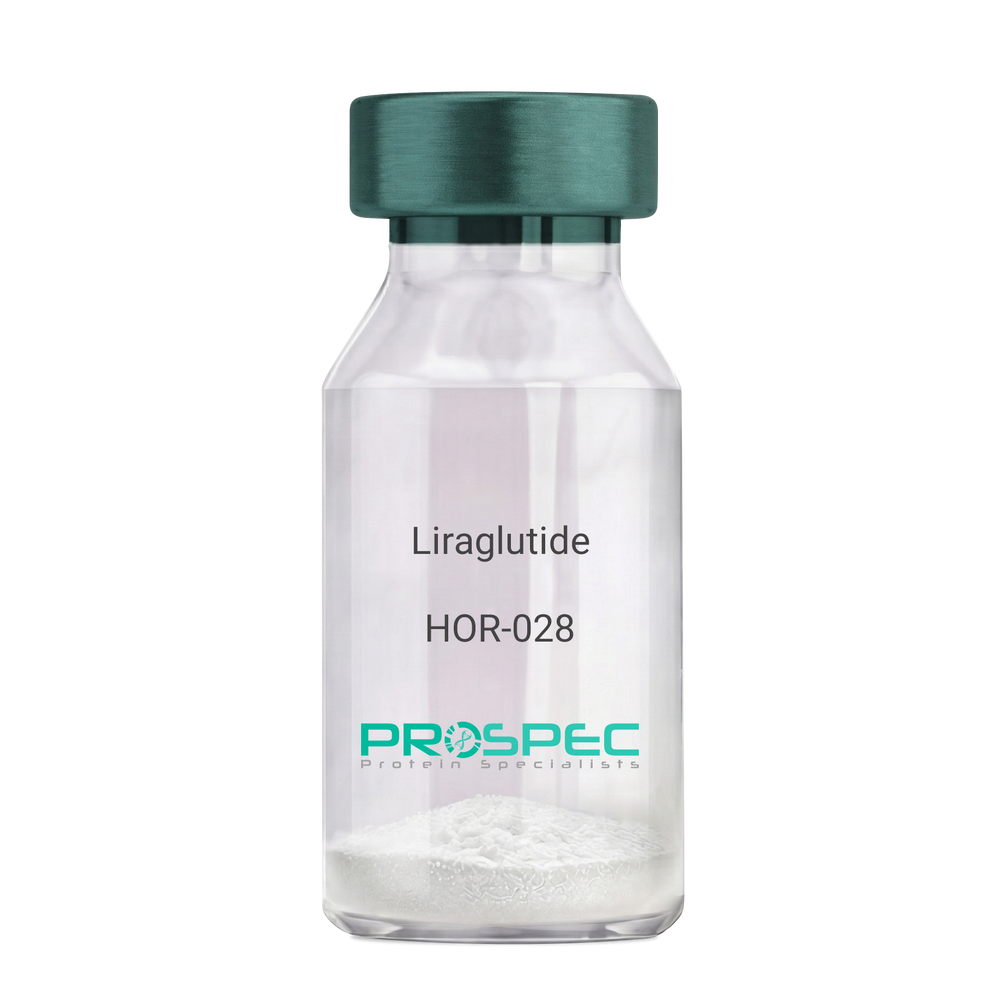
Name :
LiraglutideDescription:
Liraglutide
Product # :
HOR-028Price :
Quantity :
Shipping Method :
Shipped at Room temp
More Info
- description
- formulation
- purity
- More Info
- HPLC, MS
Description
Liraglutide peptide is a single, non-glycosylated polypeptide chain containing 31 amino acids, having a molecular mass of 3751 Dalton and a Molecular formula of C172H265N43O51.
Formulation
The protein (1mg/ml) was lyophilized with no additives.
Purity
Greater than 97% as determined by RP-HPLC.
HPLC, MS
More Info
-
Introduction
Liraglutide is a long-acting GLP1 analogue sharing 97% a.a. homology to human GLP1. Liraglutide differs from GLP1 by the substitution of arginine for lysine at position 34. While GLP1 is degraded swiftly by DPP4 resulting in short life of insulin, Liraglutide contains a fatty acid that binds to albumin and prolongs the half-life of the structure. liraglutide causes insulin to secret in the presence of increasing glucose levels. Due toits receptor site, liraglutide inhibits glucagon secretion and delays gastric emptying.
-
Physical Appearance
Sterile Filtered White lyophilized (freeze-dried) powder.
-
Stability
Store the lyophilized Liraglutide between 2-8°C, do not freeze. Upon reconstitution Lypressin should be stored at 4°C between 2-7 days and for future use below -18°C. Please prevent freeze-thaw cycles.
-
Solubility
It is recommended to reconstitute the lyophilized Liraglutide in sterile 18MΩ-cm H2O not less than 100 µg/ml, which can then be further diluted to other aqueous solutions.
Lypressin is also soluble in 1% Acetic Acid. -
Amino Acid Sequence
H-His-Ala-Glu-Gly-Thr-Phe-Thr-Ser-Asp-Val-Ser-Ser-Tyr-Leu-Glu-Gly-Gln-Ala-Ala-Lys(γ-Glu-palmitoyl)-Glu-Phe-Ile-Ala-Trp-Leu-Val-Arg-Gly-Arg-Gly-OH
ProSpec's products are furnished for LABORATORY RESEARCH USE ONLY. The product may not be used as drugs, agricultural or pesticidal products, food additives or household chemicals.
- View Data Sheet
Name :
RARRES2 Human, HisDescription:
Retinoic Acid Receptor Responder 2 Human Recombinant, His Tag
Chemerin, TIG2, Tazarotene-induced gene 2 protein, Retinoic acid receptor responder protein 2, RAR-responsive protein TIG2, RARRES2, HP10433.
Product # :
PRO-1458Price :
Quantity :
Shipping Method :
Shipped with Ice Packs
More Info
- description
- source
- formulation
- purity
- More Info
Description
RARRES2 Human Recombinant produced in E.Coli is a single, non-glycosylated, polypeptide chain containing 158 amino acids (21-157a.a) and having a total molecular mass of 18 kDa. RARRES2 is fused to a 21 amino acid His-tag at N-terminus & purified by proprietary chromatographic techniques.
Source
Escherichia Coli.
Formulation
RARRES2 protein solution (1mg/ml) containing 20mM Tris-HCl buffer (pH 8.0), 0.4M Urea and 10% glycerol.
Purity
Greater than 85.0% as determined by SDS-PAGE.
More Info
-
Introduction
RARRES2 is a secreted chemotactic protein that initiates chemotaxis through the ChemR23 G protein-coupled seven-transmembrane domain ligand. RARRES2 is upregulated by the synthetic retinoid tazarotene and found in a vast variety of tissues. RARRES2 acts as an adipokine, and is truncated on both termini from the proprotein. RARRES2 is structurally related to the cathelicidin precursors, cystatin C and kininogens. RARRES2 promotes calcium mobilization and chemotaxis of immature dendritic cells and macrophages. RARRES2 is secreted as a precursor of little biological activity, which requires proteolytic cleavage of its COOH-terminal domain to be exchangeed into a potent and highly specific agonist of ChemR23. RARRES2 signals via its receptor, ChemR23 (CMKLR1), as a positive regulator of adipocyte differentiation and metabolic function. The Chemerin receptor acts as a coreceptor for SIV and some primary HIV-1 strains. The Chemerin receptor has another ligand, called tazarotene-induced gene.
-
Synonyms
Chemerin, TIG2, Tazarotene-induced gene 2 protein, Retinoic acid receptor responder protein 2, RAR-responsive protein TIG2, RARRES2, HP10433.
-
Physical Appearance
Sterile Filtered colorless solution.
-
Stability
Store at 4°C if entire vial will be used within 2-4 weeks. Store, frozen at -20°C for longer periods of time. For long term storage it is recommended to add a carrier protein (0.1% HSA or BSA).Avoid multiple freeze-thaw cycles.
-
Amino Acid Sequence
MGSSHHHHHH SSGLVPRGSH MELTEAQRRG LQVALEEFHK HPPVQWAFQE TSVESAVDTP FPAGIFVRLE FKLQQTSCRK RDWKKPECKV RPNGRKRKCL ACIKLGSEDK VLGRLVHCPI ETQVLREAEE HQETQCLRVQ RAGEDPHSFY FPGQFAFS.
ProSpec's products are furnished for LABORATORY RESEARCH USE ONLY. The product may not be used as drugs, agricultural or pesticidal products, food additives or household chemicals.
- View Data Sheet
Name :
Adiponectin Human, HMWDescription:
Adiponectin glycosylated Human Recombinant, HMW Rich
Acrp30, AdipoQ, GBP-28, APM-1, ACDC.
Product # :
CYT-764Price :
Quantity :
Shipping Method :
Shipped at Room temp
More Info
- description
- source
- formulation
- purity
- More Info
Description
Adiponectin Human Recombinant HMW Rich produced in HEK cells is a single, glycosylated, polypeptide chain (19-244) containing a total of 226 amino acids, having a molecular mass of 24.6kDa (calculated). Human Acrp30 HMW Rich migrates on SDS-PAGE under non-reducing conditions at ~ 884 kDa.
Source
HEK293.
Formulation
Acrp30 was filtered (0.4µm) and lyophilized from 0.5mg/ml in 20mM Tris, 50mM NaCl, pH 7.5 and 1mM CaCl2.
Purity
Greater than 90% as determined by SDS-PAGE.
More Info
-
Introduction
Adiponectin is a recently discovered 244 amino acid protein, the product of the apM1 gene, which is physiologically active and specifically and highly expressed in adipose cells (Adipokine). The protein belongs to the soluble defense collagen super family; it has a collagen-like domain structurally homologous with collagen VIII and X and complement factor C1q-like globular domain. APM-1 forms homotrimers, which are the building blocks for higher order complexes found circulating in serum.
-
Synonyms
Acrp30, AdipoQ, GBP-28, APM-1, ACDC.
-
Physical Appearance
Filtered White lyophilized (freeze-dried) powder.
-
Stability
Store lyophilized protein at -20°C. Aliquot the product after reconstitution to avoid repeated freezing/thawing cycles. Reconstituted protein can be stored at 4°C for a limited period of time.
-
Solubility
It is recommended to add deionized water to a working concentration of 0.5mg/ml and let the lyophilized pellet dissolve completely. Acrp30 is not sterile! Please filter the product by an appropriate sterile filter before using it in the cell culture.
-
Amino Acid Sequence
ETTTQGPGVL LPLPKGACTG WMAGIPGHPG HNGAPGRDGR DGTPGEKGEK GDPGLIGPKG DIGETGVPGA EGPRGFPGIQ GRKGEPGEGA YVYRSAFSVG LETYVTIPNM PIRFTKIFYN QQNHYDGSTG KFHCNIPGLY YFAYHITVYM KDVKVSLFKK DKAMLFTYDQ YQENNVDQAS GSVLLHLEVG DQVWLQVYGE GERNGLYADN DNDSTFTGFL LYHDTN.
-
Background
What is the molecular weight/Mw of ADIPONECTIN Protein?
ADIPONECTIN Protein has a total Mw of 24.6 kDa.
What is the source or expression system of ADIPONECTIN Protein?
HEK293.
What is the Purity of ADIPONECTIN Protein?
ADIPONECTIN Protein is >90% pure as determined by SDS-PAGE.
What is the Biological Activity of ADIPONECTIN Protein?
The biological functionality of ADIPONECTIN Protein will be determined in the future.
What is the amino acid sequence of ADIPONECTIN Protein?
ETTTQGPGVL LPLPKGACTG WMAGIPGHPG HNGAPGRDGR DGTPGEKGEK GDPGLIGPKG DIGETGVPGA EGPRGFPGIQ GRKGEPGEGA YVYRSAFSVG LETYVTIPNM PIRFTKIFYN QQNHYDGSTG KFHCNIPGLY YFAYHITVYM KDVKVSLFKK DKAMLFTYDQ YQENNVDQAS GSVLLHLEVG DQVWLQVYGE GERNGLYADN DNDSTFTGFL LYHDTN.
What applications can ADIPONECTIN Protein be used in?
ADIPONECTIN Protein can probably be used in western blot, ELISA and Lateral Flow.
What is the endotoxin level for ADIPONECTIN Protein?
The endotoxin level is minimal, ADIPONECTIN Protein was purified using conventional chromatography techniques.
ProSpec's products are furnished for LABORATORY RESEARCH USE ONLY. The product may not be used as drugs, agricultural or pesticidal products, food additives or household chemicals.
- View Data Sheet
Name :
Erythropoietin HumanDescription:
Erythropoietin Receptor Human Recombinant
Erythropoietin Receptor, EPO-R, EPOR. Normal 0 false false false EN-US X-NONE HE MicrosoftInternetExplorer4-->
Product # :
CYT-929Price :
Quantity :
Shipping Method :
Shipped with Ice Packs
More Info
- description
- source
- formulation
- purity
- More Info
- sds-page
Description
EPOR Human Recombinant produced in Sf9 Baculovirus cells is a single, glycosylated polypeptide chain containing 232 amino acids (25-250 a.a) and having a molecular mass of 25.6kDa. (Migrates at 28-40kDa on SDS-PAGE under reducing conditions). EPOR is fused to an 6 amino acid His-tag at C-terminus & purified by proprietary chromatographic techniques.
Source
Sf9, Baculovirus cells.
Formulation
EPOR protein solution (0.5mg/ml) containing Phosphate Buffered Saline (pH 7.4) and 10% glycerol.
Purity
Greater than 95.0% as determined by SDS-PAGE.
sds-page
More Info
-
Introduction
Erythropoietin receptor, also known as EPOR arbitrates erythropoietin-induced erythroblast proliferation as well as differentiation. During EPO binding, EPOR activates Jak2 tyrosine kinase which activates various intracellular pathways including: Ras/MAP kinase, phosphatidylinositol 3-kinase and STAT transcription factors. Furthermore, stimulated EPOR has a function in erythroid cell survival. Mutations in EPOR may possibly produce erythroleukemia and familial erythrocytosis. In addition, dysregulation of EPOR can affect on the growth of selected tumors.
-
Synonyms
Erythropoietin Receptor, EPO-R, EPOR.
-
Physical Appearance
Sterile Filtered clear solution.
-
Stability
Store at 4°C if entire vial will be used within 2-4 weeks. Store, frozen at -20°C for longer periods of time. For long term storage it is recommended to add a carrier protein (0.1% HSA or BSA).Avoid multiple freeze-thaw cycles.
-
Amino Acid Sequence
APPPNLPDPK FESKAALLAA RGPEELLCFT ERLEDLVCFW EEAASAGVGP GNYSFSYQLE DEPWKLCRLH QAPTARGAVR FWCSLPTADT SSFVPLELRV TAASGAPRYH RVIHINEVVL LDAPVGLVAR LADESGHVVL RWLPPPETPM TSHIRYEVDV SAGNGAGSVQ RVEILEGRTE CVLSNLRGRT RYTFAVRARM AEPSFGGFWS AWSEPVSLLT PSDLDPHHHH HH.
-
Background
What is the molecular weight/Mw of ERYTHROPOIETIN Protein?
ERYTHROPOIETIN Protein has a total Mw of 25.6kDa.
What is the source or expression system of ERYTHROPOIETIN Protein?
Sf9, Baculovirus cells.
What is the Purity of ERYTHROPOIETIN Protein?
ERYTHROPOIETIN Protein is >95% pure as determined by SDS-PAGE.
What is the Biological Activity of ERYTHROPOIETIN Protein?
The biological functionality of ERYTHROPOIETIN Protein will be determined in the future.
What is the amino acid sequence of ERYTHROPOIETIN Protein?
APPPNLPDPK FESKAALLAA RGPEELLCFT ERLEDLVCFW EEAASAGVGP GNYSFSYQLE DEPWKLCRLH QAPTARGAVR FWCSLPTADT SSFVPLELRV TAASGAPRYH RVIHINEVVL LDAPVGLVAR LADESGHVVL RWLPPPETPM TSHIRYEVDV SAGNGAGSVQ RVEILEGRTE CVLSNLRGRT RYTFAVRARM AEPSFGGFWS AWSEPVSLLT PSDLDPHHHH HH.
What applications can ERYTHROPOIETIN Protein be used in?
ERYTHROPOIETIN Protein can probably be used in western blot, ELISA and Lateral Flow.
What is the endotoxin level for ERYTHROPOIETIN Protein?
The endotoxin level is minimal, ERYTHROPOIETIN Protein was purified using conventional chromatography techniques.
ProSpec's products are furnished for LABORATORY RESEARCH USE ONLY. The product may not be used as drugs, agricultural or pesticidal products, food additives or household chemicals.
- View Data Sheet
Name :
SNCG AntibodyDescription:
Gamma-Synuclein, Polyclonal Rabbit Anti-Human Antibody
Gamma-synuclein, Persyn, Breast cancer-specific gene 1 protein, Synoretin, SR, SNCG, BCSG1, PERSYN, PRSN, g-Synuclein.
Product # :
ANT-455Price :
Quantity :
Shipping Method :
Shipped with Ice Packs
More Info
- formulation
- More Info
Formulation
1mg/ml containing PBS, pH-7.4, & 0.1% Sodium Azide.
More Info
-
Introduction
γ-synuclein (Originally known as a breast cancer specific gene product, BCSG1) is an acidic neuronal protein of 127 amino acids. Gamma-Synuclein is a member of the Synuclein protein family, which is believed to be involved in the pathogenesis of neurodegenerative diseases. High levels of Gamma-Synuclein have been found in advanced breast carcinomas suggesting a correlation between overexpression of SNCG and breast tumor development. Synuclein-Gamma is found mostly in the peripheral nervous system (in primary sensory neurons, sympathetic neurons, and motor neurons) and retina. SNCG is also identified in the brain, ovarian tumors, and in the olfactory epithelium. SNCG expression in breast tumors is a marker for tumor progression. A modification in the expression of gamma-synuclein has been detected in the retina of Alzheimer''s patients.
-
Synonyms
Gamma-synuclein, Persyn, Breast cancer-specific gene 1 protein, Synoretin, SR, SNCG, BCSG1, PERSYN, PRSN, g-Synuclein.
-
Physical Appearance
Sterile filtered colorless solution.
-
Immunogen
Recombinant human γ-Synuclein amino acids 1-127 purified from E. coli.
-
Applications
γ-Synuclein antibody has been tested by ELISA and Western blot analysis to assure specificity and reactivity. Since application varies, however, each investigation should be titrated by the reagent to obtain optimal results. Recommended dilution range for Western blot analysis is 1:500 ~ 2,000. Recommended starting dilution is 1:1,000.
-
Type
Polyclonal Rabbit Antibody.
-
Storage Procedures
For periods up to 1 month store at 4°C, for longer periods of time, store at -20°C. Prevent freeze thaw cycles.
ProSpec's products are furnished for LABORATORY RESEARCH USE ONLY. The product may not be used as drugs, agricultural or pesticidal products, food additives or household chemicals.
- View Data Sheet
Name :
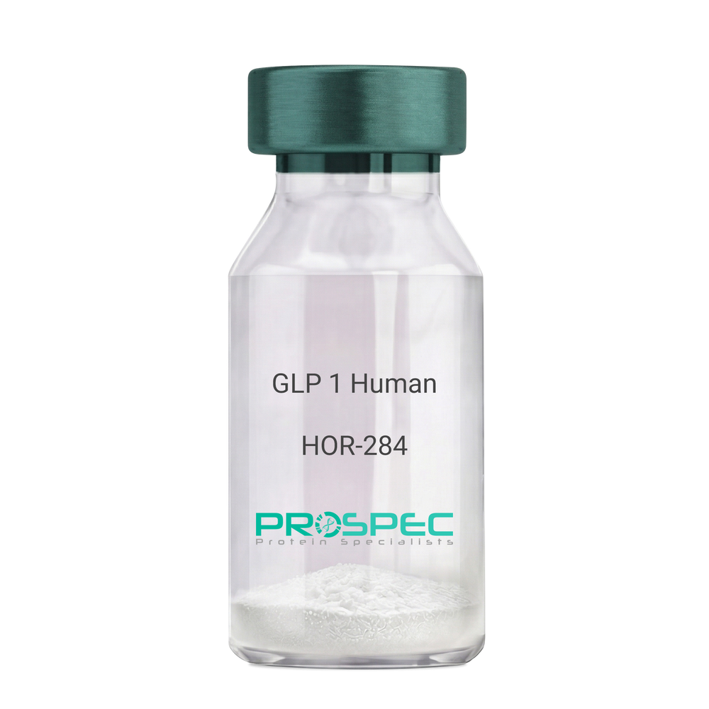
GLP 1 HumanDescription:
Human Glucagon Like Peptide-1
GLP1, Glucagon Like Peptide-1, Incretin hormone.
Product # :
HOR-284Price :
Quantity :
Shipping Method :
Shipped at Room temp
More Info
- description
- formulation
- purity
- More Info
Description
Glucagon Like Peptide-1 is a single, non-glycosylated, polypeptide chain containing 30 amino acids and having a molecular mass of 3297.7 Dalton.The GLP-1 is purified by proprietary chromatographic techniques.
Formulation
The GLP-1 peptide was lyophilized with no additives.
Purity
Greater than 96.0% as determined by RP-HPLC.
More Info
-
Introduction
Glucagon-like peptide-1 (GLP-1) is derived from the transcription product of the proglucagon gene. The major source of GLP-1 in the body is the intestinal L cell that secretes GLP-1 as a guthormone. The biologically active forms of GLP-1 are: GLP-1-(7-37) and GLP-1-(7-36)NH2.
GLP-1 secretion by L cells is dependent on the presence of nutrients in the lumen of the small intestine. The secretagogues (agents that causes or stimulates secretion) of this hormone include major nutrients like carbohydrate, proteinand lipid. Once in the circulation, GLP-1 has a half life of less than 2 minutes, due to rapid degradation by the enzyme dipeptidyl peptidase-4.
GLP-1 possesses several physiological properties that make it a subject of intensive investigation as a potential treatment of diabetes mellitus. The known physiological functions of GLP-1 include: Increases insulin secretion from the pancreas in a glucose-dependent manner, decreases glucagon secretion from the pancreas, increases beta cells mass and insulin gene expression, inhibits acid secretion and gastric emptying in the stomach, decreases food intake by increasing satiety. -
Synonyms
GLP1, Glucagon Like Peptide-1, Incretin hormone.
-
Physical Appearance
Sterile Filtered White lyophilized (freeze-dried) powder.
-
Stability
Lyophilized Glucagon Like Peptide-1 although stable at room temperature for 3 weeks, should be stored desiccated below -18°C. Upon reconstitution GLP-1 should be stored at 4°C between 2-7 days and for future use below -18°C. For long term storage it is recommended to add a carrier protein (0.1% HSA or BSA).Please prevent freeze-thaw cycles.
-
Solubility
It is recommended to reconstitute the lyophilized Glucagon Like Peptide-1 in sterile H2O at 0.5mg/ml, which can then be further diluted to other aqueous solutions.
-
Amino Acid Sequence
H-His-Ala-Glu-Gly-Thr-Phe-Thr-Ser-Asp-Val-Ser-Ser-Tyr-Leu-Glu-Gly-Gln-Ala-Ala-Lys-Glu-Phe-Ile-Ala-Trp-Leu-Val-Lys-Gly-Arg-NH2.
ProSpec's products are furnished for LABORATORY RESEARCH USE ONLY. The product may not be used as drugs, agricultural or pesticidal products, food additives or household chemicals.
- View Data Sheet
Name :
Tamm HorsfallDescription:
Recombinant Human Tamm Horsfall Glycoprotein
Tamm-Horsfall urinary glycoprotein, THP, FJHN, HNFJ, THGP, MCKD2, ADMCKD2, UMOD, Uromodulin.
Product # :
ENZ-1206Price :
Quantity :
Shipping Method :
Shipped at Room temp
More Info
- description
- source
- formulation
- purity
- More Info
- SDS-PAGE
Description
Uromodulin Human Recombinant protein produced from HEK Cells, is a polypeptide chain containing 595 amino acids ( 25-613 a.a. ) and having a total Mw of 65 kDa.
Source
HEK293
Formulation
The UMOD protein was lyophilized from 0.4μm filtered solution containing 50mM NaCl, 0.02M TRIS, pH 7.5.
Purity
Greater than 90.0% as determined by SDS-PAGE analysis.
SDS-PAGE
More Info
-
Introduction
Uromodulin (Tamm–Horsfall protein) is produced mainly by cells in the kidney’s thick ascending limb and is the most abundant protein in normal urine.
Uromodulin takes part in salt and water regulation, helps prevent urinary tract infections and kidney stones, and influences inflammation and immune activity in the kidney.
Reduced Uromodulin levels is associated with chronic kidney disease.
UMOD mutations causes unproper protein folding and thus transported incorrectly, resulting in its accumulation inside kidney tubular cells that damages the tubules and cause kidney disease (ADTKD-UMOD), associated with high uric acid, gout, and progressive kidney failure.
-
Synonyms
Tamm-Horsfall urinary glycoprotein, THP, FJHN, HNFJ, THGP, MCKD2, ADMCKD2, UMOD, Uromodulin.
-
Physical Appearance
Filtered White lyophilized (freeze-dried) powder.
-
Stability
Lyophilized UMOD although stable at room temperature for 3 weeks, should be stored at -18C. Upon reconstitution UMOD should be stored at 4C between 2-7 days and for future use below -18C.
For long term storage it is recommended to add a carrier protein (0.1% HSA or BSA).
Please prevent freeze-thaw cycles.
-
Solubility
Add deionized water to prepare a working stock solution of approximately 0.5mg/ml and let the lyophilized pellet dissolve completely. Product is not sterile! Please filter the product by an appropriate sterile filter before using it in the cell culture.
-
Amino Acid Sequence
DTSEARWCSE CHSNATCTED EAVTTCTCQE GFTGDGLTCV DLDECAIPGA HNCSANSSCV NTPGSFSCVC PEGFRLSPGL GCTDVDECAE PGLSHCHALA TCVNVVGSYL CVCPAGYRGD GWHCECSPGS CGPGLDCVPE GDALVCADPC QAHRTLDEYW RSTEYGEGYA CDTDLRGWYR FVGQGGARMA ETCVPVLRCN TAAPMWLNGT HPSSDEGIVS RKACAHWSGH CCLWDASVQV KACAGGYYVY NLTAPPECHL AYCTDPSSVE GTCEECSIDE DCKSNNGRWH CQCKQDFNIT DISLLEHRLE CGANDMKVSL GKCQLKSLGF DKVFMYLSDS RCSGFNDRDN RDWVSVVTPA RDGPCGTVLT RNETHATYSN TLYLADEIII RDLNIKINFA CSYPLDMKVS LKTALQPMVS ALNIRVGGTG MFTVRMALFQ TPSYTQPYQG SSVTLSTEAF LYVGTMLDGG DLSRFALLMT NCYATPSSNA TDPLKYFIIQ DRCPHTRDST IQVVENGESS QGRFSVQMFR FAGNYDLVYL HCEVYLCDTM NEKCKPTCSG TRFRSGSVID QSRVLNLGPI TRKGVQATVH HHHHH
-
Background
Uromodulin is the most abundant protein in normal urine. Its secretion in urine follows proteolytic cleavage of the ectodomain of its glycosyl phosphatidylinosital-anchored counterpart that is situated on the luminal cell surface of the loop of Henle. Uromodulin plays a role as a constitutive inhibitor of calcium crystallization in renal fluids. Secretion of uromodulin in urine provides protection against urinary tract infections caused by uropathogenic bacteria. Defects in Uromodulin expression are associated with the autosomal dominant renal disorders medullary cystic kidney disease-2 (MCKD2) and familial juvenile hyperuricemic nephropathy (FJHN). These disorders are characterized by juvenile onset of hyperuricemia, gout, and progressive renal failure. While several transcript variants may exist for this gene, the full-length natures of only two have been described to date. UMOD is involved in regulating the circulating activity of cytokines as it binds to il-1, il-2 and tnf with high affinity.
What is the molecular weight/Mw of UMOD Protein?
UMOD Protein has a total Mw of 65kDa.
What is the source or expression system of UMOD Protein?
HEK293.
What is the Purity of UMOD Protein?
UMOD Protein is >90% pure as determined by SDS-PAGE.
What is the Biological Activity of UMOD Protein?
The biological functionality of UMOD Protein will be determined in the future.
What is the amino acid sequence of UMOD Protein?
UMOD Protein is composed from 595 amino acids.
What applications can UMOD Protein be used in?
UMOD Protein can probably be used in western blot, ELISA and Lateral Flow.
What is the endotoxin level for UMOD Protein?
The endotoxin level is minimal, UMOD Protein was purified using conventional chromatography techniques.
ProSpec's products are furnished for LABORATORY RESEARCH USE ONLY. The product may not be used as drugs, agricultural or pesticidal products, food additives or household chemicals.
- View Data Sheet
Name :
LysostaphinDescription:
Lysostaphin Recombinant
Lysostaphin, EC 3.4.24.75, Glycyl-glycine endopeptidase.
Product # :
ENZ-269Price :
Quantity :
Shipping Method :
Shipped at Room temp
More Info
- description
- source
- formulation
- purity
- biological activity
- More Info
Description
Lysostaphin Recombinant produced in E.Coli is a non-glycosylated polypeptide chain having a molecular mass of 26.92 kDa.
Source
Escherichia Coli.
Formulation
The protein was lyophilized without any additives.
Purity
98% as determined by RP-HPLC.
Biological Activity
Assessed by the decrease in turbidity of a suspension of heat-killed Staphylococcus aureus at pH-8, 30°C. Lysostaphin is a zinc enzyme therefore EDTA is an inhibitory factor.
More Info
-
Introduction
Lysostaphin, an endopeptidase specific for the cell wall peptidoglycan of staphylococci, is an extremely potent anti-staphylococcal agent. Lysostaphin is used as a research and diagnostic tool. Because it lyses staphylococci efficiently, it is widely used when preparing staphylococcal DNA or other cellular components for genetic and biochemical studies and for the preparation of protoplasts for transformation. Preparation and analysis of bacterial DNA has become a powerful tool used by clinical and other microbiologists in epidemiological studies aimed at tracing sources of infection or bacterial contamination.
-
Synonyms
Lysostaphin, EC 3.4.24.75, Glycyl-glycine endopeptidase.
-
Physical Appearance
Sterile Filtered lyophilized powder.
-
Stability
Lyophilized Lysostaphin although stable at room temperature for 2 weeks, should be stored desiccated below -18°C. Upon reconstitution Lysostaphin can be stored at 4°C up to 3 weeks and for future use below -18°C.Please prevent freeze-thaw cycles.
-
Solubility
It is recommended to reconstitute the lyophilized Lysostaphin in 20mM sodium acetate, pH 4.5 for optimal stability, which can then be further diluted.
-
Protein content
Protein quantitation was carried out by two independent methods 1. UV spectroscopy at 280 nm using the absorbency value of 2.02 as the extinction coefficient for a 0.1% (1mg/ml) solution. This value is calculated by the PC GENE computer analysis program of protein sequences (IntelliGenetics). 2. Analysis RP-HPLC, using a calibrated solution of Lysostaphin as a Reference standard.
-
Specific Activity
Determined to be 3,540 units/mg.
ProSpec's products are furnished for LABORATORY RESEARCH USE ONLY. The product may not be used as drugs, agricultural or pesticidal products, food additives or household chemicals.
- View Data Sheet
Description:
Mouse Anti Norovirus Group-I Paired
Product # :
ANT-662Price :
Quantity :
Shipping Method :
Shipped with Ice Packs
More Info
- description
- formulation
- purity
- More Info
Description
Paired Norovirus Group-I antibodies, capture and conjugating, target the viral nuclear protein. They were developed to detect Norovirus I antigen in stool rapid test. The capture antibody is used as a coating antibody, and the conjugating antibody is used as the conjugate to bind to colloid gold. Please note that when ordering for example: 100µg antibody we ship 50µg from each of the antibodies (100µg in total).
Formulation
* Norovirus Group I capture antibody in 1xPBS, pH 7.4.
* Norovirus Group I conjugating antibody in 1xPBS, pH 7.4.Purity
Greater than 95%.
More Info
-
Introduction
Noroviruses are categorized into two groups - group 1 and group 2. Norovirus is a widespread virus which can cause human gastroenteritis, an illness characterized with symptoms such as abdominal pain, diarrhea, vomiting and sickness. In America there are about 20 million causes of infection by Nororvirus, 800 ending in deaths. Worldwide, this virus infects around 267 million people and causes over 200,000 deaths per year. Even though having norovirus is unpleasant, it is seldom dangerous and typically ends in full recovery after few days. The cases resulting in deaths are primarily very young, elderly and immuno-suppressed individuals and people from less developed countries. Norovirus is extremely contagious and is spread from person to person, by infected food or water or polluted surfaces. Outbreaks usually happen from November to April, peaking in January.
-
Physical Appearance
2 vials of sterile filtered clear colorless solution.
-
Stability
Norovirus Group I antibody although stable at 4°C for 1 week, should be stored below -18°C. For long term storage it is recommended to add a carrier protein (0.1% HSA or BSA). Please prevent freeze-thaw cycles.
-
Applications
Lateral flow immunoassay.
-
Type
Mouse antibody Monoclonal.
-
Purification Method
Purified monoclonal IgG by protein A chromatography.
ProSpec's products are furnished for LABORATORY RESEARCH USE ONLY. The product may not be used as drugs, agricultural or pesticidal products, food additives or household chemicals.
- View Data Sheet
Name :
SERPINE1 HumanDescription:
Plasminogen Activator Inhibitor-1 Human Recombinant
PAI-1, PAI1, PLANH1, SERPINE1, PAIE, PLASMINOGEN ACTIVATOR INHIBITOR, BETA-MIGRATING ENDOTHELIAL-CELL-DERIVED TYPE.
Product # :
ENZ-357Price :
Quantity :
Shipping Method :
Shipped with Ice Packs
More Info
- description
- source
- formulation
- purity
- biological activity
- More Info
Description
SERPINE1 Human Recombinant fused to an N-terminal His-Tag produced in E.Coli is a single, non-glycosylated polypeptide chain containing 400 amino acids (24-402) and having a molecular mass of 45kDa.SERPINE1 is fused to a 21 amino acid His-Tag at N-terminus and purified by proprietary chromatographic techniques.
Source
Escherichia Coli.
Formulation
50mM NaAc (pH 5.5), 10% glycerol and 0.1M NaCl.
Purity
Greater than 95.0% as determined by SDS-PAGE.
Biological Activity
The IC50 for this effect is less than 3nM, measured by its ability to inhibit uPA cleavage of the substrate Z-GGRAMC.
More Info
-
Introduction
Plasminogen activator inhibitor-1 is the principal inhibitor of tissue plasminogen activator(tPA) and uPA, the activators of plasminogenand hence fibrinolysis(the physiological breakdown of blood clots). It is a serine protease inhibitor(serpin) protein (SERPINE1). The other PAI, plasminogen activator inhibitor-2(PAI-2) is secreted by the placentaand only present in significant amounts during pregnancy. In addition, protease nexinacts as an inhibitor of tPA. SERPINE1, however, is the main inhibitor of the plasminogen activators.
-
Synonyms
PAI-1, PAI1, PLANH1, SERPINE1, PAIE, PLASMINOGEN ACTIVATOR INHIBITOR, BETA-MIGRATING ENDOTHELIAL-CELL-DERIVED TYPE.
-
Physical Appearance
Sterile Filtered clear colorless solution.
-
Stability
Store at 4°C if entire vial will be used within 2-4 weeks. Store, frozen at -20°C for longer periods of time. For long term storage it is recommended to add a carrier protein (0.1% HSA or BSA).Avoid multiple freeze-thaw cycles.
-
Amino Acid Sequence
MGSSHHHHHH SSGLVPRGSH MVHHPPSYVA HLASDFGVRV FQQVAQASKD RNVVFSPYGVASVLAMLQLT TGGETQQQIQ AAMGFKIDDK GMAPALRHLY KELMGPWNKD EISTTDAIFVQRDLKLVQGF MPHFFRLFRS TVKQVDFSEV ERARFIINDW VKTHTKGMIS NLLGKGAVDQLTRLVLVNAL YFNGQWKTPF PDSSTHRRLF HKSDGSTVSV PMMAQTNKFN YTEFTTPDGHYYDILELPYH GDTLSMFIAA PYEKEVPLSA LTNILSAQLI SHWKGNMTRL PRLLVLPKFSLETEVDLRKP LENLGMTDMF RQFQADFTSL SDQEPLHVAQ ALQKVKIEVN ESGTVASSSTAVIVSARMAP EEIIMDRPFL FVVRHNPTGT VLFMGQVMEP.
ProSpec's products are furnished for LABORATORY RESEARCH USE ONLY. The product may not be used as drugs, agricultural or pesticidal products, food additives or household chemicals.
- View Data Sheet
Name :
SNCA (1-140) AntibodyDescription:
Alpha-Synuclein (Immunogen 1-140 a.a.), Mouse Anti Human
Alpha-synuclein, Non-A beta component of AD amyloid, Non-A4 component of amyloid precursor, NACP, PD1, PARK1, PARK4, MGC110988, a-Synuclein, SNCA.
Product # :
ANT-461Price :
Quantity :
Shipping Method :
Shipped with Ice Packs
More Info
- formulation
- More Info
Formulation
1mg/ml containing PBS, pH-7.4, 10% Glycerol and 0.01% Sodium Azide.
More Info
-
Introduction
a-Synuclein (amino acids 1-140), an acidic neuronal protein of 140 amino acids, is extremely heat-resistant and is natively unfolded with an extended structure primarily composed of random coils. a-synuclein has been suggested to be implicated in the pathogenesis of Parkinson’s disease and related neurodegenerative disorders, and more recently, to be an important regulatory component of vesicular transport in neuronal cells. Moreover, recent studies have shown that a-synuclein has chaperone activity and that this activity is lost upon removing its C-terminal acidic tail (amino acids 96-140).
-
Synonyms
Alpha-synuclein, Non-A beta component of AD amyloid, Non-A4 component of amyloid precursor, NACP, PD1, PARK1, PARK4, MGC110988, a-Synuclein, SNCA.
-
Physical Appearance
Sterile Filtered colorless solution.
-
Immunogen
Anti-human SNCA mAb, clone PAT1E10A, is derived from hybridization of mouse F0 myeloma cells with spleen cells from BALB/c mice immunized with a recombinant human SNCA protein 1-140 amino acids purified from E. coli.
-
Ig Subclass
Mouse IgG1 heavy chain and Kappa light chain.
-
Clone
PAT1E10A.
-
Applications
The antibody has been tested by ELISA and Western blot analysis to assure specificity and reactivity. Since application varies, however, each investigation should be titrated by the reagent to obtain optimal results. Recommended starting dilution is 1:500.
-
Type
Mouse Anti Human Monoclonal.
-
Storage Procedures
For periods up to 1 month store at 4°C, for longer periods of time, store at -20°C. Prevent freeze thaw cycles.
-
Purification Method
SNCA antibody was purified from mouse ascitic fluids by protein-A affinity chromatography.
ProSpec's products are furnished for LABORATORY RESEARCH USE ONLY. The product may not be used as drugs, agricultural or pesticidal products, food additives or household chemicals.