Skip to content

High-quality research proteins.

  • 1-800-805-6531

  • Mon - Sat 8.00-18.00, Sun - Closed

  • info@prospecbio.com

  • PO Box 6591 East Brunswick NJ 08816

  • Products
    • Cytokines
    • Growth factors
    • Chemokines
    • Cd antigens
    • Neurotrophins
    • Hormones
    • Enzymes
    • Viral antigens
    • Recombinant proteins
    • Natural proteins
    • Monoclonal antibodies
    • Polyclonal antibodies
    • Thermal Packaging
  • About
    • Company
    • Founder
    • Contribution
  • Customers
    • Eastern Asia
    • Eastern Europe
    • North America
    • South America
    • South East Asia
    • Southern Asia
    • Western Europe
  • Ordering
    • Ordering Method
    • Ordering - Credit Card
    • Ordering - PO
    • Shipping Fees
    • FAQs
  • Services
    • DNA Cloning
    • Protein Expression
    • Monoclonal Antibody
    • Fermentation
    • Technology License
  • Contact us
Log in

Country/region

  • USD $ | Albania
  • USD $ | Andorra
  • USD $ | Argentina
  • USD $ | Armenia
  • USD $ | Australia
  • USD $ | Austria
  • USD $ | Azerbaijan
  • USD $ | Belarus
  • USD $ | Belgium
  • USD $ | Bolivia
  • USD $ | Brazil
  • USD $ | Bulgaria
  • USD $ | Cambodia
  • USD $ | Canada
  • USD $ | Chile
  • USD $ | China
  • USD $ | Colombia
  • USD $ | Costa Rica
  • USD $ | Croatia
  • USD $ | Cyprus
  • USD $ | Czechia
  • USD $ | Denmark
  • USD $ | Estonia
  • USD $ | Finland
  • USD $ | France
  • USD $ | Georgia
  • USD $ | Germany
  • USD $ | Greece
  • USD $ | Hong Kong SAR
  • USD $ | Hungary
  • USD $ | Iceland
  • USD $ | India
  • USD $ | Ireland
  • USD $ | Israel
  • USD $ | Italy
  • USD $ | Japan
  • USD $ | Kazakhstan
  • USD $ | Latvia
  • USD $ | Liechtenstein
  • USD $ | Lithuania
  • USD $ | Madagascar
  • USD $ | Malta
  • USD $ | Mexico
  • USD $ | Moldova
  • USD $ | Monaco
  • USD $ | Netherlands
  • USD $ | New Zealand
  • USD $ | North Macedonia
  • USD $ | Norway
  • USD $ | Panama
  • USD $ | Paraguay
  • USD $ | Peru
  • USD $ | Philippines
  • USD $ | Poland
  • USD $ | Portugal
  • USD $ | Romania
  • USD $ | Singapore
  • USD $ | Slovakia
  • USD $ | Slovenia
  • USD $ | South Africa
  • USD $ | South Korea
  • USD $ | Spain
  • USD $ | Sri Lanka
  • USD $ | Sweden
  • USD $ | Switzerland
  • USD $ | Taiwan
  • USD $ | Thailand
  • USD $ | Türkiye
  • USD $ | Ukraine
  • USD $ | United Kingdom
  • USD $ | United States
  • USD $ | Uruguay
  • USD $ | Vatican City
  • USD $ | Venezuela
  • USD $ | Vietnam
  • Facebook
  • Instagram
logoProSpec - Recombinant Protein Manufacturer for academic and R&D industry
Become a distributor
Account Log in
Wishlist
Cart Cart(0)
  • Products
    +
    • Cytokines
    • Growth factors
    • Chemokines
    • Cd antigens
    • Neurotrophins
    • Hormones
    • Enzymes
    • Viral antigens
    • Recombinant proteins
    • Natural proteins
    • Monoclonal antibodies
    • Polyclonal antibodies
    • Thermal Packaging
  • About
    +
    • Company
    • Founder
    • Contribution
  • Customers
    +
    • Eastern Asia
    • Eastern Europe
    • North America
    • South America
    • South East Asia
    • Southern Asia
    • Western Europe
  • Ordering
    +
    • Ordering Method
    • Ordering - Credit Card
    • Ordering - PO
    • Shipping Fees
    • FAQs
  • Services
    +
    • DNA Cloning
    • Protein Expression
    • Monoclonal Antibody
    • Fermentation
    • Technology License
  • Contact us

Item added to your cart

View cart
View All
  • Cytokines
  • Growth factors
  • Chemokines
  • Cd antigens
  • Neurotrophins
  • Hormones
  • Enzymes
  • Viral antigens
  • Recombinant proteins
  • Natural proteins
  • Monoclonal antibodies
  • Polyclonal antibodies
  • Thermal Packaging
  • Tumor Necrosis Factor

    Tumor Necrosis Factor

    View All
  • 4-1BB

    4-1BB

    View All
  • Adiponectin

    Adiponectin

    View All
  • AIF1

    AIF1

    View All
  • Other Cytokines

    Other Cytokines

    View All
  • AITRL

    AITRL

    View All
  • Angiopoietin

    Angiopoietin

    View All
  • Apolipoprotein

    Apolipoprotein

    View All
  • B-Cell Activating Factor

    B-Cell Activating Factor

    View All
  • Beta Defensin

    Beta Defensin

    View All
  • Bone Morphogenetic Protein

    Bone Morphogenetic Protein

    View All
  • B type Natriuretic Peptide

    B type Natriuretic Peptide

    View All
  • BST

    BST

    View All
  • Betacellulin

    Betacellulin

    View All
  • Cardiotrophin

    Cardiotrophin

    View All
  • Activin

    Activin

    View All
  • Fibroblast Growth Factor

    Fibroblast Growth Factor

    View All
  • Other Growth Factors

    Other Growth Factors

    View All
  • CSF

    CSF

    View All
  • CTGF

    CTGF

    View All
  • IGFBP

    IGFBP

    View All
  • VEGF Protein

    VEGF Protein

    View All
  • EGF

    EGF

    View All
  • Epigen

    Epigen

    View All
  • Erythropoietin

    Erythropoietin

    View All
  • Insulin-Like Growth Factor

    Insulin-Like Growth Factor

    View All
  • Growth Hormone

    Growth Hormone

    View All
  • HDGF

    HDGF

    View All
  • Hepatocyte Growth Factor

    Hepatocyte Growth Factor

    View All
  • Insulin

    Insulin

    View All
  • BCA-1/ BLC (CXCL13)

    BCA-1/ BLC (CXCL13)

    View All
  • BRAK (CXCL14)

    BRAK (CXCL14)

    View All
  • C-10 (CCL6)

    C-10 (CCL6)

    View All
  • Other Chemokines

    Other Chemokines

    View All
  • MEC (CCL28)

    MEC (CCL28)

    View All
  • LD78-beta (CCL3L1)

    LD78-beta (CCL3L1)

    View All
  • CTACK (CCL27)

    CTACK (CCL27)

    View All
  • CXCL16

    CXCL16

    View All
  • CXCL17

    CXCL17

    View All
  • Platelet Factor-4 (CXCL4)

    Platelet Factor-4 (CXCL4)

    View All
  • ENA-78 (CXCL5)

    ENA-78 (CXCL5)

    View All
  • Eotaxin (CCL11,24,26)

    Eotaxin (CCL11,24,26)

    View All
  • Exodus-2 (CCL21)

    Exodus-2 (CCL21)

    View All
  • Fractalkine (CX3CL1)

    Fractalkine (CX3CL1)

    View All
  • CXCL6

    CXCL6

    View All
  • Other CD Antigens

    Other CD Antigens

    View All
  • CD14

    CD14

    View All
  • CD164

    CD164

    View All
  • CD1

    CD1

    View All
  • CD2

    CD2

    View All
  • CD200

    CD200

    View All
  • CD207

    CD207

    View All
  • CD226

    CD226

    View All
  • CD23

    CD23

    View All
  • CD244

    CD244

    View All
  • CD247

    CD247

    View All
  • CD27

    CD27

    View All
  • CD274

    CD274

    View All
  • CD300

    CD300

    View All
  • CD33

    CD33

    View All
  • Other Neurotrophins

    Other Neurotrophins

    View All
  • Beta-NGF

    Beta-NGF

    View All
  • BDNF

    BDNF

    View All
  • CDNF

    CDNF

    View All
  • CNTF

    CNTF

    View All
  • GDNF

    GDNF

    View All
  • Glia Maturation Factor

    Glia Maturation Factor

    View All
  • MANF

    MANF

    View All
  • Midkine

    Midkine

    View All
  • Neuroglobin

    Neuroglobin

    View All
  • Neurotrophic factors

    Neurotrophic factors

    View All
  • Neuregulin

    Neuregulin

    View All
  • Neuropilin

    Neuropilin

    View All
  • Pigment Epithelium-Derived Factor

    Pigment Epithelium-Derived Factor

    View All
  • Pleiotrophin

    Pleiotrophin

    View All
  • Peptide Hormones

    Peptide Hormones

    View All
  • Other Hormones

    Other Hormones

    View All
  • HCG

    HCG

    View All
  • Endothelin

    Endothelin

    View All
  • Exendin

    Exendin

    View All
  • FSH

    FSH

    View All
  • Glucagon

    Glucagon

    View All
  • GHRP

    GHRP

    View All
  • GLP

    GLP

    View All
  • Thyrostimulin

    Thyrostimulin

    View All
  • Inhibin A

    Inhibin A

    View All
  • LHRH

    LHRH

    View All
  • Melanotan

    Melanotan

    View All
  • Procalcitonin

    Procalcitonin

    View All
  • PTH

    PTH

    View All
  • Peroxiredoxin

    Peroxiredoxin

    View All
  • Peptidase

    Peptidase

    View All
  • Synthetase

    Synthetase

    View All
  • Transferase

    Transferase

    View All
  • Hydrolase

    Hydrolase

    View All
  • Dehydrogenase

    Dehydrogenase

    View All
  • ACE2

    ACE2

    View All
  • Esterase

    Esterase

    View All
  • Protein Kinases

    Protein Kinases

    View All
  • Other Enzymes

    Other Enzymes

    View All
  • Phosphatase

    Phosphatase

    View All
  • Deaminase

    Deaminase

    View All
  • Oxygenase

    Oxygenase

    View All
  • Adenylate Kinase

    Adenylate Kinase

    View All
  • Lyase

    Lyase

    View All
  • Chagas

    Chagas

    View All
  • SARS

    SARS

    View All
  • Influenza

    Influenza

    View All
  • ASFV

    ASFV

    View All
  • Astrovirus

    Astrovirus

    View All
  • Borrelia

    Borrelia

    View All
  • Helicobacter Pylori

    Helicobacter Pylori

    View All
  • Chikungunya

    Chikungunya

    View All
  • Chlamydia

    Chlamydia

    View All
  • Cytomegalo

    Cytomegalo

    View All
  • Dengue

    Dengue

    View All
  • EBV

    EBV

    View All
  • Ebola

    Ebola

    View All
  • Feline Leukemia Virus

    Feline Leukemia Virus

    View All
  • Hepatitis A

    Hepatitis A

    View All
  • Other

    Other

    View All
  • Actin

    Actin

    View All
  • ADAM

    ADAM

    View All
  • Complement Component

    Complement Component

    View All
  • Ag85

    Ag85

    View All
  • Eukaryotic Translation Initiation Factor

    Eukaryotic Translation Initiation Factor

    View All
  • Anterior Gradient Protein

    Anterior Gradient Protein

    View All
  • Heat Shock Protein

    Heat Shock Protein

    View All
  • Alpha-2-HS-Glycoprotein

    Alpha-2-HS-Glycoprotein

    View All
  • Hemoglobin

    Hemoglobin

    View All
  • Allergy

    Allergy

    View All
  • Anaplasma

    Anaplasma

    View All
  • Angiogenin

    Angiogenin

    View All
  • Ankyrin Repeat Domain

    Ankyrin Repeat Domain

    View All
  • Annexin

    Annexin

    View All
  • Other Natural Proteins

    Other Natural Proteins

    View All
  • Aprotinin

    Aprotinin

    View All
  • Transferrin

    Transferrin

    View All
  • Avidin

    Avidin

    View All
  • Anti Coagulation Factors

    Anti Coagulation Factors

    View All
  • Natural Albumin

    Natural Albumin

    View All
  • Natural Coagulation Factors

    Natural Coagulation Factors

    View All
  • Fibronectin

    Fibronectin

    View All
  • Thrombin

    Thrombin

    View All
  • Anti Human Enzyme

    Anti Human Enzyme

    View All
  • Other Monoclonal Antibodies

    Other Monoclonal Antibodies

    View All
  • Anti Human Cytokine

    Anti Human Cytokine

    View All
  • Anti Human Heat Shock Protein

    Anti Human Heat Shock Protein

    View All
  • Anti Mouse Lymphocyte

    Anti Mouse Lymphocyte

    View All
  • Anti Human Chemokine

    Anti Human Chemokine

    View All
  • Anti Human Lymphocyte

    Anti Human Lymphocyte

    View All
  • Anti Viral Monoclonal

    Anti Viral Monoclonal

    View All
  • Anti Mouse Cytokine

    Anti Mouse Cytokine

    View All
  • Anti-GST Monoclonal

    Anti-GST Monoclonal

    View All
  • Other Polyclonal Antibodies

    Other Polyclonal Antibodies

    View All
  • Anti Viral

    Anti Viral

    View All
  • Anti-GST Monoclonal

    Anti-GST Monoclonal

    View All
Back

Search results

1000 results found for “NFKB Inhibitor”

Name

Description

Product #

Price

Quantity

Shipping Method

  • View Data Sheet

    Name :

    HCV NS5 Genotype-1b

    Description:

    Hepatitis C Virus NS5 Genotype-1b Recombinant

    Product # :

    HCV-228

    Price :

    Quantity :

    Shipping Method :

    Ice Icon

    Shipped with Ice Packs

    Add To Cart

    More Info

    • description
    • source
    • formulation
    • purity
    • More Info

    Description

    The E.coli derived recombinant protein contains the HCV NS5 Genotype1b immunodominant regions, amino acids 2212-2313. The protein is fused to a GST tag at N-terminus.

    Source

    Escherichia Coli.

    Formulation

    1.5M urea, 25mM Tris-HCl pH 8.0, 0.2% Triton-X and 50% Glycerol.

    Purity

    HCV NS5 Genotype-1b protein is >95% pure as determined by SDS- PAGE (coomassie staining).

    More Info

    • Introduction

      HCV is a small 50nm, enveloped, single-stranded, positive sense RNAvirus in the family Flaviviridae.
      HCV has a high rate of replication with approximately one trillion particles produced each day in an infected individual. Due to lack of proofreading by the HCV RNA polymerase, the HCV has an exceptionally high mutation rate, a factor that may help it elude the host's immune response. Hepatitis C virus is classified into six genotypes(1-6) with several subtypes within each genotype. The preponderance and distribution of HCV genotypes varies globally. Genotype is clinically important in determining potential response to interferon-based therapy and the required duration of such therapy. Genotypes 1 and 4 are less responsive to interferon-based treatment than are the other genotypes (2, 3, 5 and 6).

    • Physical Appearance

      Sterile Filtered clear solution.

    • Stability

      HCV NS5 Genotype-1b although stable at 4°C for 1 week, should be stored below -18°C. Please prevent freeze thaw cycles.

    • Applications

      HCV NS5 Genotype-1b antigen is suitable for ELISA and Western blots, excellent antigen for detection of HCV with minimal specificity problems.

    • Specificity

      Immunoreactive with sera of HCV-infected individuals.

    • Purification Method

      HCV NS5 Genotype-1b protein was purified by proprietary chromatographic technique.

    ProSpec's products are furnished for LABORATORY RESEARCH USE ONLY. The product may not be used as drugs, agricultural or pesticidal products, food additives or household chemicals.

    Hcv Ns5 Genotype 1B
  • View Data Sheet

    Name :

    OX40L Human

    Description:

    OX40 Ligand Human Recombinant

    Tumor necrosis factor ligand superfamily member 4, OX40 ligand, OX40L, OX-40L, CD252, Tnfsf4, Ox40l, Txgp1l, gp3, OX4, Ath-1, Ath1, CD134L, gp34, Tnlg2b, Txgp1l.

    Product # :

    CYT-1226

    Price :

    Quantity :

    Shipping Method :

    Ice Icon

    Shipped with Ice Packs

    Add To Cart

    More Info

    • description
    • source
    • formulation
    • purity
    • biological activity
    • More Info

    Description

    OX40L Human Recombinant is a single, glycosylated, polypeptide chain (51-183 a.a) containing a total of139 amino acids and having a molecular mass of 16.2 kDa. OX40L is fused to a 6 a.a his-Tag at C-terminus and is purified by proprietary chromatographic techniques.

    Source

    HEK293 Cells.

    Formulation

    The OX40L solution (1mg/ml) contains 10% Glycerol and Phosphate-Buffered Saline (pH 7.4).

    Purity

    Greater than 90.0% as determined by SDS-PAGE.

    Biological Activity

    Measured by its binding ability in a functional ELISA with Human OX40/TNFRSF4.

    More Info

    • Synonyms

      Tumor necrosis factor ligand superfamily member 4, OX40 ligand, OX40L, OX-40L, CD252, Tnfsf4, Ox40l, Txgp1l, gp3, OX4, Ath-1, Ath1, CD134L, gp34, Tnlg2b, Txgp1l.

    • Physical Appearance

      Sterile Filtered colorless solution.

    • Stability

      Store at 4°C if entire vial will be used within 2-4 weeks. Store, frozen at -20°C for longer periods of time. For long term storage it is recommended to add a carrier protein (0.1% HSA or BSA). Avoid multiple freeze-thaw cycles.

    • Amino Acid Sequence

      QVSHRYPRIQ SIKVQFTEYK KEKGFILTSQ KEDEIMKVQN NSVIINCDGF YLISLKGYFS QEVNISLHYQ KDEEPLFQLK KVRSVNSLMV ASLTYKDKVY LNVTTDNTSL DDFHVNGGEL ILIHQNPGEF CVLHHHHHH.

    • Background

      OX40 Ligand (OX40L), a member of the tumor necrosis factor (TNF) superfamily, plays a pivotal role in regulating immune responses and orchestrating the delicate balance between activation and tolerance. The human recombinant form of OX40L has emerged as a potent tool in immunology, offering insights into its molecular intricacies and potential applications in therapeutic interventions. This research embarks on a journey to unravel the multifaceted role of OX40L Human Recombinant, shedding light on its structural attributes, signaling pathways, and its promising avenues in immunotherapy. By delving into the properties of OX40L, scientists aim to expand our understanding of immune modulation and open new frontiers in the treatment of immune-related disorders.

      Structural Insights into OX40L Human Recombinant:

      OX40L, as a trimeric transmembrane protein, exhibits a unique structural configuration that governs its interactions with the OX40 receptor on T cells. The human recombinant form, engineered for controlled study, provides a window into the three-dimensional intricacies of the ligand. Understanding its structure is pivotal for deciphering how OX40L engages with its receptor and modulates immune responses.

      Immunomodulatory Signaling Pathways:

      OX40L binding to its cognate receptor OX40 on T cells triggers intricate signaling cascades that impact immune cell activation, proliferation, and cytokine production. The OX40-OX40L axis is a crucial regulator of T cell function, influencing both effector and regulatory T cell responses. Unraveling the specific pathways activated by OX40L Human Recombinant provides valuable insights into the modulation of immune responses in health and disease.

      Applications in Immunotherapy:

      The immunomodulatory properties of OX40L make it an attractive candidate for therapeutic interventions. OX40L Human Recombinant, in preclinical and clinical studies, is being explored for its potential in enhancing antitumor immune responses. By harnessing the ligand's ability to stimulate effector T cells and memory T cell formation, researchers aim to develop novel immunotherapies for cancer and other immune-related disorders.

      OX40L in Autoimmune Diseases:

      Conversely, OX40L's role in autoimmune diseases has spurred investigations into its inhibition as a therapeutic strategy. Blocking the OX40-OX40L interaction has shown promise in mitigating autoimmune responses, presenting a potential avenue for the development of treatments for conditions such as rheumatoid arthritis and inflammatory bowel disease.

      While the potential of OX40L Human Recombinant in immunotherapy is promising, challenges persist. Fine-tuning its applications, understanding potential side effects, and optimizing dosages are critical considerations for translational success. Additionally, comprehending the context-dependent nature of OX40L signaling is essential for tailoring therapeutic strategies to specific diseases and patient profiles.

      OX40L Human Recombinant stands at the forefront of immunomodulation research, offering a lens through which we can unravel the complexities of immune responses. Its structural insights, signaling pathways, and therapeutic applications position it as a key player in the evolving landscape of immunotherapy. As researchers continue to dissect the molecular nuances of OX40L, they not only expand our understanding of immune regulation but also pave the way for transformative advancements in the treatment of cancer and autoimmune diseases, shaping the future of precision medicine and immunotherapy.

    ProSpec's products are furnished for LABORATORY RESEARCH USE ONLY. The product may not be used as drugs, agricultural or pesticidal products, food additives or household chemicals.

    Ox40L Human
  • View Data Sheet

    Name :

    GDNF Human, Sf9

    Description:

    Glial-Derived Neurotrophic Factor Human Recombinant, Sf9

    Glial cell line-derived neurotrophic factor, hGDNF, Astrocyte-derived trophic factor, ATF, ATF1, ATF2, HFB1-GDNF, HSCR3.

    Product # :

    CYT-1162

    Price :

    Quantity :

    Shipping Method :

    Ice Icon

    Shipped with Ice Packs

    Add To Cart

    More Info

    • description
    • source
    • formulation
    • purity
    • More Info

    Description

    GDNF Human Recombinant produced in Baculovirus is a single glycosylated polypeptide chain containing 113amino acids (109-211 aa) and having a molecular mass of 12.8kDa.GDNF is fused to an 10 amino acid His tag at C-terminus and purified by proprietary chromatographic techniques.

    Source

    Sf9, Baculovirus cells.

    Formulation

    GDNF protein (0.25mg/ml) contains 10% glycerol and Phosphate-Buffered Saline (pH 7.4).

    Purity

    Greater than 90.0% as determined by SDS-PAGE.

    More Info

    • Introduction

      Glial cell-derived neurotrophic factor or GDNF is part of the GDNF group of ligands proteins. GDNF has a crucial part in numerous cell mechanisms such as neurite outgrowth, cell differentiation, cell survival and migration of cells. GDNF enhances neurons survival via GFRa receptors (mainly GFRa1). The mentioned neurons can die as a result from Parkinson's disease or ALS (amyotrophic lateral sclerosis). This protein takes part in the development of the spermatogenesis & kidney, also, it has a role in alcohol metabolism as ameliorating.

    • Synonyms

      Glial cell line-derived neurotrophic factor, hGDNF, Astrocyte-derived trophic factor, ATF, ATF1, ATF2, HFB1-GDNF, HSCR3.

    • Physical Appearance

      Sterile Filtered colorless solution.

    • Stability

      Store at 4°C if entire vial will be used within 2-4 weeks. Store, frozen at -20°C for longer periods of time. For long term storage it is recommended to add a carrier protein (0.1% HSA or BSA).Avoid multiple freeze-thaw cycles.

    • Amino Acid Sequence

      ADPMRGQRGK NRGCVLTAIH LNVTDLGLGY ETKEELIFRY CSGSCDAAET TYDKILKNLS RNRRLVSDKV GQACCRPIAF DDDLSFLDDN LVYHILRKHS AKRCGCIHHH HHH

    • Background

      What is the molecular weight/Mw of GDNF HUMAN, SF9 Protein?
      GDNF HUMAN, SF9 Protein has a total Mw of 12.8kDa.

      What is the source or expression system of GDNF HUMAN, SF9 Protein?
      Sf9, Baculovirus cells.

      What is the Purity of GDNF HUMAN, SF9 Protein?
      GDNF HUMAN, SF9 Protein is >90% pure as determined by SDS-PAGE.

      What is the Biological Activity of GDNF HUMAN, SF9 Protein?
      The biological functionality of GDNF HUMAN, SF9 Protein will be determined in the future.

      What is the amino acid sequence of GDNF HUMAN, SF9 Protein?
      ADPMRGQRGK NRGCVLTAIH LNVTDLGLGY ETKEELIFRY CSGSCDAAET TYDKILKNLS RNRRLVSDKV GQACCRPIAF DDDLSFLDDN LVYHILRKHS AKRCGCIHHH HHH

      What applications can GDNF HUMAN, SF9 Protein be used in?
      GDNF HUMAN, SF9 Protein can probably be used in western blot, ELISA and Lateral Flow.

      What is the endotoxin level for GDNF HUMAN, SF9 Protein?
      The endotoxin level is minimal, GDNF HUMAN, SF9 Protein was purified using conventional chromatography techniques.

    ProSpec's products are furnished for LABORATORY RESEARCH USE ONLY. The product may not be used as drugs, agricultural or pesticidal products, food additives or household chemicals.

    Gdnf Protein
  • View Data Sheet

    Name :

    CST6 Human

    Description:

    Cystatin E/M Human Recombinant

    Cystatin E/M, cystatin 6, Cystatin M, Cystatin-E, Cysteine proteinase inhibitor.

    Product # :

    PRO-892

    Price :

    Quantity :

    Shipping Method :

    Ice Icon

    Shipped with Ice Packs

    Add To Cart

    More Info

    • description
    • source
    • formulation
    • purity
    • More Info

    Description

    CST6 Human Recombinant produced in E.coli is a single, non-glycosylated polypeptide chain containing 142 amino acids (29-149) and having a molecular mass of 15.9 kDa.The CST6 is fused to a 21 amino acid His-Tag at N-terminus and purified by proprietary chromatographic techniques.

    Source

    E.coli.

    Formulation

    The CST6 solution (0.5mg/ml) contains 20mM Tris-HCl buffer (pH 8.0), 1mM DTT, 0.1M NaCl and 10% glycerol.

    Purity

    Greater than 95% as determined by SDS-PAGE.

    More Info

    • Introduction

      CST6 belongs to the type 2 cystatin family. Unlike other family members that act as active cysteine protease inhibitors, CST6 restrain the inhibition of cathepsin B but is not active against cathepsin C. CST6 are secretable proteins that influence osteogenesis and bone resorption, regulation of the hepatocyte growth factor receptors, and the response to systemic inflammation.

    • Synonyms

      Cystatin E/M, cystatin 6, Cystatin M, Cystatin-E, Cysteine proteinase inhibitor.

    • Physical Appearance

      Sterile Filtered colorless solution.

    • Stability

      Store at 4°C if entire vial will be used within 2-4 weeks. Store, frozen at -20°C for longer periods of time. For long term storage it is recommended to add a carrier protein (0.1% HSA or BSA).Avoid multiple freeze-thaw cycles.

    • Amino Acid Sequence

      MGSSHHHHHH SSGLVPRGSH MRPQERMVGE LRDLSPDDPQ VQKAAQAAVA SYNMGSNSIY YFRDTHIIKA QSQLVAGIKY FLTMEMGSTD CRKTRVTGDH VDLTTCPLAA GAQQEKLRCD FEVLVVPWQN SSQLLKHNCV QM.

    ProSpec's products are furnished for LABORATORY RESEARCH USE ONLY. The product may not be used as drugs, agricultural or pesticidal products, food additives or household chemicals.

    Cst6 Human
  • View Data Sheet

    Name :

    RERG Human

    Description:

    RAS-like, Estrogen-Regulated, Growth Inhibitor Human Recombinant

    Ras-related and estrogen-regulated growth inhibitor, RERG, MGC15754.

    Product # :

    PRO-106

    Price :

    Quantity :

    Shipping Method :

    Ice Icon

    Shipped with Ice Packs

    Add To Cart

    More Info

    • description
    • source
    • formulation
    • purity
    • More Info

    Description

    RERG Human Recombinant fused with a 20 amino acid His tag at N-terminus produced in E.Coli is a single, non-glycosylated, polypeptide chain containing 219 amino acids (1-199 a.a.) and having a molecular mass of 24.7kDa (Molecular size on SDS-PAGE will appear higher). The RERG is purified by proprietary chromatographic techniques.

    Source

    Escherichia Coli.

    Formulation

    The RERG solution (0.25 mg/ml) contains 20mM Tris-HCl buffer (pH8.0), 0.2M NaCl, 5mM DTT and 50% glycerol.

    Purity

    Greater than 85.0% as determined by SDS-PAGE.

    More Info

    • Introduction

      RERG is a 199 amino acid protein which localizes in the cytoplasm and is a member of the Ras subfamily of small GTPases. RERG is expressed in the pancreas, liver, skin, lung, brain, kidney and heart tissue. RERG is a vital mediator of diverse cell signaling pathways, including those leading to cell proliferation, cytoskeletal organization and secretion.

    • Synonyms

      Ras-related and estrogen-regulated growth inhibitor, RERG, MGC15754.

    • Physical Appearance

      Sterile Filtered colorless solution.

    • Stability

      Store at 4°C if entire vial will be used within 2-4 weeks. Store, frozen at -20°C for longer periods of time. For long term storage it is recommended to add a carrier protein (0.1% HSA or BSA). Avoid multiple freeze-thaw cycles.

    • Amino Acid Sequence

      MGSSHHHHHH SSGLVPRGSH MAKSAEVKLA IFGRAGVGKS ALVVRFLTKR FIWEYDPTLE STYRHQATID DEVVSMEILD TAGQEDTIQR EGHMRWGEGF VLVYDITDRG SFEEVLPLKN ILDEIKKPKN VTLILVGNKA DLDHSRQVST EEGEKLATEL ACAFYECSAC TGEGNITEIF YELCREVRRR RMVQGKTRRR SSTTHVKQAI NKMLTKISS.

    ProSpec's products are furnished for LABORATORY RESEARCH USE ONLY. The product may not be used as drugs, agricultural or pesticidal products, food additives or household chemicals.

    Rerg Human
  • View Data Sheet

    Name :

    APOB Protein

    Description:

    Apolipoprotein-B Human Recombinant

    APOB, APO-B, Apolipoprotein B.

    Product # :

    CYT-1233

    Price :

    Quantity :

    Shipping Method :

    Room Temp Icon

    Shipped at Room temp

    Add To Cart

    More Info

    • description
    • source
    • formulation
    • purity
    • More Info

    Description

    The APOBHuman is created as a recombinant protein with a 4kda N-terminal fusion of His Tag. The APOBHis-Tagged Fusion Protein, produced in E. coli, is a 31kDa protein containing 201 amino acid residues of the APOBHuman, 1406-1606 amino acids.

    Source

    Escherichia Coli.

    Formulation

    Each mg was lyophilized with 1xPBS, 0.4% SDS and 4mM DTT.

    Purity

    Greater than 80% as determined by SDS-PAGE.

    More Info

    • Synonyms

      APOB, APO-B, Apolipoprotein B.

    • Physical Appearance

      Filtered White lyophilized (freeze-dried) powder.

    • Stability

      Store lyophilized APOBat -20°C. Aliquot the product after reconstitution to avoid repeated freezing/thawing cycles. Reconstituted protein can be stored at 4°C for a limited period of time; it does not show any change after two weeks at 4°C.

    • Solubility

      It is recommended to add deionized water to prepare a working stock solution of approximately 0.5mg/ml and let the lyophilized pellet dissolve completely. Product is not sterile! Please filter the product by an appropriate sterile filter before using it on cell culture.

    • Background

      Apolipoprotein-B (ApoB) is the main apolipoprotein of LDL, VLDL, IDL and chylomicrons particles which serves as the carrier of lipids in the water surrounding the cells in every tissue across the body. Apolipoprotein B acts as the key organizing protein of all other carriers of lipids. across LDL membranes, ApoB also plays a role as a ligand for LDL receptors in many cells across the body, meaning, it shows that lipid carriers that are set to cross into cells with Apolipoprotein B receptors, this is how lipids are transported within and into cells.

      What is the molecular weight/Mw of APOB Protein?
      APOB Protein has a total Mw of 31kDa.

      What is the source or expression system of APOB Protein?
      Escherichia Coli.

      What is the Purity of APOB Protein?
      APOB Protein is >80% pure as determined by SDS-PAGE.

      What is the Biological Activity of APOB Protein?
      The biological functionality of APOB Protein will be determined in the future.

      What is the amino acid sequence of APOB Protein?
      APOB Protein is composed from 201 amino acids.

      What applications can APOB Protein be used in?
      APOB Protein can probably be used in western blot, ELISA and Lateral Flow.

      What is the endotoxin level for APOB Protein?
      The endotoxin level is minimal, APOB Protein was purified using conventional chromatography techniques.

    ProSpec's products are furnished for LABORATORY RESEARCH USE ONLY. The product may not be used as drugs, agricultural or pesticidal products, food additives or household chemicals.

    Human Apob
  • View Data Sheet

    Name :

    IBV-NP

    Description:

    Influenza B Virus Nucleoprotein Recombinant

    Product # :

    IHA-038

    Price :

    Quantity :

    Shipping Method :

    Ice Icon

    Shipped with Ice Packs

    Add To Cart

    More Info

    • description
    • source
    • formulation
    • More Info

    Description

    Recombinant Influenza B Virus Nucleoprotein produced in E. coli having a Mw of 76.8kDa. IBV-NP is fused to a 6xHis tag at its C terminal is and purified by proprietary chromatographic technique.

    Source

    E. coli.

    Formulation

    IBV-NP protein solution contains 25mM K2CO3 and PBS.

    More Info

    • Physical Appearance

      Sterile Filtered clear solution.

    • Stability

      Store at 4°C if entire vial will be used within 2-4 weeks. Store, frozen at -20°C for longer periods of time. For long term storage it is recommended to add a carrier protein (0.1% HSA or BSA). Avoid multiple freeze-thaw cycles.

    • Amino Acid Sequence

      HMSNMDIDGMNTGTIDKTPEEITSGTSGTTRPIIRPATLAPPSNKRTRNPSPDRTTTSSE

      DDVGRKAQKKQTPTEIKKSVYNMVVKLGEFYNQMMVKAGLNDDMERNLIQNAHAVERILL

      AATDDKKTEFQKKKNARDVKEGKEEIDHNKTGGTFYKMVRDDKTIYFSPIRITFLKEEVKT

      MYKTTMGSDGFSGLNHIMIGHSQMNDVCFQRSKALKRVGLDPSLISTFAGSTVPRRSGATGV

      AIKGGGTLVAEAIRFIGRAMADRGLLRDIKAKTAYEKILLNLKNKCSAPQQKALVDQVIGSRN

      PGIADIEDLTLLARSMVVVRPSVASKVVLPISIYAKIPQLGFNVEEYSMVGYEAMALYNMATP

      VSILRMGDDAKDKSQLFFMSCFGAAYEDLRVLSALTGTEFKPRSALKCKGFHVPAKEQVEGMGA

      ALMSIKLQFWAPMTRSGGNEVGGDGGSGQISCSPVFAVERPIALSKQAVRRMLSMNIEGRDADV

      KGNLLKMMNDSMAKKTSGNAFIGKKMFQISDKNKTNPIEIPIKQTIPNFFFGRDTAEDYDDLDYLE.

    • Background

      Influenza B virus, often overshadowed by its influenza A counterpart, remains a substantial contributor to seasonal influenza infections and poses a considerable public health challenge. In the quest for effective countermeasures, Influenza B Virus Nucleoprotein Recombinant has emerged as a focal point in the ongoing battle against this resilient virus. This research endeavors to unveil the intricacies of the Nucleoprotein Recombinant, delving into its structural nuances, immunogenic capabilities, and applications in vaccine development and antiviral therapies. By dissecting the properties of the Nucleoprotein Recombinant, scientists aspire to fortify our defenses against Influenza B, potentially reshaping strategies for broader influenza immunity.

      Structural Insights into Nucleoprotein Recombinant:

      Engineered to replicate key viral components, the Influenza B Virus Nucleoprotein Recombinant serves as a molecular mirror, allowing a comprehensive study of the virus's genetic material. Understanding its three-dimensional structure and conformational intricacies is crucial, not only for decoding the virus's replication mechanisms but also for tailoring interventions like vaccines and antiviral drugs to specific vulnerabilities.

      Immunogenic Potential and Vaccine Development:

      The challenge of Influenza B's seasonal variability necessitates innovative approaches to vaccine development. Nucleoprotein Recombinant, designed to induce potent immune responses, emerges as a promising candidate. By presenting conserved viral elements to the immune system, the recombinant protein seeks to instigate robust and durable immunity, countering the virus's propensity for antigenic drift and promoting broader protection.

      Application in Antiviral Therapies:

      In addition to its role in vaccination, the Influenza B Virus Nucleoprotein Recombinant holds potential in antiviral therapies. The recombinant protein's ability to provoke immune responses can be harnessed for the development of immunotherapies. Monoclonal antibodies derived from the Nucleoprotein Recombinant may offer targeted treatments, providing a dynamic approach to mitigating the impact of Influenza B infections.

      Challenges and Future Directions:

      Despite the promises held by the Nucleoprotein Recombinant, challenges persist. The virus's ability to undergo genetic reassortment and antigenic drift requires ongoing vigilance and adaptability in recombinant strategies. Considerations of vaccine safety, efficacy, and public acceptance demand continual exploration to optimize the translational success of Nucleoprotein Recombinant-based interventions.

      Influenza B Virus Nucleoprotein Recombinant stands as a beacon in the scientific quest against influenza, offering a pathway to a more comprehensive and resilient immunity. Its structural insights, immunogenic potential, and applications in vaccine development and antiviral therapies position it as a central player. As researchers delve deeper into the molecular intricacies of Nucleoprotein Recombinant, they not only fortify our defenses against Influenza B but potentially pave the way for a paradigm shift in our approach to influenza prevention and control, influencing the landscape of global health preparedness.

    ProSpec's products are furnished for LABORATORY RESEARCH USE ONLY. The product may not be used as drugs, agricultural or pesticidal products, food additives or household chemicals.

    Ibv Nuceloprotein
  • View Data Sheet

    Name :

    FGFR1OP Human (1-80)

    Description:

    FGFR1 Oncogene Partner (1-80 a.a.) Human Recombinant

    FGFR1 oncogene partner, FGFR1OP, FOP.

    Product # :

    PKA-138

    Price :

    Quantity :

    Shipping Method :

    Room Temp Icon

    Shipped at Room temp

    Add To Cart

    More Info

    • description
    • source
    • formulation
    • purity
    • More Info

    Description

    The FGFR1O PHuman is created as a recombinant protein with a 4kda N-terminal fusion of His Tag. The FGFR1OP His-Tagged Fusion Protein, produced in E. coli, is a 12kDa protein containing 80 amino acid residues of the FGFR1OP Human, 1-80 amino acids.

    Source

    Escherichia Coli.

    Formulation

    Each mg was lyophilized with 1xPBS, 0.4% SDS and 4mM DTT.

    Purity

    Greater than 90% as determined by SDS-PAGE.

    More Info

    • Synonyms

      FGFR1 oncogene partner, FGFR1OP, FOP.

    • Physical Appearance

      Filtered White lyophilized (freeze-dried) powder.

    • Stability

      Store lyophilized FGFR1OP at -20°C. Aliquot the product after reconstitution to avoid repeated freezing/thawing cycles. Reconstituted protein can be stored at 4°C for a limited period of time; it does not show any change after two weeks at 4°C.

    • Solubility

      It is recommended to add deionized water to prepare a working stock solution of approximately 0.5mg/ml and let the lyophilized pellet dissolve completely. Product is not sterile! Please filter the product by an appropriate sterile filter before using it on cell culture.

    • Amino Acid Sequence

      L MAATAAAVVA EEDTELRDLL VQTLENSGVL NRIKAELRAA VFLALEEQEK VENKTPLVNE SLRKFLNTKD GRLVASLVA

    • Background

      FGFR1 oncogene partner also known as FGFR1OP is a leucine-rich member of the FGFR1OP family. The ensuing chimeric protein contains the N-terminal leucine-rich region of the FGFR1OP protein fused to the catalytic domain of FGFR1. The FGFR1OP plays a main role in normal proliferation and differentiation of the erythroid lineage.

    ProSpec's products are furnished for LABORATORY RESEARCH USE ONLY. The product may not be used as drugs, agricultural or pesticidal products, food additives or household chemicals.

    Fgfr1Op Protein
  • View Data Sheet

    Name :

    CKMT1A Antibody

    Description:

    Creatine Kinase, Mitochondrial 1A, Mouse Anti Human

    Creatine kinase mitochondrial 1A, creatine kinase mitochondrial 1 (ubiquitous), creatine kinase U-type mitochondrial, Acidic-type mitochondrial creatine kinase, Ubiquitous mitochondrial creatine kinase, CKMT1, U-MtCK, mia-CK, EC 2.7.3, EC 2.7.3.2.

    Product # :

    ANT-519

    Price :

    Quantity :

    Shipping Method :

    Ice Icon

    Shipped with Ice Packs

    Add To Cart

    More Info

    • formulation
    • More Info

    Formulation

    1mg/ml containing PBS, pH-7.4, 10% Glycerol and 0.02% Sodium Azide.

    More Info

    • Introduction

      CKMT1A is in charge of the transfer of high energy phosphate from mitochondria to the cytosolic carrier, creatine. CKMT1A is a member of the creatine kinase isoenzyme family and exists as two isoenzymes, sarcomeric MtCK and ubiquitous MtCK, encoded by separate genes. Mitochondrial creatine kinase arises in two different oligomeric forms: dimers and octamers, unlike the exclusively dimeric cytosolic creatine kinase isoenzymes. Numerous malignant cancers with poor prognosis have displayed overexpression of ubiquitous mitochondrial creatine kinase which is linked to high energy turnover and inability to remove cancer cells through apoptosis.

    • Synonyms

      Creatine kinase mitochondrial 1A, creatine kinase mitochondrial 1 (ubiquitous), creatine kinase U-type mitochondrial, Acidic-type mitochondrial creatine kinase, Ubiquitous mitochondrial creatine kinase, CKMT1, U-MtCK, mia-CK, EC 2.7.3, EC 2.7.3.2.

    • Physical Appearance

      Sterile filtered colorless solution.

    • Immunogen

      Anti-human CKMT1A mAb, clone PAT17A2AT, is derived from hybridization of mouse F0 myeloma cells with spleen cells from BALB/c mice immunized with a recombinant human CKMT1A protein 40-417 amino acids purified from E. coli.

    • Ig Subclass

      Mouse IgG2b heavy chain and k light chain.

    • Clone

      PAT17A2AT.

    • Applications

      The antibody has been tested by ELISA, Western blot analysis to assure specificity and reactivity. Since application varies, however, each investigation should be titrated by the reagent to obtain optimal results. Recommended starting dilution is 1:1000.

    • Type

      Mouse Anti Human Monoclonal.

    • Storage Procedures

      For periods up to 1 month store at 4°C, for longer periods of time, store at -20°C. Prevent freeze thaw cycles.

    • Purification Method

      CKMT1A antibody was purified from mouse ascitic fluids by protein-A affinity chromatography.

    ProSpec's products are furnished for LABORATORY RESEARCH USE ONLY. The product may not be used as drugs, agricultural or pesticidal products, food additives or household chemicals.

    Ckmt1A Antibody
  • View Data Sheet

    Name :

    GDNF Rat

    Description:

    Glial-Derived Neurotrophic Factor Rat Recombinant

    ATF1, ATF2, HFB1-GDNF, GDNF.

    Product # :

    CYT-403

    Price :

    Quantity :

    Shipping Method :

    Room Temp Icon

    Shipped at Room temp

    Add To Cart

    More Info

    • description
    • source
    • formulation
    • purity
    • biological activity
    • More Info

    Description

    Glial derived Neurotrophic Factor Rat Recombinant produced in E.Coli is a homodimer, non-glycosylated, polypeptide chain containing 2 x 134 amino acids and having a total molecular mass of 29.8 kDa.

    Source

    Escherichia Coli.

    Formulation

    GDNF was lyophilized from a sterile solution containing 1xPBS, pH 7.4.

    Purity

    Greater than 98.0% as determined by HPLC analysis and by SDS-PAGE.

    Biological Activity

    Recombinant rat GDNF has full biological activity when compared to standards. The ED50, determined by a cell proliferation assay using rat C6 cells, is less than 0.2ng/ml corresponding to a specific activity of more than 5,000,000IU/mg.

    More Info

    • Introduction

      GDNF promotes the survival and differentiation of neurons in culture, and is able to prevent apoptosis of motor neurons induced by axotomy. The encoded protein is processed to a mature secreted form that exists as a homodimer. The mature form of the protein is a ligand for the product of the RET (rearranged during transfection) protooncogene. In addition to the transcript encoding GDNF, two additional alternative transcripts encoding distinct proteins, referred to as astrocyte-derived trophic factors, have also been described. Mutations in this gene may be associated with Hirschsprung disease.
      GDNF enhances survival and morphological differentiation of neurons and increases their high-affinity uptake.

    • Synonyms

      ATF1, ATF2, HFB1-GDNF, GDNF.

    • Physical Appearance

      Sterile Filtered White lyophilized (freeze-dried) powder.

    • Stability

      Lyophilized Glial-derived Neurotrophic Factor although stable at room temperature for 3 weeks, should be stored desiccated below -18°C. Upon reconstitution GDNF should be stored at 4°C between 2-7 days and for future use below -18°C.For long term storage it is recommended to add a carrier protein (0.1% HSA or BSA).Please prevent freeze-thaw cycles.

    • Solubility

      It is recommended to reconstitute the lyophilized Glial Derived Neurotrophic Factor in sterile 18MΩ-cm H2O not less than 100µg/ml, which can then be further diluted to other aqueous solutions.

    • Amino Acid Sequence

      SPDKQAALP RRENRNRQAAA ASPENSRGKG RRGQRGKNRG CVLTAIHLNV TDLGLGYETK EELIFRYCSG SCESAETMYD KILKNLSRSR RLTSDKVGQA CCRPVAFDDD LSFLDDNLVY HILRKHSAKR CGCI

    • Background

      What is the molecular weight/Mw of GDNF RAT Protein?
      GDNF RAT Protein has a total Mw of 29.8kDa.

      What is the source or expression system of GDNF RAT Protein?
      Escherichia Coli.

      What is the Purity of GDNF RAT Protein?
      GDNF RAT Protein is >98% pure as determined by SDS-PAGE.

      What is the Biological Activity of GDNF RAT Protein?
      Recombinant rat GDNF has full biological activity when compared to standards. The ED50, determined by a cell proliferation assay using rat C6 cells, is less than 0.2ng/ml corresponding to a specific activity of more than 5,000,000IU/mg.

      What is the amino acid sequence of GDNF RAT Protein?
      SPDKQAALP RRENRNRQAAA ASPENSRGKG RRGQRGKNRG CVLTAIHLNV TDLGLGYETK EELIFRYCSG SCESAETMYD KILKNLSRSR RLTSDKVGQA CCRPVAFDDD LSFLDDNLVY HILRKHSAKR CGCI

      What applications can GDNF RAT Protein be used in?
      GDNF RAT Protein can probably be used in western blot, ELISA and Lateral Flow.

      What is the endotoxin level for GDNF RAT Protein?
      The endotoxin level is minimal, GDNF RAT Protein was purified using conventional chromatography techniques.


    ProSpec's products are furnished for LABORATORY RESEARCH USE ONLY. The product may not be used as drugs, agricultural or pesticidal products, food additives or household chemicals.

    Gdnf Rat
  • View Data Sheet

    Name :

    KLK7 Human, sf9

    Description:

    Kallikrein-7 Human Recombinant, sf9

    Kallikrein Related Peptidase 7, Kallikrein 7 (Chymotryptic, Stratum Corneum), Stratum Corneum Chymotryptic Enzyme, Serine Protease 6, PRSS6, SCCE, HK7, Kallikrein-Related Peptidase 7, Signal Protein, EC 3.4.21.117, EC 3.4.21, HSCCE.

    Product # :

    ENZ-962

    Price :

    Quantity :

    Shipping Method :

    Ice Icon

    Shipped with Ice Packs

    Add To Cart

    More Info

    • description
    • source
    • formulation
    • purity
    • More Info

    Description

    KLK7 produced in Sf9 Baculovirus cells is a single, glycosylated polypeptide chain containing 190 amino acids (1-181 a.a.) and having a molecular mass of 20.9kDa (Migrates at 28-40kDa on SDS-PAGE under reducing conditions). KLK7 is expressed with a 6 amino acid His tag at C-Terminus and purified by proprietary chromatographic techniques.

    Source

    Sf9, Baculovirus cells.

    Formulation

    KLK7 protein solution (0.5mg/ml) contains Phosphate Buffered Saline (pH 7.4) and 10% glycerol.

    Purity

    Greater than 95.0% as determined by SDS-PAGE.

    More Info

    • Introduction

      KLK7 catalyzes the degradation of intercellular cohesive structures in the cornified layer of the skin in the continuous shedding of cells from the skin surface. Specific for amino acid residues with aromatic side chains in the P1 position. KLK7 cleaves insulin B chain at ''6-Leu- -Cys-7'', ''16-Tyr- -Leu-17'', ''25-Phe- -Tyr-26'' and ''26-Tyr--Thr-27''. KLK7 is involved in the activation of precursors to inflammatory cytokines.

    • Synonyms

      Kallikrein Related Peptidase 7, Kallikrein 7 (Chymotryptic, Stratum Corneum), Stratum Corneum Chymotryptic Enzyme, Serine Protease 6, PRSS6, SCCE, HK7, Kallikrein-Related Peptidase 7, Signal Protein, EC 3.4.21.117, EC 3.4.21, HSCCE.

    • Physical Appearance

      Sterile Filtered colorless solution.

    • Stability

      Store at 4°C if entire vial will be used within 2-4 weeks. Store, frozen at -20°C for longer periods of time. For long term storage it is recommended to add a carrier protein (0.1% HSA or BSA). Avoid multiple freeze-thaw cycles.

    • Amino Acid Sequence

      ADPMNEYTVH LGSDTLGDRR AQRIKASKSF RHPGYSTQTH VNDLMLVKLN SQARLSSMVK KVRLPSRCEP PGTTCTVSGW GTTTSPDVTF PSDLMCVDVK LISPQDCTKV YKDLLENSML CAGIPDSKKN ACNGDSGGPL VCRGTLQGLV SWGTFPCGQP NDPGVYTQVC KFTKWINDTM KKHRHHHHHH.

    ProSpec's products are furnished for LABORATORY RESEARCH USE ONLY. The product may not be used as drugs, agricultural or pesticidal products, food additives or household chemicals.

    Klk7 Human Sf9
  • View Data Sheet

    Name :

    MAGED1 Human

    Description:

    Melanoma Antigen Family D, 1 Human Recombinant

    MAGED1, Melanoma Antigen Family D 1, Neurotrophin Receptor-Interacting, MAGE Homolog, NRAGE, MAGE Tumor Antigen CCF, MAGE-D1 Antigen, DLXIN-1, Melanoma-Associated Antigen D1.

    Product # :

    PRO-1796

    Price :

    Quantity :

    Shipping Method :

    Ice Icon

    Shipped with Ice Packs

    Add To Cart

    More Info

    • description
    • source
    • formulation
    • purity
    • More Info

    Description

    MAGED1 Human Recombinant produced in E.Coli is a single, non-glycosylated polypeptide chain containing 280 amino acids (504-760 a.a) and having a molecular mass of 31.7kDa.MAGED1 is fused to a 23 amino acid His-tag at N-terminus & purified by proprietary chromatographic techniques.

    Source

    E.coli.

    Formulation

    MAGED1 protein solution (0.25mg/ml) containing 20mM Tris-HCl buffer (pH8.0), 20% glycerol and 1mM DTT.

    Purity

    Greater than 85% as determined by SDS-PAGE.

    More Info

    • Introduction

      Melanoma-associated antigen D1 (MAGED1) belongs to the melanoma antigen gene (MAGE) family and expressed in virtually all normal adult tissues. MAGED1 is involved in the p75 neurotrophin receptor mediated programmed cell death pathway. MAGED1 is involved in Prader-Willi syndrome including hyperphagia, repetitive and compulsive behaviors, and cognitive impairment. MAGED1 is involved in the apoptotic response following NGF (nerve growth factor) binding in neuronal cells. MAGED1 hinders cell cycle progression, and facilitates NGFR-mediated apoptosis. MAGED1 functions as a regulator of the function of DLX family members. MAGED1 has a role in the circadian rythm regulation.

    • Synonyms

      MAGED1, Melanoma Antigen Family D 1, Neurotrophin Receptor-Interacting, MAGE Homolog, NRAGE, MAGE Tumor Antigen CCF, MAGE-D1 Antigen, DLXIN-1, Melanoma-Associated Antigen D1.

    • Physical Appearance

      Sterile Filtered colorless solution.

    • Stability

      Store at 4°C if entire vial will be used within 2-4 weeks. Store, frozen at -20°C for longer periods of time. For long term storage it is recommended to add a carrier protein (0.1% HSA or BSA).Avoid multiple freeze-thaw cycles.

    • Amino Acid Sequence

      MGSSHHHHHH SSGLVPRGSH MGSLRPSPNS RASQNPGAAQ PRDVALLQER ANKLVKYLML KDYTKVPIKR SEMLRDIIRE YTDVYPEIIE RACFVLEKKF GIQLKEIDKE EHLYILISTP ESLAGILGTT KDTPKLGLLL VILGVIFMNG NRASEAVLWE ALRKMGLRPG VRHPLLGDLR KLLTYEFVKQ KYLDYRRVPN SNPPEYEFLW GLRSYHETSK MKVLRFIAEV QKRDPRDWTA QFMEAADEAL DALDAAAAEA EARAEARTRM GIGDEAVSGP.

    ProSpec's products are furnished for LABORATORY RESEARCH USE ONLY. The product may not be used as drugs, agricultural or pesticidal products, food additives or household chemicals.

    Maged1 Human
  • View Data Sheet

    Name :

    IL32 Antibody

    Description:

    Interleukin-32, Mouse Anti Human

    NK4, TAIF, TAIFa, TAIFb, TAIFc, TAIFd, IL-32beta, IL-32alpha, IL-32delta, IL-32gamma, Interleukin-32, IL-32, Natural killer cells protein 4, Tumor necrosis factor alpha-inducing factor, IL-32a, IL32a, IL32, Interleukin-32 alpha.

    Product # :

    ANT-007

    Price :

    Quantity :

    Shipping Method :

    Ice Icon

    Shipped with Ice Packs

    Add To Cart

    More Info

    • formulation
    • More Info

    Formulation

    1mg/ml containing PBS, pH-7.4, & 0.1% Sodium Azide.

    More Info

    • Introduction

      IL-32 is part of the cytokine family and contains a tyrosine sulfation site, 3 potential N-myristoylation sites, multiple putative phosphorylation sites, and an RGD cell-attachment sequence. IL-32 expression is elevated after the activation of T-cells by mitogens or the activation of NK cells by IL-2. IL-32 induces the production of TNF-a from macrophage cells. IL-32 pro-inflammatory pathway is activated in response to influenza A virus infection. Dysregulation of IL-32 in myelodysplastic syndrome and chronic myelomonocytic leukemia modulates apoptosis and impairs NK function. Induction of TNF, IL-1beta, and IL-6 by IL-32 is intervened by p38-MAPK. IL-32 induced monocyte-to-macrophage differentiation is mediated through nonapoptotic, caspase-3-dependent mechanisms. IL32 plays an important role in the pathogenesis of rheumatoid arthritis. IL-32 is involved in activation-induced cell death in T cells, through its intracellular actions. IL-32 is a cell-associated proinflammatory cytokine,

    • Synonyms

      NK4, TAIF, TAIFa, TAIFb, TAIFc, TAIFd, IL-32beta, IL-32alpha, IL-32delta, IL-32gamma, Interleukin-32, IL-32, Natural killer cells protein 4, Tumor necrosis factor alpha-inducing factor, IL-32a, IL32a, IL32, Interleukin-32 alpha.

    • Physical Appearance

      Sterile Filtered clear solution.

    • Immunogen

      Anti-human IL32 mAb, is derived from hybridization of mouse F0 myeloma cells with spleen cells from BALB/c mice immunized with recombinant human IL32 amino acids 1-331 purified from E. coli.

    • Ig Subclass

      Mouse IgG1 heavy chain and κ light chain.

    • Clone

      PAT2F9AT.

    • Applications

      IL32 antibody has been tested by ELISA, Western blot analysis to assure specificity and reactivity. Since application varies, however, each investigation should be titrated by the reagent to obtain optimal results.
      Recommended starting dilution is 1:500.

    • Type

      Mouse Anti Human Monoclonal.

    • Storage Procedures

      For periods up to 1 month store at 4°C, for longer periods of time, store at -20°C. Prevent freeze thaw cycles.

    • Purification Method

      IL32 antibody was purified from mouse ascitic fluids by protein-G affinity chromatography.

    ProSpec's products are furnished for LABORATORY RESEARCH USE ONLY. The product may not be used as drugs, agricultural or pesticidal products, food additives or household chemicals.

    Il32 Antibody
  • View Data Sheet

    Name :

    CACYBP Antibody

    Description:

    Calcyclin Binding Protein, Mouse Anti Human

    S100A6BP, S100A6 Binding Protein, GIG5, PNAS-107, RP1-102G20.6, SIP, CACYBP, hCacyBP, Siah-interacting protein, MGC87971.

    Product # :

    ANT-541

    Price :

    Quantity :

    Shipping Method :

    Ice Icon

    Shipped with Ice Packs

    Add To Cart

    More Info

    • formulation
    • More Info

    Formulation

    1mg/ml containing PBS, pH-7.4, 10% Glycerol and 0.02% Sodium Azide.

    More Info

    • Introduction

      CACYBP (calcyclin binding protein) participates in calcium-dependent ubiquitination and subsequent proteosomal degradation of target proteins. CACYBP acts as a molecular bridge in ubiquitin E3 complexes. CACYBP is involved in the ubiquitin-mediated degradation of beta-catenin.

    • Synonyms

      S100A6BP, S100A6 Binding Protein, GIG5, PNAS-107, RP1-102G20.6, SIP, CACYBP, hCacyBP, Siah-interacting protein, MGC87971.

    • Physical Appearance

      Sterile filtered colorless solution.

    • Immunogen

      Anti-human CACYBP mAb, is derived from hybridization of mouse F0 myeloma cells with spleen cells from BALB/c mice immunized with a recombinant human CACYBP protein 1-185 amino acids purified from E. coli.

    • Ig Subclass

      Mouse IgG2a heavy chain and k light chain.

    • Clone

      PAT3G8AT.

    • Applications

      CACYBP antibody has been tested by ELISA, Western blot analysis to assure specificity and reactivity. Since application varies, however, each investigation should be titrated by the reagent to obtain optimal results. Recommended starting dilution is 1:1000.

    • Type

      Mouse Anti Human Monoclonal.

    • Storage Procedures

      For periods up to 1 month store at 4°C, for longer periods of time, store at -20°C. Prevent freeze thaw cycles.

    • Purification Method

      CACYBP antibody was purified from mouse ascitic fluids by protein-A affinity chromatography.

    ProSpec's products are furnished for LABORATORY RESEARCH USE ONLY. The product may not be used as drugs, agricultural or pesticidal products, food additives or household chemicals.

    Cacybp Antibody
  • View Data Sheet

    Name :

    PGK1 Antibody

    Description:

    Phosphoglycerate Kinase 1, Mouse Anti Human

    Phosphoglycerate kinase 1, Primer recognition protein 2, Cell migration-inducing gene 10 protein, PRP 2, PGKA, MIG10, MGC8947, MGC117307, MGC142128, PGK1, Pgk-1.

    Product # :

    ANT-604

    Price :

    Quantity :

    Shipping Method :

    Ice Icon

    Shipped with Ice Packs

    Add To Cart

    More Info

    • formulation
    • More Info

    Formulation

    1mg/ml containing PBS, pH-7.4, 10% Glycerol and 0.02% Sodium Azide.

    More Info

    • Introduction

      PGK1 is an X-linked enzyme that has a major role in the glycolytic pathway. PGK1 is a glycolytic enzyme which catalyzes the conversion of 1,3-diphosphoglycerate to 3-phosphoglycerate, generating an ATP molecule. PGK1 may also act as a cofactor for polymerase alpha. Defects in the PGK1 gene are usually associated with chronic hemolytic anemia, though it can be accompanied by either mental retardation or muscular disease (rhabdomyolysis). Overexpression of PGK1 and its signalling targets are possibly an expression-pathway in diffuse primary gastric carcinomas promoting peritoneal dissemination. It was shown that PGK1 is differentially expressed in the dorsolateral prefrontal cortex from patients with schizophrenia.

    • Synonyms

      Phosphoglycerate kinase 1, Primer recognition protein 2, Cell migration-inducing gene 10 protein, PRP 2, PGKA, MIG10, MGC8947, MGC117307, MGC142128, PGK1, Pgk-1.

    • Physical Appearance

      Sterile filtered colorless solution.

    • Immunogen

      Anti-human PGK1 mAb, is derived from hybridization of mouse F0 myeloma cells with spleen cells from BALB/c mice immunized with a recombinant human PGK1 protein 1-417 amino acids purified from E. coli.

    • Ig Subclass

      Mouse IgG1 heavy chain and k light chain.

    • Clone

      PAT2F4AT.

    • Applications

      PGK1 antibody has been tested by ELISA, Western blot analysis, ICC/IF and Flow cytometry to assure specificity and reactivity. Since application varies, however, each investigation should be titrated by the reagent to obtain optimal results.

    • Type

      Mouse Anti Human Monoclonal.

    • Storage Procedures

      For periods up to 1 month store at 4°C, for longer periods of time, store at -20°C. Prevent freeze thaw cycles.

    • Purification Method

      PGK1 antibody was purified from mouse ascitic fluids by protein-A affinity chromatography.

    ProSpec's products are furnished for LABORATORY RESEARCH USE ONLY. The product may not be used as drugs, agricultural or pesticidal products, food additives or household chemicals.

    Pgk1 Antibody
  • View Data Sheet

    Name :

    KRT20 Antibody

    Description:

    Cytokeratin 20, Mouse Anti Human

    Keratin type I cytoskeletal 20, Cytokeratin-20, CK-20, Keratin-20, K20, Protein IT, KRT20, CD20, CK20, KRT21, MGC35423.

    Product # :

    ANT-563

    Price :

    Quantity :

    Shipping Method :

    Ice Icon

    Shipped with Ice Packs

    Add To Cart

    More Info

    • formulation
    • More Info

    Formulation

    1mg/ml containing PBS, pH-7.4, 10% Glycerol and 0.02% Sodium Azide.

    More Info

    • Introduction

      KRT20 is a member of the keratin family. The keratins are intermediate filament proteins responsible for the structural integrity of epithelial cells and are subdivided into cytokeratins and hair keratins. The type I cytokeratins consist of acidic proteins which are arranged in pairs of heterotypic keratin chains. This cytokeratin is a major cellular protein of mature enterocytes and goblet cells and is specifically expressed in the gastric and intestinal mucosa. The type I cytokeratin genes are clustered in a region of chromosome 17q12-q21.

    • Synonyms

      Keratin type I cytoskeletal 20, Cytokeratin-20, CK-20, Keratin-20, K20, Protein IT, KRT20, CD20, CK20, KRT21, MGC35423.

    • Physical Appearance

      Sterile filtered colorless solution.

    • Immunogen

      Anti-human KRT20 mAb, clone PAT10D8AT, is derived from hybridization of mouse F0 myeloma cells with spleen cells from BALB/c mice immunized with a recombinant human KRT20 protein 1-424 amino acids purified from E. coli.

    • Ig Subclass

      Mouse IgG2a heavy chain and k light chain.

    • Clone

      PAT10D8AT.

    • Applications

      The antibody has been tested by ELISA, Western blot analysis to assure specificity and reactivity. Since application varies, however, each investigation should be titrated by the reagent to obtain optimal results. Recommended starting dilution is 1:200.

    • Type

      Mouse Anti Human Monoclonal.

    • Storage Procedures

      For periods up to 1 month store at 4°C, for longer periods of time, store at -20°C. Prevent freeze thaw cycles.

    • Purification Method

      KRT20 antibody was purified from mouse ascitic fluids by protein-A affinity chromatography.

    ProSpec's products are furnished for LABORATORY RESEARCH USE ONLY. The product may not be used as drugs, agricultural or pesticidal products, food additives or household chemicals.

    Krt20 Antibody
  • View Data Sheet

    Name :

    TUBB3 Antibody

    Description:

    Tubulin beta-3, Mouse Anti Human

    Beta-4, CDCBM, CFEOM3A, TUBB4, Tubulin beta-3 chain, Tubulin beta-4 chain, Tubulin beta-III, TUBB3.

    Product # :

    ANT-569

    Price :

    Quantity :

    Shipping Method :

    Ice Icon

    Shipped with Ice Packs

    Add To Cart

    More Info

    • formulation
    • More Info

    Formulation

    1mg/ml containing PBS, pH-7.4, 10% Glycerol and 0.02% Sodium Azide.

    More Info

    • Introduction

      Tubulin, beta 3 class III (TUBB3), which is the major constituent of microtubules, is primarily Expressed in neurons and is involved in neurogenesis and in axon guidance and maintenance. TUBB3 binds two moles of GTP, one at an exchangeable site on the beta chain and one at a non-exchangeable site on the alpha-chain.

    • Synonyms

      Beta-4, CDCBM, CFEOM3A, TUBB4, Tubulin beta-3 chain, Tubulin beta-4 chain, Tubulin beta-III, TUBB3.

    • Physical Appearance

      Sterile filtered colorless solution.

    • Immunogen

      Anti-human TUBB3 mAb, is derived from hybridization of mouse F0 myeloma cells with spleen cells from BALB/c mice immunized with a recombinant human TUBB3 protein 1-450 amino acids purified from E. coli.

    • Ig Subclass

      Mouse IgG1 heavy chain and k light chain.

    • Clone

      PAT2F5AT.

    • Applications

      TUBB3 antibody has been tested by ELISA, Western blot analysis, ICC/IF and Flow cytometry to assure specificity and reactivity. Since application varies, however, each investigation should be titrated by the reagent to obtain optimal results. Recommended dilution range for Western blot analysis is 1:1000. Recommended dilution range for ICC/IF and Flow cytometry is 1:200.

    • Type

      Mouse Anti Human Monoclonal.

    • Storage Procedures

      For periods up to 1 month store at 4°C, for longer periods of time, store at -20°C. Prevent freeze thaw cycles.

    • Purification Method

      TUBB3 antibody was purified from mouse ascitic fluids by protein-A affinity chromatography.

    ProSpec's products are furnished for LABORATORY RESEARCH USE ONLY. The product may not be used as drugs, agricultural or pesticidal products, food additives or household chemicals.

    Tubb3 Antibody
  • View Data Sheet

    Name :

    Dengue Control Antibody

    Description:

    Monoclonal Mouse Anti Dengue Control for Lateral Flow Test

    Product # :

    ANT-543

    Price :

    Quantity :

    Shipping Method :

    Ice Icon

    Shipped with Ice Packs

    Add To Cart

    More Info

    • description
    • formulation
    • purity
    • More Info

    Description

    Monoclonal Mouse Anti Dengue Control for Lateral Flow Test. Used to prepare a control line for dengue IgG/IgM rapid test, coating concentration is 0.4-0.5mg/ml, 1µl/cm.

    Formulation

    10mM phosphate buffered saline pH-7.2.

    Purity

    90% pure (12% SDS-PAGE and Coomassie staining).

    More Info

    • Physical Appearance

      Sterile Filtered clear colorless solution.

    • Stability

      Store at 4°C if entire vial will be used within 2-4 weeks. Store, frozen at -20°C for longer periods of time. For long term storage it is recommended to add a carrier protein (0.1% HSA or BSA).Avoid multiple freeze-thaw cycles.

    • Applications

      Immunoassay.

    • Purification Method

      Purified monoclonal IgG by protein A chromatography.

    ProSpec's products are furnished for LABORATORY RESEARCH USE ONLY. The product may not be used as drugs, agricultural or pesticidal products, food additives or household chemicals.

    Dengue Control Antibody
  • View Data Sheet

    Name :

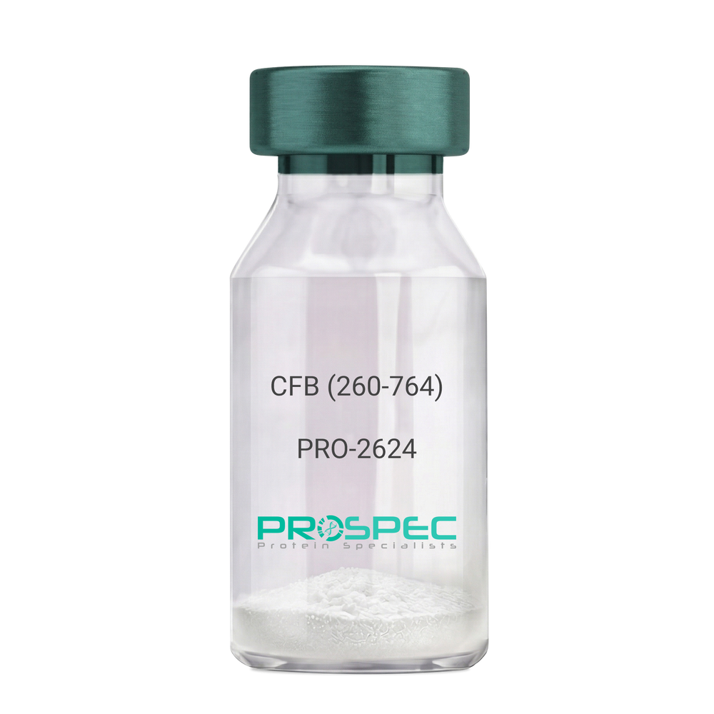
    CFB (260-764) Human, Sf9

    Description:

    Complement Factor B (260-764 a.a.) Human Recombinant, Sf9

    CFB, C3/C5 convertase, Glycine-rich beta glycoprotein, GBG, PBF2, Properdin factor B, BF, BFD, AHUS4, ARMD14, CFAB, CFBD, FB, FBI12, GBG, H2-Bf.

    Product # :

    PRO-2624

    Price :

    Quantity :

    Shipping Method :

    Ice Icon

    Shipped with Ice Packs

    Add To Cart

    More Info

    • description
    • source
    • formulation
    • purity
    • More Info

    Description

    CFB Human Recombinant produced in Sf9 Baculovirus cells is a single, glycosylated polypeptide chain containing 514 amino acids (260-764 a.a) and having a molecular mass of 58.1kDa.CFB is fused to an 9 amino acid His-tag at C-terminus & purified by proprietary chromatographic techniques.

    Source

    Sf9, Baculovirus cells.

    Formulation

    CFB protein solution (0.5mg/ml) containing Phosphate Buffered Saline (pH 7.4) and 20% glycerol.

    Purity

    Greater than 90.0% as determined by SDS-PAGE.

    More Info

    • Introduction

      Factor B is a protein, build from a single chain that circulates in the blood. Complement factor D cleaves and activates factor B, thus creating a catalytic subunit Bb and noncatalytic chain Ba. Subunit Bb is a serine protease that can bind to C3b and forms alternative pathway C3 convertase. Subunit Ba acts as an inhibitor for proliferation of lymphocytes, when Bb is part of the lymphocytes’ proliferation. The gene that codes for this protein is located on chromosome six.

    • Synonyms

      CFB, C3/C5 convertase, Glycine-rich beta glycoprotein, GBG, PBF2, Properdin factor B, BF, BFD, AHUS4, ARMD14, CFAB, CFBD, FB, FBI12, GBG, H2-Bf.

    • Physical Appearance

      Sterile Filtered colorless solution.

    • Stability

      Store at 4°C if entire vial will be used within 2-4 weeks. Store, frozen at -20°C for longer periods of time. For long term storage it is recommended to add a carrier protein (0.1% HSA or BSA).Avoid multiple freeze-thaw cycles.

    • Amino Acid Sequence

      ADPKIVLDPS GSMNIYLVLD GSDSIGASNF TGAKKCLVNL IEKVASYGVK PRYGLVTYAT
      YPKIWVKVSE ADSSNADWVT KQLNEINYED HKLKSGTNTK KALQAVYSMM SWPDDVPPEG
      WNRTRHVIIL MTDGLHNMGG DPITVIDEIR DLLYIGKDRK NPREDYLDVY VFGVGPLVNQ
      VNINALASKK DNEQHVFKVK DMENLEDVFY QMIDESQSLS LCGMVWEHRK GTDYHKQPWQ
      AKISVIRPSK GHESCMGAVV SEYFVLTAAH CFTVDDKEHS IKVSVGGEKR DLEIEVVLFH
      PNYNINGKKE AGIPEFYDYD VALIKLKNKL KYGQTIRPIC LPCTEGTTRA LRLPPTTTCQ
      QQKEELLPAQ DIKALFVSEE EKKLTRKEVY IKNGDKKGSC ERDAQYAPGY DKVKDISEVV
      TPRFLCTGGV SPYADPNTCR GDSGGPLIVH KRSRFIQVGV ISWGVVDVCK NQKRQKQVPA HARDFHINLF QVLPWLKEKL QDEDLGFLHH HHHH

    ProSpec's products are furnished for LABORATORY RESEARCH USE ONLY. The product may not be used as drugs, agricultural or pesticidal products, food additives or household chemicals.

    Complement Factor B
  • View Data Sheet

    Name :

    TGFB2 Human, HEK

    Description:

    Transforming Growth Factor-Beta 2 Human Recombinant, HEK

    Transforming growth factor, beta 2, cetermin, Glioblastoma-derived T-cell suppressor factor, polyergin, G-TSF, TGF-beta2, TGF-beta-2, transforming growth factor beta-2, BSC-1 cell growth inhibitor, TGFB-2.

    Product # :

    CYT-112

    Price :

    Quantity :

    Shipping Method :

    Room Temp Icon

    Shipped at Room temp

    Add To Cart

    More Info

    • description
    • source
    • formulation
    • purity
    • biological activity
    • More Info

    Description

    TGF-b 2 Human Recombinant produced in HEK cells is a non-glycosylated homodimer, having a total molecular weight of 25kDa.The TGF-b 2 is purified by proprietary chromatographic techniques.

    Source

    HEK.

    Formulation

    TGF-b 2 was lyophilized from a 0.2µm filtered solution containing 50mM sodium acetate pH 4.5.

    Purity

    Greater than 95% as obsereved by SDS-PAGE.

    Biological Activity

    The specific activity was determined by the dose-dependent inhibition of IL-4 induced proliferation of mouse HT-2 cells (BALB/c spleen activated by sheep erythrocytes in the presence of IL-2), the ED50 is 0.16ng/ml.

    More Info

    • Introduction

      TGFB2 is a 27.08 kDa protein having two identical 118 amino acid peptide chains linked by a single disulfide bond. TGFB2 is part of a family of five related cytokines that have an extensive variation of normal and neoplastic cells, indicating the importance of these homo-dimmer proteins as multi-functional regulators of cellular activity. The three mammalian isoforms of TGF-beta (TGFb1, TGFb2 and TGFb3) signal through the same receptor and stimulate similar biological responses. They are involved in physiological processes as embryogenesis, tissue remodelling and wound healing.

    • Synonyms

      Transforming growth factor, beta 2, cetermin, Glioblastoma-derived T-cell suppressor factor, polyergin, G-TSF, TGF-beta2, TGF-beta-2, transforming growth factor beta-2, BSC-1 cell growth inhibitor, TGFB-2.

    • Physical Appearance

      Sterile Filtered White lyophilized (freeze-dried) powder.

    • Stability

      Lyophilized TGF-b 2 although stable at room temperature for 3 weeks, should be stored desiccated below -18°C. Upon reconstitution TGF-b 2 should be stored at 4°C between 2-7 days and for future use below -18°C.For long term storage it is recommended to add a carrier protein (0.1% HSA or BSA).Please prevent freeze-thaw cycles.

    • Solubility

      It is recommended to reconstitute the lyophilized TGF-b 2 in sterile solution containing 20% ethanol, 50mM sodium acetate and 75mM acetic acid.

    ProSpec's products are furnished for LABORATORY RESEARCH USE ONLY. The product may not be used as drugs, agricultural or pesticidal products, food additives or household chemicals.

    Tgf B 2 Human Hek
  • View Data Sheet

    Name :

    NCL Human

    Description:

    Nucleolin Human Recombinant

    Nucleolin, Protein C23, NCL, C23.

    Product # :

    PRO-1508

    Price :

    Quantity :

    Shipping Method :

    Ice Icon

    Shipped with Ice Packs

    Add To Cart

    More Info

    • description
    • source
    • formulation
    • purity
    • More Info

    Description

    Nucleolin Human Recombinant produced in SF9 is a glycosylated, polypeptide chain containing the C-terminal section of the human nucleolin and missing the N-terminal histone-binding part of nucleolin, having a calculated molecular mass of 55,162 Dalton. NCL is expressed with a -6x His tag at N-terminus and purified by proprietary chromatographic techniques.

    Source

    Sf9 Insect Cells.

    Formulation

    NCL is supplied in 20mM HEPES pH-7.3, 600mM NaCl, 0.3mM Tris(2-carboxyethyl)phosphine (TCEP) and 25% glycerol.

    Purity

    Greater than 80.0% as determined by SDS-PAGE.

    More Info

    • Introduction

      Nucleolin (NCL) which is a eukaryotic nucleolar phosphoprotein, involved in the synthesis and maturation of ribosomes. Nucleolin is the key nucleolar protein of growing eukaryotic cells. NCL is found linked with intranucleolar chromatin and pre-ribosomal particles. NCL induces chromatin decondensation by binding to histone H1. Nucleolin is assumed to have a role in pre-rRNA transcription and ribosome compilation. Nucleolin may also have a role in the process of transcriptional elongation. Nucleolin is located primarily in the dense fibrillar regions of the nucleolus. The Human NCL gene consists of 14 exons with 13 introns and spans approximately 11kb.

    • Synonyms

      Nucleolin, Protein C23, NCL, C23.

    • Physical Appearance

      Sterile Filtered clear solution.

    • Stability

      Store at 4°C if entire vial will be used within 2-4 weeks. Store, frozen at -20°C for longer periods of time. Avoid multiple freeze-thaw cycles.

    ProSpec's products are furnished for LABORATORY RESEARCH USE ONLY. The product may not be used as drugs, agricultural or pesticidal products, food additives or household chemicals.

    Ncl Human
  • View Data Sheet

    Name :

    DnaK SBD

    Description:

    DnaK Substrate Binding Domain E.Coli Recombinant

    HSP-70, HSP70, DnaK, Chaperone protein dnaK, Heat shock protein 70, Heat shock 70 kDa protein, groP, grpF, seg, b0014, JW0013.

    Product # :

    HSP-008

    Price :

    Quantity :

    Shipping Method :

    Ice Icon

    Shipped with Ice Packs

    Add To Cart

    More Info

    • description
    • source
    • formulation
    • purity
    • More Info

    Description

    Recombinant DnaK Substrate Binding domain produced in E.Coli is a single, non-glycosylated polypeptide chain containing (385-546 a.a.) 163 amino acids and having a molecular mass of 17.7 kDa.

    Source

    Escherichia Coli.

    Formulation

    The protein contains 25mM Tris-HCl, pH7.5, 2mM B-ME and 1mM DTT.

    Purity

    Greater than 95.0% as determined by SDS-PAGE.

    More Info

    • Introduction

      DnaK, originally identified for its DNA replication by bacteriophage l in E. coli is the bacterial hsp70 chaperone. This protein is involved in the folding and assembly of newly synthesized polypeptide chains and in preventing the aggregation of stress-denatured proteins. Dnak(residues 508-638) of the substrate binding domain is a-helical and appears to act as a lid covering the substrate binding cleft. DnaK(amino acid 508-638) was purified to apparent homogeneity by using conventional column chromatography techniques. Additional amino acid (Met) is attached at N- terminus.

    • Synonyms

      HSP-70, HSP70, DnaK, Chaperone protein dnaK, Heat shock protein 70, Heat shock 70 kDa protein, groP, grpF, seg, b0014, JW0013.

    • Physical Appearance

      Sterile filtered colorless solution.

    • Stability

      Store at 4°C if entire vial will be used within 2-4 weeks. Store, frozen at -20°C for longer periods of time. For long term storage it is recommended to add a carrier protein (0.1% HSA or BSA).Avoid multiple freeze-thaw cycles.

    • Amino Acid Sequence

      MDVKDVLLLD VTPLSLGIET MGGVMTTLIA KNTTIPTKHS QVFSTAEDNQ SAVTIHVLQG ERKRAADNKS LGQFNLDGIN PAPRGMPQIE VTFDIDADGI LHVSAKDKNS GKEQKITIKA SSGLNEDEIQ KMVRDAEANA EADRKFEELV QTRNQGDHLL HST.

    ProSpec's products are furnished for LABORATORY RESEARCH USE ONLY. The product may not be used as drugs, agricultural or pesticidal products, food additives or household chemicals.

    Dnak Sbd
  • View Data Sheet

    Name :

    IL 12p40 Antibody

    Description:

    Interleukin-12 p40, Rat Anti-Mouse

    NKSF2, CTL maturation factor (TCMF), Cytotoxic lymphocyte maturation factor 40 kDa subunit (CLMF p40), TSF, Edodekin-alpha, IL-12 p40, IL-12B, IL-12 subunit p40, NK cell stimulatory factor chain 2.

    Product # :

    ANT-114

    Price :

    Quantity :

    Shipping Method :

    Room Temp Icon

    Shipped at Room temp

    Add To Cart

    More Info

    • More Info

    More Info

    • Introduction

      Active IL-12 is a p70 disulphide-linked dimer composed of p35 and p40 subunits. The protein is a pleiotropic cytokine produced primarily by antigen presenting cells and has multiple effects on T lymphocytes and natural killer cells in terms of stimulating cytotoxicity, proliferation, production of other cytokines and Th1 subset differentiation.

    • Synonyms

      NKSF2, CTL maturation factor (TCMF), Cytotoxic lymphocyte maturation factor 40 kDa subunit (CLMF p40), TSF, Edodekin-alpha, IL-12 p40, IL-12B, IL-12 subunit p40, NK cell stimulatory factor chain 2.

    • Solubility

      Reconstitute with sterile H20. Mix gently, wash the sides of the vial and wait 30-60 seconds before use.

    • Immunogen

      r.Murine IL-12 p40.

    • Ig Subclass

      Rat IgG2a.

    • Clone

      NYRmIL-12p40.

    • Applications

      Direct ELISA, Western Blot, Immuneprecipitation.

    • Titer

      By direct ELISA, 1:5,000 dilution will yield 0.5 O.D using alkaline phosphatase conjugated rabbit anti-mouse Ig (Jackson Laboratories).

    • Shipping Conditions

      Antibody is shipped lyophilized at ambient temperature.

    • Type

      Rat Anti Mouse Monoclonal.

    • Storage Procedures

      In lyophilized form, for long periods, store at 4oC in a dry environment. After reconstitution, if not intended for use within a month, aliquot and store at -20oC.

    • Purification Method

      Ion exchange.

    ProSpec's products are furnished for LABORATORY RESEARCH USE ONLY. The product may not be used as drugs, agricultural or pesticidal products, food additives or household chemicals.

    Il 12P40 Antibody
  • View Data Sheet

    Name :

    SERPINI1 Human

    Description:

    Serpin Peptidase Inhibitor, Clade I Member 1 Human Recombinant

    Neuroserpin, Peptidase inhibitor 12, PI-12, Serpin I1, SERPINI1, PI12.

    Product # :

    PRO-213

    Price :

    Quantity :

    Shipping Method :

    Room Temp Icon

    Shipped at Room temp

    Add To Cart

    More Info

    • description
    • source
    • formulation
    • purity
    • biological activity
    • More Info

    Description

    SERPINI1 Human Recombinant produced in E.coli is a single, non-glycosylated polypeptide chain containing 394 amino acids and having a total molecular mass of 44.7kDa. SERPINI1 is purified by proprietary chromatographic techniques.

    Source

    Escherichia Coli.

    Formulation

    The protein was Lyophilized from a 0.2µm filtered concentrated solution in 1×PBS, pH 7.4.

    Purity

    Greater than 95.0% as determined by
    (a) Analysis by HPLC.
    (b) Analysis by SDS-PAGE.

    Biological Activity

    Determined by the dose-dependent stimulation of the proliferation of rat C6 cells which is less than 0.5µg/ml, corresponding to a specific activity of >2000IU/mg.

    More Info

    • Introduction

      SERPINI1 (Neuroserpin) is an inhibitory serpin which is expressed primarily in the central nervous system. Even though the physiological target of SERPINI1 is still vague, amassed evidence suggest that SERPINI1 has an imperative role in controlling proteolytic degradation of extracellular matrix (ECM) during synaptogenesis and the subsequent development of neuronal plasticity. The neuroprotective role of SERPINI1 has been demonstrated in transgenic mice lacking SERPINI1 expression. The deficiency of SERPINI1 in these mice is linked with motor neuron disease characterized by axonal degradation. In humans, defects in SERPINI1, caused by point mutations in the neuroserpin gene, trigger a hereditary disorder known as the familial encephalopathy with neuroserpin inclusion bodies (FENIB).

    • Synonyms

      Neuroserpin, Peptidase inhibitor 12, PI-12, Serpin I1, SERPINI1, PI12.

    • Physical Appearance

      Sterile Filtered White lyophilized (freeze-dried) powder.

    • Stability

      Lyophilized SERPINI1 although stable at room temperature for 3 weeks, should be stored desiccated below -18°C. Upon reconstitution SERPINI1 should be stored at 4°C between 2-7 days and for future use below -18°C.Please prevent freeze-thaw cycles.

    • Solubility

      It is recommended to reconstitute the lyophilized SERPINI1 in sterile 18M-cm H2O not less than 100µg/ml, which can then be further diluted to other aqueous solutions.

    • Amino Acid Sequence

      TGATFPEEAI ADLSVNMYNR LRATGEDENI LFSPLSIALA MGMMELGAQG
      STQKEIRHSM GYDSLKNGEE FSFLKEFSNM VTAKESQYVM KIANSLFVQN
      GFHVNEEFLQ MMKKYFNAAV NHVDFSQNVA VANYINKWVE NNTNNLVKDL
      VSPRDFDAAT YLALINAVYF KGNWKSQFRP ENTRTFSFTK DDESEVQIPM
      MYQQGEFYYG EFSDGSNEAG GIYQVLEIPY EGDEISMMLV LSRQEVPLAT
      LEPLVKAQLV EEWANSVKKQ KVEVYLPRFT VEQEIDLKDV LKALGITEIF
      IKDANLTGLS DNKEIFLSKA IHKSFLEVNE EGSEAAAVSG MIAISRMAVL
      YPQVIVDHPF FFLIRNRRTG TILFMGRVMH PETMNTSGHD FEEL.

    ProSpec's products are furnished for LABORATORY RESEARCH USE ONLY. The product may not be used as drugs, agricultural or pesticidal products, food additives or household chemicals.

    Serpini1 Human
  • 1
  • …
  • 30
  • 31
  • 32
  • 33
  • 34
  • …
  • 42
Your browser does not support the video tag.

Products

  • Cytokines
  • Growth Factors
  • Chemokines
  • CD Antigens
  • Neurotrophins
  • Hormones
  • Enzymes
  • Viral Antigens
  • Recombinant Proteins
  • Natural Proteins
  • Monoclonal Antibodies
  • Polyclonal Antibodies
  • Thermal Packaging

About

  • Company
  • Founder
  • Contribution
  • Contact Us
  • My Account

Ordering

  • Ordering Method
  • Ordering - Credit Card
  • Ordering PO
  • Shipping Fees
  • FAQ's

Legal

  • Terms & Conditions
  • Privacy
  • Site Map
  • info@prospecbio.com
  • Facebook
  • Instagram
  • Linkedin Profile

© 2026 ProSpec-Tany TechnoGene Ltd. All Rights Reserved.

  • Choosing a selection results in a full page refresh.
  • Opens in a new window.