Search results
759 results found for “Met Proto-Oncogene”
Name
Description
Product #
Price
Quantity
Shipping Method
- View Data Sheet
Name :
VEGF EquineDescription:
Vascular Endothelial Growth Factor Equine Recombinant
VEGF-A, VPF, glioma-derived endothelial cell mitogen.
Product # :
CYT-1200Price :
Quantity :
Shipping Method :
Shipped at Room temp
More Info
- description
- source
- formulation
- purity
- biological activity
- More Info
Description
VEGF Equine Recombinant produced in E.Coli is a single, non-glycosylated, polypeptide chain containing 2 x 165 amino acids and having a total molecular mass of 38.6 kDa.The VEGF is purified by proprietary chromatographic techniques.
Source
Escherichia Coli.
Formulation
Lyophilized from a sterile (0.2 micron) filtered aqueous solution containing 10 mM Sodium Phosphate, pH 7.5.
Purity
Greater than 95.0% as determined by SDS-PAGE.
Biological Activity
The ED50 as determined by HUVEC Proliferation is ≤ 10 ng/mL, corresponding to a specific activity of ≥ 1.0 x 10^5 units/mg.
More Info
-
Introduction
Vascular endothelial growth factor is an important signaling protein involved in both vasculogenesis and angiogenesis. As its name implies, VEGF activity has been mostly studied on cells of the vascular endothelium, although it does have effects on a number of other cell types (e.g. stimulation monocyte/macrophage migration, neurons, cancer cells, kidney epithelial cells ).VEGF mediates increased vascular permeability, induces angiogenesis, vasculogenesis and endothelial cell growth, promotes cell migration, and inhibits apoptosis. In vitro, VEGF has been shown to stimulate endothelial cell mitogenesis and cell migration. VEGF is also a vasodilator and increases microvascular permeability and was originally referred to as vascular permeability factor. Elevated levels of this protein is linked to POEMS syndrome, also known as Crow-Fukase syndrome. Mutations in this gene have been associated with proliferative and nonproliferative diabetic retinopathy.
-
Synonyms
VEGF-A, VPF, glioma-derived endothelial cell mitogen.
-
Physical Appearance
Sterile Filtered White lyophilized (freeze-dried) powder.
-
Stability
Lyophilized VEGF although stable at room temperature for 3 weeks, should be stored desiccated below -18°C. Upon reconstitution VEGF Human should be stored at 4°C between 2-7 days and for future use below -18°C.For long term storage it is recommended to add a carrier protein (0.1% HSA or BSA).Please prevent freeze-thaw cycles.
-
Solubility
It is recommended to reconstitute the lyophilized VEGF in sterile water at a concentration of 0.1 mg/ml, which can then be further diluted to other aqueous solutions.
-
Amino Acid Sequence
MAPMAEGEHK THEVVKFMDV YQRSYCRPIE TLVDIFQEYP DEIEYIFKPS CVPLMRCGGC CNDEGLECVP TAEFNITMQI MRIKPHQSQH IGEMSFLQHS KCECRPKKDK ARQENPCGPC SERRKHLFVQ DPQTCKCSCK NTDSRCKARQ LELNERTCRC DKPRR
ProSpec's products are furnished for LABORATORY RESEARCH USE ONLY. The product may not be used as drugs, agricultural or pesticidal products, food additives or household chemicals.
- View Data Sheet
Name :
Protein-L, HisDescription:
Protein L Recombinant, His Tag
Product # :
PRO-1930Price :
Quantity :
Shipping Method :
Shipped at Room temp
More Info
- description
- source
- formulation
- purity
- More Info
Description
Recombinant Protein-L produced in E.Coli is a single non-glycosylated polypeptide chain fused with a 6×His tag at N-terminus. Protein-L is comprised of 5 IgG-binding regions of protein L (B1-B2-B3-B4-B5) containing 372 amino acids in total and having a molecular mass of 41.5kDa, however, it migrates with an apparent molecular mass of 46kDa on SDS-PAGE. Cell wall binding region, cell membrane binding region and albumin binding region have been eliminated from the recombinant Protein-L to guarantee the maximum specific IgG binding.
Source
Escherichia Coli.
Formulation
Protein-L was lyophilized without any additives.
Purity
Greater than 97.0% as determined by:
(a) Analysis by SEC-HPLC.
(b) Analysis by SDS-PAGE.More Info
-
Introduction
The Recombinant Protein L is comprised of 5 kappa-binding domains. Protein L has the exceptional ability to bind through kappa light chain interactions without hindering with the antibody’s antigen-binding site. This gives Protein L the capacity to bind a broader range of Ig classes and subclasses than other antibody-binding proteins. The recombinant Protein L is perfect for purification of polyclonal or monoclonal IgG antibodies. Protein L binds to IgG from humans, mice, rats and pigs.
-
Physical Appearance
Sterile Filtered White lyophilized (freeze-dried) powder.
-
Stability
Lyophilized Protein-L although stable at room temperature for 3 weeks, should be stored desiccated below -18°C. Upon reconstitution Protein-L should be stored at 4°C between 2-7 days and for future use below -18°C. For long term storage it is recommended to add a carrier protein (0.1% HSA or BSA). Please prevent freeze-thaw cycles.
-
Solubility
It is recommended to reconstitute the lyophilized Protein-L in sterile 18M-cm H2O not less than 0.1mg/ml, which can then be further diluted to other aqueous solutions.
-
Amino Acid Sequence
MHHHHHHKEE TPETPETDSE EEVTIKANLI FANGSTQTAE FKGTFEKATS EAYAYADTLK KDNGEYTVDV ADKGYTLNIK FAGKEKTPEE PKEEVTIKAN LIYADGKTQT AEFKGTFEEA TAEAYRYADA LKKDNGEYTV DVADKGYTLN IKFAGKEKTP EEPKEEVTIK ANLIYADGKT QTAEFKGTFE EATAEAYRYA DLLAKENGKY TVDVADKGYT LNIKFAGKEK TPEEPKEEVT IKANLIYADG KTQTAEFKGT FAEATAEAYR YADLLAKENG KYTADLEDGG YTINIRFAGK KVDEKPEEKE QVTIKENIYF EDGTVQTATF KGTFAEATAE AYRYADLLSK EHGKYTADLE DGGYTINIRF AG.
ProSpec's products are furnished for LABORATORY RESEARCH USE ONLY. The product may not be used as drugs, agricultural or pesticidal products, food additives or household chemicals.
- View Data Sheet
Name :
BNP ProteinDescription:
B-type Natriuretic Peptide Human Recombinant
NPPB, Natriuretic Peptide Precursor B, BNP, B-type Natriuretic Peptide.
Product # :
CYT-327Price :
Quantity :
Shipping Method :
Shipped at Room temp
More Info
- description
- source
- formulation
- purity
- More Info
Description
B-type Natriuretic Peptide Recombinant Human produced in E.Coli is a single, non-glycosylated, polypeptide chain containing 32 amino acids and having a molecular mass of 3,500 Dalton. NPPB is purified by proprietary chromatographic techniques.
Source
Escherichia Coli.
Formulation
Natriuretic Peptide Precursor B was lyophilized from a 0.2 µm filtered concentrated solution in PBS, pH 7.4.
Purity
Greater than 97.0% as determined by:
(a) Analysis by RP-HPLC.
(b) Analysis by SDS-PAGE.More Info
-
Introduction
Natriuretic Peptide Precursor B acts as a cardiac hormone with a variety of biological actions including natriuresis, diuresis, vasorelaxation, and inhibition of renin and aldosterone secretion. It is thought to play a key role in cardiovascular homeostasis. Helps restore the body's salt and water balance. Improves heart function.
-
Synonyms
NPPB, Natriuretic Peptide Precursor B, BNP, B-type Natriuretic Peptide.
-
Physical Appearance
Sterile Filtered White lyophilized (freeze-dried) powder.
-
Stability
Lyophilized B-type Natriuretic Peptide although stable at room temperature for 3 weeks, should be stored desiccated below -18°C. Upon reconstitution NPPB should be stored at 4°C between 2-7 days and for future use below -18°C.For long term storage it is recommended to add a carrier protein (0.1% HSA or BSA).Please prevent freeze-thaw cycles.
-
Solubility
It is recommended to reconstitute the lyophilized B-type Natriuretic Peptide in sterile 18MΩ-cm H2O not less than 100µg/ml, which can then be further diluted to other aqueous solutions.
-
Amino Acid Sequence
SPKMVQGSGCFGRKMDRISSSSGLGCKVLRRH.
-
Background
What is the molecular weight / Mw of BNP Protein?
BNP Protein has a total Mw of 3.5kDa.
What is the source or expression system of BNP Protein?
Escherichia Coli.
What is the Purity of BNP Protein?
BNP Protein is >97% pure as determined by SDS-PAGE.
What is the Biological Activity of BNP Protein?
The biological functionality of BNP Protein will be determined in the future.
What is the amino acid sequence of BNP Protein?
SPKMVQGSGCFGRKMDRISSSSGLGCKVLRRH.
What applications can BNP Protein Protein be used in?
BNP Protein can probably be used in western blot, ELISA and Lateral Flow.
What is the endotoxin level for BNP Protein?
The endotoxin level is minimal, BNP Protein was purified using conventional chromatography techniques.
ProSpec's products are furnished for LABORATORY RESEARCH USE ONLY. The product may not be used as drugs, agricultural or pesticidal products, food additives or household chemicals.
- View Data Sheet
Name :
PDZK1 AntibodyDescription:
PDZ Domain Containing 1, Mouse Anti Human
PDZ Domain Containing 1, PDZ-Containing Kidney Protein 1, Na(+)/H(+) Exchange Regulatory Cofactor NHE-RF3, Na/Pi Cotransporter C-Terminal-Associated Protein 1, Sodium-Hydrogen Exchanger Regulatory Factor 3, CFTR-Associated Protein Of 70 KDa, NHERF-3, CAP70, PDZD1, CLAMP, naPi-Cap1.
Product # :
ANT-460Price :
Quantity :
Shipping Method :
Shipped with Ice Packs
More Info
- formulation
- More Info
Formulation
1mg/ml containing PBS, pH-7.4, 10% Glycerol and 0.02% Sodium Azide.
More Info
-
Introduction
PDZK1 is a PDZ domain-containing scaffolding protein which facilitates localization of cell surface proteins and has a key part in cholesterol metabolism by regulating the HDL receptor, scavenger receptor class B type 1. Single nucleotide polymorphism in this gene is linked to metabolic syndrome, and overexpression results in drug resistance of multiple myeloma.
-
Synonyms
PDZ Domain Containing 1, PDZ-Containing Kidney Protein 1, Na(+)/H(+) Exchange Regulatory Cofactor NHE-RF3, Na/Pi Cotransporter C-Terminal-Associated Protein 1, Sodium-Hydrogen Exchanger Regulatory Factor 3, CFTR-Associated Protein Of 70 KDa, NHERF-3, CAP70, PDZD1, CLAMP, naPi-Cap1.
-
Physical Appearance
Sterile Filtered colorless solution.
-
Immunogen
Anti-human PDZK1 mAb, clone PAT1A2A, is derived from hybridization of mouse F0 myeloma cells with spleen cells from BALB/c mice immunized with a recombinant human PDZK1 protein 1-519 amino acids purified from E. coli.
-
Ig Subclass
Mouse IgG2b heavy chain and Kappa light chain.
-
Clone
PAT1A2AT.
-
Applications
The antibody has been tested by ELISA and Western blot analysis to assure specificity and reactivity. Since application varies, however, each investigation should be titrated by the reagent to obtain optimal results. Recommended starting dilution is 1:1000.
-
Type
Mouse Anti Human Monoclonal.
-
Storage Procedures
For periods up to 1 month store at 4°C, for longer periods of time, store at -20°C. Prevent freeze thaw cycles.
-
Purification Method
PDZK1 antibody was purified from mouse ascitic fluids by protein-A affinity chromatography.
ProSpec's products are furnished for LABORATORY RESEARCH USE ONLY. The product may not be used as drugs, agricultural or pesticidal products, food additives or household chemicals.
- View Data Sheet
Name :
IGF1 Human, GSTDescription:
Insulin-Like Growth Factor 1 Human Recombinant, GST Tag
Somatomedin C, IGF-I, IGFI, IGF1, IGF-IA, Mechano growth factor, MGF.
Product # :
CYT-690Price :
Quantity :
Shipping Method :
Shipped with Ice Packs
More Info
- description
- source
- formulation
- More Info
Description
IGF1 Human Recombinant produced in E.Coli is a single, non-glycosylated, Polypeptide chain fused to a GST tag and is purified by proprietary chromatographic techniques.
Source
Escherichia Coli.
Formulation
IGF1 is supplied in 50mM Tris-Acetate, pH-7.5, 1mM EDTA and 20% Glycerol.
More Info
-
Introduction
The somatomedins, or insulin-like growth factors (IGFs), comprise a family of peptides that play important roles in mammalian growth and development. IGF1 mediates many of the growth-promoting effects of growth hormone (GH; MIM 139250). Early studies showed that growth hormone did not directly stimulate the incorporation of sulfate into cartilage, but rather acted through a serum factor, termed 'sulfation factor,' which later became known as 'somatomedin' (Daughaday et al., 1972). Three main somatomedins have been characterized: somatomedin C (IGF1), somatomedin A (IGF2; MIM 147470), and somatomedin B (MIM 193190) (Rotwein, 1986; Rosenfeld, 2003).
-
Synonyms
Somatomedin C, IGF-I, IGFI, IGF1, IGF-IA, Mechano growth factor, MGF.
-
Physical Appearance
Sterile Filtered clear solution.
-
Stability
Store at 4°C if entire vial will be used within 2-4 weeks. Store, frozen at -20°C for longer periods of time. Please avoid freeze thaw cycles.
ProSpec's products are furnished for LABORATORY RESEARCH USE ONLY. The product may not be used as drugs, agricultural or pesticidal products, food additives or household chemicals.
- View Data Sheet
Name :
HPSE ActiveDescription:
Recombinant Human Heparanase-1 Active
Product # :
ENZ-1032Price :
Quantity :
Shipping Method :
Shipped with Ice Packs
More Info
- description
- formulation
- purity
- biological activity
- More Info
Description
Heparanase Active Enzyme is produced in CHO cells.The protein is purified by several orthogonal chromatography steps.
Formulation
Heparanase Active Enzyme is supplied in20mM Acetate buffer and 750mM NaCl pH 5.4.
Purity
Greater than 95.0% as determined by:
(a) Analysis by RP-HPLC.
(b) Analysis by SDS-PAGE.Biological Activity
The specific activity of Heparanase Active Enzyme in-house standard is about 0.7 Units (1 unit = 1 μmole of reducing ends of heparan sulfate substrate formed per minute per mg Heparanase Active Enzyme at 37°C). The enzymatic activity of each Heparanase Active Enzyme batch is comparable to the standard as determined by activity assay in which immobilized heparan, released due to heparanase activity, is quantified colorimetrically. Recommended reaction buffer: 20 mM Citrate Phosphate buffer, pH 5.4; 50mM NaCl; 1mM CaCl2.
More Info
-
Introduction
Heparanase is an endo β-D-glucuronidase, which degrades heparan sulfate side chains of heparan sulfate proteoglycans (HSPGs) in the extracellular matrix. Heparanase plays an important role in ECM degradation, facilitating the migration and extravasation of tumor cells and inflammatory leukocytes (1,2,3). Upon degradation, heparanase releases growth factors and cytokines that stimulate cell proliferation and chemotaxis (4,5). Heparanase is a heterodimer comprised of a 50 kDa subunit harboring the active site and a 8 kDa subunit. It is produced as a latent 65 kDa precursor and proteolytically processed to its active form (1,6). Heparanase is highly expressed in myeloid leukocytes (i.e. neutrophils) in platelets and in human placenta. Human heparanase was found to be upregulated in various types of primary tumors, correlating in some cases with increased tumor invasiveness and vascularity and with poor prospective survival (7,8).
-
Data Sheet
To view the FULL VERSION data sheet click Heparanase-1 Active Enzyme
-
Specificity
Heparanase Active Enzyme is identified by Western blot analysis with polyclonalrabbit anti-HPA1 antibodies as 2 subunits of 8-kDa and 50-kDa.
-
Shipping Conditions
Heparanase Active Enzyme is shipped frozen on dry ice unless stated otherwise by the customer. Shipping fees to N. America and W. Europe is $400 Shipping fees to Asia, Australia and E. Europe is $500
-
Storage Procedures
Store at –80ºC, avoid repeated freeze-thaw cycles.
ProSpec's products are furnished for LABORATORY RESEARCH USE ONLY. The product may not be used as drugs, agricultural or pesticidal products, food additives or household chemicals.
- View Data Sheet
Name :
C1QBP AntibodyDescription:
Complement Component 1, Mouse Anti Human
p32, HABP1, gC1Qr, GC1QBP, SF2p32, gC1Q-R, Complement component 1 Q subcomponent-binding protein mitochondrial, Glycoprotein gC1qBP, C1qBP, GC1q-R protein, Hyaluronan-binding protein 1, Mitochondrial matrix protein p32, p33, C1QBP.
Product # :
ANT-556Price :
Quantity :
Shipping Method :
Shipped with Ice Packs
More Info
- formulation
- More Info
Formulation
1mg/ml containing PBS, pH-7.4, 10% Glycerol and 0.02% Sodium Azide.
More Info
-
Introduction
C1QBP binds to the globular "heads" of c1q thus inhibiting c1 activation. C1QBP interacts with a wide range of ligands and is implicated in cell signaling. C1QBP associates with C1r and C1s in order to yield the first component of the serum complement system. C1QBP protein has been identified as the p32 subunit of pre-mRNA splicing factor SF2, as well as a hyaluronic acid-binding protein.
C1QBP is a new marker of tumor cells and tumor-associated macrophages/myeloid cells in hypoxic/metabolically deprived areas of tumors.
Mitochondrial C1QBP is a critical mediator of p14ARF-induced apoptosis.
C1QBP functions as a chemotactic factor for immature dendritic cells, and migration is mediated through ligation of both C1QBP and cC1qR/CR.
C1QBP overexpression successfully blocks mRNA accumulation from the adenovirus major late transcription unit (MLTU) and stimulates RNA polymerase II carboxy-terminal domain phosphorylation in virus-infected cells.
C1QBP binds with Hepacivirus core protein on CD8+ and CD4+ positive t-cells and inactivates lck and akt. -
Synonyms
p32, HABP1, gC1Qr, GC1QBP, SF2p32, gC1Q-R, Complement component 1 Q subcomponent-binding protein mitochondrial, Glycoprotein gC1qBP, C1qBP, GC1q-R protein, Hyaluronan-binding protein 1, Mitochondrial matrix protein p32, p33, C1QBP.
-
Physical Appearance
Sterile filtered colorless solution.
-
Immunogen
Anti-human C1QBP mAb, is derived from hybridization of mouse F0 myeloma cells with spleen cells from BALB/c mice immunized with a recombinant human C1QBP protein 74-282 amino acids purified from E. coli.
-
Ig Subclass
Mouse IgG1 heavy chain and k light chain.
-
Clone
PAT1G7AT.
-
Applications
C1QBP antibody has been tested by ELISA, Western blot analysis to assure specificity and reactivity. Since application varies, however, each investigation should be titrated by the reagent to obtain optimal results.
-
Type
Mouse Anti Human Monoclonal.
-
Storage Procedures
For periods up to 1 month store at 4°C, for longer periods of time, store at -20°C. Prevent freeze thaw cycles.
-
Purification Method
C1QBP antibody was purified from mouse ascitic fluids by protein-A affinity chromatography.
ProSpec's products are furnished for LABORATORY RESEARCH USE ONLY. The product may not be used as drugs, agricultural or pesticidal products, food additives or household chemicals.
- View Data Sheet
Name :
GAS7 AntibodyDescription:
Growth Arrest-Specific 7 Isoform b, Mouse Anti Human
GAS7, GAS-7, Growth arrest-specific protein 7, KIAA0394, MGC1348, MLL/GAS7.
Product # :
ANT-358Price :
Quantity :
Shipping Method :
Shipped with Ice Packs
More Info
- formulation
- More Info
Formulation
1mg/ml containing PBS, pH-7.4, & 0.1% Sodium Azide.
More Info
-
Introduction
GAS7 is mainly expressed in terminally differentiated brain cells and predominantly in mature cerebellar Purkinje neurons. GAS7 is involved in neuronal development by promoting maturation and morphological differentiation of cerebellar neurons. Inhibition of GAS7 production in terminally differentiating cultures of embryonic murine cerebellum impedes neurite outgrowth. Over-expression of GAS7 initiats the development of human osteosarcoma.
-
Synonyms
GAS7, GAS-7, Growth arrest-specific protein 7, KIAA0394, MGC1348, MLL/GAS7.
-
Immunogen
Anti-human GAS7 mAb is derived from hybridization of mouse F0 myeloma cells with spleen cells from BALB/c mice immunized with recombinant human GAS7 amino acids 1-416 purified from E. coli.
-
Ig Subclass
Mouse IgG1 heavy chain and κ light chain.
-
Clone
PAT4H8AT.
-
Applications
GAS7 antibody has been tested by ELISA and Western blot analysis to assure specificity and reactivity. Since application varies, however, each investigation should be titrated by the reagent to obtain optimal results. Recommended dilution range for Western blot analysis is 1:500 ~ 1000. Recommended starting dilution is 1:500.
-
Type
Mouse Anti Human Monoclonal.
-
Storage Procedures
For periods up to 1 month store at 4°C, for longer periods of time, store at -20°C. Prevent freeze thaw cycles.
-
Purification Method
GAS7 antibody was purified from mouse ascitic fluids by protein-G affinity chromatography.
ProSpec's products are furnished for LABORATORY RESEARCH USE ONLY. The product may not be used as drugs, agricultural or pesticidal products, food additives or household chemicals.
- View Data Sheet
Name :
Protein A/GDescription:
Protein A/G Recombinant
Product # :
PRO-646Price :
Quantity :
Shipping Method :
Shipped at Room temp
More Info
- description
- source
- formulation
- purity
- More Info
Description
The recombinant Protein A/G consists of 5 IgG-binding regions of protein A and 2 of protein G, which corresponds to the Protein A and G domains that are included in the recombinant sequence. Cell wall binding region, cell membrane binding region and albumin binding region have been removed from the recombinant Protein A/G to ensure the maximum specific IgG binding. The Protein A portion is from Staphylococcus aureus segments E, D, A, B and C. The Protein G portion is from Streptococcus segments C1 and C3. The fusion protein has a predicted molecular mass of 47.7kDa and containing 429 amino acids.
Source
Escherichia coli.
Formulation
Lyophilized white Powder containing no additives.
Purity
>97% as determined by SDS-PAGE and RP-HPLC.
More Info
-
Introduction
Recombinant Protein A/G fusion protein joins IgG binding domains of both Protein A and Protein G.
Protein A/G includes four Fc binding domains from Protein A and two from Protein G, yielding a final mass of 50.4 kDa. The binding dependency to pH of Protein A/G lower than Protein A, but has the additive properties of Protein A and G together. Protein A/G binds to all subclasses of human IgG, making it helpful for purifying polyclonal or monoclonal IgG antibodies whose subclasses have not been identifieed. Protein A/G binds to IgA, IgE, IgM and IgD. Protein A/G binds to all subclasses of mouse IgG excluding mouse IgA, IgM or serum albumin. This permits Protein A/G to be used in purification and detection of mouse monoclonal IgG antibodies, with no interference from IgA, IgM and serum albumin. Mouse monoclonal antibodies normally have a stronger affinity to the chimeric Protein A/G than to either Protein A or Protein G. Protein A/G also has been used for purification of macaque IgG. -
Stability
After reconstitution, aliquot and store at -20°C. Avoid repeated freeze/thaw cycles.
-
Solubility
It is recommended to reconstitute the lyophilized Protein-A/G in sterile 18M-cm H2O not less than 0.1mg/ml, which can then be further diluted to other aqueous solutions.
-
Amino Acid Sequence
NAAQHDEAQQ NAFYQVLNMP NLNADQRNGF IQSLKDDPSQ SANVLGEAQK LNDSQAPKAD AQQNNFNKDQ QSAFYEILNM PNLNEAQRNG FIQSLKDDPS QSTNVLGEAK KLNESQAPKA DNNFNKEQQN AFYEILNMPN LNEEQRNGFI QSLKDDPSQS ANLLSEAKKL NESQAPKADN KFNKEQQNAF YEILHLPNLN EEQRNGFIQS LKDDPSQSAN LLAEAKKLND AQAPKADNKF NKEQQNAFYE ILHLPNLTEE QRNGFIQSLK DDPSVSKEIL AEAKKLNDAQ APKEEDSLEG SGSGTYKLIL NGKTLKGETT TEAVDAATAE KVFKQYANDN GVDGEWTYDD ATKTFTVTEK PEVIDASELT PAVTTYKLVI NGKTLKGETT TKAVDAETAE KAFKQYANDN GVDGVWTYDD ATKTFTVTE.
ProSpec's products are furnished for LABORATORY RESEARCH USE ONLY. The product may not be used as drugs, agricultural or pesticidal products, food additives or household chemicals.
- View Data Sheet
Name :
ASF1B AntibodyDescription:
ASF1 Anti-Silencing Function 1 Homolog B, Mouse Anti Human
Histone chaperone ASF1B, Anti-silencing function protein 1 homolog B, hAsf1, hAsf1b, CCG1-interacting factor A-II, CIA-II, hCIA-II, ASF1B.
Product # :
ANT-542Price :
Quantity :
Shipping Method :
Shipped with Ice Packs
More Info
- formulation
- More Info
Formulation
1mg/ml containing PBS, pH-7.4, 10% Glycerol and 0.02% Sodium Azide.
More Info
-
Introduction
ASF1 Anti-Silencing Function 1 Homolog B (ASF1B) belongs to the H3/H4 family of histone chaperone proteins and is similar to the anti-silencing function-1 protein in yeast. ASF1B is the substrate of the tousled-like kinase family of cell cycle-regulated kinases, and may have a crucial role in modulating the nucleosome structure of chromatin by guaranteeing a regular supply of histones at sites of nucleosome assembly. ASF1B cooperates with CAF-1 (chromatin assembly factor 1) to stimulate replication-dependent chromatin assembly. ASF1B is highly expressed in the testis and at lower levels in colon, small intestine and thymus. ASF1B is necessary for spermatogenesis.
-
Synonyms
Histone chaperone ASF1B, Anti-silencing function protein 1 homolog B, hAsf1, hAsf1b, CCG1-interacting factor A-II, CIA-II, hCIA-II, ASF1B.
-
Physical Appearance
Sterile filtered colorless solution.
-
Immunogen
Anti-human ASF1B mAb, clone PAT1D4AT, is derived from hybridization of mouse F0 myeloma cells with spleen cells from BALB/c mice immunized with a recombinant human ASF1B protein (1-202 amino acids) purified from E. coli.
-
Ig Subclass
Mouse IgG2b heavy chain and k light chain.
-
Clone
PAT1D4AT.
-
Applications
The antibody has been tested by ELISA, Western blot analysis to assure specificity and reactivity. Since application varies, however, each investigation should be titrated by the reagent to obtain optimal results. Recommended starting dilution is 1:1000.
-
Type
Mouse Anti Human Monoclonal.
-
Storage Procedures
For periods up to 1 month store at 4°C, for longer periods of time, store at -20°C. Prevent freeze thaw cycles.
-
Purification Method
ASF1B antibody was purified from mouse ascitic fluids by protein-A affinity chromatography.
ProSpec's products are furnished for LABORATORY RESEARCH USE ONLY. The product may not be used as drugs, agricultural or pesticidal products, food additives or household chemicals.
- View Data Sheet
Name :
Protein-L CysDescription:
Protein L Cys Recombinant
Product # :
PRO-1931Price :
Quantity :
Shipping Method :
Shipped at Room temp
More Info
- description
- source
- formulation
- purity
- More Info
- sds-page, HPLC
Description
Recombinant Protein-L produced in E.Coli is a single non-glycosylated polypeptide chain fused with a Cys at C-terminus. Protein-L is comprised of 5 IgG-binding regions of protein L (B1-B2-B3-B4-B5) containing 366 amino acids in total and having a molecular mass of 40.6kDa, however, it migrates with an apparent molecular mass of 46kDa on SDS-PAGE. Cell wall binding region, cell membrane binding region and albumin binding region have been eliminated from the recombinant Protein-L to guarantee the maximum specific IgG binding.
Source
Escherichia Coli.
Formulation
Protein-L was lyophilized without any additives.
Purity
Greater than 96.0% as determined by:
(a) Analysis by SEC-HPLC.
(b) Analysis by SDS-PAGE.sds-page, HPLC
More Info
-
Introduction
The Recombinant Protein L is comprised of 5 kappa-binding domains. Protein L has the exceptional ability to bind through kappa light chain interactions without hindering with the antibody’s antigen-binding site. This gives Protein L the capacity to bind a broader range of Ig classes and subclasses than other antibody-binding proteins. The recombinant Protein L is perfect for purification of polyclonal or monoclonal IgG antibodies. Protein L binds to IgG from humans, mice, rats and pigs.
-
Physical Appearance
Sterile Filtered White lyophilized (freeze-dried) powder.
-
Stability
Lyophilized Protein-L although stable at room temperature for 3 weeks, should be stored desiccated below -18°C. Upon reconstitution Protein-L should be stored at 4°C between 2-7 days and for future use below -18°C. For long term storage it is recommended to add a carrier protein (0.1% HSA or BSA). Please prevent freeze-thaw cycles.
-
Solubility
It is recommended to reconstitute the lyophilized Protein-L in sterile 18M-cm H2O not less than 0.1mg/ml, which can then be further diluted to other aqueous solutions.
-
Amino Acid Sequence
KEETPETPET DSEEEVTIKA NLIFANGSTQ TAEFKGTFEK ATSEAYAYAD TLKKDNGEYT VDVADKGYTL NIKFAGKEKT PEEPKEEVTI KANLIYADGK TQTAEFKGTF EEATAEAYRY ADALKKDNGE YTVDVADKGY TLNIKFAGKE KTPEEPKEEV TIKANLIYAD GKTQTAEFKG TFEEATAEAY RYADLLAKEN GKYTVDVADK GYTLNIKFAG KEKTPEEPKE EVTIKANLIY ADGKTQTAEF KGTFAEATAE AYRYADLLAK ENGKYTADLE DGGYTINIRF AGKKVDEKPE EKEQVTIKEN IYFEDGTVQT ATFKGTFAEA TAEAYRYADL LSKEHGKYTA DLEDGGYTIN IRFAGC.
ProSpec's products are furnished for LABORATORY RESEARCH USE ONLY. The product may not be used as drugs, agricultural or pesticidal products, food additives or household chemicals.
- View Data Sheet
Name :
TSH ProteinDescription:
Thyroid Stimulating Hormone Human Recombinant
Glycoprotein hormones alpha chain, Anterior pituitary glycoprotein hormones common subunit alpha, Follitropin alpha chain, Follicle-stimulating hormone alpha chain, FSH-alpha, Lutropin alpha chain, Luteinizing hormone alpha chain, LSH-alpha, Thyrotropin alpha chain, Thyroid-stimulating hormone alpha chain, TSH-alpha, Choriogonadotropin alpha chain, Chorionic gonadotrophin alpha subunit, CG-alpha, Thyrotropin subunit beta, Thyroid-stimulating hormone subunit beta, TSH-beta, TSH-B, Thyrotropin beta chain, Thyrotropin alfa.
Product # :
HOR-050Price :
Quantity :
Shipping Method :
Shipped at Room temp
More Info
- description
- source
- formulation
- purity
- biological activity
- More Info
Description
Thyroid Stimulating Hormone Human Recombinant produced in HEK 293 cells.
Source
HEK 293 cells.
Formulation
Lyophilized from a concentrated 50mM ammonium bicarbonate.
Purity
Greater than 95.0% as determined by SDS-PAGE.
Biological Activity
The protein is biologically active using Siemens Centaur CP is standardized against WHO 3rd IS 81/565.
More Info
-
Synonyms
Glycoprotein hormones alpha chain, Anterior pituitary glycoprotein hormones common subunit alpha, Follitropin alpha chain, Follicle-stimulating hormone alpha chain, FSH-alpha, Lutropin alpha chain, Luteinizing hormone alpha chain, LSH-alpha, Thyrotropin alpha chain, Thyroid-stimulating hormone alpha chain, TSH-alpha, Choriogonadotropin alpha chain, Chorionic gonadotrophin alpha subunit, CG-alpha, Thyrotropin subunit beta, Thyroid-stimulating hormone subunit beta, TSH-beta, TSH-B, Thyrotropin beta chain, Thyrotropin alfa.
-
Physical Appearance
Sterile Filtered White lyophilized (freeze-dried) powder.
-
Stability
Lyophilized Recombinant TSH although stable at room temperature for 3 weeks, should be stored desiccated below -18°C. Upon reconstitution Recombinant TSH should be stored at 4°C between 2-7 days and for future use below -18°C. For long term storage it is recommended to add a carrier protein (0.1% HSA or BSA). Please prevent freeze-thaw cycles.
-
Solubility
It is recommended to reconstitute the lyophilized recombinant TSH in sterile 10 mM Sodium Phosphate, 150 mM Sodium Chloride, 1 mg/ml BSA, 0.1% Sodium Azide (optional), pH 7.4.
-
Background
Thyroid-stimulating hormone (TSH), also known as thyrotropin, is a glycoprotein hormone produced by the anterior pituitary gland. Its primary function is to regulate thyroid gland activity by stimulating the synthesis and secretion of thyroid hormones, thyroxine (T4) and triiodothyronine (T3). Recombinant human TSH (rhTSH) has emerged as a valuable tool in clinical practice, particularly in the management of thyroid disorders and in diagnostic procedures involving the thyroid gland. This paper aims to provide an overview of research on rhTSH, including its mechanism of action, activity, and therapeutic applications.
The mechanism of action of rhTSH involves binding to the TSH receptor (TSHR) on the surface of thyroid follicular cells. This interaction activates intracellular signalling pathways, including cyclic adenosine monophosphate (cAMP) production and protein kinase A (PKA) activation. These pathways stimulate various cellular processes within thyroid follicular cells, such as iodine uptake, thyroid hormone synthesis, and secretion.
What is the source or expression system of TSH PROTEIN Protein?
HEK 293 cells.
What is the Purity of TSH PROTEIN Protein?
TSH PROTEIN Protein is >95% pure as determined by SDS-PAGE.
What is the Biological Activity of TSH PROTEIN Protein?
The protein is biologically active using Siemens Centaur CP is standardized against WHO 3rd IS 81/565.
What applications can TSH PROTEIN Protein be used in?
TSH PROTEIN Protein can probably be used in western blot, ELISA and Lateral Flow.
What is the endotoxin level for TSH PROTEIN Protein?
The endotoxin level is minimal, TSH PROTEIN Protein was purified using conventional chromatography techniques.
ProSpec's products are furnished for LABORATORY RESEARCH USE ONLY. The product may not be used as drugs, agricultural or pesticidal products, food additives or household chemicals.
- View Data Sheet
Name :
ProteaseDescription:
Recombinant Protease
Product # :
ENZ-354Price :
Quantity :
Shipping Method :
Shipped with Ice Packs
More Info
- description
- source
- More Info
Description
Protease Recombinant is a fusion protein of glutathione S-transferase (GST) and human rhinovirus (HRV) type 14 3C protease. The protease specifically recognizes a subset of sequences which include the core amino acid sequence Leu-Phe-Gln/Gly-Pro cleaving between the Gln and Gly residues. Substrate recognition and cleavage are likely to be dependent not only upon primary structural signals, but also upon the secondary and tertiary structures of the fusion protein as well.The Recombinant Protease is purified by proprietary chromatographic techniques.
Source
Escherichia Coli.
More Info
-
Physical Appearance
Sterile filtered colorless solution.
-
Stability
Store at 4°C if entire vial will be used within 2-4 weeks. Store, frozen at -20°C for longer periods of time. Avoid multiple freeze-thaw cycles.
-
Cleavage Conditions
For Cleavage of a Fusion Protein: During cleavage reactions, it is recommended that samples be removed at various time points and analyzed by SDS-PAGE to estimate the yield, purity, and extent of digestion. The amount of PreScission Protease, temperature and length of incubation required for complete digestion of a given GST fusion partner may vary depending on the fusion partner. Optimal conditions for each fusion should be determined in pilot experiments. Digestion may be improved by adding TritonTM X-100, TweenTM 20, NonidetTM, or NP40 to a concentration of 0.01%. Concentrations of these detergents up to 1% do not inhibit PreScission Protease.
-
Cleavage Buffer
50mM Tris-HCl, pH-7.0 (at 25°C), 150mM NaCl, 1mM EDTA, 1mM dithiothreitol. Chill to 5°C prior to use.
-
Unit Definition
One unit will cleave ?90% of 100 µg of a test GST-fusion protein in Cleavage Buffer (50mM Tris-HCl, 150 mM NaCl, 1 mM EDTA, 1 mM DTT, pH 7.0 at 25°C) at 5°C for 16 hours.
ProSpec's products are furnished for LABORATORY RESEARCH USE ONLY. The product may not be used as drugs, agricultural or pesticidal products, food additives or household chemicals.
- View Data Sheet
Name :
GNB2L1 HumanDescription:
Guanine Nucleotide Binding Protein beta polypeptide 2-like 1 Human Recombinant
Guanine nucleotide binding protein (G protein) beta polypeptide 2-like 1, RACK1, Receptor for Activated C Kinase 1, Gnb2-rs1, H12.3, Cell proliferation-inducing gene 21 protein, Guanine nucleotide-binding protein subunit beta-like protein 12.3, Human lung cancer oncogene 7 protein, Receptor of activated protein kinase C 1, HLC-7, PIG21, proliferation-inducing gene 21, protein homologous to chicken B complex protein guanine nucleotide binding.
Product # :
PRO-1023Price :
Quantity :
Shipping Method :
Shipped with Ice Packs
More Info
- description
- source
- formulation
- purity
- More Info
Description
GNB2L1 Human Recombinant produced in E.coli is a single, non-glycosylated polypeptide chain containing 337 amino acids (1-317 and having a molecular mass of 37.2kDa.GNB2L1 is fused to a 20 amino acid His-tag at N-terminus & purified by proprietary chromatographic techniques.
Source
E.coli.
Formulation
The GNB2L1 solution (0.25mg/ml) contains 20mM Tris-HCl buffer (pH 8.0), 200mM NaCl, 5mM DTT and 50% glycerol.
Purity
Greater than 95% as determined by SDS-PAGE.
More Info
-
Introduction
GNB2L1 was first known as an anchoring protein for protein kinase C beta (PKCbeta), which it stabilizes in the active state and anchors to membranes or functional sites. But, there is now confirmation that GNB2L1 has an essential part of in critical biological responses. GNB2L1 binds specifically to the active form of PKCbeta isoforms and cooperates with numerous other vital signaling proteins such as the androgen receptor, the Src kinase family, integrin beta1, integrin beta2, integrin beta3 and integrin beta5, beta-spectrin and dynamin, RasGAP, IGF-1r, Epstein-Barr virus trans-activator protein BZLF1, p73 and pRB. GNB2L1 is highly expressed in tissues of higher mammals and humans including spleen, liver and brain.
-
Synonyms
Guanine nucleotide binding protein (G protein) beta polypeptide 2-like 1, RACK1, Receptor for Activated C Kinase 1, Gnb2-rs1, H12.3, Cell proliferation-inducing gene 21 protein, Guanine nucleotide-binding protein subunit beta-like protein 12.3, Human lung cancer oncogene 7 protein, Receptor of activated protein kinase C 1, HLC-7, PIG21, proliferation-inducing gene 21, protein homologous to chicken B complex protein guanine nucleotide binding.
-
Physical Appearance
Sterile Filtered colorless solution.
-
Stability
Store at 4°C if entire vial will be used within 2-4 weeks. Store, frozen at -20°C for longer periods of time. For long term storage it is recommended to add a carrier protein (0.1% HSA or BSA).Avoid multiple freeze-thaw cycles.
-
Amino Acid Sequence
MGSSHHHHHH SSGLVPRGSH MTEQMTLRGT LKGHNGWVTQ IATTPQFPDM ILSASRDKTI IMWKLTRDET NYGIPQRALR GHSHFVSDVV ISSDGQFALS GSWDGTLRLW DLTTGTTTRR FVGHTKDVLS VAFSSDNRQI VSGSRDKTIK LWNTLGVCKY TVQDESHSEW VSCVRFSPNS SNPIIVSCGW DKLVKVWNLA NCKLKTNHIG HTGYLNTVTV SPDGSLCASG GKDGQAMLWD LNEGKHLYTL DGGDIINALC FSPNRYWLCA ATGPSIKIWD LEGKIIVDEL KQEVISTSSK AEPPQCTSLA WSADGQTLFA GYTDNLVRVW QVTIGTR
ProSpec's products are furnished for LABORATORY RESEARCH USE ONLY. The product may not be used as drugs, agricultural or pesticidal products, food additives or household chemicals.
- View Data Sheet
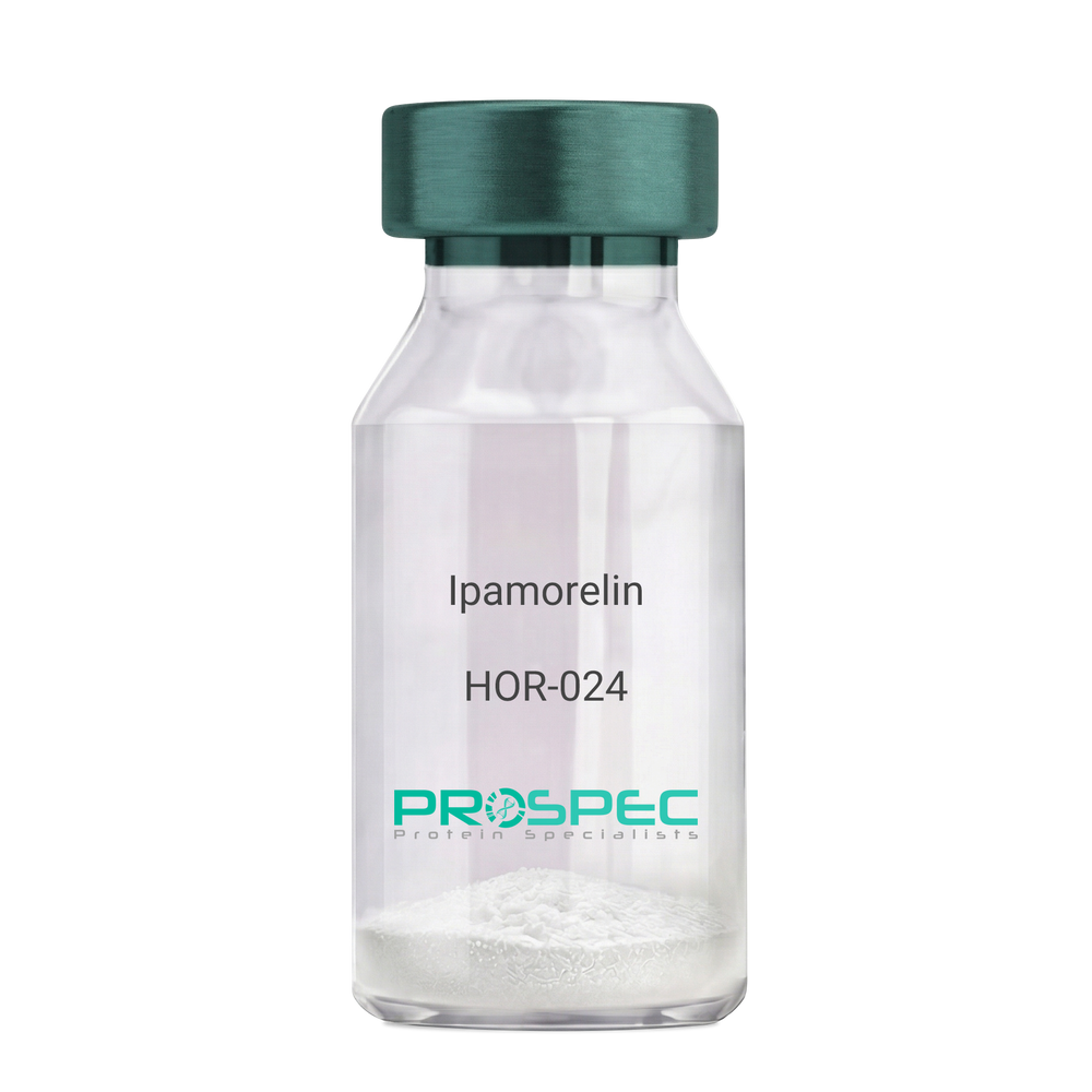
Name :
IpamorelinDescription:
Ipamorelin
Ipamorelin
Product # :
HOR-024Price :
Quantity :
Shipping Method :
Shipped at Room temp
More Info
- description
- formulation
- purity
- More Info
Description
Ipamorelin Synthetic is a single, non-glycosylated polypeptide chain containing 4 amino acids, having a molecular mass of 711.85 Dalton and a Molecular formula of C38H49N9O5.
Formulation
The protein was lyophilized with no additives.
Purity
Greater than 97.0% as determined by analysis by RP-HPLC.
More Info
-
Introduction
Ipamorelin is a peptide selective agonist of the ghrelin/growth hormone secretagogue receptor and a growth hormone secretagogue. Ipamorelin is a pentapeptide that was derived from GHRP1. Ipamorelin significantly increases plasma growth hormone levels in both animals and humans. Like pralmorelin and GHRP-6, ipamorelin does not affect prolactin, FSH, LH or TSH levels. However, unlike GHRP2 and GHRP6, but as growth hormone-releasing hormone (GHRH), ipamorelin does not stimulate the secretion of adrenocorticotropic hormone (ACTH) or cortisol, and is highly selective for inducing the secretion only of GH.
-
Synonyms
Ipamorelin
-
Physical Appearance
Sterile Filtered White lyophilized (freeze-dried) powder.
-
Stability
Lyophilized Ipamorelin although stable at room temperature for 3 weeks, should be stored desiccated below -18°C. Upon reconstitution Ipamorelin should be stored at 4°C between 2-7 days and for future use below -18°C. For long term storage it is recommended to add a carrier protein (0.1% HSA or BSA). Please prevent freeze-thaw cycles.
-
Solubility
It is recommended to reconstitute the lyophilized Ipamorelin in sterile 18MΩ-cm H2O not less than 100 µg/ml, which can then be further diluted to other aqueous solutions.
-
Amino Acid Sequence
Aib-His-D-2-Nal-D-Phe-Lys-NH2.
ProSpec's products are furnished for LABORATORY RESEARCH USE ONLY. The product may not be used as drugs, agricultural or pesticidal products, food additives or household chemicals.