Search results
1000 results found for “Ebola”
Name
Description
Product #
Price
Quantity
Shipping Method
- View Data Sheet
Name :
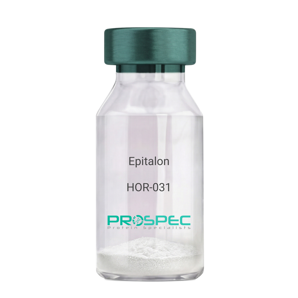
EpitalonDescription:
Epitalon
Product # :
HOR-031Price :
Quantity :
Shipping Method :
Shipped at Room temp
More Info
- description
- formulation
- purity
- More Info
- Citations
Description
Epithalon Synthetic is a single, non-glycosylated polypeptide chain containing 4 amino acids, having a molecular mass of 390.35 Dalton and a Molecular formula of C14H22N4O9.
Formulation
The protein was lyophilized with no additives.
Purity
Greater than 97.0% as determined by analysis by RP-HPLC.
Citations
More Info
-
Physical Appearance
Sterile Filtered White lyophilized (freeze-dried) powder.
-
Stability
Lyophilized Epithalon although stable at room temperature for 3 weeks, should be stored desiccated below -18°C. Upon reconstitution Epithalon should be stored at 4°C between 2-7 days and for future use below -18°C.For long term storage it is recommended to add a carrier protein (0.1% HSA or BSA). Please prevent freeze-thaw cycles.
-
Solubility
It is recommended to reconstitute the lyophilized Epithalon in sterile 18MΩ-cm H2O not less than 100 µg/ml, which can then be further diluted to other aqueous solutions.
-
Amino Acid Sequence
H-Ala-Glu-Asp-Gly-OH .
-
Background
Epitalon, also known as Epithalon, is a synthetic tetrapeptide that has been a focal point in the field of aging and longevity research. This peptide, consisting of four amino acids, is derived from the pineal gland and has been associated with a variety of biological effects, most notably its potential role in extending the lifespan of cells.
Epitalon's primary function is its interaction with telomeres, the protective caps at the ends of chromosomes that shorten as cells divide. By stimulating the production of telomerase, an enzyme that can repair and lengthen telomeres, Epitalon may slow down the aging process at a cellular level.
Numerous studies have explored the potential of Epitalon in extending the lifespan of organisms. For instance, Khavinson and colleagues (2003) found that Epitalon increased the lifespan of fruit flies, while Anisimov et al. (2003) reported similar results in mice. These findings suggest that Epitalon could potentially have similar effects in humans.
Epitalon has also been studied for its role in regulating circadian rhythms. Korkushko et al. (2011) found that Epitalon could restore disrupted circadian rhythms in elderly people, suggesting potential applications in sleep disorders and other conditions related to circadian rhythm disruption.
Beyond its potential role in aging and longevity, Epitalon has been explored for its potential therapeutic applications. Anisimov et al. (2011) found that Epitalon could reduce the incidence of spontaneous tumors in mice, suggesting potential applications in cancer prevention.
While research on Epitalon is promising, it is important to note that most studies have been conducted in animals or in vitro. More research is needed to fully understand the potential effects and applications of Epitalon in humans. However, the existing body of research suggests that Epitalon could be a promising tool in the fight against aging and age-related diseases.
ProSpec's products are furnished for LABORATORY RESEARCH USE ONLY. The product may not be used as drugs, agricultural or pesticidal products, food additives or household chemicals.
- View Data Sheet
Name :
Shiga Like Toxin 2Description:
Shiga Like Toxin-2 Subunit B Recombinant
Product # :
STX-002Price :
Quantity :
Shipping Method :
Shipped with Ice Packs
More Info
- description
- source
- formulation
- purity
- More Info
- Citations
Description
Recombinant Shiga Like Toxin-2 Subunit B from E.Coli O157:H7 produced in E.coli having an Mw of 22kDa.The Shiga Like Toxin 2 protein is fused to a 6xHis tag at its N-terminus and purified by proprietary chromatographic technique.
Source
Escherichia Coli.
Formulation
Shiga Like Toxin 2 protein solution contains PBS, 50mM arginine and 0.05% sodium azide.
Purity
Protein is >95% pure as determined by 10% PAGE (coomassie staining).
Citations
More Info
-
Introduction
Shiga-like toxin (verotoxin) is a toxin produced by some strains of Escherichia coli. Shiga-like toxin is named for its similarity to the AB5-type Shiga toxin produced by the bacteria Shigella dysenteriae. There are two known types-SLT1 and SLT2. The Shiga-like toxin is linked with hemolytic-uremic syndrome. Shiga-like toxin requires highly specific receptors on the cells' surface in order to attach and enter the cell. Species such as cattle, swine, and deer which do not carry these receptors may harbor toxigenic bacteria without any ill effect, dropping them in their feces, from where they may be distributed to humans. Shiga Like Toxin-2 Subunit B has nontoxic action, it is the functional region which binds to the receptor. Upon binding to the host cell receptor, the Shiga Like Toxin-2 Subunit B forms a pentamer. The Shiga Like Toxin-2 Subunit B can be useful in vaccine study, antibody test and other functional research.
-
Stability
Store at 4°C if entire vial will be used within 2-4 weeks. Store, frozen at -20°C for longer periods of time. For long term storage it is recommended to add a carrier protein (0.1% HSA or BSA).Avoid multiple freeze-thaw cycles.
-
Purification Method
Purified by affinity chromatographic technique.
ProSpec's products are furnished for LABORATORY RESEARCH USE ONLY. The product may not be used as drugs, agricultural or pesticidal products, food additives or household chemicals.
- View Data Sheet
Name :
HB-EGF MouseDescription:
HB-EGF Mouse Recombinant
DTR, HEGFL, diphtheria toxin receptor, DTSF.
Product # :
CYT-068Price :
Quantity :
Shipping Method :
Shipped at Room temp
More Info
- description
- source
- formulation
- purity
- biological activity
- More Info
- sds-page
- Citations
Description
HB-EGF Mouse Recombinant produced in E.Coli is a single, non-glycosylated, polypeptide chain containing 86 amino acids (63-148 a.a.) and having a molecular mass of 9.8 kDa.The HB-EGF is purified by proprietary chromatographic techniques
Source
Escherichia Coli.
Formulation
The protein was filtered (0.2µm) and lyophilized from a concentrated solution containing 10mM PB and 500mM NaCl, pH7.4.
Purity
Greater than 97.0% as determined by:
(a) Analysis by RP-HPLC.
(b) Analysis by SDS-PAGE.Biological Activity
The ED50 was determined by a cell proliferation assay using balb/c 3T3 cells is < 1.0 ng/ml, corresponding to a specific activity of > 1.0×10⁶ units/mg.sds-page
Citations
More Info
-
Introduction
HB-EGF is an EGF related growth factor which signals via the EGF receptor, and stimulates the proliferation of SMC (smooth muscle cells), fibroblasts, epithelial cells and keratinocytes. HB-EGF is expressed in various cell types and tissues, including vascular endothelial cells and SMC, macrophages, skeletal muscle, keratinocytes and particular tumor cells. HB-EGF’s ability to explicitly bind sulfate proteoglycans is dissimilar from other EGF-like molecules, and might be related to the enhanced mitogenic activity, relative to EGF, that HB-EGF exerts on smooth muscle cells.
-
Synonyms
DTR, HEGFL, diphtheria toxin receptor, DTSF.
-
Physical Appearance
Sterile Filtered White lyophilized (freeze-dried) powder.
-
Stability
Lyophilized Mouse HB-EGF Recombinant although stable at room temperature for 3 weeks, should be stored desiccated below -18°C. Upon reconstitution HB-EGF should be stored at 4°C between 2-7 days and for future use below -18°C.For long term storage it is recommended to add a carrier protein (0.1% HSA or BSA).Please prevent freeze-thaw cycles.
-
Solubility
It is recommended to reconstitute the lyophilized Mouse HB-EGF in sterile 18M-cm H2O not less than 100 µg/ml, which can then be further diluted to other aqueous solutions.
-
Amino Acid Sequence
DLEGTDLNLF KVAFSSKPQG LATPSKERNG KKKKKGKGLG KKRDPCLRKY KDYCIHGECR YLQEFRTPSC KCLPGYHGHR CHGLTL.
-
Background
What is the molecular weight/Mw of HB-EGF Protein?
HB-EGF Protein has a total Mw of 9.8kDa.
What is the source or expression system of HB-EGF Protein?
Escherichia Coli.
What is the Purity of HB-EGF Protein?
HB-EGF Protein is >97% pure as determined by SDS-PAGE.
What is the Biological Activity of HB-EGF Protein?
The ED50 was determined by a cell proliferation assay using balb/c 3T3 cells is < 1.0 ng/ml, corresponding to a specific activity of > 1.0×10⁶ units/mg.
What is the amino acid sequence of HB-EGF Protein?
DLEGTDLNLF KVAFSSKPQG LATPSKERNG KKKKKGKGLG KKRDPCLRKY KDYCIHGECR YLQEFRTPSC KCLPGYHGHR CHGLTL.
What applications can HB-EGF Protein be used in?
HB-EGF Protein can probably be used in western blot, ELISA and Lateral Flow.
What is the endotoxin level for HB-EGF Protein?
The endotoxin level is minimal, HB-EGF Protein was purified using conventional chromatography techniques.
ProSpec's products are furnished for LABORATORY RESEARCH USE ONLY. The product may not be used as drugs, agricultural or pesticidal products, food additives or household chemicals.
- View Data Sheet
Name :
DBI MouseDescription:
Diazepam Binding Inhibitor Mouse Recombinant
Acyl-CoA-binding protein, ACBP, Diazepam-binding inhibitor, DBI, Endozepine, EP, Dbi.
Product # :
PRO-2527Price :
Quantity :
Shipping Method :
Shipped with Ice Packs
More Info
- description
- source
- formulation
- purity
- More Info
- Citations
Description
DBI Mouse Recombinant produced in E.Coli is a single, non-glycosylated polypeptide chain containing 110 amino acids (1-87 a.a) and having a molecular mass of 12.4kDa. DBI is fused to a 23 amino acid His-tag at N-terminus & purified by proprietary chromatographic techniques.
Source
Escherichia Coli.
Formulation
DBI protein solution (1mg/ml) contains Phosphate buffer saline (pH7.4).
Purity
Greater than 95.0% as determined by SDS-PAGE.
Citations
More Info
-
Introduction
DBI, also known as Acyl-CoA-binding protein isoform 2, is a diazepam binding inhibitor, which is regulated by hormones. DBI participates in lipid metabolism and in the displacement of beta-carbolines & benzodiazepines, which modulate signal transduction at type A gamma-aminobutyric acid receptors located in brain synapses. Moreover, during adipocyte differentiation the expression of DBI is significantly induced. DBI preforms as an acyl-CoA pool former and regulates LCFA (long-chain fatty acids) metabolism in peripheral tissues.
-
Synonyms
Acyl-CoA-binding protein, ACBP, Diazepam-binding inhibitor, DBI, Endozepine, EP, Dbi.
-
Physical Appearance
Sterile Filtered clear solution.
-
Stability
Store at 4°C if entire vial will be used within 2-4 weeks. Store, frozen at -20°C for longer periods of time. For long term storage it is recommended to add a carrier protein (0.1% HSA or BSA).Avoid multiple freeze-thaw cycles.
-
Amino Acid Sequence
MGSSHHHHHH SSGLVPRGSH MGSMSQAEFD KAAEEVKRLK TQPTDEEMLF IYSHFKQATV GDVNTDRPGL LDLKGKAKWD SWNKLKGTSK ESAMKTYVEK VDELKKKYGI
ProSpec's products are furnished for LABORATORY RESEARCH USE ONLY. The product may not be used as drugs, agricultural or pesticidal products, food additives or household chemicals.
- View Data Sheet
Name :
SMAC/DIABLO HumanDescription:
SMAC/DIABLO Human Recombinant
Diablo homolog mitochondrial, Second mitochondria-derived activator of caspase, Smac protein, Direct IAP-binding protein with low pI, DIABLO, SMAC, SMAC3, DIABLO-S, FLJ10537, FLJ25049.
Product # :
PRO-614Price :
Quantity :
Shipping Method :
Shipped with Ice Packs
More Info
- description
- source
- formulation
- purity
- More Info
- Citations
Description
Smac/Diablo Human Recombinant fused to N-terminal T7-Tag produced in E.Coli is a single, non-glycosylated polypeptide chain containing 199 amino acids and having a molecular mass of 22 kDa.
Source
Escherichia Coli.
Formulation
The Smac/Diablo solution contains 20mM Tris pH-7.5.
Purity
Greater than 95.0% as determined by:
(a) Analysis by RP-HPLC.
(b) Analysis by SDS-PAGE.Citations
More Info
-
Introduction
Smac/Diablo is a proapoptotic protein that increases caspase activation in the cytochrome c/Apaf-1/caspase-9 pathway by its binding to the inhibitor of apoptosis proteins (IAPs) and removing their inhibitory activity. Smac/Diablo is a mitochondrial protein which enters the cytosol when cells go through apoptosis, and it moderates the caspase inhibition of IAPs.
Smac/DIABLO expression is associated with the result of renal cell carcinoma.
Dimeric form of Smac/DIABLO implies that once expressed in the cell the protein has a little probability of dissociation and, thus, loss of function.
Survivin, Smac/DIABLO, & PKC-? play an important part in the inhibition of apoptosis by FGF-2 in human small cell lung cancer cells. Mitochondrial survivin associates with Smac/DIABLO, delaying its release. Decreased expression of Smac protein takes part in ovarian carcinogenesis and chemotherapeutic resistance. Smac/DIABLO plays a role in tumor cells during the pathway of apoptosis induction. SMAC protein is regulated by XIAP and degraded by proteasome. SMAC protein takes part inleukemic cell apoptosis.
Smac is released during stress-induced apoptosis in multiple myeloma cells. -
Synonyms
Diablo homolog mitochondrial, Second mitochondria-derived activator of caspase, Smac protein, Direct IAP-binding protein with low pI, DIABLO, SMAC, SMAC3, DIABLO-S, FLJ10537, FLJ25049.
-
Physical Appearance
Sterile Filtered colorless solution.
-
Stability
Store at 4°C if entire vial will be used within 2-4 weeks. Store, frozen at -20°C for longer periods of time. For long term storage it is recommended to add a carrier protein (0.1% HSA or BSA).Avoid multiple freeze-thaw cycles.
-
Amino Acid Sequence
MASMTGGQQM GRGSMAVPIA QKSEPHSLSS EALMRRAVSL VTDSTSTFLS QTTYALIEAI TEYTKAVYTL TSLYRQYTSL LGKMNSEEED EVWQVIIGAR AEMTSKHQEY LKLETTWMTA VGLSEMAAEA AYQTGADQAS ITARNHIQLV KLQVEEVHQL SRKAETKLAE AQIEELRQKT QEEGEERAES EQEAYLRED.
ProSpec's products are furnished for LABORATORY RESEARCH USE ONLY. The product may not be used as drugs, agricultural or pesticidal products, food additives or household chemicals.
- View Data Sheet
Name :
IFN tau OvineDescription:
IFN-Tau Ovine Recombinant
IFN-tau1, Trophoblast protein 1, TP-1, Trophoblastin, Antiluteolysin, Trophoblast antiluteolytic protein, IFN-tau, IFN tau-1.
Product # :
CYT-377Price :
Quantity :
Shipping Method :
Shipped at Room temp
More Info
- description
- source
- formulation
- purity
- biological activity
- More Info
- Citations
Description
IFN-Tau Ovine Recombinant produced in E.Coli is a single, non-glycosylated, polypeptide chain containing 172 amino acids and having a molecular mass of 19914.7 Dalton.The IFN-Tau is purified by proprietary chromatographic techniques.
Source
Escherichia Coli.
Formulation
Lyophilized from (1mg/ml) solution containing PBS pH-7.4.
Purity
Greater than 95.0% as determined by both:
(a) Analysis by RP-HPLC.
(b) Analysis by SDS-PAGE.Biological Activity
The specific activity as determined in a viral resistance assay using bovine kidney MDBK cells was found to be 10,000,000IU/mg.Citations
More Info
-
Introduction
IFN-tau is also known as TP-1 (trophoblast protein-1) is a new class of type I IFN that is secreted by the trophoblast and is the signal for maternal recognition of pregnancy in sheep. IFN- tau has potent immunosuppressive and antiviral activities similar to other type I IFN but is less cytotoxic than IFN-alpha and IFN-beta. The current investigation concerns the effect of recombinant ovine IFN- tau (rOIFN- tau) on the modulation of MHC class I and II expression on cloned mouse cerebrovascular endothelial (CVE) cells.
IFN-tau induced tyrosine phosphorylation of Stat1 and upregulated the expression of MHC class I on CVE. One proposed action by which type I IFN reduces the relapse rate in MS is via interference with IFN-?-induced MHC class II expression. IFN- tau was shown to downregulate IFN-?-induced MHC class II expression on CVE and, hence, may be of potential therapeutic value in downregulating inflammation in the central nervous system (CNS). IFN- tau did not upregulate the expression of MHC class II on CVE. IFN- tau also inhibited the replication of Theiler's virus in CVE. -
Synonyms
IFN-tau1, Trophoblast protein 1, TP-1, Trophoblastin, Antiluteolysin, Trophoblast antiluteolytic protein, IFN-tau, IFN tau-1.
-
Physical Appearance
Sterile Filtered White lyophilized (freeze-dried) powder.
-
Stability
Lyophilized IFN-Tau although stable at room temperature for 3 weeks, should be stored desiccated below -18°C. Upon reconstitution IFN-Tau should be stored at 4°C between 2-7 days and for future use below -18°C.For long term storage it is recommended to add a carrier protein (0.1% HSA or BSA).Please prevent freeze-thaw cycles.
-
Solubility
It is recommended to reconstitute the lyophilized IFN Tau in sterile 18MΩ-cm H2O not less than 100µg/ml, which can then be further diluted to other aqueous solutions.
-
Amino Acid Sequence
The sequence of the first five N-terminal amino acids was determined and was found to be Cys-Tyr-Leu-Ser-Arg.
-
Background
What is the molecular weight/Mw of IFN TAU OVINE Protein?
IFN TAU OVINE Protein has a total Mw of 19.9kDa.
What is the source or expression system of IFN TAU OVINE Protein?
Escherichia Coli.
What is the Purity of IFN TAU OVINE Protein?
IFN TAU OVINE Protein is >95% pure as determined by SDS-PAGE.
What is the Biological Activity of IFN TAU OVINE Protein?
The specific activity as determined in a viral resistance assay using bovine kidney MDBK cells was found to be 10,000,000IU/mg.
What is the amino acid sequence of IFN TAU OVINE Protein?
The sequence of the first five N-terminal amino acids was determined and was found to be Cys-Tyr-Leu-Ser-Arg.
What applications can IFN TAU OVINE Protein be used in?
IFN TAU OVINE Protein can probably be used in western blot, ELISA and Lateral Flow.
What is the endotoxin level for IFN TAU OVINE Protein?
The endotoxin level is minimal, IFN TAU OVINE Protein was purified using conventional chromatography techniques.
ProSpec's products are furnished for LABORATORY RESEARCH USE ONLY. The product may not be used as drugs, agricultural or pesticidal products, food additives or household chemicals.
- View Data Sheet
Name :
IL1RA PorcineDescription:
Interleukin-1 Receptor Antagonist Porcine Recombinant
IRAP, IL1F3, IL1RA, IL-1ra3, ICIL-1RA, IL1RN, IL1 inhibitor, IL-1ra, MGC10430.
Product # :
CYT-376Price :
Quantity :
Shipping Method :
Shipped at Room temp
More Info
- description
- source
- formulation
- purity
- biological activity
- More Info
- Citations
Description
Recombinant IL 1RA Porcine produced in E.coli cells is a non-glycosylated, homodimeric protein containing 152 amino acid chain and having a molecular mass of 17.1kDa. The IL 1RA is purified by proprietary chromatographic techniques.
Source
Escherichia Coli.
Formulation
The IL 1RA was lyophilized from a 0.2µm filtered concentrated solution in 1×PBS, pH 7.4, containing 1mM DTT.
Purity
Greater than 97.0% as determined by:
(a) Analysis by RP-HPLC.
(b) Analysis by SDS-PAGE.Biological Activity
The ED50 as determined by inhibiting IL-1a-dependent proliferation of murine D10S cells is less than 50 ng/ml, corresponding to a specific activity of > 2.0 × 10⁴ IU/mg in the presence of 75 pg/ml rPoIL-1a.
Citations
More Info
-
Introduction
Interleukin-1 ra is a member of the interleukin 1 cytokine family. This protein inhibits the activities of interleukin 1, alpha (IL1A) and interleukin 1, beta (IL1B), and modulates a variety of interleukin 1 related immune and inflammatory responses. This gene and five other closely related cytokine genes form a gene cluster spanning approximately 400 kb on chromosome 2. A polymorphism of this gene is reported to be associated with increased risk of osteoporotic fractures and gastric cancer. Four alternatively spliced transcript variants encoding distinct isoforms have been reported.
-
Synonyms
IRAP, IL1F3, IL1RA, IL-1ra3, ICIL-1RA, IL1RN, IL1 inhibitor, IL-1ra, MGC10430.
-
Physical Appearance
Sterile Filtered White lyophilized (freeze-dried) powder.
-
Stability
Lyophilized IL 1RA although stable at room temperature for 3 weeks, should be stored desiccated below -18°C. Upon reconstitution IL 1RA should be stored at 4°C between 2-7 days and for future use below -18°C.For long term storage it is recommended to add a carrier protein (0.1% HSA or BSA).Please prevent freeze-thaw cycles.
-
Solubility
It is recommended to reconstitute the lyophilized IL 1RA in sterile distilled H2O not less than 100µg/ml, which can then be further diluted to other aqueous solutions.
-
Amino Acid Sequence
HPLGKRPCRM QAFRIWDVNQ KTFYLRNNQL VAGYLQGPNT KLEEKIDVVP VEPHFVFLGI HGGKLCLSCV KSGDEMKLQL DAVNITDLRK NSEQDKRFTF IRSDSGPTTS FESAACPGWF LCTALEADQP VGLTNTPKAA VKVTKFYFQQ DQ
-
Background
What is the molecular weight/Mw of IL1RA PORCINE Protein?
IL1RA PORCINE Protein has a total Mw of 17.1kDa.
What is the source or expression system of IL1RA PORCINE Protein?
Escherichia Coli.
What is the Purity of IL1RA PORCINE Protein?
IL1RA PORCINE Protein is >97% pure as determined by SDS-PAGE.
What is the Biological Activity of IL1RA PORCINE Protein?
The ED50 as determined by inhibiting IL-1a-dependent proliferation of murine D10S cells is less than 50 ng/ml, corresponding to a specific activity of > 2.0 × 10⁴ IU/mg in the presence of 75 pg/ml rPoIL-1a.
What is the amino acid sequence of IL1RA PORCINE Protein?
HPLGKRPCRM QAFRIWDVNQ KTFYLRNNQL VAGYLQGPNT KLEEKIDVVP VEPHFVFLGI HGGKLCLSCV KSGDEMKLQL DAVNITDLRK NSEQDKRFTF IRSDSGPTTS FESAACPGWF LCTALEADQP VGLTNTPKAA VKVTKFYFQQ DQ.
What applications can IL1RA PORCINE Protein be used in?
IL1RA PORCINE Protein can probably be used in western blot, ELISA and Lateral Flow.
What is the endotoxin level for IL1RA PORCINE Protein?
The endotoxin level is minimal, IL1RA PORCINE Protein was purified using conventional chromatography techniques.
Who supplies recombinant IL1RA PORCINE that is in stock and available to order?
ProSpec has the protein in stock and available for immediate delivery.
Where can I order bulk mg quantities of IL1RA PORCINE?
ProSpec supplies bulk mg quantities at affordable prices that meets research and academic budgets.
ProSpec's products are furnished for LABORATORY RESEARCH USE ONLY. The product may not be used as drugs, agricultural or pesticidal products, food additives or household chemicals.
- View Data Sheet
Name :
Influenza-B TokioDescription:
Influenza-B Virus Tokio/53/99
Product # :
IHA-017Price :
Quantity :
Shipping Method :
Shipped with Ice Packs
More Info
- description
- formulation
- purity
- More Info
- Citations
Description
Allantoic fluid of 10 days old embryonated eggs, inoculated with influenza B virus, strain B/Tokio/53/99. The Influenza B Virus was purified by Ultracentrifugation with 10-40 % sucrose gradient.
Formulation
The B/Tokio/53/99solution (1.7mg/ml) contains STE, 0.1% sodium azide (NaN3) and 0.005% thimerosal.
Purity
Greater than 90.0% as determined byAnalysis by SDS-PAGE.
Citations
More Info
-
Introduction
Influenza-B virus is a genus in the virusfamily Orthomyxoviridae. The only species in this genus is called "Influenza B virus". Influenza B virus only infects humansand seals. This limited host range is apparently in contrast with those caused by the similar Influenza virus Aas both mutate by both genetic drift and reassortment. Influenza-B virus evolves slower than A viruses and faster than C viruses. Influenza-B virus mutates at a rate 2-3 times lower than type A. However, influenza B mutates enough that lasting immunity is not possible. The Influenza B virus capsidis enveloped while its virionconsists of a matrix protein + envelope + nucleoprotein complex + nucleocapsid, and a polymerasecomplex. Influenza B is sometimes spherical and sometimes filamentous. Its 500 or so surface projections are made of hemagglutinin and neuraminidase.
The Influenza B virus is 14648 nucleotideslong and consists of eight segments of linear negative-sense, single-stranded RNA. The multipartite genome is encapsidated, each segment in a separate nucleocapsid, and the nucleocapsids are surrounded by one envelope. -
Physical Appearance
Sterile Filtered colorless solution.
-
Stability
B/Tokio/53/99 although stable 4°C for 4 weeks, should be stored below -18°C. Please prevent freeze-thaw cycles.
-
Inactivation
Thimerosal and beta propiolactone treatmentThis product has been treated in a manner consistent with methods of inactivation. Generally accepted good laboratory practices appropriate to microbiological/viral safe handling practices and techniques are required at work.
-
Immunological Activity
Serological studies of influenza B virus, immunogen for antibody production.Tested with anti-influenza B monoclonal antibodies in ELISA.
ProSpec's products are furnished for LABORATORY RESEARCH USE ONLY. The product may not be used as drugs, agricultural or pesticidal products, food additives or household chemicals.
- View Data Sheet
Name :
CD100 Human HEKDescription:
CD100 Human Recombinant HEK
Thesemaphorin family containing 1 Ig-like C2-type domain, 1 PSI domain and 1 Sema domai,Semaphorin-4D, A8, BB18, GR3, SEMA4D, C9orf164, CD100, SEMAJ.
Product # :
PRO-1640Price :
Quantity :
Shipping Method :
Shipped at Room temp
More Info
- description
- source
- formulation
- purity
- More Info
- Citations
Description
CD100 Human Recombinant produced by mammalian expression system in human cells is a single polypeptide chain containing 721 amino acids (22-734). CD100 is fused to an 8 amino acid His-tag at C-terminus & purified by proprietary chromatographic techniques.
Source
HEK293 cells.
Formulation
CD100 was lyophilized from a 0.2 µM filtered solution of 20mM PB and 150mM NaCl, PH 7.4.
Purity
Greater than 95% as determined by SDS-PAGE.
Citations
More Info
-
Introduction
Semaphorin-4D (CD100) is a member of the semaphorin family containing 1 Ig-like C2-type domain, 1 PSI domain and 1 Sema domain. CD100 is the cell surface receptor forPLXN1B and PLXNB2 which plays a central role in cell-cell signaling. CD100 stimulates the migration of cerebellar granule cells and of endothelial cells, regulates dendrite and axon branching and morphogenesis. In addition, CD100 has a role in the immune system; it promotes signaling via SRC and PTK2B/PYK2, which subsequently mediates activation of phosphatidylinositol 3-kinase and of the AKT1 signaling cascade.
-
Synonyms
Thesemaphorin family containing 1 Ig-like C2-type domain, 1 PSI domain and 1 Sema domai,Semaphorin-4D, A8, BB18, GR3, SEMA4D, C9orf164, CD100, SEMAJ.
-
Physical Appearance
Sterile Filtered White lyophilized (freeze-dried) powder.
-
Stability
Lyophilized CD100 although stable at room temperature for 3 weeks, should be stored desiccated below -18°C. Upon reconstitution CD100 should be stored at 4°C between 2-7 days and for future use below -18°C. Please prevent freeze-thaw cycles.
-
Solubility
It is recommended to reconstitute the lyophilized CCD100 in 1xPBS to a concentration no less than 100 µg/ml, which can then be further diluted to other aqueous solutions.
-
Amino Acid Sequence
MAFAPIPRITWEHREVHLVQFHEPDIYNYSALLLSEDKDTLYIGAREAVFAVNALNISEKQHE
VYWKVSEDKKAKCAEKGKSKQTECLNYIRVLQPLSATSLYVCGTNAFQPACDHLNLTSFKFLG
KNEDGKGRCPFDPAHSYTSVMVDGELYSGTSYNFLGSEPIISRNSSHSPLRTEYAIPWLNEPSF
VFADVIRKSPDSPDGEDDRVYFFFTEVSVEYEFVFRVLIPRIARVCKGDQGGLRTLQKKWTSFL
KARLICSRPDSGLVFNVLRDVFVLRSPGLKVPVFYALFTPQLNNVGLSAVCAYNLSTAEEVFSH
GKYMQSTTVEQSHTKWVRYNGPVPKPRPGACIDSEARAANYTSSLNLPDKTLQFVKDHPLMDDSV
TPIDNRPRLIKKDVNYTQIVVDRTQALDGTVYDVMFVSTDRGALHKAISLEHAVHIIEETQLFQD
FEPVQTLLLSSKKGNRFVYAGSNSGVVQAPLAFCGKHGTCEDCVLARDPYCAWSPPTATCVALHQ
TESPSRGLIQEMSGDASVCPDKSKGSYRQHFFKHGGTAELKCSQKSNSEKTMYLKSSDNRVDHHHHHH.
ProSpec's products are furnished for LABORATORY RESEARCH USE ONLY. The product may not be used as drugs, agricultural or pesticidal products, food additives or household chemicals.
- View Data Sheet
Name :
UBE2D2 Human, HisDescription:
Ubiquitin Conjugating Enzyme E2D2 Human Recombinant, His Tag
ubiquitin-conjugating enzyme E2D 2 (homologous to yeast UBC4/5), UBC4, Ubiquitin carrier protein D2, Ubiquitin-conjugating enzyme E2-17 kDa 2, Ubiquitin-protein ligase D2, UBCH5B, EC 6.3.2.19, E2(17)KB2, PUBC1, UBC4/5, UBC5B, UBCH4.
Product # :
ENZ-154Price :
Quantity :
Shipping Method :
Shipped with Ice Packs
More Info
- description
- source
- formulation
- purity
- More Info
- Citations
Description
UBE2D2 Human Recombinant produced in E.coli is a single, non-glycosylated polypeptide chain containing 167 amino acids (1-147) and having a molecular mass of 18.9 kDa.The UBE2D2 is fused to a 20 amino acid His-Tag at N-terminus and purified by proprietary chromatographic techniques.
Source
E.coli.
Formulation
The UBE2D2 solution (0.5mg/ml) contains 20mM Tris-HCl buffer (pH 8.0), 1mM DTT, 0.1M NaCl and 10% glycerol.
Purity
Greater than 95% as determined by SDS-PAGE.
Citations
More Info
-
Introduction
UBE2D2 belongs to the E2 ubiquitin-conjugating enzyme family. UBE2D2 takes part in the ubiquitination of the tumor-suppressor protein p53, which is induced by an E3 ubiquitin-protein ligase. UBE2D2 catalyzes ubiquitination of IkB-alpha in a SCFB-TRCP and phosphorylation dependent method.
-
Synonyms
ubiquitin-conjugating enzyme E2D 2 (homologous to yeast UBC4/5), UBC4, Ubiquitin carrier protein D2, Ubiquitin-conjugating enzyme E2-17 kDa 2, Ubiquitin-protein ligase D2, UBCH5B, EC 6.3.2.19, E2(17)KB2, PUBC1, UBC4/5, UBC5B, UBCH4.
-
Physical Appearance
Sterile Filtered colorless solution.
-
Stability
Store at 4°C if entire vial will be used within 2-4 weeks. Store, frozen at -20°C for longer periods of time. For long term storage it is recommended to add a carrier protein (0.1% HSA or BSA).Avoid multiple freeze-thaw cycles.
-
Amino Acid Sequence
MGSSHHHHHH SSGLVPRGSH MALKRIHKEL NDLARDPPAQ CSAGPVGDDM FHWQATIMGP NDSPYQGGVF FLTIHFPTDY PFKPPKVAFT TRIYHPNINS NGSICLDILR SQWSPALTIS KVLLSICSLL CDPNPDDPLV PEIARIYKTD REKYNRIARE WTQKYAM.
ProSpec's products are furnished for LABORATORY RESEARCH USE ONLY. The product may not be used as drugs, agricultural or pesticidal products, food additives or household chemicals.
- View Data Sheet
Name :
H3N2 Canine, MutantDescription:
Hemagglutinin-Influenza A Virus H3N2 Canine Recombinant, Mutant
Product # :
IHA-034Price :
Quantity :
Shipping Method :
Shipped with Ice Packs
More Info
- description
- source
- formulation
- purity
- More Info
- Citations
Description
H3N2 Canine produced in E. coli. is a single non-glycosylated polypeptide chain containing 336 amino acids (18-344) and having a molecular mass of 36.9kDa.H3N2 Canine is fused to a 6 amino acid His-tag at C-terminus & purified by proprietary chromatographic techniques.
Source
E. coli.
Formulation
The H3N2 Canine solution (0.5mg/ml) contains 20mM Tris-HCl buffer (pH 8.0) and 10% glycerol.
Purity
Greater than 95% as determined by SDS-PAGE.
Citations
More Info
-
Introduction
H3N2 is a subtype of the influenza A virus. Its name derives from the forms of the two kinds of proteins on the surface of its coat, hemagglutinin (H) and neuraminidase (N). H3N2 exchanges genes for internal proteins with other influenza subtypes. H3N2 has tended to dominate in prevalence over H1N1, H1N2, and influenza B. H3N2 strain descended from H2N2 by antigenic shift, in which genes from multiple subtypes re-assorted to form a new virus. Both the H2N2 and H3N2 strains contained genes from avian influenza viruses.
H3N2 viruses are able to infect mammals and birds. In pigs, humans, and birds, the virus has mutated into many strains. Hemagglutinin(HA) binds to sialic acid-containing receptors on the cell surface, generating the attachment of the virus particle to the cell. HA has a vital part in the determination of host range restriction and virulence and is in charge of the diffusion of the virus into the cell cytoplasm by facilitating the fusion of the membrane of the endocytos -
Physical Appearance
Sterile Filtered colorless solution.
-
Stability
Store at 4°C if entire vial will be used within 2-4 weeks. Store, frozen at -20°C for longer periods of time. For long term storage it is recommended to add a carrier protein (0.1% HSA or BSA).Avoid multiple freeze-thaw cycles.
-
Amino Acid Sequence
ADNLPGNENN AATLCLGHHA VPNGTIVKTI TDDQIEVTNA TELVQNSSTG KICNNPHKIL DGRDCTLIDA LLGDPHCDVF QNETWDLFVE RSNAFSNCYP YDVPDYASLR SIVASSGTLE FITEGFTWAG VTQNGGSGAC KKGPANGFFS RLNWLTKSGN TYPVLNVTMP NNNNFDKLYI WGVHHPSTNQ EQTSLYIQAS GRVKVSTRRS QQTIIPNIGS RPLVRGQSGR ISVYWTIVKP GDVLVINSNG NLIAPRGYFK MRIGKSSIMR SDAPIDTCIS ECITPNGSIP NEKPFQNVNK ITYGACPKYV KQNTLKLATG MRNVPERQTH HHHHH
ProSpec's products are furnished for LABORATORY RESEARCH USE ONLY. The product may not be used as drugs, agricultural or pesticidal products, food additives or household chemicals.
- View Data Sheet
Name :
HIV-1 p24, HRPDescription:
HIV-1 p24 Recombinant, HRP Labeled
Product # :
HIV-109Price :
Quantity :
Shipping Method :
Shipped with Ice Packs
More Info
- description
- source
- formulation
- purity
- More Info
- Citations
Description
The E.coli derived HRP recombinant protein is a non-glycosylated polypeptide chain, containing the HIV-1 p24 immunodominant regions and fused to a GST tag at N-terminus.
Source
Escherichia Coli.
Formulation
1.5M urea, 25mM Tris-HCl pH-8.0 and 50% Glycerol.
Purity
Greater than 90.0% as determined by HPLC analysis and SDS-PAGE.
Citations
More Info
-
Introduction
Human immunodeficiency virus (HIV) is a retrovirusthat can lead to a condition in which the immune systembegins to fail, leading to opportunistic infections. HIV primarily infects vital cells in the humanimmune systemsuch as helper T cells(specifically CD4+ T cells), macrophagesand dendritic cells. HIV infection leads to low levels of CD4+ T cells through three main mechanisms: firstly, direct viral killing of infected cells; secondly, increased rates of apoptosisin infected cells; and thirdly, killing of infected CD4+ T cells by CD8 cytotoxic lymphocytesthat recognize infected cells. When CD4+ T cell numbers decline below a critical level, cell-mediated immunityis lost, and the body becomes progressively more susceptible to opportunistic infections. HIV was classified as a member of the genus Lentivirus, part of the family of Retroviridae. Lentiviruses have many common morphologies and biological properties. Many species are infected by lentiviruses, which are characteristically responsible for long-duration illnesses with a long incubation period. Lentiviruses are transmitted as single-stranded, positive-sense, enveloped RNA viruses. Upon entry of the target cell, the viral RNA genomeis converted to double-stranded DNAby a virally encoded reverse transcriptasethat is present in the virus particle. This viral DNA is then integrated into the cellular DNA by a virally encoded integraseso that the genome can be transcribed. Once the virus has infected the cell, two pathways are possible: either the virus becomes latentand the infected cell continues to function, or the virus becomes active and replicates, and a large number of virus particles are liberated that can then infect other cells.
-
Physical Appearance
Sterile filtered colorless clear solution.
-
Stability
HIV-1 p24 although stable at 4°C for 1 week, should be stored below -18°C. Please prevent freeze thaw cycles.
-
Applications
ELISA, Western blots, Flow-through.
ProSpec's products are furnished for LABORATORY RESEARCH USE ONLY. The product may not be used as drugs, agricultural or pesticidal products, food additives or household chemicals.
- View Data Sheet
Name :
C3 MouseDescription:
Complement C3 Mouse
Complement C3, C3 and PZP-like alpha-2-macroglobulin domain-containing protein 1, C3, CPAMD1.
Product # :
PRO-2705Price :
Quantity :
Shipping Method :
Shipped with Ice Packs
More Info
- description
- source
- formulation
- purity
- More Info
- Citations
Description
Mouse Complement C3 produced in Mouse plasma having a molecular weight of 185kDa.
Source
Mouse Plasma.
Formulation
C3 solution contains phosphate buffer saline.
Purity
Greater than 93.0% as determined by SDS-PAGE.
Citations
More Info
-
Introduction
C3 is central to the activation of all 3 pathways of complement activation. Initiation of each pathway generates proteolytic enzyme complexes which binds the target surface. These enzymes cleave a peptide bond in C3 releasing the anaphylatoxin C3a and activating C3b. Most of the C3 activated during complement activation never attaches to the surface due to its thioester reaction with water forming fluid phase C3b which is rapidly inactivated by factors H and I forming iC3b. Surface-bound C3b is necessary in all 3 pathways for efficient activation of C5 and formation of C5b-9 complexes that lyse the target cell membrane.
-
Synonyms
Complement C3, C3 and PZP-like alpha-2-macroglobulin domain-containing protein 1, C3, CPAMD1.
-
Physical Appearance
Sterile filtered solution.
-
Stability
C3 Mouse is stable at 4°C if entire vial will be used within 2-4 weeks.Store, frozen below -20°C for longer periods of time.For long term storage it is recommended to add a carrier protein (0.1% HSA or BSA).Avoid multiple freeze-thaw cycles.
ProSpec's products are furnished for LABORATORY RESEARCH USE ONLY. The product may not be used as drugs, agricultural or pesticidal products, food additives or household chemicals.
- View Data Sheet
Name :
C3b Inactivated MouseDescription:
Complement C3b Inactivated Mouse
Complement C3, C3 and PZP-like alpha-2-macroglobulin domain-containing protein 1, C3, CPAMD1.
Product # :
PRO-2813Price :
Quantity :
Shipping Method :
Shipped with Ice Packs
More Info
- description
- source
- formulation
- purity
- More Info
- Citations
Description
Mouse Complement C3b Inactivated produced in Mouse serum having a molecular weight of 175kDa.
Source
Mouse serum.
Formulation
C3b Mouse Inactivated solution contains PBS.
Purity
Greater than 95.0% as determined by SDS-PAGE.
Citations
More Info
-
Synonyms
Complement C3, C3 and PZP-like alpha-2-macroglobulin domain-containing protein 1, C3, CPAMD1.
-
Physical Appearance
Sterile Filtered solution.
-
Stability
C3b Mouse Inactivated is stable at 4°C if entire vial will be used within 2-4 weeks. Store, frozen below -20°C for longer periods of time. For long term storage it is recommended to add a carrier protein (0.1% HSA or BSA). Avoid multiple freeze-thaw cycles.
-
Background
Complement component 3b (C3b) is a key protein in the complement system, a crucial component of the innate immune response. C3b plays a pivotal role in opsonization, inflammation, and clearance of pathogens and damaged cells. In mouse models, research on C3b has provided valuable insights into its mechanism of action, activity, and implications in immune function. This paper aims to delve into the dynamics of mouse C3b, shedding light on its functional attributes and potential applications in immunological research. C3b Inactivated protein has lost its biological capabilities due to structural changes compared to C3b that has binding sites to factor P, factor H, factor B, factorC5, and factor-I and therefore it destroys these sites.
Significantly, C3b inactivated protein cannot bind factor B and is therefore it cannot participate in complement activation.
C3b inactivated protein still binds tC50 properdin and CR3 receptor.
ProSpec's products are furnished for LABORATORY RESEARCH USE ONLY. The product may not be used as drugs, agricultural or pesticidal products, food additives or household chemicals.
- View Data Sheet
Name :
HA Monoclonal AntibodyDescription:
HA Mouse Antibody
Product # :
ANT-225Price :
Quantity :
Shipping Method :
Shipped with Ice Packs
More Info
- description
- formulation
- More Info
- Citations
Description
Monoclonal antibodies are produced by immunizing mouse with synthetic peptide (YPYDVPDYA) coupled to KLH.
Formulation
1×PBS & 50% glycerol.
Citations
More Info
-
Physical Appearance
Sterile filtered colorless solution.
-
Ig Subclass
Mouse IgG1.
-
Clone
PHASHG.
-
Applications
Western Blot, Immunoprecipitation.
-
Titer
Western Blotting: 1µg/ml.
-
Type
Mouse Antibody Monoclonal.
-
Storage Procedures
For periods up to 1 month store at 4°C, for longer periods of time, store at -20°C. Prevent freeze thaw cycles.
ProSpec's products are furnished for LABORATORY RESEARCH USE ONLY. The product may not be used as drugs, agricultural or pesticidal products, food additives or household chemicals.
- View Data Sheet
Name :
EEF1D HumanDescription:
Eukaryotic Translation Elongation Factor 1 Delta Human Recombinant
Elongation factor 1-delta, EF-1-delta, Antigen NY-CO-4, EEF1D, EF1D, EF-1D, FP1047, FLJ20897.
Product # :
PRO-170Price :
Quantity :
Shipping Method :
Shipped with Ice Packs
More Info
- description
- source
- formulation
- purity
- More Info
- Citations
Description
EEF1D Human Recombinant fused with a 20 amino acid His tag at N-terminus produced in E.Coli is a single, non-glycosylated, polypeptide chain containing 301 amino acids (1-281 a.a.) and having a molecular mass of 33.2kDa (Molecular weight on SDS-PAGE will appear higher). The EEF1D is purified by proprietary chromatographic techniques.
Source
Escherichia Coli.
Formulation
The EEF1D solution (0.5 mg/ml) contains 20mM Tris-HCl buffer (pH 8.0), 1mM DTT and 10% glycerol.
Purity
Greater than 90.0% as determined by SDS-PAGE.
Citations
More Info
-
Introduction
Eukaryotic translation elongation factor 1 delta (EEF1D) is a subunit of the elongation factor-1 complex that is responsible for the enzymatic transport of aminoacyl tRNAs to the ribosome. EEF1D functions as guanine nucleotide exchange factor. EEF1D interacts with HIV-1 Tat, and consequently represses the translation of host-cell, but not HIV-1, mRNAs. EEF1D overexpression is linked to oesophageal carcinoma and may negatively influence the outcome of medulloblastomas.
-
Synonyms
Elongation factor 1-delta, EF-1-delta, Antigen NY-CO-4, EEF1D, EF1D, EF-1D, FP1047, FLJ20897.
-
Physical Appearance
Sterile Filtered colorless solution.
-
Stability
Store at 4°C if entire vial will be used within 2-4 weeks. Store, frozen at -20°C for longer periods of time. For long term storage it is recommended to add a carrier protein (0.1% HSA or BSA).Avoid multiple freeze-thaw cycles.
-
Amino Acid Sequence
MGSSHHHHHH SSGLVPRGSH MATNFLAHEK IWFDKFKYDD AERRFYEQMN GPVAGASRQE NGASVILRDI ARARENIQKS LAGSSGPGAS SGTSGDHGEL VVRIASLEVE NQSLRGVVQE LQQAISKLEA RLNVLEKSSP GHRATAPQTQ HVSPMRQVEP PAKKPATPAE DDEDDDIDLF GSDNEEEDKE AAQLREERLR QYAEKKAKKP ALVAKSSILL DVKPWDDETD MAQLEACVRS IQLDGLVWGA SKLVPVGYGI RKLQIQCVVE DDKVGTDLLE EEITKFEEHV QSVDIAAFNK I.
ProSpec's products are furnished for LABORATORY RESEARCH USE ONLY. The product may not be used as drugs, agricultural or pesticidal products, food additives or household chemicals.
- View Data Sheet
Name :
PDCD1 HumanDescription:
Programmed Cell Death 1 Human Recombinant
Programmed Cell Death 1, Systemic Lupus Erythematosus Susceptibility 2, Protein PD-1, HPD-1, PD1, Programmed Cell Death 1 Protein, Programmed Cell Death Protein 1, CD279 Antigen, CD279, HPD-L, HSLE1, SLEB2, PD-1.
Product # :
PRO-2442Price :
Quantity :
Shipping Method :
Shipped with Ice Packs
More Info
- description
- source
- formulation
- purity
- More Info
- Citations
Description
PDCD1 Human Recombinant produced in Sf9 Baculovirus cells is a single, glycosylated polypeptide chain containing 392 amino acids (21-170a.a.) and having a molecular mass of 44.0kDa (Molecular size on SDS-PAGE will appear at approximately 40-57kDa).PDCD1 is expressed with a 239 amino acids hIgG-His tag at C-Terminus and purified by proprietary chromatographic techniques.
Source
Sf9, Baculovirus cells.
Formulation
PDCD1 protein solution (0.5mg/ml) contains Phosphate Buffered Saline (pH 7.4) and 10% glycerol.
Purity
Greater than 90.0% as determined by SDS-PAGE.
Citations
More Info
-
Introduction
PDCD1 is a cell surface membrane protein of the immunoglobulin superfamily. PDCD1 is expressed in pro-B-cells and is thought to have a role in their differentiation. In mice, expression of PDCD1 gene is induced in the thymus when anti-CD3 antibodies are injected and large numbers of thymocytes undergo apoptosis. Mice deficient for the PDCD1 gene bred on a BALB/c background developed dilated cardiomyopathy and died from congestive heart failure. These studies suggest that the PDCD1 gene product may also be important in T cell function and contribute to the prevention of autoimmune diseases.
-
Synonyms
Programmed Cell Death 1, Systemic Lupus Erythematosus Susceptibility 2, Protein PD-1, HPD-1, PD1, Programmed Cell Death 1 Protein, Programmed Cell Death Protein 1, CD279 Antigen, CD279, HPD-L, HSLE1, SLEB2, PD-1.
-
Physical Appearance
Sterile Filtered colorless solution.
-
Stability
Store at 4°C if entire vial will be used within 2-4 weeks. Store, frozen at -20°C for longer periods of time. For long term storage it is recommended to add a carrier protein (0.1% HSA or BSA). Avoid multiple freeze-thaw cycles.
-
Amino Acid Sequence
ADPPGWFLDS PDRPWNPPTF SPALLVVTEG DNATFTCSFS NTSESFVLNW YRMSPSNQTD KLAAFPEDRS QPGQDCRFRV TQLPNGRDFH MSVVRARRND SGTYLCGAIS LAPKAQIKES LRAELRVTER RAEVPTAHPS PSPRPAGQFQ TLVLEPKSCD KTHTCPPCPA PELLGGPSVF LFPPKPKDTL MISRTPEVTC VVVDVSHEDP EVKFNWYVDG VEVHNAKTKP REEQYNSTYR VVSVLTVLHQ DWLNGKEYKC KVSNKALPAP IEKTISKAKG QPREPQVYTL PPSRDELTKN QVSLTCLVKG FYPSDIAVEW ESNGQPENNY KTTPPVLDSD GSFFLYSKLT VDKSRWQQGN VFSCSVMHEA LHNHYTQKSL SLSPGKHHHH HH.
ProSpec's products are furnished for LABORATORY RESEARCH USE ONLY. The product may not be used as drugs, agricultural or pesticidal products, food additives or household chemicals.
- View Data Sheet
Name :
CETN1 HumanDescription:
Centrin-1 Human Recombinant
Centrin EF-hand protein 1, calcium binding protein, Caltractin isoform 2, CETN.
Product # :
PRO-1104Price :
Quantity :
Shipping Method :
Shipped with Ice Packs
More Info
- description
- source
- formulation
- purity
- More Info
- Citations
Description
CETN1 Human Recombinant produced E. coli is a single polypeptide chain containing 196 amino acids (1-172) and having a molecular mass of 22.1kDa.CETN1 is fused to a 24 amino acid His-tag at N-terminus & purified by proprietary chromatographic techniques.
Source
E.coli.
Formulation
The CETN1 solution (0.5mg/1ml) contains 20mM Tris-HCl buffer (pH 8.0), 0.1M NaCl, 1mM DTT and 10% glycerol.
Purity
Greater than 90% as determined by SDS-PAGE.
Citations
More Info
-
Introduction
CETN1 is an EF-hand type Ca2+-binding protein. CETN1 is localized to the centrosome of interphase cells, and reorganizes the region of the spindle poles during mitosis, reflecting the dynamic behavior of the centrosome during the cell cycle. CETN1 has a vital part in the determination of centrosome position and isolation, and in the course of microtubule separating.
-
Synonyms
Centrin EF-hand protein 1, calcium binding protein, Caltractin isoform 2, CETN.
-
Physical Appearance
Sterile Filtered colorless solution.
-
Stability
Store at 4°C if entire vial will be used within 2-4 weeks. Store, frozen at -20°C for longer periods of time. For long term storage it is recommended to add a carrier protein (0.1% HSA or BSA).Avoid multiple freeze-thaw cycles.
-
Amino Acid Sequence
MGSSHHHHHH SSGLVPRGSH MGSHMASGFK KPSAASTGQK RKVAPKPELT EDQKQEVREA FDLFDVDGSG TIDAKELKVA MRALGFEPRK EEMKKMISEV DREGTGKISF NDFLAVMTQK MSEKDTKEEI LKAFRLFDDD ETGKISFKNL KRVANELGEN LTDEELQEMI DEADRDGDGE VNEEEFLRIM KKTSLY
ProSpec's products are furnished for LABORATORY RESEARCH USE ONLY. The product may not be used as drugs, agricultural or pesticidal products, food additives or household chemicals.
- View Data Sheet
Name :
GYPA HumanDescription:
Glycophorin A Human Recombinant
Glycophorin A (MNS Blood Group), Glycophorin A (MN Blood Group), Sialoglycoprotein Alpha, MN Sialoglycoprotein, PAS-2, GPA, Erythroid-Lineage-Specific Membrane Sialoglycoprotein, Recombinant Glycophorin A-B Miltenberger-DR, Glycophorin A (Includes MN Blood Group), Mi.V Glycoprotein (24 AA), Glycophorin Sta Type C, Glycophorin A, GPA, Glycophorin Erik, Glycophorin MiV, Glycophorin SAT, CD235a Antigen, Glycophorin-A, HGpSta(C), HGpMiXI, CD235a, GPErik, HGpMiV, GPSAT, MNS, MN.
Product # :
PRO-2426Price :
Quantity :
Shipping Method :
Shipped with Ice Packs
More Info
- description
- source
- formulation
- purity
- More Info
- Citations
Description
GYPA Human Recombinant produced in Sf9 Insect cells is a single, glycosylated polypeptide chain containing 81 amino acids (20-91a.a.) and having a molecular mass of 9.1kDa. (Molecular size on SDS-PAGE under reducing conditions 18-28kDa).GYPA is expressed with a 9 amino acids His tag at C-Terminus and purified by proprietary chromatographic techniques.
Source
Sf9, Insect cells.
Formulation
GYPA protein solution (1mg/ml) containing Phosphate Buffered Saline (pH 7.4) and 10% glycerol.
Purity
Greater than 85.0% as determined by SDS-PAGE.
Citations
More Info
-
Introduction
Glycophorins A & B (GYPA &GYPB) are the main sialoglycoproteins of the human erythrocyte membrane which carry the antigenic determinants for the MN and Ss blood groups. Along with the M or N and S or s antigens which normally occur in all populations, approximately 40 related variant phenotypes were identified. These variants comprise all the variants of the Miltenberger complex and some isoforms of Sta, as well as Dantu, Sat, He, Mg, and deletion variants Ena, S-s-U- and Mk. GYPA is significant for the function of SLC4A1 and is necessary for high activity of SLC4A1. GYPA is involved in translocation of SLC4A1 to the plasma membrane. GYPA is also a receptor for: the influenza virus, Plasmodium falciparum erythrocyte-binding antigen 175 (EBA-175); binding of EBA-175 is dependent on sialic acid residues of the O-linked glycans and is also a receptor for Hepatitis A virus (HAV).
-
Synonyms
Glycophorin A (MNS Blood Group), Glycophorin A (MN Blood Group), Sialoglycoprotein Alpha, MN Sialoglycoprotein, PAS-2, GPA, Erythroid-Lineage-Specific Membrane Sialoglycoprotein, Recombinant Glycophorin A-B Miltenberger-DR, Glycophorin A (Includes MN Blood Group), Mi.V Glycoprotein (24 AA), Glycophorin Sta Type C, Glycophorin A, GPA, Glycophorin Erik, Glycophorin MiV, Glycophorin SAT, CD235a Antigen, Glycophorin-A, HGpSta(C), HGpMiXI, CD235a, GPErik, HGpMiV, GPSAT, MNS, MN.
-
Physical Appearance
Sterile Filtered colorless solution.
-
Stability
Store at 4°C if entire vial will be used within 2-4 weeks. Store, frozen at -20°C for longer periods of time. For long term storage it is recommended to add a carrier protein (0.1% HSA or BSA).Avoid multiple freeze-thaw cycles.
-
Amino Acid Sequence
ADPLSTTEVA MHTSTSSSVT KSYISSQTND THKRDTYAAT PRAHEVSEIS VRTVYPPEEE TGERVQLAHH FSEPEHHHHH H
ProSpec's products are furnished for LABORATORY RESEARCH USE ONLY. The product may not be used as drugs, agricultural or pesticidal products, food additives or household chemicals.
- View Data Sheet
Name :
CA1 E.ColiDescription:
Carbonic Anhydrase-1 E.Coli Recombinant
Carbonate dehydratase, CAN, ECK0125, JW0122, yadF, CA 1, CA I, CA1, CAI, Car 1, Car1, Carbonate dehydratase I, Carbonic anhydrase 1, Carbonic anhydrase B.
Product # :
ENZ-1138Price :
Quantity :
Shipping Method :
Shipped with Ice Packs
More Info
- description
- source
- formulation
- purity
- More Info
- Citations
Description
CA1 E.Coli Recombinant produced in E.Coli is a single, non-glycosylated polypeptide chain containing 240 amino acids (1-220) and having a molecular mass of 27.0 kDa. CA1 E.Coli is fused to a 20 amino acid His-Tag at N-terminus and purified by proprietary chromatographic techniques.
Source
Escherichia Coli.
Formulation
CA1 E.Coli protein (1mg/ml) is formulated in 20mM Tris-HCl buffer (pH 8.0) containing 1mM DTT, 10% glycerol.
Purity
Greater than 95.0% as determined by SDS-PAGE.
Citations
More Info
-
Introduction
CA, also known as carbonic anhydrase is an enzyme that functions around the involvement of CO2 + H2O to HCO3- + H+ (conversion of CO2 to bicarbonate & protons). This enzyme holds a Zn ion as part of its active site. The central function of CA is to preserve the acid-base homeostasis within the blood and various organs. carbonic anhydrase assists Carbonic Anhydrase I to move CO2 to and from tissues.
-
Synonyms
Carbonate dehydratase, CAN, ECK0125, JW0122, yadF, CA 1, CA I, CA1, CAI, Car 1, Car1, Carbonate dehydratase I, Carbonic anhydrase 1, Carbonic anhydrase B.
-
Physical Appearance
Sterile Filtered clear solution.
-
Stability
Store at 4°C if entire vial will be used within 2-4 weeks. Store, frozen at -20°C for longer periods of time. For long term storage it is recommended to add a carrier protein (0.1% HSA or BSA). Avoid multiple freeze-thaw cycles.
-
Amino Acid Sequence
MGSSHHHHHH SSGLVPRGSH MKDIDTLISN NALWSKMLVE EDPGFFEKLA QAQKPRFLWI GCSDSRVPAE RLTGLEPGEL FVHRNVANLV IHTDLNCLSV VQYAVDVLEV EHIIICGHYG CGGVQAAVEN PELGLINNWL LHIRDIWFKH SSLLGEMPQE RRLDTLCELN VMEQVYNLGH STIMQSAWKR GQKVTIHGWA YGIHDGLLRD LDVTATNRET LEQRYRHGIS NLKLKHANHK.
ProSpec's products are furnished for LABORATORY RESEARCH USE ONLY. The product may not be used as drugs, agricultural or pesticidal products, food additives or household chemicals.
- View Data Sheet
Name :
HIV-2 gagDescription:
HIV-2 gag Recombinant
Product # :
HIV-015Price :
Quantity :
Shipping Method :
Shipped with Ice Packs
More Info
- description
- source
- formulation
- purity
- More Info
- Citations
Description
Recombinant HIV-2 gag produced in E. coli containing a total of 231 amino acids and having a Mw of 24kDa. Recombinant HIV-2 gag may appear as a dimer on SDS-PAGE gel and is purified by proprietary chromatographic technique.
Source
Escherichia Coli.
Formulation
HIV-2 gag solution contains PBS & 25mM K2CO3.
Purity
Protein is >90% pure as determined by 10% PAGE (coomassie staining).
Citations
More Info
-
Physical Appearance
Sterile filtered colorless clear solution.
-
Stability
Store at 4°C if entire vial will be used within 2-4 weeks. Store, frozen at -20°C for longer periods of time. For long term storage it is recommended to add a carrier protein (0.1% HSA or BSA). Avoid multiple freeze-thaw cycles.
-
Applications
ELISA, WB & LFA.
-
Background
Human Immunodeficiency Virus type 2 (HIV-2) is a closely related retrovirus to HIV-1, and it also causes AIDS. However, HIV-2 exhibits distinct clinical and virological characteristics, including a lower transmission rate and slower disease progression. Despite its relative lower prevalence compared to HIV-1, HIV-2 remains a significant public health concern, particularly in West Africa and parts of India. Understanding the molecular mechanisms underlying HIV-2 replication is crucial for developing effective antiviral strategies against this virus.
The HIV-2 Gag protein plays a central role in the assembly and maturation of viral particles. Gag is a polyprotein precursor that undergoes a series of proteolytic cleavages to yield the mature structural proteins essential for viral assembly and infectivity. Research on HIV-2 Gag has been limited compared to HIV-1, and a comprehensive characterization of HIV-2 Gag recombinant is necessary to shed light on its role in viral replication.
The primary objective of this study is to express and purify recombinant HIV-2 Gag protein using various expression systems. Recombinant DNA techniques will be employed to construct expression vectors harboring the HIV-2 gag gene, followed by expression in bacterial, yeast, or mammalian cell-based systems. The recombinant Gag protein will be purified using affinity chromatography or other suitable methods, allowing for subsequent biochemical and biophysical characterization.
The second objective is to investigate the assembly and maturation processes of HIV-2 Gag protein. In vitro assays will be conducted to analyze the ability of the purified Gag protein to self-assemble into virus-like particles (VLPs). The formation and morphology of VLPs will be characterized using techniques such as electron microscopy and dynamic light scattering. Additionally, the effect of potential inhibitors or small molecules on Gag assembly and maturation will be evaluated.
The third objective is to determine the three-dimensional structure of HIV-2 Gag recombinant through X-ray crystallography or cryo-electron microscopy. Structural information will provide crucial insights into the molecular interactions involved in Gag assembly and maturation, offering a basis for rational drug design targeting HIV-2 Gag.
By characterizing the HIV-2 Gag recombinant, this research aims to enhance our understanding of the assembly and maturation processes underlying HIV-2 replication. The findings from this study may contribute to the development of novel antiretroviral strategies specifically targeting HIV-2, thereby improving the treatment options available for individuals infected with this virus.
ProSpec's products are furnished for LABORATORY RESEARCH USE ONLY. The product may not be used as drugs, agricultural or pesticidal products, food additives or household chemicals.
- View Data Sheet
Name :
Norovirus Group-1 P-DomainDescription:
Norovirus Group-1 Capsid P-Domain Recombinant
Product # :
NRV-215Price :
Quantity :
Shipping Method :
Shipped with Ice Packs
More Info
- description
- source
- formulation
- purity
- More Info
- Citations
Description
The Recombinant Norovirus Group-1 Capsid P-Domain, E.Coli derived, Norwalk strain contains a.a. from 225-520 having a Mw of 30kDa. The protein is fused to a 6xHis tag at N-terminal and purified by chromatography techniques. P-domain (225-520 a.a.) forms P1- P2-P1 structure. P-domain has a receptor binding region to recognize human histo-blood group antigens (HBGAs). P-domain expressed in bacteria can spontaneously form a P dime and a P particle aggregated by 12 P dimmers. P-particle displays an enhanced binding activity to HBGAs higher than virus-like particle (VLP) formed by the full length capsid.
Source
Escherichia Coli.
Formulation
Phosphate buffer and 10 mM K2CO3.
Purity
Protein is >95% pure as determined by 12% PAGE (coomassie staining).
Citations
More Info
-
Introduction
Human norovirus is classified into two groups, group 1& group 2. Norwalk virus is the species which belongs to group 1 and was discovered in 1968 at Ohio. Norovirus is a familiar virus which causes human gastroenteritis with the following symptoms vomiting, diarrhea and sickness. CDC report revealed that there are 19-21 million Americans infected by Nororvirus annually with 800 deaths, 1 in 15 people with infection. Around the world, this virus affects about 267 million people and causes over 200,000 deaths each year; these losses are mostly in less developed countries and in the very young, elderly and immuno-suppressed population, though, most cases are self-limited with a full recovery within just a few days. Norovirus is extremely contagious and can spread from human to human through infected food, water or contaminated surfaces. The outbreaks usually occur from November-April, while the peak is in January. Norovirus is a positive sense RNA virus with 7.5 kb nucleotides, encoding a major structural protein VP1 with 50-55kDa. The full length of VP1 capsid comprises the internal N-terminal, Hinge, shell (S) and protruding (P) domains. P domain from 225 to 520 forms P1- P2-P1 structure. Moreover, P domain has a receptor binding region which recognizes human histo-blood group antigens (HBGAs). P domain expressed in bacteria can spontaneously form a P dime as well as a P particle aggregated by 12 P dimmers. P particle displays an increased binding activity to HBGAs higher than virus-like particle (VLP) formed by the full-length capsid. For norovirus vaccine development, we consider P domain as a good candidate.
-
Physical Appearance
Sterile Filtered clear solution.
-
Stability
The Recombinant Norovirus Group-1 P-Domain although stable at 4°C for 1 week, should be stored below -18°C. Please prevent freeze thaw cycles.
ProSpec's products are furnished for LABORATORY RESEARCH USE ONLY. The product may not be used as drugs, agricultural or pesticidal products, food additives or household chemicals.
- View Data Sheet
Name :
Toxoplasma P22Description:
Toxoplasma Gondii P22 (SAG2) Recombinant
Product # :
TOX-265Price :
Quantity :
Shipping Method :
Shipped with Ice Packs
More Info
- description
- source
- formulation
- purity
- More Info
- Citations
Description
Recombinant Toxoplasma Gondii P22 (SAG2) produced in E.coli is a full length Surface antigen 2 (SAG2) and fused with a 6xHis tag at C- terminus.
Source
Escherichia Coli.
Formulation
Sterile Filtered solution containing Phosphate buffered saline.
Purity
Toxoplasma protein is >95% pure as determined by 10% PAGE (coomassie staining).
Citations
More Info
-
Introduction
The life cycle of Toxoplasma gondii has two phases. The coccidia like takes place only in members of the Felidae family which makes these animals the parasite's primary host. The asexual part of the life cycle can take place in any warm-blooded animal, like other mammals(including felines) and birds. T. gondii constructing daughter scaffolds within the mother cell. In the intermediate hosts (including felines), the parasite invades cells, forming intracellular so-called parasitophorous vacuoles containing bradyzoites, the slowly replicating form of the parasit. Vacuoles form tissue cysts mainly within the muscles and brain. Since they are within cells, the host's immune system does not detect these cysts. Resistance to antibiotics varies, but the cysts are very difficult to eradicate entirely. Within these vacuoles T. gondii propagates by a series of binary fissions until the infected cell eventually bursts and tachyzoites are released. Tachyzoites are the motile, asexually reproducing form of the parasite. Unlike the bradyzoites, the free tachyzoites are usually efficiently cleared by the host's immune response, although some manage to infect cells and form bradyzoites, thus maintaining the infection.
-
Physical Appearance
Sterile Filtered solution.
-
Stability
Toxoplasma P22 although stable at 4°C for 1 week, should be stored below -18°C. Please prevent freeze thaw cycles.
-
Applications
Immunoassay.
-
Specificity
Recombinant SAG2 is effective in detecting IgG to Toxoplasma gondii in human patients with acute toxoplasmosis. It can be used as an antigen in ELISA to test T. gondii infection in domestic cats as well.
-
Purification Method
Toxoplasma protein was purified by proprietary chromatographic technique.
ProSpec's products are furnished for LABORATORY RESEARCH USE ONLY. The product may not be used as drugs, agricultural or pesticidal products, food additives or household chemicals.
- View Data Sheet
Name :
Toxoplasma P32Description:
Toxoplasma Gondii P32 (GRA6) Recombinant
Product # :
TOX-267Price :
Quantity :
Shipping Method :
Shipped with Ice Packs
More Info
- description
- source
- formulation
- purity
- More Info
- Citations
Description
Recombinant Toxoplasma Gondii P32 (GRA6) produced in E.coli containing 180 amino acids of the Dense Granule Antigen 6 (GRA6) and fused with a 6xHis tag at C- terminus. This antigen forms dimers on SDS-PAGE under denatured conditions, compared to other toxoplasma monomer antigens, it has better immunoreactions.
Source
Escherichia Coli.
Formulation
Sterile Filtered solution containing containing PBS and 25mM arginine.
Purity
Toxoplasma protein is >95% pure as determined by 10% PAGE (coomassie staining).
Citations
More Info
-
Introduction
The life cycle of Toxoplasma gondii has two phases. The coccidia like takes place only in members of the Felidae family which makes these animals the parasite's primary host. The asexual part of the life cycle can take place in any warm-blooded animal, like other mammals(including felines) and birds. T. gondii constructing daughter scaffolds within the mother cell. In the intermediate hosts (including felines), the parasite invades cells, forming intracellular so-called parasitophorous vacuoles containing bradyzoites, the slowly replicating form of the parasit. Vacuoles form tissue cysts mainly within the muscles and brain. Since they are within cells, the host's immune system does not detect these cysts. Resistance to antibiotics varies, but the cysts are very difficult to eradicate entirely. Within these vacuoles T. gondii propagates by a series of binary fissions until the infected cell eventually bursts and tachyzoites are released. Tachyzoites are the motile, asexually reproducing form of the parasite. Unlike the bradyzoites, the free tachyzoites are usually efficiently cleared by the host's immune response, although some manage to infect cells and form bradyzoites, thus maintaining the infection.
-
Physical Appearance
Sterile Filtered solution.
-
Stability
Toxoplasma P32 although stable at 4°C for 1 week, should be stored below -18°C. Please prevent freeze thaw cycles.
-
Applications
Immunoassay.
-
Purification Method
Toxoplasma protein was purified by proprietary chromatographic technique.
ProSpec's products are furnished for LABORATORY RESEARCH USE ONLY. The product may not be used as drugs, agricultural or pesticidal products, food additives or household chemicals.