Search results
1000 results found for “Calcyphosine”
Name
Description
Product #
Price
Quantity
Shipping Method
- View Data Sheet
Name :
PTH HumanDescription:
Parathyroid Hormone (1-34) Human Recombinant
Parathyrin, PTH, Parathormone.
Product # :
HOR-247Price :
Quantity :
Shipping Method :
Shipped at Room temp
More Info
- description
- source
- formulation
- purity
- biological activity
- More Info
Description
Parathyroid Hormone Human Recombinant (C181H290N55O51S2) produced in E.Coli is a single, non-glycosylated, polypeptide chain containing 34 amino acids and having a molecular mass of 4117.8 Dalton. The PTH is purified by proprietary chromatographic techniques.
Source
Escherichia Coli.
Formulation
The protein (1 mg/ml) was lyophilized after extensive dialyses against 1.15 mg sodium citrate, sodium chloride 7.31 mg, 0.21 mg citric acid, 0.1117 EDTA-Na2, 0.2 mg Tween 80 and 50 mg Mannitol.
Purity
Greater than 98.0% as determined by:
(a) Analysis by RP-HPLC.
(b) Analysis by SDS-PAGE.Biological Activity
The activity calculated by UMR106 cell/cAMP method corresponding to a specific activity of 10,000 Units/mg.More Info
-
Introduction
Parathyroid hormone (PTH), or parathormone, is secreted by the parathyroid glands as a polypeptide containing 84 amino acids. It acts to increase the concentration of calciumin the blood, whereas calcitonin (a hormone produced by the parafollicular cells of the thyroid gland) acts to decrease calcium concentration. PTH acts to increase the concentration of calcium in the blood by acting upon parathyroid hormone receptorin three parts of the body: In the bones- It enhances the release of calcium from the large reservoir contained in the bones. Bone resorption is the normal destruction of bone by osteoclasts, which are indirectly stimulated by PTH. Stimulation is indirect since osteoclasts do not have a receptor for PTH; rather, PTH binds to osteoblasts, the cells responsible for creating bone. Binding stimulates osteoblasts to increase their expression of RANKL, which can bind to osteoclast precursors containing RANK, a receptor for RANKL. The binding of RANKL to RANK stimulates these precursors to fuse, forming new osteoclasts which ultimately enhances the resorption of bone.
In the kidney- It enhances active reabsorption of calcium from distal tubules and the thick ascending limb.
In the intestine- It enhances the absorption of calcium in the intestine by increasing the production of vitamin D and upregulating the enzyme responsible for 1-alpha hydroxylationof 25-hydroxy vitamin D, converting vitamin D to its active form (1,25-dihydroxy vitamin D) which effects the actual absorption of calcium (as Ca2+ ions) by the intestine via calbindin.
Recombinant Human full length PTH 1-84 has potential as an anti-osteoporotic agent, due to its properties as a bone formation stimulant, it increases bone turnover, stimulating osteoblasts and reducing both vertebral and non vertebral fractures. -
Synonyms
Parathyrin, PTH, Parathormone.
-
Physical Appearance
Sterile Filtered White lyophilized (freeze-dried) powder.
-
Stability
Lyophilized Parathyrin although stable at room temperature for 3 weeks, should be stored desiccated below -18°C. Upon reconstitution PTH should be stored at 4°C between 2-7 days and for future use below -18°C.For long term storage it is recommended to add a carrier protein (0.1% HSA or BSA).Please prevent freeze-thaw cycles.
-
Solubility
It is recommended to reconstitute the lyophilized Parathormone in sterile 18MΩ-cm H2O not less than 100 µg/ml, which can then be further diluted to other aqueous solutions.
-
Amino Acid Sequence
Ser-Val-Ser-Glu-Ile-Gln-Leu-Met-His-Asn-Leu-Gly-Lys-His-Leu-Asn-Ser-Met-Glu-Arg-Val-Glu-Trp-Leu-Arg-Lys-Lys-Leu-Gln-Asp-Val-His-Asn-Phe.
ProSpec's products are furnished for LABORATORY RESEARCH USE ONLY. The product may not be used as drugs, agricultural or pesticidal products, food additives or household chemicals.
- View Data Sheet
Name :
NEFH BovineDescription:
Neurofilament Heavy Chain Bovine
Neurofilament light polypeptide, NF-L, NEFL, NF68, NFL, 68 kDa neurofilament protein.
Product # :
PRO-2787Price :
Quantity :
Shipping Method :
Shipped at Room temp
More Info
- description
- source
- formulation
- purity
- More Info
Description
NEFH Bovine having a calculated molecular mass of 200 kDa, pI-5.5.
Source
Bovine spinal cord.
Formulation
NEFH was lyophilized from a 1mg/ml solution containing 10mM sodium phosphate buffer pH 7.5, 6M urea, 1mM EDTA, 2mM DTT and 10mM methylammonium chloride.
Purity
Greater than 95.0% as determined by SDS-PAGE.
More Info
-
Synonyms
Neurofilament light polypeptide, NF-L, NEFL, NF68, NFL, 68 kDa neurofilament protein.
-
Physical Appearance
Filtered White lyophilized (freeze-dried) powder.
-
Stability
Store the lyophilized NEFH between 2-8°C, do not freeze. Upon reconstitution NEFH should be stored at -20°C.Please prevent freeze-thaw cycles.
-
Solubility
It is recommended to reconstitute the lyophilized NEFH in sterile 18MΩ-cm H2O not less than 100µg/ml, which can then be further diluted to other aqueous solutions.
-
Background
Neurofilament heavy chain (NEFH) is a vital structural protein in neurons, predominantly found in the central and peripheral nervous systems. Although extensive research has been conducted on NEFH in humans and rodents, the investigation of NEFH in bovine nervous tissues presents an emerging area with substantial potential for advancing our understanding of neuronal biology in larger mammals and its applications in veterinary medicine and neurobiology.
Bovine nervous tissues, including the brain and spinal cord, are of particular interest due to their relevance in cattle health, neuroscience, and the food industry. This research aims to provide a comprehensive exploration of NEFH in bovine nervous tissues, elucidating its functions, structural significance, and potential applications.
The primary objective of this research is to elucidate the role of NEFH in bovine nervous tissues, particularly in maintaining neuronal structural integrity and axonal function. In vitro and ex vivo experiments, utilizing bovine neuronal cell cultures and tissue specimens, will be conducted to investigate how NEFH contributes to neuronal morphology, axonal transport, and overall neuronal resilience. Understanding these mechanisms is fundamental for deciphering the complexities of neuronal biology in bovine species.
The second objective is to assess the relevance of bovine NEFH in veterinary medicine. Studies involving bovine models will be conducted to evaluate the impact of NEFH mutations or variations on neuronal health, disease susceptibility, and neurodegenerative conditions. These investigations may provide valuable insights into potential applications in cattle health, the development of diagnostic tools for neurological disorders, and strategies for enhancing animal welfare.
The third objective is to explore the potential applications of bovine NEFH in neurobiology and biotechnology. Research will investigate the use of bovine NEFH-expressing cells as models for studying neuronal-related diseases and for developing tissue engineering approaches in veterinary medicine and biotechnology.
By delving into the functions and roles of NEFH in bovine nervous tissues, this research aims to expand our knowledge of neuronal biology, its implications for veterinary medicine, and its potential applications in neurobiology, cattle health, and biotechnology.
ProSpec's products are furnished for LABORATORY RESEARCH USE ONLY. The product may not be used as drugs, agricultural or pesticidal products, food additives or household chemicals.
- View Data Sheet
Name :
S100A12 HumanDescription:
S100 Calcium Binding Protein A12 Human Recombinant
Extracellular newly identified RAGE-binding protein, Protein S100-A12, CGRP, Calcium-binding protein in amniotic fluid 1, CAAF1, Calgranulin-C, CAGC, EN-RAGE, Neutrophil S100 protein, S100 calcium-binding protein A12, p6, S100A12, MRP6, ENRAGE.
Product # :
PRO-152Price :
Quantity :
Shipping Method :
Shipped at Room temp
More Info
- description
- source
- formulation
- More Info
Description
The Recombinant Human S100A12 produced in E.coli has a molecular mass of 11.63kDa containing 101 amino acid residues of the human S100A12 and fused to a 10 a.a. His tag at N-terminus.
Source
Escherichia Coli.
Formulation
S100A12 was filtered (0.4 µm) and lyophilized in 0.5 mg/ml in 20mM Tris and 50mM NaCl, pH 7.5.
More Info
-
Introduction
S100A12 belongs to the S100 family of proteins containing 2 EF-hand calcium-binding motifs. S100 family members are localized in the cytoplasm and/or nucleus of a wide range of cells, and are involved in the regulation of a number of cellular processes such as cell cycle progression and differentiation. S100A12 may be involved in specific calcium-dependent signal transduction pathways and its regulatory effect on cytoskeletal components may modulate various neutrophil activities.
-
Synonyms
Extracellular newly identified RAGE-binding protein, Protein S100-A12, CGRP, Calcium-binding protein in amniotic fluid 1, CAAF1, Calgranulin-C, CAGC, EN-RAGE, Neutrophil S100 protein, S100 calcium-binding protein A12, p6, S100A12, MRP6, ENRAGE.
-
Stability
Store lyophilized protein at -20°C. Aliquot the product after reconstitution to avoid repeated freezing/thawing cycles. Reconstituted protein can be stored at 4°C for a limited period of time; it does not show any change after two weeks at 4°C.
-
Solubility
It is recommended to add deionized water to prepare a working stock solution of approximately 0.5 mg/ml and let the lyophilized pellet dissolve completely. Product is not sterile! Please filter the product by an appropriate sterile filter before using it in the cell culture.
-
Amino Acid Sequence
MKHHHHHHAS TKLEEHLEGI VNIFHQYSVR KGHFDTLSKG ELKQLLTKEL ANTIKNIKDK AVIDEIFQGL DANQDEQVDFQEFISLVAIA LKAAHYHTHK E.
ProSpec's products are furnished for LABORATORY RESEARCH USE ONLY. The product may not be used as drugs, agricultural or pesticidal products, food additives or household chemicals.
- View Data Sheet
Name :
CRMP1 MouseDescription:
Collapsin Response Mediator Protein-1 Mouse Recombinant
Dihydropyrimidinase-related protein 1, DRP-1, Collapsin response mediator protein 1, CRMP-1, Unc-33-like phosphoprotein 3, ULIP-3.
Product # :
PRO-2521Price :
Quantity :
Shipping Method :
Shipped with Ice Packs
More Info
- description
- source
- formulation
- purity
- More Info
Description
CRMP1 Mouse Recombinant produced in E.Coli is a single, non-glycosylated polypeptide chain containing 597 amino acids (1-572 a.a) and having a molecular mass of 64.8kDa.CRMP1 is fused to a 25 amino acid His-tag at N-terminus & purified by proprietary chromatographic techniques.
Source
Escherichia Coli.
Formulation
CRMP1 protein solution (0.25mg/ml) contains 40% glycerol, 20mM Tris-HCl (pH 8.5), 0.2M NaCl & 0.1mM PMSF.
Purity
Greater than 95.0% as determined by SDS-PAGE.
More Info
-
Introduction
Collapsin response mediator proteins (CRMPs) are cytosolic phosphoproteins involved in neuronal differentiation as well as axonal guidance. CRMP2 was previously shown to mediate the repulsive effect of Sema3A on axons and to participate in axonal specification. The X-ray crystal structure of murine CRMP1 was determined at 2.1 resolution and demonstrates that CRMP1 is a bilobed ‘lung-shaped’ protein forming a tetrameric assembly.
-
Synonyms
Dihydropyrimidinase-related protein 1, DRP-1, Collapsin response mediator protein 1, CRMP-1, Unc-33-like phosphoprotein 3, ULIP-3.
-
Physical Appearance
Sterile Filtered clear solution.
-
Stability
Store at 4°C if entire vial will be used within 2-4 weeks. Store, frozen at -20°C for longer periods of time. For long term storage it is recommended to add a carrier protein (0.1% HSA or BSA).Avoid multiple freeze-thaw cycles.
-
Amino Acid Sequence
MGSSHHHHHH SSGLVPRGSH MGSEFMSHQG KKSIPHITSD RLLIRGGRII NDDQSFYADV YLEDGLIKQI GENLIVPGGV KTIEANGRMV IPGGIDVNTY LQKPSQGMTS ADDFFQGTKA ALAGGTTMII DHVVPEPGSS LLTSFEKWHE AADTKSCCDY SLHVDITSWY DGVREELEVL VQDKGVNSFQ VYMAYKDLYQ MSDSQLYEAF TFLKGLGAVI LVHAENGDLI AQEQKRILEM GITGPEGHAL SRPEELEAEA VFRAIAIAGR INCPVYITKV MSKSAADIIA LARKKGPLVF GEPIAASLGT DGTHYWSKNW AKAAAFVTSP PLSPDPTTPD YLTSLLACGD LQVTGSGHCP YSTAQKAVGK DNFTLIPEGV NGIEERMTVV WDKAVATGKM DENQFVAVTS TNAAKIFNLY PRKGRIAVGS DADVVIWDPD KMKTITAKSH KSTVEYNIFE GMECHGSPLV VISQGKIVFE DGNISVSKGM GRFIPRKPFP EHLYQRVRIR SKVFGLHSVS RGMYDGPVYE VPATPKHAAP APSAKSSPSK HQPPPIRNLH QSNFSLSGAQ IDDNNPRRTG HRIVAPPGGR SNITSLG.
ProSpec's products are furnished for LABORATORY RESEARCH USE ONLY. The product may not be used as drugs, agricultural or pesticidal products, food additives or household chemicals.
- View Data Sheet
Name :
CRYAB HumanDescription:
Crystallin Alpha B Human Recombinant
CRYA2, CTPP2, HSPB5, Crystallin Alpha B, CRYAB.
Product # :
HSP-003Price :
Quantity :
Shipping Method :
Shipped with Ice Packs
More Info
- description
- source
- formulation
- purity
- More Info
Description
Recombinant CRYAB produced in E.Coli is a single,non-glycosylated polypeptide chain containing 175 amino acids and having a molecular mass of 20.1kDa. CRYAB is purified by proprietary chromatographic techniques.
Source
Escherichia Coli.
Formulation
The CRYAB protein (1mg/ml) solution contains 20mM Tris-HCl buffer pH-7.5, 50mM NaCl and 1mM EDTA.
Purity
Greater than 95.0% as determined by SDS-PAGE.
More Info
-
Introduction
Alpha crystallins are composed of two gene products ; alpha-A and alpha-B, for acidic and basic, respectively. Alpha crystallins can be induced by heat shock and are members of the small heat shock protein (sHSP also known as the HSP20). They act as molecular chaperones and hold them in in large soluble aggregates. These heterogeneous aggregates consist of 30-40 subunits; the alpha-A and alpha-B subunits have a 3:1 ratio, respectively. Two additional function of a-crystallins are an autokinase activity and participation in the intracellular architecture. Alpha-B is expressed widely in many tissues and organs and occurs in many neurological diseases.
-
Synonyms
CRYA2, CTPP2, HSPB5, Crystallin Alpha B, CRYAB.
-
Physical Appearance
Sterile filtered colorless solution.
-
Stability
Store at 4°C if entire vial will be used within 2-4 weeks. Store, frozen at -20°C for longer periods of time. For long term storage it is recommended to add a carrier protein (0.1% HSA or BSA).Avoid multiple freeze-thaw cycles.
-
Amino Acid Sequence
MDIAIHHPWI RRPFFPFHSP SRLFDQFFGE HLLESDLFPT STSLSPFYLR PPSFLRAPSWFDTGLSEMRL EKDRFSVNLD VKHFSPEELK VKVLGDVIEV HGKHEERQDE HGFISREFHRKYRIPADVDP LTITSSLSSD GVLTVNGPRK QVSGPERTIP ITREEKPAVT AAPKK.
ProSpec's products are furnished for LABORATORY RESEARCH USE ONLY. The product may not be used as drugs, agricultural or pesticidal products, food additives or household chemicals.
- View Data Sheet
Name :
PCOLCE HumanDescription:
Procollagen C-Endopeptidase Enhancer Human Recombinant
Procollagen C-Endopeptidase Enhancer, Procollagen C-Proteinase Enhancer 1, Type I Procollagen COOH-Terminal Proteinase Enhancer, Type 1 Procollagen C-Proteinase Enhancer Protein, Procollagen COOH-Terminal Proteinase Enhancer 1, PCPE-1, PCPE1, Procollagen, Type 1, COOH-Terminal Proteinase Enhancer, Procollagen C-Endopeptidase Enhancer 1, COOH-Terminal Proteinase Enhancer, Procollagen, Type 1, PCPE, Procollagen C-endopeptidase enhancer 1.
Product # :
ENZ-863Price :
Quantity :
Shipping Method :
Shipped with Ice Packs
More Info
- description
- source
- formulation
- purity
- More Info
Description
PCOLCE Human Recombinant produced in E.Coli is a single, non-glycosylated polypeptide chain containing 447 amino acids (26-449 a.a) and having a molecular mass of 47.9kDa. PCOLCE is fused to a 23 amino acid His-tag at N-terminus & purified by proprietary chromatographic techniques.
Source
Escherichia Coli.
Formulation
PCOLCE protein solution (1mg/ml) containing 20mM Tris-HCl buffer (pH 8.0) and 10% glycerol.
Purity
Greater than 85.0% as determined by SDS-PAGE.
More Info
-
Introduction
Procollagen C-endopeptidase enhancer 1, also known as PCOLCE binds to the C-terminal propeptide of type I procollagen and enhances procollagen C-proteinase activity. In addition, C-terminal processed part of PCPE (CT-PCPE) has a metalloproteinase inhibitory activity. Among the diseases which are associated with PCOLCE: bone fracture & oculopharyngeal muscular dystrophy.
-
Synonyms
Procollagen C-Endopeptidase Enhancer, Procollagen C-Proteinase Enhancer 1, Type I Procollagen COOH-Terminal Proteinase Enhancer, Type 1 Procollagen C-Proteinase Enhancer Protein, Procollagen COOH-Terminal Proteinase Enhancer 1, PCPE-1, PCPE1, Procollagen, Type 1, COOH-Terminal Proteinase Enhancer, Procollagen C-Endopeptidase Enhancer 1, COOH-Terminal Proteinase Enhancer, Procollagen, Type 1, PCPE, Procollagen C-endopeptidase enhancer 1.
-
Physical Appearance
Sterile filtered colorless solution.
-
Stability
Store at 4°C if entire vial will be used within 2-4 weeks. Store, frozen at -20°C for longer periods of time. For long term storage it is recommended to add a carrier protein (0.1% HSA or BSA).Avoid multiple freeze-thaw cycles.
-
Amino Acid Sequence
MGSSHHHHHH SSGLVPRGSH MGSQTPNYTR PVFLCGGDVK GESGYVASEG FPNLYPPNKE CIWTITVPEG QTVSLSFRVF DLELHPACRY DALEVFAGSG TSGQRLGRFC GTFRPAPLVA PGNQVTLRMT TDEGTGGRGF LLWYSGRATS GTEHQFCGGR LEKAQGTLTT PNWPESDYPP GISCSWHIIA PPDQVIALTF EKFDLEPDTY CRYDSVSVFN GAVSDDSRRL GKFCGDAVPG SISSEGNELL VQFVSDLSVT ADGFSASYKT LPRGTAKEGQ GPGPKRGTEP KVKLPPKSQP PEKTEESPSA PDAPTCPKQC RRTGTLQSNF CASSLVVTAT VKSMVREPGE GLAVTVSLIG AYKTGGLDLP SPPTGASLKF YVPCKQCPPM KKGVSYLLMG QVEENRGPVL PPESFVVLHR PNQDQILTNL SKRKCPSQPV RAAASQD.
ProSpec's products are furnished for LABORATORY RESEARCH USE ONLY. The product may not be used as drugs, agricultural or pesticidal products, food additives or household chemicals.
- View Data Sheet
Name :
Hemopexin Human, Sf9Description:
Hemopexin Human Recombinant, Sf9
Hemopexin, Beta-1B-Glycoprotein, HX, Beta-1B-glycoprotein.
Product # :
PRO-2544Price :
Quantity :
Shipping Method :
Shipped with Ice Packs
More Info
- description
- source
- formulation
- purity
- More Info
Description
Hemopexin produced in Sf9 Baculovirus cells is a single, glycosylated polypeptide chain containing 448 amino acids (24-462a.a.) and having a molecular mass of 50.4kDa. (Molecular size on SDS-PAGE will appear at approximately 50-70kDa).Hemopexin is expressed with a 9 amino acid His tag at C-Terminus and purified by proprietary chromatographic techniques.
Source
Sf9, Baculovirus cells.
Formulation
Hemopexin protein solution (0.5mg/ml) contains 10% glycerol & Phosphate Buffered Saline (pH 7.4).
Purity
Greater than 90.0% as determined by SDS-PAGE.
More Info
-
Introduction
Hemopexin (or haemopexin) is a plasma protein that binds heme with the highest affinity of any known protein. Hemopexin is generally expressed in liver, and belongs to acute phase reactants, the synthesis of which is induced after inflammation. Heme is potentially very toxic because of its ability to intercalate into lipid membrane and to generate hydroxyl radicals. Hemopexin’s function of scavenging the heme released or lost by the turnover of heme proteins such as hemoglobin defends the body from the oxidative damage that free heme can cause. Additionally, hemopexin discharges its bound ligand for internalisation upon interacting with a specific receptor located on the surface of liver cells. This hemopexin function is in order to preserve the body's iron. Hemopexin’s levels in the serum are an indication of how much heme is present in the blood. Low Hemopexin levels show that there is a lot of it in the serum. For that reason, low hemopexin levels indicate that there has been consid
-
Synonyms
Hemopexin, Beta-1B-Glycoprotein, HX, Beta-1B-glycoprotein.
-
Physical Appearance
Sterile filtered colorless solution.
-
Stability
Store at 4°C if entire vial will be used within 2-4 weeks.Store, frozen at -20°C for longer periods of time. For long term storage it is recommended to add a carrier protein (0.1% HSA or BSA). Avoid multiple freeze-thaw cycles.
-
Amino Acid Sequence
ADPTPLPPTS AHGNVAEGET KPDPDVTERC SDGWSFDATT LDDNGTMLFF KGEFVWKSHK WDRELISERW KNFPSPVDAA FRQGHNSVFL IKGDKVWVYP PEKKEKGYPK LLQDEFPGIP SPLDAAVECH RGECQAEGVL FFQGDREWFW DLATGTMKER SWPAVGNCSS ALRWLGRYYC FQGNQFLRFD PVRGEVPPRY PRDVRDYFMP CPGRGHGHRN GTGHGNSTHH GPEYMRCSPH LVLSALTSDN HGATYAFSGT HYWRLDTSRD GWHSWPIAHQ WPQGPSAVDA AFSWEEKLYL VQGTQVYVFL TKGGYTLVSG YPKRLEKEVG TPHGIILDSV DAAFICPGSS RLHIMAGRRL WWLDLKSGAQ ATWTELPWPH EKVDGALCME KSLGPNSCSA NGPGLYLIHG PNLYCYSDVE KLNAAKALPQ PQNVTSLLGC THHHHHHH.
ProSpec's products are furnished for LABORATORY RESEARCH USE ONLY. The product may not be used as drugs, agricultural or pesticidal products, food additives or household chemicals.
- View Data Sheet
Name :
CoV-2 Spike (318-542)Description:
Coronavirus 2019 Spike Receptor Binding Domain (318-542 a.a.) Recombinant
Product # :
SARS-057Price :
Quantity :
Shipping Method :
Shipped with Ice Packs
More Info
- description
- source
- formulation
- purity
- More Info
Description
Recombinant Coronavirus 2019 Spike Receptor Binding Domain (318-542 aa) having a Mw of 25.7kDa was purified from E. coli.The CoV-2 Spike is fused to a 6xHis tag at its C terminal and purified by proprietary chromatographic technique.
Source
Escherichia Coli.
Formulation
CoV-2 Spike protein solution contains PBS and 25mM K2CO3.
Purity
Protein is >95% pure as determined by 10% PAGE (coomassie staining).
More Info
-
Introduction
A human infecting coronavirus (viral pneumonia) called 2019 novel coronavirus, 2019-nCoV was found in the fish market at the city of Wuhan, Hubei province of China on December 2019.The 2019-nCoV shares an 87% identity to the 2 bat-derived severe acute respiratory syndrome 2018 SARS-CoV-2 located in Zhoushan of eastern China. 2019-nCoV has an analogous receptor-BD-structure to that of 2018 SARS-CoV, even though there is a.a. diversity so thus the 2019-nCoV might bind to ACE2 receptor protein (angiotensin-converting enzyme 2) in humans.While bats are possibly the host of 2019-nCoV, researchers suspect that animal from the ocean sold at the seafood market was an intermediate host. RSCU analysis proposes that the 2019-nCoV is a recombinant within the viral spike glycoprotein between the bat coronavirus and an unknown coronavirus.
-
Physical Appearance
Sterile Filtered clear solution.
-
Stability
Store at 4°C if entire vial will be used within 2-4 weeks.Store, frozen at -20°C for longer periods of time.For long term storage it is recommended to add a carrier protein (0.1% HSA or BSA).Avoid multiple freeze-thaw cycles.
-
Amino Acid Sequence
HMRVQPTESI VRFPNITNLC PFGEVFNATR FASVYAWNRK RISNCVADYS VLYNSASFS TFKCYGVSPT KLNDLCFTNV YADSFVIRGD EVRQIAPGQT GKIADYNYKL PDDFTGCVI AWNSNNLDSKV GGNYNYLYRL FRKSNLKPFE RDISTEIYQA GSTPCNGVEG FNCYFPLQSY GFQPTNGVGY QPYRVVVLSF ELLHAPATVC GPKKSTNLVK NKCVNFNLE
-
Applications
ELISA, Western blot and lateral flow immunoassay.
ProSpec's products are furnished for LABORATORY RESEARCH USE ONLY. The product may not be used as drugs, agricultural or pesticidal products, food additives or household chemicals.
- View Data Sheet
Name :
CNRIP1 HumanDescription:
Cannabinoid Receptor Interacting Protein 1 Human Recombinant
Cannabinoid Receptor Interacting Protein 1, Cannabinoid Receptor CB1-Interacting Protein 1, Chromosome 2 Open Reading Frame 32, C2orf32, CRIP1, CRIP1a, CRIP1b, DKFZP566K1924.
Product # :
PRO-1214Price :
Quantity :
Shipping Method :
Shipped with Ice Packs
More Info
- description
- source
- formulation
- purity
- More Info
Description
CNRIP1 Human Recombinant produced in E. coli is a single polypeptide chain containing 187 amino acids (1-164) and having a molecular mass of 21.0 kDa.CNRIP1 is fused to a 23 amino acid His-tag at N-terminus & purified by proprietary chromatographic techniques.
Source
E.coli.
Formulation
The CNRIP1 solution (1mg/1ml) contains 20mM Tris-HCl buffer (pH 8.0), 0.1M NaCl, 1mM DTT and 20% glycerol.
Purity
Greater than 90% as determined by SDS-PAGE.
More Info
-
Introduction
CB1 cannabinoid receptor-interacting protein 1 (CNRIP1) is a 164 amino acid protein and G-protein coupled receptor which is a member of the CNRIP family. The CNRIP1 interacts with the C-terminal tail of cannabinoid receptor 1. CNRIP1 is expressed in the brain tissue and, at low levels, in the testis. CNRIP1 is involved in appetite, synaptic plasticity, neuroprotection and analgesia.
-
Synonyms
Cannabinoid Receptor Interacting Protein 1, Cannabinoid Receptor CB1-Interacting Protein 1, Chromosome 2 Open Reading Frame 32, C2orf32, CRIP1, CRIP1a, CRIP1b, DKFZP566K1924.
-
Physical Appearance
Sterile Filtered colorless solution.
-
Stability
Store at 4°C if entire vial will be used within 2-4 weeks. Store, frozen at -20°C for longer periods of time. For long term storage it is recommended to add a carrier protein (0.1% HSA or BSA).Avoid multiple freeze-thaw cycles.
-
Amino Acid Sequence
MGSSHHHHHH SSGLVPRGSH MGSMGDLPGL VRLSIALRIQ PNDGPVFYKV DGQRFGQNRT IKLLTGSSYK VEVKIKPSTL QVENISIGGV LVPLELKSKE PDGDRVVYTG TYDTEGVTPT KSGERQPIQI TMPFTDIGTF ETVWQVKFYN YHKRDHCQWG SPFSVIEYEC KPNETRSLMW VNKESFL
ProSpec's products are furnished for LABORATORY RESEARCH USE ONLY. The product may not be used as drugs, agricultural or pesticidal products, food additives or household chemicals.
- View Data Sheet
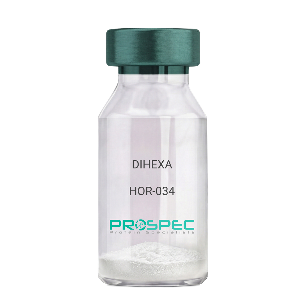
Name :
DIHEXADescription:
DIHEXA
Product # :
HOR-034Price :
Quantity :
Shipping Method :
Shipped at Room temp
More Info
- description
- formulation
- purity
- More Info
Description
DIHEXA Synthetic is a single, non-glycosylated polypeptide chain containing 3 amino acids, having a molecular mass of 504.28 Dalton and a Molecular formula of C27H44N4O5.
Formulation
The protein was lyophilized with no additives.
Purity
Greater than 97.0% as determined by analysis by RP-HPLC.
More Info
-
Physical Appearance
Sterile Filtered White lyophilized (freeze-dried) powder.
-
Stability
Lyophilized DIHEXA although stable at room temperature for 3 weeks, should be stored desiccated below -18°C. Upon reconstitution DIHEXA should be stored at 4°C between 2-7 days and for future use below -18°C. For long term storage it is recommended to add a carrier protein (0.1% HSA or BSA). Please prevent freeze-thaw cycles.
-
Solubility
It is recommended to reconstitute the lyophilized DIHEXA in sterile 18MΩ-cm H2O not less than 100 µg/ml, which can then be further diluted to other aqueous solutions.
-
Amino Acid Sequence
Hexanoyl-Tyr-Ile-Ahx-NH2.
-
Background
Dihexa, also known as N-hexanoic-Tyr-Ile-(6) aminohexanoic amide, is a potent and orally active small peptide that has been the focus of significant research due to its potential neurogenic and neuroprotective effects. This compound, derived from angiotensin IV, has been shown to possess a wide range of biological activities, including enhancing cognitive function, promoting neurogenesis, and potentially mitigating the effects of neurodegenerative diseases.
Dihexa's primary mechanism of action involves its interaction with hepatocyte growth factor (HGF) and its receptor, c-Met. By mimicking the effects of HGF, Dihexa can stimulate the c-Met receptor, leading to a cascade of events that promote neurogenesis and synaptic plasticity. Studies by Benoist et al. (2014) have demonstrated that Dihexa can enhance cognitive function in rats, suggesting potential applications in cognitive enhancement and the treatment of cognitive disorders.
In addition to its neurogenic effects, Dihexa has been shown to possess neuroprotective properties. Research by Kawas et al. (2017) found that Dihexa could protect neurons from apoptosis, suggesting potential applications in the treatment of neurodegenerative diseases such as Alzheimer's and Parkinson's disease.
Given its neurogenic and neuroprotective effects, Dihexa has been proposed as a potential therapeutic agent for a variety of conditions, including cognitive disorders, neurodegenerative diseases, and stroke. For instance, a study by Harding et al. (2018) found that Dihexa could improve outcomes in animal models of stroke, indicating its potential as a therapeutic agent in stroke recovery.
While research on Dihexa is promising, it is important to note that most studies have been conducted in animals or in vitro. More research is needed to fully understand the potential effects and applications of Dihexa in humans. However, the existing body of research suggests that Dihexa could be a promising tool in the treatment of cognitive disorders and neurodegenerative diseases.
ProSpec's products are furnished for LABORATORY RESEARCH USE ONLY. The product may not be used as drugs, agricultural or pesticidal products, food additives or household chemicals.
- View Data Sheet
Name :
CXCL8 Human, PichiaDescription:
Interleukin-8 (1-77 a.a.) Human Recombinant, (CXCL8) Pichia
IL-8, CXCL8, Monocyte-derived neutrophil chemotactic factor, MDNCF, T-cell chemotactic factor, Neutrophil-activating protein 1, NAP-1, Protein 3-10C, Granulocyte chemotactic protein 1, GCP-1, Monocyte-derived neutrophil-activating peptide, MONAP, Emoctakin, K60, NAF, LECT, LUCT, 3-10C, LYNAP, SCYB8, TSG-1, AMCF-I, b-ENAP.
Product # :
CHM-349Price :
Quantity :
Shipping Method :
Shipped at Room temp
More Info
- description
- source
- formulation
- purity
- biological activity
- More Info
Description
Interleukin-8 Human Recombinant produced in Yeast is a single, glycosylated polypeptide chain containing 79 amino acids and having a molecular mass of 9 kDa. The IL-8 is purified by proprietary chromatographic techniques.
Source
Pichia Pastoris.
Formulation
Lyophilized from a concentrated (1mg/ml) solution in water containing 20mM sodium phosphate buffer pH-8.
Purity
Greater than 98.0% as determined by SDS-PAGE.
Biological Activity
Chemotactic activity was reached at 25ng/ml on human neutrophils.
More Info
-
Introduction
Interleukin-8 (IL-8) is a chemokine produced by macrophages and other cell types such as epithelial cells. It is also synthesized by endothelial cells, which store IL-8 in their storage vesicles, the Weibel-Palade bodies.
When first encountering an antigen, the primary cells to encounter it are the macrophages who phagocytose the particle. Upon processing, they release chemokines to signal other immune cells to come in to the site of inflammation. IL-8 is one such chemokine. It serves as a chemical signal that attracts neutrophils at the site of inflammation, and therefore is also known as Neutrophil Chemotactic Factor. -
Synonyms
IL-8, CXCL8, Monocyte-derived neutrophil chemotactic factor, MDNCF, T-cell chemotactic factor, Neutrophil-activating protein 1, NAP-1, Protein 3-10C, Granulocyte chemotactic protein 1, GCP-1, Monocyte-derived neutrophil-activating peptide, MONAP, Emoctakin, K60, NAF, LECT, LUCT, 3-10C, LYNAP, SCYB8, TSG-1, AMCF-I, b-ENAP.
-
Physical Appearance
Sterile Filtered White lyophilized (freeze-dried) powder.
-
Stability
Lyophilized Interleukin-8 although stable at room temperature for 3 weeks, should be stored desiccated below -18°C. Upon reconstitution CXCL8 should be stored at 4°C between 2-7 days and for future use below -18°C.For long term storage it is recommended to add a carrier protein (0.1% HSA or BSA).Please prevent freeze-thaw cycles.
-
Solubility
It is recommended to reconstitute the lyophilized Interleukin 8 in sterile 18MΩ-cm H2O not less than 100µg/ml, which can then be further diluted to other aqueous solutions.
-
Background
What is the molecular weight/Mw of CXCL8 HUMAN, PICHIA Protein?
CXCL8 HUMAN, PICHIA Protein has a total Mw of 9kDa.
What is the source or expression system of CXCL8 HUMAN, PICHIA Protein?
Pichia Pastoris.
What is the Purity of CXCL8 HUMAN, PICHIA Protein?
CXCL8 HUMAN, PICHIA Protein is >98% pure as determined by SDS-PAGE.
What is the Biological Activity of CXCL8 HUMAN, PICHIA Protein?
Chemotactic activity was reached at 25ng/ml on human neutrophils.
What is the amino acid sequence of CXCL8 HUMAN, PICHIA Protein?
CXCL8 HUMAN, PICHIA Protein is composed from 79 amino acids.
What applications can CXCL8 HUMAN, PICHIA Protein be used in?
CXCL8 HUMAN, PICHIA Protein can probably be used in western blot, ELISA and Lateral Flow.
What is the endotoxin level for CXCL8 HUMAN, PICHIA Protein?
The endotoxin level is minimal, CXCL8 HUMAN, PICHIA Protein was purified using conventional chromatography techniques.
ProSpec's products are furnished for LABORATORY RESEARCH USE ONLY. The product may not be used as drugs, agricultural or pesticidal products, food additives or household chemicals.
- View Data Sheet
Name :
PPIF AntibodyDescription:
Cyclophilin-F, Mouse Anti Human
Oeptidylprolyl Isomerase F, PPIF, CYP-D, CYP3, SnuCyp-20, Peptidyl-prolyl cis-trans isomerase mitochondrial, Cyclophilin F, FLJ90798, MGC117207, peptidylprolyl isomerase F.
Product # :
ANT-389Price :
Quantity :
Shipping Method :
Shipped with Ice Packs
More Info
- formulation
- More Info
Formulation
1mg/ml containing PBS, pH-7.4, & 0.1% Sodium Azide.
More Info
-
Introduction
PPIF is a part of the peptidyl-prolyl cis-trans isomerase (PPIase) family. PPIF accelerates the folding of proteins. It catalyzes the cis-trans isomerization of proline imidic peptide bonds in oligopeptides. PPIF is key component of the mitochondrial permeability transition pore in the inner mitochondrial membrane. Activation of this pore is thought to be involved in the induction of apoptotic and necrotic cell death.
-
Synonyms
Oeptidylprolyl Isomerase F, PPIF, CYP-D, CYP3, SnuCyp-20, Peptidyl-prolyl cis-trans isomerase mitochondrial, Cyclophilin F, FLJ90798, MGC117207, peptidylprolyl isomerase F.
-
Immunogen
Anti-human Cyclophilin-F mAb is derived from hybridization of mouse F0 myeloma cells with spleen cells from BALB/c mice immunized with recombinant human Cyclophilin-F amino acids 30-207 purified from E. coli.
-
Ig Subclass
Mouse IgG2b heavy chain and κ light chain.
-
Clone
PAT1F5AT.
-
Applications
Cyclophilin-F antibody has been tested by ELISA, Western blot and immunohistochemistry analysis to assure specificity and reactivity. Since application varies, however, each investigation should be titrated by the reagent to obtain optimal results. Recommended dilution range for Western blot is 1:500~1:1,000 and immunohistochemistry analysis is 1:50~100. Recommended starting dilution for Western blot is 1:500 and Immunohistochemistry is 1:50.
-
Type
Mouse Anti Human Monoclonal.
-
Storage Procedures
For periods up to 1 month store at 4°C, for longer periods of time, store at -20°C. Prevent freeze thaw cycles.
-
Purification Method
Cyclophilin-F antibody was purified from mouse ascitic fluids by protein-G affinity chromatography.
ProSpec's products are furnished for LABORATORY RESEARCH USE ONLY. The product may not be used as drugs, agricultural or pesticidal products, food additives or household chemicals.
- View Data Sheet
Name :
Fra e 1.0101Description:
Allergen Fra e 1.0101 Recombinant
Allergen Fra e 1.0101, Fra e 1.0101, Fra e 1.
Product # :
PRO-2286Price :
Quantity :
Shipping Method :
Shipped with Ice Packs
More Info
- description
- source
- formulation
- purity
- More Info
Description
Recombinant Allergen Fra e 1.0101 produced in SF9 is a glycosylated, polypeptide chain having a calculated molecular mass of 17,794 Dalton. Fra e 1.0101 is expressed with a 10xHis tag at N-terminus and purified by proprietary chromatographic techniques.
Source
Sf9 insect cells.
Formulation
Fra e 1.0101 is supplied in 20mM HEPES buffer pH-7.9 and 6M Urea.
Purity
Greater than 80.0% as determined by SDS-PAGE.
More Info
-
Introduction
Allergen Fra e 1.0101 (Fra e 1.0101) causes an allergic reaction in humans.
-
Synonyms
Allergen Fra e 1.0101, Fra e 1.0101, Fra e 1.
-
Physical Appearance
Sterile Filtered clear solution.
-
Stability
Store at 4°C if entire vial will be used within 2-4 weeks. Store, frozen at -20°C for longer periods of time. Avoid multiple freeze-thaw cycles.
-
Immunological Functions
1. Binds IgE type human antibodies. 2. Immunodot test with positive/negative sera panels.
-
Molar extinction coefficient
14815; A280(1mg/ml)=0.833
ProSpec's products are furnished for LABORATORY RESEARCH USE ONLY. The product may not be used as drugs, agricultural or pesticidal products, food additives or household chemicals.
- View Data Sheet
Name :
ALDH2 Mouse, ActiveDescription:
Aldehyde Dehydrogenase 2 Mouse Recombinant, Active
Aldehyde dehydrogenase, mitochondrial, AHD-M1, ALDH class 2, ALDH-E2, ALDHI.
Product # :
ENZ-1093Price :
Quantity :
Shipping Method :
Shipped with Ice Packs
More Info
- description
- source
- formulation
- purity
- biological activity
- More Info
Description
ALDH2 Mouse Recombinant produced in E.Coli is a single, non-glycosylated polypeptide chain containing 523 amino acids (20-519) and having a molecular mass of 56.8kDa. ALDH2 Mouse is fused to a 23 amino acid His-Tag at N-terminus and purified by proprietary chromatographic techniques.
Source
Escherichia Coli.
Formulation
ALDH2 Mouse protein (0.5mg/ml) is formulated in Phosphate buffered saline (pH7.4), 20% glycerol and 1mM DTT.
Purity
Greater than 95% as determined by SDS-PAGE.
Biological Activity
Specific activity is > 180 pmol/min/ug, and was obtained by measuring the increase of NADH in absorbance at 340 nm resulting from the reduction of NAD at pH 8.0 at 25C.
More Info
-
Introduction
ALDH2 is part of the aldehyde dehydrogenase family of proteins which catalyze the chemical transformation from acetaldehyde to acetic acid. ALDH2 is the second enzyme of the major oxidative pathway of alcohol metabolism. ALDH2 has 2 major liver isoforms: cytosolic and mitochondrial, which differ by their electrophoretic mobilities, kinetic properties, and subcellular localizations. Nearly all Caucasians have 2 major isozymes, whereas roughly 50% of Orientals have only the cytosolic isozyme, omitting the mitochondrial isozyme. The extremely higher rate of acute alcohol intoxication with Orientals compared to Caucasians is due to the fact of the absence of mitochondrial isozyme. ALDH2 has a low Km for acetaldehydes, and is localized in mitochondrial matrix.
-
Synonyms
Aldehyde dehydrogenase, mitochondrial, AHD-M1, ALDH class 2, ALDH-E2, ALDHI.
-
Physical Appearance
Sterile Filtered clear solution.
-
Stability
Store at 4°C if entire vial will be used within 2-4 weeks. Store, frozen at -20°C for longer periods of time. For long term storage it is recommended to add a carrier protein (0.1% HSA or BSA).Avoid multiple freeze-thaw cycles.
-
Amino Acid Sequence
MGSSHHHHHH SSGLVPRGSH MGSSAAATSA VPAPNHQPEV FCNQIFINNE WHDAVSRKTF PTVNPSTGEV ICQVAEGNKE DVDKAVKAARAAFQLGSPWR RMDASDRGRL LYRLADLIER DRTYLAALET LDNGKPYVIS YLVDLDMVLK CLRYYAGWAD KYHGKTIPID GDFFSYTRHEPVGVCGQIIP WNFPLLMQAW KLGPALATGN VVVMKVAEQT PLTALYVANL IKEAGFPPGV VNIVPGFGPT AGAAIASHEG VDKVAFTGSTEVGHLIQVAA GSSNLKRVTL ELGGKSPNII MSDADMDWAV EQAHFALFFN QGQCCCAGSR TFVQENVYDE FVERSVARAK SRVVGNPFDSRTEQGPQVDE TQFKKILGYI KSGQQEGAKL LCGGGAAADR GYFIQPTVFG DVKDGMTIAK EEIFGPVMQI LKFKTIEEVV GRANDSKYGLAAAVFTKDLD KANYLSQALQ AGTVWINCYD VFGAQSPFGG YKMSGSGREL GEYGLQAYTE VKTVTVKVPQ KNS.
ProSpec's products are furnished for LABORATORY RESEARCH USE ONLY. The product may not be used as drugs, agricultural or pesticidal products, food additives or household chemicals.
- View Data Sheet
Name :
SARS Mosaic S(C)Description:
SARS-Associated Coronavirus Spike Mosaic S(C) Recombinant
Product # :
SARS-257Price :
Quantity :
Shipping Method :
Shipped with Ice Packs
More Info
- description
- formulation
- purity
- More Info
Description
The E.Coli derived 37 kDa recombinant mosaic protein contains the C-terminal t section of the Spike protein 1051-1076, 1121-1154, 1162-1190 amino acids immunodominant regions.
Formulation
25mM Tris-HCl, 0.4% sarcosyl, 0.25% Triton –100 and 50% glycerol.
Purity
SARS Mosaic Protein is >95% pure as determined by 10% PAGE (coomassie staining).
More Info
-
Introduction
SARS Coronavirus is an enveloped virus containing three outer structural proteins, namely the membrane (M), envelope (E), and spike (S) proteins. Spike (S)-glycoprotein of the virus interacts with a cellular receptor and mediates membrane fusion to allow viral entry into susceptible target cells. Accordingly, S-protein plays an important role in virus infection cycle and is the primary target of neutralizing antibodies.
-
Stability
SARS Mosaic protein is shipped at ambient temperature.Upon arrival, store at -20C.
-
Applications
SARS Mosaic antigen is suitable for ELISA and Western blots, excellent antigen for detection of SARS with minimal specificity problems.
-
Specificity
SARS Mosaic protein is Immunoreactive with sera of SARS-infected individuals.
-
Purification Method
SARS Mosaic protein was purified by proprietary chromatographic technique.
ProSpec's products are furnished for LABORATORY RESEARCH USE ONLY. The product may not be used as drugs, agricultural or pesticidal products, food additives or household chemicals.
- View Data Sheet
Name :
CoV-2 Spike (1000-1200)Description:
Coronavirus 2019 Spike (1000-1200 a.a.) Recombinant
Product # :
SARS-017Price :
Quantity :
Shipping Method :
Shipped with Ice Packs
More Info
- description
- source
- formulation
- purity
- More Info
Description
The E.Coli derived recombinant protein contains the Coronavirus 2019 Spike (1000-1200 a.a.) immunodominant regions, fused to 6xHis tag at C-terminal.
Source
Escherichia Coli.
Formulation
CoV 2019 Spike Protein 1mg/ml solution is supplied in 1x PBS.
Purity
CoV 2019 Spike Protein is >90% pure as determined SDS-PAGE.
More Info
-
Introduction
A human infecting coronavirus (viral pneumonia) called 2019 novel coronavirus, 2019-nCoV was found in the fish market at the city of Wuhan, Hubei province of China on December 2019.
The 2019-nCoV shares an 87% identity to the 2 bat-derived severe acute respiratory syndrome 2018 SARS-CoV-2 located in Zhoushan of eastern China. 2019-nCoV has an analogous receptor-BD-structure to that of 2018 SARS-CoV, even though there is a.a. diversity so thus the 2019-nCoV might bind to ACE2 receptor protein (angiotensin-converting enzyme 2) in humans.
While bats are possibly the host of 2019-nCoV, researchers suspect that animal from the ocean sold at the seafood market was an intermediate host. RSCU analysis proposes that the 2019-nCoV is a recombinant within the viral spike glycoprotein between the bat coronavirus and an unknown coronavirus.
-
Physical Appearance
Sterile Filtered clear solution.
-
Stability
CoV 2019 Spike Protein is shipped on ice packs. Upon arrival, Store at -20°C.
ProSpec's products are furnished for LABORATORY RESEARCH USE ONLY. The product may not be used as drugs, agricultural or pesticidal products, food additives or household chemicals.
- View Data Sheet
Name :
T.pallidum p41Description:
Treponema pallidum p41 Recombinant
Product # :
TRP-242Price :
Quantity :
Shipping Method :
Shipped with Ice Packs
More Info
- description
- source
- formulation
- purity
- More Info
Description
The E.coli derived recombinant protein contains the outer membrane T.Pallidum p41 immunodominant regions. The protein is fused with a Beta-galactosidase tag.
Source
Escherichia Coli.
Formulation
10mM Tris-HCl pH8.0, 1mM EDTA, 1mMDTT and 8M urea.
Purity
Treponema pallidum protein is >90% pure as determined by 10% PAGE (coomassie staining).
More Info
-
Introduction
Treponema pallidum is a gram-negative spirochaete bacterium and is considered to be metabolically crippled. There are at least four known subspecies: T. pallidum pallidum, T. pallidum pertenue, T. pallidum carateum and T. pallidum endemicum. The helical structure of T. pallidum pallidum allows it to move in a corkscrew motion through viscous mediums such as mucus. Treponema pallidum sub sp. pallidum has one of the smallest bacterial genomes at 1.14 million base pairs (Mb) and has limited metabolic capabilities, reflecting its adaptation through genome reduction to the rich environment of mammalian tissue.
-
Stability
T.pallidum p41 although stable at 4°C for 1 week, should be stored below -18°C. Please prevent freeze thaw cycles.
-
Applications
Treponema pallidum is suitable for ELISA and Western blots, excellent antigen for detection of T. Pallidum with minimal specificity problems.
-
Specificity
Immunoreactive with sera of T.Pallidum infected individuals.
-
Purification Method
Treponema pallidum protein was purified by proprietary chromatographic technique.
ProSpec's products are furnished for LABORATORY RESEARCH USE ONLY. The product may not be used as drugs, agricultural or pesticidal products, food additives or household chemicals.
- View Data Sheet
Name :
SERPINA7 HumanDescription:
Serpin Peptidase Inhibitor, Clade A Member 7 Human
Product # :
PRO-2739Price :
Quantity :
Shipping Method :
Shipped at Room temp
More Info
- description
- source
- formulation
- purity
- More Info
Description
Human Serpin Peptidase Inhibitor, Clade A Member 7 Protein produced in Human plasma having a molecular mass of approximately 55kD.
Source
Human serum.
Formulation
The protein was lyophilized (0.2 µm filtered) from 20mM NH4HCO3.
Purity
Greater than 98.0%.
More Info
-
Introduction
Thyroxine binding globulin, also known as SERPINA7, is synthesized in the liver and carried in the blood.
SERPINA7 may be decreased or increased in a number of diseases. Its main value in diagnosis is to assess thyroid function by determining the amount of free T3/T4.
Thyroxine binding globulin, binds and transports thyroid hormones in the circulation. SERPINA7 carries the majority of the T3/T4 in the blood. SERPINA7 has only one binding site for T3/T4 and about 25% of Thyroxine binding globulin is usually saturated. -
Physical Appearance
Sterile Filtered White lyophilized (freeze-dried) powder.
-
Stability
SERPINA7 Human although stable at room temperature for 3 weeks, should be stored between 2-8°C.
-
Solubility
It is recommended to reconstitute the lyophilized SERPINA7 Human in phosphate buffer containing 0.15M NaCl.
-
Human Virus Test
Starting material donor has been tested and certified negative for antibodies to HIV-1, HIV-2, HCV, HBSAG, Parvovirus B19, Syphilis and HIV/HBV/HCV (PCR).
ProSpec's products are furnished for LABORATORY RESEARCH USE ONLY. The product may not be used as drugs, agricultural or pesticidal products, food additives or household chemicals.
- View Data Sheet
Name :
OmpADescription:
Outer Membrane Protein-A Bacterial Recombinant
Outer Membrane Protein-A, OmpA.
Product # :
PRO-571Price :
Quantity :
Shipping Method :
Shipped at Room temp
More Info
- description
- source
- formulation
- purity
- biological activity
- More Info
Description
The recombinant form was found to be undistinguishable from the wild type when examined by SDS-PAGE and gel filtration chromatography yielding a 50.5 kDa monomeric protein. The immunological similarity of the protein samples was demonstrated by employing polyclonal and monoclonal antibodies in ELISA and Western Blot techniques. All forms of A-protein were found to activate the secretion of tumour necrosis factor alpha from murine macrophage. For ref see Maurice et al. (1999) Protein Expression and Purification 16, 396-404.The OmpA is purified by proprietary chromatographic techniques.
Source
Escherichia Coli.
Formulation
The OmpA protein was lyophilized from a concentrated (1mg/ml) solution with 0.02% NaHCO3.
Purity
Greater than 98.0% as determined by:
(a) Analysis by Gel filtration.
(b) Analysis by SDS-PAGE.Biological Activity
The interaction of bacterial and recombinant A-layer protein with murine macrophages was directed at determining the effect of A-protein on intracellular events that occur in primed macrophages. This was accomplished by measuring the cytotoxic product produced by peritoneal macrophages when exposed to A-protein coated latex beads. Thioglycolate elicited macrophages exhibited a low level of activation (18% cytotoxicity) that was significantly increased (48% cytotoxicity) in the presence of latex beads. Coating of the latex beads with each of the three A-protein products resulted in an increase of cytoxicity (mean +/- SEM) from 48% to 91%.More Info
-
Introduction
The OmpA protein is one of the main outer-membrane proteins of a large array of Gram-negative bacteria such as A.salmonicida, Shigella dysenteriae and E.coli.OmpA’s major physiological functions include maintenance of the structural integrity and morphology of the cells and porin activity, as well as a role in conjugation and bacteriophage binding.Achromogenic atypical Aeromonas salmonicida is the causative agent of goldfish ulcer disease.Virulence of this bacterium is associated with the production of a paracrystalline outer membrane A-layer protein.The species specific structural gene for the monomeric form of A-protein was cloned into a pET-3d plasmid in order to express and produce a recombinant form of the protein in E.coli BL21(DE3). The induced protein was isolated from inclusion bodies by a simple solubilization-renaturation procedure and purified by ion exchange chromatography on Q-Sepharose to over 95% pure monomeric protein.Recombinant A-protein was compared by biochemical, immunological and molecular methods with the A-protein isolated from atypical A.salmonicida bacterial cells by the glycine and the membrane extraction methods.
-
Synonyms
Outer Membrane Protein-A, OmpA.
-
Physical Appearance
Sterile Filtered White lyophilized (freeze-dried) powder.
-
Stability
Lyophilized Bacterial Outer Membrane Protein-A although stable at room temperature for 3 weeks, should be stored desiccated below -18°C. Upon C between 2-7 days and for future use reconstituted OmpA should be stored at 4 below -18°C. For long term storage it is recommended to add a carrier protein (0.1% HSA or BSA). Please avoid freeze-thaw cycles.
-
Solubility
It is recommended to reconstitute the lyophilized OmpA in sterile 0.4% NaHCO3, not less than 100µg/ml, which can then be further diluted to other aqueous solutions.
-
Amino Acid Sequence
mdvvispndn tfvttslasv tkqpvldfst aqqnltlnfs evgdlknngf ivleiqgegq fndaeirqwl sngfwrrpft gllvnpndhg nfansgevnd vrkffkiisd gtqltivhti dsngkrlrla lasdveetin fadaevelkl nlanqafklt sgsqgtvalt agalwnasyt adpvatkplf klgklfqlsl tnagkatalv segflklnig danisatdfa itnvttnqti qrdkvnltlt gdvsafkkda ngnlvnkaga sigwkaaadg qsatavlgag nmaggvqnal aafgtlyvaa dntvpvpavn fnvkaeiqgd sqatynyfkd eladlfiltr dgmkfdtitt gttsanlihi rdvsnilpte ggkifvtite yadhaangrg egtvlvtrka lsvtlpsgga vtlkpadvaa dvgasitagr qarlvfevet nqgevavkks naegvdiqng trgtaplvdf tl.
ProSpec's products are furnished for LABORATORY RESEARCH USE ONLY. The product may not be used as drugs, agricultural or pesticidal products, food additives or household chemicals.
- View Data Sheet
Name :
SARS MERS, HEKDescription:
SARS MERS Spike Glycoprotein-S1, Recombinant
Product # :
SARS-023Price :
Quantity :
Shipping Method :
Shipped with Ice Packs
More Info
- description
- source
- formulation
- purity
- More Info
Description
The HEK293 derived recombinant protein contains the SARS MERS Spike Glycoprotein S1, amino acids 18-725 fused to dimeric Fc tag at N-terminal having a total Mw of 215.7 kDa.
Source
HEK293
Formulation
SARS MERS S1 protein solution is supplied in PBS.
Purity
Protein is >85% pure as determined SDS-PAGE.
More Info
-
Introduction
SARS Coronavirus is an enveloped virus containing three outer structural proteins, namely the membrane (M), envelope (E), and spike (S) proteins. Spike (S)-glycoprotein of the virus interacts with a cellular receptor and mediates membrane fusion to allow viral entry into susceptible target cells. Accordingly, S-protein plays an important role in virus infection cycle and is the primary target of neutralizing antibodies.
-
Physical Appearance
Sterile Filtered clear solution.
-
Stability
SARS MERS Protein is shipped on ice packs. Upon arrival, Store at -20°C.
-
Purification Method
Purified by Protein-G chromatographic technique.
ProSpec's products are furnished for LABORATORY RESEARCH USE ONLY. The product may not be used as drugs, agricultural or pesticidal products, food additives or household chemicals.
- View Data Sheet
Name :
EGF Long HumanDescription:
Epidermal Growth Factor Long Human Recombinant
Urogastrone, URG, EGF.
Product # :
CYT-798Price :
Quantity :
Shipping Method :
Shipped at Room temp
More Info
- description
- source
- formulation
- purity
- biological activity
- More Info
Description
Recombinant Human EGF Long produced in E.coli cells is a single non-glycosylated, polypeptide chain containing 106 amino acids and having a molecular mass of 12.3kDa. The EGF Long is purified by proprietary chromatographic techniques.
Source
Escherichia Coli.
Formulation
The EGF Long was lyophilized from a 0.2µm filtered concentrated solution in 10mM HCl.
Purity
Greater than 95.0% as determined by:
(a) Analysis by RP-HPLC.
(b) Analysis by SDS-PAGE.Biological Activity
The ED50 as determined by a cell proliferation assay using murine Balb/c 3T3 cells is less than 1.0 ng/ml, corresponding to a specific activity of > 1.0 × 10⁶ IU/mg.More Info
-
Introduction
Epidermal growth factor has a profound effect on the differentiation of specific cells in vivo and is a potent mitogenic factor for a variety of cultured cells of both ectodermal and mesodermal origin. The EGF precursor is believed to exist as a membrane-bound molecule which is proteolytically cleaved to generate the 53-amino acid peptide hormone that stimulates cells to divide. EGF stimulates the growth of various epidermal and epithelial tissues in vivo and in vitro and of some fibroblasts in cell culture. Long EGF is a recombinant analog of Human EGF developed as a replacement for use in therapeutic cell culture applications as a like-for-like supplement for Recombinant Human or native EGF. It includes the Human EGF amino acid sequence plus a 53 amino acid N-terminal extension peptide.
-
Synonyms
Urogastrone, URG, EGF.
-
Physical Appearance
Sterile Filtered White lyophilized (freeze-dried) powder.
-
Stability
Lyophilized EGF Long although stable at room temperature for 3 weeks, should be stored desiccated below -18°C. Upon reconstitution EGF Long should be stored at 4°C between 2-7 days and for future use below -18°C.For long term storage it is recommended to add a carrier protein (0.1% HSA or BSA).Please prevent freeze-thaw cycles.
-
Solubility
It is recommended to reconstitute the lyophilized EGF Long in sterile 100mM AcOH (acetic Acid) not less than 100µg/ml, which can then be further diluted to other aqueous solutions.
-
Amino Acid Sequence
MFPAMPLSSL FANAVLRAQH LHQLAADTYK EFERAYIPEG QRYSIQVNFA HYGNSDSECP LSHDGYCLHD GVCMYIEALD KYACNCVVGY IGERCQYRDL KWWELR
-
Background
What is the molecular weight/Mw of EGF Protein?
EGF Protein has a total Mw of 12.3kDa.
What is the source or expression system of EGF Protein?
Escherichia Coli.
What is the Purity of EGF Protein?
EGF Protein is >95% pure as determined by SDS-PAGE.
What is the Biological Activity of EGF Protein?
The ED50 as determined by a cell proliferation assay using murine Balb/c 3T3 cells is less than 1.0 ng/ml, corresponding to a specific activity of > 1.0 × 10⁶ IU/mg.
What is the amino acid sequence of EGF Protein?
MFPAMPLSSL FANAVLRAQH LHQLAADTYK EFERAYIPEG QRYSIQVNFA HYGNSDSECP LSHDGYCLHD GVCMYIEALD KYACNCVVGY IGERCQYRDL KWWELR
What applications can EGF Protein be used in?
EGF Protein can probably be used in western blot, ELISA and Lateral Flow.
What is the endotoxin level for EGF Protein?
The endotoxin level is minimal, EGF Protein was purified using conventional chromatography techniques.
ProSpec's products are furnished for LABORATORY RESEARCH USE ONLY. The product may not be used as drugs, agricultural or pesticidal products, food additives or household chemicals.
- View Data Sheet
Name :
CEA HumanDescription:
Carcinoembryonic Antigen Human Recombinant
CEACAM5, Meconium Antigen 100, Carcinoembryonic Antigen, CD66e Antigen, CD66e, Carcinoembryonic Antigen, CEA, oncofetal antigen.
Product # :
PRO-287Price :
Quantity :
Shipping Method :
Shipped with Ice Packs
More Info
- description
- source
- formulation
- purity
- More Info
Description
CEA Human Recombinant is glycosylated with N-linked sugars and produced using baculovirus vectors in insect cells. CEA is a well-known tumor marker corresponding to the full length human CEA which is approximately 120,000 Dalton.
Source
Baculovirus Insect Cells.
Formulation
The sterile protein solution contains 10mM NaH2PO4, pH 7 and 150mM NaCl.
Purity
Greater than 90.0% as determined by SDS-PAGE.
More Info
-
Introduction
Carcinoembryonic antigen (CEA) is a glycoprotein present in fetal digestive-tract tissues; it’s involved in cell adhesion. The production of CEA stops before birth. CEA is called tumor marker since its elevated levels are found in the serum from individuals with colorectal, gastric, pancreatic, lung and breast carcinomas and in heavy smokers.
There are also benign conditions that elevate CEA levels such as smoking, infection, inflammatory bowel disease, pancreatitis, cirrhosis of the liver, and some benign tumors (in the equivalent organs which have cancers with elevated CEA). Typically, higher levels of CEA are found in men, smokers, and older individuals.
The presence of CEA assists in screening, in evaluating recurrent or disseminated disease, and in determining the success of surgical removal of malignant tumors.
CEA levels can be used as indicators of treatment success. The normal values range from 0.0 to 2.5 ng/ml of serum (from blood), in non-smokers, a greater amount than that may be suggestive of cancer. Levels above 20 ng/ml before treatment are associated with cancer which has already metastasized. Benign conditions do not usually cause a CEA increase over 10 ng/ml.
The high levels of CEA should return to normal after successful therapy, however if during follow up there’s an elevation in CEA levels it indicates a recurrence of tumor.
Carcinoembryonic antigen family belongs to the immunoglobulin superfamily; it consists of 29 genes, 18 of which are normally expressed. -
Synonyms
CEACAM5, Meconium Antigen 100, Carcinoembryonic Antigen, CD66e Antigen, CD66e, Carcinoembryonic Antigen, CEA, oncofetal antigen.
-
Physical Appearance
Sterile Filtered colourless solution.
-
Stability
CEA should be stored at 2-8°C.Avoid freezing.
ProSpec's products are furnished for LABORATORY RESEARCH USE ONLY. The product may not be used as drugs, agricultural or pesticidal products, food additives or household chemicals.
- View Data Sheet
Name :
PTH (1-84) HumanDescription:
Parathyroid Hormone (1-84) Human Recombinant
Parathyrin, PTH, Parathormone.
Product # :
HOR-263Price :
Quantity :
Shipping Method :
Shipped at Room temp
More Info
- description
- source
- formulation
- purity
- biological activity
- More Info
- sds-page
Description
Parathyroid Hormone 1-84 (full length) Human Recombinant produced in E.Coli is a single, non-glycosylated, polypeptide chain containing 84 amino acids, having an MW of ~9.4kDa.The PTH is purified by proprietary chromatographic techniques.
Source
Escherichia Coli.
Formulation
Lyophilized from a 0.2µm filtered concentrated solution in PBS pH7.4.
Purity
Greater than 97.0% as determined by:
(a) Analysis by RP-HPLC.
(b) Analysis by SDS-PAGE.Biological Activity
The activity is calculated by its ability to induce cAMP accumulation in murine MC3T3E1 cells and is <50ng/ml, corresponding to a specific activity of greater than 2.0 x 10⁴ Units/mg.
sds-page
More Info
-
Introduction
Parathyroid hormone (PTH), or parathormone, is secreted by the parathyroid glands as a polypeptide containing 84 amino acids. It acts to increase the concentration of calciumin the blood, whereas calcitonin (a hormone produced by the parafollicular cells of the thyroid gland) acts to decrease calcium concentration. PTH acts to increase the concentration of calcium in the blood by acting upon parathyroid hormone receptorin three parts of the body: In the bones- It enhances the release of calcium from the large reservoir contained in the bones. Bone resorption is the normal destruction of bone by osteoclasts, which are indirectly stimulated by PTH. Stimulation is indirect since osteoclasts do not have a receptor for PTH; rather, PTH binds to osteoblasts, the cells responsible for creating bone. Binding stimulates osteoblasts to increase their expression of RANKL, which can bind to osteoclast precursors containing RANK, a receptor for RANKL. The binding of RANKL to RANK stimulates these precursors to fuse, forming new osteoclasts which ultimately enhances the resorption of bone.
In the kidney- It enhances active reabsorption of calcium from distal tubules and the thick ascending limb.
In the intestine- It enhances the absorption of calcium in the intestine by increasing the production of vitamin D and upregulating the enzyme responsible for 1-alpha hydroxylationof 25-hydroxy vitamin D, converting vitamin D to its active form (1,25-dihydroxy vitamin D) which effects the actual absorption of calcium (as Ca2+ ions) by the intestine via calbindin.
Recombinant Human full length PTH 1-84 has potential as an anti-osteoporotic agent, due to its properties as a bone formation stimulant, it increases bone turnover, stimulating osteoblasts and reducing both vertebral and non vertebral fractures. -
Synonyms
Parathyrin, PTH, Parathormone.
-
Physical Appearance
Sterile Filtered White lyophilized (freeze-dried) powder.
-
Stability
Lyophilized Parathyrin although stable at room temperature for 3 weeks, should be stored desiccated below -18°C. Upon reconstitution PTH should be stored at 4°C between 2-7 days and for future use below -18°C.For long term storage it is recommended to add a carrier protein (0.1% HSA or BSA).Please prevent freeze-thaw cycles.
-
Solubility
It is recommended to reconstitute the lyophilized Parathormone in sterile 18MΩ-cm H2O not less than 100 µg/ml, which can then be further diluted to other aqueous solutions.
-
Amino Acid Sequence
SVSEIQLMHN LGKHLNSMER VEWLRKKLQD VHNFVALGAP LAPRDAGSQR PRKKEDNVLV ESHEKSLGEA DKADVNVLTK AKSQ.
ProSpec's products are furnished for LABORATORY RESEARCH USE ONLY. The product may not be used as drugs, agricultural or pesticidal products, food additives or household chemicals.
- View Data Sheet
Name :
DeslorelinDescription:
Deslorelin
Product # :
HOR-240Price :
Quantity :
Shipping Method :
Shipped at Room temp
More Info
- description
- formulation
- purity
- More Info
Description
Deslorelin is a potent LHRH/GnRH agonist has a molecular formula of C64H83N17O12, pGlu-His-Trp-Ser-Tyr-D-Trp-Leu-Arg-Pro-NHC2H5 having an Mw of 1284.4 Dalton.
Formulation
The Deslorelin peptide was lyophilized with no additives.
Purity
Greater than 99.0% as determined by RP-HPLC.
More Info
-
Introduction
Deslorelin is being studied in the treatment of cancer as a way to block sex hormones made by the ovaries or testicles. It belongs to the family of drugs called gonadotropin-releasing hormone analogs. It is used for the induction of ovulation in mares. Deslorelin binds to and activates pituitary gonadotropin releasing hormone (GnRH) receptors. Continuous, prolonged administration of goserelin in males results in pituitary GnRH receptor desensitization and inhibition of pituitary secretion of follicle stimulating hormone (FSH) and luteinizing hormone (LH), leading to a significant decline in testosterone production; in females, prolonged administration results in a decrease in estradiol production.
-
Physical Appearance
Sterile Filtered White lyophilized (freeze-dried) powder.
-
Stability
Lyophilized Deslorelin although stable at room temperature for 3 weeks, should be stored desiccated below -18°C. Upon reconstitution Deslorelin should be stored at 4°C between 2-7 days and for future use below -18°C. For long term storage it is recommended to add a carrier protein (0.1% HSA or BSA).Please prevent freeze-thaw cycles.
-
Solubility
It is recommended to reconstitute the lyophilized Deslorelin in sterile 18MΩ-cm H2O not less than 100µg/ml, which can then be further diluted to other aqueous solutions.
ProSpec's products are furnished for LABORATORY RESEARCH USE ONLY. The product may not be used as drugs, agricultural or pesticidal products, food additives or household chemicals.