Skip to content

High-quality research proteins.

  • 1-800-805-6531

  • Mon - Sat 8.00-18.00, Sun - Closed

  • info@prospecbio.com

  • PO Box 6591 East Brunswick NJ 08816

  • Products
    • Cytokines
    • Growth factors
    • Chemokines
    • Cd antigens
    • Neurotrophins
    • Hormones
    • Enzymes
    • Viral antigens
    • Recombinant proteins
    • Natural proteins
    • Monoclonal antibodies
    • Polyclonal antibodies
    • Thermal Packaging
  • About
    • Company
    • Founder
    • Contribution
  • Customers
    • Eastern Asia
    • Eastern Europe
    • North America
    • South America
    • South East Asia
    • Southern Asia
    • Western Europe
  • Ordering
    • Ordering Method
    • Ordering - Credit Card
    • Ordering - PO
    • Shipping Fees
    • FAQs
  • Services
    • DNA Cloning
    • Protein Expression
    • Monoclonal Antibody
    • Fermentation
    • Technology License
  • Contact us
Log in

Country/region

  • USD $ | Albania
  • USD $ | Andorra
  • USD $ | Argentina
  • USD $ | Armenia
  • USD $ | Australia
  • USD $ | Austria
  • USD $ | Azerbaijan
  • USD $ | Belarus
  • USD $ | Belgium
  • USD $ | Bolivia
  • USD $ | Brazil
  • USD $ | Bulgaria
  • USD $ | Cambodia
  • USD $ | Canada
  • USD $ | Chile
  • USD $ | China
  • USD $ | Colombia
  • USD $ | Costa Rica
  • USD $ | Croatia
  • USD $ | Cyprus
  • USD $ | Czechia
  • USD $ | Denmark
  • USD $ | Estonia
  • USD $ | Finland
  • USD $ | France
  • USD $ | Georgia
  • USD $ | Germany
  • USD $ | Greece
  • USD $ | Hong Kong SAR
  • USD $ | Hungary
  • USD $ | Iceland
  • USD $ | India
  • USD $ | Ireland
  • USD $ | Israel
  • USD $ | Italy
  • USD $ | Japan
  • USD $ | Kazakhstan
  • USD $ | Latvia
  • USD $ | Liechtenstein
  • USD $ | Lithuania
  • USD $ | Madagascar
  • USD $ | Malta
  • USD $ | Mexico
  • USD $ | Moldova
  • USD $ | Monaco
  • USD $ | Netherlands
  • USD $ | New Zealand
  • USD $ | North Macedonia
  • USD $ | Norway
  • USD $ | Panama
  • USD $ | Paraguay
  • USD $ | Peru
  • USD $ | Philippines
  • USD $ | Poland
  • USD $ | Portugal
  • USD $ | Romania
  • USD $ | Singapore
  • USD $ | Slovakia
  • USD $ | Slovenia
  • USD $ | South Africa
  • USD $ | South Korea
  • USD $ | Spain
  • USD $ | Sri Lanka
  • USD $ | Sweden
  • USD $ | Switzerland
  • USD $ | Taiwan
  • USD $ | Thailand
  • USD $ | Türkiye
  • USD $ | Ukraine
  • USD $ | United Kingdom
  • USD $ | United States
  • USD $ | Uruguay
  • USD $ | Vatican City
  • USD $ | Venezuela
  • USD $ | Vietnam
  • Facebook
  • Instagram
logoProSpec - Recombinant Protein Manufacturer for academic and R&D industry
Become a distributor
Account Log in
Wishlist
Cart Cart(0)
  • Products
    +
    • Cytokines
    • Growth factors
    • Chemokines
    • Cd antigens
    • Neurotrophins
    • Hormones
    • Enzymes
    • Viral antigens
    • Recombinant proteins
    • Natural proteins
    • Monoclonal antibodies
    • Polyclonal antibodies
    • Thermal Packaging
  • About
    +
    • Company
    • Founder
    • Contribution
  • Customers
    +
    • Eastern Asia
    • Eastern Europe
    • North America
    • South America
    • South East Asia
    • Southern Asia
    • Western Europe
  • Ordering
    +
    • Ordering Method
    • Ordering - Credit Card
    • Ordering - PO
    • Shipping Fees
    • FAQs
  • Services
    +
    • DNA Cloning
    • Protein Expression
    • Monoclonal Antibody
    • Fermentation
    • Technology License
  • Contact us

Item added to your cart

View cart
View All
  • Cytokines
  • Growth factors
  • Chemokines
  • Cd antigens
  • Neurotrophins
  • Hormones
  • Enzymes
  • Viral antigens
  • Recombinant proteins
  • Natural proteins
  • Monoclonal antibodies
  • Polyclonal antibodies
  • Thermal Packaging
  • Tumor Necrosis Factor

    Tumor Necrosis Factor

    View All
  • 4-1BB

    4-1BB

    View All
  • Adiponectin

    Adiponectin

    View All
  • AIF1

    AIF1

    View All
  • Other Cytokines

    Other Cytokines

    View All
  • AITRL

    AITRL

    View All
  • Angiopoietin

    Angiopoietin

    View All
  • Apolipoprotein

    Apolipoprotein

    View All
  • B-Cell Activating Factor

    B-Cell Activating Factor

    View All
  • Beta Defensin

    Beta Defensin

    View All
  • Bone Morphogenetic Protein

    Bone Morphogenetic Protein

    View All
  • B type Natriuretic Peptide

    B type Natriuretic Peptide

    View All
  • BST

    BST

    View All
  • Betacellulin

    Betacellulin

    View All
  • Cardiotrophin

    Cardiotrophin

    View All
  • Activin

    Activin

    View All
  • Fibroblast Growth Factor

    Fibroblast Growth Factor

    View All
  • Other Growth Factors

    Other Growth Factors

    View All
  • CSF

    CSF

    View All
  • CTGF

    CTGF

    View All
  • IGFBP

    IGFBP

    View All
  • VEGF Protein

    VEGF Protein

    View All
  • EGF

    EGF

    View All
  • Epigen

    Epigen

    View All
  • Erythropoietin

    Erythropoietin

    View All
  • Insulin-Like Growth Factor

    Insulin-Like Growth Factor

    View All
  • Growth Hormone

    Growth Hormone

    View All
  • HDGF

    HDGF

    View All
  • Hepatocyte Growth Factor

    Hepatocyte Growth Factor

    View All
  • Insulin

    Insulin

    View All
  • BCA-1/ BLC (CXCL13)

    BCA-1/ BLC (CXCL13)

    View All
  • BRAK (CXCL14)

    BRAK (CXCL14)

    View All
  • C-10 (CCL6)

    C-10 (CCL6)

    View All
  • Other Chemokines

    Other Chemokines

    View All
  • MEC (CCL28)

    MEC (CCL28)

    View All
  • LD78-beta (CCL3L1)

    LD78-beta (CCL3L1)

    View All
  • CTACK (CCL27)

    CTACK (CCL27)

    View All
  • CXCL16

    CXCL16

    View All
  • CXCL17

    CXCL17

    View All
  • Platelet Factor-4 (CXCL4)

    Platelet Factor-4 (CXCL4)

    View All
  • ENA-78 (CXCL5)

    ENA-78 (CXCL5)

    View All
  • Eotaxin (CCL11,24,26)

    Eotaxin (CCL11,24,26)

    View All
  • Exodus-2 (CCL21)

    Exodus-2 (CCL21)

    View All
  • Fractalkine (CX3CL1)

    Fractalkine (CX3CL1)

    View All
  • CXCL6

    CXCL6

    View All
  • Other CD Antigens

    Other CD Antigens

    View All
  • CD14

    CD14

    View All
  • CD164

    CD164

    View All
  • CD1

    CD1

    View All
  • CD2

    CD2

    View All
  • CD200

    CD200

    View All
  • CD207

    CD207

    View All
  • CD226

    CD226

    View All
  • CD23

    CD23

    View All
  • CD244

    CD244

    View All
  • CD247

    CD247

    View All
  • CD27

    CD27

    View All
  • CD274

    CD274

    View All
  • CD300

    CD300

    View All
  • CD33

    CD33

    View All
  • Other Neurotrophins

    Other Neurotrophins

    View All
  • Beta-NGF

    Beta-NGF

    View All
  • BDNF

    BDNF

    View All
  • CDNF

    CDNF

    View All
  • CNTF

    CNTF

    View All
  • GDNF

    GDNF

    View All
  • Glia Maturation Factor

    Glia Maturation Factor

    View All
  • MANF

    MANF

    View All
  • Midkine

    Midkine

    View All
  • Neuroglobin

    Neuroglobin

    View All
  • Neurotrophic factors

    Neurotrophic factors

    View All
  • Neuregulin

    Neuregulin

    View All
  • Neuropilin

    Neuropilin

    View All
  • Pigment Epithelium-Derived Factor

    Pigment Epithelium-Derived Factor

    View All
  • Pleiotrophin

    Pleiotrophin

    View All
  • Peptide Hormones

    Peptide Hormones

    View All
  • Other Hormones

    Other Hormones

    View All
  • HCG

    HCG

    View All
  • Endothelin

    Endothelin

    View All
  • Exendin

    Exendin

    View All
  • FSH

    FSH

    View All
  • Glucagon

    Glucagon

    View All
  • GHRP

    GHRP

    View All
  • GLP

    GLP

    View All
  • Thyrostimulin

    Thyrostimulin

    View All
  • Inhibin A

    Inhibin A

    View All
  • LHRH

    LHRH

    View All
  • Melanotan

    Melanotan

    View All
  • Procalcitonin

    Procalcitonin

    View All
  • PTH

    PTH

    View All
  • Peroxiredoxin

    Peroxiredoxin

    View All
  • Peptidase

    Peptidase

    View All
  • Synthetase

    Synthetase

    View All
  • Transferase

    Transferase

    View All
  • Hydrolase

    Hydrolase

    View All
  • Dehydrogenase

    Dehydrogenase

    View All
  • ACE2

    ACE2

    View All
  • Esterase

    Esterase

    View All
  • Protein Kinases

    Protein Kinases

    View All
  • Other Enzymes

    Other Enzymes

    View All
  • Phosphatase

    Phosphatase

    View All
  • Deaminase

    Deaminase

    View All
  • Oxygenase

    Oxygenase

    View All
  • Adenylate Kinase

    Adenylate Kinase

    View All
  • Lyase

    Lyase

    View All
  • Chagas

    Chagas

    View All
  • SARS

    SARS

    View All
  • Influenza

    Influenza

    View All
  • ASFV

    ASFV

    View All
  • Astrovirus

    Astrovirus

    View All
  • Borrelia

    Borrelia

    View All
  • Helicobacter Pylori

    Helicobacter Pylori

    View All
  • Chikungunya

    Chikungunya

    View All
  • Chlamydia

    Chlamydia

    View All
  • Cytomegalo

    Cytomegalo

    View All
  • Dengue

    Dengue

    View All
  • EBV

    EBV

    View All
  • Ebola

    Ebola

    View All
  • Feline Leukemia Virus

    Feline Leukemia Virus

    View All
  • Hepatitis A

    Hepatitis A

    View All
  • Other

    Other

    View All
  • Actin

    Actin

    View All
  • ADAM

    ADAM

    View All
  • Complement Component

    Complement Component

    View All
  • Ag85

    Ag85

    View All
  • Eukaryotic Translation Initiation Factor

    Eukaryotic Translation Initiation Factor

    View All
  • Anterior Gradient Protein

    Anterior Gradient Protein

    View All
  • Heat Shock Protein

    Heat Shock Protein

    View All
  • Alpha-2-HS-Glycoprotein

    Alpha-2-HS-Glycoprotein

    View All
  • Hemoglobin

    Hemoglobin

    View All
  • Allergy

    Allergy

    View All
  • Anaplasma

    Anaplasma

    View All
  • Angiogenin

    Angiogenin

    View All
  • Ankyrin Repeat Domain

    Ankyrin Repeat Domain

    View All
  • Annexin

    Annexin

    View All
  • Other Natural Proteins

    Other Natural Proteins

    View All
  • Aprotinin

    Aprotinin

    View All
  • Transferrin

    Transferrin

    View All
  • Avidin

    Avidin

    View All
  • Anti Coagulation Factors

    Anti Coagulation Factors

    View All
  • Natural Albumin

    Natural Albumin

    View All
  • Natural Coagulation Factors

    Natural Coagulation Factors

    View All
  • Fibronectin

    Fibronectin

    View All
  • Thrombin

    Thrombin

    View All
  • Anti Human Enzyme

    Anti Human Enzyme

    View All
  • Other Monoclonal Antibodies

    Other Monoclonal Antibodies

    View All
  • Anti Human Cytokine

    Anti Human Cytokine

    View All
  • Anti Human Heat Shock Protein

    Anti Human Heat Shock Protein

    View All
  • Anti Mouse Lymphocyte

    Anti Mouse Lymphocyte

    View All
  • Anti Human Chemokine

    Anti Human Chemokine

    View All
  • Anti Human Lymphocyte

    Anti Human Lymphocyte

    View All
  • Anti Viral Monoclonal

    Anti Viral Monoclonal

    View All
  • Anti Mouse Cytokine

    Anti Mouse Cytokine

    View All
  • Anti-GST Monoclonal

    Anti-GST Monoclonal

    View All
  • Other Polyclonal Antibodies

    Other Polyclonal Antibodies

    View All
  • Anti Viral

    Anti Viral

    View All
  • Anti-GST Monoclonal

    Anti-GST Monoclonal

    View All
Back

Search results

1000 results found for “other polyclonal antibodies”

Name

Description

Product #

Price

Quantity

Shipping Method

  • View Data Sheet

    Name :

    CD21 Antibody, FITC

    Description:

    CD21, Mouse Anti-Human, FITC

    Complement receptor type 2, Cr2, Complement C3d receptor, Epstein-Barr virus receptor, EBV receptor, CD21 antigen, CR2, C3DR, CD21, SLEB9.

    Product # :

    ANT-259

    Price :

    Quantity :

    Shipping Method :

    Room Temp Icon

    Shipped at Room temp

    Add To Cart

    More Info

    • formulation
    • More Info
    • Citations

    Formulation

    1mg/ml in PBS (after reconstitution).

    Citations

    More Info

    • Introduction

      CD21 is expressed strongly on mature B cells, follicular dentritic cells and weakly on immature thymocytes and T lymphocytes. In B-cell ontogeny, CD21 appears after the pre-B-stage, is maintained during peripheral B-cell development and is lost upon terminal differentiation into plasma cells. CD21 expression is also gradually lost after stimulation of B cells in vitro. CD21 functions as receptor for C3d, C3dg and iC3b Complement components, for EBV and for IFNalpha. CD21 binds to CD23 and associates with CD19, CD81 and Leu13 to form a large signal-transduction complex involved in B cell activation.

    • Synonyms

      Complement receptor type 2, Cr2, Complement C3d receptor, Epstein-Barr virus receptor, EBV receptor, CD21 antigen, CR2, C3DR, CD21, SLEB9.

    • Solubility

      Reconstitute with of H20. Mix gently, wash the sides of the vial and wait 30-60 seconds before use.

    • Immunogen

      Purified human B-Cells.

    • Ig Subclass

      Mouse IgG2a.

    • Clone

      hCD21.

    • Applications

      Staining antibody. For staining, use 10µl/1,000,000 cells.

    • Available Conjugates

      This antibody is also available unconjugated and Biotin. For staining with biotin or FITC-conjugated antibody use 5-10µl/106 cells.

    • Type

      Mouse Anti Human Monoclonal.

    • Storage Procedures

      Lyophilized: store at 4°C. After reconstitution, if not intended for use within a month, aliquot and store at -20°C.

    • Purification Method

      Protein-A.

    ProSpec's products are furnished for LABORATORY RESEARCH USE ONLY. The product may not be used as drugs, agricultural or pesticidal products, food additives or household chemicals.

    Cd21 Antibody Fitc
  • View Data Sheet

    Name :

    VZV ORF26

    Description:

    Varicella Zoster Virus ORF26 Recombinant

    Product # :

    VZV-233

    Price :

    Quantity :

    Shipping Method :

    Ice Icon

    Shipped with Ice Packs

    Add To Cart

    More Info

    • description
    • source
    • formulation
    • purity
    • More Info
    • Citations

    Description

    The E.Coli derived recombinant protein contains the VZV ORF26 immunodominant regions, amino acids 9-33, 184-208.

    Source

    Escherichia Coli.

    Formulation

    25mM Tris-Hcl pH 7.2, 1mM EDTA and 50% glycerol.

    Purity

    Varicella protein is >95% pure as determined by 10% PAGE (coomassie staining).

    Citations

    More Info

    • Introduction

      VZV is closely related to the herpes simplex viruses(HSV), sharing much genomehomology. The known envelope glycoproteins (gB, gC, gE, gH, gI, gK, gL) correspond with those in HSV, however there is no equivalent of HSV gD. VZV virons are spherical and 150-200 nm in diameter. Their lipidenvelope encloses the nucleocapsid of 162 capsomeres arranged in a hexagonalform. Its DNAis a single, linear, double-stranded molecule, 125,000 nt long. The virus is very susceptible to disinfectants, notably sodium hypochlorite. Within the body it can be treated by a number of drugs and therapeutic agents including aciclovir, zoster-immune globulin(ZIG), and vidarabine.

    • Stability

      Varicella protein although stable at 4°C for 1 week, should be stored below -18°C. Please prevent freeze thaw cycles.

    • Specificity

      Immunoreactive with sera of VZV-infected individuals.

    • Purification Method

      Varicella was purified by proprietary chromatographic technique.

    ProSpec's products are furnished for LABORATORY RESEARCH USE ONLY. The product may not be used as drugs, agricultural or pesticidal products, food additives or household chemicals.

    Vzv Orf26
  • View Data Sheet

    Name :

    SARS CoV-2 IgM S1

    Description:

    Recombinant Anti Human SARS CoV-2 IgM Spike S1

    Product # :

    ANT-761

    Price :

    Quantity :

    Shipping Method :

    Ice Icon

    Shipped with Ice Packs

    Add To Cart

    More Info

    • description
    • source
    • formulation
    • More Info
    • Citations

    Description

    Recombinant Anti Human SARS CoV-2 IgM Kappa Spike S1 is a recombinant monoclonal antibody derived from HEK293 cells which recognizes the SARS-CoV and SARS-CoV-2 Spike glycoprotein. CoV-2 IgM S1 Antibody binds to both SARS-CoV and SARS-CoV-2 with high affinity at amino acids 318-510 (RBD, Receptor Binding Domain) in the S1 subunit of the Spike protein.

    Source

    HEK293 Cells

    Formulation

    1mg/ml in PBS and 0.02% Proclin 300.

    Citations

    More Info

    • Introduction

      SARS Coronavirus is an enveloped virus containing three outer structural proteins, namely the membrane (M), envelope (E), and spike (S) proteins. Spike (S)-glycoprotein of the virus interacts with a cellular receptor and mediates membrane fusion to allow viral entry into susceptible target cells. Accordingly, S-protein plays an important role in virus infection cycle and is the primary target of neutralizing antibodies.It has recently been shown that SARS is caused by a human coronavirus. Human coronaviruses are the major cause of upper respiratory tract illness in humans, such as the common cold. Coronaviruses are positive-stranded RNA viruses, featuring the largest viral RNA genomes known to date (27-31 kb). The first step in coronavirus infection is binding of the viral spike protein, a 139-kDa protein, to certain receptors on host cells. The spike protein is the main surface antigen of the coronavirus. The most prominent protein in the culture supernatants infected with SARS virus is a 46 kDa nucleocapsid protein. This suggests that the nucleocapsid protein is a major immunogen that may be useful for early diagnostics.

    • Physical Appearance

      Sterile Filtered liquid formulation.

    • Stability

      Store at 4°C, stable for 2 weeks. For long-term storage, store at -20°C.

    • Immunogen

      The native monoclonal antibody was generated by sequencing peripheral blood lymphocytes of a patient exposed to the SARS-CoV. Mouse Anti Human SARS CoV-2 IgM Kappa Spike S1 Reformatted using the variable domain sequences of the original Human IgG1 format.

    • Applications

      ELISA

    • Type

      Mouse antibody Monoclonal.

    • Purification Method

      Affinity purified via recombinant lectin column.

    ProSpec's products are furnished for LABORATORY RESEARCH USE ONLY. The product may not be used as drugs, agricultural or pesticidal products, food additives or household chemicals.

    Cov2 Antibody
  • View Data Sheet

    Name :

    Interleukin 2r Antibody

    Description:

    Interleukin-2 Receptor (CD25), Rat Anti-Mouse

    CD25, IL2R, TCGFR, IL-2RA, sIL-2RA, TAC antigen, sIL-2R, IDDM10, p55.

    Product # :

    ANT-103

    Price :

    Quantity :

    Shipping Method :

    Room Temp Icon

    Shipped at Room temp

    Add To Cart

    More Info

    • formulation
    • More Info
    • Citations

    Formulation

    1mg/ml in PBS (after reconstitution).

    Citations

    More Info

    • Introduction

      IL2-Ra is one of the three constituent subunits of the IL2 receptor. IL-2Ra is released into the serum after increased cellular expression such as increased activation of B and T cells. Clinical manifestations of IL2-Ra elevation include autoimmune conditions and some leukemias and lymphomas.

    • Synonyms

      CD25, IL2R, TCGFR, IL-2RA, sIL-2RA, TAC antigen, sIL-2R, IDDM10, p55.

    • Solubility

      Reconstitute with sterile H20. Mix gently, wash the sides of the vial and wait 30-60 seconds before use.

    • Immunogen

      Con A-activated T cells.

    • Ig Subclass

      Rat IgG1.

    • Clone

      NYRmIL-2R.

    • Titer

      1:1,000 dilution will block T cell proliferation induced by specific Ag or concanavaline A. 10µl will stain 106 IL-R positive cells for FACS analysis.

    • Shipping Conditions

      Antibody is shipped lyophilized at ambient temperature.

    • Type

      Rat Anti Mouse Monoclonal.

    • Storage Procedures

      In lyophilized form, for long periods, store at 4°C in a dry environment. After reconstitution, if not intended for use within a month, aliquot and store at -20°C.

    • Purification Method

      Ion exchange.

    ProSpec's products are furnished for LABORATORY RESEARCH USE ONLY. The product may not be used as drugs, agricultural or pesticidal products, food additives or household chemicals.

    Il 2R Mouse Antibody
  • View Data Sheet

    Name :

    AHA1 Antibody

    Description:

    Activator of HSP90 ATPase-1, Mouse Anti Human

    AHSA1, ACA1, p38, C14orf3, Activator of HSP90 ATPase-1.

    Product # :

    ANT-611

    Price :

    Quantity :

    Shipping Method :

    Ice Icon

    Shipped with Ice Packs

    Add To Cart

    More Info

    • formulation
    • More Info
    • Citations

    Formulation

    1mg/ml containing PBS, pH-7.4, 10% Glycerol and 0.02% Sodium Azide.

    Citations

    More Info

    • Introduction

      AHA1 is a cochaperone that stimulates hsp90 ATPase activity. AHSA1 binds the middle domain of Hsp90 (amino acid 272-627) and functions as an ATPase-activating protein that competes with p23 and other co-chaperones for Hsp90 binding.
      AHA1 affects a step in the endoplasmic reticulum to golgi trafficking.

    • Synonyms

      AHSA1, ACA1, p38, C14orf3, Activator of HSP90 ATPase-1.

    • Physical Appearance

      Sterile filtered colorless solution.

    • Immunogen

      Anti-human AHA1 mAb, is derived from hybridization of mouse F0 myeloma cells with spleen cells from BALB/c mice immunized with a recombinant human AHA1 protein 19-337 amino acids purified from E. coli.

    • Ig Subclass

      Mouse IgG2b heavy chain and k light chain.

    • Clone

      PAT3E9AT.

    • Applications

      AHA1 antibody has been tested by ELISA, Western blot analysis and ICC/IF to assure specificity and reactivity. Since application varies, however, each investigation should be titrated by the reagent to obtain optimal results. Recommended starting dilution for Western blot analysis is 1:1000.

    • Type

      Mouse Anti Human Monoclonal.

    • Storage Procedures

      For periods up to 1 month store at 4°C, for longer periods of time, store at -20°C. Prevent freeze thaw cycles.

    • Purification Method

      AHA1 antibody was purified from mouse ascitic fluids by protein-A affinity chromatography.

    ProSpec's products are furnished for LABORATORY RESEARCH USE ONLY. The product may not be used as drugs, agricultural or pesticidal products, food additives or household chemicals.

    Aha1 Antibody
  • View Data Sheet

    Name :

    TDP1 Antibody

    Description:

    Tyrosyl-DNA phosphodiesterase 1, Mouse Anti Human

    Tyrosyl-DNA phosphodiesterase 1, Tyr-DNA phosphodiesterase 1, TDP1, FLJ11090, MGC104252.

    Product # :

    ANT-447

    Price :

    Quantity :

    Shipping Method :

    Ice Icon

    Shipped with Ice Packs

    Add To Cart

    More Info

    • formulation
    • More Info
    • Citations

    Formulation

    1mg/ml containing PBS, pH-7.4, & 0.1% Sodium Azide.

    Citations

    More Info

    • Introduction

      TDP1 belongs to the phospholipase D family and contains two PLD phosphodiesterase domains. TDP1 is involved in repairing stalled topoisomerase I-DNA complexes by catalyzing the hydrolysis of the phosphodiester bond between the tyrosine residue of topoisomerase I and the 3-prime phosphate of DNA. TDP1 may also remove glycolate from single-stranded DNA containing 3-prime phosphoglycolate, suggesting a role in repair of free-radical mediated DNA double-strand breaks. Mutations in the TDP1 gene are linked to the disease spinocerebellar ataxia with axonal neuropathy (SCAN1).

    • Synonyms

      Tyrosyl-DNA phosphodiesterase 1, Tyr-DNA phosphodiesterase 1, TDP1, FLJ11090, MGC104252.

    • Immunogen

      Anti-human TDP1 mAb, is derived from hybridization of mouse F0 myeloma cells with spleen cells from BALB/c mice immunized with recombinant human TDP1 amino acids 1-298 purified from E. coli.

    • Ig Subclass

      Mouse IgG2b heavy chain and κ light chain.

    • Clone

      PAT1F2AT.

    • Applications

      TDP1 antibody has been tested by ELISA and Western blot analysis to assure specificity and reactivity. Since application varies, however, each investigation should be titrated by the reagent to obtain optimal results. Recommended dilution range for Western blot analysis is 1:1000 ~ 2000. Recommended starting dilution is 1:1000.

    • Type

      Mouse Anti Human Monoclonal.

    • Storage Procedures

      For periods up to 1 month store at 4°C, for longer periods of time, store at -20°C. Prevent freeze thaw cycles.

    • Purification Method

      TDP1 antibody was purified from mouse ascitic fluids by protein-G affinity chromatography.

    ProSpec's products are furnished for LABORATORY RESEARCH USE ONLY. The product may not be used as drugs, agricultural or pesticidal products, food additives or household chemicals.

    Tdp1 Antibody
  • View Data Sheet

    Name :

    BD 1 Mouse

    Description:

    BD 1 Mouse

    Beta-defensin 1, BD-1, Defensin beta 1, hBD-1, HBD1, HBP1, DEFB1, HBD-1, HBP-1, DEFB101, DEFB-1, MGC51822.

    Product # :

    CYT-044

    Price :

    Quantity :

    Shipping Method :

    Room Temp Icon

    Shipped at Room temp

    Add To Cart

    More Info

    • description
    • source
    • formulation
    • purity
    • biological activity
    • More Info
    • Citations

    Description

    BD 1 Mouse Recombinant produced in E.Coli is a single, non-glycosylated, polypeptide chain containing 37 amino acids and having a molecular mass of 4.1 KDa.The BD 1 Mouse is purified by proprietary chromatographic techniques.

    Source

    Escherichia Coli.

    Formulation

    Lyophilized from a 0.2µm filtered concentrated solution in PBS, pH7.4.

    Purity

    Greater than 97.0% as determined by:
    (a) Analysis by RP-HPLC.
    (b) Analysis by SDS-PAGE.

    Biological Activity

    Measured by its ability to chemoattract CD34+ dendritic cells using a concentration range of 0.1-1.0 ug/ml, corresponding to a specific activity of 1,000-10,000units/mg.

    Citations

    More Info

    • Introduction

      The Defensin family are highly similar in their protein sequence and are microbicidal & cytotoxic peptides made by neutrophils. Beta Defensin-1 is an antimicrobial peptide having the resistance of epithelial surfaces to microbial colonization. Beta Defensin-1 has close proximity to Defensin Alpha-1 and has been implicated in the pathogenesis of cystic fibrosis.
      Skin of patients having atopic dermatitis patients and mycosis fungoides (non-lesional and lesional) show lower human Beta Defensin-1 mRNA expression and higher human Beta Defensin-2 and human Beta Defensin-3 mRNA expression.
      Beta Defensin is highly expressed by epithelial cells.
      Beta-defensin 1 may play a role in the pathogenesis of severe sepsis.

    • Synonyms

      Beta-defensin 1, BD-1, Defensin beta 1, hBD-1, HBD1, HBP1, DEFB1, HBD-1, HBP-1, DEFB101, DEFB-1, MGC51822.

    • Physical Appearance

      Sterile Filtered White lyophilized (freeze-dried) powder.

    • Stability

      Lyophilized Mouse BD 1 although stable at room temperature for 3 weeks, should be stored desiccated below -18°C. Upon reconstitution BD 1 should be stored at 4°C between 2-7 days and for future use below -18°C.For long term storage it is recommended to add a carrier protein (0.1% HSA or BSA).Please prevent freeze-thaw cycles.

    • Solubility

      It is recommended to reconstitute the lyophilized Mouse BD-1 in sterile 18M-cm H2O not less than 100µg/ml, which can then be further diluted to other aqueous solutions.

    • Amino Acid Sequence

      DQYKCLQHGG FCLRSSCPSN TKLQGTCKPD KPNCCKS.

    • Background

      What is the molecular weight/Mw of BD1 Protein?
      BD1 Protein has a total Mw of 4.1kDa.

      What is the source or expression system of BD1 Protein?
      Escherichia Coli.

      What is the Purity of BD1 Protein?
      BD1 Protein is >97% pure as determined by SDS-PAGE.

      What is the Biological Activity of BD1 Protein?
      Measured by its ability to chemoattract CD34+ dendritic cells using a concentration range of 0.1-1.0 ug/ml, corresponding to a specific activity of 1,000-10,000units/mg.

      What is the amino acid sequence of BD1 Protein?
      DQYKCLQHGG FCLRSSCPSN TKLQGTCKPD KPNCCKS.

      What applications can BD1 Protein be used in?
      BD1 Protein can probably be used in western blot, ELISA and Lateral Flow.

      What is the endotoxin level for BD1 Protein?
      The endotoxin level is minimal, BD1 Protein was purified using conventional chromatography techniques.

    ProSpec's products are furnished for LABORATORY RESEARCH USE ONLY. The product may not be used as drugs, agricultural or pesticidal products, food additives or household chemicals.

    Bd 1 Mouse
  • View Data Sheet

    Name :

    HTLV-1 mosaic

    Description:

    HTLV-1 Mosaic Recombinant

    Product # :

    HIV-144

    Price :

    Quantity :

    Shipping Method :

    Ice Icon

    Shipped with Ice Packs

    Add To Cart

    More Info

    • description
    • formulation
    • purity
    • More Info
    • Citations

    Description

    The E.Coli derived recombinant mosiac protein contains the gp21 and gp46 immunodominant regions, 374-400 amino acids and 190-201 amino acids. The protein is fused with GST at N-terminus.

    Formulation

    20mM Phosphate buffer PH 7.5.

    Purity

    Protein is >95% pure as determined by SDS-PAGE.

    Citations

    More Info

    • Introduction

      Human T-lymphotropic virus (HTLV) is a human, single-stranded RNA retrovirus that causes T-cell leukemia and T-cell lymphoma. The virus activates a subset of T-helper cellscalled Th1cells. The result is a proliferation of Th1 cells and overproduction of Th1 related cytokines (mainly IFN-gamma and TNF-alpha). Feedback mechanisms of these cytokines cause a suppression of the Th2 lymphocytes and a reduction of Th2 cytokine production (mainly IL-4, IL-5, IL-10 and IL-13). The end result is a reduction in the ability of the infected host to mount an adequate immune response to invading organisms that require a predominantly Th2 dependant response (these include parasitic infections and production of mucosal and humoral antibodies).

    • Stability

      HTLV-1 Mosaic although stable at 4°C for 1 week, should be stored below -18°C. Please prevent freeze thaw cycles.

    • Applications

      HTLV-1 Mosaic can be used as an antigen in ELISA and Western Blots. Excellent reagent for correct detection of HTLV infections, with minimal specificity problems.

    • Specificity

      Immunoreactive with sera of HTLV-I infected individuals

    • Purification Method

      HTLV-1 Mosaic was purified by proprietary chromatographic technique.

    ProSpec's products are furnished for LABORATORY RESEARCH USE ONLY. The product may not be used as drugs, agricultural or pesticidal products, food additives or household chemicals.

    Htlv 1 Mosaic
  • View Data Sheet

    Name :

    LTA Antibody

    Description:

    Lymphotoxin-alpha, Mouse Anti Human

    Lymphotoxin-alpha, LT-alpha, TNF-beta, Tumor necrosis factor ligand superfamily member 1, LTA, TNFB, TNFSF1, LT.

    Product # :

    ANT-014

    Price :

    Quantity :

    Shipping Method :

    Ice Icon

    Shipped with Ice Packs

    Add To Cart

    More Info

    • formulation
    • More Info
    • Citations

    Formulation

    1mg/ml containing PBS, pH-7.4, & 0.1% Sodium Azide.

    Citations

    More Info

    • Introduction

      Lymphotoxin-alpha (LT-alpha or LTA) belongs to the TNF ligand superfamily, which bind the same TNF receptor and mediate similar pleiotropic effect. LTA is a proinflammatory cytokine with important biological activity and immunomodulatory function and is known to influence a variety of cellular response. Furthermore, LTA mediates a large variety of inflammatory, immunostimulatory, and antiviral responses, is involved in the formation of secondary lymphoid organs during development and has a role in apoptosis. LTA is highly inducible, secreted, and forms heterotrimers with lymphotoxin-beta which anchors lymphotoxin-alpha to the cell surface. LTA, which is located within the MHC III region of chromosome 6, shows close relation to the HLA class I (HLA-B) and class II (HLA-DR) genes. Polymorphism in the LTA gene appears to contribute to infectious disease susceptibility and infection. Genetic variations in the LTA gene are linked to susceptibility to leprosy type 4 and psoriatic arthritis.

    • Synonyms

      Lymphotoxin-alpha, LT-alpha, TNF-beta, Tumor necrosis factor ligand superfamily member 1, LTA, TNFB, TNFSF1, LT.

    • Physical Appearance

      Sterile Filtered clear solution.

    • Immunogen

      Anti-human LTA mAb, is derived from hybridization of mouse F0 myeloma cells with spleen cells from BALB/c mice immunized with recombinant human LTA amino acids 35-205 purified from E. coli.

    • Ig Subclass

      Mouse IgG2b heavy chain and ? light chain.

    • Clone

      PAT15A3AT.

    • Applications

      LTA antibody has been tested by ELISA, Western blot and Immunofluorescence analysis to assure specificity and reactivity. Since application varies, however, each investigation should be titrated by the reagent to obtain optimal results. Recommended dilution range for Western blot analysis and Immunofluorescence is 1:250 ~ 500.
      Recommended starting dilution is 1:250.

    • Type

      Mouse Anti Human Monoclonal.

    • Storage Procedures

      For periods up to 1 month store at 4°C, for longer periods of time, store at -20°C. Prevent freeze thaw cycles.

    • Purification Method

      LTA antibody was purified from mouse ascitic fluids by protein-G affinity chromatography.

    ProSpec's products are furnished for LABORATORY RESEARCH USE ONLY. The product may not be used as drugs, agricultural or pesticidal products, food additives or household chemicals.

    Lta Antibody
  • View Data Sheet

    Name :

    ACP1 Antibody

    Description:

    Acid Phosphatase-1, Mouse Anti Human

    HAAP, MGC3499, MGC111030, ACP1, Low molecular weight phosphotyrosine protein phosphatase, LMW-PTPase, LMW-PTP, Low molecular weight cytosolic acid phosphatase, Red cell acid phosphatase 1, Adipocyte acid phosphatase, Acid Phosphatase-1.

    Product # :

    ANT-610

    Price :

    Quantity :

    Shipping Method :

    Ice Icon

    Shipped with Ice Packs

    Add To Cart

    More Info

    • formulation
    • More Info
    • Citations

    Formulation

    1mg/ml containing PBS, pH-7.4, 10% Glycerol and 0.02% Sodium Azide.

    Citations

    More Info

    • Introduction

      ACP1 is part of the phosphotyrosine protein. ACP1 functions as an acid phosphatase and a protein tyrosine phosphatase (PTPase) existing in all human tissues, including adipocytes. ACP1 enzyme hydrolyzes protein tyrosine phosphate to protein tyrosine and orthophosphate, and also orthophosphoric monoesters to alcohol and orthophosphate. ACP1 is present in adipocytes, thus playing a specific role in the regulation of adipose tissue. High levels of the ACP1 negatively regulate cell proliferation and growth of leiomyomas during dephosphorylation of the PDGF receptor. High significant differences in birth weight-placental weight relationships were observed among acid phosphatase locus 1 phenotypes.

    • Synonyms

      HAAP, MGC3499, MGC111030, ACP1, Low molecular weight phosphotyrosine protein phosphatase, LMW-PTPase, LMW-PTP, Low molecular weight cytosolic acid phosphatase, Red cell acid phosphatase 1, Adipocyte acid phosphatase, Acid Phosphatase-1.

    • Physical Appearance

      Sterile filtered colorless solution.

    • Immunogen

      Anti-human ACP1 mAb, is derived from hybridization of mouse F0 myeloma cells with spleen cells from BALB/c mice immunized with a recombinant human ACP1 protein 1-158 amino acids purified from E. coli.

    • Ig Subclass

      Mouse IgG1 heavy chain and k light chain.

    • Clone

      PAT5C11AT.

    • Applications

      ACP1 antibody has been tested by ELISA, Western blot analysis, Flow cytometry and ICC/IF to assure specificity and reactivity. Since application varies, however, each investigation should be titrated by the reagent to obtain optimal results.

    • Type

      Mouse Anti Human Monoclonal.

    • Storage Procedures

      For periods up to 1 month store at 4°C, for longer periods of time, store at -20°C. Prevent freeze thaw cycles.

    • Purification Method

      ACP1 antibody was purified from mouse ascitic fluids by protein-A affinity chromatography.

    ProSpec's products are furnished for LABORATORY RESEARCH USE ONLY. The product may not be used as drugs, agricultural or pesticidal products, food additives or household chemicals.

    Acp1 Antibody
  • View Data Sheet

    Name :

    LDHB Antibody

    Description:

    Lactate Dehydrogenase B, Mouse Anti Human

    Lactate Dehydrogenase, LDH, Lactate Dehydrogenase B, LDHB.

    Product # :

    ANT-585

    Price :

    Quantity :

    Shipping Method :

    Ice Icon

    Shipped with Ice Packs

    Add To Cart

    More Info

    • formulation
    • More Info
    • Citations

    Formulation

    1mg/ml containing PBS, pH-7.4, 10% Glycerol and 0.02% Sodium Azide.

    Citations

    More Info

    • Introduction

      Lactate dehydrogenase (LDH) is an enzyme(EC1.1.1.27) present in a wide variety of organisms, including plants and animals.
      A tetrameric enzyme that catalyses the interconversion of pyruvateand lactate with concomitant interconversion of NADH and NAD+. At high concentrations of pyruvate, the enzyme exhibits feedback inhibition and the rate of conversion of pyruvate to lactate is decreased. In vertebrates, genes for three different subunits (LDH-A, LDH-B and LDH-C) exist.

    • Synonyms

      Lactate Dehydrogenase, LDH, Lactate Dehydrogenase B, LDHB.

    • Physical Appearance

      Sterile filtered colorless solution.

    • Immunogen

      Anti-human LDHB mAb, is derived from hybridization of mouse F0 myeloma cells with spleen cells from BALB/c mice immunized with recombinant human LDHB amino acids 1-334 purified from E. coli.

    • Ig Subclass

      Mouse IgG2a heavy chain and k light chain.

    • Clone

      PAT14D7AT.

    • Applications

      LDHB antibody has been tested by ELISA and Western blot analysis to assure specificity and reactivity. Since application varies, however, each investigation should be titrated by the reagent to obtain optimal results.

    • Type

      Mouse Anti Human Monoclonal.

    • Storage Procedures

      For periods up to 1 month store at 4°C, for longer periods of time, store at -20°C. Prevent freeze thaw cycles.

    • Purification Method

      LDHB antibody was purified from mouse ascitic fluids by protein-A affinity chromatography.

    ProSpec's products are furnished for LABORATORY RESEARCH USE ONLY. The product may not be used as drugs, agricultural or pesticidal products, food additives or household chemicals.

    Ldhb Antibody
  • View Data Sheet

    Name :

    CD34 Antibody

    Description:

    CD34, Mouse Anti Human

    Hematopoietic progenitor cell antigen CD34, CD34.

    Product # :

    ANT-360

    Price :

    Quantity :

    Shipping Method :

    Ice Icon

    Shipped with Ice Packs

    Add To Cart

    More Info

    • formulation
    • More Info
    • Citations

    Formulation

    1mg/ml containing PBS, pH-7.4, & 0.1% Sodium Azide.

    Citations

    More Info

    • Introduction

      CD34 is a single chain transmembrane glycoprotein which is selectively expressed on human lymphoid and myeloid hemapoietic progenitor cells. CD34 has been used to measure angiogenesis, which reportedly predicts tumor recurrence.

    • Synonyms

      Hematopoietic progenitor cell antigen CD34, CD34.

    • Physical Appearance

      Sterile Filtered colorless solution.

    • Immunogen

      Anti-human CD34 mAb , is derived from hybridization of mouse F0 myeloma cells with spleen cells from BALB/c mice immunized with recombinant human CD34 amino acids 32-290 purified from E. coli.

    • Ig Subclass

      Mouse IgG1 heavy chain and κ light chain.

    • Clone

      PAT4C2AT.

    • Applications

      CD34 antibody has been tested by ELISA and Western blot analysis to assure specificity and reactivity. Since application varies, however, each investigation should be titrated by the reagent to obtain optimal results. Recommended dilution range for Western blot analysis is 1:500 ~ 1000. Recommended starting dilution is 1:500.

    • Type

      Mouse Anti Human Monoclonal.

    • Storage Procedures

      For periods up to 1 month store at 4°C, for longer periods of time, store at -20°C. Prevent freeze thaw cycles.

    • Purification Method

      CD34 antibody was purified from mouse ascitic fluids by protein-G affinity chromatography.

    ProSpec's products are furnished for LABORATORY RESEARCH USE ONLY. The product may not be used as drugs, agricultural or pesticidal products, food additives or household chemicals.

    Cd34 Antibody
  • View Data Sheet

    Name :

    UBE2L6 Antibody

    Description:

    Ubiquitin Conjugating Enzyme E2L 6, Mouse Anti Human

    RIG-B, UBCH8, MGC40331, UBE2L6, Ubiquitin/ISG15-conjugating enzyme E2 L6, Ubiquitin-protein ligase L6, Ubiquitin carrier protein L6, Retinoic acid-induced gene B protein.

    Product # :

    ANT-386

    Price :

    Quantity :

    Shipping Method :

    Ice Icon

    Shipped with Ice Packs

    Add To Cart

    More Info

    • formulation
    • More Info
    • Citations

    Formulation

    1mg/ml containing PBS, pH-7.4, & 0.1% Sodium Azide.

    Citations

    More Info

    • Introduction

      UBE2L6 belongs to the E2 ubiquitin-conjugating enzyme family. UBE2L6 enzyme is very similar in its primary structure to the enzyme encoded by UBE2L3 gene. UBE2L6 catalyzes the covalent attachment of ubiquitin to other proteins. UBE2L6 functions in the e6/e6-ap-induced ubiquitination of p53/tp53.

    • Synonyms

      RIG-B, UBCH8, MGC40331, UBE2L6, Ubiquitin/ISG15-conjugating enzyme E2 L6, Ubiquitin-protein ligase L6, Ubiquitin carrier protein L6, Retinoic acid-induced gene B protein.

    • Immunogen

      Anti-human UBE2L6 mAb is derived from hybridization of mouse F0 myeloma cells with spleen cells from BALB/c mice immunized with recombinant human UBE2L6 amino acids 1-152 purified from E. coli.

    • Ig Subclass

      Mouse IgG2b heavy chain and κ light chain.

    • Clone

      Pk1H3AT.

    • Applications

      UBE2L6 antibody has been tested by ELISA, Western blot and immunohistochemistry analysis to assure specificity and reactivity. Since application varies, however, each investigation should be titrated by the reagent to obtain optimal results. Recommended dilution range for Western blot is 1:1,000~1:3,000 and immunohistochemistry analysis is 1:50~100. Recommended starting dilution for Western blot is 1:2,000 and Immunohistochemistry is 1:50.

    • Type

      Mouse Anti Human Monoclonal.

    • Storage Procedures

      For periods up to 1 month store at 4°C, for longer periods of time, store at -20°C. Prevent freeze thaw cycles.

    • Purification Method

      UBE2L6 antibody was purified from mouse ascitic fluids by protein-G affinity chromatography.

    ProSpec's products are furnished for LABORATORY RESEARCH USE ONLY. The product may not be used as drugs, agricultural or pesticidal products, food additives or household chemicals.

    Ube2L6 Antibody
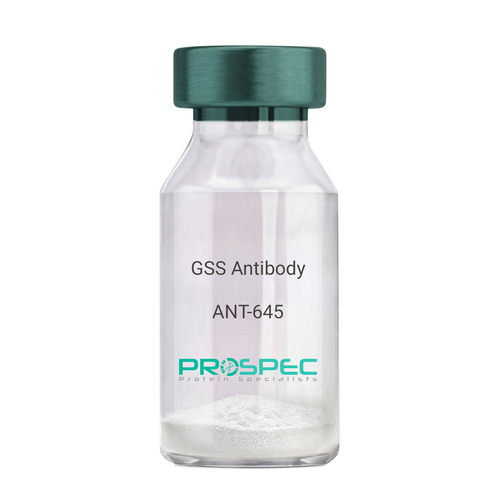
  • View Data Sheet

    Name :

    GSS Antibody

    Description:

    Glutathione Synthetase, Mouse Anti Human

    Glutathione synthetase, GSH synthetase, GSH-S, Glutathione synthase, GSHS, MGC14098, GSS.

    Product # :

    ANT-645

    Price :

    Quantity :

    Shipping Method :

    Ice Icon

    Shipped with Ice Packs

    Add To Cart

    More Info

    • formulation
    • More Info
    • Citations

    Formulation

    1mg/ml containing PBS, pH-7.4, 10% Glycerol and 0.02% Sodium Azide.

    Citations

    More Info

    • Introduction

      Glutathione synthetase (GSS) is the second enzyme in the glutathione biosynthesis pathway. GSS catalyses the condensation of gamma-glutamylcysteine and glycine to form glutathione. Defects in the GSS are the cause of glutathione synthetase deficiency aka GSS deficiency or 5-oxoprolinuria or pyroglutamic aciduria, which is a severe form characterized by an increased rate of hemolysis and defective function of the central nervous system.

    • Synonyms

      Glutathione synthetase, GSH synthetase, GSH-S, Glutathione synthase, GSHS, MGC14098, GSS.

    • Physical Appearance

      Sterile filtered colorless solution.

    • Immunogen

      Anti-human GSS mAb, is derived from hybridization of mouse F0 myeloma cells with spleen cells from BALB/c mice immunized with recombinant human GSS amino acids 1-474 purified from E. coli.

    • Ig Subclass

      Mouse IgG1 heavy chain and k light chain.

    • Clone

      PAT12F2AT.

    • Applications

      GSS antibody has been tested by ELISA, Western blot analysis and ICC/IF to assure specificity and reactivity. Since application varies, however, each investigation should be titrated by the reagent to obtain optimal results.

    • Type

      Mouse Anti Human Monoclonal.

    • Storage Procedures

      For periods up to 1 month store at 4°C, for longer periods of time, store at -20°C. Prevent freeze thaw cycles.

    • Purification Method

      GSS antibody was purified from mouse ascitic fluids by protein-A affinity chromatography.

    ProSpec's products are furnished for LABORATORY RESEARCH USE ONLY. The product may not be used as drugs, agricultural or pesticidal products, food additives or household chemicals.

    Gss Antibody
  • View Data Sheet

    Name :

    GLUL Antibody

    Description:

    Glutamine Synthetase, Mouse Anti Human

    GLNS, EC 6.3.1.2, EC 4.1.1.15, GLUL, Glutamine Synthetase, GS, Glutamate decarboxylase, Glutamate--ammonia ligase, PIG43, PIG59.

    Product # :

    ANT-705

    Price :

    Quantity :

    Shipping Method :

    Ice Icon

    Shipped with Ice Packs

    Add To Cart

    More Info

    • formulation
    • More Info
    • Citations

    Formulation

    1mg/ml containing PBS, pH-7.4, 10% Glycerol and 0.02% Sodium Azide.

    Citations

    More Info

    • Introduction

      GLUL catalyzes the synthesis of glutamine from glutamate and ammonia. Glutamine is a major source of energy and that takes part in cell proliferation, inhibition of apoptosis, and cell signaling. GLUL is expressed during early fetal stages, and has a role in maintaining body pH by removing ammonia from circulation. Mutations in GLUL gene are related with congenital glutamine deficiency.

    • Synonyms

      GLNS, EC 6.3.1.2, EC 4.1.1.15, GLUL, Glutamine Synthetase, GS, Glutamate decarboxylase, Glutamate--ammonia ligase, PIG43, PIG59.

    • Physical Appearance

      Sterile filtered colorless solution.

    • Immunogen

      Anti-human GLUL mAb, is derived from hybridization of mouse F0 myeloma cells with spleen cells from BALB/c mice immunized with recombinant human GLUL amino acids 1-373 purified from E. coli.

    • Ig Subclass

      Mouse IgG1 heavy chain and κ light chain.

    • Clone

      PAT8D7AT.

    • Applications

      GLUL antibody has been tested by ELISA and Western blot analysis to assure specificity and reactivity. Since application varies, however, each investigation should be titrated by the reagent to obtain optimal results.

    • Type

      Mouse Anti Human Monoclonal.

    • Storage Procedures

      For periods up to 1 month store at 4°C, for longer periods of time, store at -20°C. Prevent freeze thaw cycles.

    • Purification Method

      GLUL antibody was purified from mouse ascitic fluids by protein-A affinity chromatography.

    ProSpec's products are furnished for LABORATORY RESEARCH USE ONLY. The product may not be used as drugs, agricultural or pesticidal products, food additives or household chemicals.

    Glul Antibody
  • View Data Sheet

    Name :

    PPP1CA Antibody

    Description:

    Protein Phosphatase 1 Catalytic Subunit Alpha, Mouse Anti Human

    Serine/threonine-protein phosphatase PP1-alpha catalytic subunit, PP-1A, PPP1CA, PPP1A, protein phosphatase 1 catalytic subunit alpha isozyme, MGC1674, MGC15877, PP1alpha.

    Product # :

    ANT-415

    Price :

    Quantity :

    Shipping Method :

    Ice Icon

    Shipped with Ice Packs

    Add To Cart

    More Info

    • formulation
    • More Info
    • Citations

    Formulation

    1mg/ml containing PBS, pH-7.4, & 0.1% Sodium Azide.

    Citations

    More Info

    • Introduction

      PPP1CA is one of the 3 catalytic subunits of protein phosphatase 1 (PP1). PP1 is a serine/threonine specific protein phosphatase which is involved in the regulation of a variety of cellular processes, such as cell division, glycogen metabolism, muscle contractility, protein synthesis, and HIV-1 viral transcription. Increased PP1 activity is detected in the end stage of heart failure. Studies in both human and mice suggest that PP1 is a significant regulator of cardiac function. Mouse studies also propose that PP1 functions as a suppressor of learning and memory.

    • Synonyms

      Serine/threonine-protein phosphatase PP1-alpha catalytic subunit, PP-1A, PPP1CA, PPP1A, protein phosphatase 1 catalytic subunit alpha isozyme, MGC1674, MGC15877, PP1alpha.

    • Physical Appearance

      Sterile Filtered clear solution.

    • Immunogen

      Anti-human PPP1CA mAb, is derived from hybridization of mouse FO myeloma cells with spleen cells from BALB/c mice immunized with recombinant human PPP1CA amino acids 30-299 purified from E. coli.

    • Ig Subclass

      Mouse IgG2a heavy chain and κ light chain.

    • Clone

      P4G3AT.

    • Applications

      PPP1CA antibody has been tested by ELISA and Western blot analysis to assure specificity and reactivity. Since application varies, however, each investigation should be titrated by the reagent to obtain optimal results. Recommended dilution range for Western blot analysis is 1:500 ~ 2,000. Recommended starting dilution is 1:1,000.

    • Type

      Mouse Anti Human Monoclonal.

    • Storage Procedures

      For periods up to 1 month store at 4°C, for longer periods of time, store at -20°C. Prevent freeze thaw cycles.

    • Purification Method

      PPP1CA antibody was purified from mouse ascitic fluids by protein-G affinity chromatography.

    ProSpec's products are furnished for LABORATORY RESEARCH USE ONLY. The product may not be used as drugs, agricultural or pesticidal products, food additives or household chemicals.

    Ppp1Ca Antibody
  • View Data Sheet

    Name :

    CD14 Antibody, FITC

    Description:

    CD14, Mouse Anti-Human FITC

    Monocyte differentiation antigen CD14, Myeloid cell-specific leucine-rich glycoprotein.

    Product # :

    ANT-253

    Price :

    Quantity :

    Shipping Method :

    Room Temp Icon

    Shipped at Room temp

    Add To Cart

    More Info

    • formulation
    • More Info
    • Citations

    Formulation

    1mg/ml in PBS (after reconstitution).

    Citations

    More Info

    • Introduction

      CD14 (also known lipopolysaccharide (LPS) receptor) is expressed strongly on monocytes and macrophage and weakly on the surface of neutrophils. CD14 is anchored to cells by linkage to glycosylphosphatidylinositol (GPI) and functions as a high affinity receptor for complexes of LPS and LPS binding protein (LBP). Soluble CD14, also binding to LPS, acts at physiological concentration as an LPS agonist and has, at higher concentrations, an LPS antagonizing effect in cell activation. CD14 has been shown to bind apoptotic cells.

    • Synonyms

      Monocyte differentiation antigen CD14, Myeloid cell-specific leucine-rich glycoprotein.

    • Solubility

      Reconstitute with of H20. Mix gently, wash the sides of the vial and wait 30-60 seconds before use.

    • Immunogen

      Purified human B Cells.

    • Ig Subclass

      Mouse IgG1.

    • Clone

      hCD4.

    • Applications

      For staining, use 10 µl/1,000,000 cells.

    • Available Conjugates

      This antibody is also available as purified antibody and conjugated to biotin.

    • Type

      Mouse Anti Human Monoclonal.

    • Storage Procedures

      Lyophilized: store at 4°C. After reconstitution, if not intended for use within a month, aliquot and store at -20°C.

    • Purification Method

      Ion exchange column.

    ProSpec's products are furnished for LABORATORY RESEARCH USE ONLY. The product may not be used as drugs, agricultural or pesticidal products, food additives or household chemicals.

    Cd14 Antibody Fitc
  • View Data Sheet

    Name :

    Shiga Like Toxin 1

    Description:

    Shiga Like Toxin-1 Subunit B Recombinant

    Product # :

    STX-001

    Price :

    Quantity :

    Shipping Method :

    Ice Icon

    Shipped with Ice Packs

    Add To Cart

    More Info

    • description
    • source
    • formulation
    • purity
    • More Info
    • Citations

    Description

    Recombinant Shiga Like Toxin-1 Subunit B is produced from E.Coli O157:H7 amino acids 2-90 of the Shiga Like Toxin-1 Subunit B.The Shiga Like Toxin 1 protein is fused to a 6xHis tag at its N-terminus and purified by proprietary chromatographic technique.

    Source

    Escherichia Coli.

    Formulation

    Phosphate buffered saline and 25mM K₂CO₃.

    Purity

    Protein is >95% pure as determined by 12% PAGE (coomassie staining).

    Citations

    More Info

    • Introduction

      Shiga-like toxin (verotoxin) is a toxin produced by some strains of Escherichia coli. Shiga-like toxin is named for its similarity to the AB5-type Shiga toxin produced by the bacteria Shigella dysenteriae. There are two known types-SLT1 and SLT2. The Shiga-like toxin is linked with hemolytic-uremic syndrome. Shiga-like toxin requires highly specific receptors on the cells' surface in order to attach and enter the cell. Species such as cattle, swine, and deer which do not carry these receptors may harbor toxigenic bacteria without any ill effect, dropping them in their feces, from where they may be distributed to humans. Shiga Like Toxin-1 Subunit B has nontoxic action, it is the functional region which binds to the receptor. The Shiga Like Toxin-1 Subunit B can be useful in vaccine study, antibody test and other functional research.

    • Stability

      Store at 4°C if entire vial will be used within 2-4 weeks. Store, frozen at -20°C for longer periods of time. For long term storage it is recommended to add a carrier protein (0.1% HSA or BSA).Avoid multiple freeze-thaw cycles.

    • Purification Method

      Purified by affinity chromatographic technique.

    ProSpec's products are furnished for LABORATORY RESEARCH USE ONLY. The product may not be used as drugs, agricultural or pesticidal products, food additives or household chemicals.

    Shiga Like Toxin 1
  • View Data Sheet

    Name :

    GSTT1 Antibody

    Description:

    Glutathione S-Transferase Theta-1, Mouse Anti Human

    Glutathione S-transferase theta-1, GST class-theta-1, Glutathione transferase T1-1, GSTT1.

    Product # :

    ANT-591

    Price :

    Quantity :

    Shipping Method :

    Ice Icon

    Shipped with Ice Packs

    Add To Cart

    More Info

    • formulation
    • More Info
    • Citations

    Formulation

    1mg/ml containing PBS, pH-7.4, 10% Glycerol and 0.02% Sodium Azide.

    Citations

    More Info

    • Introduction

      GSTT1 belongs to a superfamily of proteins which catalyze the conjugation of reduced glutathione to a variety of electrophilic and hydrophobic compounds. GSTT1 is one of the GSTs’ four main classes: alpha, mu, pi and theta (which includes GSTT1 and GSTT2). GSTT1 is involved in activation and detoxification reactions and catalyzes the conjugation of industrial chemicals, such as epoxybutane, ethylene oxides, halomethane with glutathione. GSTT1 is found in erythrocytes, at low levels in the liver as well as in Clara and ciliated cells at the alveolar/bronchiolar junction in the lung. The GSTT1 gene is deficient in 38% of the population. The GSTTI enzyme deficiency might influence the individual risk for development of acquired aplastic anemia and acute myeloid leukemia. The presence or absence of the GSTT1 gene is concurrent with GSST1+ (the conjugator) and GSTT1- (the non-conjugator) phenotypes correspondingly. The GSTT1+ phenotype is able to catalyze the glutathione conjugation of dichloromethane. GSTT1-null genotypes are seen as having a higher risk of developing leukoplakia. Germline genetic polymorphism in GSTT1 is linked to breast cancer.

    • Synonyms

      Glutathione S-transferase theta-1, GST class-theta-1, Glutathione transferase T1-1, GSTT1.

    • Physical Appearance

      Sterile filtered colorless solution.

    • Immunogen

      Anti-human GSTT1 mAb, is derived from hybridization of mouse F0 myeloma cells with spleen cells from BALB/c mice immunized with recombinant human GSTT1 amino acids 1-240 purified from E. coli.

    • Ig Subclass

      Mouse IgG2b heavy chain and k light chain.

    • Clone

      PAT38D11AT.

    • Applications

      GSTT1 antibody has been tested by ELISA, Western blot analysis, ICC/IF and Flow cytometry to assure specificity and reactivity. Since application varies, however, each investigation should be titrated by the reagent to obtain optimal results.

    • Type

      Mouse Anti Human Monoclonal.

    • Storage Procedures

      For periods up to 1 month store at 4°C, for longer periods of time, store at -20°C. Prevent freeze thaw cycles.

    • Purification Method

      GSTT1 antibody was purified from mouse ascitic fluids by protein-A affinity chromatography.

    ProSpec's products are furnished for LABORATORY RESEARCH USE ONLY. The product may not be used as drugs, agricultural or pesticidal products, food additives or household chemicals.

    Gstt1 Antibody
  • View Data Sheet

    Name :

    CD11b Antibody, FITC

    Description:

    CD11b, Mouse Anti-Human, FITC

    Integrin alpha-M, Cell surface glycoprotein MAC-1 subunit alpha, CR-3 alpha chain, Leukocyte adhesion receptor MO1, Neutrophil adherence receptor, CD11b antigen, ITGAM, CR3A, MO1A, CD11B, MAC-1, MAC1A, MGC117044.

    Product # :

    ANT-276

    Price :

    Quantity :

    Shipping Method :

    Room Temp Icon

    Shipped at Room temp

    Add To Cart

    More Info

    • formulation
    • More Info
    • Citations

    Formulation

    1mg/ml in PBS (after reconstitution).

    Citations

    More Info

    • Introduction

      Integrins are heterodimeric integral membrane proteins composed of an alpha chain and a beta chain. This I-domain containing alpha integrin combines with the beta 2 chain (ITGB2) to form a leukocyte-specific integrin referred to as macrophage receptor 1 ('Mac-1'), or inactivated-C3b (iC3b) receptor 3 ('CR3'). The alpha M beta 2 integrin is important in the adherence of neutrophils and monocytes to stimulated endothelium, and also in the phagocytosis of complement coated particles. Integrin alpha-m/beta-2 is implicated in various adhesive interactions of monocytes, macrophages and granulocytes as well as in mediating the uptake of complement-coated particles. CD-11b is identical with cr-3, the receptor for the ic3b fragment of the third complement component. CD-11b probably recognizes the r-g-d peptide in c3b. Integrin alpha-m/beta-2 is also a receptor for fibrinogen, factor x and icam1. it recognizes p1 and p2 peptides of fibrinogen gamma chain.

    • Synonyms

      Integrin alpha-M, Cell surface glycoprotein MAC-1 subunit alpha, CR-3 alpha chain, Leukocyte adhesion receptor MO1, Neutrophil adherence receptor, CD11b antigen, ITGAM, CR3A, MO1A, CD11B, MAC-1, MAC1A, MGC117044.

    • Solubility

      Reconstitute with of H20. Mix gently, wash the sides of the vial and wait 30-60 seconds before use.

    • Immunogen

      Purified human PBL Monocytes.

    • Ig Subclass

      Mouse IgG1.

    • Clone

      hCD11b.

    • Applications

      Blocking and staining antibody. For staining, use 10µl/1,000,000 cells. Titer for blocking LPS binding should be determined by the investigator.

    • Available Conjugates

      This antibody is also available unconjugated and conjugated to Biotin. For staining with biotin or FITC-conjugated antibody use 5-10µl/1,000,000 cells.

    • Type

      Mouse Anti Human Monoclonal.

    • Storage Procedures

      Lyophilized: store at 4°C. After reconstitution, if not intended for use within a month, aliquot and store at -20°C.

    • Purification Method

      Ion exchange column.

    ProSpec's products are furnished for LABORATORY RESEARCH USE ONLY. The product may not be used as drugs, agricultural or pesticidal products, food additives or household chemicals.

    Cd11B Antibody Fitc
  • View Data Sheet

    Name :

    CD11b Antibody, Biotin

    Description:

    CD11b, Mouse Anti-Human, Biotin

    Integrin alpha-M, Cell surface glycoprotein MAC-1 subunit alpha, CR-3 alpha chain, Leukocyte adhesion receptor MO1, Neutrophil adherence receptor, CD11b antigen, ITGAM, CR3A, MO1A, CD11B, MAC-1, MAC1A, MGC117044.

    Product # :

    ANT-275

    Price :

    Quantity :

    Shipping Method :

    Room Temp Icon

    Shipped at Room temp

    Add To Cart

    More Info

    • formulation
    • More Info
    • Citations

    Formulation

    1mg/ml in PBS (after reconstitution).

    Citations

    More Info

    • Introduction

      Integrins are heterodimeric integral membrane proteins consisting of an ? chain and a ? chain. CD11b is an I-domain including ? integrin combines with the ? 2 chain (ITGB2) to form a leukocyte-specific integrin referred to as macrophage receptor also called MAC1. The ? M beta 2 integrin is crucial in the adherence of neutrophils and monocytes to stimulated endothelium, and also in the phagocytosis of complement coated particles. Multiple transcript variants encoding different isoforms have been found for this gene.

    • Synonyms

      Integrin alpha-M, Cell surface glycoprotein MAC-1 subunit alpha, CR-3 alpha chain, Leukocyte adhesion receptor MO1, Neutrophil adherence receptor, CD11b antigen, ITGAM, CR3A, MO1A, CD11B, MAC-1, MAC1A, MGC117044.

    • Solubility

      Reconstitute with of H20. Mix gently, wash the sides of the vial and wait 30-60 seconds before use.

    • Immunogen

      Purified human PBL Monocytes.

    • Ig Subclass

      Mouse IgG1.

    • Clone

      hCD11b.

    • Applications

      Blocking and staining antibody. For staining, use 10µl/1,000,000 cells. Titer for blocking LPS binding should be determined by the investigator.

    • Available Conjugates

      This antibody is also available unconjugated and conjugated to FITC. For staining with biotin or FITC-conjugated antibody use 5-10µl/1,000,000 cells.

    • Type

      Mouse Anti Human Monoclonal.

    • Storage Procedures

      Lyophilized: store at 4°C. After reconstitution, if not intended for use within a month, aliquot and store at -20°C.

    • Purification Method

      Ion exchange column.

    ProSpec's products are furnished for LABORATORY RESEARCH USE ONLY. The product may not be used as drugs, agricultural or pesticidal products, food additives or household chemicals.

    Cd11B Antibody Biotin
  • View Data Sheet

    Name :

    PPM1A Antibody

    Description:

    Protein Phosphatase 1A Alpha Isoform, Mouse Anti Human

    Protein phosphatase 1A, EC 3.1.3.16, Protein phosphatase 2C isoform alpha, PP2C-alpha, IA, PPM1A, PP2CA, MGC9201.

    Product # :

    ANT-309

    Price :

    Quantity :

    Shipping Method :

    Ice Icon

    Shipped with Ice Packs

    Add To Cart

    More Info

    • formulation
    • More Info
    • Citations

    Formulation

    1mg/ml containing PBS, pH-7.4, & 0.1% Sodium Azide.

    Citations

    More Info

    • Introduction

      Protein Phosphatase 2C alpha is a member of the PP2C family of Ser/Thr protein phosphatases. PP2C family members are known to be negative regulators of cell stress response pathways. This phosphatase dephosphorylates, and negatively regulates the activities of, MAP kinases and MAP kinase kinases. It has been shown to inhibit the activation of p38 and JNK kinase cascades induced by environmental stresses. This phosphatase can also dephosphorylate cyclin-dependent kinases, and thus may be involved in cell cycle control. Overexpression of this phosphatase is reported to activate the expression of the tumor suppressor gene TP53/p53, which leads to G2/M cell cycle arrest and apoptosis. Three alternatively spliced transcript variants encoding two distinct isoforms have been described. Protein phosphatase 2C(PP2Cα) is a Mn2+- or Mg2+-dependent protein serine/threonine phosphatase that is essential for regulating cellular stress response in eukaryotes.

    • Synonyms

      Protein phosphatase 1A, EC 3.1.3.16, Protein phosphatase 2C isoform alpha, PP2C-alpha, IA, PPM1A, PP2CA, MGC9201.

    • Immunogen

      Anti-human PPM1A mAb, is derived from hybridization of mouse SP2/O myeloma cells with spleen cells from BALB/c mice immunized with recombinant human PPM1A amino acids 1-382 purified from E. coli.

    • Ig Subclass

      Mouse IgG2b heavy chain and κ light chain.

    • Clone

      Pp6c7AT.

    • Applications

      PPM1A antibody has been tested by ELISA and Western blot analysis to assure specificity and reactivity. Since application varies, however, each investigation should be titrated by the reagent to obtain optimal results. Recommended dilution range for Western blot analysis is 1:250 ~ 500. Recommended starting dilution is 1:250.

    • Type

      Mouse Anti Human Monoclonal.

    • Storage Procedures

      For periods up to 1 month store at 4°C, for longer periods of time, store at -20°C. Prevent freeze thaw cycles.

    • Purification Method

      PPM1A antibody was purified from mouse ascitic fluids by protein-G affinity chromatography.

    ProSpec's products are furnished for LABORATORY RESEARCH USE ONLY. The product may not be used as drugs, agricultural or pesticidal products, food additives or household chemicals.

    Ppm1A Antibody
  • View Data Sheet

    Name :

    HSF1 Antibody

    Description:

    Heat Shock Transcription Factor-1, Mouse Anti Human

    HSF-1, HSF1, HSTF-1, HSTF1, Heat shock factor protein 1, Heat shock transcription factor 1.

    Product # :

    ANT-328

    Price :

    Quantity :

    Shipping Method :

    Ice Icon

    Shipped with Ice Packs

    Add To Cart

    More Info

    • formulation
    • More Info
    • Citations

    Formulation

    1mg/ml containing PBS, pH-7.4, 0.02% Sodium Azide and 10% Glycerol.

    Citations

    More Info

    • Introduction

      The transcription of heat-shock genes is rapidly induced after temperature stress in response to environmental changes, and it is also involved in oogenesis, spermatogenesis, and placental development. Hsp90, is a major repressor of HSF1 gene. HSF1 is known for its participation in heat shock response. HSF1 protein regulates the transcription of hundreds of targets, such as genes involved in protein folding, detoxification, energy generation, carbohydrate metabolism, and cell wall organization. HSF1 binds to MTA1 in vitro & in breast carcinoma. Suppression of estrogen-dependent transcription may contribute to role of HSF1 in cancer. Human cancer lines of diverse origins express higher dependence on HSF1 function to maintain proliferation and survival than their non-transformed counterparts. HSF1 induces anticoagulation and relaxation factors in vascular endothelial cells and could therefore be used to treat cardiovascular disease. HSF1 is directly involved in the regulation of HO1 havi

    • Synonyms

      HSF-1, HSF1, HSTF-1, HSTF1, Heat shock factor protein 1, Heat shock transcription factor 1.

    • Immunogen

      Anti-human HSF1 mAb, is derived from hybridization of mouse FO myeloma cells with spleen cells from BALB/c mice immunized with recombinant human HSF1 amino acids 1-529 purified from E. coli.

    • Ig Subclass

      Mouse IgG1 heavy chain and κ light chain.

    • Clone

      PJ7F9AT.

    • Applications

      HSF1 antibody has been tested by ELISA and Western blot analysis to assure specificity and reactivity. Since application varies, however, each investigation should be titrated by the reagent to obtain optimal results. Recommended dilution range for Western blot analysis is 1:1,000~ 2,000. Recommended starting dilution is 1:1,000.

    • Type

      Mouse Anti Human Monoclonal.

    • Storage Procedures

      For periods up to 1 month store at 4°C, for longer periods of time, store at -20°C. Prevent freeze thaw cycles.

    • Purification Method

      HSF1 antibody was purified from mouse ascitic fluids by protein-G affinity chromatography.

    ProSpec's products are furnished for LABORATORY RESEARCH USE ONLY. The product may not be used as drugs, agricultural or pesticidal products, food additives or household chemicals.

    Hsf1 Antibody
  • View Data Sheet

    Name :

    ENO2 Antibody

    Description:

    Enolase-2, Mouse Anti Human

    Gamma-enolase, EC 4.2.1.11, 2-phospho-D-glycerate hydro-lyase, Neural enolase, Neuron-specific enolase, NSE, Enolase 2, ENO2.

    Product # :

    ANT-497

    Price :

    Quantity :

    Shipping Method :

    Ice Icon

    Shipped with Ice Packs

    Add To Cart

    More Info

    • formulation
    • More Info
    • Citations

    Formulation

    1mg/ml containing PBS, pH-7.4, 10% Glycerol and 0.02% Sodium Azide.

    Citations

    More Info

    • Introduction

      Neuron-specificenolase also caled NSE is a glycolytic isoenzyme which is situated in central and peripheral neurons and neuroendocrine cells. Enolase-2 is released into the CSF when neural tissue is injured. Neoplasms derived from neural or neuroendocrine tissue release Enolase-2 into the blood. Enolase-2 is a useful substance that has been detected in patients with certain tumors, such as neuroblastoma, small cell lung cancer, medullary thyroid cancer, carcinoid tumors, pancreatic endocrine tumors, and melanoma. ENO2 is 1 of the 3 enolase isoenzymes found in mammals. ENO2 isoenzyme, is found in mature neurons and cells of neuronal origin. An exchange from alpha enolase to gamma enolase occurs in neural tissue during development in rats and primates.

    • Synonyms

      Gamma-enolase, EC 4.2.1.11, 2-phospho-D-glycerate hydro-lyase, Neural enolase, Neuron-specific enolase, NSE, Enolase 2, ENO2.

    • Physical Appearance

      Sterile filtered colorless solution.

    • Immunogen

      Anti-human ENO2 mAb, clone PAT17D10A, is derived from hybridization of mouse F0 myeloma cells with spleen cells from BALB/c mice immunized with a recombinant human ENO2 protein 1-434 amino acids purified from E. coli.

    • Ig Subclass

      Mouse IgG2b heavy chain and Kappa light chain.

    • Clone

      PAT17D10A

    • Applications

      The antibody has been tested by ELISA, Western blot analysis to assure specificity and reactivity. Since application varies, however, each investigation should be titrated by the reagent to obtain optimal results. Recommended dilution range for Western blot analysis is 1:500 ~ 1:5000. Recommended starting dilution is 1:1000.

    • Type

      Mouse Anti Human Monoclonal.

    • Storage Procedures

      For periods up to 1 month store at 4°C, for longer periods of time, store at -20°C. Prevent freeze thaw cycles.

    • Purification Method

      ENO2 antibody was purified from mouse ascitic fluids by protein-A affinity chromatography.

    ProSpec's products are furnished for LABORATORY RESEARCH USE ONLY. The product may not be used as drugs, agricultural or pesticidal products, food additives or household chemicals.

    Eno2 Antibody
  • 1
  • …
  • 29
  • 30
  • 31
  • 32
  • 33
  • …
  • 42
Your browser does not support the video tag.

Products

  • Cytokines
  • Growth Factors
  • Chemokines
  • CD Antigens
  • Neurotrophins
  • Hormones
  • Enzymes
  • Viral Antigens
  • Recombinant Proteins
  • Natural Proteins
  • Monoclonal Antibodies
  • Polyclonal Antibodies
  • Thermal Packaging

About

  • Company
  • Founder
  • Contribution
  • Contact Us
  • My Account

Ordering

  • Ordering Method
  • Ordering - Credit Card
  • Ordering PO
  • Shipping Fees
  • FAQ's

Legal

  • Terms & Conditions
  • Privacy
  • Site Map
  • info@prospecbio.com
  • Facebook
  • Instagram
  • Linkedin Profile

© 2026 ProSpec-Tany TechnoGene Ltd. All Rights Reserved.

  • Choosing a selection results in a full page refresh.
  • Opens in a new window.