Skip to content

High-quality research proteins.

  • 1-800-805-6531

  • Mon - Sat 8.00-18.00, Sun - Closed

  • info@prospecbio.com

  • PO Box 6591 East Brunswick NJ 08816

  • Products
    • Cytokines
    • Growth factors
    • Chemokines
    • Cd antigens
    • Neurotrophins
    • Hormones
    • Enzymes
    • Viral antigens
    • Recombinant proteins
    • Natural proteins
    • Monoclonal antibodies
    • Polyclonal antibodies
    • Thermal Packaging
  • About
    • Company
    • Founder
    • Contribution
  • Customers
    • Eastern Asia
    • Eastern Europe
    • North America
    • South America
    • South East Asia
    • Southern Asia
    • Western Europe
  • Ordering
    • Ordering Method
    • Ordering - Credit Card
    • Ordering - PO
    • Shipping Fees
    • FAQs
  • Services
    • DNA Cloning
    • Protein Expression
    • Monoclonal Antibody
    • Fermentation
    • Technology License
  • Contact us
Log in

Country/region

  • USD $ | Albania
  • USD $ | Andorra
  • USD $ | Argentina
  • USD $ | Armenia
  • USD $ | Australia
  • USD $ | Austria
  • USD $ | Azerbaijan
  • USD $ | Belarus
  • USD $ | Belgium
  • USD $ | Bolivia
  • USD $ | Brazil
  • USD $ | Bulgaria
  • USD $ | Cambodia
  • USD $ | Canada
  • USD $ | Chile
  • USD $ | China
  • USD $ | Colombia
  • USD $ | Costa Rica
  • USD $ | Croatia
  • USD $ | Cyprus
  • USD $ | Czechia
  • USD $ | Denmark
  • USD $ | Estonia
  • USD $ | Finland
  • USD $ | France
  • USD $ | Georgia
  • USD $ | Germany
  • USD $ | Greece
  • USD $ | Hong Kong SAR
  • USD $ | Hungary
  • USD $ | Iceland
  • USD $ | India
  • USD $ | Ireland
  • USD $ | Israel
  • USD $ | Italy
  • USD $ | Japan
  • USD $ | Kazakhstan
  • USD $ | Latvia
  • USD $ | Liechtenstein
  • USD $ | Lithuania
  • USD $ | Madagascar
  • USD $ | Malta
  • USD $ | Mexico
  • USD $ | Moldova
  • USD $ | Monaco
  • USD $ | Netherlands
  • USD $ | New Zealand
  • USD $ | North Macedonia
  • USD $ | Norway
  • USD $ | Panama
  • USD $ | Paraguay
  • USD $ | Peru
  • USD $ | Philippines
  • USD $ | Poland
  • USD $ | Portugal
  • USD $ | Romania
  • USD $ | Singapore
  • USD $ | Slovakia
  • USD $ | Slovenia
  • USD $ | South Africa
  • USD $ | South Korea
  • USD $ | Spain
  • USD $ | Sri Lanka
  • USD $ | Sweden
  • USD $ | Switzerland
  • USD $ | Taiwan
  • USD $ | Thailand
  • USD $ | Türkiye
  • USD $ | Ukraine
  • USD $ | United Kingdom
  • USD $ | United States
  • USD $ | Uruguay
  • USD $ | Vatican City
  • USD $ | Venezuela
  • USD $ | Vietnam
  • Facebook
  • Instagram
logoProSpec - Recombinant Protein Manufacturer for academic and R&D industry
Become a distributor
Account Log in
Wishlist
Cart Cart(0)
  • Products
    +
    • Cytokines
    • Growth factors
    • Chemokines
    • Cd antigens
    • Neurotrophins
    • Hormones
    • Enzymes
    • Viral antigens
    • Recombinant proteins
    • Natural proteins
    • Monoclonal antibodies
    • Polyclonal antibodies
    • Thermal Packaging
  • About
    +
    • Company
    • Founder
    • Contribution
  • Customers
    +
    • Eastern Asia
    • Eastern Europe
    • North America
    • South America
    • South East Asia
    • Southern Asia
    • Western Europe
  • Ordering
    +
    • Ordering Method
    • Ordering - Credit Card
    • Ordering - PO
    • Shipping Fees
    • FAQs
  • Services
    +
    • DNA Cloning
    • Protein Expression
    • Monoclonal Antibody
    • Fermentation
    • Technology License
  • Contact us

Item added to your cart

View cart
View All
  • Cytokines
  • Growth factors
  • Chemokines
  • Cd antigens
  • Neurotrophins
  • Hormones
  • Enzymes
  • Viral antigens
  • Recombinant proteins
  • Natural proteins
  • Monoclonal antibodies
  • Polyclonal antibodies
  • Thermal Packaging
  • Tumor Necrosis Factor

    Tumor Necrosis Factor

    View All
  • 4-1BB

    4-1BB

    View All
  • Adiponectin

    Adiponectin

    View All
  • AIF1

    AIF1

    View All
  • Other Cytokines

    Other Cytokines

    View All
  • AITRL

    AITRL

    View All
  • Angiopoietin

    Angiopoietin

    View All
  • Apolipoprotein

    Apolipoprotein

    View All
  • B-Cell Activating Factor

    B-Cell Activating Factor

    View All
  • Beta Defensin

    Beta Defensin

    View All
  • Bone Morphogenetic Protein

    Bone Morphogenetic Protein

    View All
  • B type Natriuretic Peptide

    B type Natriuretic Peptide

    View All
  • BST

    BST

    View All
  • Betacellulin

    Betacellulin

    View All
  • Cardiotrophin

    Cardiotrophin

    View All
  • Activin

    Activin

    View All
  • Fibroblast Growth Factor

    Fibroblast Growth Factor

    View All
  • Other Growth Factors

    Other Growth Factors

    View All
  • CSF

    CSF

    View All
  • CTGF

    CTGF

    View All
  • IGFBP

    IGFBP

    View All
  • VEGF Protein

    VEGF Protein

    View All
  • EGF

    EGF

    View All
  • Epigen

    Epigen

    View All
  • Erythropoietin

    Erythropoietin

    View All
  • Insulin-Like Growth Factor

    Insulin-Like Growth Factor

    View All
  • Growth Hormone

    Growth Hormone

    View All
  • HDGF

    HDGF

    View All
  • Hepatocyte Growth Factor

    Hepatocyte Growth Factor

    View All
  • Insulin

    Insulin

    View All
  • BCA-1/ BLC (CXCL13)

    BCA-1/ BLC (CXCL13)

    View All
  • BRAK (CXCL14)

    BRAK (CXCL14)

    View All
  • C-10 (CCL6)

    C-10 (CCL6)

    View All
  • Other Chemokines

    Other Chemokines

    View All
  • MEC (CCL28)

    MEC (CCL28)

    View All
  • LD78-beta (CCL3L1)

    LD78-beta (CCL3L1)

    View All
  • CTACK (CCL27)

    CTACK (CCL27)

    View All
  • CXCL16

    CXCL16

    View All
  • CXCL17

    CXCL17

    View All
  • Platelet Factor-4 (CXCL4)

    Platelet Factor-4 (CXCL4)

    View All
  • ENA-78 (CXCL5)

    ENA-78 (CXCL5)

    View All
  • Eotaxin (CCL11,24,26)

    Eotaxin (CCL11,24,26)

    View All
  • Exodus-2 (CCL21)

    Exodus-2 (CCL21)

    View All
  • Fractalkine (CX3CL1)

    Fractalkine (CX3CL1)

    View All
  • CXCL6

    CXCL6

    View All
  • Other CD Antigens

    Other CD Antigens

    View All
  • CD14

    CD14

    View All
  • CD164

    CD164

    View All
  • CD1

    CD1

    View All
  • CD2

    CD2

    View All
  • CD200

    CD200

    View All
  • CD207

    CD207

    View All
  • CD226

    CD226

    View All
  • CD23

    CD23

    View All
  • CD244

    CD244

    View All
  • CD247

    CD247

    View All
  • CD27

    CD27

    View All
  • CD274

    CD274

    View All
  • CD300

    CD300

    View All
  • CD33

    CD33

    View All
  • Other Neurotrophins

    Other Neurotrophins

    View All
  • Beta-NGF

    Beta-NGF

    View All
  • BDNF

    BDNF

    View All
  • CDNF

    CDNF

    View All
  • CNTF

    CNTF

    View All
  • GDNF

    GDNF

    View All
  • Glia Maturation Factor

    Glia Maturation Factor

    View All
  • MANF

    MANF

    View All
  • Midkine

    Midkine

    View All
  • Neuroglobin

    Neuroglobin

    View All
  • Neurotrophic factors

    Neurotrophic factors

    View All
  • Neuregulin

    Neuregulin

    View All
  • Neuropilin

    Neuropilin

    View All
  • Pigment Epithelium-Derived Factor

    Pigment Epithelium-Derived Factor

    View All
  • Pleiotrophin

    Pleiotrophin

    View All
  • Peptide Hormones

    Peptide Hormones

    View All
  • Other Hormones

    Other Hormones

    View All
  • HCG

    HCG

    View All
  • Endothelin

    Endothelin

    View All
  • Exendin

    Exendin

    View All
  • FSH

    FSH

    View All
  • Glucagon

    Glucagon

    View All
  • GHRP

    GHRP

    View All
  • GLP

    GLP

    View All
  • Thyrostimulin

    Thyrostimulin

    View All
  • Inhibin A

    Inhibin A

    View All
  • LHRH

    LHRH

    View All
  • Melanotan

    Melanotan

    View All
  • Procalcitonin

    Procalcitonin

    View All
  • PTH

    PTH

    View All
  • Peroxiredoxin

    Peroxiredoxin

    View All
  • Peptidase

    Peptidase

    View All
  • Synthetase

    Synthetase

    View All
  • Transferase

    Transferase

    View All
  • Hydrolase

    Hydrolase

    View All
  • Dehydrogenase

    Dehydrogenase

    View All
  • ACE2

    ACE2

    View All
  • Esterase

    Esterase

    View All
  • Protein Kinases

    Protein Kinases

    View All
  • Other Enzymes

    Other Enzymes

    View All
  • Phosphatase

    Phosphatase

    View All
  • Deaminase

    Deaminase

    View All
  • Oxygenase

    Oxygenase

    View All
  • Adenylate Kinase

    Adenylate Kinase

    View All
  • Lyase

    Lyase

    View All
  • Chagas

    Chagas

    View All
  • SARS

    SARS

    View All
  • Influenza

    Influenza

    View All
  • ASFV

    ASFV

    View All
  • Astrovirus

    Astrovirus

    View All
  • Borrelia

    Borrelia

    View All
  • Helicobacter Pylori

    Helicobacter Pylori

    View All
  • Chikungunya

    Chikungunya

    View All
  • Chlamydia

    Chlamydia

    View All
  • Cytomegalo

    Cytomegalo

    View All
  • Dengue

    Dengue

    View All
  • EBV

    EBV

    View All
  • Ebola

    Ebola

    View All
  • Feline Leukemia Virus

    Feline Leukemia Virus

    View All
  • Hepatitis A

    Hepatitis A

    View All
  • Other

    Other

    View All
  • Actin

    Actin

    View All
  • ADAM

    ADAM

    View All
  • Complement Component

    Complement Component

    View All
  • Ag85

    Ag85

    View All
  • Eukaryotic Translation Initiation Factor

    Eukaryotic Translation Initiation Factor

    View All
  • Anterior Gradient Protein

    Anterior Gradient Protein

    View All
  • Heat Shock Protein

    Heat Shock Protein

    View All
  • Alpha-2-HS-Glycoprotein

    Alpha-2-HS-Glycoprotein

    View All
  • Hemoglobin

    Hemoglobin

    View All
  • Allergy

    Allergy

    View All
  • Anaplasma

    Anaplasma

    View All
  • Angiogenin

    Angiogenin

    View All
  • Ankyrin Repeat Domain

    Ankyrin Repeat Domain

    View All
  • Annexin

    Annexin

    View All
  • Other Natural Proteins

    Other Natural Proteins

    View All
  • Aprotinin

    Aprotinin

    View All
  • Transferrin

    Transferrin

    View All
  • Avidin

    Avidin

    View All
  • Anti Coagulation Factors

    Anti Coagulation Factors

    View All
  • Natural Albumin

    Natural Albumin

    View All
  • Natural Coagulation Factors

    Natural Coagulation Factors

    View All
  • Fibronectin

    Fibronectin

    View All
  • Thrombin

    Thrombin

    View All
  • Anti Human Enzyme

    Anti Human Enzyme

    View All
  • Other Monoclonal Antibodies

    Other Monoclonal Antibodies

    View All
  • Anti Human Cytokine

    Anti Human Cytokine

    View All
  • Anti Human Heat Shock Protein

    Anti Human Heat Shock Protein

    View All
  • Anti Mouse Lymphocyte

    Anti Mouse Lymphocyte

    View All
  • Anti Human Chemokine

    Anti Human Chemokine

    View All
  • Anti Human Lymphocyte

    Anti Human Lymphocyte

    View All
  • Anti Viral Monoclonal

    Anti Viral Monoclonal

    View All
  • Anti Mouse Cytokine

    Anti Mouse Cytokine

    View All
  • Anti-GST Monoclonal

    Anti-GST Monoclonal

    View All
  • Other Polyclonal Antibodies

    Other Polyclonal Antibodies

    View All
  • Anti Viral

    Anti Viral

    View All
  • Anti-GST Monoclonal

    Anti-GST Monoclonal

    View All
Back

Search results

1000 results found for “enterokinase”

Name

Description

Product #

Price

Quantity

Shipping Method

  • View Data Sheet

    Name :

    PRKACA Human, sf9

    Description:

    c-AMP dependant Protein Kinase A catalytic subunit alpha Human Recombinant, Sf9

    cAMP-dependent protein kinase alpha-catalytic subunit, EC 2.7.11.11, PKA C-alpha, PKACA, PRKACA,  MGC48865, MGC102831.

    Product # :

    PKA-093

    Price :

    Quantity :

    Shipping Method :

    Ice Icon

    Shipped with Ice Packs

    Add To Cart

    More Info

    • description
    • source
    • formulation
    • purity
    • More Info

    Description

    PRKACA Human Recombinant produced in Sf9 Baculovirus cells is a single, glycosylated polypeptide chain containing 578 amino acids (1-351 a.a.) and having a molecular mass of 67kDa (Migrates at 50-70kDa on SDS-PAGE under reducing conditions). PRKACA is expressed with a 227 amino acid GST Tag at N-Terminus and purified by proprietary chromatographic techniques.

    Source

    Sf9, Baculovirus cells.

    Formulation

    PRKACA protein solution (0.25mg/ml) contains Phosphate Buffered Saline (pH 7.4) and 10% glycerol.

    Purity

    Greater than 85% as determined by SDS-PAGE.

    More Info

    • Introduction

      cAMP-dependent protein kinase catalytic subunit alpha isoform Calpha1 (PRKACA) belongs to the Ser/Thr protein kinase family. PRKACA is responsible for phosphorylating other proteins and substrates, changing their activity. The PRKACA protein is a member of the AGC kinase family, and contributes to the regulation of cellular processes which include glucose metabolism, cell division, and contextual memory.

    • Synonyms

      cAMP-dependent protein kinase alpha-catalytic subunit, EC 2.7.11.11, PKA C-alpha, PKACA, PRKACA, MGC48865, MGC102831.

    • Physical Appearance

      Sterile Filtered colorless solution.

    • Stability

      Store at 4°C if entire vial will be used within 2-4 weeks. Store, frozen at -20°C for longer periods of time. For long term storage it is recommended to add a carrier protein (0.1% HSA or BSA).Avoid multiple freeze-thaw cycles.

    • Amino Acid Sequence

      MSPILGYWKI KGLVQPTRLL LEYLEEKYEE HLYERDEGDK WRNKKFELGL EFPNLPYYID GDVKLTQSMA IIRYIADKHN MLGGCPKERA EISMLEGAVL DIRYGVSRIA YSKDFETLKV DFLSKLPEML KMFEDRLCHK TYLNGDHVTH PDFMLYDALD VVLYMDPMCL DAFPKLVCFK KRIEAIPQID KYLKSSKYIA WPLQGWQATF GGGDHPPKSD LVPRGSHMGN AAAAKKGSEQ ESVKEFLAKA KEDFLKKWES PAQNTAHLDQ FERIKTLGTG SFGRVMLVKH KETGNHYAMK ILDKQKVVKL KQIEHTLNEK RILQAVNFPF LVKLEFSFKD NSNLYMVMEY VPGGEMFSHL RRIGRFSEPH ARFYAAQIVL TFEYLHSLDL IYRDLKPENL LIDQQGYIQV TDFGFAKRVK GRTWTLCGTP EYLAPEIILS KGYNKAVDWW ALGVLIYEMA AGYPPFFADQ PIQIYEKIVS GKVRFPSHFS SDLKDLLRNL LQVDLTKRFG NLKNGVNDIK NHKWFATTDW IAIYQRKVEA PFIPKFKGPG DTSNFDDYEE EEIRVSINEK CGKEFSEF.

    ProSpec's products are furnished for LABORATORY RESEARCH USE ONLY. The product may not be used as drugs, agricultural or pesticidal products, food additives or household chemicals.

    Prkaca Human Sf9
  • View Data Sheet

    Name :

    MAPK3 Antibody

    Description:

    Mitogen-Activated Protein Kinase 3, Mouse Anti Human

    Mitogen-activated protein kinase 3, EC 2.7.11.24, Extracellular signal-regulated kinase 1, ERK-1, Insulin-stimulated MAP2 kinase, MAP kinase 1, MAPK 1, p44-ERK1, ERT2, p44-MAPK, Microtubule-associated protein 2 kinase, ERK1, PRKM3, P44ERK1, P44MAPK, HS44KDAP, HUMKER1A, MGC20180.

    Product # :

    ANT-023

    Price :

    Quantity :

    Shipping Method :

    Ice Icon

    Shipped with Ice Packs

    Add To Cart

    More Info

    • formulation
    • More Info

    Formulation

    1mg/ml containing PBS, pH-7.4, & 0.1% Sodium Azide.

    More Info

    • Introduction

      Extracellular signal-regulated kinases (ERKs) or classical MAP kinases are widely expressed protein kinaseintracellular signalingmolecules which are involved in functions including the regulation of meiosis, mitosis, and postmitotic functions in differentiated cells. Many different stimuli, including growth factors, cytokines, virusinfection, ligands for heterotrimeric G protein-coupled receptors, transforming agents, and carcinogens, activate the ERK pathway. The term, "extracellular signal-regulated kinases", is sometimes used as a synonym for mitogen-activated protein kinase(MAPK), but has more recently been adopted for a specific subset of the mammalianMAPK family. In the MAPK/ERK pathway, Rasactivates c-Raf, followed by MEKand then MAPK1/2 (below). Ras is typically activated by growth hormones through receptor tyrosine kinasesand GRB2/SOS, but may also receive other signals. ERKs are known to activate many transcription factors and some downstream protein kinases. Disruption of th

    • Synonyms

      Mitogen-activated protein kinase 3, EC 2.7.11.24, Extracellular signal-regulated kinase 1, ERK-1, Insulin-stimulated MAP2 kinase, MAP kinase 1, MAPK 1, p44-ERK1, ERT2, p44-MAPK, Microtubule-associated protein 2 kinase, ERK1, PRKM3, P44ERK1, P44MAPK, HS44KDAP, HUMKER1A, MGC20180.

    • Immunogen

      Anti-human MAPK3 mAb, is derived from hybridization of mouse FO myeloma cells with spleen cells from BALB/c mice immunized with recombinant human MAPK3 amino acids 1-137 purified from E. coli.

    • Ig Subclass

      Mouse IgG2b heavy chain and κ light chain.

    • Clone

      PAT1A2AT.

    • Applications

      MAPK3 antibody has been tested by ELISA, Western blot, and Immunofluorescence to assure specificity and reactivity. Since applications vary, however, each investigation should be titrated by a reagent to obtain optimal results. Recommended dilution range for Western blot analysis and Immunofluorescence is 1:250 ~ 500.
      Recommended starting dilution is 1:500.

    • Type

      Mouse Anti Human Monoclonal.

    • Storage Procedures

      For periods up to 1 month store at 4°C, for longer periods of time, store at -20°C. Prevent freeze thaw cycles.

    • Purification Method

      MAPK3 antibody was purified from mouse ascitic fluids by protein-G affinity chromatography.

    ProSpec's products are furnished for LABORATORY RESEARCH USE ONLY. The product may not be used as drugs, agricultural or pesticidal products, food additives or household chemicals.

    Mapk3 Antibody
  • View Data Sheet

    Name :

    CTSZ Mouse

    Description:

    Cathepsin-Z Mouse Recombinant

    Cathepsin Z, CTSZ.

    Product # :

    ENZ-934

    Price :

    Quantity :

    Shipping Method :

    Ice Icon

    Shipped with Ice Packs

    Add To Cart

    More Info

    • description
    • source
    • formulation
    • purity
    • More Info

    Description

    CTSZ produced in Sf9 Baculovirus cells is a single, glycosylated polypeptide chain containing 292 amino acids (23-306a.a.) and having a molecular mass of 32.8kDa. (Molecular size on SDS-PAGE will appear at approximately 28-40kDa). CTSZ is expressed with an 8 amino acid His tag at C-Terminus and purified by proprietary chromatographic techniques.

    Source

    Sf9, Baculovirus cells.

    Formulation

    CTSZ protein solution (0.5mg/ml) contains Phosphate Buffered Saline (pH 7.4) and 10% glycerol.

    Purity

    Greater than 95% as determined by SDS-PAGE.

    More Info

    • Introduction

      Cathepsin-Z (CTSZ) is a lysosomal cysteine proteinase and member of the peptidase C1 family. CTSZ, which has been also known as cathepsin X and cathepsin P, exhibits carboxy-monopeptidase and carboxy-dipeptidase activities. CTSZ is expressed ubiquitously in cancer cell lines and primary tumors and, similar to other members of this family, takes part in tumorigenesis.

    • Synonyms

      Cathepsin Z, CTSZ.

    • Physical Appearance

      Sterile Filtered clear solution.

    • Stability

      Store at 4°C if entire vial will be used within 2-4 weeks. Store, frozen at -20°C for longer periods of time.For long term storage it is recommended to add a carrier protein (0.1% HSA or BSA).Please avoid freeze thaw cycles.

    • Amino Acid Sequence

      ARARLYFRSG QTCYHPIRGD QLALLGRRTY PRPHEYLSPA DLPKNWDWRN VNGVNYASVT RNQHIPQYCG SCWAHGSTSA MADRINIKRK GAWPSILLSV QNVIDCGNAG SCEGGNDLPV WEYAHKHGIP DETCNNYQAK DQDCDKFNQC GTCTEFKECH TIQNYTLWRV GDYGSLSGRE KMMAEIYANG PISCGIMATE MMSNYTGGIY AEHQDQAVIN HIISVAGWGV SNDGIEYWIV RNSWGEPWGE KGWMRIVTST YKGGTGDSYN LAIESACTFG DPIVLEHHHH HH.

    ProSpec's products are furnished for LABORATORY RESEARCH USE ONLY. The product may not be used as drugs, agricultural or pesticidal products, food additives or household chemicals.

    Ctsz Mouse
  • View Data Sheet

    Name :

    EG VEGF Mouse

    Description:

    Endocrine Gland Vascular Endothelial Growth Factor Mouse Recombinant

    PK1, Prokineticin 1, EG-VEGF, Prok1, Endocrine-gland-derived vascular endothelial growth factor.

    Product # :

    CYT-825

    Price :

    Quantity :

    Shipping Method :

    Room Temp Icon

    Shipped at Room temp

    Add To Cart

    More Info

    • description
    • source
    • formulation
    • purity
    • More Info

    Description

    EG-VEGF Mouse Recombinant produced in E.Coli is a single, non-glycosylated, polypeptide chain containing 86 amino acids and having a molecular mass of 9.6kDa.

    Source

    Escherichia Coli.

    Formulation

    Lyophilized from a 0.2µm filtered concentrated solution in PBS pH7.4 and 3% Trehalose.

    Purity

    Greater than 95.0% as determined by:
    (a) Analysis by RP-HPLC.
    (b) Analysis by SDS-PAGE.

    More Info

    • Introduction

      Endocrine gland-derived vascular endothelial growth factor (EG-VEGF) induces proliferation, migration, and fenestration in capillary endothelial cells derived from endocrine glands. Its expression is induced by hypoxia and is restricted to the steroidogenic glands (ovary, testis, adrenal, and placenta). Its expression is often complementary to the expression of VEGF (MIM 192240), suggesting that these molecules function in a coordinated manner. EG-VEGF potently contracts gastrointestinal (gi) smooth muscle. Induces proliferation, migration and fenestration (the formation of membrane discontinuities) in capillary endothelial cells derived from endocrine glands. Has little or no effect on a variety of other endothelial and non-endothelial cell types.

    • Synonyms

      PK1, Prokineticin 1, EG-VEGF, Prok1, Endocrine-gland-derived vascular endothelial growth factor.

    • Physical Appearance

      Sterile Filtered White lyophilized (freeze-dried) powder.

    • Stability

      Lyophilized EG-VEGF Human Recombinant although stable at room temperature for 3 weeks, should be stored desiccated below -18°C. Upon reconstitution EG-VEGF should be stored at 4°C between 2-7 days and for future use below -18°C.Please prevent freeze-thaw cycles.

    • Solubility

      It is recommended to reconstitute the lyophilized Endocrine Gland Vascular Endothelial Growth Factor in sterile 18MΩ-cm H2O not less than 100µg/ml, which can then be further diluted to other aqueous solutions.

    • Amino Acid Sequence

      AVITGACERD IQCGAGTCCA ISLWLRGLRL CTPLGREGEE CHPGSHKIPF LRKRQHHTCP CSPSLLCSRF PDGRYRCFRD LKNANF.

    • Background

      What is the molecular weight/Mw of EG-VEGF Protein?
      EG-VEGF Protein has a total Mw of 9.6kDa.

      What is the source or expression system of EG-VEGF Protein?
      Escherichia Coli.

      What is the Purity of EG-VEGF Protein?
      EG-VEGF Protein is >95% pure as determined by SDS-PAGE.

      What is the Biological Activity of EG-VEGF Protein?
      The biological functionality of EG-VEGF Protein will be determined in the future.

      What is the amino acid sequence of EG-VEGF Protein?
      AVITGACERD IQCGAGTCCA ISLWLRGLRL CTPLGREGEE CHPGSHKIPF LRKRQHHTCP CSPSLLCSRF PDGRYRCFRD LKNANF.

      What applications can EG-VEGF Protein be used in?
      EG-VEGF Protein can probably be used in western blot, ELISA and Lateral Flow.

      What is the endotoxin level for EG-VEGF Protein?
      The endotoxin level is minimal, EG-VEGF Protein was purified using conventional chromatography techniques.

    ProSpec's products are furnished for LABORATORY RESEARCH USE ONLY. The product may not be used as drugs, agricultural or pesticidal products, food additives or household chemicals.

    Eg Vegf Mouse
  • View Data Sheet

    Name :

    Placental Lactogen Bovine

    Description:

    Placental Lactogen Bovine Recombinant

    Chorionic Somatomammotropin Hormone 1, CSH1, CSB, CS-1, hCS-B, BPL, BPLP-I.

    Product # :

    CYT-511

    Price :

    Quantity :

    Shipping Method :

    Room Temp Icon

    Shipped at Room temp

    Add To Cart

    More Info

    • description
    • source
    • formulation
    • purity
    • biological activity
    • More Info

    Description

    Placental Lactogen Bovine Recombinant, is a single polypeptide chain containing 199 amino acids and an additional Ala at the N-terminus having a molecular mass of 23 kDa. Placental Lactogen Recombinant is purified by proprietary chromatographic techniques.

    Source

    Escherichia Coli.

    Formulation

    The protein was lyophilized from a concentrated (1mg/ml) solution with 0.02-0.03% NaHCO3.

    Purity

    Greater than 99.0% as determined by:
    (a) Analysis by SEC-HPLC.
    (b) Analysis by SDS-PAGE.

    Biological Activity

    Placental Lactogen Bovine is biologically active as evidenced by inducing proliferation of Nb2 cells.

    More Info

    • Introduction

      Placental Lactogen is a polypeptide hormone that is produced by the Syncytiotrophoblasts of the Placenta, also known as chorionic somatomammotropin. It has both Growth Hormone and Prolactin activities on growth, lactation, and luteal steroid production. In women, placental lactogen secretion begins soon after implantation and increases to 1 g or more a day in late pregnancy. Placental lactogen is also an insulin antagonist.
      Placental Lactogen Bovine is also capable of activating human and other heterologous GH receptors but not ruminat GH receptors.

    • Synonyms

      Chorionic Somatomammotropin Hormone 1, CSH1, CSB, CS-1, hCS-B, BPL, BPLP-I.

    • Physical Appearance

      Sterile Filtered White lyophilized (freeze-dried) powder.

    • Stability

      Lyophilized Placental Lactogen Bovine Recombinant although stable at room temperature for 3 weeks, should be stored desiccated below -18°C. Upon reconstitution Placental Lactogen should be stored at 4°C between 2-7 days and for future use below -18°C. For long term storage it is recommended to add a carrier protein (0.1% HSA or BSA).Please prevent freeze-thaw cycles.

    • Solubility

      It is recommended to reconstitute the lyophilized Placental Lactogen in sterile 18MΩ-cm H2O not less than 100µg/ml, which can then be further diluted to other aqueous solutions.

    • Amino Acid Sequence

      The sequence of the first six N-terminal amino acids was determined and was found to be Ala-Glu-Asp-Tyr-Ala-Pro.

    • Protein content

      UV spectroscopy at 280 nm using the absorbency value of 0.86 as the extinction coefficient for a 0.1% (1mg/ml) solution at pH 8. This value is calculated by the DNAman computer analysis program of protein sequences.

    ProSpec's products are furnished for LABORATORY RESEARCH USE ONLY. The product may not be used as drugs, agricultural or pesticidal products, food additives or household chemicals.

    Placental Lactogen Bovine
  • View Data Sheet

    Name :

    BLVRB Mouse

    Description:

    Biliverdin Reductase B Mouse Recombinant

    Flavin reductase (NADPH), FR, Biliverdin reductase B, BVR-B, Biliverdin-IX beta-reductase, NADPH-dependent diaphorase, NADPH-flavin reductase, FLR.

    Product # :

    ENZ-1074

    Price :

    Quantity :

    Shipping Method :

    Ice Icon

    Shipped with Ice Packs

    Add To Cart

    More Info

    • description
    • source
    • formulation
    • purity
    • More Info

    Description

    BLVRB Mouse Recombinant produced in E. coli is a single, non-glycosylated polypeptide chain containing 229 amino acids (1-206 a.a) and having a molecular mass of 24.6kDa.BLVRB is fused to a 23 amino acid His-tag at N-terminus & purified by proprietary chromatographic techniques.

    Source

    Escherichia Coli.

    Formulation

    BLVRB protein solution (0.5mg/ml) containing 20mM Tris-HCl buffer (pH8.0) containing 10% glycerol.

    Purity

    Greater than 95.0% as determined by SDS-PAGE.

    More Info

    • Introduction

      BLVRB (EC 1.3.1.24) catalyzes electron transfer from reduced pyridine nucleotides to flavins as well as methylene blue, pyrroloquinoline quinone, riboflavin, or methemoglobin. BLVRB is involved in protecting cells from oxidative damage or in regulating iron metabolism. BLVRB converts biliverdin to bilirubin in the liver, converting a double-bond between the second and third pyrrole ring into a single-bond. BLVRB plays a role as in human erythrocytic heme catabolic pathway and most mammalian species. Biliverdin reductase is abundantly expressed in kidney, spleen, liver and brain as well as at lower levels in the thymus and minimal levels being detected in testis.

    • Synonyms

      Flavin reductase (NADPH), FR, Biliverdin reductase B, BVR-B, Biliverdin-IX beta-reductase, NADPH-dependent diaphorase, NADPH-flavin reductase, FLR.

    • Physical Appearance

      Sterile filtered colorless solution.

    • Stability

      Store at 4°C if entire vial will be used within 2-4 weeks. Store, frozen at -20°C for longer periods of time. For long term storage it is recommended to add a carrier protein (0.1% HSA or BSA).Avoid multiple freeze-thaw cycles.

    • Amino Acid Sequence

      MGSSHHHHHH SSGLVPRGSH MGSMTVKKIA IFGATGRTGL TTLAQAVQAG YEVTVLVRDS SRLPSEGPQP AHVVVGDVRQ AADVDKTVAG QEAVIVLLGT GNDLSPTTVM SEGTRNIVTA MKAHGVDKVV ACTSAFLLWD PTKVPPRLQD VTDDHIRMHK ILQESGLKYV AVMPPHIGDQ PLTGAYTVTL DGRGPSRVIS KHDLGHFMLR CLTTNEYDGH TTYPSHQYD.

    ProSpec's products are furnished for LABORATORY RESEARCH USE ONLY. The product may not be used as drugs, agricultural or pesticidal products, food additives or household chemicals.

    Blvrb Mouse
  • View Data Sheet

    Name :

    Dopa Decarboxylase Human

    Description:

    Dopa Decarboxylase Human Recombinant

    DDC, AADC, Aromatic-L-amino-acid decarboxylase, DOPA decarboxylase.

    Product # :

    ENZ-413

    Price :

    Quantity :

    Shipping Method :

    Ice Icon

    Shipped with Ice Packs

    Add To Cart

    More Info

    • description
    • source
    • formulation
    • purity
    • More Info

    Description

    Dopa decarboxylase human recombinant produced in E.Coli is a single, non-glycosylated, polypeptide chain containing 503 amino acids (1-480 a.a.) and having a molecular mass of 56.4 kDa. The Dopa decarboxylase is fused to a 23 amino acid His Tag at N-terminus and purified by conventional chromatpgraphy.

    Source

    Escherichia Coli.

    Formulation

    The Dopa decarboxylase protein solution (1mg/ml) contains 20mM Tris-HCl, pH-8, 2mM DTT and 10% glycerol.

    Purity

    Greater than 95.0% as determined by SDS-PAGE.

    More Info

    • Introduction

      Dopa decarboxylase is a homodimeric, pyridoxal phosphate dependent enzyme.
      Dopa decarboxylase is involved in 2 metabolic pathways, synthesizing 2 significant neurotransmitters the take part in numerous clinical disorders, including Parkinson’s disease. Dopa decarboxylase is located in different areas of the brain and is mostly found in basal ganglia. Dopa decarboxylase catalyzes the decarboxylation of L-3,4-dihydroxyphenylalanine (DOPA) to dopa, L-5-hydroxytryptophan to serotonin and L-tryptophan to tryptamine. Defects in Dopa decarboxylase leads to aromatic L-amino-acid decarboxylase deficiency (AADCD). AADCD deficiency is an inborn error in neurotransmitter metabolism that causes combined serotonin and catecholamine deficiency.

    • Synonyms

      DDC, AADC, Aromatic-L-amino-acid decarboxylase, DOPA decarboxylase.

    • Physical Appearance

      Sterile filtered colorless solution.

    • Stability

      Store at 4°C if entire vial will be used within 2-4 weeks. Store, frozen at -20°C for longer periods of time. For long term storage it is recommended to add a carrier protein (0.1% HSA or BSA).Avoid multiple freeze-thaw cycles.

    • Amino Acid Sequence

      MGSSHHHHHH SSGLVPRGSH TRSMNASEFR RRGKEMVDYV ANYMEGIEGR QVYPDVEPGY LRPLIPAAAP QEPDTFEDII NDVEKIIMPG VTHWHSPYFF AYFPTASSYP AMLADMLCGA IGCIGFSWAA SPACTELETV MMDWLGKMLE LPKAFLNEKA GEGGGVIQGS ASEATLVALL AARTKVIHRL QAASPELTQA AIMEKLVAYS SDQAHSSVER AGLIGGVKLK AIPSDGNFAM RASALQEALE RDKAAGLIPF FMVATLGTTT CCSFDNLLEV GPICNKEDIW LHVDAAYAGS AFICPEFRHL LNGVEFADSF NFNPHKWLLV NFDCSAMWVK KRTDLTGAFR LDPTYLKHSH QDSGLITDYR HWQIPLGRRF RSLKMWFVFR MYGVKGLQAY IRKHVQLSHE FESLVRQDPR FEICVEVILG LVCFRLKGSN KVNEALLQRI NSAKKIHLVP CHLRDKFVLR FAICSRTVES AHVQRAWEHI KELAADVLRA ERE.

    ProSpec's products are furnished for LABORATORY RESEARCH USE ONLY. The product may not be used as drugs, agricultural or pesticidal products, food additives or household chemicals.

    Dopa Decarboxylase Human
  • View Data Sheet

    Name :

    EREG Human

    Description:

    Epiregulin Human Recombinant

    EREG, Epiregulin, ER.

    Product # :

    CYT-609

    Price :

    Quantity :

    Shipping Method :

    Room Temp Icon

    Shipped at Room temp

    Add To Cart

    More Info

    • description
    • source
    • formulation
    • purity
    • biological activity
    • More Info

    Description

    Epiregulin Human Recombinant produced in E.Coli is a single, non-glycosylated, polypeptide chain containing 49 amino acids and having a molecular mass of 5.6 kDa. Epiregulin is purified by proprietary chromatographic techniques.

    Source

    Escherichia Coli.

    Formulation

    Epiregulin was lyophilized from 0.5mg/ml solution ciontaing 20mM PBS buffer pH-7.4 containing 20mM sodium chloride.

    Purity

    Greater than 97.0% as determined by:
    (a) Analysis by RP-HPLC.
    (b) Analysis by SDS-PAGE.

    Biological Activity

    The ED50 was determined by the dose-dependent stimulation of the proliferation of murine Balb/3T3 cells is < 2.0 ng/ml, corresponding to a specific activity of > 500,000 units/mg.

    More Info

    • Introduction

      Epiregulin is part of the EGF family. Epiregulin functions as a ligand of EGFR, as well as a ligand of most members of the ERBB (v-erb-b2 oncogene homolog) family of tyrosine-kinase receptors.Epiregulin is expressed mostly in the placenta and peripheral blood leukocytes and in specific carcinomas of the bladder, lung, kidney and colon. Epiregulin stimulates the proliferation of keratinocytes, hepatocytes, fibroblasts and vascular smooth muscle cells. Epiregulin inhibits the growth of several tumor-derived epithelial cell lines. Human Epiregulin is initially synthesized as a glycosylated 19.0 kDa transmembrane precursor protein, which is processed by proteolytic cleavage to produce a 6.0 kDa mature secreted sequence.

    • Synonyms

      EREG, Epiregulin, ER.

    • Physical Appearance

      Sterile Filtered White lyophilized (freeze-dried) powder.

    • Stability

      Lyophilized Epiregulin although stable at room temperature for 3 weeks, should be stored desiccated below -18°C. Upon reconstitution Epiregulin should be stored at 4°C between 2-7 days and for future use below -18°C.For long term storage it is recommended to add a carrier protein (0.1% HSA or BSA).Please prevent freeze-thaw cycles.

    • Solubility

      It is recommended to reconstitute the lyophilized Epiregulin in sterile 18MΩ-cm H2O not less than 100µg/ml, which can then be further diluted to other aqueous solutions.

    • Amino Acid Sequence

      VAQVSITKC SSDMNGYCLH GQCIYLVDMS QNYCRCEVGY TGVRCEHFFL.

    • Background

      What is the molecular weight/Mw of EREG Protein?
      EREG Protein has a total Mw of 5.6kDa.

      What is the source or expression system of EREG Protein?
      Escherichia Coli.

      What is the Purity of EREG Protein?
      EREG Protein is >97% pure as determined by SDS-PAGE.

      What is the Biological Activity of EREG Protein?
      The ED50 was determined by the dose-dependent stimulation of the proliferation of murine Balb/3T3 cells is < 2.0 ng/ml, corresponding to a specific activity of > 500,000 units/mg.

      What is the amino acid sequence of EREG Protein?
      VAQVSITKC SSDMNGYCLH GQCIYLVDMS QNYCRCEVGY TGVRCEHFFL.

      What applications can EREG Protein be used in?
      EREG Protein can probably be used in western blot, ELISA and Lateral Flow.

      What is the endotoxin level for EREG Protein?
      The endotoxin level is minimal, EREG Protein was purified using conventional chromatography techniques.

    ProSpec's products are furnished for LABORATORY RESEARCH USE ONLY. The product may not be used as drugs, agricultural or pesticidal products, food additives or household chemicals.

    Epiregulin Human
  • View Data Sheet

    Name :

    ErbB2 (146-192) Human

    Description:

    Tyrosine Kinase ErbB-2 (146-192) Human Recombinant

    erbB-2, EC 2.7.10.1, p185erbB2, C-erbB-2.

    Product # :

    PKA-136

    Price :

    Quantity :

    Shipping Method :

    Room Temp Icon

    Shipped at Room temp

    Add To Cart

    More Info

    • description
    • source
    • formulation
    • purity
    • More Info

    Description

    The ErbB2 Human is created as a recombinant protein with a 4kda N-terminal fusion of His Tag. The ErbB2 His-Tagged Fusion Protein, produced in E. coli, is a 14kDa protein containing 47 amino acid residues of the ErbB2 Human, 146-192amino acids.

    Source

    Escherichia Coli.

    Formulation

    Each mg was lyophilized with 1xPBS, 0.4% SDS and 4mM DTT.

    Purity

    Greater than 90% as determined by SDS-PAGE.

    More Info

    • Synonyms

      erbB-2, EC 2.7.10.1, p185erbB2, C-erbB-2.

    • Physical Appearance

      Filtered White lyophilized (freeze-dried) powder.

    • Stability

      Store lyophilized ErbB2 at -20°C. Aliquot the product after reconstitution to avoid repeated freezing/thawing cycles. Reconstituted protein can be stored at 4°C for a limited period of time; it does not show any change after two weeks at 4°C.

    • Solubility

      It is recommended to add deionized water to prepare a working stock solution of approximately 0.5mg/ml and let the lyophilized pellet dissolve completely. Product is not sterile! Please filter the product by an appropriate sterile filter before using it on cell culture.

    • Background

      HER2 / ErbB2 (human epidermal growth factor receptor 2) is a member of the ErbB family of receptor tyrosine kinases. It plays a role in cell signalling related to growth, differentiation and proliferation.

      erbB-2 encodes a 185-kDa orphan receptor tyrosine kinase that is constitutively active as a dimer and displays potent oncogenic activity when overexpressed.

      ErbB2 works as a receptor that, upon ligand binding activates downstream signalling pathways such as the MAPK and PI3K/AKT pathways, promoting cell proliferation and survival.

      The activation of ErbB2 leads to enhanced cell growth and survival, making it critical in normal cellular functions and in the pathogenesis of cancer.

      Herstatin, as the product of alternative HER-2 transcript, retains intron 8.
      The herstatin mRNA is expressed in normal human fetal kidney and liver, but is at reduced levels relative to p185HER-2 mRNA in carcinoma cells that contain an amplified HER-2 gene. Herstatin appears to be an inhibitor of p185HER-2, because it disrupts dimers, reduces tyrosine phosphorylation of p185, and inhibits the anchorage-independent growth of transformed cells that overexpress HER-2.

    ProSpec's products are furnished for LABORATORY RESEARCH USE ONLY. The product may not be used as drugs, agricultural or pesticidal products, food additives or household chemicals.

    Erbb2 Protein
  • View Data Sheet

    Name :

    CoV-2 3CL

    Description:

    Coronavirus 2019 3CL Protease Recombinant

    Product # :

    SARS-058

    Price :

    Quantity :

    Shipping Method :

    Room Temp Icon

    Shipped at Room temp

    Add To Cart

    More Info

    • description
    • source
    • formulation
    • purity
    • biological activity
    • More Info

    Description

    Recombinant Coronavirus 2019 3CL Protease having a Mw of approximately 33.8 kDa was purified from E. coli. The CoV-2 3CL contains 306 amino acids and purified by proprietary chromatographic technique.

    Source

    Escherichia Coli.

    Formulation

    CoV-2 3CL solution contains 5 % Trehalose, 0.02 % Tween-20 and PBS, pH 7.4.

    Purity

    Protein is >98% pure as determined by SDS-PAGE and HPLC analyses.

    Biological Activity

    CoV-2 3CL is a cysteine protease that cleaves proteins with sequences including LQ(S/A/G).  The cleavage takes c-terminal to the glutamine residue. 

    More Info

    • Introduction

      The SARS CoV-2 3CL protease (C30 Endopeptidase) derived from human SARS-CoV-2 is a C30-type cysteine protease situated in the NS3 segment. SARS CoV-2 3CL protease biological functionality is needed to process viral polyproteins to functional complete units. SARS CoV-2 3CL protease (main protease) is the core protease protein in CoV and corresponds to non-structural protein 5. SARS CoV-2 3CL cleaves the CoV polyprotein at 11 locations. SARS CoV-2 3CL protease has a cysteine-histidine catalytic dyad at its active site and cleaves a Gln–(Ser/Ala/Gly) peptide bond. SARS CoV-2 3CL protease at position P1 cleaves the bond between the glutamine and serine/alanine/glycine. SARS CoV-2 3CL protease is necessary in the processing of the CoV replicase polyprotein.

    • Physical Appearance

      Filtered White lyophilized (freeze-dried) powder.

    • Stability

      Store lyophilized protein at -20°C. Aliquot the product after reconstitution to avoid repeated freezing/thawing cycles. Reconstituted protein can be stored at 4°C for a limited period of time; it does not show any change after two weeks at 4°C.

    • Solubility

      It is recommended to add deionized water to prepare a working stock solution of approximately 0.5mg/ml and let the lyophilized pellet dissolve completely. CoV-2 3CL is not sterile! Please filter the product by an appropriate sterile filter before using it in the cell culture.

    • Amino Acid Sequence

      SGFRKMAFPS GKVEGCMVQV TCGTTTLNGL WLDDVVYCPR HVICTSEDML NPNYEDLLIR KSNHNFLVQA GNVQLRVIGH SMQNCVLKLK VDTANPKTPK YKFVRIQPGQ TFSVLACYNG SPSGVYQCAM RPNFTIKGSF LNGSCGSVGF NIDYDCVSFC YMHHMELPTG VHAGTDLEGN FYGPFVDRQT AQAAGTDTTI TVNVLAWLYA AVINGDRWFL NRFTTTLNDF NLVAMKYNYE PLTQDHVDIL GPLSAQTGIA VLDMCASLKE LLQNGMNGRT ILGSALLEDE FTPFDVVRQC SGVTFQ

    ProSpec's products are furnished for LABORATORY RESEARCH USE ONLY. The product may not be used as drugs, agricultural or pesticidal products, food additives or household chemicals.

    product_image.jpg
  • View Data Sheet

    Name :

    PRSS3 Human, sf9

    Description:

    Recombinant Human Protease Serine 3, sf9

    Protease, Serine, 3, Protease, Serine, 4 (Trypsin 4, Brain), Brain Trypsinogen, Mesotrypsinogen, Mesotrypsin, Trypsin III, EC 3.4.21.4 4, Trypsin IV, PRSS4, TRY3, TRY4 Protease, Serine, 3 (Mesotrypsin), Pancreatic Trypsinogen III, Serine Protease 3, Serine Protease 4, Trypsinogen IV, Trypsinogen 4, Trypsinogen 5, EC 3.4.21, MTG, T9.

    Product # :

    ENZ-925

    Price :

    Quantity :

    Shipping Method :

    Ice Icon

    Shipped with Ice Packs

    Add To Cart

    More Info

    • description
    • source
    • formulation
    • purity
    • More Info

    Description

    PRSS3 produced in Sf9 Insect cells is a single, glycosylated polypeptide chain containing 233 amino acids (81-304a.a.) and having a molecular mass of 25.3kDa (Molecular size on SDS-PAGE will appear at approximately 28-40kDa).PRSS3 is expressed with a 6 amino acid His tag at C-Terminus and purified by proprietary chromatographic techniques.

    Source

    Sf9, Insect cells.

    Formulation

    PRSS3 protein solution (0.5mg/ml) contains Phosphate Buffered Saline (pH 7.4) and 10% glycerol.

    Purity

    Greater than 95.0% as determined by SDS-PAGE.

    More Info

    • Introduction

      PRSS3 is a trypsinogen, and a member of the trypsin family of serine proteases. PRSS3 is expressed in the pancreas and brain and is unaffected by common trypsin inhibitors. It is active on peptide linkages involving the carboxyl group of lysine or arginine. PRSS3 is restricted to the locus of T cell receptor beta variable orphans on chromosome 9. 4 different isoforms encoded by 4 transcript variants were identified for this gene.

    • Synonyms

      Protease, Serine, 3, Protease, Serine, 4 (Trypsin 4, Brain), Brain Trypsinogen, Mesotrypsinogen, Mesotrypsin, Trypsin III, EC 3.4.21.4 4, Trypsin IV, PRSS4, TRY3, TRY4 Protease, Serine, 3 (Mesotrypsin), Pancreatic Trypsinogen III, Serine Protease 3, Serine Protease 4, Trypsinogen IV, Trypsinogen 4, Trypsinogen 5, EC 3.4.21, MTG, T9.

    • Physical Appearance

      Sterile filtered colorless solution.

    • Stability

      Store at 4°C if entire vial will be used within 2-4 weeks.Store, frozen at -20°C for longer periods of time. For long term storage it is recommended to add a carrier protein (0.1% HSA or BSA).Avoid multiple freeze-thaw cycles.

    • Amino Acid Sequence

      ADPIVGGYTC EENSLPYQVS LNSGSHFCGG SLISEQWVVS AAHCYKTRIQ VRLGEHNIKV LEGNEQFINA AKIIRHPKYN RDTLDNDIML IKLSSPAVIN ARVSTISLPT APPAAGTECL ISGWGNTLSF GADYPDELKC LDAPVLTQAE CKASYPGKIT NSMFCVGFLE GGKDSCQRDS GGPVVCNGQL QGVVSWGHGC AWKNRPGVYT KVYNYVDWIK DTIAANSHHH HHH.

    ProSpec's products are furnished for LABORATORY RESEARCH USE ONLY. The product may not be used as drugs, agricultural or pesticidal products, food additives or household chemicals.

    Prss3 Human Sf9
  • View Data Sheet

    Name :

    UBE2V2 Human

    Description:

    Ubiquitin-Conjugating Enzyme E2 Variant 2 Human Recombinant

    DDVit-1, DDVIT1, EDAF-1, EDPF-1, EDPF1, MMS2, UEV-2, UEV2, Ubiquitin-conjugating enzyme E2 variant 2, Enterocyte differentiation-associated factor 1, Enterocyte differentiation-promoting factor 1, Vitamin D3-inducible protein, UBE2V2.

    Product # :

    ENZ-550

    Price :

    Quantity :

    Shipping Method :

    Ice Icon

    Shipped with Ice Packs

    Add To Cart

    More Info

    • description
    • source
    • formulation
    • purity
    • More Info

    Description

    UBE2V2 Recombinant Human produced in E.Coli is a single, non-glycosylated polypeptide chain containing 165 amino acids (1-145 a.a.) and having a molecular mass of 18.5 kDa. The UBE2V2 is fused to 20 amino acid His-Tag at N-terminus and purified by proprietary chromatographic techniques.

    Source

    Escherichia Coli.

    Formulation

    UBE2V2 Human solution containing 20mM Tris pH-8, 0.1M NaCl & 10% glycerol.

    Purity

    Greater than 95.0% as determined by SDS-PAGE.

    More Info

    • Introduction

      UBE2V2 comprises a distinct subfamily within the E2 protein family. They have sequence similarity to other ubiquitin-conjugating enzymes however require the conserved cysteine residue that is vital for the catalytic activity of E2s. UBE2V2 shares homology with ubiquitin-conjugating enzyme E2 variant 1 and yeast MMS2 gene product. UBE2V2 participates in the differentiation of monocytes and enterocytes.

    • Synonyms

      DDVit-1, DDVIT1, EDAF-1, EDPF-1, EDPF1, MMS2, UEV-2, UEV2, Ubiquitin-conjugating enzyme E2 variant 2, Enterocyte differentiation-associated factor 1, Enterocyte differentiation-promoting factor 1, Vitamin D3-inducible protein, UBE2V2.

    • Physical Appearance

      Sterile filtered colorless solution.

    • Stability

      Store at 4°C if entire vial will be used within 2-4 weeks. Store, frozen at -20°C for longer periods of time. For long term storage it is recommended to add a carrier protein (0.1% HSA or BSA).Avoid multiple freeze-thaw cycles.

    • Amino Acid Sequence

      MGSSHHHHHH SSGLVPRGSH MAVSTGVKVP RNFRLLEELE EGQKGVGDGT VSWGLEDDED MTLTRWTGMI IGPPRTNYEN RIYSLKVECG PKYPEAPPSV RFVTKINMNG INNSSGMVDA RSIPVLAKWQ NSYSIKVVLQ ELRRLMMSKE NMKLPQPPEG QTYNN.

    ProSpec's products are furnished for LABORATORY RESEARCH USE ONLY. The product may not be used as drugs, agricultural or pesticidal products, food additives or household chemicals.

    Ube2V2 Human
  • View Data Sheet

    Name :

    Midkine Rat

    Description:

    Midkine Rat Recombinant

    Midkine, MK, Mdk.

    Product # :

    CYT-747

    Price :

    Quantity :

    Shipping Method :

    Room Temp Icon

    Shipped at Room temp

    Add To Cart

    More Info

    • description
    • source
    • formulation
    • purity
    • biological activity
    • More Info

    Description

    Midkine Rat Recombinant produced in E.Coli is a single, non-glycosylated, polypeptide chain containing 120 amino acids and having a molecular mass of 13.2kDa.The Midkine Rat is purified by proprietary chromatographic techniques.

    Source

    Escherichia Coli.

    Formulation

    Lyophilized from a 0.2µm filtered concentrated solution in 1xPBS, pH 7.4.

    Purity

    Greater than 97.0% as determined by:
    (a) Analysis by RP-HPLC.
    (b) Analysis by SDS-PAGE.

    Biological Activity

    The biological activity determined by a chemotaxis bioassay using human neutrophils is in a concentration range of 10-100 ng/ml.

    More Info

    • Introduction

      Midkine (MK) is the product of a retinoic acid responsive gene, MK, and is a member of a family of heparin binding factors. It contains 121 amino acid residues including 10 conserved cysteine residues, all of which appear to be disulphide linked.
      Midkine is expressed during embryogenesis, showing an expression pattern that suggests functions in neurogenesis, cell migration, secondary organogenetic induction, and mesoderm-epithelial interaction.
      The widespread downregulation of MK in the adult human is reverted in a number of cancers, in which polypeptides are able to act as both transforming growth factors and promoters of angiogenesis.
      Midkine (MK), induces chemotaxis of human neutrophils and was found to trigger mobilization of intracellular calcium of these cells.
      Midkine induces histamine release from rat peritoneal mast cells with a rapid response in a dose dependent manner.
      Midkine is also a potent stimulator of collagen and glycosaminoglycan synthesis.

    • Synonyms

      Midkine, MK, Mdk.

    • Physical Appearance

      Sterile Filtered White lyophilized (freeze-dried) powder.

    • Stability

      Lyophilized Midkine Rat although stable at room temperature for 3 weeks, should be stored desiccated below -18°C. Upon reconstitution Midkine Rat should be stored at 4°C between 2-7 days and for future use below -18°C.Please prevent freeze-thaw cycles.

    • Solubility

      It is recommended to reconstitute the lyophilized Midkine Rat in sterile 18M-cm H2O not less than 100µg/ml, which can then be further diluted to other aqueous solutions.

    • Amino Acid Sequence

      VAKKKDKVKK GSECSEWTWG PCTPSSKDCG MGFREGTCGA QTQRIHCKVP CNWKKEFGAD CKYKFESWGA CDGSTGTKAR QGTLKKARYN AQCQETIRVT KPCTSKTKSK AKAKKGKGKD.

    ProSpec's products are furnished for LABORATORY RESEARCH USE ONLY. The product may not be used as drugs, agricultural or pesticidal products, food additives or household chemicals.

    Midkine Rat
  • View Data Sheet

    Name :

    UBE2H Human

    Description:

    Ubiquitin-Conjugating Enzyme E2H Human Recombinant

    Ubiquitin-conjugating enzyme E2 H, UbcH2, Ubiquitin carrier protein H, Ubiquitin-conjugating enzyme E2-20K, Ubiquitin-protein ligase H, UBE2H, GID3, UBC8, UBCH.

    Product # :

    ENZ-603

    Price :

    Quantity :

    Shipping Method :

    Ice Icon

    Shipped with Ice Packs

    Add To Cart

    More Info

    • description
    • source
    • formulation
    • purity
    • More Info

    Description

    UBE2H Human Recombinant produced in E.coli is a single, non-glycosylated polypeptide chain containing 206 amino acids (1-183) and having a molecular mass of 23.1kDa.UBE2H is fused to a 23 amino acid His-tag at N-terminus & purified by proprietary chromatographic techniques.

    Source

    Escherichia Coli.

    Formulation

    The UBE2H solution (1mg/ml) contains 20mM Tris-HCl buffer, pH8.0, 10% glycerol, 1mM DTT and 50mM NaCl.

    Purity

    Greater than 90.0% as determined by SDS-PAGE.

    More Info

    • Introduction

      Ubiquitin-conjugating enzyme E2 H (UBE2H) is a member of the ubiquitin-conjugating enzyme family. Protein modification with ubiquitin is a vital cellular apparatus for directing abnormal or short-lived proteins for degradation. Ubiquitination requires at least 3 classes of enzymes: ubiquitin-activating enzymes (E1s) ubiquitin-conjugating enzymes (E2s) and ubiquitin-protein ligases (E3s). UBE2H receives ubiquitin from the E1 complex and catalyzes its covalent attachment to other proteins. UBE2H protein sequence is 100% identical to the mouse homolog and 98% identical to the frog and zebrafish homologs.

    • Synonyms

      Ubiquitin-conjugating enzyme E2 H, UbcH2, Ubiquitin carrier protein H, Ubiquitin-conjugating enzyme E2-20K, Ubiquitin-protein ligase H, UBE2H, GID3, UBC8, UBCH.

    • Physical Appearance

      Sterile filtered colorless solution.

    • Stability

      Store at 4°C if entire vial will be used within 2-4 weeks. Store, frozen at -20°C for longer periods of time. For long term storage it is recommended to add a carrier protein (0.1% HSA or BSA).Avoid multiple freeze-thaw cycles.

    • Amino Acid Sequence

      MGSSHHHHHH SSGLVPRGSH MGSMSSPSPG KRRMDTDVVK LIESKHEVTI LGGLNEFVVK FYGPQGTPYE GGVWKVRVDL PDKYPFKSPS IGFMNKIFHP NIDEASGTVC LDVINQTWTA LYDLTNIFES FLPQLLAYPN PIDPLNGDAA AMYLHRPEEY KQKIKEYIQK YATEEALKEQ EEGTGDSSSE SSMSDFSEDE AQDMEL.

    ProSpec's products are furnished for LABORATORY RESEARCH USE ONLY. The product may not be used as drugs, agricultural or pesticidal products, food additives or household chemicals.

    Ube2H Human
  • View Data Sheet

    Name :

    GIP Human

    Description:

    Gastric Inhibitory Polypeptide Human Recombinant

    Gastric inhibitory polypeptide, GIP, Incretin hormone.

    Product # :

    PRO-1438

    Price :

    Quantity :

    Shipping Method :

    Ice Icon

    Shipped with Ice Packs

    Add To Cart

    More Info

    • description
    • source
    • formulation
    • purity
    • More Info

    Description

    GIP Human Recombinant produced in E. coli is a single polypeptide chain containing 155 amino acids (22-153) and having a molecular mass of 17.3kDa. GIP is fused to a 23 amino acid His-tag at N-terminus & purified by proprietary chromatographic techniques.

    Source

    E.coli.

    Formulation

    The GIP solution (0.25mg/ml) contains 20mM Tris-HCl buffer (pH8.0), 50% glycerol 0.1M NaCl and 2mM DTT.

    Purity

    Greater than 90% as determined by SDS-PAGE.

    More Info

    • Introduction

      Gastric Inhibitory Polypeptide (GIP) which is a significant hormone of the enteroinsular axis has a functional profile of possible therapeutic value for type 2 diabetes. GIP is an important incretin hormone released into the circulation from endocrine K-cells of the duodenum and jejunum after ingestion of food1. GIP was evaluated for his ability to elevate cellular cAMP production. GIP promotes plasma triglyceride clearance in response to oral fat loading.

    • Synonyms

      Gastric inhibitory polypeptide, GIP, Incretin hormone.

    • Physical Appearance

      Sterile Filtered colorless solution.

    • Stability

      Store at 4°C if entire vial will be used within 2-4 weeks. Store, frozen at -20°C for longer periods of time. For long term storage it is recommended to add a carrier protein (0.1% HSA or BSA).Avoid multiple freeze-thaw cycles.

    • Amino Acid Sequence

      MGSSHHHHHH SSGLVPRGSH MGSEKKEGHF SALPSLPVGS HAKVSSPQPR GPRYAEGTFI SDYSIAMDKI HQQDFVNWLL AQKGKKNDWK HNITQREARA LELAGQANRK EEEAVEPQSS PAKNPSDEDL LRDLLIQELL ACLLDQTNLC RLRSR.

    ProSpec's products are furnished for LABORATORY RESEARCH USE ONLY. The product may not be used as drugs, agricultural or pesticidal products, food additives or household chemicals.

    Gip Human
  • View Data Sheet

    Name :

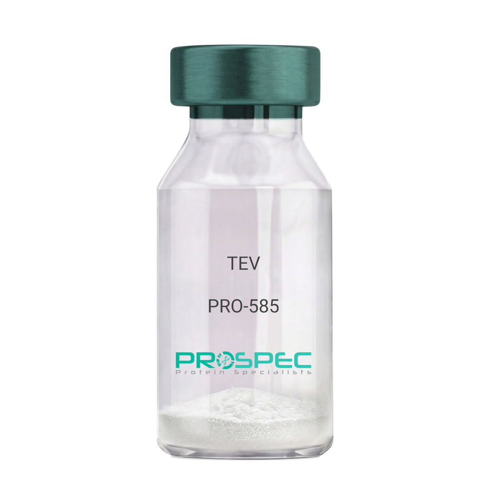
    TEV

    Description:

    Tobacco Etch Virus Protease Recombinant

    rTEV, TEV, P1 protease.

    Product # :

    PRO-585

    Price :

    Quantity :

    Shipping Method :

    Ice Icon

    Shipped with Ice Packs

    Add To Cart

    More Info

    • description
    • source
    • formulation
    • purity
    • More Info

    Description

    Recombinant TEV Protease (rTEV) is a site-specific protease purified from E. coli. The protease can be used for the removal of affinity tags from fusion proteins. The seven-amino-acid recognition site for rTEV is Glu-Asn-Leu-Tyr-Phe-Gln-Gly with cleavage occurring between Gln and Gly. The optimal temperature for cleavage is 30°C; however, the enzyme can be used at temperatures as low as 4°C. The rTEV contains His tag.The rTEV is purified by proprietary chromatographic techniques.

    Source

    Escherichia Coli.

    Formulation

    The rTEV contains 25mM Tris, Ph 8.0, 75mM NaCl, 5mM EDTA, 10mM GSH, 50% Glycerol.

    Purity

    Greater than 90.0% as determined by analysis by SDS-PAGE.

    More Info

    • Introduction

      TEV protease is the common name for the 27 kDa catalytic domain of the Nuclear Inclusion a (NIa) protein encoded by the tobacco etch virus (TEV). Because its sequence specificity is far more stringent than that of factor Xa, thrombin, or enterokinase, TEV protease is a very useful reagent for cleaving fusion proteins. TEV protease recognizes a linear epitope of the general form E-Xaa-Xaa-Y -Xaa-Q-(G/S), with cleavage occurring between Q and G or Q and S. The most commonly used sequence is ENLYFQG.

    • Synonyms

      rTEV, TEV, P1 protease.

    • Physical Appearance

      Sterile liquid formulation.

    • Stability

      rTEV although stable at 4°C for 1 week, should be stored below -18°C. Please prevent freeze thaw cycles.

    • Cleavage Conditions

      A number of variables can be changed to optimize the cleavage of any specific protein. The amount of rTEV, the temperature of the incubation, and the time needed for cleavage may be examined. If the protein of interest is heat-labile, then 4°C incubations are recommended. Reactions at 4°C will require longer incubation times and/or more rTEV.

    • Unit Definition

      One unit is defined as the amount of enzyme needed to cleave 3 ug of fusion protein in 1 hour to 85 % completion at 30°C in a buffer containing 50 mM Tris-HCl, pH 8.0, 0.5 mM EDTA, and 1 mM DTT.

    ProSpec's products are furnished for LABORATORY RESEARCH USE ONLY. The product may not be used as drugs, agricultural or pesticidal products, food additives or household chemicals.

    Tev Protease
  • View Data Sheet

    Name :

    UBE2G Human

    Description:

    Ubiquitin-Conjugating Enzyme E2G Human Recombinant

    E217K, UBC7, UBE2G, Ubiquitin-conjugating enzyme E2 G1, E2 ubiquitin-conjugating enzyme G1, E217K, UBC7, Ubiquitin carrier protein G1, Ubiquitin-protein ligase G1.

    Product # :

    ENZ-838

    Price :

    Quantity :

    Shipping Method :

    Ice Icon

    Shipped with Ice Packs

    Add To Cart

    More Info

    • description
    • source
    • formulation
    • purity
    • More Info

    Description

    UBE2G Human Recombinant produced in E.Coli is a single, non-glycosylated polypeptide chain containing 193 amino acids (1-170 a.a) and having a molecular mass of 21.9kDa.UBE2G is fused to a 23 amino acid His-tag at N-terminus & purified by proprietary chromatographic techniques.

    Source

    Escherichia Coli.

    Formulation

    UBE2G protein solution (0.5mg/ml) containing Phosphate buffered saline, (pH7.4) 30% glycerol and 1mM DTT.

    Purity

    Greater than 95.0% as determined by SDS-PAGE.

    More Info

    • Introduction

      Protein modification with ubiquitin is an essential cellular mechanism for targeting abnormal or short-lived proteins for degradation. Ubiquitination involves at least 3 classes of enzymes: ubiquitin-activating enzymes (E1s) ubiquitin-conjugating enzymes (E2s) and ubiquitin-protein ligases (E3s). Ubiquitin-Conjugating Enzyme E2G (UBE2G1) belongs to the E2 ubiquitin-conjugating enzyme family and catalyzes the covalent attachment of ubiquitin to other proteins. UE2G1 protein is involved in degradation of muscle-specific proteins.

    • Synonyms

      E217K, UBC7, UBE2G, Ubiquitin-conjugating enzyme E2 G1, E2 ubiquitin-conjugating enzyme G1, E217K, UBC7, Ubiquitin carrier protein G1, Ubiquitin-protein ligase G1.

    • Physical Appearance

      Sterile Filtered colorless solution.

    • Stability

      Store at 4°C if entire vial will be used within 2-4 weeks. Store, frozen at -20°C for longer periods of time. For long term storage it is recommended to add a carrier protein (0.1% HSA or BSA).Avoid multiple freeze-thaw cycles.

    • Amino Acid Sequence

      MGSSHHHHHH SSGLVPRGSH MGSMTELQSA LLLRRQLAEL NKNPVEGFSA GLIDDNDLYR WEVLIIGPPD TLYEGGVFKA HLTFPKDYPL RPPKMKFITE IWHPNVDKNG DVCISILHEP GEDKYGYEKP EERWLPIHTV ETIMISVISM LADPNGDSPA NVDAAKEWRE DRNGEFKRKV ARCVRKSQET AFE.

    ProSpec's products are furnished for LABORATORY RESEARCH USE ONLY. The product may not be used as drugs, agricultural or pesticidal products, food additives or household chemicals.

    Ube2G Human
  • View Data Sheet

    Name :

    AKT3 Human

    Description:

    AKT Serine/Threonine Kinase 3 Human Recombinant

    RAC-gamma serine/threonine-protein kinase, Protein kinase Akt-3, Protein kinase B gamma, PKB gamma, RAC-PK-gamma, STK-2, AKT3, PKBG.

    Product # :

    PKA-130

    Price :

    Quantity :

    Shipping Method :

    Ice Icon

    Shipped with Ice Packs

    Add To Cart

    More Info

    • description
    • source
    • formulation
    • purity
    • More Info

    Description

    AKT3 Human Recombinant produced in HEK293 Cells is a single, glycosylated polypeptide chain containing 485 amino acids (1-479a.a) and having a molecular mass of 56.5kDa.AKT3 is fused to a 6 amino acid His-tag at C-terminus and is purified by proprietary chromatographic techniques.

    Source

    HEK293 Cells.

    Formulation

    The AKT3 solution (0.25mg/ml) contains PBS (pH7.4) and 10% glycerol.

    Purity

    Greater than 90.0% as determined by SDS-PAGE.

    More Info

    • Introduction

      AKT Serine/Threonine Kinase 3 (Akt3) is a member of the Akt subfamily of serine/threonine protein kinases. Akt3is 1/3 closely related serine/threonine-protein kinases (Akt1, Akt2 and Akt3) called the Akt kinase and which regulate various processes including proliferation, metabolism, cell survival, growth and angiogenesis. The mentioned above is mediated through serine and/or threonine phosphorylation of a range of downstream substrates. Akt3 takes part in brain development and is critical for the viability of malignant glioma cells.

    • Synonyms

      RAC-gamma serine/threonine-protein kinase, Protein kinase Akt-3, Protein kinase B gamma, PKB gamma, RAC-PK-gamma, STK-2, AKT3, PKBG.

    • Physical Appearance

      Sterile Filtered colorless solution.

    • Stability

      Store at 4°C if entire vial will be used within 2-4 weeks. Store, frozen at -20°C for longer periods of time. For long term storage it is recommended to add a carrier protein (0.1% HSA or BSA).Avoid multiple freeze-thaw cycles.

    • Amino Acid Sequence

      MSDVTIVKEG WVQKRGEYIK NWRPRYFLLK TDGSFIGYKE KPQDVDLPYP LNNFSVAKCQ LMKTERPKPN TFIIRCLQWT TVIERTFHVD TPEEREEWTE AIQAVADRLQ RQEEERMNCS PTSQIDNIGE EEMDASTTHH KRKTMNDFDY LKLLGKGTFG KVILVREKAS GKYYAMKILK KEVIIAKDEV AHTLTESRVL KNTRHPFLTS LKYSFQTKDR LCFVMEYVNG GELFFHLSRE RVFSEDRTRF YGAEIVSALD YLHSGKIVYR DLKLENLMLD KDGHIKITDF GLCKEGITDA ATMKTFCGTP EYLAPEVLED NDYGRAVDWW GLGVVMYEMM CGRLPFYNQD HEKLFELILM EDIKFPRTLS SDAKSLLSGL LIKDPNKRLG GGPDDAKEIM RHSFFSGVNW QDVYDKKLVP PFKPQVTSET DTRYFDEEFT AQTITITPPE KYDEDGMDCM DNERRPHFPQ FSYSASGREH HHHHH.

    ProSpec's products are furnished for LABORATORY RESEARCH USE ONLY. The product may not be used as drugs, agricultural or pesticidal products, food additives or household chemicals.

    Akt3 Human
  • View Data Sheet

    Name :

    MBP E.Coli, His

    Description:

    Maltose Binding Protein E.coli Recombinant, His Tag

    Maltose-binding periplasmic protein, MBP, MMBP, Maltodextrin-binding protein, malE, b4034, JW3994.

    Product # :

    PRO-2322

    Price :

    Quantity :

    Shipping Method :

    Ice Icon

    Shipped with Ice Packs

    Add To Cart

    More Info

    • description
    • source
    • formulation
    • purity
    • More Info

    Description

    Recombinant E.Coli MBP produced in E.Coli is a single, non-glycosylated polypeptide chain containing 410 amino acids (27-392 a.a) and having a molecular mass of 44.9kDa. MBP is fused to a 24 amino acid His-tag at N-terminus & purified by proprietary chromatographic techniques.

    Source

    Escherichia Coli.

    Formulation

    MBP protein solution (0.5mg/ml) containing 20mM Tris-HCl buffer (pH 8.0), 0.1M NaCl and 10% glycerol.

    Purity

    Greater than 95.0% as determined by SDS-PAGE.

    More Info

    • Introduction

      Maltose Binding Protein is a member of the maltose/maltodextrin system of E.Coli, which is accountable for the uptake and efficient catabolism of maltodextrins. The maltose/maltodextrin is a complex regulatory and transport system involving many proteins and protein complexes.
      MBP elevates the yield of its fusion partner in many cases and is often able to promote the solubility of polypeptides to which it is fused.

    • Synonyms

      Maltose-binding periplasmic protein, MBP, MMBP, Maltodextrin-binding protein, malE, b4034, JW3994.

    • Physical Appearance

      Sterile Filtered colorless solution.

    • Stability

      Store at 4°C if entire vial will be used within 2-4 weeks. Store, frozen at -20°C for longer periods of time. For long term storage it is recommended to add a carrier protein (0.1% HSA or BSA).Avoid multiple freeze-thaw cycles.

    • Amino Acid Sequence

      MGSSHHHHHH SSGLVPRGSH MGSMKIEEGK LVIWINGDKG YNGLAEVGKK FEKDTGIKVT VEHPDKLEEK FPQVAATGDG PDIIFWAHDR FGGYAQSGLL AEITPDKAFQ DKLYPFTWDA VRYNGKLIAY PIAVEALSLI YNKDLLPNPP KTWEEIPALD KELKAKGKSA LMFNLQEPYF TWPLIAADGG YAFKYENGKY DIKDVGVDNA GAKAGLTFLV DLIKNKHMNA DTDYSIAEAA FNKGETAMTI NGPWAWSNID TSKVNYGVTV LPTFKGQPSK PFVGVLSAGI NAASPNKELA KEFLENYLLT DEGLEAVNKD KPLGAVALKS YEEELAKDPR IAATMENAQK GEIMPNIPQM SAFWYAVRTA VINAASGRQT VDEALKDAQT NSSSNNNNNN NNNNLGIEGR.

    ProSpec's products are furnished for LABORATORY RESEARCH USE ONLY. The product may not be used as drugs, agricultural or pesticidal products, food additives or household chemicals.

    Mbp Ecoli His
  • View Data Sheet

    Name :

    GLO1 Mouse

    Description:

    Glyoxalase-I Mouse Recombinant

    Lactoylglutathione lyase, Aldoketomutase, Glyoxalase I, Glx I, Ketone-aldehyde mutase, Methylglyoxalase, S-D-lactoylglutathione methylglyoxal lyase.

    Product # :

    ENZ-953

    Price :

    Quantity :

    Shipping Method :

    Ice Icon

    Shipped with Ice Packs

    Add To Cart

    More Info

    • description
    • source
    • formulation
    • purity
    • biological activity
    • More Info

    Description

    GLO1 Mouse Recombinant produced in Sf9 Baculovirus cells is a single, glycosylated polypeptide chain containing 192 amino acids (1-184a.a.) and having a molecular mass of 21.8kDa (Molecular size on SDS-PAGE will appear at approximately 28-40kDa). GLO1 is expressed with an 8 amino acid His tag at C-Terminus and purified by proprietary chromatographic techniques.

    Source

    Sf9, Baculovirus cells.

    Formulation

    GLO1 protein solution (0.5mg/ml) containing Phosphate Buffered Saline (pH 7.4) and 10% glycerol.

    Purity

    Greater than 95.0% as determined by SDS-PAGE.

    Biological Activity

    Specific activity is > 210 units/mg, and is defined as the amount of enzyme that will form 1.0 µmol of S-lactoylglutathione from methylglyoxal and reduced glutathione per minute at pH 6.5 at 25C.

    More Info

    • Introduction

      GLO1 is involved in the catalysis and formation of S-lactoyl-glutathione from methylglyoxal condensation and reduced glutatione. GLO1 is linked to HLA and is localized to 6p21.3-p21.1, between HLA and the centromere. GLO1 enzyme is ubundantly expressed and present in numerous tumor cell lines, in which its concentration is often upregulated ubiquitisly. GLO1 is a major susceptible gene for autism in an ethnic Chinese population from Taiwan. GLO1 might be involved in the pathophysiology of mood disorders. GLO1 plays a role in the pathophysiology of mood disorders. Overexpression of GLO1 is associated with kidney tumor.

    • Synonyms

      Lactoylglutathione lyase, Aldoketomutase, Glyoxalase I, Glx I, Ketone-aldehyde mutase, Methylglyoxalase, S-D-lactoylglutathione methylglyoxal lyase.

    • Physical Appearance

      Sterile Filtered colorless solution.

    • Stability

      Store at 4°C if entire vial will be used within 2-4 weeks. Store, frozen at -20°C for longer periods of time. For long term storage it is recommended to add a carrier protein (0.1% HSA or BSA).Avoid multiple freeze-thaw cycles.

    • Amino Acid Sequence

      MAEPQPASSG LTDETAFSCC SDPDPSTKDF LLQQTMLRIK DPKKSLDFYT RVLGLTLLQK LDFPAMKFSL YFLAYEDKND IPKDKSEKTA WTFSRKATLE LTHNWGTEDD ETQSYHNGNS DPRGFGHIGI AVPDVYSACK RFEELGVKFV KKPDDGKMKG LAFIQDPDGY WIEILNPNKI ATIILEHHHH HH.

    ProSpec's products are furnished for LABORATORY RESEARCH USE ONLY. The product may not be used as drugs, agricultural or pesticidal products, food additives or household chemicals.

    Glo1 Mouse
  • View Data Sheet

    Name :

    RNASE7 Human

    Description:

    Ribonuclease 7 Human Recombinant

    Ribonuclease, RNase A Family, 7, Skin-Derived Antimicrobial Protein 2, RNase 7, SAP-2, EC 3.1.27.5, EC 3.1.27., EC 3.1.27, Ribonuclease 7.

    Product # :

    ENZ-704

    Price :

    Quantity :

    Shipping Method :

    Ice Icon

    Shipped with Ice Packs

    Add To Cart

    More Info

    • description
    • source
    • formulation
    • purity
    • More Info

    Description

    RNASE7 Human Recombinant produced in E.Coli is a single, non-glycosylated polypeptide chain containing 151 amino acids (29-156 a.a) and having a molecular mass of 16.9kDa. RNASE7 is fused to a 23 amino acid His-tag at N-terminus & purified by proprietary chromatographic techniques.

    Source

    Escherichia Coli.

    Formulation

    RNASE7 protein solution (0.5mg/ml) containing 20mM Tris-HCl buffer (pH8.0) and 10% glycerol 1mM DTT.

    Purity

    Greater than 90% as determined by SDS-PAGE.

    More Info

    • Introduction

      Ribonuclease 7 (RNASE7) is one of the final RNase A superfamily ribonucleases. RNASE7 was isolated from skin-derived stratum corneum. RNASE7 protein demonstrated potent ribonuclease activity and hence may contribute to the well-known ribonuclease activity of human skin. RNASE7 has revealed a broad spectrum antimicrobial activity against many pathogenic microorganisms and extraordinarily potent activity.

    • Synonyms

      Ribonuclease, RNase A Family, 7, Skin-Derived Antimicrobial Protein 2, RNase 7, SAP-2, EC 3.1.27.5, EC 3.1.27., EC 3.1.27, Ribonuclease 7.

    • Physical Appearance

      Sterile Filtered colorless solution.

    • Stability

      Store at 4°C if entire vial will be used within 2-4 weeks. Store, frozen at -20°C for longer periods of time. For long term storage it is recommended to add a carrier protein (0.1% HSA or BSA). Avoid multiple freeze-thaw cycles.

    • Amino Acid Sequence

      MGSSHHHHHH SSGLVPRGSH MGSKPKGMTS SQWFKIQHMQ PSPQACNSAM KNINKHTKRC KDLNTFLHEP FSSVAATCQT PKIACKNGDK NCHQSHGPVS LTMCKLTSGK YPNCRYKEKR QNKSYVVACK PPQKKDSQQF HLVPVHLDRV L.

    ProSpec's products are furnished for LABORATORY RESEARCH USE ONLY. The product may not be used as drugs, agricultural or pesticidal products, food additives or household chemicals.

    Rnase7 Human
  • View Data Sheet

    Name :

    CEA Protein

    Description:

    Carcinoembryonic Antigen Human

    CEACAM5, Meconium Antigen 100, Carcinoembryonic Antigen, CD66e Antigen, CD66e, Carcinoembryonic Antigen, CEA, oncofetal antigen.

    Product # :

    PRO-2801

    Price :

    Quantity :

    Shipping Method :

    Ice Icon

    Shipped with Ice Packs

    Add To Cart

    More Info

    • description
    • source
    • formulation
    • purity
    • More Info

    Description

    CEA produced from patient source colon carcinoma liver metastatic tissue can be used as general marker in screening and monitoring malignant disease states.

    Source

    Liver tissue.

    Formulation

    CEA protein solution contains 0.1M PBS, pH 7.4, 0.09 % NaN3 and 2 % methyl-mannoside.

    Purity

    Greater than 95.0% as determined by SDS-PAGE.

    More Info

    • Synonyms

      CEACAM5, Meconium Antigen 100, Carcinoembryonic Antigen, CD66e Antigen, CD66e, Carcinoembryonic Antigen, CEA, oncofetal antigen.

    • Physical Appearance

      Sterile Filtered colorless solution.

    • Stability

      Store at 4°C if entire vial will be used within 2-4 weeks. Store, frozen at -20°C for longer periods of time. For long term storage it is recommended to add a carrier protein (0.1% HSA or BSA). Avoid multiple freeze-thaw cycles.

    • Applications

      Blood samples from tissue donors were tested and found to be negative for HBsAg, HIV-1 and HIV-2 antibodies and HCV.

    • Background

      Carcinoembryonic Antigen, commonly known as CEA, is a glycoprotein that was initially identified as a tumor marker. Over the years, research into CEA has unveiled its intricate involvement in various physiological processes, not only in cancer but also in the context of normal development and inflammatory conditions. This research aims to delve into the multifaceted roles of CEA, exploring its structural intricacies, regulatory mechanisms, and its implications in health, disease, and beyond.

      Structural Complexity of CEA:

      CEA, belonging to the immunoglobulin superfamily, is a complex glycoprotein featuring multiple structural domains. Its diverse forms and glycosylation patterns contribute to its functional versatility. CEA is primarily expressed in fetal tissues, but its presence is often detected in adults under pathological conditions, especially in various types of cancer.

      CEA in Cancer Biology:

      CEA was first recognized as a biomarker for colorectal cancer, but its overexpression is not limited to this context. Elevated CEA levels have been associated with several other malignancies, including breast, lung, and pancreatic cancers. CEA’s involvement in cancer biology ranges from promoting angiogenesis and metastasis to inhibiting immune responses, making it a critical player in tumor progression and evasion.

      Beyond Cancer: CEA in Development and Inflammation:

      While CEA’s role in cancer is prominent, recent studies have uncovered its participation in normal physiological processes. During embryonic development, CEA is involved in cell adhesion, contributing to tissue organization and morphogenesis. Additionally, CEA expression can be induced in inflammatory conditions, suggesting its involvement in immune responses and tissue repair mechanisms.

      CEA as a Diagnostic and Therapeutic Target:

      The diverse expression patterns of CEA in various diseases make it a valuable diagnostic tool. CEA assays are widely used for cancer screening, monitoring disease progression, and assessing treatment efficacy. Moreover, CEA’s presence on the surface of cancer cells has made it a target for immunotherapy, enabling the development of targeted therapies aimed at specifically eradicating CEA-positive tumor cells.

    ProSpec's products are furnished for LABORATORY RESEARCH USE ONLY. The product may not be used as drugs, agricultural or pesticidal products, food additives or household chemicals.

    Cea Human
  • View Data Sheet

    Name :

    PRKACA Human

    Description:

    cAMP-Dependent Protein Kinase A catalytic subunit α Human Recombinant

    cAMP-dependent protein kinase alpha-catalytic subunit, EC 2.7.11.11, PKA C-alpha, PKACA, PRKACA, MGC48865, MGC102831.

    Product # :

    PKA-200

    Price :

    Quantity :

    Shipping Method :

    Ice Icon

    Shipped with Ice Packs

    Add To Cart

    More Info

    • description
    • source
    • formulation
    • purity
    • More Info

    Description

    cAMP-dependent PKA is an ubiquitous serine/threonine protein kinase present in a variety of tissues (e.g. brain, skeletal muscle, heart). The intracellular cAMP level regulates cellular responses by altering the interaction between the catalytic C and regulatory R subunits of PKA. The inactive tetrameric PKA holoenzyme R2C2 is activated when cAMP binds to R2, which dissociates the tetramer to R2 cAMP 4 and two active catalytic subunits. Free Catalytic subunits of PKA can phosphorylate a wide variety of intracellular target proteins. In response to hormone- induced high cAMP levels, PKA phosphorylates glycogen synthetase (inhibition of the enzyme activity) and phosphorylase kinase to block glycogen synthesis. Different isoforms of catalytic and regulatory subunits suggest specific functions. The recombinant PKA catalytic subunit a is a 41kDa protein. The a-isoform is the predominant form with a broad tissue distribution and can be used for in vitro enzymological studies of neural and hormonal signal transduction or to phosphorylate target proteins in vivo including Ion channels, transcriptional activator proteins and regulatory enzymes of glycogen metabolism.

    Source

    Escherichia Coli.

    Formulation

    PKA catalytic subunit a is supplied in a buffer containing 20mM MOPS pH7, 150mM NaCl, 1mM DTT, 1mM EDTA and 50% Glycerin.

    Purity

    Greater than 95% as determined by SDS-PAGE.

    More Info

    • Synonyms

      cAMP-dependent protein kinase alpha-catalytic subunit, EC 2.7.11.11, PKA C-alpha, PKACA, PRKACA, MGC48865, MGC102831.

    • Stability

      Store at 4°C if entire vial will be used within 2-4 weeks. Store, frozen at -20°C for longer periods of time. Avoid multiple freeze-thaw cycles.

    • Amino Acid Sequence

      MGNAAAAKKG SEQESVKEFL AKAKEDFLKK WESPAQNTAH LDQFERIKTL GTGSFGRVML VKHKETGNHY AMKILDKQKV VKLKQIEHTL NEKRILQAVN FPFLVKLEFS FKDNSNLYMV MEYVPGGEMF SHLRRIGRFS EPHARFYAAQ IVLTFEYLHS LDLIYRDLKP ENLLIDQQGY IQVTDFGFAK RVKGRTWTLC GTPEYLAPEI ILSKGYNKAV DWWALGVLIY EMAAGYPPFF ADQPIQIYEK IVSGKVRFPS HFSSDLKDLL RNLLQVDLTK RFGNLKNGVN DIKNHKWFAT TDWIAIYQRK VEAPFIPKFK GPGDTSNFDD YEEEEIRVSI NEKCGKEFSE F.

    • Assay Conditions

      Roskoski-AssayProtein kinase activity can be measured using a modified radioactive assay according to Roskoski et al.The assay will be performed in a mixture containing 50mM MOPS (pH7.0), 10mM MgCI2, 0.25 mg/ml bovine serum albumin, 100 IJM Kemptide (peptide substrate), 100 IJM unlabeled ATP mixed with [y_32p] ATP (500-1000 cpm/pmol) and Ca subunit in a final volume of 50 IJI. Reaction is started by addition of the Ca subunit and can be stopped after 5 minutes incubation at 30°C by spotting the reaction mix onto Whatman P-81 filters and soaking the filters four times in 75mM phosphoric acid (10 ml per sample) for at least 5 minutes. After four washing steps rinse filters with ethanol, dry and count. Roskoski, R., Jr. (1983) Methods Enzymol. 99, 3-6For the detection of phosphorylation in substrate proteins the phosphotransferase reaction can alternatively be stopped by taking aliquots of the mixture and adding SDS sample buffer. The phosphorylation status of the substrate proteins can subsequently be analysed using SDS PAGE and autoradiography. Zimmermann, B. (1999) Journal of Biological Chemistry.274, 9, 5370-78.

    • Unit Definition

      One unit is defined as the amount of cAMP-Dependent Protein Kinase, recombinant C? catalytic subunit, required to incorporate 1 pmol of phosphate into the specific substrate peptide kemptide (LRRASLG) in one minute at 30°C.

    • Specific Activity

      The specific activity of the recombinant PKA catalytic subunit alpha, is >10,000,000 U/mg.

    ProSpec's products are furnished for LABORATORY RESEARCH USE ONLY. The product may not be used as drugs, agricultural or pesticidal products, food additives or household chemicals.

    Prkaca
  • View Data Sheet

    Name :

    MAPK1 Human, His

    Description:

    Recombinant Human Mitogen-Activated Protein Kinase 1, His Tag

    Mitogen-activated protein kinase 1, EC 2.7.11.24, Extracellular signal-regulated kinase 2, ERK-2, Mitogen-activated protein kinase 2, MAP kinase 2, MAPK 2, p42-MAPK, ERT1, ERK, p38, p40, p41, ERK2, MAPK2, PRKM1, PRKM2, P42MAPK, p41mapk.

    Product # :

    PKA-360

    Price :

    Quantity :

    Shipping Method :

    Ice Icon

    Shipped with Ice Packs

    Add To Cart

    More Info

    • description
    • source
    • formulation
    • purity
    • More Info

    Description

    MAPK1 Human Recombinant produced in E.Coli is a single, non-glycosylated, polypeptide chain containing 380 amino acids (1-360 a.a.) and having a molecular mass of 43.5 kDa. MAPK1 is fused to a 20 amino acids His-tag at N-terminus & purified by proprietary chromatographic techniques.

    Source

    Escherichia Coli.

    Formulation

    The MAPK1 solution contains 20 mM Tris-HCl buffer (pH 8.0), 1mM DTT and 10% glycerol.

    Purity

    Greater than 95.0% as determined by SDS-PAGE.

    More Info

    • Introduction

      Mitogen-activated protein kinase 1 (MAPK1) is also known as "extracellular signal-regulated kinase 2" (ERK2). Two similar (85% sequence identity) protein kinases were originally called ERK1 and ERK2. They were found during a search for protein kinases that are rapidly phosphorylated after activation of cell surface tyrosine kinases such as the epidermal growth factor receptor. Phosphorylation of ERKs leads to the activation of their kinase activity.The molecular events linking cell surface receptors to activation of ERKs are complex. It was found that Ras GTP-binding proteins are involved in the activation of ERKs. Another protein kinase, Raf-1, was shown to phosphorylate a "MAPK kinase", thus qualifying as a "MAPK kinase kinase". The MAPK kinase was named "MAPK/ERK kinase" (MEK). Receptor-linked tyrosine kinases, Ras, Raf, MEK and MAPK could be fitted into a signaling cascade linking an extracellular signal to MAPK activation Transgenic gene knockout mice lacking MAPK1 have major defe

    • Synonyms

      Mitogen-activated protein kinase 1, EC 2.7.11.24, Extracellular signal-regulated kinase 2, ERK-2, Mitogen-activated protein kinase 2, MAP kinase 2, MAPK 2, p42-MAPK, ERT1, ERK, p38, p40, p41, ERK2, MAPK2, PRKM1, PRKM2, P42MAPK, p41mapk.

    • Physical Appearance

      Sterile Filtered colorless solution.

    • Stability

      Store at 4°C if entire vial will be used within 2-4 weeks. Store, frozen at -20°C for longer periods of time. For long term storage it is recommended to add a carrier protein (0.1% HSA or BSA).Avoid multiple freeze-thaw cycles.

    • Amino Acid Sequence

      MGSSHHHHHH SSGLVPRGSH MAAAAAAGAG PEMVRGQVFD VGPRYTNLSY IGEGAYGMVC SAYDNVNKVR VAIKKISPFE HQTYCQRTLR EIKILLRFRH ENIIGINDII RAPTIEQMKD VYIVQDLMET DLYKLLKTQH LSNDHICYFL YQILRGLKYI HSANVLHRDL KPSNLLLNTT CDLKICDFGL ARVADPDHDH TGFLTEYVAT RWYRAPEIML NSKGYTKSID IWSVGCILAE MLSNRPIFPG KHYLDQLNHI LGILGSPSQE MGSSHHHHHH SSGLVPRGSH MAAAAAAGAG PEMVRGQVFD VGPRYTNLSY IGEGAYGMVC SAYDNVNKVR VAIKKISPFE HQTYCQRTLR EIKILLRFRH ENIIGINDII RAPTIEQMKD VYIVQDLMET DLYKLLKTQH LSNDHICYFL YQILRGLKYI HSANVLHRDL KPSNLLLNTT CDLKICDFGL ARVADPDHDH TGFLTEYVAT RWYRAPEIML NSKGYTKSID IWSVGCILAE MLSNRPIFPG KHYLDQLNHI LGILGSPSQE DLNCIINLKA RNYLLSLPHK NKVPWNRLFP NADSKALDLL DKMLTFNPHK RIEVEQALAH PYLEQYYDPS DEPIAEAPFK FDMELDDLPK EKLKELIFEE TARFQPGYRS.

    ProSpec's products are furnished for LABORATORY RESEARCH USE ONLY. The product may not be used as drugs, agricultural or pesticidal products, food additives or household chemicals.

    Mapk1 Human His
  • 1
  • …
  • 29
  • 30
  • 31
  • 32
  • 33
  • …
  • 42
Your browser does not support the video tag.

Products

  • Cytokines
  • Growth Factors
  • Chemokines
  • CD Antigens
  • Neurotrophins
  • Hormones
  • Enzymes
  • Viral Antigens
  • Recombinant Proteins
  • Natural Proteins
  • Monoclonal Antibodies
  • Polyclonal Antibodies
  • Thermal Packaging

About

  • Company
  • Founder
  • Contribution
  • Contact Us
  • My Account

Ordering

  • Ordering Method
  • Ordering - Credit Card
  • Ordering PO
  • Shipping Fees
  • FAQ's

Legal

  • Terms & Conditions
  • Privacy
  • Site Map
  • info@prospecbio.com
  • Facebook
  • Instagram
  • Linkedin Profile

© 2026 ProSpec-Tany TechnoGene Ltd. All Rights Reserved.

  • Choosing a selection results in a full page refresh.
  • Opens in a new window.