Search results
1000 results found for “KCTD”
Name
Description
Product #
Price
Quantity
Shipping Method
- View Data Sheet
Name :
CD59 HumanDescription:
CD59 Human Recombinant
CD59 Molecule Complement Regulatory Protein, CD59 Antigen P18-20, CD59 Glycoprotein, Surface Anitgen Recognized By Monoclonal Antibody 16.3A5, Membrane Attack Complex (MAC) Inhibition Factor, Lymphocytic Antigen CD59/MEM43, Human Leukocyte Antigen MIC11, Membrane Attack Complex Inhibition Factor, 20 KDa Homologous Restriction Factor, Membrane Inhibitor Of Reactive Lysis, MAC-Inhibitory Protein, T Cell-Activating Protein, Ly-6-Like Protein, Protectin, MIC11, MSK21, MEM43, MIN1/2/3, MIRL, MAC-IP, MACIF, HRF-20, G344, EJ16/30/32, 1F5.
Product # :
PRO-1648Price :
Quantity :
Shipping Method :
Shipped with Ice Packs
More Info
- description
- source
- formulation
- purity
- More Info
Description
CD59 Human Recombinant produced in E.coli is a single, non-glycosylated polypeptide chain containing 100 amino acids (26-102) and having a molecular mass of 11.3kDa.CD59 is fused to a 23 amino acid His-tag at N-terminus & purified by proprietary chromatographic techniques.
Source
Escherichia Coli.
Formulation
The CD59 solution (0.5mg/ml) contains 20mM Tris-HCl buffer (pH 8.0), 0.15M NaCl, 1mM DTT and 10% glycerol.
Purity
Greater than 85% as determined by SDS-PAGE.
More Info
-
Introduction
CD59 is a cell surface glycoprotein which regulates complement-mediated cell lysis, and it is involved in lymphocyte signal transduction. CD59 is an effective inhibitor of the complement membrane attack complex, whereby it binds complement C8 and/or C9 during the compilation of this complex, thus inhibiting the incorporation of multiple copies of C9 into the complex, which is essential for osmolytic pore formation. In addition, CD59 has a role in signal transduction pathways in the activation of T cells. CD59 gene mutations cause CD59 deficiency, a disease resulting in hemolytic anemia and thrombosis, and which causes cerebral infarction.
-
Synonyms
CD59 Molecule Complement Regulatory Protein, CD59 Antigen P18-20, CD59 Glycoprotein, Surface Anitgen Recognized By Monoclonal Antibody 16.3A5, Membrane Attack Complex (MAC) Inhibition Factor, Lymphocytic Antigen CD59/MEM43, Human Leukocyte Antigen MIC11, Membrane Attack Complex Inhibition Factor, 20 KDa Homologous Restriction Factor, Membrane Inhibitor Of Reactive Lysis, MAC-Inhibitory Protein, T Cell-Activating Protein, Ly-6-Like Protein, Protectin, MIC11, MSK21, MEM43, MIN1/2/3, MIRL, MAC-IP, MACIF, HRF-20, G344, EJ16/30/32, 1F5.
-
Physical Appearance
Sterile filtered colorless solution.
-
Stability
Store at 4°C if entire vial will be used within 2-4 weeks. Store, frozen at -20°C for longer periods of time. For long term storage it is recommended to add a carrier protein (0.1% HSA or BSA).Avoid multiple freeze-thaw cycles.
-
Amino Acid Sequence
MGSSHHHHHH SSGLVPRGSH MGSLQCYNCP NPTADCKTAV NCSSDFDACL ITKAGLQVYN KCWKFEHCNF NDVTTRLREN ELTYYCCKKD LCNFNEQLEN.
ProSpec's products are furnished for LABORATORY RESEARCH USE ONLY. The product may not be used as drugs, agricultural or pesticidal products, food additives or household chemicals.
- View Data Sheet
Name :
GLTPD1 HumanDescription:
Glycolipid Transfer Protein Domain Containing 1 Human Recombinant
Ceramide-1-Phosphate Transfer Protein, Glycolipid Transfer Protein Domain-Containing Protein 1, Glycolipid Transfer Protein Domain Containing 1, GLTP Domain-Containing Protein 1, GLTPD1, GLTPD1, Ceramide-1-phosphate transfer protein, CPTP.
Product # :
PRO-2093Price :
Quantity :
Shipping Method :
Shipped with Ice Packs
More Info
- description
- source
- formulation
- purity
- More Info
Description
GLTPD1 Human Recombinant produced in E.Coli is a single, non-glycosylated polypeptide chain containing 237 amino acids (1-214 a.a) and having a molecular mass of 26.8kDa. GLTPD1 is fused to a 23 amino acid His-tag at N-terminus & purified by proprietary chromatographic techniques.
Source
Escherichia Coli.
Formulation
GLTPD1 protein solution (0.25mg/ml) containing Phosphate buffered saline (pH7.4), 50% glycerol and 1mM DTT.
Purity
Greater than 90.0% as determined by SDS-PAGE.
More Info
-
Introduction
Glycolipid Transfer Protein Domain Containing 1 (GLTPD1) belongs to the GLTP family. GLTPD1 mediates the intracellular transfer of ceramide-1-phosphate between organelle membranes and the cell membrane. GLTPD1 is essential for normal structure of the Golgi stacks. In addition, GLTPD1 is able to bind phosphoceramides with a diversity of aliphatic chains, however GLTPD1 has a preference for lipids with saturated C16:0 or monounsaturated C18:1 aliphatic chains. Moreover, GLTPD1 is inefficient with phosphoceramides which contains lignoceryl (C24:0). GLTPD1 participates in the regulation of the cellular levels of ceramide-1-phosphate, and in that way contributes to the regulation of phospholipase PLA2G4A activity and the release of arachidonic acid.
-
Synonyms
Ceramide-1-Phosphate Transfer Protein, Glycolipid Transfer Protein Domain-Containing Protein 1, Glycolipid Transfer Protein Domain Containing 1, GLTP Domain-Containing Protein 1, GLTPD1, GLTPD1, Ceramide-1-phosphate transfer protein, CPTP.
-
Physical Appearance
Sterile Filtered clear solution.
-
Stability
Store at 4°C if entire vial will be used within 2-4 weeks. Store, frozen at -20°C for longer periods of time.For long term storage it is recommended to add a carrier protein (0.1% HSA or BSA).Please avoid freeze thaw cycles.
-
Amino Acid Sequence
MGSSHHHHHH SSGLVPRGSH MGSMDDSETG FNLKVVLVSF KQCLDEKEEV LLDPYIASWK GLVRFLNSLG TIFSFISKDV VSKLRIMERL RGGPQSEHYR SLQAMVAHEL SNRLVDLERR SHHPESGCRT VLRLHRALHW LQLFLEGLRT SPEDARTSAL CADSYNASLA AYHPWVVRRA VTVAFCTLPT REVFLEAMNV GPPEQAVQML GEALPFIQRV YNVSQKLYAE HSLLDLP.
ProSpec's products are furnished for LABORATORY RESEARCH USE ONLY. The product may not be used as drugs, agricultural or pesticidal products, food additives or household chemicals.
- View Data Sheet
Name :
CKMBITII HumanDescription:
Creatine Kinase MB Isoenzyme Type-II Human Recombinant
Creatine Kinase MB Isoenzyme Type-II, CKMBITII, CKMBI, CKMB.
Product # :
CKI-270Price :
Quantity :
Shipping Method :
Shipped with Ice Packs
More Info
- description
- source
- formulation
- biological activity
- More Info
Description
CKMBITII Human Recombinant produced in Pichia Pastoris reacts with polyclonal antibodies to MB Isoenzyme in ELISA.
Source
Pichia Pastoris.
Formulation
Each mg of protein contains 10mM Tris-HCl, pH-6.8, 0.5mM EDTA and 0.5mM DTT, 50% (v/v) glycerol.
Biological Activity
The enzyme activity measured by kinetic assay at 340nm was 650 IU/mg at 37 degrees.
More Info
-
Introduction
CK-MB Type II possesses the naturally occurring carboxy-terminal amino acid lysine.
This occurs during a myocardial infarct (MI or heart attack) when CK-MB Type II is released from damaged heart muscle, and the C-terminal lysine is cleaved in the blood stream, thus creating CK-MB Type I. This difference can be exploited in diagnosis of an MI. -
Synonyms
Creatine Kinase MB Isoenzyme Type-II, CKMBITII, CKMBI, CKMB.
-
Physical Appearance
Sterile Filtered colourless liquid formulation.
-
Stability
CKMBITII although stable at 15°C for 7 days, should be stored below -18°C. Please prevent freeze-thaw cycles.
ProSpec's products are furnished for LABORATORY RESEARCH USE ONLY. The product may not be used as drugs, agricultural or pesticidal products, food additives or household chemicals.
- View Data Sheet
Name :
MCP1 Human, HEKDescription:
Monocyte Chemotactic Protein-1/MCAF (CCL2) Human Recombinant, HEK
CCL2, C-C motif chemokine 2, GDCF-2, HSMCR30, JE, HC11, MCAF, MCP-1, MCP1, Scya2, Sigje, SMC-CF, Immediate-early serum-responsive protein JE, Monocyte chemoattractant protein 1, Small-inducible cytokine A2.
Product # :
CHM-048Price :
Quantity :
Shipping Method :
Shipped with Ice Packs
More Info
- description
- source
- formulation
- purity
- biological activity
- More Info
Description
MCP1 Human HEK Recombinant produced in HEK293 Cells is a single, glycosylated polypeptide chain containing 82 amino acids (24-99a.a) and having a molecular mass of 9.5 kDa.MCP1 is expressed with a 6 amino acid His tag at C-Terminus and purified by proprietary chromatographic techniques.
Source
HEK293 Cells.
Formulation
The MCP1 solution (0.25mg/1ml) contains phosphate buffered saline (pH7.4) and 10% glycerol.
Purity
Greater than 90.0% as determined by SDS-PAGE.
Biological Activity
Measured its ability to chemoattract using THP-1 human acute monocytic leukemia cells. The ED50 range ≤ 30 ng/ml.
More Info
-
Introduction
Chemokine (C-C motif) ligand 2 (CCL2) is a small cytokine belonging to the CC chemokine family that is also known as monocyte chemotactic protein-1 (MCP-1). It is found at the site of tooth eruption and bone degradation. In the bone, CCL2 is expressed by mature osteoclasts and osteoblasts and is under the control of nuclear factor ?B (NF?B).CCL2 recruits immune cells, such as monocytes, to sites of tissue injury and infection.This chemokine is produced as a protein precursor containing signal peptide of 23 amino acids and a mature peptide of 76 amino acids. It is a monomeric polypeptide, with a molecular weight of approximately 13kDa. As with many other CC chemokines, CCL2 is located on chromosome 17 in humans.The cell surface receptors that bind CCL2 are CCR2 and CCR5
-
Synonyms
CCL2, C-C motif chemokine 2, GDCF-2, HSMCR30, JE, HC11, MCAF, MCP-1, MCP1, Scya2, Sigje, SMC-CF, Immediate-early serum-responsive protein JE, Monocyte chemoattractant protein 1, Small-inducible cytokine A2.
-
Physical Appearance
Sterile filtered colorless solution.
-
Stability
Store at 4°C if entire vial will be used within 2-4 weeks.Store, frozen at -20°C for longer periods of time. For long term storage it is recommended to add a carrier protein (0.1% HSA or BSA).Avoid multiple freeze-thaw cycles.
-
Amino Acid Sequence
QPDAINAPVT CCYNFTNRKI SVQRLASYRR ITSSKCPKEA VIFKTIVAKE ICADPKQKWV QDSMDHLDKQ TQTPKTHHHH HH
ProSpec's products are furnished for LABORATORY RESEARCH USE ONLY. The product may not be used as drugs, agricultural or pesticidal products, food additives or household chemicals.
- View Data Sheet
Name :
MECOM HumanDescription:
MDS1 And EVI1 Complex Locus Human Recombinant
MDS1 And EVI1 Complex Locus, Ecotropic Virus Integration Site 1 Protein Homolog, Myelodysplasia Syndrome-Associated Protein 1, AML1-EVI-1, MDS1-EVI1, EVI1, MDS1, PRDM3, AML1-EVI-1 Fusion Protein, MDS1 And EVI1 Complex Locus Protein EVI1, MDS1 And EVI1 Complex Locus Protein MDS1, Oncogene EVI1, Zinc Finger Protein Evi1.
Product # :
PRO-1766Price :
Quantity :
Shipping Method :
Shipped with Ice Packs
More Info
- description
- source
- formulation
- purity
- More Info
Description
MECOM Human Recombinant produced in E.Coli is a single, non-glycosylated polypeptide chain containing 192 amino acids (1-169 a.a) and having a molecular mass of 21.1kDa.MECOM is fused to a 23 amino acid His-tag at N-terminus & purified by proprietary chromatographic techniques.
Source
Escherichia Coli.
Formulation
MECOM protein solution (0.25mg/ml) containing 20mM Tris-HCl buffer (pH8.0), 0.4M Urea and 10% glycerol.
Purity
Greater than 85.0% as determined by SDS-PAGE.
More Info
-
Introduction
MDS1 is a transcriptional regulator and oncoprotein which takes part in hematopoiesis, development, cell differentiation, apoptosis and proliferation. MDS1 is able to interact with CTBP1, SMAD3, CREBBP, KAT2B, MAPK8, and MAPK9. Overexpression, as a result of translocation with the AML1 gene, is known to cause leukemia. A number of transcript variants encoding several different isoforms were found.
-
Synonyms
MDS1 And EVI1 Complex Locus, Ecotropic Virus Integration Site 1 Protein Homolog, Myelodysplasia Syndrome-Associated Protein 1, AML1-EVI-1, MDS1-EVI1, EVI1, MDS1, PRDM3, AML1-EVI-1 Fusion Protein, MDS1 And EVI1 Complex Locus Protein EVI1, MDS1 And EVI1 Complex Locus Protein MDS1, Oncogene EVI1, Zinc Finger Protein Evi1.
-
Physical Appearance
Sterile Filtered clear solution.
-
Stability
Store at 4°C if entire vial will be used within 2-4 weeks. Store, frozen at -20°C for longer periods of time.For long term storage it is recommended to add a carrier protein (0.1% HSA or BSA).Please avoid freeze thaw cycles.
-
Amino Acid Sequence
MGSSHHHHHH SSGLVPRGSH MGSMRSKGRA RKLATNNECV YGNYPEIPLE EMPDADGVAS TPSLNIQEPC SPATSSEAFT PKEGSPYKAP IYIPDDIPIP AEFELRESNM PGAGLGIWTK RKIEVGEKFG PYVGEQRSNL KDPSYGWEVH LPRSRRVSVH SWLYLGKRSS DVGIAFSQAD VYMPGLQCAF LS
ProSpec's products are furnished for LABORATORY RESEARCH USE ONLY. The product may not be used as drugs, agricultural or pesticidal products, food additives or household chemicals.
- View Data Sheet
Name :
ACAT2 AntibodyDescription:
Acetyl-Coenzyme A acetyltransferase 2, Mouse Anti Human
Acetyl-CoA acetyltransferase cytosolic, Cytosolic acetoacetyl-CoA thiolase, ACAT2, Acetyl CoA transferase-like protein, ACAT-2.
Product # :
ANT-561Price :
Quantity :
Shipping Method :
Shipped with Ice Packs
More Info
- formulation
- More Info
Formulation
1mg/ml containing PBS, pH-7.4, 10% Glycerol and 0.02% Sodium Azide.
More Info
-
Introduction
ACAT2 enzyme participates in lipid metabolism. ACAT2 takes part in lipoprotein assembly, catalyzing cholesterol esterification in mammalian cells. ACAT2 is an integral membrane protein that localizes to the endoplasmic reticulum of human intestinal cells. ACAT2 deficiency contributes to severe mental retardation and hypotonus.
-
Synonyms
Acetyl-CoA acetyltransferase cytosolic, Cytosolic acetoacetyl-CoA thiolase, ACAT2, Acetyl CoA transferase-like protein, ACAT-2.
-
Physical Appearance
Sterile filtered colorless solution.
-
Immunogen
Anti-human ACAT2 mAb, clone PAT55D5AT, is derived from hybridization of mouse F0 myeloma cells with spleen cells from BALB/c mice immunized with a recombinant human ACAT2 protein 1-397 amino acids purified from E. coli.
-
Ig Subclass
Mouse IgG2b heavy chain and k light chain.
-
Clone
PAT55D5AT.
-
Applications
The antibody has been tested by ELISA, Western blot analysis to assure specificity and reactivity. Since application varies, however, each investigation should be titrated by the reagent to obtain optimal results. Recommended starting dilution is 1:1000.
-
Type
Mouse Anti Human Monoclonal.
-
Storage Procedures
For periods up to 1 month store at 4°C, for longer periods of time, store at -20°C. Prevent freeze thaw cycles.
-
Purification Method
ACAT2 antibody was purified from mouse ascitic fluids by protein-A affinity chromatography.
ProSpec's products are furnished for LABORATORY RESEARCH USE ONLY. The product may not be used as drugs, agricultural or pesticidal products, food additives or household chemicals.
- View Data Sheet
Name :
ANGPTL3 (PAT23D8AT) AntibodyDescription:
Mouse Anti Human Angiopoietin Like Protein 3 Clone PAT23D8AT
Angiopoietin Like 3, Angiopoietin 5, ANGPT5, ANG-5, Angiopoietin-Related Protein 3 , Angiopoietin-Like Protein 3, Angiopoietin-Like 3, Angiopoietin-5, FHBL2, ANL3.
Product # :
ANT-709Price :
Quantity :
Shipping Method :
Shipped with Ice Packs
More Info
- formulation
- More Info
Formulation
1mg/ml containing PBS, pH-7.4, 10% Glycerol and 0.02% Sodium Azide.
More Info
-
Introduction
ANGPTL3 and ANGPTL4 are angiopoietin-like proteins secreted and expressed mainly by the liver, their role being the regulation of triglyceride metabolism by inhibiting the lipolysis of triglyceride-rich lipoproteins. During different nutritional states (feeding/fasting) the levels of the circulating triglycerides are regulated by Angptl3 and Angptl4 through differential inhibition of Lipoprotein lipase (LPL) as shown by the experimental data. The molecular structure of ANGPTL3 is similar to that of the angiopoietins (vascular endothelial growth factors). Deletion mutants of human Angiopoietin 5 were used in order to demonstrate that the N-terminal domain (fragment 17-207) and not the C-terminal fibrinogen-like domain (fragment 207-460) increased the plasma triglyceride levels in mice.
-
Synonyms
Angiopoietin Like 3, Angiopoietin 5, ANGPT5, ANG-5, Angiopoietin-Related Protein 3 , Angiopoietin-Like Protein 3, Angiopoietin-Like 3, Angiopoietin-5, FHBL2, ANL3.
-
Physical Appearance
Sterile filtered colorless solution.
-
Immunogen
Anti-human ANGPTL3 mAb, is derived from hybridization of mouse F0 myeloma cells with spleen cells from BALB/c mice immunized with a recombinant human ANGPTL3 protein 243-460 amino acids purified from E. coli.
-
Ig Subclass
Mouse IgG2a heavy chain and κ light chain.
-
Clone
PAT23D8AT.
-
Applications
ANGPTL3 antibody has been tested by ELISA, Western blot analysis and ICC/IF to assure specificity and reactivity. Since application varies, however, each investigation should be titrated by the reagent to obtain optimal results.
-
Type
Mouse Anti Human Monoclonal.
-
Storage Procedures
For periods up to 1 month store at 4°C, for longer periods of time, store at -20°C. Prevent freeze thaw cycles.
-
Purification Method
ANGPTL3 antibody was purified by protein-A affinity chromatography.
ProSpec's products are furnished for LABORATORY RESEARCH USE ONLY. The product may not be used as drugs, agricultural or pesticidal products, food additives or household chemicals.
- View Data Sheet
Name :
CBR1 PAT2D6AT AntibodyDescription:
Carbonyl Reductase-1 PAT2D6AT, Mouse Anti Human
CBR, hCBR1, SDR21C1, CBR1, Carbonyl reductase [NADPH] 1, NADPH-dependent carbonyl reductase 1, Prostaglandin-E(2) 9-reductase, Prostaglandin 9-ketoreductase, 15-hydroxyprostaglandin dehydrogenase [NADP+], CRN.
Product # :
ANT-584Price :
Quantity :
Shipping Method :
Shipped with Ice Packs
More Info
- formulation
- More Info
Formulation
1mg/ml containing PBS, pH-7.4, 10% Glycerol and 0.02% Sodium Azide.
More Info
-
Introduction
CBR1 is one of numerous monomeric, NADPH-dependent oxidoreductases having ubiquistly specificity for carbonyl compounds. CBR1 is broadly distributed in human tissues. CBR1 metabolizes toxic environmental quinones and pharmacological relevant substrates such as the anticancer doxorubicin. CBR1 converts prostaglandin E2 to prostaglandin F2-alpha.
-
Synonyms
CBR, hCBR1, SDR21C1, CBR1, Carbonyl reductase [NADPH] 1, NADPH-dependent carbonyl reductase 1, Prostaglandin-E(2) 9-reductase, Prostaglandin 9-ketoreductase, 15-hydroxyprostaglandin dehydrogenase [NADP+], CRN.
-
Physical Appearance
Sterile filtered colorless solution.
-
Immunogen
Anti-human CBR1 mAb, is derived from hybridization of mouse F0 myeloma cells with spleen cells from BALB/c mice immunized with recombinant human CBR1 amino acids 1-277 purified from E. coli.
-
Ig Subclass
Mouse IgG2a heavy chain and k light chain.
-
Clone
PAT2D6AT.
-
Applications
CBR1 antibody has been tested by ELISA, Western blot analysis, ICC/IF and Flow cytometry to assure specificity and reactivity. Since application varies, however, each investigation should be titrated by the reagent to obtain optimal results.
-
Type
Mouse Anti Human Monoclonal.
-
Storage Procedures
For periods up to 1 month store at 4°C, for longer periods of time, store at -20°C. Prevent freeze thaw cycles.
-
Purification Method
CBR1 antibody was purified from mouse ascitic fluids by protein-A affinity chromatography.
ProSpec's products are furnished for LABORATORY RESEARCH USE ONLY. The product may not be used as drugs, agricultural or pesticidal products, food additives or household chemicals.
- View Data Sheet
Name :
PKM2 AntibodyDescription:
Tumor Type M2 Pyruvate Kinase, Mouse Anti Human
Pyruvate kinase isozymes M1/M2, EC 2.7.1.40, Pyruvate kinase muscle isozyme, Pyruvate kinase 2/3, Cytosolic thyroid hormone-binding protein, CTHBP, THBP1, M2PK, PKM2, PK3, PK2, PKM, TCB, OIP3, MGC3932, Tumor Type M2 Pyruvate Kinase.
Product # :
ANT-009Price :
Quantity :
Shipping Method :
Shipped with Ice Packs
More Info
- formulation
- More Info
Formulation
1mg/ml containing PBS, pH-7.4, & 0.02% Sodium Azide and 10% glycerol.
More Info
-
Introduction
Pyruvate kinase is a key enzyme in the glycolytic pathway. The M2 isoenzyme of pyruvate kinase is specifically expressed at high levels in tumor cells, and can be measured in plasma of patients with advanced breast cancer. The marker is useful for measuring disease activity, sensitivity to chemotherapy and recurrence.
-
Synonyms
Pyruvate kinase isozymes M1/M2, EC 2.7.1.40, Pyruvate kinase muscle isozyme, Pyruvate kinase 2/3, Cytosolic thyroid hormone-binding protein, CTHBP, THBP1, M2PK, PKM2, PK3, PK2, PKM, TCB, OIP3, MGC3932, Tumor Type M2 Pyruvate Kinase.
-
Immunogen
Anti-human PKM2 mAb, is derived from hybridization of mouse F0 myeloma cells with spleen cells from BALB/c mice immunized with recombinant human PKM2 amino acids 1-531 purified from E. coli.
-
Ig Subclass
Mouse IgG1 heavy chain and κ light chain.
-
Clone
PAT1B10AT.
-
Applications
PKM2 antibody has been tested by ELISA, Western blot analysis, Flow cytometry and ICC/IF to assure specificity and reactivity. Since application varies, however, each investigation should be titrated by the reagent to obtain optimal results.
-
Type
Mouse Anti Human Monoclonal.
-
Storage Procedures
For periods up to 1 month store at 4°C, for longer periods of time, store at -20°C. Prevent freeze thaw cycles.
-
Purification Method
PKM2 antibody was purified from mouse ascitic fluids by protein-G affinity chromatography.
ProSpec's products are furnished for LABORATORY RESEARCH USE ONLY. The product may not be used as drugs, agricultural or pesticidal products, food additives or household chemicals.
- View Data Sheet
Name :
CRK HumanDescription:
V-crk Sarcoma Virus CT10 Oncogene Human Recombinant
Adapter molecule crk, Proto-oncogene c-Crk, p38, CRK, CRKII.
Product # :
PRO-275Price :
Quantity :
Shipping Method :
Shipped with Ice Packs
More Info
- description
- source
- formulation
- purity
- More Info
Description
CRK Human Recombinant fused with a 20 amino acid His tag at N-terminus produced in E.Coli is a single, non-glycosylated, polypeptide chain containing 224 amino acids (1-204 a.a.) and having a molecular mass of 25kDa. The CRK is purified by proprietary chromatographic techniques.
Source
Escherichia Coli.
Formulation
The CRK solution (0.5 mg/ml) contains 20mM Tris-HCl Buffer (pH 8.0) and 10% Glycerol.
Purity
Greater than 95.0% as determined by SDS-PAGE.
More Info
-
Introduction
CRK belongs to the signaling adapter protein family which binds to several tyrosine-phosphorylated proteins. CRK is involved in many cellular processes such as apoptosis, proliferation, and differentiation. CRK has a modular domain architecture consisting of an SH2 followed by two SH3 domains (src-homology domains). The N-terminal SH2 domain of the CRK protein functions as a positive regulator of transformation whereas the C-terminal SH3 domain functions as a negative regulator of transformation.
-
Synonyms
Adapter molecule crk, Proto-oncogene c-Crk, p38, CRK, CRKII.
-
Physical Appearance
Sterile Filtered colorless solution.
-
Stability
Store at 4°C if entire vial will be used within 2-4 weeks. Store, frozen at -20°C for longer periods of time. For long term storage it is recommended to add a carrier protein (0.1% HSA or BSA).Please prevent freeze-thaw cycles.
-
Amino Acid Sequence
MGSSHHHHHH SSGLVPRGSH MAGNFDSEER SSWYWGRLSR QEAVALLQGQ RHGVFLVRDS STSPGDYVLS VSENSRVSHY IINSSGPRPP VPPSPAQPPP GVSPSRLRIG DQEFDSLPAL LEFYKIHYLD TTTLIEPVSR SRQGSGVILR QEEAEYVRAL FDFNGNDEED LPFKKGDILR IRDKPEEQWW NAEDSEGKRG MIPVPYVEKY RPASASVSAL IGGR.
ProSpec's products are furnished for LABORATORY RESEARCH USE ONLY. The product may not be used as drugs, agricultural or pesticidal products, food additives or household chemicals.
- View Data Sheet
Name :
NKp46 MouseDescription:
Natural Cytotoxicity Receptor NKp46 Mouse Recombinant
Natural cytotoxicity triggering receptor 1, Activating receptor 1, mAR-1, Lymphocyte antigen 94, Natural killer cell p46-related protein, NK-p46, NKp46, mNKp46, CD335, Ncr1, Ly94.
Product # :
PRO-2365Price :
Quantity :
Shipping Method :
Shipped with Ice Packs
More Info
- description
- source
- formulation
- purity
- More Info
Description
NKp46 Mouse Recombinant produced in Sf9 Baculovirus cells is a single, glycosylated polypeptide chain containing 481 amino acids (17-255 a.a) and having a molecular mass of 54.4kDa (Molecular size on SDS-PAGE will appear at approximately 50-70kDa).NKp46 is fused to a 239 amino acid hIgG-His Tag at C-terminus & purified by proprietary chromatographic techniques.
Source
Sf9, Baculovirus cells.
Formulation
NKp46 protein solution (0.25mg/ml) contains Phosphate Buffered Saline (pH 7.4) and 10% glycerol.
Purity
Greater than 85.0% as determined by SDS-PAGE.
More Info
-
Introduction
A natural cytotoxicity receptor (NCR) NKp46 has been shown to represent a novel NK cell-specific molecule involved in human NK cell activation. The natural cytotoxicity receptors (NCRs) are a recently characterized family of Ig-like activation receptors that appear to be major triggering receptors in tumor cell recognition. The three known NCRs include NKp46 and NKp30, which are expressed on circulating NKcells, and NKp44, which is expressed only on activating NK cells. NKp46 has been implicated in NK cell-mediated lysis of several autologous tumor cells, pathogen-infected cell lines and mononuclear phagocytes infected with an intracellular bacterium. The Lysis of tumor cells by NK-cells involves recognition by NKp46 of heparan sulfate moieties of membrane heparan sulfate proteoglycans. Furthermore, NKp46 is a surface receptor involved in NK-cell cell death by apoptosis. NKp46 has two extracellular Ig-like domains followed by a ~40 residue stalk region, a type I transmembrane domain, and a short cytoplasmic tail. The extracellular Ig-like domain of NKp46 (22-255aa) is purified by FPLC gel-filtration chromatography, after refolding of the isolated inclusion bodies in a redox buffer. In addition, engagement of the antigen with the monoclonal antibody stimulates intracellular calcium levels and the synthesis of cytokines. CD59 is an NKp46 coreceptor (by physical association) together they activate cytotoxicity of human NK-cells, their engagement results in tyrosine phosphorylation of CD3-zeta chains associated with NKp46. Reduced cell surface expression of NKp46 and other NK-cell receptors is linked to the impaired NK-cell cytolytic function in viremic HIV-1 infection.
-
Synonyms
Natural cytotoxicity triggering receptor 1, Activating receptor 1, mAR-1, Lymphocyte antigen 94, Natural killer cell p46-related protein, NK-p46, NKp46, mNKp46, CD335, Ncr1, Ly94.
-
Physical Appearance
Sterile Filtered colorless solution.
-
Stability
Store at 4°C if entire vial will be used within 2-4 weeks. Store, frozen at -20°C for longer periods of time. For long term storage it is recommended to add a carrier protein (0.1% HSA or BSA). Avoid multiple freeze-thaw cycles.
-
Amino Acid Sequence
ADLQRINTEK ETLPKPIIWA KPSIMVTNGN SVNIWCQGAQ SASEYQLYFE GSFFALERPK PSRSMNKVRF FISQMTSHTA GIYTCFYQSG ELWSKSSNPL KLVVTGLYDT PNLWVYPRPE VTLGENVTFF CQLKTATSKF FLLKERGSNH IQNKYGNIQA EFPMGPVTRA HRGTYRCFGSYNDYAWSFPS EPVTLLITGG VENSSLAPTD PTSSLDYWEF DLSTNESGLQ KDSAFWDHTT QNLEPKSCDK THTCPPCPAP ELLGGPSVFL FPPKPKDTLM ISRTPEVTCV VVDVSHEDPE VKFNWYVDGV EVHNAKTKPR EEQYNSTYRV VSVLTVLHQD WLNGKEYKCK VSNKALPAPIEKTISKAKGQ PREPQVYTLP PSRDELTKNQ VSLTCLVKGF YPSDIAVEWE SNGQPENNYK TTPPVLDSDG SFFLYSKLTV DKSRWQQGNV FSCSVMHEAL HNHYTQKSLS LSPGKHHHHH H
ProSpec's products are furnished for LABORATORY RESEARCH USE ONLY. The product may not be used as drugs, agricultural or pesticidal products, food additives or household chemicals.
- View Data Sheet
Name :
ATP5D HumanDescription:
ATP Synthase Subunit D, Mitochondrial Human Recombinant
ATP synthase subunit delta mitochondrial, F-ATPase delta subunit, ATP5D.
Product # :
ENZ-143Price :
Quantity :
Shipping Method :
Shipped with Ice Packs
More Info
- description
- source
- formulation
- purity
- More Info
Description
ATP5D produced in E.Coli is a single, non-glycosylated polypeptide chain containing 167 amino acids (23-168 a.a.) and having a molecular mass of 17.3kDa.ATP5D is fused to a 21 amino acid His-tag at N-terminus & purified by proprietary chromatographic techniques.
Source
Escherichia Coli.
Formulation
ATP5D protein solution (0.25mg/ml) containing 20mM Tris-HCl buffer (pH8.0), 20% glycerol and 0.1M NaCl.
Purity
Greater than 95.0% as determined by SDS-PAGE.
More Info
-
Introduction
F-ATPase delta (ATP5D) catalyzes ATP synthesis, employing an electrochemical gradient of protons across the inner membrane during oxidative phosphorylation. ATP synthase is composed of 2 linked multi-subunit complexes: F1-the soluble catalytic core and Fo- the membrane-spanning component, which comprise the proton channel. The catalytic portion of mitochondrial ATP synthase consists of five different subunits (alpha, beta, gamma, delta, and epsilon) assembled with a stoichiometry of 3 alphas, 3 betas, and a single representative of the other three. The proton channel consists of three main subunits-a, b, c. ATP5D is the delta subunit of the catalytic core.
-
Synonyms
ATP synthase subunit delta mitochondrial, F-ATPase delta subunit, ATP5D.
-
Physical Appearance
Sterile filtered colorless solution.
-
Stability
ATP5D Human Recombinant although stable at 4°C for 1 week, should be stored below -18°C. Please prevent freeze thaw cycles.
-
Amino Acid Sequence
MGSSHHHHHH SSGLVPRGSH MAEAAAAPAA ASGPNQMSFT FASPTQVFFN GANVRQVDVP TLTGAFGILA AHVPTLQVLR PGLVVVHAED GTTSKYFVSS GSIAVNADSS VQLLAEEAVT LDMLDLGAAK ANLEKAQAEL VGTADEATRA EIQIRIEANE ALVKALE.
ProSpec's products are furnished for LABORATORY RESEARCH USE ONLY. The product may not be used as drugs, agricultural or pesticidal products, food additives or household chemicals.
- View Data Sheet
Name :
IgG-Fc antibodyDescription:
IgG-Fc, Mouse Anti-Human
Product # :
ANT-173Price :
Quantity :
Shipping Method :
Shipped at Room temp
More Info
- formulation
- More Info
Formulation
1mg/ml in PBS (after reconstitution).
More Info
-
Solubility
Reconstitute with H20 to get a 1 mg/ml concentration. Mix gently, wash the sides of the vial and wait 30-60 seconds before use.
-
Immunogen
Purified hIgG.
-
Ig Subclass
Mouse IgG2a.
-
Clone
NYRhIgGFc.
-
Note
This antibody does not cross reacts with Fab or F(ab')2 of IgG. Some cross reaction with IgG of other species such as horse and sheep was observed. No cross reaction with human IgA, IgE or IgM.
-
Titer
By direct ELISA, 1:10,000 dilution will yield 0.75 O.D using alkaline phosphatase conjugated rabbit anti-mouse Ig (Jackson Laboratories).
-
Shipping Conditions
Antibody is shipped lyophilized at ambient temperature.
-
Type
Mouse Anti Human Monoclonal.
-
Storage Procedures
In lyophilized form, for long periods, store at 4°C in a dry environment. After reconstitution, if not intended for use within a month, aliquot and store at -20°C.
-
Purification Method
Ion exchange column.
ProSpec's products are furnished for LABORATORY RESEARCH USE ONLY. The product may not be used as drugs, agricultural or pesticidal products, food additives or household chemicals.
- View Data Sheet
Name :
FCGRT MouseDescription:
Fc Fragment Of IgG Receptor And Transporter Mouse Recombinant
IgG receptor FcRn large subunit p51, FcRn, IgG Fc fragment receptor transporter alpha chain, Neonatal Fc receptor, Fcgrt, Fcrn.
Product # :
PRO-2376Price :
Quantity :
Shipping Method :
Shipped with Ice Packs
More Info
- description
- source
- formulation
- purity
- More Info
Description
FCGRT Mouse Recombinant produced in Sf9 Baculovirus cells is a single, glycosylated polypeptide chain containing 285 amino acids (22-297 a.a) and having a molecular mass of 32.1kDa (Molecular size on SDS-PAGE will appear at approximately 40-57kDa).FCGRT is fused to a 6 amino acid His-tag at C-terminus & purified by proprietary chromatographic techniques.
Source
Sf9, Baculovirus cells.
Formulation
FCGRT protein solution (0.5mg/ml) contains Phosphate buffered saline (pH7.4) and 10% glycerol.
Purity
Greater than 85.0% as determined by SDS-PAGE.
More Info
-
Introduction
Fc Fragment Of IgG Receptor And Transporter also known as FCGRT is a transmembrane glycoprotein with structural homology to MHC class 1 proteins. FCGRT is widely expressed in endothelial and epithelial cells and takes a vital part in IgG homeostasis. Moreover, FCGRT is expressed in neutrophils in addition myeloid antigen presenting cells. FCGRT can enhance IgG-meditated phagocytosis as well as antigen presentation by heses cells, however it promotes the degradation of opsonizing IgG rather than returning it to the circulation.
-
Synonyms
IgG receptor FcRn large subunit p51, FcRn, IgG Fc fragment receptor transporter alpha chain, Neonatal Fc receptor, Fcgrt, Fcrn.
-
Physical Appearance
Sterile filtered colorless solution.
-
Stability
Store at 4°C if entire vial will be used within 2-4 weeks. Store, frozen at -20°C for longer periods of time. For long term storage it is recommended to add a carrier protein (0.1% HSA or BSA).Avoid multiple freeze-thaw cycles.
-
Amino Acid Sequence
ADPSETRPPL MYHLTAVSNP STGLPSFWAT GWLGPQQYLT YNSLRQEADP CGAWMWENQV SWYWEKETTD LKSKEQLFLE ALKTLEKILN GTYTLQGLLG CELASDNSSV PTAVFALNGE EFMKFNPRIG NWTGEWPETE IVANLWMKQP DAARKESEFL LNSCPERLLG HLERGRRNLE WKEPPSMRLK ARPGNSGSSV LTCAAFSFYP PELKFRFLRN GLASGSGNCS TGPNGDGSFH AWSLLEVKRG DEHHYQCQVE HEGLAQPLTV DLDSSARSSH HHHHH.
ProSpec's products are furnished for LABORATORY RESEARCH USE ONLY. The product may not be used as drugs, agricultural or pesticidal products, food additives or household chemicals.
- View Data Sheet
Name :
CD105 Human, Sf9Description:
Endoglin Human Recombinant, Sf9
CD105, ENG, END, ORW, HHT1, ORW1, FLJ41744, Endoglin.
Product # :
CYT-389Price :
Quantity :
Shipping Method :
Shipped at Room temp
More Info
- description
- source
- formulation
- purity
- biological activity
- More Info
Description
CD105 Human Recombinant extracellular domain produced in baculovirus is a homodimeric, glycosylated, Polypeptide containing 586 amino acids and having a molecular mass of 61 kDa but as a result of glycosylation, migrates at 90 kDa under reducing conditions in SDS-PAGE. The CD105 is fused to a C-terminal His-tag (6xHis) and purified by proprietary chromatographic techniques.
Source
Sf9 Insect Cells.
Formulation
Endoglin was lyophilized from a concentrated (1mg/ml) sterile solution containing 1xPBS.
Purity
Greater than 95.0% as determined by:
(a) Analysis by RP-HPLC.
(b) Analysis by SDS-PAGE.Biological Activity
Measured by its ability to bind with rhTGF-beta RII/Fc in a functional ELISA. Optimal dilutions should be determined by each laboratory for each application.More Info
-
Introduction
Endoglin is a type I membrane glycoprotein located on cell surfaces and is part of the TGF beta receptor complex.
The protein consists of a homodimer of 180 kDA with disulfide links. It has been found on endothelial cells, activated macrophages, fibroblasts, and smooth muscle cells. Endoglin has been found to be part of the TGF-beta1 receptor complex. It thus may be involved in the binding of TGF-beta1, TGF-beta3, activin-A, BMP-2, and BMP-7. Beside TGF-beta signaling endoglin may have other functions. It has been postulated that endoglin is involved in the cytoskeletal organization affecting cell morphology and migration. Endoglin has a role in the development of the cardiovascular system and in vascular remodeling. Its expression is regulated during heart development . Experimental mice without the endoglin gene die due to cardiovascular abnormalities. -
Synonyms
CD105, ENG, END, ORW, HHT1, ORW1, FLJ41744, Endoglin.
-
Physical Appearance
Sterile Filtered White lyophilized (freeze-dried) powder.
-
Stability
Lyophilized Endoglin although stable at room temperature for 3 weeks, should be stored desiccated below -18°C. Upon reconstitution CD105 should be stored at 4°C between 2-7 days and for future use below -18°C.For long term storage it is recommended to add a carrier protein (0.1% HSA or BSA).Please prevent freeze-thaw cycles.
-
Solubility
It is recommended to reconstitute the lyophilized CD-105 in sterile PBS not less than 100µg/ml, which can then be further diluted to other aqueous solutions.
-
Amino Acid Sequence
MDRGTLPLAVALLLASCSLSPTSLAETVHCDLQPVGPERGEVTY TTSQVSKGCVAQAPNAILEVHVLFLEFPTGPSQLELTLQASKQNGTWPREVLLVL SVNSSVFLHLQALGIPLHLAYNSSLVTFQEPPGVNTTELPSFPKTQILEWAAERGPI TSAAELNDPQSILLRLGQAQGSLSFCMLEASQDMGRTLEWRPRTPALVRGCHLE GVAGHKEAHILRVLPGHSAGPRTVTVKVELSCAPGDLDAVLILQGPPYVSWLID ANHNMQIWTTGEYSFKIFPEKNIRGFKLPDTPQGLLGEARMLNASIVASFVELPL ASIVSLHASSCGGRLQTSPAPIQTTPPKDTCSPELLMSLIQTKCADDAMTLVLKKE LVAHLKCTITGLTFWDPSCEAEDRGDKFVLRSAYSSCGMQVSASMISNEAVVNI LSSSSPQRKKVHCLNMDSLSFQLGLYLSPHFLQASNTIEPGQQSFVQVRVSPSVSE FLLQLDSCHLDLGPEGGTVELIQGRAAKGNCVSLLSPSPEGDPRFSFLLHFYTVPI PKTGTLSCTVALRPKTGS.
-
Background
What is the molecular weight/Mw of CD105 Protein?
CD105 Protein has a total Mw of 90kDa.
What is the source or expression system of CD105 Protein?
Sf9 Insect Cells.
What is the Purity of CD105 Protein?
CD105 Protein is >95% pure as determined by SDS-PAGE.
What is the Biological Activity of CD105 Protein?
Measured by its ability to bind with rhTGF-beta RII/Fc in a functional ELISA. Optimal dilutions should be determined by each laboratory for each application.
What is the amino acid sequence of CD105 Protein?
MDRGTLPLAVALLLASCSLSPTSLAETVHCDLQPVGPERGEVTY TTSQVSKGCVAQAPNAILEVHVLFLEFPTGPSQLELTLQASKQNGTWPREVLLVL SVNSSVFLHLQALGIPLHLAYNSSLVTFQEPPGVNTTELPSFPKTQILEWAAERGPI TSAAELNDPQSILLRLGQAQGSLSFCMLEASQDMGRTLEWRPRTPALVRGCHLE GVAGHKEAHILRVLPGHSAGPRTVTVKVELSCAPGDLDAVLILQGPPYVSWLID ANHNMQIWTTGEYSFKIFPEKNIRGFKLPDTPQGLLGEARMLNASIVASFVELPL ASIVSLHASSCGGRLQTSPAPIQTTPPKDTCSPELLMSLIQTKCADDAMTLVLKKE LVAHLKCTITGLTFWDPSCEAEDRGDKFVLRSAYSSCGMQVSASMISNEAVVNI LSSSSPQRKKVHCLNMDSLSFQLGLYLSPHFLQASNTIEPGQQSFVQVRVSPSVSE FLLQLDSCHLDLGPEGGTVELIQGRAAKGNCVSLLSPSPEGDPRFSFLLHFYTVPI PKTGTLSCTVALRPKTGS.
What applications can CD105 Protein be used in?
CD105 Protein can probably be used in western blot, ELISA and Lateral Flow.
What is the endotoxin level for CD105 Protein?
The endotoxin level is minimal, CD105 Protein was purified using conventional chromatography techniques.
ProSpec's products are furnished for LABORATORY RESEARCH USE ONLY. The product may not be used as drugs, agricultural or pesticidal products, food additives or household chemicals.
- View Data Sheet
Name :
SIRPA HumanDescription:
Signal-Regulatory Protein Alpha Human Recombinant
Signal-Regulatory Protein Alpha, SHPS1, CD172 Antigen-Like Family Member A, Inhibitory Receptor SHPS-1, Macrophage Fusion Receptor, PTPNS1, SIRP, P84, BIT, MFR, Brain-Immunoglobulin-Like Molecule With Tyrosine-Based Activation Motifs, Brain Ig-Like Molecule With Tyrosine-Based Activation Motifs, Protein Tyrosine Phosphatase, Non-Receptor Type Substrate 1, Tyrosine-Protein Phosphatase Non-Receptor Type Substrate 1, Tyrosine Phosphatase SHP Substrate 1, Signal-Regulatory Protein Alpha-1 , ignal-Regulatory Protein Alpha-2, Signal-Regulatory Protein Alpha-3, SHP Substrate 1, CD172a Antigen, Myd-1 Antigen, MyD-1 Antigen, Sirp-Alpha-1, Sirp-Alpha-2, Sirp-Alpha-3, CD172A, SHPS-1, MYD-1, MYD1, Tyrosine-protein phosphatase non-receptor type substrate 1.
Product # :
PRO-2080Price :
Quantity :
Shipping Method :
Shipped with Ice Packs
More Info
- description
- source
- formulation
- purity
- More Info
Description
SIRPA Human Recombinant produced in E.Coli is a single, non-glycosylated polypeptide chain containing 370 amino acids (27-373 a.a) and having a molecular mass of 40.4kDa. SIRPA is fused to a 23 amino acid His-tag at N-terminus & purified by proprietary chromatographic techniques.
Source
Escherichia Coli.
Formulation
SIRPA protein solution (0.5mg/ml) containing PBS buffer (pH 7.4), 10% glycerol and 1mM DTT.
Purity
Greater than 90.0% as determined by SDS-PAGE.
More Info
-
Introduction
Signal-Regulatory Protein Alpha, SIRPA belongs to the signal-regulatory-protein (SIRP) family, as well as theimmunoglobulin super family. The members of the SIRP family are receptor-type transmembrane glycoproteins which areinvolved in the negative regulation of receptor tyrosine kinase-coupled signaling processes. SIRPA can bephosphorylated by tyrosine kinases. The phospho-tyrosine residues of this PTP have been shown to recruit SH2domain containing tyrosine phosphatases (PTP), and perform as substrates of PTPs. SIRPA take part insignal transduction mediated by a variety of growth factor receptors. CD47 has been shown to be a ligand for SIRPA.
-
Synonyms
Signal-Regulatory Protein Alpha, SHPS1, CD172 Antigen-Like Family Member A, Inhibitory Receptor SHPS-1, Macrophage Fusion Receptor, PTPNS1, SIRP, P84, BIT, MFR, Brain-Immunoglobulin-Like Molecule With Tyrosine-Based Activation Motifs, Brain Ig-Like Molecule With Tyrosine-Based Activation Motifs, Protein Tyrosine Phosphatase, Non-Receptor Type Substrate 1, Tyrosine-Protein Phosphatase Non-Receptor Type Substrate 1, Tyrosine Phosphatase SHP Substrate 1, Signal-Regulatory Protein Alpha-1 , ignal-Regulatory Protein Alpha-2, Signal-Regulatory Protein Alpha-3, SHP Substrate 1, CD172a Antigen, Myd-1 Antigen, MyD-1 Antigen, Sirp-Alpha-1, Sirp-Alpha-2, Sirp-Alpha-3, CD172A, SHPS-1, MYD-1, MYD1, Tyrosine-protein phosphatase non-receptor type substrate 1.
-
Physical Appearance
Sterile Filtered colorless solution.
-
Stability
Store at 4°C if entire vial will be used within 2-4 weeks. Store, frozen at -20°C for longer periods of time. For long term storage it is recommended to add a carrier protein (0.1% HSA or BSA).Avoid multiple freeze-thaw cycles.
-
Amino Acid Sequence
MGSSHHHHHH SSGLVPRGSH MGSGVAGEEE LQVIQPDKSV LVAAGETATL RCTATSLIPV GPIQWFRGAG PGRELIYNQK EGHFPRVTTV SDLTKRNNMD FSIRIGNITP ADAGTYYCVK FRKGSPDDVE FKSGAGTELS VRAKPSAPVV SGPAARATPQ HTVSFTCESH GFSPRDITLK WFKNGNELSD FQTNVDPVGE SVSYSIHSTA KVVLTREDVH SQVICEVAHV TLQGDPLRGT ANLSETIRVP PTLEVTQQPV RAENQVNVTC QVRKFYPQRL QLTWLENGNV SRTETASTVT ENKDGTYNWM SWLLVNVSAH RDDVKLTCQV EHDGQPAVSK SHDLKVSAHP KEQGSNTAAE NTGSNERNIY.
ProSpec's products are furnished for LABORATORY RESEARCH USE ONLY. The product may not be used as drugs, agricultural or pesticidal products, food additives or household chemicals.
- View Data Sheet
Name :
ACE HumanDescription:
Angiotensin Converting Enzyme Human Recombinant
Angiotensin-converting enzyme, ACE, Dipeptidyl carboxypeptidase I, Kininase II, CD_antigen: CD143, DCP, DCP1, ACE1, CD143
Product # :
ENZ-1156Price :
Quantity :
Shipping Method :
Shipped with Ice Packs
More Info
- description
- source
- formulation
- purity
- biological activity
- More Info
Description
ACE Human produced in Sf9 Baculovirus cells is a single, glycosylated polypeptide chain containing 1235 amino acids (30-1256 a.a.) and having a molecular mass of 142kDa. ACE is expressed with an 8 amino acid His tag at C-Terminus and purified by proprietary chromatographic techniques.
Source
Sf9, Baculovirus cells.
Formulation
ACE protein solution (0.25mg/ml) contains Phosphate Buffered Saline (pH 7.4) and 10% glycerol.
Purity
Greater than 90.0% as determined by SDS-PAGE.
Biological Activity
Specific activity is > 1,000 pmol/min/mg. Defined by the amount of enzyme that cleaves 1pmol of McaRPPGFSAFK(Dnp)-OH per minute at 25C˚.
More Info
-
Introduction
Angiotensin Converting Enzyme (ACE) is a zinc metallopeptidase vital for blood pressure control and salt and water metabolism.ACE converts angiotensin I to angiotensin II by release of the terminal His-Leu which causes the vasoconstrictor activity of angiotensin to increase.ACE inactivates bradykinin, a potent vasodilator and has also a glycosidase activity which releases GPI-anchored proteins from the membrane by cleaving the mannose linkage in the GPI moiety.
-
Synonyms
Angiotensin-converting enzyme, ACE, Dipeptidyl carboxypeptidase I, Kininase II, CD_antigen: CD143, DCP, DCP1, ACE1, CD143
-
Physical Appearance
Sterile Filtered colorless solution.
-
Stability
Store at 4°C if entire vial will be used within 2-4 weeks. Store, frozen at -20°C for longer periods of time. For long term storage it is recommended to add a carrier protein (0.1% HSA or BSA).Avoid multiple freeze-thaw cycles.
-
Amino Acid Sequence
LDPGLQPGNF SADEAGAQLF AQSYNSSAEQ VLFQSVAASW AHDTNITAEN ARRQEEAALL SQEFAEAWGQ KAKELYEPIW QNFTDPQLRR IIGAVRTLGS ANLPLAKRQQ YNALLSNMSR IYSTAKVCLP NKTATCWSLD PDLTNILASS RSYAMLLFAW EGWHNAAGIP LKPLYEDFTA LSNEAYKQDG FTDTGAYWRS WYNSPTFEDD LEHLYQQLEP LYLNLHAFVR RALHRRYGDR YINLRGPIPA HLLGDMWAQS WENIYDMVVP FPDKPNLDVT STMLQQGWNA THMFRVAEEF FTSLELSPMP PEFWEGSMLE KPADGREVVC HASAWDFYNR KDFRIKQCTR VTMDQLSTVH HEMGHIQYYL QYKDLPVSLR RGANPGFHEA IGDVLALSVS TPEHLHKIGL LDRVTNDTES DINYLLKMAL EKIAFLPFGY LVDQWRWGVF SGRTPPSRYN FDWWYLRTKY QGICPPVTRN ETHFDAGAKF HVPNVTPYIR YFVSFVLQFQ FHEALCKEAG YEGPLHQCDI YRSTKAGAKL RKVLQAGSSR PWQEVLKDMV GLDALDAQPL LKYFQPVTQW LQEQNQQNGE VLGWPEYQWH PPLPDNYPEG IDLVTDEAEA SKFVEEYDRT SQVVWNEYAE ANWNYNTNIT TETSKILLQK NMQIANHTLK YGTQARKFDV NQLQNTTIKR IIKKVQDLER AALPAQELEE YNKILLDMET TYSVATVCHP NGSCLQLEPD LTNVMATSRK YEDLLWAWEG WRDKAGRAIL QFYPKYVELI NQAARLNGYV DAGDSWRSMY ETPSLEQDLE RLFQELQPLY LNLHAYVRRA LHRHYGAQHI NLEGPIPAHL LGNMWAQTWS NIYDLVVPFP SAPSMDTTEA MLKQGWTPRR MFKEADDFFT SLGLLPVPPE FWNKSMLEKP TDGREVVCHA SAWDFYNGKD FRIKQCTTVN LEDLVVAHHE MGHIQYFMQY KDLPVALREG ANPGFHEAIG DVLALSVSTP KHLHSLNLLS SEGGSDEHDI NFLMKMALDK IAFIPFSYLV DQWRWRVFDG SITKENYNQE WWSLRLKYQG LCPPVPRTQG DFDPGAKFHI PSSVPYIRYF VSFIIQFQFH EALCQAAGHT GPLHKCDIYQ SKEAGQRLAT AMKLGFSRPW PEAMQLITGQ PNMSASAMLS YFKPLLDWLR TENELHGEKL GWPQYNWTPN SARSEGPLPD SGRVSFLGLD LDAQQARVEH HHHHH
ProSpec's products are furnished for LABORATORY RESEARCH USE ONLY. The product may not be used as drugs, agricultural or pesticidal products, food additives or household chemicals.
- View Data Sheet
Name :
CTSW HumanDescription:
Cathepsin-W Human Recombinant
Cathepsin W (Lymphopain), LYPN, lymphopain, EC 3.4.22.-.
Product # :
ENZ-762Price :
Quantity :
Shipping Method :
Shipped with Ice Packs
More Info
- description
- source
- formulation
- purity
- More Info
Description
CTSW Human Recombinant produced in E.coli is a single, non-glycosylated polypeptide chain containing 378 amino acids (22-376) and having a molecular mass of 42.0 kDa. CTSW is fused to a 23 amino acid His-tag at N-terminus.
Source
Escherichia Coli.
Formulation
The CTSW solution (0.5mg/ml) contains 20mM Tris-HCl buffer (pH 8.0), 0.4M Urea and 10% glycerol.
Purity
Greater than 85% as determined by SDS-PAGE.
More Info
-
Introduction
CTSW protein belongs to the peptidase C1 family. CTSW is a cysteine proteinase with a detailed role in the regulation and mechanism of T-cell cytolytic activity. The encoded CTSW is linked to the membrane inside the endoplasmic reticulum of natural killer and cytotoxic T-cells. CTSW expression is up-regulated by interleukin-2.
-
Synonyms
Cathepsin W (Lymphopain), LYPN, lymphopain, EC 3.4.22.-.
-
Physical Appearance
Sterile Filtered clear solution.
-
Stability
Store at 4°C if entire vial will be used within 2-4 weeks. Store, frozen at -20°C for longer periods of time. For long term storage it is recommended to add a carrier protein (0.1% HSA or BSA).Avoid multiple freeze-thaw cycles.
-
Amino Acid Sequence
MGSSHHHHHH SSGLVPRGSH MGSIRGPLRA QDLGPQPLEL KEAFKLFQIQ FNRSYLSPEE HAHRLDIFAH NLAQAQRLQE EDLGTAEFGV TPFSDLTEEE FGQLYGYRRA AGGVPSMGRE IRSEEPEESV PFSCDWRKVA GAISPIKDQK NCNCCWAMAA AGNIETLWRI SFWDFVDVSV QELLDCGRCG DGCHGGFVWD AFITVLNNSG LASEKDYPFQ GKVRAHRCHP KKYQKVAWIQ DFIMLQNNEH RIAQYLATYG PITVTINMKP LQLYRKGVIK ATPTTCDPQL VDHSVLLVGF GSVKSEEGIW AETVSSQSQP QPPHPTPYWI LKNSWGAQWG EKGYFRLHRG SNTCGITKFP LTARVQKPDM KPRVSCPP.
ProSpec's products are furnished for LABORATORY RESEARCH USE ONLY. The product may not be used as drugs, agricultural or pesticidal products, food additives or household chemicals.
- View Data Sheet
Name :
EPCAM Human, sf9Description:
Epithelial Cell Adhesion Molecule Human Recombinant, Sf9
Epithelial Cell Adhesion Molecule , Tumor-Associated Calcium Signal Transducer 1, Major Gastrointestinal Tumor-Associated Protein GA733-2, Adenocarcinoma-Associated Antigen, Cell Surface Glycoprotein Trop-1, Epithelial Glycoprotein 314, TACSTD1, EGP314, MIC18, TROP1, M4S1, KSA, Membrane Component, Chromosome 4, Surface Marker (35kD Glycoprotein), Antigen Identified By Monoclonal Antibody AUA1, Human Epithelial Glycoprotein-2, Epithelial Cell Surface Antigen, Epithelial Glycoprotein, KS 1/4 Antigen, CD326 Antigen, GA733-2, HEGP314, HNPCC8, Ep-CAM, DIAR5, EGP-2, EGP40, KS1/4, MK-1, M1S2, ESA, EGP, EPCAM.
Product # :
PRO-2238Price :
Quantity :
Shipping Method :
Shipped with Ice Packs
More Info
- description
- source
- formulation
- purity
- More Info
Description
EPCAM produced in Sf9 Baculovirus cells is a single, glycosylated polypeptide chain (24-265 a.a.) and fused to a 6 aa His Tag at C-terminus containing a total of 248 amino acids and having a molecular mass of 28.2kDa.EPCAM shows multiple bands between 28-40kDa on SDS-PAGE, reducing conditions and purified by proprietary chromatographic techniques.
Source
Sf9, Baculovirus cells.
Formulation
EPCAM protein solution (1mg/ml) contains Phosphate buffered saline (pH7.4) and 10% glycerol.
Purity
Greater than 95.0% as determined by SDS-PAGE.
More Info
-
Introduction
EPCAM is a carcinoma-associated antigen and belongs to a family which includes at least 2 type I membrane proteins. The EPCAM protein has a role in embryonic stem cells proliferation and differentiation. EPCAM is used as a target for immunotherapy treatment of human carcinomas. EPCAM is expressed on most normal epithelial cells and gastrointestinal carcinomas and acts as a homotypic calcium-independent cell adhesion molecule. Epithelial cell adhesion molecules (EPCAM) can act as a physical homophilic interaction molecule between intestinal epithelial cells (IECs) and intraepithelial lymphocytes (IELs) at the mucosal epithelium for supplying immunological barrier as a first line of defense against mucosal infection. EPCAM gene mutations result in congenital tufting enteropathy.
-
Synonyms
Epithelial Cell Adhesion Molecule , Tumor-Associated Calcium Signal Transducer 1, Major Gastrointestinal Tumor-Associated Protein GA733-2, Adenocarcinoma-Associated Antigen, Cell Surface Glycoprotein Trop-1, Epithelial Glycoprotein 314, TACSTD1, EGP314, MIC18, TROP1, M4S1, KSA, Membrane Component, Chromosome 4, Surface Marker (35kD Glycoprotein), Antigen Identified By Monoclonal Antibody AUA1, Human Epithelial Glycoprotein-2, Epithelial Cell Surface Antigen, Epithelial Glycoprotein, KS 1/4 Antigen, CD326 Antigen, GA733-2, HEGP314, HNPCC8, Ep-CAM, DIAR5, EGP-2, EGP40, KS1/4, MK-1, M1S2, ESA, EGP, EPCAM.
-
Physical Appearance
Sterile filtered colorless solution.
-
Stability
Store at 4°C if entire vial will be used within 2-4 weeks. Store, frozen at -20°C for longer periods of time. For long term storage it is recommended to add a carrier protein (0.1% HSA or BSA).Avoid multiple freeze-thaw cycles.
-
Amino Acid Sequence
QEECVCENYK LAVNCFVNNN RQCQCTSVGA QNTVICSKLA AKCLVMKAEM NGSKLGRRAK PEGALQNNDG LYDPDCDESG LFKAKQCNGT STCWCVNTAG VRRTDKDTEI TCSERVRTYW IIIELKHKAR EKPYDSKSLR TALQKEITTR YQLDPKFITS ILYENNVITI DLVQNSSQKT QNDVDIADVA YYFEKDVKGE SLFHSKKMDL TVNGEQLDLD PGQTLIYYVD EKAPEFSMQG LKHHHHHH
ProSpec's products are furnished for LABORATORY RESEARCH USE ONLY. The product may not be used as drugs, agricultural or pesticidal products, food additives or household chemicals.
- View Data Sheet
Name :
TACI Human, HisDescription:
Tumor Necrosis Factor Receptor 13B Human Recombinant, His Tag
CD267, CVID, CVID2, TACI, TNFRSF14B, Tumor necrosis factor receptor superfamily, member 13B, Tumor necrosis factor receptor superfamily, member 13B, isoform CRA_a, TNFRSF13B.
Product # :
CYT-032Price :
Quantity :
Shipping Method :
Shipped with Ice Packs
More Info
- description
- source
- formulation
- purity
- More Info
Description
TACI Human Recombinant fused with a 23 amino acid His tag at N-terminus produced in E.Coli is a single, non-glycosylated, polypeptide chain containing 188 amino acids (1- 165a.a.) and having a molecular mass of 20.9kDa. The TACI is purified by proprietary chromatographic techniques.
Source
E.coli.
Formulation
The TACI solution (1mg/1ml) contains 20mM Tris-HCl buffer (pH8.0), 10% glycerol and 0.4M Urea.
Purity
Greater than 90% as determined by SDS-PAGE.
More Info
-
Introduction
TNFRSF13B (TACI) is a transmembrane receptor protein found predominantly on the surface of B cells (a significant part of the immune system). TACI was at first discovered owing to its ability to interact with calcium-modulator and cyclophilin ligand (CAML). Later on, it was found that TACI plays a key role in humoral immunity by interacting with two members of the TNF family. Also, TACI controls T cell-independent B cell antibody responses, isotype switching, and B cell homeostasis.
-
Synonyms
CD267, CVID, CVID2, TACI, TNFRSF14B, Tumor necrosis factor receptor superfamily, member 13B, Tumor necrosis factor receptor superfamily, member 13B, isoform CRA_a, TNFRSF13B.
-
Physical Appearance
Sterile Filtered colorless solution.
-
Stability
Store at 4°C if entire vial will be used within 2-4 weeks. Store, frozen at -20°C for longer periods of time. For long term storage it is recommended to add a carrier protein (0.1% HSA or BSA).Avoid multiple freeze-thaw cycles.
-
Amino Acid Sequence
MGSSHHHHHH SSGLVPRGSH MGSMSGLGRS RRGGRSRVDQ EERFPQGLWT GVAMRSCPEE QYWDPLLGTC MSCKTICNHQ SQRTCAAFCR SLSCRKEQGK FYDHLLRDCI SCASICGQHP KQCAYFCENK LRSPVNLPPE LRRQRSGEVE NNSDNSGRYQ GLEHRGSEAS PALPGLKLSA DQVALVYS.
ProSpec's products are furnished for LABORATORY RESEARCH USE ONLY. The product may not be used as drugs, agricultural or pesticidal products, food additives or household chemicals.
- View Data Sheet
Name :
FKBP4 HumanDescription:
FK506 Binding Protein 4 Human Recombinant
HBI, p52, Hsp56, FKBP52, FKBP59, PPIase, FKBP4, FK506-binding protein 4, Peptidyl-prolyl cis-trans isomerase, HSP-binding immunophilin, FKBP52 protein, 52 kDa FK506-binding protein, p59 protein.
Product # :
ENZ-412Price :
Quantity :
Shipping Method :
Shipped with Ice Packs
More Info
- description
- source
- formulation
- purity
- biological activity
- More Info
Description
FKBP4 produced in E.Coli is a single,non-glycosylated polypeptide chain containing 479 amino acids (1-459 a.a.) and having a molecular mass of 53.9 kDa.FKBP4 is fused to a 20 amino acid His Tag at N-terminus and purified by proprietary chromatographic techniques.
Source
Escherichia Coli.
Formulation
The FKBP4 protein solution contains 20mM Tris-HCl, pH-8 and 10% glycerol.
Purity
Greater than 90.0% as determined by SDS-PAGE.
Biological Activity
Specific activity is > 300 nmoles/min/mg, and is defined as the amount of enzyme that cleaves 1umole of suc-AAFP-pNA per minute at 25C in Tris-Hcl pH 8.0 using chymotrypsin.More Info
-
Introduction
FKBP4 is part of the immunophilin protein family, which takes part in immunoregulation and necessary cellular processes concerning protein folding and trafficking. FKBP4 is a cis-trans prolyl isomerase that connects to the immunosuppressants FK506 and rapamycin. FKBP4 has high structural and functional similarity to FKBP1A, though, FKBP4 does not have immunosuppressant activity when complexed with FK506. FKBP4 is known to connect with phytanoyl-CoA alpha-hydroxylase. FKBP4 associates with HSP90 & HSP70 thus takes part in the intracellular trafficking of hetero-oligomeric forms of the steroid hormone receptors. FKBP4 highly associates with adeno-associated virus type 2 vectors (AAV) resulting in a considerable increase in AAV-mediated transgene expression in human cell lines. FKBP4 is involved in the optimal use of AAV vectors in human gene therapy.
-
Synonyms
HBI, p52, Hsp56, FKBP52, FKBP59, PPIase, FKBP4, FK506-binding protein 4, Peptidyl-prolyl cis-trans isomerase, HSP-binding immunophilin, FKBP52 protein, 52 kDa FK506-binding protein, p59 protein.
-
Physical Appearance
Sterile filtered colorless solution.
-
Stability
Store at 4°C if entire vial will be used within 2-4 weeks. Store, frozen at -20°C for longer periods of time. For long term storage it is recommended to add a carrier protein (0.1% HSA or BSA). Avoid multiple freeze-thaw cycles.
-
Amino Acid Sequence
MGSSHHHHHH SSGLVPRGSH MTAEEMKATE SGAQSAPLPM EGVDISPKQD EGVLKVIKRE GTGTEMPMIG DRVFVHYTGW LLDGTKFDSS LDRKDKFSFDLGKGEVIKAW DIAIATMKVG EVCHITCKPE YAYGSAGSPP KIPPNATLVF EVELFEFKGE DLTEEEDGGI IRRIQTRGEG YAKPNEGAIV EVALEGYYKDKLFDQRELRF EIGEGENLDL PYGLERAIQR MEKGEHSIVY LKPSYAFGSV GKEKFQIPPN AELKYELHLK SFEKAKESWE MNSEEKLEQS TIVKERGTVYFKEGKYKQAL LQYKKIVSWL EYESSFSNEE AQKAQALRLA SHLNLAMCHL KLQAFSAAIE SCNKALELDS NNEKGLFRRG EAHLAVNDFE LARADFQKVLQLYPNNKAAK TQLAVCQQRI RRQLAREKKL YANMFERLAE EENKAKAEAS SGDHPTDTEM KEEQKSNTAG SQSQVETEA.
ProSpec's products are furnished for LABORATORY RESEARCH USE ONLY. The product may not be used as drugs, agricultural or pesticidal products, food additives or household chemicals.
- View Data Sheet
Name :
CD27 HumanDescription:
CD27 Human Recombinant
Tumor Necrosis Factor Receptor Superfamily Member 7,T-Cell Activation Antigen CD27, CD27 Molecule, CD27 Antigen,T Cell Activation Antigen S152, CD27L Receptor, TNFRSF7, S152, Tp55, T14.
Product # :
PRO-1213Price :
Quantity :
Shipping Method :
Shipped with Ice Packs
More Info
- description
- source
- formulation
- purity
- More Info
Description
CD27 Human Recombinant produced in E. coli is a single polypeptide chain containing 196 amino acids (21-191) and having a molecular mass of 21.8 kDa.CD27 is fused to a 25 amino acid His-tag at N-terminus & purified by proprietary chromatographic techniques.
Source
E.coli.
Formulation
The CD27 solution (1mg/1ml) contains 20mM Tris-HCl buffer (pH 8.0), 0.4M Urea and 10% glycerol.
Purity
Greater than 90% as determined by SDS-PAGE.
More Info
-
Introduction
CD27 belongs to the TNF-receptor superfamily. CD27 is necessary for initiation and long-term maintenance of T cell immunity. CD27 binds to ligand CD70, and has a crucial role in regulating B-cell activation and immunoglobulin synthesis. The CD27 receptor transduces signals which result in the activation of NF-kappaB and MAPK8/JNK. Adaptor proteins TRAF2 and TRAF5 mediate the signaling process of CD27. CD27-binding protein (SIVA), which is a proapoptotic protein, can bind to the CD27 receptor and is believed to have a significant role in the apoptosis induced by CD27.
-
Synonyms
Tumor Necrosis Factor Receptor Superfamily Member 7,T-Cell Activation Antigen CD27, CD27 Molecule, CD27 Antigen,T Cell Activation Antigen S152, CD27L Receptor, TNFRSF7, S152, Tp55, T14.
-
Physical Appearance
Sterile Filtered colorless solution.
-
Stability
Store at 4°C if entire vial will be used within 2-4 weeks. Store, frozen at -20°C for longer periods of time. For long term storage it is recommended to add a carrier protein (0.1% HSA or BSA).Avoid multiple freeze-thaw cycles.
-
Amino Acid Sequence
MGSSHHHHHH SSGLVPRGSH MGSHMTPAPK SCPERHYWAQ GKLCCQMCEP GTFLVKDCDQ HRKAAQCDPC IPGVSFSPDH HTRPHCESCR HCNSGLLVRN CTITANAECA CRNGWQCRDK ECTECDPLPN PSLTARSSQA LSPHPQPTHL PYVSEMLEAR TAGHMQTLAD FRQLPARTLS THWPPQRSLC SSDFIR.
ProSpec's products are furnished for LABORATORY RESEARCH USE ONLY. The product may not be used as drugs, agricultural or pesticidal products, food additives or household chemicals.
- View Data Sheet
Name :
TNFSF8 HumanDescription:
CD30 Ligand Human Recombinant
Tumor necrosis factor ligand superfamily member 8, CD30 ligand, CD30-L, CD153, TNFSF8, CD30L, CD30LG, CD30 Antigen Ligand, CD153 Antigen.
Product # :
CYT-824Price :
Quantity :
Shipping Method :
Shipped with Ice Packs
More Info
- description
- source
- formulation
- purity
- More Info
Description
TNFSF8 Human Recombinant produced in E.Coli is a single, non-glycosylated polypeptide chain containing 195 amino acids (63-234 a.a.) and having a molecular mass of 22kDa.TNFSF8 is fused to a 23 amino acid His-tag at N-terminus & purified by proprietary chromatographic techniques.
Source
Escherichia Coli.
Formulation
TNFSF8 protein solution (0.5mg/ml) contains 20mM Tris-HCl buffer (pH 8.0), 10% glycerol and 0.4M Urea.
Purity
Greater than 80% as determined by SDS-PAGE.
More Info
-
Introduction
CD30 Ligand (TNFSF8) is a cytokine which is a member of the tumor necrosis factor (TNF) ligand family. The TNFSF8 cytokine is a ligand for TNFRSF8/CD30, which is a cell surface antigen and a marker for Hodgkin lymphoma and related hematologic malignancies. The employment of the TNFSF8 cytokine expressed on B cell surface has an inhibitory role in modulating Ig class switch. TNFSF8 enhances cell proliferation of some lymphoma cell lines, while inducing cell death and reducing cell proliferation of other lymphoma cell lines. The pleiotropic biological activities of the TNFSF8 cytokine on different CD30+ lymphoma cell lines has a pathophysiologic role in Hodgkin's and some non-Hodgkin's lymphomas.
-
Synonyms
Tumor necrosis factor ligand superfamily member 8, CD30 ligand, CD30-L, CD153, TNFSF8, CD30L, CD30LG, CD30 Antigen Ligand, CD153 Antigen.
-
Physical Appearance
Sterile Filtered clear solution.
-
Stability
Store at 4°C if entire vial will be used within 2-4 weeks. Store, frozen at -20°C for longer periods of time.For long term storage it is recommended to add a carrier protein (0.1% HSA or BSA).Please avoid freeze thaw cycles.
-
Amino Acid Sequence
MGSSHHHHHH SSGLVPRGSH MGSQRTDSIP NSPDNVPLKG GNCSEDLLCI LKRAPFKKSW AYLQVAKHLN KTKLSWNKDG ILHGVRYQDG NLVIQFPGLY FIICQLQFLV QCPNNSVDLK LELLINKHIK KQALVTVCES GMQTKHVYQN LSQFLLDYLQ VNTTISVNVD TFQYIDTSTF PLENVLSIFL YSNSD.
ProSpec's products are furnished for LABORATORY RESEARCH USE ONLY. The product may not be used as drugs, agricultural or pesticidal products, food additives or household chemicals.
- View Data Sheet
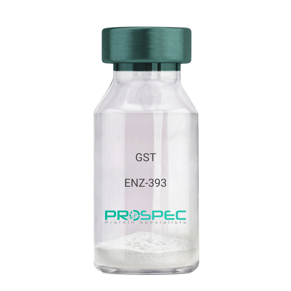
Name :
GSTDescription:
Glutathione S-Transferase Recombinant
Glutathione S-Transferase, GST, Glutathione S-transferase class-mu 26 kDa isozyme, GST 26, Sj26 antigen, SjGST.
Product # :
ENZ-393Price :
Quantity :
Shipping Method :
Shipped with Ice Packs
More Info
- description
- source
- formulation
- purity
- biological activity
- More Info
Description
Recombinant Glutathione S-Transferase full length protein (1-218a.a.) expressed in E.coli, having a molecular mass of 26kDa. GST was isolated from an E. coli strain that carries the coding sequence for Schistosoma japonicum GST under the control of a T7 promoter. The GST is purified by proprietary chromatographic techniques.
Source
Escherichia Coli.
Formulation
GST supplied in Phosphate Buffered Saline pH 7.4.
Purity
Greater than 95% as determined by SDS-PAGE.
Biological Activity
Specific activity is >20 units/mg. A unit is defined as the amount of enzyme that conjugate 1.0 u mole of 1-chloro-2,4-dinitrobenzene (CDNB) with reduced glutathione per minute at pH 6.5 at 25°C.More Info
-
Introduction
Antioxidant enzyme Glutathione S- Transferase (GST) is thought to do the primary cellular defense mechanism against reactive oxygen species. GST reduces lipid hydroperoxides through its Se-independent glutathione peroxidase activity. The enzyme also detoxifies lipid peroxidation end products such as 4-hydroxynonenal (4-HNE).
The soluble GST is a 26 kDa protein which occurs as a dimer in all aerobic organisms. Each monomer has two domains, one that binds GSH and is an /-structure similar to thioredoxin and the other, all helical, that binds the hydrophobic substrate. The GST -fusion protein expression system is a widely used recombinant protein expression system that allows a peptide or a regulatory protein domain to be expressed as a fusion to the C-terminus of Schistosoma japonicum GST. Fusion proteins also possess GST -enzymatic activity and can undergo dimerization similar to in vivo. The fusion protein can be purified via GST -affinity column chromatography. In most cases, the desired peptides or domains are removed from GST by applying a specific protease that recognizes and cleaves the linker between the protein domain and GST. The technique has been widely used to generate different kinds of proteins for crystallization, molecular immunology studies, the production of vaccines and studies involving protein-protein and protein-DNA interactions. -
Synonyms
Glutathione S-Transferase, GST, Glutathione S-transferase class-mu 26 kDa isozyme, GST 26, Sj26 antigen, SjGST.
-
Physical Appearance
Sterile Filtered clear solution.
-
Stability
Store at 4°C if entire vial will be used within 2-4 weeks. Store, frozen at -20°C for longer periods of time. For long term storage it is recommended to add a carrier protein (0.1% HSA or BSA).Avoid multiple freeze-thaw cycles.
-
Amino Acid Sequence
MSPILGYWKI KGLVQPTRLL LEYLEEKYEE HLYERDEGDK WRNKKFELGL EFPNLPYYID GDVKLTQSMA IIRYIADKHN MLGGCPKERA EISMLEGAVL DIRYGVSRIA YSKDFETLKV DFLSKLPEML KMFEDRLCHK TYLNGDHVTH PDFMLYDALD VVLYMDPMCL DAFPKLVCFK KRIEAIPQID KYLKSSKYIA WPLQGWQATF GGGDHPPKSD LVPR.
ProSpec's products are furnished for LABORATORY RESEARCH USE ONLY. The product may not be used as drugs, agricultural or pesticidal products, food additives or household chemicals.