Search results
1000 results found for “fibronectin”
Name
Description
Product #
Price
Quantity
Shipping Method
- View Data Sheet
Name :
CCL13 HumanDescription:
Monocyte Chemotactic Protein-4 Human Recombinant (CCL13)
Small inducible cytokine A13, CCL13, Monocyte chemotactic protein 4, MCP-4, Monocyte chemoattractant protein 4, CK-beta-10, NCC-1, chemokine (C-C motif) ligand 13, NCC1, CKb10, SCYL1, SCYA13, MGC17134.
Product # :
CHM-319Price :
Quantity :
Shipping Method :
Shipped at Room temp
More Info
- description
- source
- formulation
- purity
- biological activity
- More Info
Description
Monocyte Chemotactic Protein-4 Human Recombinant produced in E.Coli is a non-glycosylated, Polypeptide chain containing 75 amino acids and having a molecular mass of 8.6 kDa. The MCP-4 is purified by proprietary chromatographic techniques.
Source
Escherichia Coli.
Formulation
The protein was lyophilized from a concentrated (1mg/ml) sterile solution in 20mM PB, pH 7.4, 130mM NaCl.
Purity
Greater than 96.0% as determined by:
(a) Analysis by RP-HPLC.
(b) Analysis by SDS-PAGE.Biological Activity
The specific activity as determined by the ability of MCP-4 to chemoattaract human monocytes at 10-100ng/ml, corresponding to a Specific Activity of 10,000-100,000 units/mg.More Info
-
Introduction
Chemokine (C-C motif) ligand 13 (CCL13 / MCP-4) is a small cytokine belonging to the CC chemokine family. The MCP-4 gene is located on human chromosome 17 within a large cluster of other CC chemokines. MCP-4 induces chemotaxis in monocytes, eosinophils, T lymphocytes, and basophils by binding cell surface G-protein linked chemokine receptors such as CCR2, CCR3 and CCR5. Activity of the MCP-4 chemokine has been implicated in allergic reactions such as asthma. MCP-4 can be induced by the inflammatory cytokines interleukin-1 and TNF-a.
-
Synonyms
Small inducible cytokine A13, CCL13, Monocyte chemotactic protein 4, MCP-4, Monocyte chemoattractant protein 4, CK-beta-10, NCC-1, chemokine (C-C motif) ligand 13, NCC1, CKb10, SCYL1, SCYA13, MGC17134.
-
Physical Appearance
Sterile Filtered White lyophilized (freeze-dried) powder.
-
Stability
Lyophilized MCP-4 although stable at room temperature for 3 weeks, should be stored desiccated below -18°C. Upon reconstitution MCP-4 should be stored at 4°C between 2-7 days and for future use below -18°C. For long term storage it is recommended to add a carrier protein (0.1% HSA or BSA).Please prevent freeze-thaw cycles.
-
Solubility
It is recommended to reconstitute the lyophilized MCP-4 in sterile 18M-cm H2O not less than 100µg/ml, which can then be further diluted to other aqueous solutions.
-
Amino Acid Sequence
QPDALNVPSTCCFTFSSKKISLQRLKSYVITTSRCPQKAV
IFRTKLGKEICADPKEKWVQNYMKHLGRKAHTLKT. -
Background
What is the molecular weight/Mw of CCL13 HUMAN Protein?
CCL13 HUMAN Protein has a total Mw of 8.6kDa.
What is the source or expression system of CCL13 HUMAN Protein?
Escherichia Coli.
What is the Purity of CCL13 HUMAN Protein?
CCL13 HUMAN Protein is >96% pure as determined by SDS-PAGE.
What is the Biological Activity of CCL13 HUMAN Protein?
The specific activity as determined by the ability of MCP-4 to chemoattaract human monocytes at 10-100ng/ml, corresponding to a Specific Activity of 10,000-100,000 units/mg.
What is the amino acid sequence of CCL13 HUMAN Protein?
QPDALNVPSTCCFTFSSKKISLQRLKSYVITTSRCPQKAV
IFRTKLGKEICADPKEKWVQNYMKHLGRKAHTLKT.
What applications can CCL13 HUMAN Protein be used in?
CCL13 HUMAN Protein can probably be used in western blot, ELISA and Lateral Flow.
What is the endotoxin level for CCL13 HUMAN Protein?
The endotoxin level is minimal, CCL13 HUMAN Protein was purified using conventional chromatography techniques.
ProSpec's products are furnished for LABORATORY RESEARCH USE ONLY. The product may not be used as drugs, agricultural or pesticidal products, food additives or household chemicals.
- View Data Sheet
Name :
ID1 HumanDescription:
Inhibitor of DNA Binding 1 Human Recombinant
bHLHb24, ID, DNA-binding protein inhibitor ID-1, Class B basic helix-loop-helix protein 24, Inhibitor of DNA binding 1, ID1.
Product # :
PRO-1426Price :
Quantity :
Shipping Method :
Shipped with Ice Packs
More Info
- description
- source
- formulation
- purity
- More Info
Description
ID1 Human Recombinant produced in E. coli is a single polypeptide chain containing 178 amino acids (1-155) and having a molecular mass of 18.5kDa. ID1 is fused to a 23 amino acid His-tag at N-terminus & purified by proprietary chromatographic techniques.
Source
E.coli.
Formulation
The ID1 solution (0.5mg/ml) contains 20mM Tris-HCl buffer (pH 8.0), 0.15M NaCl, 10% glycerol and 1mM DTT.
Purity
Greater than 85% as determined by SDS-PAGE.
More Info
-
Introduction
Inhibitor of DNA Binding 1 (ID1) is a helix-loop-helix protein which can form heterodimers with members of the basic HLH family of transcription factors. ID1 is lacking DNA binding activity and thus can inhibit the DNA binding and transcriptional activation ability of basic HLH proteins with which it interacts. ID1 participates in cell growth, senescence, and differentiation.
-
Synonyms
bHLHb24, ID, DNA-binding protein inhibitor ID-1, Class B basic helix-loop-helix protein 24, Inhibitor of DNA binding 1, ID1.
-
Physical Appearance
Sterile Filtered colorless solution.
-
Stability
Store at 4°C if entire vial will be used within 2-4 weeks. Store, frozen at -20°C for longer periods of time. For long term storage it is recommended to add a carrier protein (0.1% HSA or BSA).Avoid multiple freeze-thaw cycles.
-
Amino Acid Sequence
MGSSHHHHHH SSGLVPRGSH MGSMKVASGS TATAAAGPSC ALKAGKTASG AGEVVRCLSE QSVAISRCAG GAGARLPALL DEQQVNVLLY DMNGCYSRLK ELVPTLPQNR KVSKVEILQH VIDYIRDLQL ELNSESEVGT PGGRGLPVRA PLSTLNGEIS ALTAEAACVP ADDRILCR.
ProSpec's products are furnished for LABORATORY RESEARCH USE ONLY. The product may not be used as drugs, agricultural or pesticidal products, food additives or household chemicals.
- View Data Sheet
Name :
Leptin AntibodyDescription:
Leptin, Mouse Anti-Human
OB Protein, Obesity Protein, OBS, Obesity factor.
Product # :
ANT-172Price :
Quantity :
Shipping Method :
Shipped at Room temp
More Info
- More Info
More Info
-
Introduction
A 16-kDa peptide hormone secreted from white adipocytes and implicated in the regulation of food intake and energy balance. Leptin provides the key afferent signal from fat cells in the feedback system that controls body fat stores.
-
Synonyms
OB Protein, Obesity Protein, OBS, Obesity factor.
-
Solubility
Reconstitute with sterile H20. Mix gently, wash the sides of the vial and wait 30-60 seconds before use.
-
Immunogen
r.Human Leptin.
-
Ig Subclass
Mouse IgG1.
-
Clone
YD-HLEP4B6.
-
Applications
Direct ELISA, Western Blot, Immuneprecipitation.
-
Titer
By direct ELISA, 1:10,000 dilution will yield 0.8 O.D using alkaline phosphatase conjugated rabbit anti-mouse Ig (Jackson Laboratories).
-
Shipping Conditions
Antibody is shipped lyophilized at ambient temperature.
-
Type
Mouse Anti Human Monoclonal.
-
Storage Procedures
In lyophilized form, for long periods, store at 4C in a dry environment. After reconstitution, if not intended for use within a month, aliquot and store at -20C.
-
Purification Method
Ion exchange.
ProSpec's products are furnished for LABORATORY RESEARCH USE ONLY. The product may not be used as drugs, agricultural or pesticidal products, food additives or household chemicals.
- View Data Sheet
Name :
Thyroglobulin Human, BiotinDescription:
Thyroglobulin Human, Biotinylated
Thyroglobulin, TGN, AITD3, TG.
Product # :
PRO-2563Price :
Quantity :
Shipping Method :
Shipped with Ice Packs
More Info
- description
- source
- formulation
- purity
- More Info
Description
Human Thyroglobulin is a biotinylated, glycosylated, polypeptide chain having a total molecular mass of 662 kDa (331 kDa per subunit).
Source
Native, Isolated from human thyroid glands.
Formulation
Human Thyroglobulin biotinylated is supplied at a 20mM HEPES buffer pH-7.6, 150mM NaCl and 40% Sucrose (w/v).
Purity
Greater than 80% as determined by SDS-PAGE.
More Info
-
Introduction
Thyroglobulin (TG) represents one of the main autoantigenic targets in autoimmune thyroid disease of humans. TG is a large globular dimeric glycoprotein with a total molecular weight of 660 kDa, which occupies a key precursor role in the biosynthesis of the thyroid hormones. Approximately 75% of the total protein content of the thyroid follicle consists of TG. The thyroid gland uses the Thyroglobulin in order to produce the thyroid hormones thyroxine (T4) and triiodothyronine (T3). Thyroglobulin is produced by the thyroid epithelial cells (thyrocytes) which form spherical follicles. Thyroglobulin is subsequently secreted and stored in the follicular lumen.
Patients with Hashimoto's thyroiditis or Graves' disease, frequently develop antibodies against Thyroglobulin. Tg-specific antibodies help in the diagnosis of the above diseases, however they also may be present in apparently healthy euthyroid individuals. Blood Thyroglobulin levels can be used as a tumor marker for certain kinds of thyroid cancer, and the may also be elevated in cases of Graves' disease. -
Synonyms
Thyroglobulin, TGN, AITD3, TG.
-
Physical Appearance
Sterile Filtered solution.
-
Stability
Store at 4°C if entire vial will be used within 2-4 weeks. Store, frozen at -20°C for longer periods of time. Avoid multiple freeze-thaw cycles.
-
Immunological Functions
1. Binds IgG type human auto antibodies.2. Auto antibodies to thyroglobulin recognize conformation dependent epitopes. 3.Functional Streptavidin based ELISA test (analysis of positive/negative samples.)
ProSpec's products are furnished for LABORATORY RESEARCH USE ONLY. The product may not be used as drugs, agricultural or pesticidal products, food additives or household chemicals.
- View Data Sheet
Name :
Desmin ChickenDescription:
Desmin Chicken Gizzard
Desmin, DES, CSM1, CSM2, CMD1I, FLJ12025, FLJ39719, FLJ41013, FLJ41793.
Product # :
PRO-2783Price :
Quantity :
Shipping Method :
Shipped at Room temp
More Info
- description
- source
- formulation
- purity
- More Info
Description
Desmin Chicken having a calculated molecular mass of 53 kDa, pI-5.4.
Source
Chicken gizzard.
Formulation
Desmin was lyophilized from a 1mg/ml solution containing 10 mM sodium phosphate buffer pH 7.5, 6M urea, 1mM EDTA, 2mM DTT and 10mM methylammonium chloride.
Purity
Greater than 98.0% as determined by SDS-PAGE.
More Info
-
Synonyms
Desmin, DES, CSM1, CSM2, CMD1I, FLJ12025, FLJ39719, FLJ41013, FLJ41793.
-
Physical Appearance
Sterile Filtered White lyophilized (freeze-dried) powder.
-
Stability
Store the lyophilized Desmin between 2-8°C, do not freeze. Upon reconstitution Desmin should be stored at -20°C. Please prevent freeze-thaw cycles.
-
Solubility
It is recommended to reconstitute the lyophilized Desmin in sterile 18MΩ-cm H2O not less than 100µg/ml, which can then be further diluted to other aqueous solutions.
-
Background
Desmin, an intermediate filament protein, plays a fundamental role in maintaining the structural integrity and function of muscle cells. While extensive research has been conducted on desmin in mammals, the study of desmin in chickens is an emerging area with considerable potential for advancing our understanding of muscle biology. Chickens are valuable model organisms for studying muscle development, growth, and regeneration due to their relatively simple muscular system and economic significance in poultry production. This research aims to provide a comprehensive exploration of desmin in chickens, shedding light on its functions and implications for muscle structure and function.
The primary objective of this research is to elucidate the role of desmin in chicken muscle structure and development. In vitro and in vivo experiments, utilizing chicken cell cultures and embryonic models, will be conducted to investigate how desmin contributes to the organization of muscle fibers, sarcomere assembly, and myofibrillogenesis. Understanding these mechanisms is fundamental for deciphering the complexities of muscle development in chickens.
The second objective is to assess the clinical and economic relevance of desmin in poultry production. Studies involving broiler chickens will be conducted to evaluate the impact of desmin mutations or variations on muscle growth, meat quality, and disease susceptibility. These investigations may provide valuable insights into potential strategies for enhancing poultry production efficiency and meat quality.
The third objective is to explore the potential applications of desmin in biotechnology and tissue engineering. Research will investigate the use of desmin-expressing chicken cells as models for studying muscle-related diseases and for developing tissue engineering approaches for muscle repair and regeneration.
By delving into the functions and roles of desmin in chickens, this research aims to expand our knowledge of muscle biology, its implications for poultry production, and its potential applications in biotechnology and regenerative medicine.
ProSpec's products are furnished for LABORATORY RESEARCH USE ONLY. The product may not be used as drugs, agricultural or pesticidal products, food additives or household chemicals.
- View Data Sheet
Name :
FLT1 D4 HumanDescription:
Vascular Endothelial Growth Factor Receptor-1 D4 Human Recombinant
FLT-1, FLT1, Tyrosine-protein kinase receptor FLT, Flt-1, Tyrosine-protein kinase FRT, Fms-like tyrosine kinase 1, VEGFR-1.
Product # :
PKA-235Price :
Quantity :
Shipping Method :
Shipped at Room temp
More Info
- description
- source
- formulation
- purity
- biological activity
- More Info
Description
Soluble FLT1 D1-4 Human Recombinant produced in baculovirus is monomeric, glycosylated, polypeptide containing 457 amino acids and having a molecular mass of 55 kDa. The soluble receptor protein contains only the first 4 extracellular domains, which contain all the information necessary for binding of VEGF.The VEGFR1 is purified by proprietary chromatographic techniques.
Source
Insect Cells.
Formulation
FLT1 D1-4 was lyophilized from a concentrated (1mg/ml) sterile solution containing no additives.
Purity
Greater than 90.0% as determined by(a)Analysis by RP-HPLC.
(b)Analysis by SDS-PAGE.Biological Activity
The activity of FLT1D1-4 was determined by its ability to abolish the binding of iodinated VEGF to solid surfaces or cell surfaces, and in Far-Western and cross-linking experiments with iodinated VEGF.More Info
-
Introduction
Endothelial cells express three different vascular endothelial growth factor (VEGF) receptors, belonging to the family of receptor tyrosine kinases (RTKs). They are named VEGFR-1 (Flt-1), VEGFR-2 (KDR/Flk-1), VEGFR-3 (Flt-4). Their expression is almost exclusively restricted to endothelial cells, but VEGFR-1 can also be found on monocytes, dendritic cells and on trophoblast cells. The flt-1 gene was first described in 1990. The receptor contains seven immunoglobulin-like extracellular domains, a single transmembrane region and an intracellular splited tyrosine kinase domain. Compared to VEGFR-2 the Flt-1 receptor has a higher affinity for VEGF but a weaker signaling activity. VEGFR-1 thus leads not to proliferation of endothelial cells, but mediates signals for differentiation. Interestingly a naturally occuring soluble variant of VEGFR-1 (sVEGFR-1) was found in HUVE supernatants in 1996, which is generated by alternative splicing of the flt-1 mRNA. The biological functions of sVEGFR-1 still are not clear, but it seems to be an endogenous regulator of angiogenesis, binding VEGF with the same affinity as the full-length receptor.
-
Synonyms
FLT-1, FLT1, Tyrosine-protein kinase receptor FLT, Flt-1, Tyrosine-protein kinase FRT, Fms-like tyrosine kinase 1, VEGFR-1.
-
Physical Appearance
Sterile Filtered White lyophilized (freeze-dried) powder.
-
Stability
Lyophilized FLT-1 although stable at room temperature for 3 weeks, should be stored desiccated below -18°C. Upon reconstitution FLT1 should be stored at 4°C between 2-7 days and for future use below -18°C.For long term storage it is recommended to add a carrier protein (0.1% HSA or BSA).Please prevent freeze-thaw cycles.
-
Solubility
It is recommended to reconstitute the lyophilized FLT1 D4 in sterile water not less than 100 µg/ml, which can then be further diluted to other aqueous solutions.
ProSpec's products are furnished for LABORATORY RESEARCH USE ONLY. The product may not be used as drugs, agricultural or pesticidal products, food additives or household chemicals.
- View Data Sheet
Name :
MFGE8 MouseDescription:
Milk Fat Globule-EGF Factor 8 Protein Mouse Recombinant
Milk fat globule-EGF factor 8 protein, isoform CRA_a, Putative uncharacterized protein, Mfge8, mCG_6301.
Product # :
CYT-1000Price :
Quantity :
Shipping Method :
Shipped with Ice Packs
More Info
- description
- source
- formulation
- purity
- More Info
Description
MFGE8 Mouse Recombinant produced in Sf9 Baculovirus cells is a single, glycosylated polypeptide chain containing 413 amino acids (23-426a.a.) and having a molecular mass of 46kDa (Molecular size on SDS-PAGE will appear at approximately 40-57kDa).MFGE8 is expressed with a 6 amino acid His-tag at C-Terminus and purified by proprietary chromatographic techniques.
Source
Sf9, Baculovirus cells.
Formulation
MFGE8 protein solution (0.25mg/ml) contains Phosphate Buffered Saline (pH 7.4) and 10% glycerol.
Purity
Greater than 90.0% as determined by SDS-PAGE.
More Info
-
Introduction
Milk fat globule-EGF factor 8 protein (Mfge8) is pleiotropic secreted glycoprotein which promotes mammary gland morphogenesis, angiogenesis, and tumor progression. Mfge8 has also an imperative role in tissue homeostasis and the prevention of inflammation. Mfge8 functions as a bridge between phosphatidylserine on apoptotic cells and Integrin alpha V beta 3 on phagocytes, leading to the clearance of apoptotic debris.
-
Synonyms
Milk fat globule-EGF factor 8 protein, isoform CRA_a, Putative uncharacterized protein, Mfge8, mCG_6301.
-
Physical Appearance
Sterile filtered colorless solution.
-
Stability
Store at 4°C if entire vial will be used within 2-4 weeks. Store, frozen at -20°C for longer periods of time. For long term storage it is recommended to add a carrier protein (0.1% HSA or BSA).Avoid multiple freeze-thaw cycles.
-
Amino Acid Sequence
ADLASGDFCD SSLCLNGGTC LTGQDNDIYC LCPEGFTGLV CNETERGPCS PNPCYNDAKC LVTLDTQRGD IFTEYICQCP VGYSGIHCET GCSTQLGMEG GAIADSQISA SSVYMGFMGL QRWGPELARL YRTGIVNAWT ASNYDSKPWI QVNLLRKMRV SGVMTQGASR AGRAEYLKTF KVAYSLDGRK FEFIQDESGG DKEFLGNLDN NSLKVNMFNP TLEAQYIKLY PVSCHRGCTL RFELLGCELH GCSEPLGLKN NTIPDSQMSA SSSYKTWNLR AFGWYPHLGR LDNQGKINAW TAQSNSAKEW LQVDLGTQRQ VTGIITQGAR DFGHIQYVAS YKVAHSDDGV QWTVYEEQGS SKVFQGNLDN NSHKKNIFEK PFMARYVRVL PVSWHNRITL RLELLGCHHH HHH.
-
Background
An Insight into Milk Fat Globule-EGF Factor 8 Protein (Mouse Recombinant): Characteristics, Applications, and Future Directions
Abstract
The Milk Fat Globule-EGF Factor 8 (MFG-E8) protein, particularly in its mouse recombinant form, has emerged as a pivotal player in multiple physiological processes. This paper aims to elucidate its unique characteristics, delve into current methodologies employed in its study, and chart the trajectory for future research directions.
Introduction
Milk Fat Globule-EGF Factor 8 (MFG-E8) is a glycoprotein known for its vital role in several cellular processes, including cell signaling, apoptosis, and phagocytosis. The recombinant form of MFG-E8 derived from mouse models offers a valuable tool for researchers in deciphering its biological relevance.
Characteristics of MFG-E8 (Mouse Recombinant)
1. Structural Profile: The MFG-E8 protein harbors EGF-like domains, which enable its participation in numerous cellular signaling events.
2. Expression Spectrum: While originally identified in mammary epithelial cells, its expression spectrum extends to macrophages, dendritic cells, and other tissues.
3. Biochemical Activity: It plays a key role in facilitating the phagocytic clearance of apoptotic cells by bridging these cells to phagocytes.
Methodologies Employed in MFG-E8 Research
1. Production of Recombinant MFG-E8: Using bacterial expression systems, such as E. coli, mouse MFG-E8 DNA is introduced, followed by protein purification techniques like gel filtration chromatography.
2. Assays: The phagocytosis assays employing fluorescently tagged apoptotic cells and phagocytes enable researchers to quantify MFG-E8's effectiveness in apoptotic cell clearance.
3. Immunoblotting: Through SDS-PAGE and Western blotting, the expression and purification of MFG-E8 can be monitored and validated.
4. Knockout Models: MFG-E8 knockout mice models help decipher its in vivo significance, especially concerning its immune-regulatory roles.
Original Ideas & Implications
The potential for MFG-E8, particularly in its mouse recombinant form, to serve as a therapeutic agent in autoimmune disorders remains a tantalizing prospect. Considering its role in apoptotic cell clearance, dysregulation in MFG-E8 might be implicated in the etiology of autoimmune disorders. Thus, targeting this protein therapeutically may pave the way for innovative treatments.
Conclusions & Future Directions
While the recombinant MFG-E8 protein has elucidated much about the biological implications of this protein, the horizon is rife with potential. Future research might focus on its therapeutic potential, particularly in autoimmunity and inflammation.
ProSpec's products are furnished for LABORATORY RESEARCH USE ONLY. The product may not be used as drugs, agricultural or pesticidal products, food additives or household chemicals.
- View Data Sheet
Name :
SEC61B HumanDescription:
SEC61B Human Recombinant
Protein transport protein Sec61 subunit beta, SEC61B, Sec61 Beta Subunit, Protein Translocation Complex Beta, Protein Transport Protein SEC61 Beta Subunit, Sec61 Complex Beta Subunit.
Product # :
PRO-1795Price :
Quantity :
Shipping Method :
Shipped with Ice Packs
More Info
- description
- source
- formulation
- purity
- More Info
Description
SEC61B Human Recombinant produced in E.Coli is a single, non-glycosylated polypeptide chain containing 93 amino acids (1-70 a.a) and having a molecular mass of 9.4kDa.SEC61B is fused to a 23 amino acid His-tag at N-terminus & purified by proprietary chromatographic techniques.
Source
Escherichia Coli.
Formulation
SEC61B protein solution (0.25mg/ml) containing 20mM Tris-HCl buffer (pH 8.0), 0.2M NaCl, 50% glycerol and 2mM DTT.
Purity
Greater than 85.0% as determined by SDS-PAGE.
More Info
-
Introduction
The Sec61 complex is the key component of the protein translocation apparatus of the ER (endoplasmic reticulum) membrane. Oligomers of the Sec61 complex construct a transmembrane channel where proteins are translocated across and incorporated into the ER membrane. This complex consists of 3 membrane proteins- alpha, beta, and gamma. The SEC61B gene encodes the beta-subunit protein. The Sec61 subunits are also detected in the post-ER compartment, suggesting that these proteins can get out of the ER and recycle back.
-
Synonyms
Protein transport protein Sec61 subunit beta, SEC61B, Sec61 Beta Subunit, Protein Translocation Complex Beta, Protein Transport Protein SEC61 Beta Subunit, Sec61 Complex Beta Subunit.
-
Physical Appearance
Sterile Filtered colorless solution.
-
Stability
Store at 4°C if entire vial will be used within 2-4 weeks. Store, frozen at -20°C for longer periods of time. For long term storage it is recommended to add a carrier protein (0.1% HSA or BSA).Avoid multiple freeze-thaw cycles.
-
Amino Acid Sequence
MGSSHHHHHH SSGLVPRGSH MGSMPGPTPS GTNVGSSGRS PSKAVAARAA GSTVRQRKNA SCGTRSAGRT TSAGTGGMWR FYTEDSPGLK VGP.
ProSpec's products are furnished for LABORATORY RESEARCH USE ONLY. The product may not be used as drugs, agricultural or pesticidal products, food additives or household chemicals.
- View Data Sheet
Name :
PCSK1N HumanDescription:
Proprotein Convertase Subtilisin/Kexin Type 1 Inhibitor Human Recombinant
ProSAAS precursor, Proprotein convertase subtilisin/kexin type 1 inhibitor, PROSAAS; SAAS, PCSK1N.
Product # :
PRO-1819Price :
Quantity :
Shipping Method :
Shipped with Ice Packs
More Info
- description
- source
- formulation
- purity
- More Info
Description
PCSK1N Human Recombinant produced in E.coli is a single, non-glycosylated polypeptide chain containing 251 amino acids (34-260) and having a molecular mass of 26.6 kDa.PCSK1N is fused to a 23 amino acid His-Tag at N-terminus and purified by proprietary chromatographic techniques.
Source
Escherichia Coli.
Formulation
The PCSK1N solution (0.25mg/ml) contains 20mM Tris-HCl buffer (pH 8.0), 10% glycerol and 0.4M Urea.
Purity
Greater than 85.0% as determined by SDS-PAGE.
More Info
-
Introduction
Proprotein Convertase Subtilisin/Kexin Type 1 Inhibitor (PCSK1N) takes part in the control of the neuroendocrine secretory pathway. PCSK1N inhibits prohormone convertase 1, which regulates the proteolytic cleavage of neuroendocrine peptide precursors. PCSK1Nslows down convertase-mediated processing of proopiomelanocortin and proenkephalin and also monitors the intracellular timing of PCSK1.
-
Synonyms
ProSAAS precursor, Proprotein convertase subtilisin/kexin type 1 inhibitor, PROSAAS; SAAS, PCSK1N.
-
Physical Appearance
Sterile Filtered colorless solution.
-
Stability
Store at 4°C if entire vial will be used within 2-4 weeks. Store, frozen at -20°C for longer periods of time. For long term storage it is recommended to add a carrier protein (0.1% HSA or BSA).Avoid multiple freeze-thaw cycles.
-
Amino Acid Sequence
MGSSHHHHHH SSGLVPRGSH MGSMARPVKE PRGLSAASPP LAETGAPRRF RRSVPRGEAA GAVQELARAL AHLLEAERQE RARAEAQEAE DQQARVLAQL LRVWGAPRNS DPALGLDDDP DAPAAQLARA LLRARLDPAA LAAQLVPAPV PAAALRPRPP VYDDGPAGPD AEEAGDETPD VDPELLRYLL GRILAGSADS EGVAAPRRLR RAADHDVGSE LPPEGVLGAL LRVKRLETPA PQVPARRLLP P.
ProSpec's products are furnished for LABORATORY RESEARCH USE ONLY. The product may not be used as drugs, agricultural or pesticidal products, food additives or household chemicals.
- View Data Sheet
Name :
ANXA10 Human (1-162)Description:
Annexin A10 (1-162 a.a.) Human Recombinant
anxa-10.
Product # :
PRO-2837Price :
Quantity :
Shipping Method :
Shipped at Room temp
More Info
- description
- source
- formulation
- purity
- More Info
Description
The ANXA10 Human is created as a recombinant protein with a 4kda N-terminal fusion of His Tag. The ANXA10 His-Tagged Fusion Protein, produced in E. coli, is a 21kDa protein containing 162 amino acid residues of the ANXA10 Human, 1-162 amino acids.
Source
Escherichia Coli.
Formulation
Each mg was lyophilized with 1xPBS, 0.4% SDS and 4mM DTT.
Purity
Greater than 90% as determined by SDS-PAGE.
More Info
-
Synonyms
anxa-10.
-
Physical Appearance
Filtered White lyophilized (freeze-dried) powder.
-
Stability
Store lyophilized ANXA10 at -20°C. Aliquot the product after reconstitution to avoid repeated freezing/thawing cycles. Reconstituted protein can be stored at 4°C for a limited period of time; it does not show any change after two weeks at 4°C.
-
Solubility
It is recommended to add deionized water to prepare a working stock solution of approximately 0.5mg/ml and let the lyophilized pellet dissolve completely. Product is not sterile! Please filter the product by an appropriate sterile filter before using it on cell culture.
-
Background
Annexin A10 also known as ANXA10 is a part of the annexin family of calcium-binding proteins which members own a conserved core domain and a unique amino-terminal region which may convene binding specificity. The ANXA10 protein contains 4 annexin domains and plays a role in the regulation of cellular growth and signal transduction pathways throughout the cell.
ProSpec's products are furnished for LABORATORY RESEARCH USE ONLY. The product may not be used as drugs, agricultural or pesticidal products, food additives or household chemicals.
- View Data Sheet
Name :
Omentin 298 a.a. HumanDescription:
Omentin 298 a.a. Human Recombinant
Intelectin-1, HL1, LFR, HL-1, INTL, ITLN, hIntL.
Product # :
CYT-061Price :
Quantity :
Shipping Method :
Shipped with Ice Packs
More Info
- description
- source
- formulation
- purity
- More Info
Description
Omentin Human Recombinant produced in E.coli is a single, non-glycosylated polypeptide chain containing 298 amino acids (17-313) and having a molecular mass of 33.2 kDa.The Omentin is purified by proprietary chromatographic techniques.
Source
Escherichia Coli.
Formulation
Omentin protein (1mg/ml) is supplied in 20mM Tris-HCL, pH-8, 0.4M Urea and 10% Glycerol.
Purity
Greater than 90.0% as determined by SDS-PAGE.
More Info
-
Introduction
Omentin is a recently recognized gene highly localized to the mental tissue (visceral adipose tissue). Omentin is present in the stromal vascular cells in the adipose tissue rather than in the adipocytes. Omentin is predominantly expressed in the visceral adipose tissue than the subcutaneous tissue, with the omentin mRNA being 150 times higher in the visceral adipose tissue. Omentin has also been detected in human blood using western blot analysis, and seems to increase insulin-stimulated glucose uptake in 3T3-L1 adipocytes in mice. Omentin seems to increase Akt phosphorylation irrespective of insulin presence. Its role in glucose metabolism and obesity remains to be described; an insulin-sensitizing action is possible.Differences in Omentin expression has been noted in adipose tissue from normals and patients with inflammatory bowel disease although its significance is unknown.
-
Synonyms
Intelectin-1, HL1, LFR, HL-1, INTL, ITLN, hIntL.
-
Physical Appearance
Sterile Filtered colorless solution.
-
Stability
Store at 4°C if entire vial will be used within 2-4 weeks. Store, frozen at -20°C for longer periods of time. For long term storage it is recommended to add a carrier protein (0.1% HSA or BSA).Avoid multiple freeze-thaw cycles.
-
Amino Acid Sequence
MWSTDEANTY FKEWTCSSSP SLPRSCKEIK DECPSAFDGL YFLRTENGVI YQTFCDMTSG GGGWTLVASV HENDMRGKCT VGDRWSSQQG SKAVYPEGDG NWANYNTFGS AEAATSDDYK NPGYYDIQAK DLGIWHVPNK SPMQHWRNSS LLRYRTDTGF LQTLGHNLFG IYQKYPVKYG EGKCWTDNGP VIPVVYDFGD AQKTASYYSP YGQREFTAGF VQFRVFNNER AANALCAGMR VTGCNTEHHC IGGGGYFPEA SPQQCGDFSG FDWSGYGTHV GYSSSREITE AAVLLFYR
ProSpec's products are furnished for LABORATORY RESEARCH USE ONLY. The product may not be used as drugs, agricultural or pesticidal products, food additives or household chemicals.
- View Data Sheet
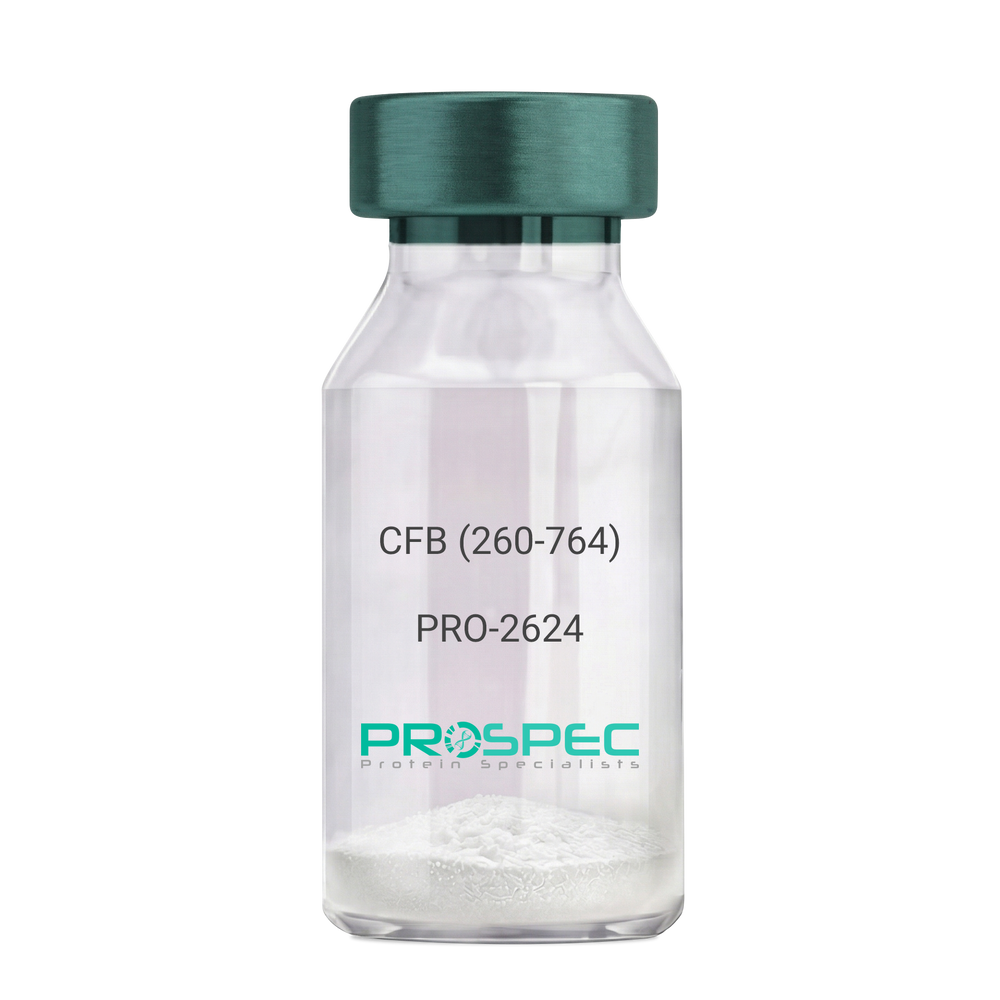
Name :
CFB (260-764) Human, Sf9Description:
Complement Factor B (260-764 a.a.) Human Recombinant, Sf9
CFB, C3/C5 convertase, Glycine-rich beta glycoprotein, GBG, PBF2, Properdin factor B, BF, BFD, AHUS4, ARMD14, CFAB, CFBD, FB, FBI12, GBG, H2-Bf.
Product # :
PRO-2624Price :
Quantity :
Shipping Method :
Shipped with Ice Packs
More Info
- description
- source
- formulation
- purity
- More Info
Description
CFB Human Recombinant produced in Sf9 Baculovirus cells is a single, glycosylated polypeptide chain containing 514 amino acids (260-764 a.a) and having a molecular mass of 58.1kDa.CFB is fused to an 9 amino acid His-tag at C-terminus & purified by proprietary chromatographic techniques.
Source
Sf9, Baculovirus cells.
Formulation
CFB protein solution (0.5mg/ml) containing Phosphate Buffered Saline (pH 7.4) and 20% glycerol.
Purity
Greater than 90.0% as determined by SDS-PAGE.
More Info
-
Introduction
Factor B is a protein, build from a single chain that circulates in the blood. Complement factor D cleaves and activates factor B, thus creating a catalytic subunit Bb and noncatalytic chain Ba. Subunit Bb is a serine protease that can bind to C3b and forms alternative pathway C3 convertase. Subunit Ba acts as an inhibitor for proliferation of lymphocytes, when Bb is part of the lymphocytes’ proliferation. The gene that codes for this protein is located on chromosome six.
-
Synonyms
CFB, C3/C5 convertase, Glycine-rich beta glycoprotein, GBG, PBF2, Properdin factor B, BF, BFD, AHUS4, ARMD14, CFAB, CFBD, FB, FBI12, GBG, H2-Bf.
-
Physical Appearance
Sterile Filtered colorless solution.
-
Stability
Store at 4°C if entire vial will be used within 2-4 weeks. Store, frozen at -20°C for longer periods of time. For long term storage it is recommended to add a carrier protein (0.1% HSA or BSA).Avoid multiple freeze-thaw cycles.
-
Amino Acid Sequence
ADPKIVLDPS GSMNIYLVLD GSDSIGASNF TGAKKCLVNL IEKVASYGVK PRYGLVTYAT
YPKIWVKVSE ADSSNADWVT KQLNEINYED HKLKSGTNTK KALQAVYSMM SWPDDVPPEG
WNRTRHVIIL MTDGLHNMGG DPITVIDEIR DLLYIGKDRK NPREDYLDVY VFGVGPLVNQ
VNINALASKK DNEQHVFKVK DMENLEDVFY QMIDESQSLS LCGMVWEHRK GTDYHKQPWQ
AKISVIRPSK GHESCMGAVV SEYFVLTAAH CFTVDDKEHS IKVSVGGEKR DLEIEVVLFH
PNYNINGKKE AGIPEFYDYD VALIKLKNKL KYGQTIRPIC LPCTEGTTRA LRLPPTTTCQ
QQKEELLPAQ DIKALFVSEE EKKLTRKEVY IKNGDKKGSC ERDAQYAPGY DKVKDISEVV
TPRFLCTGGV SPYADPNTCR GDSGGPLIVH KRSRFIQVGV ISWGVVDVCK NQKRQKQVPA HARDFHINLF QVLPWLKEKL QDEDLGFLHH HHHH
ProSpec's products are furnished for LABORATORY RESEARCH USE ONLY. The product may not be used as drugs, agricultural or pesticidal products, food additives or household chemicals.
- View Data Sheet
Name :
F8 HumanDescription:
Coagulation Factor-VIII Human
Coagulation factor VIII, Procoagulant component, Antihemophilic factor, AHF, F8, F8C, F8B, HEMA, FVIII, DXS1253E, F8 protein.
Product # :
PRO-317Price :
Quantity :
Shipping Method :
Shipped at Room temp
More Info
- description
- source
- formulation
- biological activity
- More Info
Description
Human Factor VIII produced from Human Plasma contains 2332 amino acids and having a molecular mass of 330kDa. Factor-VIII is effective in the correction and prevention of severe bleeding episodes attributed to Factor VIII deficiency. The Factor-VIII is purified by proprietary chromatographic techniques.
Source
Human Plasma.
Formulation
The lyophilized protein 200IU/ml was lyophilized from a sterile solution containing 1.5% Glycine, 160mM Calcium chloride and 25mM NaCitrate and 25mM NaCl.
Biological Activity
The potency was found to be 10 Units/mg.
More Info
-
Introduction
Coagulation factor VIII participates in the intrinsic pathway of blood coagulation; factor VIII is a cofactor for factor IXa which, in the presence of Ca+2 and phospholipids, converts factor X to the activated form Xa. This gene produces two alternatively spliced transcripts. Transcript variant 1 encodes a large glycoprotein, isoform a, which circulates in plasma and associates with von Willebrand factor in a noncovalent complex. This protein undergoes multiple cleavage events. Transcript variant 2 encodes a putative small protein, isoform b, which consists primarily of the phospholipid binding domain of factor VIIIc. This binding domain is essential for coagulant activity. Defects in this gene results in hemophilia A, a common recessive X-linked coagulation disorder.
-
Synonyms
Coagulation factor VIII, Procoagulant component, Antihemophilic factor, AHF, F8, F8C, F8B, HEMA, FVIII, DXS1253E, F8 protein.
-
Physical Appearance
Sterile Filtered White lyophilized (freeze-dried) powder.
-
Stability
Lyophilized Factor-VIII although stable at room temperature for 1 week, should be stored desiccated between 2-8°C. Upon reconstitution Factor-VIII should be stored at 4°C.
-
Solubility
It is recommended to reconstitute the lyophilized Factor-VIII in sterile 18MΩ-cm H2O at a concentration of 200IU/ml, which can then be further diluted to other aqueous solutions.
Make sure that the vial has reached room temperature prior to its reconstitution, otherwise it might precipitate.
-
Human Virus Test
The plasma is collected from donors with Hepatitis B vaccinated. Each unit of plasma has been tested for HBsAg, Anti-HIV-1/2 plus O and Anti-HCV by using the imported kits which are approved by Federal Drug Administration (FDA).
ProSpec's products are furnished for LABORATORY RESEARCH USE ONLY. The product may not be used as drugs, agricultural or pesticidal products, food additives or household chemicals.
- View Data Sheet
Name :
SERPINA3 HumanDescription:
Alpha-1 AntiChymotrypsin Human
Alpha-1-antichymotrypsin, ACT, Cell growth-inhibiting gene 24/25 protein, SERPINA3, AACT, A1ACT, GIG24, GIG25, MGC88254.
Product # :
PRO-378Price :
Quantity :
Shipping Method :
Shipped at Room temp
More Info
- description
- source
- formulation
- purity
- More Info
Description
Human Alpha-1 AntiChymotrypsin produced from normal human serum having a molecular mass of 68kDa.
Source
Human Serum.
Formulation
Lyophilized from 0.02M Tris-buffer,pH-7.5 and 0.15M Nacl.
Purity
Greater than 90.0%.
More Info
-
Introduction
Alpha 1 ACT is an early-stage acute-phase plasma protein and a serpin that preferentially inactivates chymotrypsin, cathepsin G, and chymase. Alpha-1- ACT, a serine protease inhibitor, is tightly associated with amyloid plaques in Alzheimer's disease (AD) and in normal aged human and monkey brain.
Regulation of the serine proteases and serine protease inhibitors plays an important role in neuromuscular differentiation. Prostate specific antigen (PSA), a chymotrypsin-like serine protease, is predominantly complexed to Alpha-1-ACT. -
Synonyms
Alpha-1-antichymotrypsin, ACT, Cell growth-inhibiting gene 24/25 protein, SERPINA3, AACT, A1ACT, GIG24, GIG25, MGC88254.
-
Physical Appearance
Sterile Filtered White lyophilized (freeze-dried) powder.
-
Stability
Human SERPINA3 although stable at room temperature for 3 weeks, should be stored between 2-8°C. Do not freeze.
-
Solubility
It is recommended to reconstitute the lyophilized human A1ACT in sterile 18MΩ-cm H2O at 1mg/ml, which can then be further diluted to other aqueous solutions.
-
Human Virus Test
Starting material tested and found negative for HIV I & II antibodies, Hepatitis B surface antigen, and Hepatitis C antibodies.
ProSpec's products are furnished for LABORATORY RESEARCH USE ONLY. The product may not be used as drugs, agricultural or pesticidal products, food additives or household chemicals.
- View Data Sheet
Name :
FABP1 AntibodyDescription:
Fatty Acid Binding Protein-1, Mouse Anti Human
Fatty acid-binding protein 1 liver, L-FABP, FABPL, FABP-1, FABP1, Z-protein.
Product # :
ANT-346Price :
Quantity :
Shipping Method :
Shipped with Ice Packs
More Info
- formulation
- More Info
Formulation
1mg/ml containing PBS, pH-7.4, 10% Glycerol and 0.02% Sodium Azide.
More Info
-
Introduction
FABP1 (Fatty acid binding protein1) encodes the fatty acid binding protein found in liver. FABP1 is composed of ten antiparallel β strands that form a barrel with a bigger binding pocket than the other FABPs allowing it to accommodate two fatty acids. This protein binds free fatty acids and their coenzyme A derivatives, bilirubin, and some other small molecules in the cytoplasm; it may be involved in intracellular lipid transport and metabolism.
-
Synonyms
Fatty acid-binding protein 1 liver, L-FABP, FABPL, FABP-1, FABP1, Z-protein.
-
Immunogen
Anti-human FABP1 mAb is derived from hybridization of mouse F0 myeloma cells with spleen cells from BALB/c mice immunized with recombinant human FABP1 amino acids 1-127 purified from E. coli.
-
Ig Subclass
Mouse IgG1 heavy chain and k light chain.
-
Clone
P2G4AT.
-
Applications
FABP1 antibody has been tested by ELISA, Western blot and immunohistochemistry analysis to assure specificity and reactivity. Since application varies, however, each investigation should be titrated by the reagent to obtain optimal results. Recommended dilution range for Western blot is 1:1,000 ~ 1:2,000 and immunohistochemistry analysis is 1:100~300. Recommended starting dilution for Western blot is 1:1,000 and Immunohistochemistry is 1:100.
-
Type
Mouse Anti Human Monoclonal.
-
Storage Procedures
For periods up to 1 month store at 4°C, for longer periods of time, store at -20°C. Prevent freeze thaw cycles.
-
Purification Method
FABP1 antibody was purified from mouse ascitic fluids by protein-G affinity chromatography.
ProSpec's products are furnished for LABORATORY RESEARCH USE ONLY. The product may not be used as drugs, agricultural or pesticidal products, food additives or household chemicals.
- View Data Sheet
Name :
FABP7 AntibodyDescription:
Fatty Acid Binding Protein-7, Mouse Anti Human
MRG, BLBP, FABPB, B-FABP, DKFZp547J2313, Fatty acid-binding protein brain, Fatty acid-binding protein 7, Brain lipid-binding protein, Mammary-derived growth inhibitor related, FABP7.
Product # :
ANT-355Price :
Quantity :
Shipping Method :
Shipped with Ice Packs
More Info
- formulation
- More Info
Formulation
1mg/ml containing PBS, pH-7.4, & 0.1% Sodium Azide.
More Info
-
Introduction
FABP7 is a brain fatty acid binding protein. Fatty acid binding proteins (FABPs) are a family of small, highly conserved, cytoplasmic proteins that bind long-chain fatty acids and other hydrophobic ligands. FABPs are are inovlved in fatty acid uptake, transport, and metabolism. FABP7 is expressed in radial glia by the activation of Notch receptors and binds DHA with the highest affinity among all of FABPs. FABP7 plays an important role in transport of hydrophobic ligand with potential morphogenic activity during cns development. FABP7 is required for the establishment of the radial glial fiber system in developing brain, a system that is necessary for the migration of immature neurons to establish cortical layers (by similarity).
-
Synonyms
MRG, BLBP, FABPB, B-FABP, DKFZp547J2313, Fatty acid-binding protein brain, Fatty acid-binding protein 7, Brain lipid-binding protein, Mammary-derived growth inhibitor related, FABP7.
-
Immunogen
Anti-human FABP7 mAb is derived from hybridization of mouse F0 myeloma cells with spleen cells from BALB/c mice immunized with recombinant human FABP7 amino acids 1-132 purified from E. coli.
-
Ig Subclass
Mouse IgG2b heavy chain and κ light chain.
-
Clone
PAT1D1AT.
-
Applications
FABP7 antibody has been tested by ELISA and Western blot analysis to assure specificity and reactivity. Since application varies, however, each investigation should be titrated by the reagent to obtain optimal results. Recommended dilution range for Western blot analysis is 1:1000 ~ 2000. Recommended starting dilution is 1:1000.
-
Type
Mouse Anti Human Monoclonal.
-
Storage Procedures
For periods up to 1 month store at 4°C, for longer periods of time, store at -20°C. Prevent freeze thaw cycles.
-
Purification Method
FABP7 antibody was purified from mouse ascitic fluids by protein-G affinity chromatography.
ProSpec's products are furnished for LABORATORY RESEARCH USE ONLY. The product may not be used as drugs, agricultural or pesticidal products, food additives or household chemicals.
- View Data Sheet
Name :
IL-6 PAT1F10AT AntibodyDescription:
Interleukin-6 Clone PAT1F10AT, Mouse Anti Human
IFN-b2, B cell differentiation factor, BCDF, BSF-2, HPGF, HSF, MGI-2, B-cell stimulatory factor 2, Interferon beta-2, Hybridoma growth factor, CTL differentiation factor, CDF, IL-6, HGF.
Product # :
ANT-540Price :
Quantity :
Shipping Method :
Shipped with Ice Packs
More Info
- formulation
- More Info
Formulation
1mg/ml containing PBS, pH-7.4, 10% Glycerol and 0.02% Sodium Azide.
More Info
-
Introduction
Il-6 is a cytokine with a wide variety of biological functions: it plays an essential role in the final differentiation of b-cells into ig-secreting cells, it induces myeloma and plasmacytoma growth, it induces nerve cells differentiation, in hepatocytes it induces acute phase reactants.
-
Synonyms
IFN-b2, B cell differentiation factor, BCDF, BSF-2, HPGF, HSF, MGI-2, B-cell stimulatory factor 2, Interferon beta-2, Hybridoma growth factor, CTL differentiation factor, CDF, IL-6, HGF.
-
Physical Appearance
Sterile filtered colorless solution.
-
Immunogen
Anti-human IL-6, clone PAT1F10AT mAb, is derived from hybridization of mouse F0 myeloma cells with spleen cells from BALB/c mice immunized with a recombinant human IL-6 protein 30-212 amino acids purified from E. coli.
-
Ig Subclass
Mouse IgG1 heavy chain and k light chain.
-
Clone
PAT1F10AT.
-
Applications
The antibody has been tested by ELISA, Western blot analysis to assure specificity and reactivity. Since application varies, however, each investigation should be titrated by the reagent to obtain optimal results. Recommended starting dilution is 1:1000.
-
Background
Research Paper on Interleukin-6 Clone PAT1F10AT, Mouse Anti-Human
Abstract:
Interleukin-6 (IL-6) Clone PAT1F10AT, a Mouse Anti-Human monoclonal antibody, plays a pivotal role in deciphering the complexities of immune modulation. This research paper provides an insightful exploration of its molecular attributes and its profound implications in immunological research. We examine its interactions, functions, and synonyms, including DIF, TNFA, and TNFSF2, shedding light on its significance in understanding immune responses.
Introduction:
IL-6 Clone PAT1F10AT, a Mouse Anti-Human antibody, has emerged as a cornerstone in immunology research. This paper delves into its unique characteristics and its critical role in unraveling immune mechanisms.
Molecular Precision and Specificity:
The exceptional affinity and specificity of IL-6 Clone PAT1F10AT for human IL-6 distinguish it as an invaluable tool for immunodetection and functional studies. Its targeted binding facilitates accurate identification of IL-6 in various experimental contexts.
Unveiling Immune Dynamics:
IL-6 is a central player in immune orchestration, and IL-6 Clone PAT1F10AT offers a window into its world. By investigating its involvement in immune responses, inflammation, and signaling pathways, researchers gain insights into immune regulation.
Synonyms and Interactions:
Understanding IL-6 synonyms, such as DIF, TNFA, and TNFSF2, underscores its intricate web of interactions. IL-6 Clone PAT1F10AT's ability to decipher these connections enhances our understanding of cytokine-mediated immune modulation.
Disease Implications and Therapeutic Avenues:
With its relevance in diseases like autoimmune disorders and inflammatory conditions, IL-6 Clone PAT1F10AT assumes significance in disease-focused investigations. It enables researchers to dissect disease mechanisms and evaluate potential therapeutic interventions.
Clinical Utility and Diagnostic Precision:
IL-6 Clone PAT1F10AT's utility transcends research settings, finding application in clinical diagnostics. Its role in immunoassays facilitates accurate disease diagnosis and monitoring, contributing to improved patient management.
Conclusion:
In the realm of immunology, IL-6 Clone PAT1F10AT stands as a potent ally in unraveling immune intricacies. Its molecular precision, diverse functions, and potential therapeutic implications make it a cornerstone in advancing our understanding of immune responses and diseases.
-
Type
Mouse Anti Human Monoclonal.
-
Storage Procedures
For periods up to 1 month store at 4°C, for longer periods of time, store at -20°C. Prevent freeze thaw cycles.
-
Purification Method
IL-6 antibody was purified from mouse ascitic fluids by protein-A affinity chromatography.
ProSpec's products are furnished for LABORATORY RESEARCH USE ONLY. The product may not be used as drugs, agricultural or pesticidal products, food additives or household chemicals.
- View Data Sheet
Name :
FKBP4 AntibodyDescription:
FK506 Binding Protein 4, Mouse Anti Human
HBI, p52, Hsp56, FKBP52, FKBP59, PPIase, FKBP4, FK506-binding protein 4, Peptidyl-prolyl cis-trans isomerase, HSP-binding immunophilin, FKBP52 protein, 52 kDa FK506-binding protein, p59 protein.
Product # :
ANT-390Price :
Quantity :
Shipping Method :
Shipped with Ice Packs
More Info
- formulation
- More Info
Formulation
1mg/ml containing PBS, pH-7.4, 10% glycerol & 0.02% Sodium Azide.
More Info
-
Introduction
FKBP4 is part of the immunophilin protein family, which takes part in immunoregulation and necessary cellular processes concerning protein folding and trafficking. FKBP4 is a cis-trans prolyl isomerase that connects to the immunosuppressants FK506 and rapamycin. FKBP4 has high structural and functional similarity to FKBP1A, though, FKBP4 does not have immunosuppressant activity when complexed with FK506. FKBP4 interacts with interferon regulatory factor-4 and is involved in immunoregulatory gene expression in B and T lymphocytes. FKBP4 is known to connect with phytanoyl-CoA alpha-hydroxylase. FKBP4 associates with HSP90 & HSP70 thus takes part in the intracellular trafficking of hetero-oligomeric forms of the steroid hormone receptors. FKBP4 highly associates with adeno-associated virus type 2 vectors (AAV) resulting in a considerable increase in AAV-mediated transgene expression in human cell lines. FKBP4 is involved in the optimal use of AAV vectors in human gene therapy.
-
Synonyms
HBI, p52, Hsp56, FKBP52, FKBP59, PPIase, FKBP4, FK506-binding protein 4, Peptidyl-prolyl cis-trans isomerase, HSP-binding immunophilin, FKBP52 protein, 52 kDa FK506-binding protein, p59 protein.
-
Immunogen
Anti-human FKBP4 mAb is derived from hybridization of mouse F0 myeloma cells with spleen cells from BALB/c mice immunized with recombinant human FKBP4 amino acids 1-459 purified from E. coli.
-
Ig Subclass
Mouse IgG2b heavy chain and κ light chain.
-
Clone
PAT4D3AT.
-
Applications
FKBP4 antibody has been tested by ELISA, Western blot and immunohistochemistry analysis to assure specificity and reactivity. Since application varies, however, each investigation should be titrated by the reagent to obtain optimal results.
-
Type
Mouse Anti Human Monoclonal.
-
Storage Procedures
For periods up to 1 month store at 4°C, for longer periods of time, store at -20°C. Prevent freeze thaw cycles.
-
Purification Method
FKBP4 antibody was purified from mouse ascitic fluids by protein-G affinity chromatography.
ProSpec's products are furnished for LABORATORY RESEARCH USE ONLY. The product may not be used as drugs, agricultural or pesticidal products, food additives or household chemicals.
- View Data Sheet
Name :
TREM2 AntibodyDescription:
Triggering receptor expressed on myeloid cells 2, Mouse Anti Human
Triggering receptor expressed on myeloid cells 2, Triggering receptor expressed on monocytes 2, TREM-2, TREM2, Trem2a, Trem2b, Trem2c.
Product # :
ANT-429Price :
Quantity :
Shipping Method :
Shipped with Ice Packs
More Info
- formulation
- More Info
Formulation
1mg/ml containing PBS, pH-7.4, & 0.1% Sodium Azide.
More Info
-
Introduction
TREM2 is a membrane protein that forms a receptor signaling complex with TYROBP. TREM2 may be involved in chronic inflammation by triggering the production of constitutive inflammatory cytokines. Defects in the TREM2 gene are a source of polycystic lipomembranous osteodysplasia with sclerosing leukoencephalopathy (PLOSL).
-
Synonyms
Triggering receptor expressed on myeloid cells 2, Triggering receptor expressed on monocytes 2, TREM-2, TREM2, Trem2a, Trem2b, Trem2c.
-
Immunogen
Anti-human TREM2 mAb, is derived from hybridization of mouse F0 myeloma cells with spleen cells from BALB/c mice immunized with recombinant human TREM2 amino acids 19-161 purified from E. coli.
-
Ig Subclass
Mouse IgG3 heavy chain and κ light chain.
-
Clone
P2B5AT.
-
Applications
TREM2 antibody has been tested by ELISA and Western blot analysis to assure specificity and reactivity. Since application varies, however, each investigation should be titrated by the reagent to obtain optimal results. Recommended dilution range for Western blot analysis is 1:500 ~ 1,000.Recommended starting dilution is 1:500.
-
Type
Mouse Anti Human Monoclonal.
-
Storage Procedures
For periods up to 1 month store at 4°C, for longer periods of time, store at -20°C. Prevent freeze thaw cycles.
-
Purification Method
TREM2 antibody was purified from mouse ascitic fluids by protein-G affinity chromatography.
ProSpec's products are furnished for LABORATORY RESEARCH USE ONLY. The product may not be used as drugs, agricultural or pesticidal products, food additives or household chemicals.
- View Data Sheet
Name :
TREM2 PAT1ETAT AntibodyDescription:
Triggering receptor expressed on myeloid cells 2 Clone PAT1ETAT, Mouse Anti Human
Triggering receptor expressed on myeloid cells 2, Triggering receptor expressed on monocytes 2, TREM-2, TREM2, Trem2a, Trem2b, Trem2c.
Product # :
ANT-777Price :
Quantity :
Shipping Method :
Shipped with Ice Packs
More Info
- formulation
- More Info
Formulation
1mg/ml containing PBS, pH-7.4, 10% Glycerol and 0.02% Sodium Azide.
More Info
-
Synonyms
Triggering receptor expressed on myeloid cells 2, Triggering receptor expressed on monocytes 2, TREM-2, TREM2, Trem2a, Trem2b, Trem2c.
-
Immunogen
Anti-human TREM2 mAb, is derived from hybridization of mouse F0 myeloma cells with spleen cells from BALB/c mice immunized with recombinant human TREM2 amino acids 19-161 purified from E. coli.
-
Ig Subclass
Mouse IgG1 heavy chain and κ light chain.
-
Clone
PAT1ETAT.
-
Applications
TREM2 antibody has been tested by ELISA, ICC/IF, FACS and Western blot analysis to assure specificity and reactivity. Since application varies, however, each investigation should be titrated by the reagent to obtain optimal results.
-
Background
TREM2 is a membrane protein which forms a receptor signaling complex with TYROBP. TREM2 takes part in chronic inflammation by triggering the production of constitutive inflammatory cytokines. TREM2 deficiency is a source of polycystic lipomembranous osteodysplasia with sclerosing leukoencephalopathy.
-
Type
Mouse Anti Human Monoclonal.
-
Storage Procedures
For periods up to 1 month store at 4°C, for longer periods of time, store at -20°C. Prevent freeze thaw cycles.
-
Purification Method
TREM2 antibody was purified from mouse ascitic fluids by protein-A affinity chromatography.
ProSpec's products are furnished for LABORATORY RESEARCH USE ONLY. The product may not be used as drugs, agricultural or pesticidal products, food additives or household chemicals.
- View Data Sheet
Name :
GPC4 AntibodyDescription:
Glypican-4, Mouse Anti Human
Glypican 4, Glypican Proteoglycan 4, K-glypican, DJ900E8.1 (Glypican 4), glypican-4.
Product # :
ANT-572Price :
Quantity :
Shipping Method :
Shipped with Ice Packs
More Info
- formulation
- More Info
Formulation
1mg/ml containing PBS, pH-7.4, 10% Glycerol and 0.02% Sodium Azide.
More Info
-
Introduction
Glypican 4, also known as GPC4, is part of a family of glycosylphosphatidylinositol (GPI)-anchored heparan sulphate proteoglycans (HSPGs) which take part in the control of cell division and growth regulation. GPC4 is broadly expressed in human tissues, including lung, kidney, heart, placenta, skeletal muscle, and pancreas. In addition, GPC4 has been shown to be present in astrocytes, haematopoietic-progenitor and bone-marrow-stromal cells.
-
Synonyms
Glypican 4, Glypican Proteoglycan 4, K-glypican, DJ900E8.1 (Glypican 4), glypican-4.
-
Physical Appearance
Sterile filtered colorless solution.
-
Immunogen
Anti-human GPC4 mAb, is derived from hybridization of mouse F0 myeloma cells with spleen cells from BALB/c mice immunized with a recombinant human GPC4 protein 401-529 amino acids purified from E. coli.
-
Ig Subclass
Mouse IgG2a heavy chain and k light chain.
-
Clone
PAT51E3AT.
-
Applications
GPC4 antibody has been tested by ELISA, Western blot analysis, ICC/IF and Flow cytometry to assure specificity and reactivity. Since application varies, however, each investigation should be titrated by the reagent to obtain optimal results. Recommended dilution range for Western blot analysis is 1:1000, ICC/IF and Flow cytometry is 1:200.
-
Type
Mouse Anti Human Monoclonal.
-
Storage Procedures
For periods up to 1 month store at 4°C, for longer periods of time, store at -20°C. Prevent freeze thaw cycles.
-
Purification Method
GPC4 antibody was purified from mouse ascitic fluids by protein-A affinity chromatography.
ProSpec's products are furnished for LABORATORY RESEARCH USE ONLY. The product may not be used as drugs, agricultural or pesticidal products, food additives or household chemicals.
- View Data Sheet
Name :
Leptin qA Ovine, PEGDescription:
Leptin Quadruple Antagonist Pegylated Ovine Recombinant
Product # :
CYT-1246Price :
Quantity :
Shipping Method :
Shipped at Room temp
More Info
- description
- source
- formulation
- purity
- biological activity
- More Info
Description
Leptin Antagonist Quadruple Mutant Ovine Recombinant is a single non-glycosilated polypeptide chain containing 146 amino acids and additional Ala at N-terminus. The Ovine Leptin antagonist was mutated, resulting in D23L/L39A/D40A/F41A mutant. The Ovine Leptin antagonist is bound to 20 kDa mono-PEG at N-terminus, resulting in 35.6 kDa. The Ovine Leptin Quadruple anatagonist Pegylated runs as a 48 kDa due to enlarged hydrodymanic volume. Leptin Antagonist Quadruple Mutant Ovine Recombinant was purified by proprietary chromatographic techniques.
Source
Escherichia coli.
Formulation
The Ovine Leptin Quadruple anatagonist was lyophilized from a concentrated (0.65mg/ml) solution with 0.003mM NaHCO3.
Purity
Greater than 95.0% as determined by:
(a) Gel filtration analysis.
(b) Analysis by SDS-PAGE.
Biological Activity
Pegylated Ovine Leptin Quadruple Antagonist inhibits leptin-induced proliferation of BAF/3 cells stably transfected with the long form of human leptin receptor. Pegylated recombinant Ovine leptin antagonist in vitro activity is 6-8 fold lower than the non-pegylated recombinant super Ovine leptin antagonist but is 15 fold higher as compared to pegylated recombinant super active ovine leptin antagonist.
More Info
-
Physical Appearance
White lyophilized (freeze-dried) powder.
-
Stability
Lyophilized Leptin Antagonist Quadruple Mutant Ovine Recombinant although stable at room temperature for several weeks, should be stored desiccated below -18°C. Upon reconstitution at > 0.1 mg/ml and up to 2mM and filter sterilization LEP mutant can be stored at 4°C or even room temperature for several weeks making it suitable for long term infusion studies using osmotic pumps. At lower concentration addition of a carrier protein (0.1% HSA or BSA) is suggested. Please prevent freeze-thaw cycles.
-
Solubility
It is recommended to reconstitute the lyophilized Leptin Antagonist Quadruple Mutant Ovine Recombinant in sterile water or sterile 0.4% NaHCO3adjusted to pH 8-9, not less than 100µg/ml, which can then be further diluted with other aqueous solutions.
-
Background
Leptin is mainly produced by adipocytes. Leptin’s main part is to regulate long-term energy balance. Leptin is encoded by the LEP gene. Leptin effects mostly on leptin receptors in the cell mambrane of various cells in the human body. The leptin receptor can be found on a various cell types. The leptin receptor is a single-transmembrane-domain type 1 cytokine receptor. leptin levels influence satiety, appetite and triggers behaviours which save energy. High leptin levels are interpreted by the brain that energy reserves are high, whereas low leptin levels means that energy reserves are low, in the process adapting the organism to starvation through a variety of metabolic, neurobiochemical, endocrine and behavioral change.
-
Protein content
Protein quantization was carried out by UV spectroscopy at 280 nm using the absorbency value of 0.2 as the extinction coefficient for a 0.1% (1mg/ml) solution at pH 8.0. This value is calculated by the PC GENE computer analysis program of protein sequences (IntelliGenetics).
ProSpec's products are furnished for LABORATORY RESEARCH USE ONLY. The product may not be used as drugs, agricultural or pesticidal products, food additives or household chemicals.
- View Data Sheet
Name :
Flt3 Ligand Human, HEKDescription:
Flt3-Ligand Human Recombinant, HEK derived
Fms-related tyrosine kinase 3 ligand, FLK2, STK1, CD135, Stem Cell Tyrosine Kinase 1, FLT3LG, Flt3.
Product # :
CYT-706Price :
Quantity :
Shipping Method :
Shipped at Room temp
More Info
- description
- source
- formulation
- purity
- biological activity
- More Info
Description
Flt3-Ligand Human Recombinant produced in HEK cells is a glycosylated monomer, having a molecular weight range of 24-30kDa due to glycosylation. The Flt3-Ligand is purified by proprietary chromatographic techniques.
Source
HEK293 (Human Embryonic Kidney cell line).
Formulation
Flt3-Ligand was lyophilized from a 0.2µm filtered solution containing 1xPBS.
Purity
Greater than 95.0% as determined by: (a) Analysis by RP-HPLC. (b) Analysis by SDS-PAGE.
Biological Activity
The activity was determined by the dose- dependent stimulation of the proliferation of the human acute myeloid leukemia cell line OCI-AML5. The ED50 is 0.56ng/ml.
More Info
-
Introduction
FLT3 ligand is a receptor for the fl cytokine has a tyrosine-protein kinase activity & a growth factor that regulates proliferation of early hematopoietic cells. Flt3-Ligand synergizes with other CSFs and interleukins to induce growth and differentiation.
-
Synonyms
Fms-related tyrosine kinase 3 ligand, FLK2, STK1, CD135, Stem Cell Tyrosine Kinase 1, FLT3LG, Flt3.
-
Physical Appearance
Sterile Filtered White lyophilized (freeze-dried) powder.
-
Stability
Lyophilized Flt3-Ligand although stable at room temperature for 3 weeks, should be stored desiccated below -18°C. Upon reconstitution Flt3-Ligand should be stored at 4°C between 2-7 days and for future use below -18°C. For long term storage it is recommended to add a carrier protein (0.1% HSA or BSA). Please prevent freeze-thaw cycles.
-
Solubility
It is recommended to reconstitute the lyophilized Flt3-Ligand in sterile 18MΩ-cm H2O not less than 100µg/ml, which can then be further diluted to other aqueous solutions.
-
Amino Acid Sequence
TQDCSFQHSP ISSDFAVKIR ELSDYLLQDY PVTVASNLQD EELCGGLWRL VLAQRWMERL KTVAGSKMQG LLERVNTEIH FVTKCAFQPP PSCLRFVQTN ISRLLQETSE QLVALKPWIT RQNFSRCLEL QCQPDSSTLP PPWSPRPLEA TAPTAPQP.
-
Background
What is the molecular weight/Mw of FLT3 LIGAND HUMAN, HEK Protein?
FLT3 LIGAND HUMAN, HEK Protein has a total Mw of 27kDa.
What is the source or expression system of FLT3 LIGAND HUMAN, HEK Protein?
HEK293 (Human Embryonic Kidney cell line).
What is the Purity of FLT3 LIGAND HUMAN, HEK Protein?
FLT3 LIGAND HUMAN, HEK Protein is >95% pure as determined by SDS-PAGE.
What is the Biological Activity of FLT3 LIGAND HUMAN, HEK Protein?
The activity was determined by the dose- dependent stimulation of the proliferation of the human acute myeloid leukemia cell line OCI-AML5. The ED50 is 0.56ng/ml.
What is the amino acid sequence of FLT3 LIGAND HUMAN, HEK Protein?
TQDCSFQHSP ISSDFAVKIR ELSDYLLQDY PVTVASNLQD EELCGGLWRL VLAQRWMERL KTVAGSKMQG LLERVNTEIH FVTKCAFQPP PSCLRFVQTN ISRLLQETSE QLVALKPWIT RQNFSRCLEL QCQPDSSTLP PPWSPRPLEA TAPTAPQP.
What applications can FLT3 LIGAND HUMAN, HEK Protein be used in?
FLT3 LIGAND HUMAN, HEK Protein can probably be used in western blot, ELISA and Lateral Flow.
What is the endotoxin level for FLT3 LIGAND HUMAN, HEK Protein?
The endotoxin level is minimal, FLT3 LIGAND HUMAN, HEK Protein was purified using conventional chromatography techniques.
ProSpec's products are furnished for LABORATORY RESEARCH USE ONLY. The product may not be used as drugs, agricultural or pesticidal products, food additives or household chemicals.
- View Data Sheet
Name :
AngiotensinDescription:
Angiotensin
Angiotensinogen, Serpin A8, ANHU, SERPINA8.
Product # :
ENZ-283Price :
Quantity :
Shipping Method :
Shipped at Room temp
More Info
- description
- source
- purity
- More Info
Description
Angiotensin contains a total of 8 amino acids having a molecular weight of 1031.2 Dalton and a molecular formula of C49H70N14O11.
Source
Synthetic.
Purity
Greater than 98.0% as determined by RP-HPLC.
More Info
-
Introduction
Angiotensin is an oligopeptide in the blood that causes vasoconstriction, increased blood pressure, and release of aldosterone from the adrenal cortex. It is a powerful dipsogen. It is derived from the precursor molecule angiotensinogen, a serum globulin produced in the liver. It plays an important role in the renin-angiotensin system.
The protein encoded by this gene, pre-angiotensinogen or angiotensinogen precursor, is expressed in the liver and is cleaved by the enzyme renin in response to lowered blood pressure. The resulting product, angiotensin I is then cleaved by angiotensin converting enzyme (ACE) to generate the physiologically active enzyme angiotensin II. The protein is involved in maintaining blood pressure and in the pathogenesis of essential hypertension and preeclampsia. -
Synonyms
Angiotensinogen, Serpin A8, ANHU, SERPINA8.
-
Physical Appearance
Sterile Filtered white lyophilized powder.
-
Stability
Lyophilized Angiotensin although stable at room temperature for 3 weeks, should be stored desiccated below -18°C. Upon reconstitution Serpin A8 should be stored at 4°C between 2-7 days and for future use below -18°C.Please prevent freeze-thaw cycles.
-
Solubility
It is recommended to reconstitute the lyophilized Angiotensin in sterile 18MΩ-cm H2O not less than 100 µg/ml or more than 10 mg/ml solutions.
-
Amino Acid Sequence
Asn-Arg-Val-Tyr-Val-His-Pro-Phe-OH.
ProSpec's products are furnished for LABORATORY RESEARCH USE ONLY. The product may not be used as drugs, agricultural or pesticidal products, food additives or household chemicals.