Search results
1000 results found for “adp-ribosylation factor”
Name
Description
Product #
Price
Quantity
Shipping Method
- View Data Sheet
Name :
FGFR3 HumanDescription:
Fibroblast Growth Factor Receptor 3 Fc Chimera Human Recombinant
Achondroplasia, Thanatophoric Dwarfism, CD333, ACH, CEK2, JTK4, HSFGFR3EX, FGFR3.
Product # :
PKA-232Price :
Quantity :
Shipping Method :
Shipped at Room temp
More Info
- description
- source
- formulation
- purity
- More Info
- Citations
Description
Soluble FGFR3 Human recombinant fused using a Xa cleavage site with the Fc part of human IgG1 produced in Fc Chimera is a heterodimeric, glycosylated, Polypeptide chain containing 593aa and having a molecular mass of 170kDa.
Source
Insect Cells.
Formulation
FGFR3 was lyophilized from a concentrated (1 mg/ml) sterile solution containing PBS.
Purity
Greater than 90.0% as determined by:(a) Analysis by silver stain.(b) Analysis by SDS-PAGE.
Citations
More Info
-
Introduction
Fibroblast Growth Factors (FGFs) comprise a family of at least eighteen structurally realted proteins that are involved in a multitude of physiological and pathological cellular processes, including cell growth, differentation, angiogenesis, wound healing and tumorgenesis. The biological activities of the FGFs are mediated by a family if type I transmembrane tyrosine kinases which undergo dimerization and autophosphorylation after ligand binding. Four distinct genes encoding closely related FGF receptors, FGFR-1to -4 are known. Multiple forms of FGFR-1 to -3 are generated by alternative splicing of the mRNAs. A frequent splicing event involving FGFR-1 and -2 results in receptors containing all three Ig domains, referred to as the alpha isoform, or only IgII and IgIII, referred to as the ? isoform. Only the alpha isoform has been identified for FGFR-3 and FGFR-4. Additional splicing events for FGFR-1 to -3, involving the C-terminal half of the IgIII domain encoded by two mutually exclusive alternative exons, generate FGF receptors with alternative IgIII domains (IIIb and IIIc). A IIIa isoform which is a secreted FGF binding protein containing only the N-terminal half of the IgIII domain plus some intron sequences has also been reported for FGFR-1. Mutations in FGFR-1 to -3 have been found in patients with birth defects involving craniosynostosis.
-
Synonyms
Achondroplasia, Thanatophoric Dwarfism, CD333, ACH, CEK2, JTK4, HSFGFR3EX, FGFR3.
-
Physical Appearance
Sterile Filtered White lyophilized (freeze-dried) powder.
-
Stability
Lyophilized FGFR3 although stable at room temperature for 3 weeks, should be stored desiccated below -18oC. Upon reconstitution FGFR3 should be stored at 4oC between 2-7 days and for future use below -18oC.For long term storage it is recommended to add a carrier protein (0.1% HSA or BSA).Please prevent freeze-thaw cycles.
-
Solubility
It is recommended to reconstitute the lyophilized FGFR-3 in sterile PBS not less than 50µg/ml, which can then be further diluted to other aqueous solutions.
-
Amino Acid Sequence
ESLGTEQRVV GRAAEVPGPE PGQQEQLVFG SGDAVELSCP PPGGGPMGPT VWVKDGTGLV PSERVLVGPQ RLQVLNASHE DSGAYSCRQR LTQRVLCHFS VRVTDAPSSG DDEDGEDEAE DTGVDTGAPY WTRPERMDKK LLAVPAANTV RFRCPAAGNP TPSISWLKNG REFRGEHRIG GIKLRHQQWS LVMESVVPSD RGNYTCVVEN KFGSIRQTYT LDVLERSPHR PILQAGLPAN QTAVLGSDVE FHCKVYSDAQ PHIQWLKHVE VNGSKVGPDG TPYVTVLKTA GANTTDKELE VLSLHNVTFE DAGEYTCLAG NSIGFSHHSA WLVVLPAEEE LVEADEAGDP RRASIEGRGD PEEPKSCDKT HTCPPCPAPE LLGGPSVFLF PPKPKDTLMI SRTPEVTCVV VDVSHEDPEV KFNWYVDGVE VHNAKTKPRE EQYNSTYRVV SVTVLHQDWL NGKEYKCKVS NKALPAPIEK TISKAKGQPR EPQVYTLPPS RDELTKNQVS LTCLVKGFYP SDIAVEWESN GQPENNYKTT PPVLDSDGSF FLYSKLTVDK SRWQQGNVFS CSVMHEALHN HYTQKSLSLS PGK.
ProSpec's products are furnished for LABORATORY RESEARCH USE ONLY. The product may not be used as drugs, agricultural or pesticidal products, food additives or household chemicals.
- View Data Sheet
Name :
ProNGF HumanDescription:
Pro-Nerve Growth Factor Human Recombinant
Human Pro-NGF, ProNGF, NGFB.
Product # :
CYT-426Price :
Quantity :
Shipping Method :
Shipped at Room temp
More Info
- description
- source
- formulation
- purity
- More Info
- Citations
Description
Pro-NGF Human Recombinant produced in E.Coli is a single, non-glycosylated, polypeptide chain containing 224 amino acids and having a molecular mass of 25 kDa.ProNGF Human Recombinant is purified by proprietary chromatographic techniques.
Source
Escherichia Coli.
Formulation
ProNGF was lyophilized from a 0.2 μM filtered solution of 20m Tris-HCL, 0.5M NaCl, 5% Trehalose, 5% Mannitol. 0.01% Tween-80 and 1mM EDTA pH-8.
Purity
Greater than 95.0% as determined by SDS-PAGE.
Citations
More Info
-
Synonyms
Human Pro-NGF, ProNGF, NGFB.
-
Physical Appearance
Sterile Filtered White lyophilized (freeze-dried) powder.
-
Stability
Lyophilized ProNGF although stable at room temperature for 3 weeks, should be stored desiccated below -18°C. Upon reconstitution ProNGF should be stored at 4°C between 2-7 days and for future use below -18°C. Please prevent freeze-thaw cycles.
-
Solubility
It is recommended to reconstitute the lyophilized ProNGF in distilled water to a concentration no less than 100 µg/ml, which can then be further diluted to other aqueous solutions.
-
Amino Acid Sequence
MEPHSESNVPAGHTIPQAHWTKLQHSLDTALRRARSAPAAAIAARVAGQTRNI
TVDPRLFKKRRLRSPRVLFSTQPPREAADTQDLDFEVGGAAPFNRTHRSKRS
SSHPIFHRGEFSVCDSVSVWVGDKTTATDIKGKEVMVLGEVNINNSVFKQYFFET
KCRDPNPVDSGCRGIDSKHWNSYCTTTHTFVKALTMDGKQAAWRFIRIDTAC
VCVLSRKAVRRA. -
Background
Pro-Nerve Growth Factor Human Recombinant: Unveiling its Potential in Neuroregulation and Disease Pathogenesis
Abstract:
Pro-Nerve Growth Factor (Pro-NGF) human recombinant is a crucial precursor protein involved in neuronal development, survival, and degenerative processes. This research paper aims to provide a comprehensive analysis of Pro-NGF, including its characteristics, processing mechanisms, and implications in neuroregulation and disease pathogenesis. Additionally, innovative methodologies for the production and manipulation of Pro-NGF human recombinant are proposed, highlighting its potential as a therapeutic target for neurological disorders and neurodegenerative diseases.
Introduction:
Neuroregulation and maintenance of neuronal health are intricate processes governed by a network of signaling molecules. Pro-NGF, the precursor form of Nerve Growth Factor (NGF), acts as a key player in neuronal development, synaptic plasticity, and cell survival. This paper delves into the distinctive features of Pro-NGF and presents novel approaches for the production and manipulation of Pro-NGF human recombinant, aiming to unravel its role in neuroregulation and disease pathogenesis.
Characteristics and Processing Mechanisms:
Pro-NGF is initially synthesized as an inactive precursor, requiring proteolytic cleavage for conversion into mature NGF. The processing of Pro-NGF involves the action of proteases, such as furin, and the formation of distinct protein complexes. The balance between Pro-NGF and mature NGF levels plays a critical role in modulating neuronal function and fate, influencing processes such as neuronal survival, axonal growth, and synaptic plasticity.
Production and Manipulation of Pro-NGF Human Recombinant:
Efficient production methodologies and manipulation strategies are crucial for studying the role of Pro-NGF in neuroregulation and disease pathogenesis. Recombinant protein expression systems, including mammalian cell culture and bacterial expression systems, have been employed to produce functional Pro-NGF human recombinant. Techniques such as mutagenesis, protein purification, and specific inhibitors targeting Pro-NGF processing pathways enable the manipulation of Pro-NGF levels and investigation of its downstream effects.
Implications in Neuroregulation and Disease Pathogenesis:
Pro-NGF human recombinant holds significant potential in understanding the intricate mechanisms underlying neuroregulation and disease pathogenesis. Dysregulation of Pro-NGF processing and altered Pro-NGF/mature NGF ratios have been implicated in various neurological disorders, including Alzheimer's disease, Parkinson's disease, and ischemic stroke. Manipulating Pro-NGF levels and the balance between its mature form may offer therapeutic strategies for modulating neurotrophic signaling and promoting neuronal health in these conditions.
Conclusion:
Pro-NGF human recombinant emerges as a key regulator in neuroregulation and disease pathogenesis, offering promising avenues for therapeutic intervention. Enhancing our understanding of Pro-NGF processing mechanisms and its downstream signaling cascades will provide valuable insights into neurodevelopment, neurodegeneration, and potential therapeutic strategies. Targeting Pro-NGF as a therapeutic intervention may hold immense promise in treating neurological disorders and promoting neuronal health.
What is the molecular weight / Mw of ProNGF Protein?
ProNGF Protein has a total Mw of 25kDa.
What is the source or expression system of ProNGF Protein?
Escherichia Coli.
What is the Purity of ProNGF Protein?
ProNGF Protein is >95% pure as determined by SDS-PAGE.
What is the Biological Activity of ProNGF Protein?
The biological functionality of ProNGF Protein will be determined in the future.
What is the amino acid sequence of ProNGF Protein?
MEPHSESNVPAGHTIPQAHWTKLQHSLDTALRRARSAPAAAIAARVAGQTRNI
TVDPRLFKKRRLRSPRVLFSTQPPREAADTQDLDFEVGGAAPFNRTHRSKRS
SSHPIFHRGEFSVCDSVSVWVGDKTTATDIKGKEVMVLGEVNINNSVFKQYFFET
KCRDPNPVDSGCRGIDSKHWNSYCTTTHTFVKALTMDGKQAAWRFIRIDTAC
VCVLSRKAVRRA
What applications can ProNGF Protein be used in?
Tissue Factor Protein can probably be used in western blot, ELISA and Lateral Flow.
What is the endotoxin level for ProNGF Protein?
The endotoxin level is minimal, ProNGF Protein was purified using conventional chromatography techniques
ProSpec's products are furnished for LABORATORY RESEARCH USE ONLY. The product may not be used as drugs, agricultural or pesticidal products, food additives or household chemicals.
- View Data Sheet
Name :
TGFB1 (113 a.a.) HumanDescription:
Transforming Growth Factor-Beta 1 (113 a.a.) Human Recombinant
Transforming growth factor beta-1, TGF-beta-1, CED, DPD1, TGFB, TGF-b 1.
Product # :
CYT-679Price :
Quantity :
Shipping Method :
Shipped with Ice Packs
More Info
- description
- source
- formulation
- purity
- More Info
- Citations
Description
TGF-b 1 Human Recombinant produced in E.Coli is a single, non-glycosylated, polypeptide chain containing 113 amino acids (279-390 a.a.) and having a total molecular mass of 12.9 kDa. TGF-b 1 (113 a.a.) is purified by proprietary chromatographic techniques.
Source
Escherichia Coli.
Formulation
The TGF-b 1 solution contains 10mM Sodium Citrate (pH3.5) and 10% glycerol.
Purity
Greater than 95.0% as determined by SDS-PAGE.
Citations
More Info
-
Introduction
Transforming growth factor betas (TGFBetas) mediate many cell-cell interactions that occur during embryonic development. Three TGFBetas have been identified in mammals. TGFBeta1, TGFBeta2 and TGFBeta3 are each synthesized as precursor proteins that are very similar in that each is cleaved to yield a 112 amino acid polypeptide that remains associated with the latent portion of the molecule.
-
Synonyms
Transforming growth factor beta-1, TGF-beta-1, CED, DPD1, TGFB, TGF-b 1.
-
Physical Appearance
Sterile Filtered colorless solution.
-
Stability
Store at 4°C if entire vial will be used within 2-4 weeks. Store, frozen at -20°C for longer periods of time. For long term storage it is recommended to add a carrier protein (0.1% HSA or BSA).Avoid multiple freeze-thaw cycles.
-
Amino Acid Sequence
MALDTNYCFS STEKNCCVRQ LYIDFRKDLG WKWIHEPKGY HANFCLGPCP YIWSLDTQYS KVLALYNQHN PGASAAPCCV PQALEPLPIVYYVGRKPKVE QLSNMIVRSC KCS.
-
Background
Title: Transforming Growth Factor-Beta 1 (113 a.a.) Human Recombinant: A Key Regulator of Cellular Processes with Therapeutic Potential
Abstract:
Transforming Growth Factor-Beta 1 (TGF-β1) is a multifunctional cytokine that plays a crucial role in various cellular processes, including cell growth, differentiation, and immune modulation. The development of TGF-β1 human recombinant proteins has provided valuable tools for studying its biological functions and therapeutic applications. This research paper explores the production process, characteristics, and potential therapeutic uses of TGF-β1 human recombinant, highlighting its importance and clinical significance.Introduction:
TGF-β1 is a pivotal cytokine involved in numerous physiological and pathological processes, such as embryonic development, tissue repair, and immune regulation. Harnessing the therapeutic potential of TGF-β1 has been facilitated by the development of TGF-β1 human recombinant proteins using recombinant DNA technology. These recombinant proteins have become valuable tools for investigating the biological functions of TGF-β1 and exploring its therapeutic applications.Production Process and Characteristics:
TGF-β1 human recombinant proteins are produced using recombinant DNA technology, allowing for the expression of the TGF-β1 gene in different host systems. The resulting recombinant proteins possess similar structural and functional characteristics to native TGF-β1. They exhibit the ability to bind to the TGF-β receptor, initiate intracellular signaling pathways, and modulate various cellular responses.Therapeutic Applications:
TGF-β1 human recombinant proteins have shown promise in a wide range of therapeutic applications. They have been investigated for their potential in tissue regeneration and wound healing, as TGF-β1 plays a crucial role in promoting cell proliferation and extracellular matrix production. Additionally, TGF-β1 has been studied in the context of fibrotic diseases, such as pulmonary fibrosis and liver fibrosis, where it is implicated in the fibrotic cascade. Furthermore, TGF-β1 has been explored as a potential target for antitumor therapies due to its involvement in tumor progression and immune evasion.Advantages and Challenges:
The use of TGF-β1 human recombinant proteins offers several advantages, including the ability to study and manipulate its biological functions in a controlled manner. Recombinant proteins also provide a consistent and reproducible source of TGF-β1, overcoming the challenges associated with sourcing native TGF-β1 from biological samples. However, challenges remain in optimizing production processes, ensuring correct protein folding, and maintaining protein stability.Conclusion:
TGF-β1 human recombinant proteins have emerged as valuable tools for studying the biological functions of TGF-β1 and exploring its therapeutic applications. The production of TGF-β1 recombinant proteins using recombinant DNA technology allows for the investigation of its diverse roles in cellular processes. The therapeutic potential of TGF-β1 human recombinant proteins extends to tissue regeneration, fibrotic diseases, and cancer research. Continued research and development efforts are essential to further optimize production processes, address challenges, and fully exploit the clinical benefits of TGF-β1 human recombinant proteins.
ProSpec's products are furnished for LABORATORY RESEARCH USE ONLY. The product may not be used as drugs, agricultural or pesticidal products, food additives or household chemicals.
- View Data Sheet
Name :
NRG1 HumanDescription:
Heregulin-B2 Human Recombinant
Neuregulin-1, NRG1, GGF, HGL, HRGA, NDF, SMDF, HRG, ARIA, GGF2, HRG1.
Product # :
CYT-407Price :
Quantity :
Shipping Method :
Shipped at Room temp
More Info
- description
- source
- formulation
- purity
- biological activity
- More Info
- Citations
Description
Recombinant Human Neuregulin-1 beta 2 produced in E.Coli is a single, non-glycosylated, polypeptide chain containing 61 amino acids and having a total molecular mass of 7.0kDa. NRG-1 is purified by proprietary chromatographic techniques.
Source
Escherichia Coli.
Formulation
Lyophilized from a 0.2µm filtered solution in PBS, pH 7.4.
Purity
Greater than 96.0% as determined by:
(a) Analysis by RP-HPLC.
(b) Analysis by SDS-PAGE.Biological Activity
The ED50 as determined by a cell proliferation assay using serum free human MCF-7 cells is less than 5ng/ml, corresponding to a specific activity of > 2.0 × 10⁵ U/mg.
Citations
More Info
-
Introduction
Neuregulin is a signaling protein for ErbB2/ErbB4 receptor heterodimers on the cardiac muscle cells, playing an important role in heart structure and function through inducing ErbB2/ErbB4 receptor phosphorylation and cardiomyocyte differentiation. Research on molecular level discovered that neuregulin recombinant could make disturbed myocardial cell structure into order and strengthen the connection between myocardial cells by intercalated discs re-organization. Pharmacodynamic experiments in animals showed that neuregulin (NRG1) recombinant can reduce the degree of damage on myocardial cells caused by ischemia, hypoxia and viral infection.
-
Synonyms
Neuregulin-1, NRG1, GGF, HGL, HRGA, NDF, SMDF, HRG, ARIA, GGF2, HRG1.
-
Physical Appearance
Sterile Filtered White lyophilized (freeze-dried) powder.
-
Stability
Lyophilized NRG1 although stable at room temperature for 3 weeks, should be stored desiccated below -18°C. Upon reconstitution Heregulin should be stored at 4°C between 2-7 days and for future use below -18°C.Please prevent freeze-thaw cycles.
-
Solubility
It is recommended to reconstitute the lyophilized NRG1 in sterile 18MΩ-cm H2O not less than 100µg/ml, which can then be further diluted to other aqueous solutions.
-
Amino Acid Sequence
SHLVKCAEKEKTFCVNGGECFMVKDLSNPSRYLCKCPNEFTGDRCQNYVMASFYKAEELYQ.
ProSpec's products are furnished for LABORATORY RESEARCH USE ONLY. The product may not be used as drugs, agricultural or pesticidal products, food additives or household chemicals.
- View Data Sheet
Name :
TGFA HumanDescription:
Transforming Growth Factor-Alpha Human Recombinant
Transforming Growth Factor Alpha, Protransforming Growth Factor Alpha, TGF-Alpha, TGFA.
Product # :
CYT-871Price :
Quantity :
Shipping Method :
Shipped at Room temp
More Info
- description
- source
- formulation
- purity
- biological activity
- More Info
- Citations
Description
TGFA Human Recombinant (40-89) produced in E.Coli is a single, non-glycosylated, polypeptide chain containing 50 amino acids and having a molecular mass of 5.6kDa. The TGFA is purified by proprietary chromatographic techniques.
Source
Escherichia Coli.
Formulation
Lyophilized from a 0.2µm filtered solution in 0.1% TFA.
Purity
Greater than 95.0% as determined by SDS-PAGE.
Biological Activity
The ED50, as measured in a proliferation assay using mouse BALB/c 3T3 cells, is 0.395ng/ml.
Citations
More Info
-
Introduction
Transforming Growth Factor-Alpha (TGF-alpha) belongs to the EGF family of cytokines. TGFA soluble form is discharged from the membrane by proteolytic cleavage. Membrane-bound proTGF-alpha is biologically active and has a role in cell-cell adhesion or in the stimulation of adjacent cells. TGFA expression is common in transformed cells. Additionally, TGFA is expressed in normal tissues during embryogenesis and in adult cells/tissues, including the pituitary, keratinocytes, and macrophages.
-
Synonyms
Transforming Growth Factor Alpha, Protransforming Growth Factor Alpha, TGF-Alpha, TGFA.
-
Physical Appearance
Sterile Filtered White lyophilized (freeze-dried) powder.
-
Stability
Lyophilized TGFA although stable at room temperature for 3 weeks, should be stored desiccated below -18°C. Upon reconstitution TGFA should be stored at 4°C between 2-7 days and for future use below -18°C.For long term storage it is recommended to add a carrier protein (0.1% HSA or BSA).Please prevent freeze-thaw cycles.
-
Solubility
It is recommended to reconstitute the lyophilized TGFA in sterile 18M-cm H2O not less than 100µg/ml, which can then be further diluted to other aqueous solutions.
-
Amino Acid Sequence
VVSHFNDCPD SHTQFCFHGT CRFLVQEDKP ACVCHSGYVG ARCEHADLLA.
-
Background
Title: Transforming Growth Factor-Alpha Human Recombinant, Yeast: A Versatile Biopharmaceutical for Therapeutic Applications
Abstract:
Transforming Growth Factor-Alpha (TGF-α) is a potent growth factor involved in numerous physiological processes, including cell proliferation, differentiation, and tissue repair. The development of TGF-α human recombinant using yeast expression systems has provided a valuable biopharmaceutical tool for therapeutic applications. This research paper explores the production process, characteristics, and potential therapeutic applications of TGF-α human recombinant derived from yeast, highlighting its versatility and clinical significance.Introduction:
TGF-α is a crucial growth factor that regulates cellular functions and plays a vital role in tissue development and repair. Harnessing the therapeutic potential of TGF-α has been limited by challenges in its production and stability. However, the development of TGF-α human recombinant using yeast expression systems has overcome these limitations, making it an attractive biopharmaceutical for therapeutic interventions.Production Process and Characteristics:
TGF-α human recombinant derived from yeast is produced through recombinant DNA technology, utilizing yeast cells as expression hosts. Yeast expression systems offer several advantages, including high expression yields, cost-effectiveness, and the ability to produce correctly folded and biologically active TGF-α. The resulting TGF-α human recombinant closely resembles native TGF-α in terms of structure and function, allowing for effective therapeutic intervention.Therapeutic Applications:
TGF-α human recombinant derived from yeast has shown promise in various therapeutic applications. It has been investigated for its wound-healing properties, where it promotes tissue regeneration and accelerates the healing process. Additionally, TGF-α has been explored in tissue engineering and regenerative medicine, playing a crucial role in stimulating cell proliferation and tissue development. Furthermore, TGF-α has been studied in the context of cancer research, as it is involved in tumor growth and angiogenesis, making it a potential target for anticancer therapies.Advantages and Challenges:
The use of yeast expression systems for producing TGF-α human recombinant offers several advantages, including scalability, cost-effectiveness, and the ability to produce bioactive protein. However, challenges remain, such as optimizing production processes, purification methods, and ensuring product consistency and stability. Further research is needed to address these challenges and maximize the clinical potential of TGF-α human recombinant derived from yeast.Conclusion:
TGF-α human recombinant derived from yeast represents a versatile biopharmaceutical tool with significant therapeutic potential. Its production using yeast expression systems offers advantages in terms of scalability, cost-effectiveness, and bioactivity. The therapeutic applications of TGF-α human recombinant extend to wound healing, tissue engineering, and cancer research. Continued research and development efforts are crucial to optimizing production processes, overcoming challenges, and fully exploiting the clinical benefits of TGF-α human recombinant as a therapeutic agent.
ProSpec's products are furnished for LABORATORY RESEARCH USE ONLY. The product may not be used as drugs, agricultural or pesticidal products, food additives or household chemicals.
- View Data Sheet
Name :
C1QTNF7 HumanDescription:
Complement C1q Tumor Necrosis Factor-Related Protein 7 Human Recombinant
CTRP7, C1QTNF7, ZACRP7, Complement C1q tumor necrosis factor-related protein 7.
Product # :
PRO-514Price :
Quantity :
Shipping Method :
Shipped at Room temp
More Info
- description
- source
- formulation
- purity
- More Info
- Citations
Description
C1QTNF7 Human Recombinant produced in E.Coli is a signle, non-glycosylated, Polypeptide chain containing 283 amino acids and having a molecular mass of 30.22 kDa. The protein contains an extra 10 amino acid His tag at N-terminus. The C1QTNF7 amino acid sequence is identical to UniProtKB/Swiss-Prot entry Q9BXJ2 amino acids 17–289.The C1QTNF7 is purified by proprietary chromatographic techniques.
Source
Escherichia Coli.
Formulation
Human C1QTNF7 was filtered (0.4µm) and lyophilized from a 0.5mg/ml solution containing 30mM Acetate Buffer, pH-4 and 5% (w/v) trehalose.
Purity
Greater than 95% as determined by SDS PAGE.
Citations
More Info
-
Introduction
C1QTNF7 is a collagen type-IV specific for basement membranes.
-
Synonyms
CTRP7, C1QTNF7, ZACRP7, Complement C1q tumor necrosis factor-related protein 7.
-
Physical Appearance
Filtered White lyophilized (freeze-dried) powder.
-
Stability
Store lyophilized protein at -20°C. Aliquot the product after reconstitution to avoid repeated freezing/thawing cycles. Reconstituted protein can be stored at 4°C for a limited period of time; it does not show any change after two weeks at 4°C.
-
Solubility
Add 0.1M Acetate buffer pH4 to prepare a working stock solution of approximately 0.5mg/mL and let the lyophilized pellet dissolve completely. For conversion into higher pH value, we recommend intensive dilution by relevant buffer to a concentration of 10μg/ml. In higher concentrations the solubility of this antigen is limited. Product is not sterile! Please filter the product by an appropriate sterile filter before using it in the cell culture.
-
Amino Acid Sequence
MKHHHHHHAS QPRGNQLKGE NYSPRYICSI PGLPGPPGPP GANGSPGPHG RIGLPGRDGR DGRKGEKGEK GTAGLRGKTG PLGLAGEKGD QGETGKKGPI GPEGEKGEVG PIGPPGPKGD RGEQGDPGLP GVCRCGSIVL KSAFSVGITT SYPEERLPII FNKVLFNEGE HYNPATGKFI CAFPGIYYFS YDITLANKHL AIGLVHNGQY RIKTFDANTG NHDVASGSTV IYLQPEDEVW LEIFFTDQNG LFSDPGWADS LFSGFLLYVD TDYLDSISEDDEL.
ProSpec's products are furnished for LABORATORY RESEARCH USE ONLY. The product may not be used as drugs, agricultural or pesticidal products, food additives or household chemicals.
- View Data Sheet
Name :
EGF (1-51), HumanDescription:
Epidermal Growth Factor (1-51 a.a.)Human Recombinant
Urogastrone, URG, EGF.
Product # :
CYT-1115Price :
Quantity :
Shipping Method :
Shipped at Room temp
More Info
- description
- source
- formulation
- purity
- biological activity
- More Info
- Citations
Description
Epidermal Growth Factor (1-51 a.a.) Human Recombinant produced in yeast is a single, glycosylated polypeptide chain containing 51 amino acids and having a molecular mass of 6.0kDa. The EGF is purified by proprietary chromatographic techniques.
Source
Saccharomyces cerevisiae
Formulation
Lyophilized from a 0.2μm filtered concentrated solution in PBS, pH 7.4.
Purity
Greater than 98.0% as determined by:
(a) Analysis by RP-HPLC.
(b) Analysis by SDS-PAGE.Biological Activity
The ED50 is determined by a cell proliferation assay using murine Balb/c 3T3 cells and is < than 0.1 ng/ml, corresponding to a specific activity of > 1.0 × 10⁷ IU/mg.
Citations
More Info
-
Introduction
Epidermal growth factor has a profound effect on the differentiation of specific cells in vivo and is a potent mitogenic factor for a variety of cultured cells of both ectodermal and mesodermal origin. The EGF precursor is believed to exist as a membrane-bound molecule which is proteolytically cleaved to generate the 53-amino acid peptide hormone that stimulates cells to divide. EGF stimulates the growth of several epidermal and epithelial tissues in vivo and in vitro and of some fibroblasts in cell culture.
-
Synonyms
Urogastrone, URG, EGF.
-
Physical Appearance
Sterile Filtered White lyophilized (freeze-dried) powder.
-
Stability
Lyophilized EGF although stable at room temperature for 3 weeks, should be stored desiccated below -18°C. Upon reconstitution Epidermal Growth Factor should be stored at 4°C between 2-7 days and for future use below -18°C.Please prevent freeze-thaw cycles.
-
Solubility
It is recommended to reconstitute the lyophilized Epidermal Growth Factor in sterile 18MΩ-cm H2O not less than 100µg/ml, which can then be further diluted to other aqueous solutions.
-
Amino Acid Sequence
NSDSECPLSH DGYCLHDGVC MYIEALDKYA CNCVVGYIGE RCQYRDLKWW E.
-
Background
Exploring the Potential of Epidermal Growth Factor (1-51 a.a.) Human Recombinant: Novel Insights and Therapeutic Prospects
Abstract:
Epidermal Growth Factor (EGF) stands as a pivotal cytokine orchestrating essential cellular processes. This concise research paper delves into the unique realm of Epidermal Growth Factor (1-51 a.a.) Human Recombinant, unveiling its intricate molecular dynamics, signaling cascades, and therapeutic promise. Employing cutting-edge methodologies encompassing in vitro assays and animal models, this study elucidates the multifaceted cellular responses sparked by this truncated EGF variant, paving the way for potential clinical applications.
Introduction:
The truncated form of EGF, spanning amino acids 1 to 51 (a.a.), carries distinct attributes that set it apart from the full-length counterpart. This paper centers on exploring the intriguing dimensions of Epidermal Growth Factor (1-51 a.a.) Human Recombinant, offering new insights into its interactions and potential utility.
Molecular Insights and Signaling Dynamics:
At the heart of its function lies the interplay between EGF (1-51 a.a.) and the epidermal growth factor receptor (EGFR). High-resolution structural analyses unveil the nuances of their binding interface, initiating a cascade of phosphorylation events that trigger canonical and non-canonical signaling pathways. The MAPK pathway and the PI3K/Akt pathway, intricately modulated by EGF (1-51 a.a.), propel cellular processes like proliferation, migration, and evasion of apoptosis.
In Vitro Profiling and Cellular Responses:
In dissecting the cellular responses, diverse in vitro assays have been employed. These encompass cell viability assays, wound healing assays, and intricate fluorescence resonance energy transfer (FRET) studies. These assays converge to illuminate the dynamic orchestration of EGF-induced cellular behaviors, showcasing its role in promoting cellular migration, division, and wound closure.
In Vivo Implications and Therapeutic Horizons:
Translating these insights into tangible therapeutic possibilities, in vivo studies present a compelling narrative. In animal models, EGF (1-51 a.a.) emerges as a potent player in cutaneous wound healing, fostering accelerated tissue regeneration. Moreover, its potential extends to oncology, as it not only influences tumor microenvironments but also demonstrates anti-apoptotic effects, hinting at its role in tailored cancer interventions.
Future Prospects and Challenges:
While these discoveries hold immense promise, challenges persist. The intricate network of signaling events demands further scrutiny, considering potential cross-talk and off-target effects. Refining delivery mechanisms and dosing regimens is essential for realizing the clinical potential of EGF (1-51 a.a.).
Conclusion:
In a synthesis of complex molecular insights and tangible therapeutic potential, Epidermal Growth Factor (1-51 a.a.) Human Recombinant emerges as a captivating subject. Its truncated structure and distinctive signaling cascades paint a canvas of cellular orchestration. As research advances, harnessing its therapeutic benefits could usher in novel interventions for wound healing and cancer therapy.
What is the molecular weight/Mw of EGF Protein?
EGF Protein has a total Mw of 6kDa.
What is the source or expression system of EGF Protein?
Saccharomyces cerevisiae
What is the Purity of EGF Protein?
EGF Protein is >98% pure as determined by SDS-PAGE.
What is the Biological Activity of EGF Protein?
The ED50 is determined by a cell proliferation assay using murine Balb/c 3T3 cells and is < than 0.1 ng/ml, corresponding to a specific activity of > 1.0 × 10⁷ IU/mg.
What is the amino acid sequence of EGF Protein?
NSDSECPLSH DGYCLHDGVC MYIEALDKYA CNCVVGYIGE RCQYRDLKWW E.
What applications can EGF Protein be used in?
EGF Protein can probably be used in western blot, ELISA and Lateral Flow.
What is the endotoxin level for EGF Protein?
The endotoxin level is minimal, EGF Protein was purified using conventional chromatography techniques.
ProSpec's products are furnished for LABORATORY RESEARCH USE ONLY. The product may not be used as drugs, agricultural or pesticidal products, food additives or household chemicals.
- View Data Sheet
Name :
CFH HumanDescription:
Complement Factor H Human
Complement factor H, H factor 1, CFH, HF, HF1, HF2.
Product # :
PRO-2700Price :
Quantity :
Shipping Method :
Shipped with Ice Packs
More Info
- description
- source
- formulation
- purity
- More Info
- Citations
Description
Human Complement Factor H produced in Human plasma having a total molecular mass of 155kDa.
Source
Human Plasma.
Formulation
CFH protein solution contains PBS, pH 7.2.
Purity
Greater than 95.0% as determined by SDS-PAGE.
Citations
More Info
-
Introduction
Complement factor H (CFH) is an important regulatory component of the alternative pathway of complement. CFH is prevents complement activation on host cells and tissues, mainly the kidney. CFH controls the formation and decay of the alternative pathway C3/C5 convertase and acts as a cofactor for factor I which proteolytically inactivates C3b when C3b is bound to factor H. The N-terminal 5 domains of CFH bind to C3b and inhibit binding of factor B thus reducing the formation of C3/C5 convertase. CFH also binds to preformed C3/C5 convertases and causes quick release of the catalytic subunit Bb. These activities are necessary for controlling the spontaneous activation of the alternative pathway amplification process in plasma. In addition, CFH controls the formation and decay of these enzymes when C3b is attached to the surface of particles.
-
Synonyms
Complement factor H, H factor 1, CFH, HF, HF1, HF2.
-
Physical Appearance
Sterile filtered solution.
-
Stability
CFH Human is stable at 4°C if entire vial will be used within 2-4 weeks.Store, frozen below -20°C for longer periods of time.For long term storage it is recommended to add a carrier protein (0.1% HSA or BSA).Avoid multiple freeze-thaw cycles.
-
Human Virus Test
Plasma from each donor has been tested and found negative for antibody to HIV-1, HIV-2, HCV and HBSAG.
ProSpec's products are furnished for LABORATORY RESEARCH USE ONLY. The product may not be used as drugs, agricultural or pesticidal products, food additives or household chemicals.
- View Data Sheet
Name :
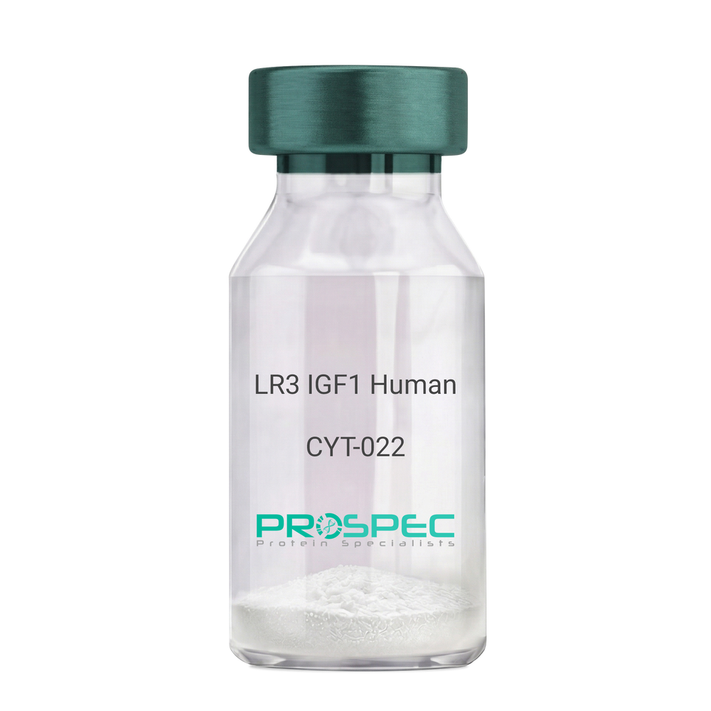
IGF1 LR3 HumanDescription:
LR3 Insulin Like Growth Factor-1 Human Recombinant
R3 IGF1, R3 IGF-1, R3IGF1, R3IGF-1, LONG IGF1, LONG IGF-1, LONG R3 IGF1, LONG R3IGF1, LONG R3 IGF-1, LONG R3IGF-1.
Product # :
CYT-022Price :
Quantity :
Shipping Method :
Shipped at Room temp
More Info
- description
- source
- formulation
- purity
- biological activity
- More Info
- sds-page
- Citations
Description
The LR3 is a long-term analog of human IGF-1, specifically designed and manufactured for mammalian cell culture to support large-scale manufacturing of recombinant biopharmaceuticals. Recombinant Human LR3 Insulin Like Growth Factor-1 produced in E.Coli is a single, non-glycosylated, polypeptide chain containing 83 amino acids and having a molecular mass of 9.1kDa.
Source
Escherichia Coli.
Formulation
Lyophilized from a 0.2µm filtered concentrated solution in 20mM PB, pH 7.2.
Purity
Greater than 97.0% as determined by SDS-PAGE.
Biological Activity
The ED50 as determined by the stimulation of protein synthesis in L6 myoblasts is less than 10ng/ml, corresponding to a specific activity of 100,000units/mg.sds-page
Citations
More Info
-
Introduction
IGF-1 (Insulin-like growth factor-1) is a major hormonal mediator of statural growth. Under regular circumstances, GH (growth hormone) binds to its receptor in the liver, and other tissues, and stimulates the synthesis/secretion of IGF-1. In target tissues, the Type 1 IGF receptor, that is homologous to the insulin receptor, is activated by IGF-1, leading to intracellular signaling which stimulates multiple processes leading to statural growth. IGF-1 metabolic actions are partly directed at stimulating the uptake of glucose, fatty acids, and amino acids so that metabolism supports growing tissues.
-
Synonyms
R3 IGF1, R3 IGF-1, R3IGF1, R3IGF-1, LONG IGF1, LONG IGF-1, LONG R3 IGF1, LONG R3IGF1, LONG R3 IGF-1, LONG R3IGF-1.
-
Physical Appearance
Sterile Filtered White lyophilized (freeze-dried) powder.
-
Stability
Lyophilized LR3 IGF1 although stable at room temperature for 3 weeks, should be stored desiccated below -18°C. Upon reconstitution the LR3 IGF1 should be stored at 4°C between 2-7 days and for future use below -18°C.For long term storage it is recommended to add a carrier protein (0.1% HSA or BSA).Please prevent freeze-thaw cycles.
-
Solubility
It is recommended to reconstitute the lyophilized LR3 IGF1 in sterile 18M-cm H2O at a concentration of 100µg/ml, which can then be further diluted to other aqueous solutions.
-
Amino Acid Sequence
MFPAMPLSSLFVNGPRTLCGAELVDALQFVCGDRGFYFNKPTGYGSSSRRAPQTGIV DECCFRSCDLRRLEMYCAPLKPAKSA.
-
Background
What is the molecular weight/Mw of IGF1 LR3 HUMAN Protein?
IGF1 LR3 HUMAN Protein has a total Mw of 9.1kDa.
What is the source or expression system of IGF1 LR3 HUMAN Protein?
Escherichia Coli.
What is the Purity of IGF1 LR3 HUMAN Protein?
IGF1 LR3 HUMAN Protein is >98% pure as determined by SDS-PAGE.
What is the Biological Activity of IGF1 LR3 HUMAN Protein?
The ED50 as determined by the stimulation of protein synthesis in L6 myoblasts is less than 10ng/ml, corresponding to a specific activity of 100,000units/mg.
What is the amino acid sequence of IGF1 LR3 HUMAN Protein?
MFPAMPLSSLFVNGPRTLCGAELVDALQFVCGDRGFYFNKPTGYGSSSRRAPQTGIV DECCFRSCDLRRLEMYCAPLKPAKSA.
What applications can IGF1 LR3 HUMAN Protein be used in?
IGF1 LR3 HUMAN Protein can probably be used in western blot, ELISA and Lateral Flow.
What is the endotoxin level for IGF1 LR3 HUMAN Protein?
The endotoxin level is minimal, IGF1 LR3 HUMAN Protein was purified using conventional chromatography techniques.
ProSpec's products are furnished for LABORATORY RESEARCH USE ONLY. The product may not be used as drugs, agricultural or pesticidal products, food additives or household chemicals.
- View Data Sheet
Name :
RELT HumanDescription:
RELT Human Recombinant
RELT, TNF Receptor, Receptor Expressed In Lymphoid Tissues, Tumor Necrosis Factor Receptor Superfamily Member 19L, RELT Tumor Necrosis Factor Receptor, TNFRSF19L, Tumor Necrosis Factor Receptor Superfamily, Member 19-Like, TRLT.
Product # :
CYT-1101Price :
Quantity :
Shipping Method :
Shipped with Ice Packs
More Info
- description
- source
- formulation
- purity
- More Info
- Citations
Description
RELT produced in Sf9 Baculovirus cells is a single, glycosylated polypeptide chain containing 376 amino acids (26-162a.a.) and having a molecular mass of 41.4kDa. (Molecular size on SDS-PAGE will appear at approximately 40-57kDa).RELT is expressed with a 239 amino acid hIgG-His-tag at C-Terminus and purified by proprietary chromatographic techniques.
Source
Sf9, Baculovirus cells.
Formulation
RELT protein solution (0.5mg/ml) contains 10% glycerol & Phosphate Buffered Saline (pH 7.4).
Purity
Greater than 90% as determined by SDS-PAGE.
Citations
More Info
-
Introduction
RELTis expressed in hematopoietic tissues and peripheral blood leukocytesand is a part of thetumor necrosis factor receptor superfamily. RELT mediates activation of NF-kappa-B and takes part in T-cell activation.overexpression of RELT in HEK-293 cells induces p38 and JNK signalling and leads to apoptosis. it can also costimulate T-cell proliferation in the presence of CD3 signalling.
-
Synonyms
RELT, TNF Receptor, Receptor Expressed In Lymphoid Tissues, Tumor Necrosis Factor Receptor Superfamily Member 19L, RELT Tumor Necrosis Factor Receptor, TNFRSF19L, Tumor Necrosis Factor Receptor Superfamily, Member 19-Like, TRLT.
-
Physical Appearance
Sterile filtered colorless solution.
-
Stability
Store at 4°C if entire vial will be used within 2-4 weeks. Store, frozen at -20°C for longer periods of time. For long term storage it is recommended to add a carrier protein (0.1% HSA or BSA).Avoid multiple freeze-thaw cycles.
-
Amino Acid Sequence
STTLWQCPPG EEPDLDPGQG TLCRPCPPGT FSAAWGSSPC QPHARCSLWR RLEAQVGMAT
RDTLCGDCWP GWFGPWGVPR VPCQPCSWAPLGTHGCDEWG RRARRGVEVA AGASSGGETR QPGNGTRAGG PEETAAQVEP KSCDKTHTCP PCPAPELLGG PSVFLFPPKP KDTLMISRTP EVTCVVVDVS HEDPEVKFNW YVDGVEVHNA KTKPREEQYN STYRVVSVLT VLHQDWLNGK EYKCKVSNKA LPAPIEKTIS KAKGQPREPQ VYTLPPSRDE LTKNQVSLTC LVKGFYPSDI AVEWESNGQP ENNYKTTPPV LDSDGSFFLY SKLTVDKSRW QQGNVFSCSV MHEALHNHYT QKSLSLSPGK HHHHHH
ProSpec's products are furnished for LABORATORY RESEARCH USE ONLY. The product may not be used as drugs, agricultural or pesticidal products, food additives or household chemicals.
- View Data Sheet
Name :
Activin A HumanDescription:
Activin A Human Recombinant
Activin Beta-A chain, Erythroid differentiation protein, EDF, FRP, Activin-A, Inhibin-B, Inihibin-Beta A chain.
Product # :
CYT-569Price :
Quantity :
Shipping Method :
Shipped with Ice Packs
More Info
- description
- source
- formulation
- purity
- More Info
- sds-page
- Citations
Description
Activin-A Human Recombinant produced in E.coli is a single, non-glycosylated polypeptide chain containing 137 amino acids (311-426) and having a molecular mass of 15.2kDa.Activin-A is fused to a 21 amino acid His-tag at N-terminus & purified by proprietary chromatographic techniques.
Source
Escherichia Coli.
Formulation
Activin-A protein solution (1mg/ml) contains 20mM Tris pH 8.0 and 10% glycerol.
Purity
Greater than 90.0% as determined by SDS-PAGE.
sds-page
Citations
More Info
-
Introduction
Inhibins are dimeric peptide hormones produced by female ovarian granulose cells and male Sertoli cells as well as a variety of other tissues. Inhibins have two isoforms, A and B, with the same alpha subunit but different beta subunits. Inhibin A is a dimer of alpha and beta A subunits, inhibin B is a dimer of alpha and beta B subunits.
Inhibins are thought to inhibit the production of follicle-stimulating hormone (FSH) by the pituitary gland. In addition, Inhibins are also thought to play a role in the control of gametogenesis, and embryonic and fetal development. -
Synonyms
Activin Beta-A chain, Erythroid differentiation protein, EDF, FRP, Activin-A, Inhibin-B, Inihibin-Beta A chain.
-
Physical Appearance
Sterile Filtered clear solution.
-
Stability
Store at 4°C if entire vial will be used within 2-4 weeks. Store, frozen at -20°C for longer periods of time. For long term storage it is recommended to add a carrier protein (0.1% HSA or BSA).Avoid multiple freeze-thaw cycles.
-
Amino Acid Sequence
MGSSHHHHHH SSGLVPRGSH MGLECDGKVN ICCKKQFFVS FKDIGWNDWI IAPSGYHANY CEGECPSHIA GTSGSSLSFH STVINHYRMR GHSPFANLKS CCVPTKLRPM SMLYYDDGQN IIKKDIQNMI VEECGCS.
-
Background
What is the molecular weight / Mw of Activin A Protein?
Activin A Protein has a total Mw of 15.2 kDa.What is the source or expression system of Activin A Protein?
E.ColiWhat is the Purity of Activin A Protein?
Activin A Protein is >90% pure as determined by SDS-PAGE.
What is the Biological Activity of Activin A Protein?
The biological functionality of Activin-A Protein will be determined in the future.What is the endotoxin level for Activin A Protein?
The endotoxin level is minimal, ACTIVIN A Protein was purified using conventional chromatography techniques.What is the amino acid sequence of ACTIVIN A Protein?MGSSHHHHHH SSGLVPRGSH MGLECDGKVN ICCKKQFFVS FKDIGWNDWI IAPSGYHANY CEGECPSHIA GTSGSSLSFH STVINHYRMR GHSPFANLKS CCVPTKLRPM SMLYYDDGQN IIKKDIQNMI VEECGCS
What applications can ACTIVIN-A Protein be used in?
ACTIVIN A Protein can probably be used in western blot, ELISA and Lateral Flow.
ProSpec's products are furnished for LABORATORY RESEARCH USE ONLY. The product may not be used as drugs, agricultural or pesticidal products, food additives or household chemicals.
- View Data Sheet
Name :
HGFR MouseDescription:
Hepatocyte Growth Factor Receptor Mouse Recombinant
hepatocyte growth factor receptor, HGF R/c-MET, Met, AI838057, c-Met, HGF, HGFR, Par4, HGF receptor, HGF/SF receptor, Proto-oncogene c-Met, Scatter factor receptor, SF receptor, Tyrosine-protein kinase Met.
Product # :
CYT-1139Price :
Quantity :
Shipping Method :
Shipped with Ice Packs
More Info
- description
- source
- formulation
- purity
- More Info
- SDS-PAGE
- Citations
Description
HGF Receptor Mouse Recombinant produced in Baculovirus is a single glycosylated polypeptide chain containing 1146 amino acids (25-931aa) and having a molecular mass of 127.8kDa. HGF is fused to a 239 amino acid hIgG-His-Tag at C-terminus and purified by proprietary chromatographic techniques.
Source
Sf9, Baculovirus cells.
Formulation
HGF Receptor protein (0.25mg/ml) contains Phosphate Buffered Saline (pH 7.4) and 10% glycerol.
Purity
Greater than 85.0% as determined by SDS-PAGE.
SDS-PAGE
Citations
More Info
-
Introduction
HGF, also known as scatter factor (SF) and Hepatocyte growth factor, is secreted from mesenchymal cells, targets and acts mainly on epithelial and endothelial cells. The protein is a paracrine cellular growth, motility and morphogenic factor. HGF can also be part of haemopoietic progenitor cells and T cells. HGF has a crucial role in embryonic organ development, mainly in myogenesis. In adults HGF takes part in organ regeneration and wound healing.
-
Synonyms
hepatocyte growth factor receptor, HGF R/c-MET, Met, AI838057, c-Met, HGF, HGFR, Par4, HGF receptor, HGF/SF receptor, Proto-oncogene c-Met, Scatter factor receptor, SF receptor, Tyrosine-protein kinase Met.
-
Physical Appearance
Sterile Filtered colorless solution.
-
Stability
Store at 4°C if entire vial will be used within 2-4 weeks. Store, frozen at -20°C for longer periods of time. For long term storage it is recommended to add a carrier protein (0.1% HSA or BSA).Avoid multiple freeze-thaw cycles.
-
Amino Acid Sequence
ECKEALVKSE MNVNMKYQLP NFTAETPIQN VVLHGHHIYL GATNYIYVLN DKDLQKVSEF KTGPVLEHPD CLPCRDCSSK ANSSGGVWKD NINMALLVDT YYDDQLISCG SVNRGTCQRH VLPPDNSADI QSEVHCMFSP EEESGQCPDC VVSALGAKVL LSEKDRFINF FVGNTINSSY PPGYSLHSIS VRRLKETQDG FKFLTDQSYI DVLPEFQDSY PIKYIHAFES NHFIYFLTVQ KETLDAQTFH TRIIRFCSVD SGLHSYMEMP LECILTEKRR KRSTREEVFN ILQAAYVSKP GANLAKQIGA SPSDDILFGV FAQSKPDSAE PVNRSAVCAF PIKYVNDFFN KIVNKNNVRC LQHFYGPNHE HCFNRTLLRN SSGCEARSDE YRTEFTTALQ RVDLFMGRLN QVLLTSISTF IKGDLTIANL GTSEGRFMQV VLSRTAHLTP HVNFLLDSHP VSPEVIVEHP SNQNGYTLVV TGKKITKIPL NGLGCGHFQS CSQCLSAPYF IQCGWCHNQC VRFDECPSGT WTQEICLPAV YKVFPTSAPL EGGTVLTICG WDFGFRKNNK FDLRKTKVLL GNESCTLTLS ESTTNTLKCT VGPAMSEHFN VSVIISNSRE TTQYSAFSYV DPVITSISPR YGPQAGGTLL TLTGKYLNSG NSRHISIGGK TCTLKSVSDS ILECYTPAQT TSDEFPVKLK IDLANRETSS FSYREDPVVY EIHPTKSFIS GGSTITGIGK TLNSVSLPKL VIDVHEVGVN YTVACQHRSN SEIICCTTPS LKQLGLQLPL KTKAFFLLDG ILSKHFDLTY VHNPVFEPFE KPVMISIGNE NVVEIKGNNI DPEAVKGEVL KVGNQSCESL HWHSGAVLCT VPSDLLKLNS ELNIEWKQAV SSTVLGKVIV QPDQNFALEP KSCDKTHTCP PCPAPELLGG PSVFLFPPKP KDTLMISRTP EVTCVVVDVS HEDPEVKFNW YVDGVEVHNA KTKPREEQYN STYRVVSVLT VLHQDWLNGK EYKCKVSNKA LPAPIEKTIS KAKGQPREPQ VYTLPPSRDE LTKNQVSLTC LVKGFYPSDI AVEWESNGQP ENNYKTTPPV LDSDGSFFLY SKLTVDKSRW QQGNVFSCSV MHEALHNHYT QKSLSLSPGK HHHHHH.
-
Background
What is the molecular weight/Mw of HGFR MOUSE Protein?
HGFR MOUSE Protein has a total Mw of 127.8kDa.
What is the source or expression system of HGFR MOUSE Protein?
Sf9, Baculovirus cells.
What is the Purity of HGFR MOUSE Protein?
HGFR MOUSE Protein is >85% pure as determined by SDS-PAGE.
What is the Biological Activity of HGFR MOUSE Protein?
The biological functionality of HGFR MOUSE Protein will be determined in the future.
What is the amino acid sequence of HGFR MOUSE Protein?
ECKEALVKSE MNVNMKYQLP NFTAETPIQN VVLHGHHIYL GATNYIYVLN DKDLQKVSEF KTGPVLEHPD CLPCRDCSSK ANSSGGVWKD NINMALLVDT YYDDQLISCG SVNRGTCQRH VLPPDNSADI QSEVHCMFSP EEESGQCPDC VVSALGAKVL LSEKDRFINF FVGNTINSSY PPGYSLHSIS VRRLKETQDG FKFLTDQSYI DVLPEFQDSY PIKYIHAFES NHFIYFLTVQ KETLDAQTFH TRIIRFCSVD SGLHSYMEMP LECILTEKRR KRSTREEVFN ILQAAYVSKP GANLAKQIGA SPSDDILFGV FAQSKPDSAE PVNRSAVCAF PIKYVNDFFN KIVNKNNVRC LQHFYGPNHE HCFNRTLLRN SSGCEARSDE YRTEFTTALQ RVDLFMGRLN QVLLTSISTF IKGDLTIANL GTSEGRFMQV VLSRTAHLTP HVNFLLDSHP VSPEVIVEHP SNQNGYTLVV TGKKITKIPL NGLGCGHFQS CSQCLSAPYF IQCGWCHNQC VRFDECPSGT WTQEICLPAV YKVFPTSAPL EGGTVLTICG WDFGFRKNNK FDLRKTKVLL GNESCTLTLS ESTTNTLKCT VGPAMSEHFN VSVIISNSRE TTQYSAFSYV DPVITSISPR YGPQAGGTLL TLTGKYLNSG NSRHISIGGK TCTLKSVSDS ILECYTPAQT TSDEFPVKLK IDLANRETSS FSYREDPVVY EIHPTKSFIS GGSTITGIGK TLNSVSLPKL VIDVHEVGVN YTVACQHRSN SEIICCTTPS LKQLGLQLPL KTKAFFLLDG ILSKHFDLTY VHNPVFEPFE KPVMISIGNE NVVEIKGNNI DPEAVKGEVL KVGNQSCESL HWHSGAVLCT VPSDLLKLNS ELNIEWKQAV SSTVLGKVIV QPDQNFALEP KSCDKTHTCP PCPAPELLGG PSVFLFPPKP KDTLMISRTP EVTCVVVDVS HEDPEVKFNW YVDGVEVHNA KTKPREEQYN STYRVVSVLT VLHQDWLNGK EYKCKVSNKA LPAPIEKTIS KAKGQPREPQ VYTLPPSRDE LTKNQVSLTC LVKGFYPSDI AVEWESNGQP ENNYKTTPPV LDSDGSFFLY SKLTVDKSRW QQGNVFSCSV MHEALHNHYT QKSLSLSPGK HHHHHH.
What applications can HGFR MOUSE Protein be used in?
HGFR MOUSE Protein can probably be used in western blot, ELISA and Lateral Flow.
What is the endotoxin level for HGFR MOUSE Protein?
The endotoxin level is minimal, HGFR MOUSE Protein was purified using conventional chromatography techniques.
ProSpec's products are furnished for LABORATORY RESEARCH USE ONLY. The product may not be used as drugs, agricultural or pesticidal products, food additives or household chemicals.
- View Data Sheet
Name :
VEGF AntibodyDescription:
Vascular Endothelial Cell Growth Factor, Mouse Anti-Human
Vascular endothelial growth factor A, VEGF-A, Vascular permeability factor, VPF, VEGF, MGC70609.
Product # :
ANT-125Price :
Quantity :
Shipping Method :
Shipped at Room temp
More Info
- More Info
- Citations
Citations
More Info
-
Introduction
Vascular endothelial growth factoris an important signaling proteininvolved in both vasculogenesisand angiogenesis. As its name implies, VEGF activity has been mostly studied on cells of the vascular endothelium, although it does have effects on a number of other cell types (e.g. stimulation monocyte/macrophagemigration, neurons, cancer cells, kidney epithelial cells ).VEGF mediates increased vascular permeability, induces angiogenesis, vasculogenesis and endothelial cell growth, promotes cell migration, and inhibits apoptosis. In vitro, VEGF has been shown to stimulate endothelial cell mitogenesisand cell migration. VEGF is also a vasodilator and increases microvascular permeability and was originally referred to as vascular permeability factor.
Elevated levels of this protein is linked to POEMS syndrome, also known as Crow-Fukase syndrome. Mutations in this gene have been associated with proliferative and nonproliferative diabetic retinopathy. -
Synonyms
Vascular endothelial growth factor A, VEGF-A, Vascular permeability factor, VPF, VEGF, MGC70609.
-
Solubility
Reconstitute with sterile H20. Mix gently, wash the sides of the vial and wait 30-60 seconds before use.
-
Immunogen
r.HumanVEGF.
-
Ig Subclass
Mouse IgM.
-
Clone
NYRhVEGF.
-
Applications
Direct ELISA, Western Blot, Immuneprecipitation.
-
Titer
By direct ELISA, 1:10,000 dilution will yield 0.15 O.D using alkaline phosphatase conjugated rabbit anti-mouse Ig (Jackson Laboratories).
-
Shipping Conditions
Antibody is shipped lyophilized at ambient temperature.
-
Type
Mouse Anti Human Monoclonal.
-
Storage Procedures
In lyophilized form, for long periods, store at 4oC in a dry environment. After reconstitution, if not intended for use within a month, aliquot and store at -20oC.
-
Purification Method
Boric acid precipitation Protein concentration1mg/ml in PBS (after reconstitution).
ProSpec's products are furnished for LABORATORY RESEARCH USE ONLY. The product may not be used as drugs, agricultural or pesticidal products, food additives or household chemicals.
- View Data Sheet
Name :
G CSF HumanDescription:
Granulocyte-Colony Stimulating Factor Human Recombinant
CSF-3, MGI-1G, GM-CSF beta, Pluripoietin, Lenograstim, G-CSF, MGC45931, GCSF.
Product # :
CYT-220Price :
Quantity :
Shipping Method :
Shipped at Room temp
More Info
- description
- source
- formulation
- purity
- biological activity
- More Info
- Citations
Description
Granulocyte Colony Stimulating Factor Human Recombinant produced in E.coli is a single, non-glycosylated, polypeptide chain containing 175 amino acids and having a molecular mass of 18.8 KD.GCSF is purified by proprietary chromatographic techniques.
Source
Escherichia Coli.
Formulation
GCSF was lyophilized after extensive dialysis against 10mM sodium acetate buffer pH= 4.
Purity
Greater than 98.0% as determined by:
(a) Analysis by RP-HPLC.
(b) Analysis by SDS-PAGE.Biological Activity
The ED50, calculated by the dose-dependant proliferation of murine NFS-60 indicator cells (measured by 3H-thymidine uptake) is < 0.1 ng/ml, corresponding to a Specific Activity of 100,000,000 IU/mg.Citations
More Info
-
Introduction
GCSF is a cytokine that controls the production, differentiation, and function of granulocytes. The active protein is found extracellularly. Three transcript variants encoding three different isoforms have been found for the GCSF gene. Granulocyte/macrophage colony-stimulating factors are cytokines that act in hematopoiesis by controlling the production, differentiation, and function of 2 related white cell populations of the blood, the granulocytes and the monocytes-macrophages. This csf induces granulocytes.
-
Synonyms
CSF-3, MGI-1G, GM-CSF beta, Pluripoietin, Lenograstim, G-CSF, MGC45931, GCSF.
-
Physical Appearance
Sterile Filtered White lyophilized (freeze-dried) powder.
-
Stability
Lyophilized GCSF although stable at room temperature for 3 weeks, should be stored desiccated below -18°C. Upon reconstitution GCSF should be stored at 4°C between 2-7 days and for future use below -18°C.For long term storage it is recommended to add a carrier protein (0.1% HSA or BSA).Please prevent freeze-thaw cycles.
-
Solubility
It is recommended to reconstitute the lyophilized GCSF in sterile 20mM AcOH not less than 100µg/ml, which can then be further diluted to other aqueous solutions.
-
Amino Acid Sequence
The sequence of the first five N-terminal amino acids of GCSF was determined and was found to be Met-Thr-Pro-Leu-Gly.
-
Background
What is the molecular weight/Mw of GDF15 HUMAN, HIS Protein?
GDF15 HUMAN, HIS Protein has a total Mw of 18.8kDa.
What is the source or expression system of GDF15 HUMAN, HIS Protein?
Escherichia Coli.
What is the Purity of GDF15 HUMAN, HIS Protein?
GDF15 HUMAN, HIS Protein is >98% pure as determined by SDS-PAGE.
What is the Biological Activity of GDF15 HUMAN, HIS Protein?
The ED50, calculated by the dose-dependant proliferation of murine NFS-60 indicator cells (measured by 3H-thymidine uptake) is < 0.1 ng/ml, corresponding to a Specific Activity of 100,000,000 IU/mg.
What is the amino acid sequence of GDF15 HUMAN, HIS Protein?
GDF15 HUMAN, HIS Protein is composed from 175 amino acids.
What applications can GDF15 HUMAN, HIS Protein be used in?
GDF15 HUMAN, HIS Protein can probably be used in western blot, ELISA and Lateral Flow.
What is the endotoxin level for GDF15 HUMAN, HIS Protein?
The endotoxin level is minimal, GDF15 HUMAN, HIS Protein was purified using conventional chromatography techniques.
-
Protein content
GCSF quantitation was carried out by two independent methods:1. UV spectroscopy at 280 nm using the absorbency value of 0.815 as the extinction coefficient for a 0.1% (1mg/ml) solution. This value is calculated by the PC GENE computer analysis program of protein sequences (IntelliGenetics). 2. Analysis by RP-HPLC, using a calibrated solution of GCSF as a Reference Standard.
ProSpec's products are furnished for LABORATORY RESEARCH USE ONLY. The product may not be used as drugs, agricultural or pesticidal products, food additives or household chemicals.
- View Data Sheet
Name :
KGF MouseDescription:
Keratinocye Growth Factor Mouse Recombinant
HBGF-7, FGF7, FGF-7, KGF, Keratinocyte growth factor, Fibroblast growth factor 7, Heparin-binding growth factor 7.
Product # :
CYT-721Price :
Quantity :
Shipping Method :
Shipped at Room temp
More Info
- description
- source
- formulation
- purity
- biological activity
- More Info
- Citations
Description
Keratinocyte Growth Factor-1 Mouse Recombinant produced in E.Coli is a single, non-glycosylated, polypeptide chain containing 164 amino acids and having a molecular mass of 18.9 kDa. The FGF-7 is purified by proprietary chromatographic techniques.
Source
Escherichia Coli.
Formulation
The protein was lyophilized from a concentrated (1mg/ml) solution containing 20mM Phosphate buffer pH-8 and 0.1M NaCl.
Purity
Greater than 95.0% as determined by: (a) Analysis by HPLC. (b) Analysis by SDS-PAGE.
Biological Activity
The ED50, calculated by the dose-dependant stimulation of KGF-responsive BaF3 indicator cells (measured by 3H-thymidine uptake) is < 10ng/ml corresponding to a specific activity of 100,000 Units/mg.Citations
More Info
-
Introduction
KGF is a member of the fibroblast growth factor (FGF) family. FGF family members possess broad mitogenic and cell survival activities, and are involved in a variety of biological processes, including embryonic development, cell growth, morphogenesis, tissue repair, tumor growth and invasion. FGF7 is a potent epithelial cell-specific growth factor, whose mitogenic activity is predominantly exhibited in keratinocytes but not in fibroblasts and endothelial cells. Studies of mouse and rat homologs of this gene implicated roles in morphogenesis of epithelium, reepithelialization of wounds, hair development and early lung organogenesis.
-
Synonyms
HBGF-7, FGF7, FGF-7, KGF, Keratinocyte growth factor, Fibroblast growth factor 7, Heparin-binding growth factor 7.
-
Physical Appearance
Sterile Filtered White lyophilized (freeze-dried) powder.
-
Stability
Lyophilized KGF although stable at room temperature for 3 weeks, should be stored desiccated below -18°C. Upon reconstitution FGF7 should be stored at 4°C between 2-7 days and for future use below -18°C. For long term storage it is recommended to add a carrier protein (0.1% HSA or BSA). Please prevent freeze-thaw cycles.
-
Solubility
It is recommended to reconstitute the lyophilized KGF in sterile 18MΩ-cm H2O not less than 100µg/ml, which can then be further diluted to other aqueous solutions.
-
Amino Acid Sequence
MCNDMSPEQT ATSVNCSSPE RHTRSYDYME GGDIRVRRLF CRTQWYLRID KRGKVKGTQE MKNSYNIMEI RTVAVGIVAI KGVESEYYLA MNKEGKLYAK KECNEDCNFK ELILENHYNT YASAKWTHSG GEMFVALNQK GIPVKGKKTK KEQKTAHFLP MAIT.
ProSpec's products are furnished for LABORATORY RESEARCH USE ONLY. The product may not be used as drugs, agricultural or pesticidal products, food additives or household chemicals.
- View Data Sheet
Name :
VEGI Human, HisDescription:
Vascular Endothelial Growth InhibitorHuman Recombinant, His Tag
Tumor necrosis factor ligand superfamily member 15, TNFSF-15, TNFSF15, TNF ligand-related molecule 1, VEGI, TL-1, TL1, TL1A, VEGI192A, VEGI-192, MGC129934, MGC129935.
Product # :
CYT-589Price :
Quantity :
Shipping Method :
Shipped with Ice Packs
More Info
- description
- source
- formulation
- purity
- More Info
- Citations
Description
VEGI Human Recombinant fused with a 21 amino acid His tag at N-terminus produced in E.Coli is a single, non-glycosylated, polypeptide chain containing 201 amino acids(72-251a.a.) and having a molecular mass of 22.7kDa. The VEGI is purified by proprietary chromatographic techniques.
Source
Escherichia Coli.
Formulation
The VEGI solution (1mg/1ml) contains 20mM Tris-HCl buffer (pH 8.0), 50% glycerol, 1mM DTT and 0.2M NaCl.
Purity
Greater than 95.0% as determined by SDS-PAGE.
Citations
More Info
-
Introduction
TNFSF15 is a cytokine that belongs to the tumor necrosis factor (TNF) ligand family. This protein is abundantly expressed in endothelial cells, but is not expressed in either B or T cells. The expression of TNFSF15 is inducible by TNF and IL-1 alpha. This cytokine is a ligand for receptor TNFRSF25 and decoy receptor TNFRSF21/DR6. It can activate NF-kappaB and MAP kinases, and acts as an autocrine factor to induce apoptosis in endothelial cells. TNFSF15 is also found to inhibit endothelial cell proliferation, and thus may function as an angiogenesis inhibitor. An additional isoform encoded by an alternatively spliced transcript variant has been reported but the sequence of this transcript has not been determined.
-
Synonyms
Tumor necrosis factor ligand superfamily member 15, TNFSF-15, TNFSF15, TNF ligand-related molecule 1, VEGI, TL-1, TL1, TL1A, VEGI192A, VEGI-192, MGC129934, MGC129935.
-
Physical Appearance
Sterile Filtered colorless solution.
-
Stability
Store at 4°C if entire vial will be used within 2-4 weeks. Store, frozen at -20°C for longer periods of time. For long term storage it is recommended to add a carrier protein (0.1% HSA or BSA).Avoid multiple freeze-thaw cycles.
-
Amino Acid Sequence
MGSSHHHHHH SSGLVPRGSH MLKGQEFAPS HQQVYAPLRA DGDKPRAHLT VVRQTPTQHF KNQFPALHWE HELGLAFTKN RMNYTNKFLL IPESGDYFIY SQVTFRGMTS ECSEIRQAGR PNKPDSITVV ITKVTDSYPE PTQLLMGTKS VCEVGSNWFQ PIYLGAMFSL QEGDKLMVNV SDISLVDYTK EDKTFFGAFL L
ProSpec's products are furnished for LABORATORY RESEARCH USE ONLY. The product may not be used as drugs, agricultural or pesticidal products, food additives or household chemicals.
- View Data Sheet
Name :
Insulin Human, HisDescription:
Insulin Human Recombinant, His Tag
Insulin, Insulin-Dependent Diabetes Mellitus 2, Preproinsulin, Proinsulin, MODY10, IDDM1, IDDM2, IDDM, ILPR, IRDN.
Product # :
CYT-1076Price :
Quantity :
Shipping Method :
Shipped with Ice Packs
More Info
- description
- source
- formulation
- purity
- biological activity
- More Info
- Citations
Description
Insulin Recombinant produced in E.Coli is a single, non-glycosylated polypeptide chain containing 109 amino acids (25-110 a.a) and having a molecular mass of 11.8kDa. Insulin is fused to a 23 amino acid His-tag at N-terminus & purified by proprietary chromatographic techniques.
Source
Escherichia Coli.
Formulation
Insulin protein solution (0.5mg/ml) containing Phosphate-Buffered Saline (pH 7.4) and 10% Glycerol.
Purity
Greater than 95.0% as determined by SDS-PAGE.
Biological Activity
Measured in a cell proliferation assay using MCF7 human breast cancer cell. The ED50 for this effect is less or equal to 4 ug/ml.
Citations
More Info
-
Introduction
Insulin decreases blood glucose concentration. it increases cell permeability to monosaccharides, amino acids and fatty acids. it accelerates glycolysis, the pentose phosphate cycle, and glycogen synthesis in liver.
-
Synonyms
Insulin, Insulin-Dependent Diabetes Mellitus 2, Preproinsulin, Proinsulin, MODY10, IDDM1, IDDM2, IDDM, ILPR, IRDN.
-
Physical Appearance
Sterile Filtered clear solution.
-
Stability
Store at 4°C if entire vial will be used within 2-4 weeks. Store, frozen at -20°C for longer periods of time. For long term storage it is recommended to add a carrier protein (0.1% HSA or BSA).Avoid multiple freeze-thaw cycles.
-
Amino Acid Sequence
MGSSHHHHHH SSGLVPRGSH MGSFVNQHLC GSHLVEALYL VCGERGFFYT PKTRREAEDL QVGQVELGGG PGAGSLQPLA LEGSLQKRGI VEQCCTSICS LYQLENYCN
ProSpec's products are furnished for LABORATORY RESEARCH USE ONLY. The product may not be used as drugs, agricultural or pesticidal products, food additives or household chemicals.
- View Data Sheet
Name :
CXCL4 Variant 1 HumanDescription:
Platelet Factor-4 Variant 1 Human Recombinant
CXCL4, PF-4, PF4, Iroplact, Oncostatin-A, SCYB4, MGC138298.
Product # :
CHM-243Price :
Quantity :
Shipping Method :
Shipped at Room temp
More Info
- description
- source
- formulation
- purity
- More Info
- Citations
Description
CXCL4 Variant-1 Human Recombinant produced in E.Coli is a single, non-glycosylated polypeptide chain containing 77 amino acids and having a molecular mass of 8.7 kDa. The CXCL4 Variant-1 is fused to 6xHis tag at N-Terminus and purified by standard chromatography techniques.
Source
Escherichia Coli.
Formulation
The protein was lyophilized in PBS, pH 8.5.
Purity
Greater than 95.0% as determined by SDS-PAGE.
Citations
More Info
-
Introduction
Platelet factor-4 is a 70-amino acid protein that is
released from the alpha-granules of activated platelets. Its major physiologic
role appears to be neutralization of molecules on the endothelial surface of
blood vessels, thereby inhibiting local antithrombin III activity and promoting
coagulation. As a strong chemoattractant for neutrophils and fibroblasts, PF4
probably has a role in inflammation and wound repair. Oncostatin-A is a member
of the CXC chemokine family. Furthermore it is used as an inhibitor in the
angiogenesis during tumor therapy. -
Synonyms
CXCL4, PF-4, PF4, Iroplact, Oncostatin-A, SCYB4, MGC138298.
-
Physical Appearance
Sterile Filtered white lyophilized powder.
-
Stability
Human CXCL4 although stable at 25°C 1 week, should be stored desiccated below -18°C. Please prevent freeze-thaw cycles.
-
Solubility
It is recommended to reconstitute the lyophilized CXCL4 in sterile 18MΩ-cm H2O not less than 100µg/ml, which can then be further diluted to other aqueous solutions.
-
Amino Acid Sequence
MHHHHHHEAE EDGDLQCLCV KTTSQVRPRH ITSLEVIKAG PHCPTAQLIA TLKNGRKICL DLQALLYKKI IKEHLES.
-
Background
What is the molecular weight/Mw of CXCL4 VARIANT 1 HUMAN Protein?
CXCL4 VARIANT 1 HUMAN Protein has a total Mw of 8.7kDa.
What is the source or expression system of CXCL4 VARIANT 1 HUMAN Protein?
Escherichia Coli.
What is the Purity of CXCL4 VARIANT 1 HUMAN Protein?
CXCL4 VARIANT 1 HUMAN Protein is > 95% pure as determined by SDS-PAGE.
What is the Biological Activity of CXCL4 VARIANT 1 HUMAN Protein?
The biological functionality of CXCL4 VARIANT 1 HUMAN Protein will be determined in the future.
What is the amino acid sequence of CXCL4 VARIANT 1 HUMAN Protein?
MHHHHHHEAE EDGDLQCLCV KTTSQVRPRH ITSLEVIKAG PHCPTAQLIA TLKNGRKICL DLQALLYKKI IKEHLES.
What applications can CXCL4 VARIANT 1 HUMAN Protein be used in?
CXCL4 VARIANT 1 HUMAN Protein can probably be used in western blot, ELISA and Lateral Flow.
What is the endotoxin level for CXCL4 VARIANT 1 HUMAN Protein?
The endotoxin level is minimal, CXCL4 VARIANT 1 HUMAN Protein was purified using conventional chromatography techniques.
Who supplies recombinant CXCL4 VARIANT 1 HUMAN that is in stock and available to order?
ProSpec has the protein in stock and available for immediate delivery.
Where can I order bulk mg quantities of CXCL4 VARIANT 1 HUMAN?
ProSpec supplies bulk mg quantities at affordable prices that meets research and academic budgets.
ProSpec's products are furnished for LABORATORY RESEARCH USE ONLY. The product may not be used as drugs, agricultural or pesticidal products, food additives or household chemicals.
- View Data Sheet
Name :
Myostatin Propeptide HumanDescription:
Myostatin Propeptide Human Recombinant
GDF-8, MSTN, Growth Differentiation Factor 8, MSTN Muscle Hypertrophy.
Product # :
CYT-448Price :
Quantity :
Shipping Method :
Shipped at Room temp
More Info
- description
- source
- formulation
- purity
- biological activity
- More Info
- Citations
Description
Recombinant Human Myostatin Propeptide is a 27.8kDa protein containing 244 amino acid residues of the human Myostatin Propeptide.
Source
Escherichia Coli.
Formulation
Lyophilized with no additives.
Purity
Greater than 95.0% as determined by SDS-PAGE.
Biological Activity
The protein has full biological activity when compared to a standard. The activity is determined by its ability to inhibit 50ng/ml of Myostatin on MPC-11 cells and is typically 0.13-0.2 μg/ml.Citations
More Info
-
Introduction
Myostatin (GDF-8), a member of the TGFbeta superfamily, is a potent and specific negative regulator of skeletal muscle mass. In serum, myostatin circulates as part of a latent complex containing myostatin propeptide and/or follistatin-related gene. The myostatin propeptide is known to bind and inhibit myostatin in vitro. This interaction is relevant in vivo, with a majority (>70%) of myostatin in serum bound to its propeptide. The myostatin propeptide is negative regulator of myostatin in vivo.
-
Synonyms
GDF-8, MSTN, Growth Differentiation Factor 8, MSTN Muscle Hypertrophy.
-
Physical Appearance
Sterile Filtered white lyophilized (freeze-dried) powder.
-
Stability
Store lyophilized protein at -20°C. Aliquot the product after reconstitution to avoid repeated freezing/thawing cycles. Reconstituted protein can be stored at 4°C for a limited period of time.
-
Solubility
It is recommended to reconstitute the lyophilized Myostatin Propeptide in sterile 20mM HCl at 0.1mg/ml, which can then be further diluted to other aqueous solutions.
-
Amino Acid Sequence
MNENSEQKE NVEKEGLCNA CTWRQNTKSS RIEAIKIQIL SKLRLETAPN ISKDVIRQLL PKAPPLRELI DQYDVQRDDS SDGSLEDDDY HATTETIITM PTESDFLMQV DGKPKCCFFK FSSKIQYNKV VKAQLWIYLR PVETPTTVFV QILRLIKPMK DGTRYTGIRS LKLDMNPGTG IWQSIDVKTV LQNWLKQPES NLGIEIKALD ENGHDLAVTF PGPGEDGLNP FLEVKVTDTP KRSRR.
ProSpec's products are furnished for LABORATORY RESEARCH USE ONLY. The product may not be used as drugs, agricultural or pesticidal products, food additives or household chemicals.
- View Data Sheet
Name :
TNFSF8 Human, Sf9Description:
CD30 Ligand Human Recombinant, Sf9
Tumor Necrosis Factor Superfamily Member 8, Tumor Necrosis Factor (Ligand) Superfamily, Member 8, CD153 Antigen, CD30 Ligand, CD30LG, CD30-L, CD30L, Tumor Necrosis Factor (Ligand) Superfamily Member 8, Tumor Necrosis Factor Ligand 3A, CD30 Antigen Ligand, TNLG3A, CD153, Tumor necrosis factor ligand superfamily member 8, TNFSF8, CD30 ligand.
Product # :
CYT-954Price :
Quantity :
Shipping Method :
Shipped with Ice Packs
More Info
- description
- source
- formulation
- purity
- More Info
- Citations
Description
TNFSF8 produced in Sf9 Baculovirus cells is a single, glycosylated polypeptide chain containing 181 amino acids (63-234a.a.) and having a molecular mass of 20.7kDa (Molecular size on SDS-PAGE will appear at approximately 18-40kDa). TNFSF8 is expressed with a 6 amino acid His tag at C-Terminus and purified by proprietary chromatographic techniques.
Source
Sf9, Baculovirus cells.
Formulation
TNFSF8 protein solution (0.5mg/ml) contains Phosphate Buffered Saline (pH 7.4) and 10% glycerol.
Purity
Greater than 85% as determined by SDS-PAGE.
Citations
More Info
-
Introduction
CD30 Ligand (TNFSF8) is a cytokine which is a member of the tumor necrosis factor (TNF) ligand family. The TNFSF8 cytokine is a ligand for TNFRSF8/CD30, which is a cell surface antigen and a marker for Hodgkin lymphoma and related hematologic malignancies. The employment of the TNFSF8 cytokine expressed on B cell surface has an inhibitory role in modulating Ig class switch. TNFSF8 enhances cell proliferation of some lymphoma cell lines, while inducing cell death and reducing cell proliferation of other lymphoma cell lines. The pleiotropic biological activities of the TNFSF8 cytokine on different CD30+ lymphoma cell lines has a pathophysiologic role in Hodgkin's and some non-Hodgkin's lymphomas.
-
Synonyms
Tumor Necrosis Factor Superfamily Member 8, Tumor Necrosis Factor (Ligand) Superfamily, Member 8, CD153 Antigen, CD30 Ligand, CD30LG, CD30-L, CD30L, Tumor Necrosis Factor (Ligand) Superfamily Member 8, Tumor Necrosis Factor Ligand 3A, CD30 Antigen Ligand, TNLG3A, CD153, Tumor necrosis factor ligand superfamily member 8, TNFSF8, CD30 ligand.
-
Physical Appearance
Sterile filtered colorless solution.
-
Stability
Store at 4°C if entire vial will be used within 2-4 weeks. Store, frozen at -20°C for longer periods of time. For long term storage it is recommended to add a carrier protein (0.1% HSA or BSA).Avoid multiple freeze-thaw cycles.
-
Amino Acid Sequence
ADPQRTDSIP NSPDNVPLKG GNCSEDLLCI LKRAPFKKSW AYLQVAKHLN KTKLSWNKDG ILHGVRYQDG NLVIQFPGLY FIICQLQFLV QCPNNSVDLK LELLINKHIK KQALVTVCES GMQTKHVYQN LSQFLLDYLQ VNTTISVNVD TFQYIDTSTF PLENVLSIFL YSNSDHHHHH H.
ProSpec's products are furnished for LABORATORY RESEARCH USE ONLY. The product may not be used as drugs, agricultural or pesticidal products, food additives or household chemicals.
- View Data Sheet
Name :
PAFAH2 HumanDescription:
Platelet-Activating Factor Acetylhydrolase 2 Human Recombinant
HSD-PLA2, Platelet-activating factor acetylhydrolase 2, cytoplasmic, Serine-dependent phospholipase A2, SD-PLA2, hSD-PLA2.
Product # :
ENZ-899Price :
Quantity :
Shipping Method :
Shipped with Ice Packs
More Info
- description
- source
- formulation
- purity
- More Info
- Citations
Description
PAFAH2 Human Recombinant produced in E.Coli is a single, non-glycosylated polypeptide chain containing 415 amino acids (1-392 a.a) and having a molecular mass of 46.4kDa.PAFAH2 is fused to a 23 amino acid His-tag at N-terminus & purified by proprietary chromatographic techniques.
Source
Escherichia Coli.
Formulation
PAFAH2 protein solution (1mg/ml) in Phosphate buffered saline (pH7.4), 20% glycerol and 1mM DTT.
Purity
Greater than 90.0% as determined by SDS-PAGE.
Citations
More Info
-
Introduction
Platelet-activating factor acetylhydrolase 2 cytoplasmic (PAFAH2) has a marked selectivity for phospholipids with short acyl chains at the sn-2 position. PAFAH2 may share a mutual physiologic function with the plasma-type enzyme.
-
Synonyms
HSD-PLA2, Platelet-activating factor acetylhydrolase 2, cytoplasmic, Serine-dependent phospholipase A2, SD-PLA2, hSD-PLA2.
-
Physical Appearance
Sterile Filtered colorless solution.
-
Stability
Store at 4°C if entire vial will be used within 2-4 weeks. Store, frozen at -20°C for longer periods of time. For long term storage it is recommended to add a carrier protein (0.1% HSA or BSA).Avoid multiple freeze-thaw cycles.
-
Amino Acid Sequence
MGSSHHHHHH SSGLVPRGSH MGSMGVNQSV GFPPVTGPHL VGCGDVMEGQ NLQGSFFRLF YPCQKAEETM EQPLWIPRYE YCTGLAEYLQ FNKRCGGLLF NLAVGSCRLP VSWNGPFKTK DSGYPLIIFS HGLGAFRTLY SAFCMELASR GFVVAVPEHR DRSAATTYFC KQAPEENQPT NESLQEEWIP FRRVEEGEKE FHVRNPQVHQ RVSECLRVLK ILQEVTAGQT VFNILPGGLD LMTLKGNIDM SRVAVMGHSF GGATAILALA KETQFRCAVA LDAWMFPLER DFYPKARGPV FFINTEKFQT MESVNLMKKI CAQHEQSRII TVLGSVHRSQ TDFAFVTGNL IGKFFSTETR GSLDPYEGQE VMVRAMLAFL QKHLDLKEDY NQWNNLIEGI GPSLTPGAPH HLSSL.
ProSpec's products are furnished for LABORATORY RESEARCH USE ONLY. The product may not be used as drugs, agricultural or pesticidal products, food additives or household chemicals.
- View Data Sheet
Name :
Leptin-A TilapiaDescription:
Leptin-A Tilapia Recombinant
Product # :
CYT-1109Price :
Quantity :
Shipping Method :
Shipped at Room temp
More Info
- description
- source
- formulation
- purity
- biological activity
- More Info
- Citations
Description
Leptin-A Tilapia Recombinant produced in E.Coli is a single, non-glycosylated polypeptide chain containing 161 amino acids and having a molecular mass of 16,491 Dalton. The Leptin-A Tilapia is purified by proprietary chromatographic techniques.
Source
Escherichia Coli.
Formulation
Lyophilized from a concentrated (1mg/ml) solution containing NaHCO3 at 1:2 salt: protein ratio.
Purity
Greater than 95.0% as determined by:
(a) Gel filtration analysis.
(b) Analysis by SDS-PAGE.Biological Activity
Tilapia leptins were found to be biologically active in promoting proliferation of BAF/3 cells stably transfected with the long form of human leptin receptor, but their activity was lower than that of mammalian leptin. Furthermore, the Tilapia leptins were biologically active in promoting STAT‐LUC activation in COS7 cells transfected with Tilapia leptin receptor but not in cells transfected with human leptin receptor. Tilapia Leptin A was more active than Tilapia Leptin B.
Citations
More Info
-
Introduction
Leptin is a protein hormone. It is mainly produced in adipose cells that regulate energy homeostasis by restraining hunger. Leptin ties to nuclear receptors in the hypothalamus (arcuate nucleus). Similar to insulin resistance in type II diabetes, in obesity there is a decrease in the sensitivity towards leptin, ending in a failure to identify satiety, even in high levels of energy stores or leptin itself. Full-length cDNA encoding 2 leptin sequences (tLepA and tLepB) and 1 leptin receptor sequence (tLepR) exists in tilapia (Oreochromis niloticus). The full-length cDNA of tLepR is 3423 bp, encoding a protein of 1140 amino acid which contained all functionally important domains conserved among vertebrate leptin receptors. The cDNAs of tLepA and tLepB are 486 bp and 459 bp in length, encoding proteins of 161 aa and 152 aa, respectively. The three-dimensional structures of tLepA and tLepB demonstrates strong conservation of tertiary structure with that of human leptin comprised of 4 helixes.
-
Physical Appearance
Sterile Filtered White lyophilized (freeze-dried) powder.
-
Stability
Lyophilized Leptin-A Tilapia although stable at room temperature for 3 weeks, should be stored desiccated below -18°C. Upon reconstitution Leptin-A Tilapia should be stored at 4°C between 2-7 days and for future use below -18°C. For long term storage it is recommended to add a carrier protein (0.1% HSA or BSA). Please prevent freeze-thaw cycles.
-
Solubility
It is recommended to reconstitute the lyophilized Leptin-A Tilapia in sterile water or 0.4% NaHCO3 adjusted to pH 8-9, not less than 100µg/ml, which can then be further diluted to other aqueous solutions.
-
Amino Acid Sequence
The first six N-terminal amino acids of recombinant Tilapia leptin A are Ala-Pro-Leu-Pro-Val-Glu.
-
Protein content
Protein quantitation was carried out by UV spectroscopy at 280 nm using the absorbency value of 1.33 for 1 mg/ml Leptin-A Tilapia as the extinction coefficient for a 0.1% (1mg/ml) solution at pH 8.0. This value is calculated by the DNAman computer analysis program of protein sequences.
ProSpec's products are furnished for LABORATORY RESEARCH USE ONLY. The product may not be used as drugs, agricultural or pesticidal products, food additives or household chemicals.
- View Data Sheet
Name :
TFF2 HumanDescription:
Trefoil Factor-2 Human Recombinant
TFF-2, Spasmolytic polypeptide, Spasmolysin, SML1, Trefoil factor 2, SP, TFF2.
Product # :
CYT-612Price :
Quantity :
Shipping Method :
Shipped at Room temp
More Info
- description
- source
- formulation
- purity
- biological activity
- More Info
- Citations
Description
TFF-2 Human Recombinant produced in E.Coli is a single, non-glycosylated, polypeptide chain containing 106 amino acids (24-129) and having a total molecular mass of 12 kDa. TFF2 Human Recombinant includes a 40-amino acid trefoil motif containing three conserved intramolecular disulfide bonds and was purified by proprietary chromatographic techniques.
Source
Escherichia Coli.
Formulation
The TFF2 protein was lyophilized from 0.4μm filtered solution at a concentration of 1mg/mL containing 1x PBS pH-7.4.
Purity
Greater than 97.0% as determined by(a) Analysis by RP-HPLC.
(b) Analysis by SDS-PAGE.Biological Activity
Determined by its ability to chemoattract human MCF-7 cells using a concentration 1.0-10.0 ng/ml.Citations
More Info
-
Introduction
Proteins of the TFF family are characterized by obtaining a minimum of 1 copy of the trefoil motif, a 40-amino acid domain that contains 3 conserved disulfides. Trefoil Factors are stable secretory proteins expressed in gastrointestinal mucosa which protect the mucosa from insults, stabilize the mucus layer and affect healing of the epithelium.TFF2 inhibits gastric acid motility & secretion. TFF2 stabilizes glycoproteins in the mucus gel through interactions with carbohydrate side chains.
-
Synonyms
TFF-2, Spasmolytic polypeptide, Spasmolysin, SML1, Trefoil factor 2, SP, TFF2.
-
Physical Appearance
Sterile Filtered White lyophilized (freeze-dried) powder.
-
Stability
Lyophilized TFF2 although stable at room temperature for 3 weeks, should be stored desiccated below -18°C. Upon reconstitution TFF2 should be stored at 4°C between 2-7 days and for future use below -18°C.For long term storage it is recommended to add a carrier protein (0.1% HSA or BSA).Please prevent freeze-thaw cycles.
-
Solubility
It is recommended to reconstitute the lyophilized TFF2 in sterile 18MΩ-cm H2O not less than 100µg/ml, which can then be further diluted to other aqueous solutions.
-
Amino Acid Sequence
EKPSPCQCSR LSPHNRTNCG FPGITSDQCF DNGCCFDSSV TGVPWCFHPL PKQESDQCVM EVSDRRNCGY PGISPEECAS RKCCFSNFIF EVPWCFFPKSVEDCHY.
ProSpec's products are furnished for LABORATORY RESEARCH USE ONLY. The product may not be used as drugs, agricultural or pesticidal products, food additives or household chemicals.
- View Data Sheet
Name :
FAS AntibodyDescription:
FAS Blocking/Activating antibody (CD95), Mouse anti Human
FASLG receptor, Apoptosis-mediating surface antigen FAS, Apo-1 antigen, CD95, Tumor necrosis factor receptor superfamily member 6 TNR6, APT1, FAS1, TNFRSF6.
Product # :
ANT-204Price :
Quantity :
Shipping Method :
Shipped at Room temp
More Info
- formulation
- More Info
- Citations
Formulation
1 mg/ml in PBS (after reconstitution).
Citations
More Info
-
Introduction
The Fas receptor (CD95) mediates apoptotic signaling by Fas-ligand expressed on the surface of other cells. The Fas-FasL interaction plays an important role in the immune system and lack of this system leads to autoimmunity, indicating that Fas-mediated apoptosis removes self-reactive lymphocytes. Fas signaling is also involved in immune surveillance to remove transformed cells and virus infected cells. Binding of FAS to oligimerized FasL on another cell activates apoptotic signaling through a cytoplasmic domain termed the death domain that interacts with signaling adaptors including FAF, FADD and DAX to activate the caspase proteolytic cascade. Caspase-8 and caspase-10 are first activated, to then cleave and activate downstream caspases, and a variety of cellular substrates that lead to cell death. Caspases cleave nuclear lamins, causing the nucleus to break down and lose its normal structure and another caspase substrate is DFF, inducing cleavage and degradation of the genome. Other caspase substrates are involved in cytoskeletal structure, cell cycle regulation and signaling pathways. Activation of JNK kinase, activation of Jun, and production of ceramide may also play roles in Fas-mediated apoptosis. Activation of fas-mediated apoptosis is opposed by I-FLICE and FAP. Viruses and tumors may escape immune surveillance in part through suppression of fas-mediated apoptosis using similar mechanisms.
-
Synonyms
FASLG receptor, Apoptosis-mediating surface antigen FAS, Apo-1 antigen, CD95, Tumor necrosis factor receptor superfamily member 6 TNR6, APT1, FAS1, TNFRSF6.
-
Solubility
Reconstitute with H20. Mix gently, wash the sides of the vial and wait 30-60 seconds before use.
-
Immunogen
Recombinant Human FAS.
-
Ig Subclass
mouse IgG1.
-
Clone
NYRhFAS.
-
Note
This antibody is a BLOCKING antibody when in soluble form and will block FAS-mediated apoptosis. It will ACTIVATE FAS-mediated killing when immobilized.
-
Titer
By direct ELISA, 1:10,000 dilution will yield 0.5 O.D using alkaline phosphatase conjugated rabbit anti-mouse Ig (Jackson Laboratories).
-
Shipping Conditions
Antibody is shipped lyophilized at ambient temperature.
-
Type
Mouse Anti Human Monoclonal.
-
Storage Procedures
In lyophilized form, for long periods, store at 4°C in a dry environment. After reconstitution, if not intended for use within a month, aliquot and store at -20°C.
-
Purification Method
Protein-A column.
ProSpec's products are furnished for LABORATORY RESEARCH USE ONLY. The product may not be used as drugs, agricultural or pesticidal products, food additives or household chemicals.